Embed Size (px)
Citation preview

Keratinocyte Apoptosis Induced by Ultraviolet B Radiationand CD95 Ligation ± Differential Protection throughEpidermal Growth Factor Receptor Activation and Bcl-xL
Expression
Monika Jost, Francis P. Gasparro, Pamela J. Jensen,* and Ulrich RodeckDepartment of Dermatology and Cutaneous Biology, Thomas Jefferson University, Philadelphia, and *Department of Dermatology, University of
Pennsylvania, Philadelphia, Pennsylvania, U.S.A.
Previous work has shown that activation of the epi-dermal growth factor receptor by endogenous orexogenous signals markedly enhances survival ofcultured keratinocytes upon cellular stress such aspassaging. This is due, in part, to epidermal-growth-factor-receptor-dependent expression of Bcl-xL, anantiapoptotic Bcl-2 homolog. In this study we testedwhether epidermal-growth-factor-receptor-depend-ent signal transduction and attendant Bcl-xL expres-sion affected survival of human keratinocytes uponexposure to a frequently encountered apoptoticstimulus, radiation with ultraviolet B. We describethat blocking epidermal-growth-factor-receptor-dependent signal transduction sensitized normal ker-atinocytes to undergo apoptosis upon ultraviolet B
radiation with solar light characteristics. Forcedexpression of Bcl-xL partially but signi®cantly inhib-ited ultraviolet-B-induced apoptosis of immortalizedkeratinocytes (HaCaT). Bcl-xL overexpressionafforded no protection to HaCaT cells against apop-tosis induced by binding of an agonist antibody tothe death receptor CD95, however. CD95 activationhas previously been shown to functionally contributeto apoptosis in ultraviolet-irradiated keratinocytes.These results indicate that epidermal growth factorreceptor activation and attendant Bcl-xL expressionprovided a physiologically relevant protective path-way of keratinocytes against ultraviolet-induced butnot CD95-dependent apoptosis. J Invest Dermatol116:860±866, 2001
Solar light presents a major environmental challenge tothe skin contributing not only to the aging process butalso to cancer development. Ultraviolet (UV) radiationis thought to be responsible for most of thecarcinogenic effects of sunlight on epidermal keratino-
cytes either by direct DNA damage or by generation of reactiveoxygen species (for review see de Gruijl, 1999). The solarspectrum at sea level consists mainly of UVB (290±320 nm) andUVA (320±400 nm) whereas short wavelength UVC (200±290 nm) is absorbed ef®ciently by the ozone layer in the upperearth atmosphere (Gasparro and Brown, 2000). Carcinogeniceffects of solar radiation might be thwarted by UV-inducedprogrammed cell death, a feature occasionally observed as``sunburn cells'' in skin acutely exposed to UV light (Ziegler etal, 1994).
The mechanisms of UV-induced apoptosis in keratinocytesare poorly understood. Recent evidence has shown that UVBradiation activates multiple cell surface receptors, which mayeither contribute to or antagonize entry of keratinocytes intodeath programs. Examples for pro-apoptotic receptors activated
by UV exposure are CD95/FasR (Leverkus et al, 1997;Rehemtulla et al, 1997) and tumor necrosis factor receptor(Tobin et al, 1998). Other cell surface receptors activated byUV radiation, however, including the epidermal growth factorreceptor (EGFR) (Sachsenmaier et al, 1994; Coffer et al, 1995;Huang et al, 1996; Rosette and Karin, 1996; Peus et al, 1998),have been implicated in protection of keratinocytes fromapoptosis (Rodeck et al, 1997a, b; Stoll et al, 1998). Therelative contribution of single receptor classes to the propensityof keratinocytes to undergo apoptosis following UV radiation isunknown. Protection of keratinocytes against other pro-apoptotic agents has been traced, in part, to upregulation ofthe antiapoptotic protein Bcl-xL through activation of theEGFR tyrosine kinase moiety (Rodeck et al, 1997a; Stoll et al,1998; Jost et al, 1999). Furthermore, Bcl-xL expression bykeratinocytes has been reported to contribute to resistance ofthese cells to UVB radiation (Taylor et al, 1999). Takentogether, these ®ndings raised the questions whether EGFRactivation protects keratinocytes from apoptosis induced by thefrequently encountered environmental stress factor UVB andwhether Bcl-xL expression plays a signi®cant role in thisprocess.
Here we describe protection of cultured normal humankeratinocytes against UVB-induced apoptosis by EGFR activationand by forced expression of high levels of Bcl-xL. Interestingly, thisprotection did not extend to apoptosis induced by a CD95-agonistic antibody, which was unaffected by Bcl-xL overexpression.
Manuscript received September 14, 2001; revised February 19, 2001;accepted for publication February 20, 2001.
Reprint requests to: Dr. Ulrich Rodeck, Department of Dermatologyand Cutaneous Biology, Thomas Jefferson University, BLSB 319, 233 S10th Street, Philadelphia, PA 19107. Email: [email protected]
Abbreviations: EGFR, epidermal growth factor receptor; PARP, polyADP-ribose-polymerase.
0022-202X/01/$15.00 ´ Copyright # 2001 by The Society for Investigative Dermatology, Inc.
860

MATERIALS AND METHODS
Cell culture conditions and reagents Human neonatal foreskinkeratinocytes were initiated and propagated as described previously(McNeill and Jensen, 1990). Complete MCDB153 (Sigma, St. Louis,MO) contained 30 mM Ca2+ and was supplemented with amino acids,ethanolamine, phosphorylethanolamine, hydrocortisone, insulin (all fromSigma), puri®ed EGF (10 ng per ml; Collaborative Research, Bedford,MA), and bovine pituitary extract (prepared from pituitaries obtainedfrom Pel-Freez, Rogers, WI). Base MCDB153 contained allnonproteinaceous components but no EGF, insulin, or bovine pituitaryextract. The agonist CD95 antibody IPO-4 (IgM; k) was obtained fromKamiya Biomedical, Seattle, WA. Antigen speci®city and EGFR-antagonistic characteristics of monoclonal antibody MoAb 425 have beendescribed previously (Murthy et al, 1987, 1990; Rodeck et al, 1987,1990). Tyrphostins AG1478 and AG1295 and the caspase inhibitor Ac-DEVD-CHO were from Calbiochem (San Diego, CA). Establishmentand culture of HaCaT cells stably transfected with a tetracyclinecontrollable episomal Bcl-xL construct was described previously (Jost etal, 1997); in this system tetracycline represses transgene expression (Tet-Off). For the experiments described, cells were seeded at a density of1.5±2 3 104 cells per cm2 and pretreated as indicated for 48±72 h. Priorto UV exposure, the culture medium was replaced with phosphate-buffered saline (PBS). After radiation, PBS was removed and freshmedium was added as indicated. Control cells were subjected to thesame treatment without exposure to UVB.
UVB radiation source and dosimetry UV radiation was performedusing a bank of FS-40 lamps (Westinghouse Electric, Pittsburgh, PA)with a UVB emission maximum near 310 nm. To mimic the UVBspectrum encountered in sunlight wavelengths below 290 nm (UVCrange) were blocked by using a UVC Kodacel ®lter (Eastman Kodak,Rochester, NY). The use of this ®lter may explain why a comparativelyhigher dose was necessary to induce apoptosis in this study compared tothe dose range used by other investigators (Aragane et al, 1998; Peus etal, 1998; Kulms et al, 1999). Prior to radiation, the lamp output wasquanti®ed with a calibrated IL 1700 radiometer (International Light,Newburyport, MA) and exposure times were calculated accordingly.Cells were radiated in PBS without lid at a distance of 27 cm to thelamps from the top of the plates. Doses ranged from 50 to 300 mJ percm2.
Detection of cell death and apoptosis To determine cell viabilitytrypan blue exclusion assays were performed. Non-attached cells were
collected by centrifugation at 400 3 g and attached cells were dislodgedwith trypsin/ethylenediamine tetraacetic acid; both fractions werepooled. Aliquots of cell suspensions were diluted 2-fold with 0.4%trypan blue in PBS and examined using an inverted light microscope.Detection of apoptotic cell death was performed using the M30CytoDEATH detection kit (Roche Molecular Biochemicals,Indianapolis, IN) or by TUNEL staining, followed by ¯uorescence-activated cell sorter (FACS) analysis. Prior to staining cell suspensionswere ®xed and permeabilized in 70% ethanol at 4°C. The M30CytoDEATH mouse MoAb (clone M30) detects a caspase cleavageproduct of cytokeratin 18. The M30 CytoDEATH assay was performedfollowing the manufacturer's instructions. Brie¯y, ®xed cells wereblocked in PBS containing 1% (wt/vol) bovine serum albumin (BSA)and then stained with the M30 MoAb for 1 h at room temperature,followed by incubation with a ¯uorescein isothiocyanate (FITC)conjugated secondary antibody for 30 min at room temperature. Sampleswere washed between incubations with 0.1% (vol/vol) Tween-20 inPBS containing 1% BSA and analyzed immediately. Prior to TUNELstaining, cells were treated with PBS containing 1% (wt/vol) BSA.Staining was performed using TdT enzyme, biotin-16-dUTP, or¯uorescein-12-dUTP and buffers from Roche Molecular Biochemicalsfor 1 h at 37°C. For detection of biotin-labeled nuclei cells wereincubated with FITC-avidin (2.5 mg per ml; Vector Laboratories,Burlingame, CA) in 4 3 SCC (1 3 SSC is 0.15 M NaCl, 0.015 M Nacitrate). Labeled cells were washed once with PBS containing 1% BSAand 0.1% Triton-X-100. Prior to counterstaining with propidium iodide,intracellular RNA was removed by treatment with 1 mg per ml RNAse
Figure 1. EGFR blockade with antagonistic MoAb 425 reducesviability of keratinocytes upon UVB radiation. Keratinocytes werepretreated for 3 d with either EGF (10 ng per ml) or MoAb 425 (10 mgper ml) in base medium supplemented with insulin and were thenirradiated with UVB at 50 or 100 mJ per cm2 in PBS, as indicated. Afterradiation, PBS was replaced with complete MCDB. Viability wasassessed by trypan blue exclusion assay 2 d after treatment. Resultsrepresent mean 6 standard deviation of the number of viable cells;results of three different experiments using keratinocytes derived fromdifferent donors are shown.
Figure 2. EGFR blockade enhances UVB-induced apoptosis inkeratinocytes. Cells were treated with EGF (A, B), MoAb 425 (66 nM;C, D), or AG1478 (10 mM; D, E) in base medium supplemented withinsulin for 3 d and then exposed to 100 mJ per cm2 UVB. Apoptosiswas analyzed by M30 staining 24 h after radiation by FACS analysis.Density plots of M30 ¯uorescence over forward scatter were used toidentify apoptotic (R2) and necrotic (R3) cells. MoAb-425- andAG1478-treated cultures showed a higher fraction of apoptotic cells,shown as a percentage of the whole cell population in brackets. Note theincrease in necrotic cells after AG1478 treatment.
VOL. 116, NO. 6 JUNE 2001 UVB-INDUCED KERATINOCYTE APOPTOSIS 861

(Roche Molecular Biochemicals) for 5 min at 37°C, followed by stainingwith 5 mg per ml propidium iodide (Molecular Probes, Eugene, OR) inPBS for 20 min at 4°C. A minimum of 105 cells were analyzed with aFACSort cyto¯uorograph (Beckton Dickinson, San Jose, CA) and LysisII Software (Beckton Dickinson). To assess the proportion of cells thatnot only survived but regained proliferative competence, cultures wereallowed to grow for 1 wk after radiation, ®xed with 1%paraformaldehyde, and stained with crystal violet (0.2% wt/vol in 2%ethanol).
Immunoblot analysis For analysis of poly ADP-ribose-polymerase(PARP) cleavage, attached and nonattached cells were pooled and lysedas described previously (Rodeck et al, 1997a). Equal amounts of totalcellular protein were separated on 7.5% sodium dodecyl sulfate/polyacrylamide gels followed by immunoblotting and detection using anantibody to PARP (Santa Cruz, Santa Cruz, CA) that crossreacts withfull-length and the longer cleavage product (p85) diluted according tothe manufacturer's recommendation. Signals were visualized bychemiluminescence using reagents from Pierce Chemical (Rockford, IL)according to the manufacturer's instructions. For detection of Bcl-xL anantibody from Transduction Laboratories (Lexington, KY) was used.
RESULTS
Enhancement of UVB-induced apoptosis by EGFRblockade Previously we demonstrated that inhibition of theEGFR tyrosine kinase activity either by antagonistic MoAb 425 orEGFR-selective tyrphostin AG1478 reduces viability of normalhuman keratinocytes upon cellular stress such as passaging (Rodecket al, 1997a, b; Jost et al, 1999). In this study, we examined whetherEGFR engagement also protected keratinocytes against a commonenvironmental stressor for keratinocytes, i.e., UVB radiation. First,we determined dose-dependent effects of UVB light on viability oflow-passage primary keratinocyte cultures by exposing these cells toUVB at various intensities (50±300 mJ per cm2). After radiation,cultures were further incubated in medium fully supplementedwith growth factors and cytokines including EGF. Under theseexperimental conditions, keratinocytes underwent massiveapoptosis at dosages above 100 mJ per cm2 as determined by
light microscopy inspection and trypan blue exclusion assay. Bycontrast, only marginal loss of viability or apoptosis was observedwithin 48 h after treatment when cells were exposed to 100 mJ percm2 UVB or less. Next, we exposed keratinocytes pretreated withreceptor saturating amounts of EGFR antagonistic MoAb 425(10 mg per ml) to UVB at increasing dosages. At subapoptotic UVBdoses (50 and 100 mJ per cm2), MoAb-425-pretreated culturesdisplayed morphologic features of cell death, such as cell shrinkage,blebbing, and detachment from the substratum. Figure 1 showsthe percentage of viable cells after EGFR blockade relative to EGF-treated controls as determined by trypan blue staining. AlthoughEGFR blockade had little effect on the viability in nonexposed cells(see also Rodeck et al, 1997a, b), it drastically reduced viability ofUVB-treated cells in a UVB-dose-dependent manner to only about50% of controls maintained in EGF-containing medium at 100 mJper cm2.
Further experiments were performed to demonstrate that thereduction in cell viability in keratinocytes treated with MoAb 425prior to UVB radiation was due to induction of apoptosis.Figure 2 shows that MoAb 425 treatment sensitized keratinocytesto UVB-induced apoptosis as determined by the appearance ofcaspase cleaved cytokeratin 18. UVB radiation combined withEGFR inhibition clearly increased the number of apoptotic cells asindicated by the increase in the population of cells in the R2 regionof the scattergraphs (arrows).
Protection of HaCaT cells against UVB-induced apoptosisby forced expression of Bcl-xL Previously, an antiapoptoticmember of the Bcl-2 family, Bcl-xL, was shown to be upregulatedby EGFR activation in normal and immortalized keratinocytes(Rodeck et al, 1997a; Stoll et al, 1998). In immortalizedkeratinocytes (HaCaT), overexpression of Bcl-xL protectedagainst apoptosis induced by EGFR blockade and passaging,consistent with a functional role for Bcl-xL in EGFR-dependentprotection from apoptosis. In light of these results, we questionedwhether EGFR-dependent Bcl-xL expression is similarly important
Figure 3. Forced expression of Bcl-xL
protects immortalized keratinocytes (HaCaT)from UVB-induced cell death. HaCaT cellsexpressing tetracycline-regulated Bcl-xL andmock-transfected cells were irradiated with 0 (A),150 (B), or 300 (C) mJ per cm2 UVB and thenincubated for 24 h, all in the absence oftetracycline. Phase-contrast microscopy 24 h afterirradiation shows enhanced survival in Bcl-xL-expressing cultures.
862 JOST ET AL THE JOURNAL OF INVESTIGATIVE DERMATOLOGY

in protecting keratinocytes against UVB-induced apoptosis. Toaddress this issue, we analyzed the effect of overexpression of Bcl-xL on sensitivity of HaCaT keratinocytes to apoptosis induction byUVB radiation.
First, we assessed the effect of Bcl-xL overexpression on short-term survival of UVB-exposed HaCaT cells (Fig 3). Mock andBcl-xL-transfected HaCaT cells were morphologically indistin-guishable in the absence of UVB treatment (Fig 3A). Twenty-fourhours after irradiation with 150 mJ per cm2 UVB, however, mock-transfected cells displayed morphologic features of apoptosis,
whereas Bcl-xL-expressing HaCaT cells were protected up to 300mJ per cm2 (Fig 3B, C). To further assess protection of HaCaT±Bcl-xL cells against UVB-radiation-induced apoptotic cell deathTUNEL staining was performed to detect UVB-induced apoptoticDNA cleavage. As shown in Fig 4(A), overexpression of Bcl-xL
reduced apoptotic DNA cleavage after UVB radiation. Althoughprotection was greatest after induction of Bcl-xL by removal oftetracycline from the culture medium, some protection was alsoobserved in noninduced Bcl-xL-transfected cells. This was probablydue to leakiness as we observed a higher basal Bcl-xL expression
Figure 4. Expression of Bcl-xL reduces apoptotic DNA and PARP cleavage in HaCaT cells. (A) HaCaT±mock and HaCaT±Bcl-xL cells weremaintained for 48 h prior to UVB irradiation in medium with or without tetracycline (1 mg per ml). Twenty-four hours after treatment, cultures wereharvested and processed for TUNEL staining. Reduced apoptotic DNA cleavage was evident in cells overexpressing Bcl-xL. (B) Bcl-xL expression inmock-transfected and Bcl-xL-transfected HaCaT keratinocytes prior to UV radiation. Slightly higher levels of Bcl-xL were observed in noninduced Bcl-xL transfectants compared to mock-transfected cells consistent with some leakiness of the inducible expression system. (C) Untransfected HaCaT cells(upper panel) and mock- or Bcl-xL-transfected HaCaT cells (lower panel) were induced for 48 h and then exposed to 0, 100, or 150 mJ per cm2 UVB,and PARP expression was determined by Western blot analysis 24 h after radiation. UVB exposure induced PARP cleavage in untransfected andmock-transfected HaCaT cells but only marginally in Bcl-xL-transfected cells.
VOL. 116, NO. 6 JUNE 2001 UVB-INDUCED KERATINOCYTE APOPTOSIS 863

level (50%) in noninduced Bcl-xL-transfected cells compared to thelevels of endogenous Bcl-xL in mock-transfected HaCaT cells(Fig 4B). We also examined apoptotic cleavage of the caspase 3substrate PARP to its 85 kDa form by immunoblot analysis(Fig 4B). Irradiation with 100±150 mJ per cm2 induced signi®cantPARP cleavage in HaCaT keratinocytes (Fig 4C). Similar resultswere obtained in mock-transfected cells, whereas Bcl-xL-trans-fected cells showed markedly attenuated PARP cleavage at 100±150 mJ per cm2 UVB. These results con®rm that Bcl-xL conferspartial protection against UVB-induced apoptosis to HaCaT cells.To test whether Bcl-xL expression only delayed but did not preventthe onset of apoptosis we determined long-term survival of Bcl-xL-overexpressing HaCaT cells after UVB radiation by performingclonal growth assays. Figure 5 shows mock- and Bcl-xL-transfected cells cultured for 1 wk after irradiation with UVB asindicated. Compared to mock-transfected cells, Bcl-xL-overex-pressing HaCaT cells exposed to up to 200 mJ per cm2 UVBreadily formed colonies indicating that they retained proliferativepotential. This experiment shows that Bcl-xL overexpression doesnot simply delay UV-induced cell death but leads to survival ofproliferation-competent HaCaT cells.
Bcl-xL overexpression does not protect HaCaT cells againstFas-mediated apoptosis It has been reported previously that
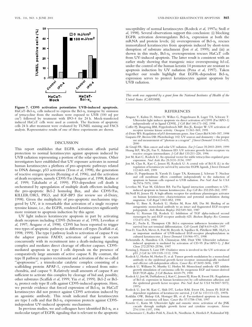
CD95 activation by Fas ligand occurs in keratinocytes upon UVradiation, and that this death receptor contributes to the demise ofthese cells (Aragane et al, 1998). In some cell types, Bcl-xL canprotect against Fas-mediated apoptosis whereas in others it does not(Scaf®di et al, 1998). Based on these observations we determinedwhether forced expression of Bcl-xL protected HaCaT cells againstFas-mediated apoptosis. Consistent with earlier ®ndings in prostateepithelial cells (Rokhlin et al, 1997), activation of CD95 by use ofan agonist antibody (IPO-4) induced apoptosis in HaCaT cells(Fig 6); the antibody used is of the IgM class and thus is capable ofextensively crosslinking CD95. Overexpression of Bcl-xL,however, did not affect the rate at which these cells underwentIPO-4-induced apoptosis. By contrast, treatment with the broad-spectrum caspase inhibitor Ac-DEVD-CHO blocked IPO-4-induced apoptosis in HaCaT±Bcl-xL cells (not shown). IPO-4treatment and UVB radiation had additive effects on apoptosis inmock-transfected HaCaT cells (Fig 7). As expected Bcl-xL
overexpression provided only partial protection against apoptosisinduced by combined UVB radiation (100 mJ per cm2) and IPO-4treatment.
Figure 5. Effect of Bcl-xL expression on long-term survival ofHaCaT cells after UVB radiation. Cells were treated with UVB asindicated, maintained in culture for 1 wk post exposure, ®xed, andstained with crystal violet. Bcl-xL-transfected cultures show a higherfraction of surviving cells than mock-transfected cells.
Figure 6. Forced Bcl-xL expression does not protect HaCaT cellsagainst CD95-mediated apoptosis. Noninduced and induced Bcl-xL-and mock-transfected cultures were treated for 24 h with a CD95agonist antibody (IPO-4, 1 mg per ml) in the presence and absence oftetracycline. The fractions of apoptotic cells were evaluated by TUNELstaining and FACS analysis.
864 JOST ET AL THE JOURNAL OF INVESTIGATIVE DERMATOLOGY
DISCUSSION
This report establishes that EGFR activation affords partialprotection to normal keratinocytes against apoptosis induced byUVB radiation representing a portion of the solar spectrum. Otherinvestigators have established that UV exposure activates in normalhuman keratinocytes a plethora of pro-apoptotic pathways relatedto DNA damage, p53 activation (Tron et al, 1998), the generationof reactive oxygen species (Renzing et al, 1996), and the activationof death receptors, namely CD95/Fas (Aragane et al, 1998; Kulms etal, 1999; Takahashi et al, 1999). P53-dependent cell death isorchestrated by upregulation of multiple death effectors includingthe pro-apoptotic Bcl-2 homolog Bax, and also CD95/Fas,KILLER/DR5, PIG3, and IGF-BP3 (for review see el-Deiry,1998). Given the multiplicity of pro-apoptotic mechanisms trig-gered by UV, it is remarkable that activation of a single receptortyrosine kinase, i.e., the EGFR, rendered keratinocytes signi®cantlymore resistant to apoptosis induction by this agent.
UV light induces keratinocyte apoptosis in part by activatingdeath receptors including CD95 (Schwarz et al, 1995; Leverkus etal, 1997; Aragane et al, 1998). CD95 has been reported to triggertwo types of apoptotic pathways in different cell types (Scaf®di et al,1998; 1999). The type I pathway leads to activation of caspase 8 viathe adaptor protein FADD; activation of caspase 8 occursconcurrently with its recruitment into a death-inducing signalingcomplex and mediates direct cleavage of effector caspases. CD95-mediated apoptosis in type I cells depends on the presence ofcomparatively large amounts of active caspase 8. By contrast, thetype II pathway requires recruitment and activation of the so-called``apoptosome'', a trimolecular catalytic complex consisting of ascaffolding protein APAF-1, cytochrome c released from mito-chondria, and caspase 9. Relatively small amounts of caspase 8 aresuf®cient to activate this complex by cleavage of bid and, possibly,other substrates (Scaf®di et al, 1999; Yin et al, 1999). Bcl-2 or Bcl-xL protect only type II cells against CD95-induced apoptosis. Here,we provide evidence that forced expression of Bcl-xL in HaCaTkeratinocytes did not protect against CD95 activation induced byan agonistic antibody. This result indicated that keratinocytesare type I cells and that Bcl-xL expression protects against CD95-independent UV-induced apoptotic mechanisms.
In previous studies, we and colleagues have identi®ed Bcl-xL as amolecular target of EGFR signaling that is relevant to the apoptotic
susceptibility of normal keratinocytes (Rodeck et al, 1997a; Stoll etal, 1998). Several observations support this conclusion: (i) blockingEGFR activation downregulates Bcl-xL expression at both themRNA and protein levels; (ii) overexpression of Bcl-xL rescuesimmortalized keratinocytes from apoptosis induced by short-termdisruption of substrate attachment (Jost et al, 1999); and (iii) asshown in this study, Bcl-xL overexpression rescues HaCaT cellsfrom UV-induced apoptosis. The latter result is consistent with anearlier study showing that transgenic mice overexpressing bcl-xLunder the control of the human keratin 14 promoter are resistant toapoptosis induction by UV radiation (Pena et al, 1997). Takentogether our results highlight that EGFR-dependent Bcl-xL
expression serves to protect keratinocytes against apoptosis byUVB radiation.
This work was supported by a grant from the National Institutes of Health of the
United States (CA81008).
REFERENCES
Aragane Y, Kulms D, Metze D, Wilkes G, Poppelmann B, Luger TA, Schwarz T:Ultraviolet light induces apoptosis via direct activation of CD95 (Fas/APO-1)independently of its ligand CD95L. J Cell Biol 140:171±182, 1998
Coffer PJ, Burgering BM, Peppelenbosch MP, Bos JL, Kruijer W: UV activation ofreceptor tyrosine kinase activity. Oncogene 11:561±569, 1995
el-Deiry WS: Regulation of p53 downstream genes. Sem Cancer Biol 8:345±357, 1998Gasparro FP, Brown DB: Photobiology 102: UV sources and dosimetry ± the proper
use and measurement of ``photons as a reagent''. J Invest Dermatol 114:613±615,2000
de Gruijl FR: Skin cancer and solar UV radiation. Eur J Cancer 35:2003±2009, 1999Huang RP, Wu JX, Fan Y, Adamson ED: UV activates growth factor receptors via
reactive oxygen intermediates. J Cell Biol 133:211±220, 1996Jost M, Kari C, Rodeck U: An episomal vector for stable tetracycline-regulated gene
expression. Nucl Acids Res 25:3131±3134, 1997Jost M, Class R, Kari C, Jensen PJ, Rodeck U: A central role of Bcl-X (L) in the
regulation of keratinocyte survival by autocrine EGFR ligands. J Invest Dermatol112:443±449, 1999
Kulms D, Poppelmann B, Yarosh D, Luger TA, Krutmann J, Schwarz T: Nuclearand cell membrane effects contribute independently to the induction ofapoptosis in human cells exposed to UVB radiation. Proc Natl Acad Sci USA96:7974±7979, 1999
Leverkus M, Yaar M, Gilchrest BA: Fas/Fas ligand interaction contributes to UV-induced apoptosis in human keratinocytes. Exp Cell Res 232:255±262, 1997
McNeill H, Jensen PJ: A high-af®nity receptor for urokinase plasminogen activatoron human keratinocytes: characterization and potential modulation duringmigration. Cell Regul 1:843±852, 1990
Murthy U, Basu A, Rodeck U, Herlyn M, Ross AH, Das M: Binding of anantagonistic monoclonal antibody to an intact and fragmented EGF-receptorpolypeptide. Arch Biochem Biophys 252:549±560, 1987
Murthy U, Rieman DJ, Rodeck U: Inhibition of TGF alpha-induced secondmessengers by anti-EGF receptor antibody-425. Biochem Biophys Res Commun172:471±476, 1990
Pena JC, Fuchs E, Thompson CB: Bcl-x expression in¯uences keratinocyte cellsurvival but not terminal differentiation. Cell Growth Diff 8:619±629, 1997
Peus D, Vasa RA, Meves A, Pott M, Beyerle A, Squillace K, Pittelkow MR: H2O2 isan important mediator of UVB-induced EGF-receptor phosphorylation incultured keratinocytes. J Invest Dermatol 110:966±971, 1998
Rehemtulla A, Hamilton CA, Chinnaiyan AM, Dixit VM: Ultraviolet radiation-induced apoptosis is mediated by activation of CD-95 (Fas/APO-1). J BiolChem 272:25783±25786, 1997
Renzing J, Hansen S, Lane DP: Oxidative stress is involved in the UV activation ofp53. J Cell Sci 109:1105±1112, 1996
Rodeck U, Herlyn M, Herlyn D, et al: Tumor growth modulation by a monoclonalantibody to the epidermal growth factor receptor: immunologically mediatedand effector cell-independent effects. Cancer Res 47:3692±3696, 1987
Rodeck U, Williams N, Murthy U, Herlyn M: Monoclonal antibody 425 inhibitsgrowth stimulation of carcinoma cells by exogenous EGF and tumor-derivedEGF/TGF-alpha. J Cell Biochem 44:69±79, 1990
Rodeck U, Jost M, DuHadaway J, Kari C, Jensen PJ, Risse B, Ewert DL: Regulationof Bcl-xL expression in human keratinocytes by cell-substratum adhesion andthe epidermal growth factor receptor. Proc Natl Acad Sci USA 94:5067±5072,1997a
Rodeck U, Jost M, Kari C, Shih DT, Lavker RM, Ewert DL, Jensen PJ: EGF-Rdependent regulation of keratinocyte survival. J Cell Sci 110:113±121, 1997b
Rokhlin OW, Bishop GA, Hostager BS, et al: Fas-mediated apoptosis in humanprostatic carcinoma cell lines. Cancer Res 57:1758±1768, 1997
Rosette C, Karin M: Ultraviolet light and osmotic stress: activation of the JNKcascade through multiple growth factor and cytokine receptors. Science274:1194±1197, 1996
Sachsenmaier C, Radler-Pohl A, Zinck R, Nordheim A, Herrlich P, Rahmsdorf HJ:
Figure 7. CD95 activation potentiates UVB-induced apoptosis.HaCaT±Bcl-xL cells induced to express the Bcl-xL transgene by omissionof tetracycline from the medium were exposed to UVB (100 mJ percm2) followed by treatment with IPO-4 for 24 h. Mock-transfectedinduced HaCaT cells were used as controls. The fractions of apoptoticcells 24 h after treatment were evaluated by TUNEL staining and FACSanalysis. Representative results of one of three experiments are shown.
VOL. 116, NO. 6 JUNE 2001 UVB-INDUCED KERATINOCYTE APOPTOSIS 865
Involvement of growth factor receptors in the mammalian UVC response. Cell78:963±972, 1994
Scaf®di C, Fulda S, Srinivasan A, et al: Two CD95 (APO-1/Fas) signaling pathways.EMBO J 17:1675±1687, 1998
Scaf®di C, Schmitz I, Zha J, Korsmeyer SJ, Krammer PH, Peter ME: Differentialmodulation of apoptosis sensitivity in CD95 type I and type II cells. J Biol Chem274:22532±22538, 1999
Schwarz A, Bhardwaj R, Aragane Y, et al: Ultraviolet-B-induced apoptosis ofkeratinocytes: evidence for partial involvement of tumor necrosis factor-alphain the formation of sunburn cells. J Invest Dermatol 104:922±927, 1995
Stoll SW, Benedict M, Mitra R, Hiniker A, Elder JT, Nunez G: EGF receptorsignaling inhibits keratinocyte apoptosis: evidence for mediation by Bcl-XL.Oncogene 16:1493±1499, 1998
Takahashi H, Nakamura S, Asano K, Kinouchi M, Ishida-Yamamoto A, Iizuka H:Fas antigen modulates ultraviolet B-induced apoptosis of SVHK cells:
sequential activation of caspases 8, 3, and 1 in the apoptotic process. ExpCell Res 249:291±298, 1999
Taylor JK, Zhang QQ, Monia BP, Marcusson EG, Dean NM: Inhibition of Bcl-xLexpression sensitizes normal human keratinocytes and epithelial cells toapoptotic stimuli. Oncogene 18:4495±4504, 1999
Tobin D, van Hogerlinden M, Toftgard R: UVB-induced association of tumornecrosis factor (TNF) receptor 1/TNF receptor-associated factor-2 mediatesactivation of Rel proteins. Proc Natl Acad Sci USA 95:565±569, 1998
Tron VA, Trotter MJ, Tang L, Krajewska M, Reed JC, Ho VC, Li G: p53-regulatedapoptosis is differentiation dependent in ultraviolet B-irradiated mousekeratinocytes. Am J Pathol 153:579±585, 1998
Yin XM, Wang K, Gross A, et al: Bid-de®cient mice are resistant to Fas-inducedhepatocellular apoptosis. Nature 400:886±891, 1999
Ziegler A, Jonason AS, Leffell DJ, et al: Sunburn and p53 in the onset of skin cancer.Nature 372:773±776, 1994
866 JOST ET AL THE JOURNAL OF INVESTIGATIVE DERMATOLOGY