Embed Size (px)
Citation preview
K E I T H C L A F F E Y , M A , A P N
G E O R G E G O M B A S , M D
I L E A N A H O W A R D , M D
Advanced Management of Spasticity
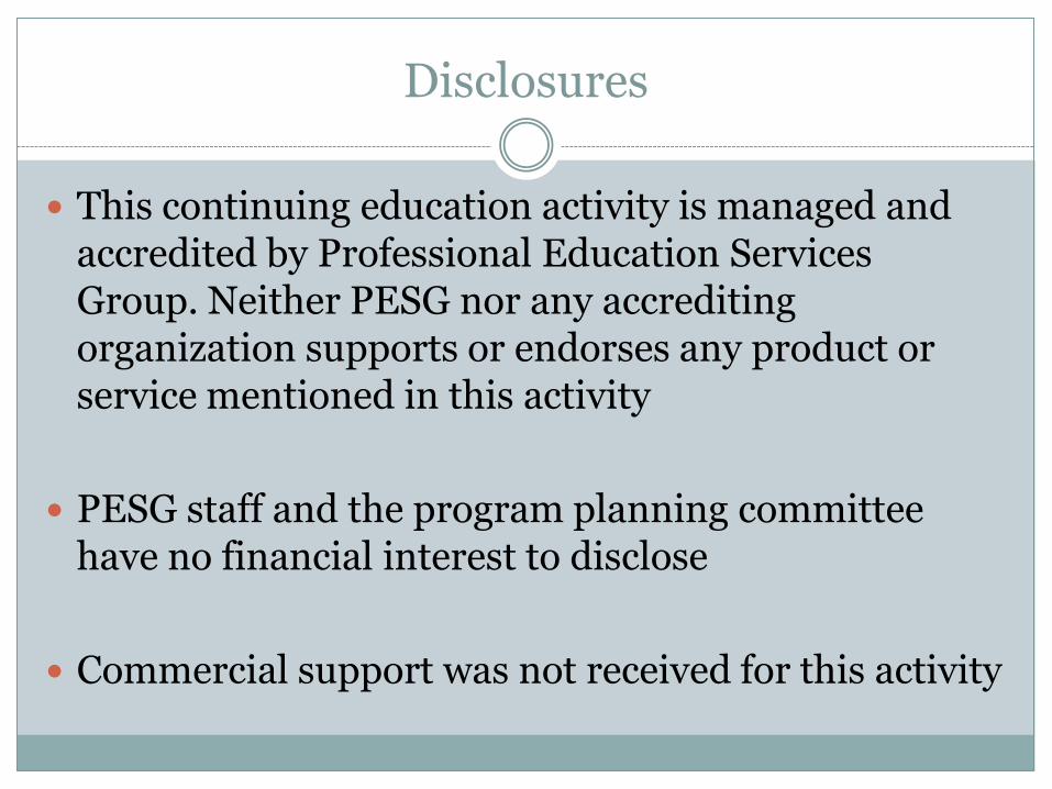
Disclosures
This continuing education activity is managed and accredited by Professional Education Services Group. Neither PESG nor any accrediting organization supports or endorses any product or service mentioned in this activity
PESG staff and the program planning committee have no financial interest to disclose
Commercial support was not received for this activity
Disclosures
Ileana Howard, MD
has no financial interest to disclose
Keith Claffey, MA, APN
has no financial interest to disclose
George Gombas, MD
has no financial interest to disclose
Learning Objectives
1. Describe a clinical algorithm for spasticity management
2. Discuss indications for advanced spasticity management- including chemodenervation and intrathecal baclofen pump
3. Recognize cases in which advanced spasticity management improves the patient’s outcome beyond what can be achieved through conservative measures
THE PROBLEM
Spasticity
What is spasticity?
Velocity dependent increase in tonic stretch reflexes with exaggerated tendon jerks
Lance 1980
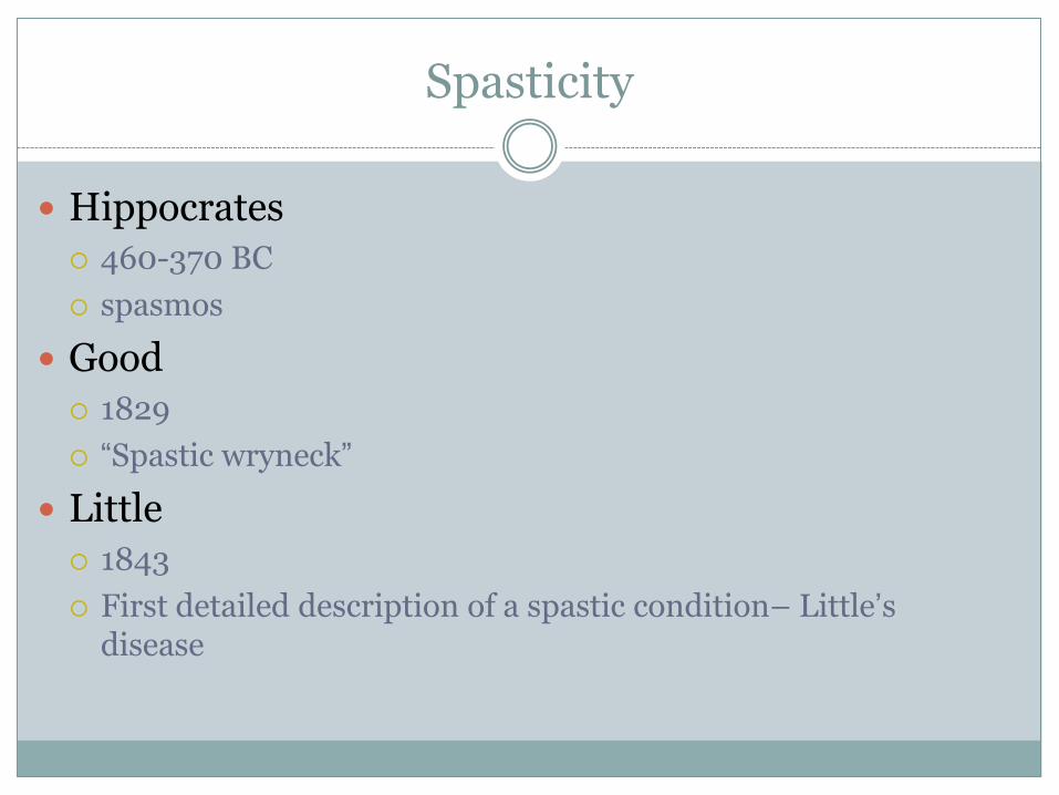
Spasticity
Hippocrates
460-370 BC
spasmos
Good
1829
“Spastic wryneck”
Little
1843
First detailed description of a spastic condition– Little’s disease
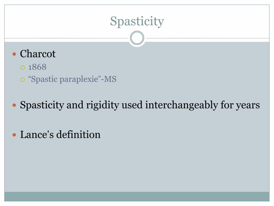
Spasticity
Charcot
1868
“Spastic paraplexie”-MS
Spasticity and rigidity used interchangeably for years
Lance’s definition
Spasticity
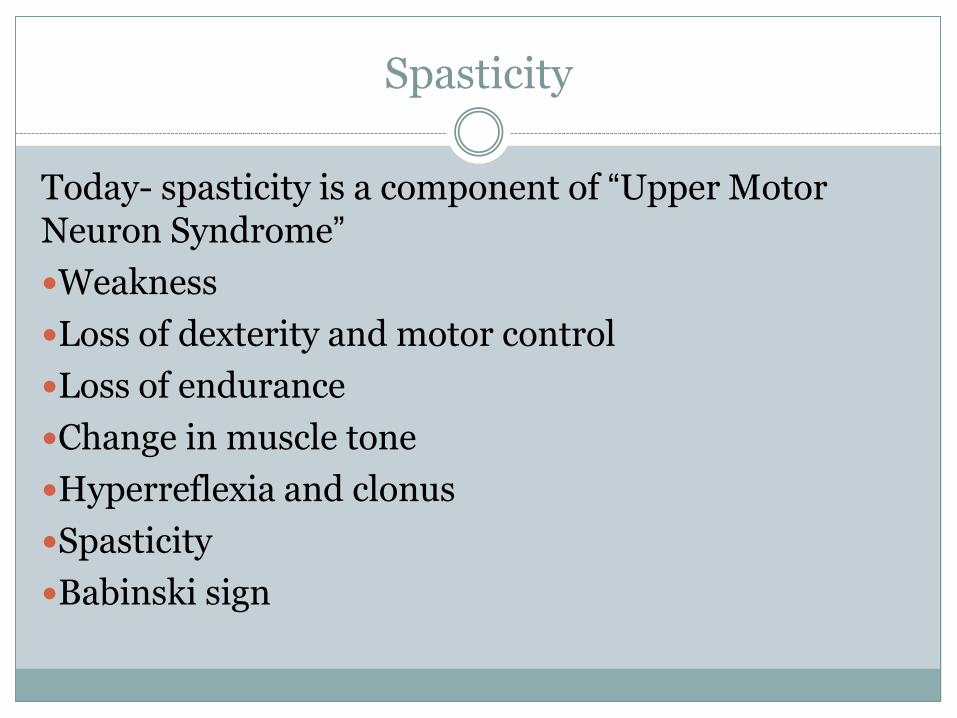
Today- spasticity is a component of “Upper Motor Neuron Syndrome”
Weakness
Loss of dexterity and motor control
Loss of endurance
Change in muscle tone
Hyperreflexia and clonus
Spasticity
Babinski sign

Spasticity
Spasticity
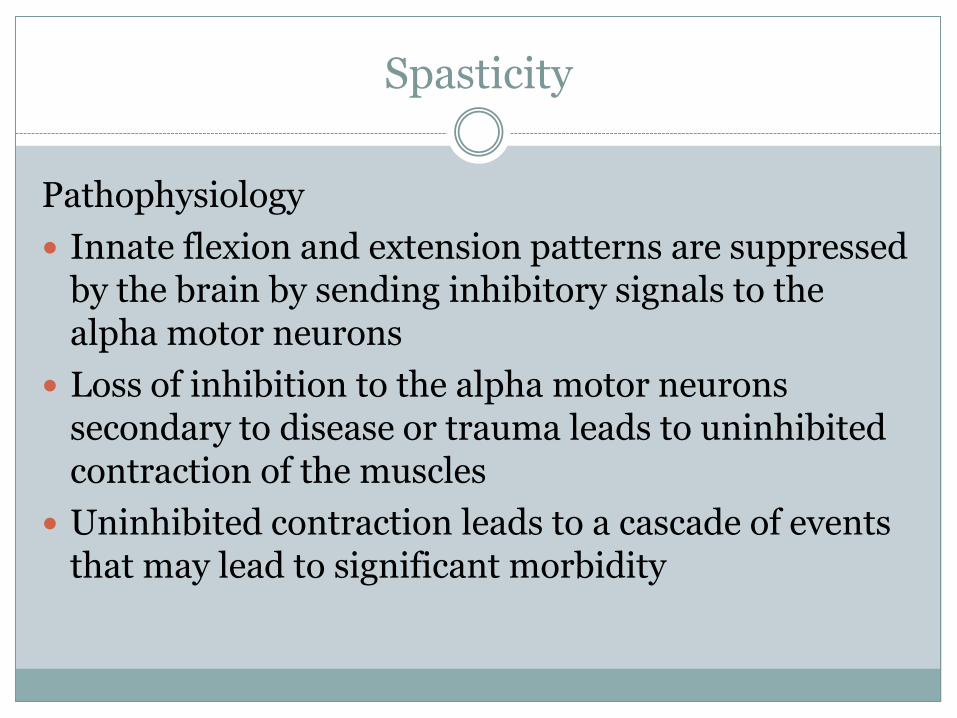
Pathophysiology
Innate flexion and extension patterns are suppressed by the brain by sending inhibitory signals to the alpha motor neurons
Loss of inhibition to the alpha motor neurons secondary to disease or trauma leads to uninhibited contraction of the muscles
Uninhibited contraction leads to a cascade of events that may lead to significant morbidity
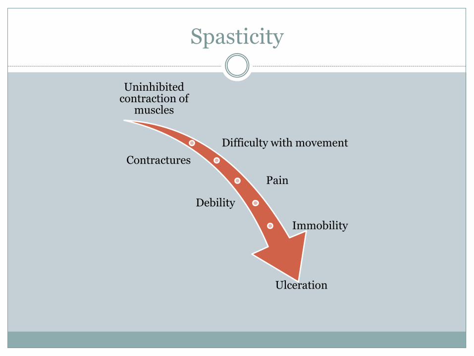
Spasticity
Uninhibited contraction of
muscles
Difficulty with movement
Contractures
Pain
Debility
Immobility
Ulceration
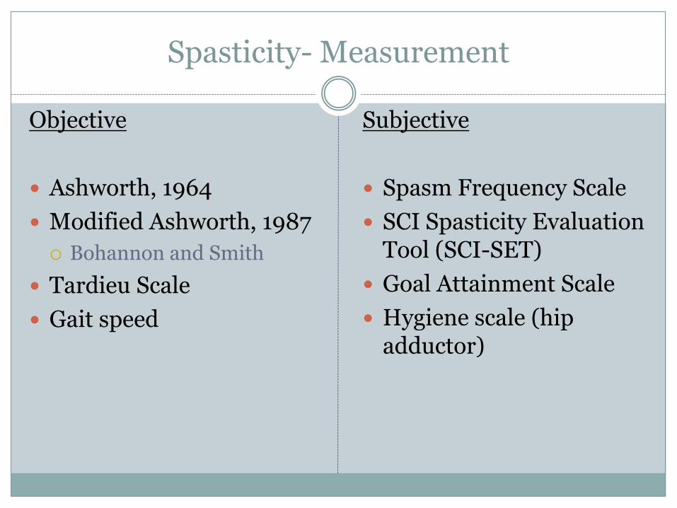
Spasticity- Measurement
Objective
Ashworth, 1964
Modified Ashworth, 1987
Bohannon and Smith
Tardieu Scale
Gait speed
Subjective
Spasm Frequency Scale
SCI Spasticity Evaluation Tool (SCI-SET)
Goal Attainment Scale
Hygiene scale (hip adductor)
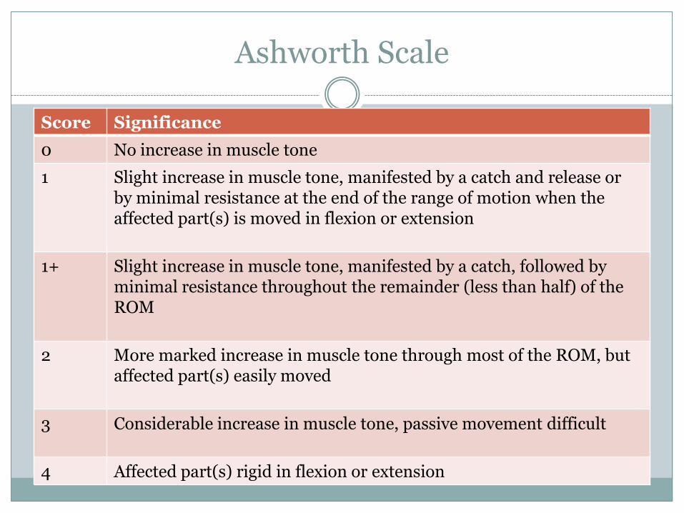
Ashworth Scale
Score Significance
0 No increase in muscle tone
1 Slight increase in muscle tone, manifested by a catch and release or by minimal resistance at the end of the range of motion when the affected part(s) is moved in flexion or extension
1+ Slight increase in muscle tone, manifested by a catch, followed by minimal resistance throughout the remainder (less than half) of the ROM
2 More marked increase in muscle tone through most of the ROM, but affected part(s) easily moved
3 Considerable increase in muscle tone, passive movement difficult
4 Affected part(s) rigid in flexion or extension
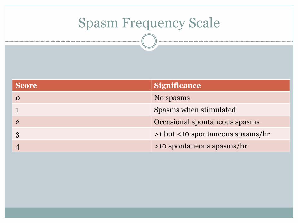
Spasm Frequency Scale
Score Significance
0 No spasms
1 Spasms when stimulated
2 Occasional spontaneous spasms
3 >1 but <10 spontaneous spasms/hr
4 >10 spontaneous spasms/hr
SPECTRUM OF DISORDERS
Spasticity
Spasticity
Conditions that may exhibit spasticity include
Spinal cord injury
Multiple Sclerosis
Stroke
Brain injury
ALS
Cerebral Palsy
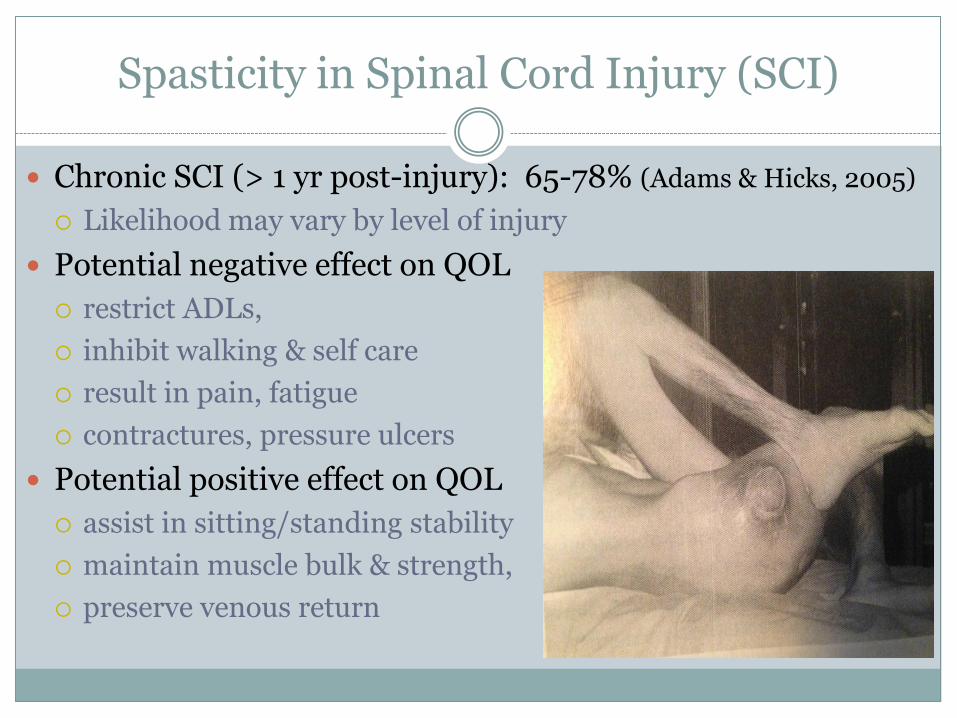
Spasticity in Spinal Cord Injury (SCI)
Chronic SCI (> 1 yr post-injury): 65-78% (Adams & Hicks, 2005)
Likelihood may vary by level of injury
Potential negative effect on QOL
restrict ADLs,
inhibit walking & self care
result in pain, fatigue
contractures, pressure ulcers
Potential positive effect on QOL
assist in sitting/standing stability
maintain muscle bulk & strength,
preserve venous return
Spasticity in MS
Prevalence: up to 84% (Rizzo, 2004) patients with MS report some spasticity
>30% report moderate to severe symptoms (frequent to daily interference with activities)
Males and those with more severe disability had more severe spasticity symptoms
Spasticity-related pain complaints very prevalent in persons with MS
Fine balance between alleviating symptoms and not limiting functional aspects of spasticity
Spasticity in ALS
Spasticity is common and decreases quality of life in persons with ALS
Spasticity management in ALS is different from other spinal cord disorders- botulinum toxin injections are relatively contraindicated in progressive neuromuscular disease
C O N S E R V A T I V E T R E A T M E N T
M E D I C A T I O N S
I N J E C T I O N S
I N T R A T H E C A L P U M P
S U R G E R Y
Spasticity Management Toolkit
S T R E T C H I N G
S P L I N T I N G
M O D A L I T I E S
Conservative Treatment for Spasticity
Stretching for Spasticity Management
Why:
Maintains/improves range of motion
Reduces the stretch input from the muscle spindle which triggers spasticity
Additive benefit of stretching combined with chemodenervation
How:
Weightbearing stretches may be superior to non-weightbearing stretches to diminish spasticity
Prolonged stretching >1 minute may be more effective
Splinting for Spasticity
Why:
Prolonged stretch
Diminished sensory input while in the splint
Maintains range of motion
How:
Serial casting
Static splints
Dynamic splints
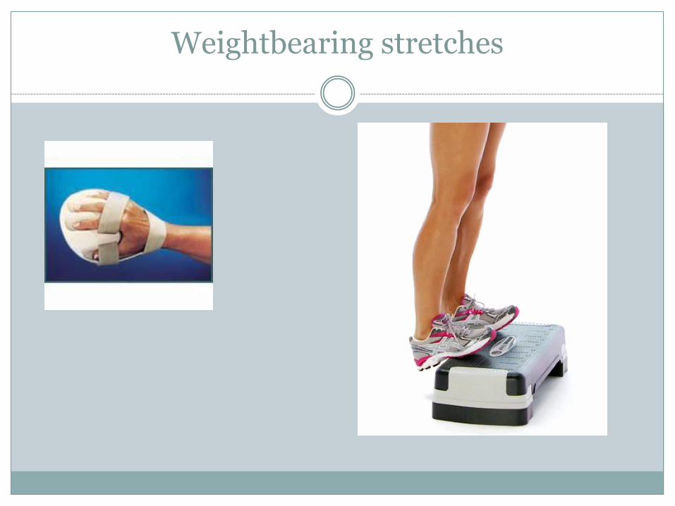
Weightbearing stretches
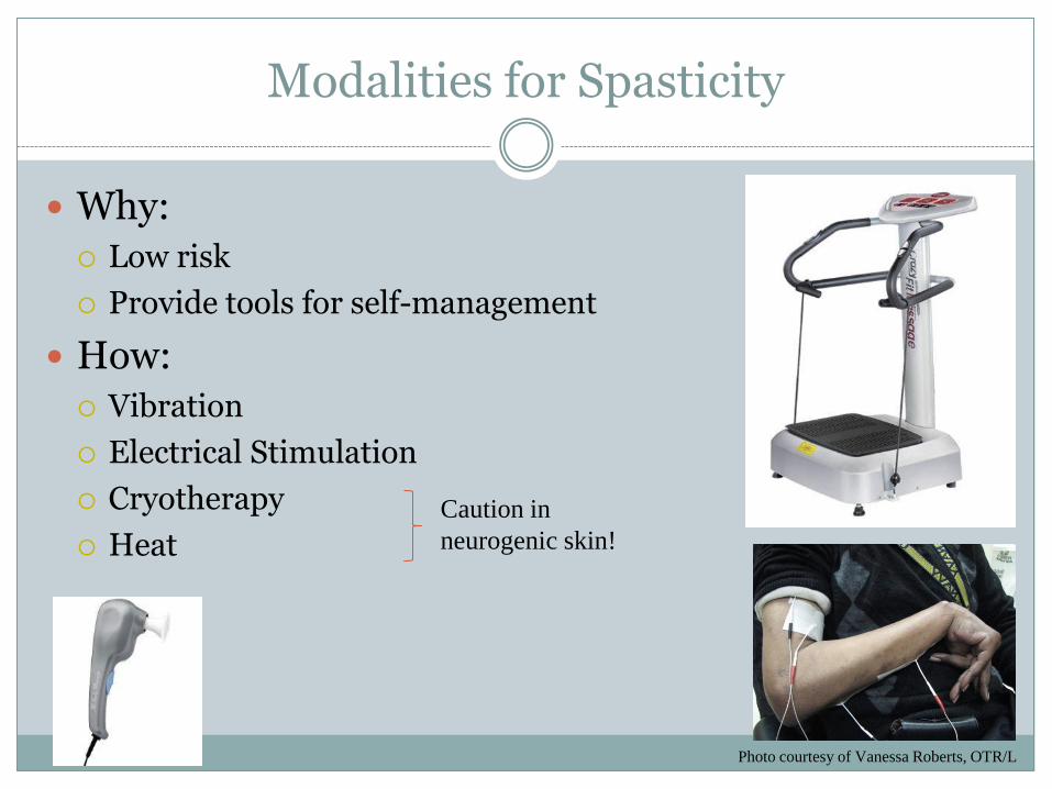
Modalities for Spasticity
Why:
Low risk
Provide tools for self-management
How:
Vibration
Electrical Stimulation
Cryotherapy
Heat
Caution in
neurogenic skin!
Photo courtesy of Vanessa Roberts, OTR/L
Exercise
Evidence for exercise as a primary or adjunctive treatment for spasticity
Land-based
Aquatic exercise
Functional Electrical Stimulation
Includes ALS among conditions benefiting from exercise with improved spasticity outcomes
Conservative Interventions: Summary
Multiple interventions available for spasticity management:
Stretching
Splinting
Exercise
Vibration
Electrical Stimulation
Heat/cold
Optimal advanced spasticity management requires:
Coordination of appointments with therapies/”pathway”
Communication/feedback with interdisciplinary team
Medications
Medications
Principles
One of the first interventions
Global as opposed to local involvement
Considerations
Adverse effects
“Pill burden”
Medications
Most commonly used types
Central acting
GABA analogs
Alpha 2 adrenergic
Peripheral acting
Medications- central acting
Gama-aminobutyric acid (GABA) receptor system
Chief inhibitory neurotransmitter of the central nervous system
Two main classes-A and B
Indirectly inhibits action potential formation in alpha motor neuron
Medications
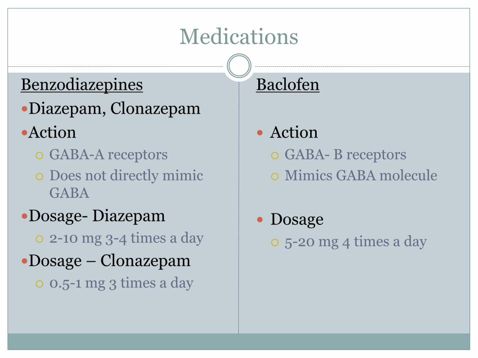
Benzodiazepines
Diazepam, Clonazepam
Action
GABA-A receptors
Does not directly mimic GABA
Dosage- Diazepam
2-10 mg 3-4 times a day
Dosage – Clonazepam
0.5-1 mg 3 times a day
Baclofen
Action
GABA- B receptors
Mimics GABA molecule
Dosage
5-20 mg 4 times a day
Medications
Benzodiazepine cont.
Adverse effects
sedation
cognitive impairment
dependence
withdrawal
seizures
Baclofen cont.
Adverse effects
drowsiness
fatigue
weakness
nausea
dizziness
hallucinations
seizures with stoppage of medication
Medications- central acting
Alpha 2 adrenergic receptor system
Receptors found throughout the body
Central - causes sedation and analgesia
Peripheral - causes bradycardia, diuresis, and vasoconstriction or dilation
Mechanism of action
Inhibit the release of excitatory amino acids in spinal interneurons
Medications
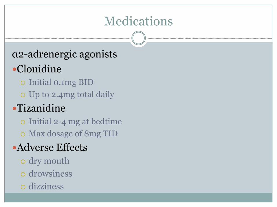
α2-adrenergic agonists
Clonidine
Initial 0.1mg BID
Up to 2.4mg total daily
Tizanidine
Initial 2-4 mg at bedtime
Max dosage of 8mg TID
Adverse Effects
dry mouth
drowsiness
dizziness
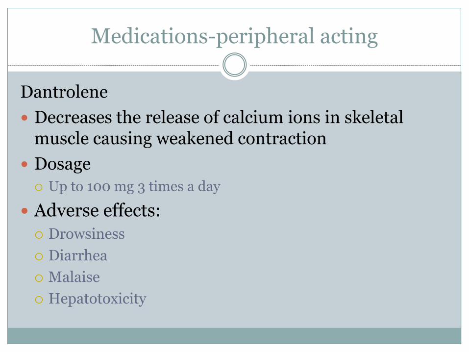
Medications-peripheral acting
Dantrolene
Decreases the release of calcium ions in skeletal muscle causing weakened contraction
Dosage
Up to 100 mg 3 times a day
Adverse effects:
Drowsiness
Diarrhea
Malaise
Hepatotoxicity
Advanced Spasticity Management
Chemodenervation: Botulinum toxin
Injections
Principles:
Focal treatment
Reduce exposure to side effects of systemic medications
Considerations
Invasive
Painful
May require repeated treatments
Botulinum Toxin
Most common question

Botulinum Toxin
Botulinum Toxin
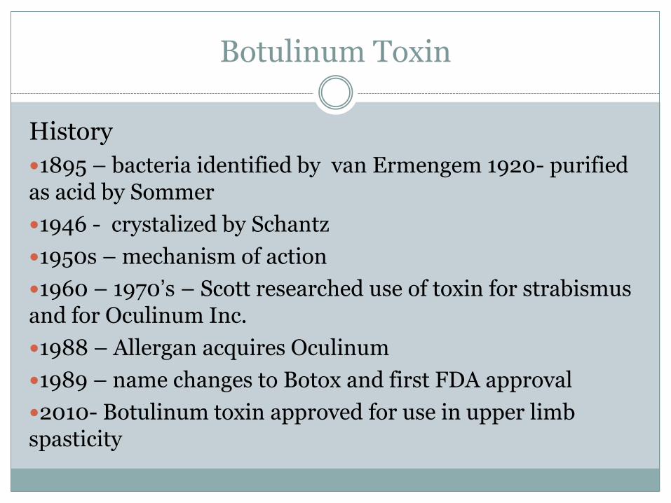
History
1895 – bacteria identified by van Ermengem 1920- purified as acid by Sommer
1946 - crystalized by Schantz
1950s – mechanism of action
1960 – 1970’s – Scott researched use of toxin for strabismus and for Oculinum Inc.
1988 – Allergan acquires Oculinum
1989 – name changes to Botox and first FDA approval
2010- Botulinum toxin approved for use in upper limb spasticity
Botulinum Toxin
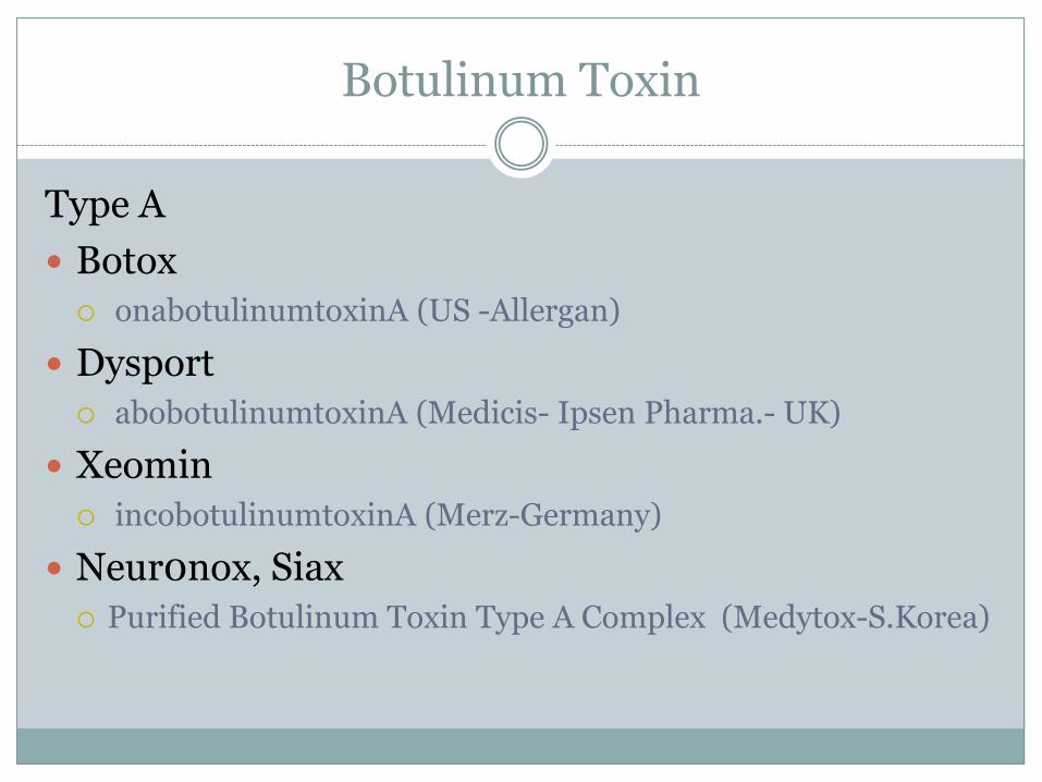
Type A
Botox
onabotulinumtoxinA (US -Allergan)
Dysport
abobotulinumtoxinA (Medicis- Ipsen Pharma.- UK)
Xeomin
incobotulinumtoxinA (Merz-Germany)
Neur0nox, Siax
Purified Botulinum Toxin Type A Complex (Medytox-S.Korea)
Botulinum Toxin
Type B
Myobloc, Neurobloc
rimabotulinumtoxinB (Solstice-US)
Botulinum Toxin
Most common treatment for focal spasticity
Inhibits release of acetylcholine at the neuromuscular junction
Weakens muscle
Starts to work in 3-7 days and can last up to 6 months
Cost – expensive - $500/100 units for BOTOX®
Botulinum Toxin
Sold as crystalline and diluted at time of use in normal saline
Wide variation in dilution practice
2-4 cc / 100 units
Proteins are delicate, caution during reconstitution
Units of one type botulinum toxin are not convertible to another
Typically 100-400 Units of BOTOX® Type A per session
Recommended maximum of BOTOX® is 360 Units/3 months.
Botulinum Toxin
Contraindications
Prior allergic reaction
Injection into areas of infection or inflammation
Pregnancy
Breast feeding
Caution
Myasthenia Gravis, ALS
Medications
Aminoglycosides, calcium channel blockers, penicillamine, quinine
Botulinum Toxin
Complications
dry mouth
reduced sweating
dysphagia
weakness
death
Botulinum Toxin
Dosing Adjustments
Voluntary control
Ashworth score
Patient weight
Muscle bulk
Botulinum Toxin
Recommended dilution of BOTOX® is 100 U/ 2cc Normal Saline
No more than 50 U or 0.5-1cc volume/site
25-30g needle for superficial muscles
22 g for deeper muscles
EMG for localization
Botulinum Toxin
Selected upper extremity conditions treated with BOTOX®
Flexed elbow:
Biceps, Brachialis, Brachioradialis
100-200 U divided in 4 sites
Flexed wrist
Flexor Carpi Radialis and Ulnaris
12.5-50 Units in each muscle in 1 site
Clenched fist
Flexor Digit. Profundus and Superficialis
30-50 U in each muscle in 1 site
Boutlinum Toxin
Selected lower extremity conditions treated with BOTOX® - not currently approved by FDA
Flexed hip
Iliopsoas and Rectus Femoris
100 U in each muscle in 2 sites
Flexed knee
Medial and lateral hamstrings
100 U in each muscle in 2-3 sites
Plantar flexed foot
Medial/lateral Gastrocnemius and Soleus
100 U in each muscle in 2-4 sites
Advanced Spasticity Management
Chemodenervation: phenol neurolysis
Phenol neurolysis
Lost art? Secret tool?
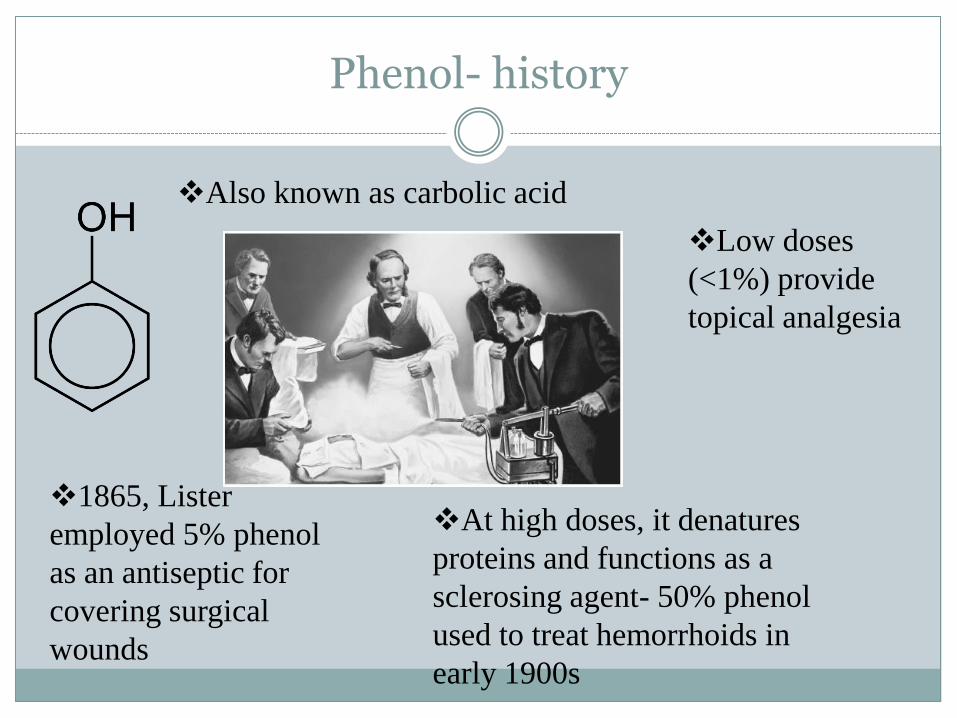
Phenol- history
Also known as carbolic acid
1865, Lister
employed 5% phenol
as an antiseptic for
covering surgical
wounds
At high doses, it denatures
proteins and functions as a
sclerosing agent- 50% phenol
used to treat hemorrhoids in
early 1900s
Low doses
(<1%) provide
topical analgesia
Phenol
Long history of use for spasticity and pain:
1950: Mandle injected phenol for sympathectomy
1955: intrathecal phenol used for intractable cancer pain
1959: Nathan, Kelly & Gauthier-Smith- injected intrathecal phenol for spasticity
1965: Halpern and Meelhuysen described motor point blocks with 5-7% phenol
No incremental benefit from 7% over 5%
1967: Khalili performed selective nerve blocks with 2-3% phenol
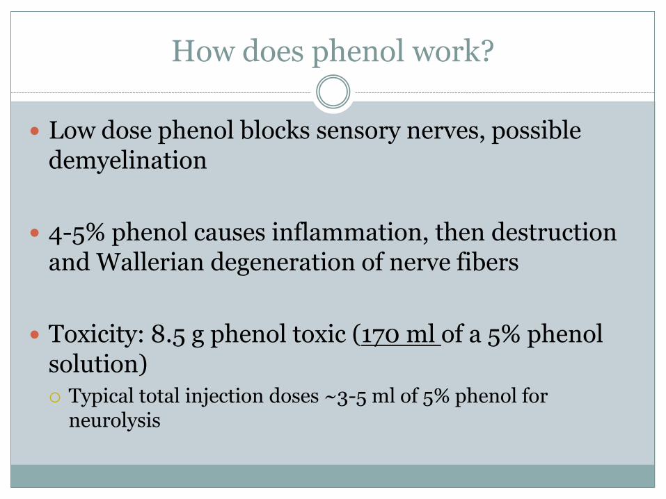
How does phenol work?
Low dose phenol blocks sensory nerves, possible demyelination
4-5% phenol causes inflammation, then destruction and Wallerian degeneration of nerve fibers
Toxicity: 8.5 g phenol toxic (170 ml of a 5% phenol solution)
Typical total injection doses ~3-5 ml of 5% phenol for neurolysis
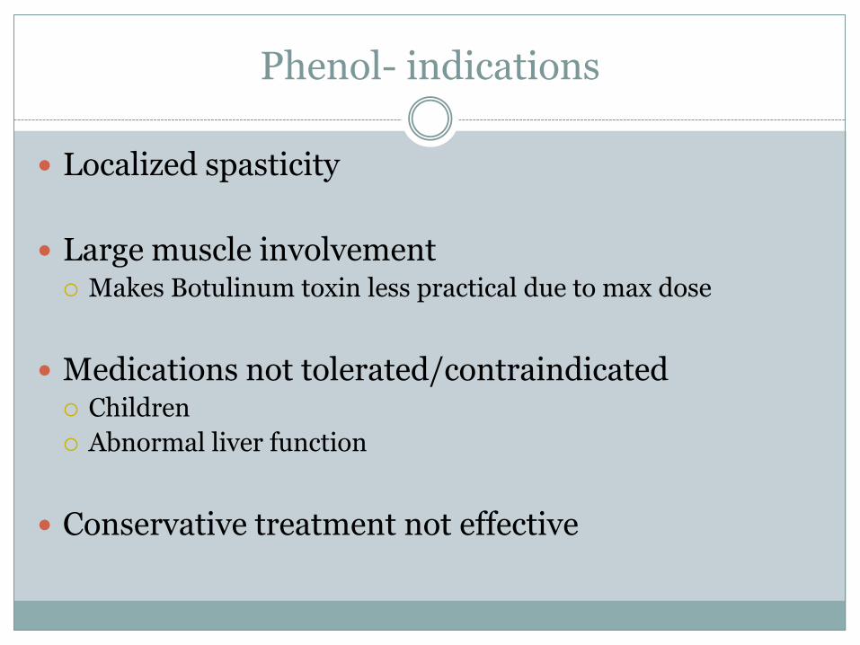
Phenol- indications
Localized spasticity
Large muscle involvement Makes Botulinum toxin less practical due to max dose
Medications not tolerated/contraindicated Children
Abnormal liver function
Conservative treatment not effective
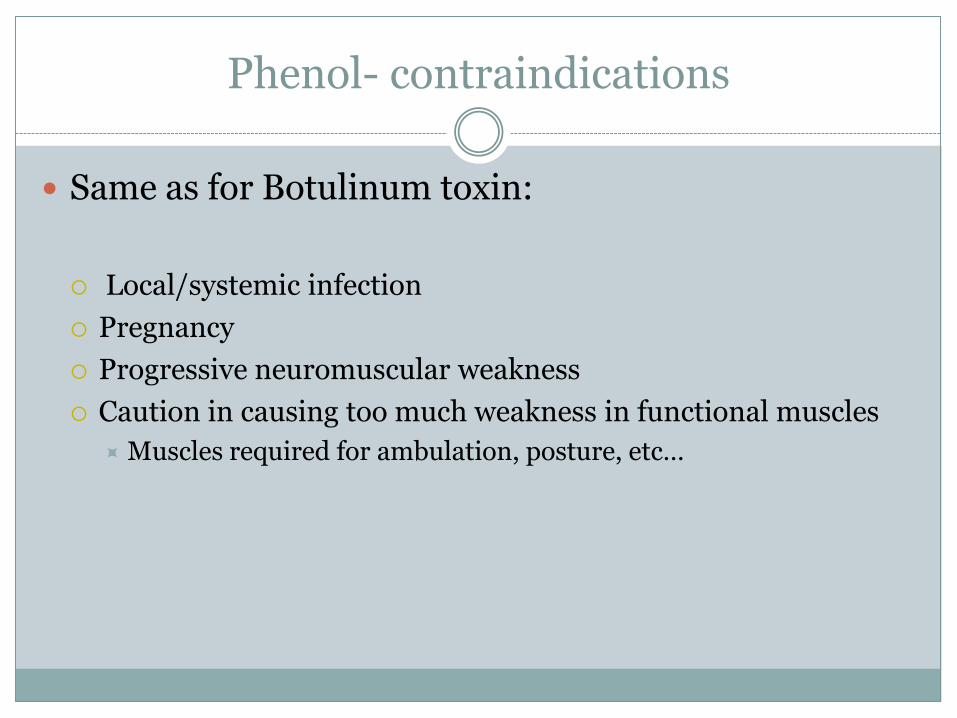
Phenol- contraindications
Same as for Botulinum toxin:
Local/systemic infection
Pregnancy
Progressive neuromuscular weakness
Caution in causing too much weakness in functional muscles
Muscles required for ambulation, posture, etc…

Phenol- benefits
Cost
Longer duration of effect (6-9 months vs. 3-4 months from BoNT)
Possibly beneficial for patients who live far from the medical center
Immediate effect- helpful in the acute rehab setting
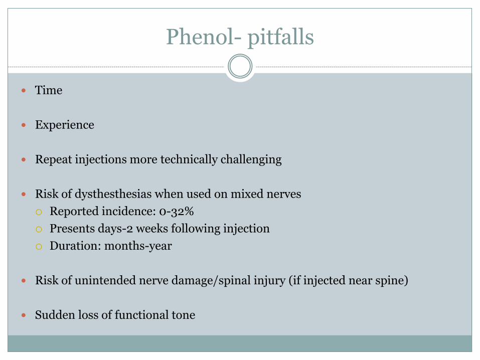
Phenol- pitfalls
Time
Experience
Repeat injections more technically challenging
Risk of dysthesthesias when used on mixed nerves
Reported incidence: 0-32%
Presents days-2 weeks following injection
Duration: months-year
Risk of unintended nerve damage/spinal injury (if injected near spine)
Sudden loss of functional tone
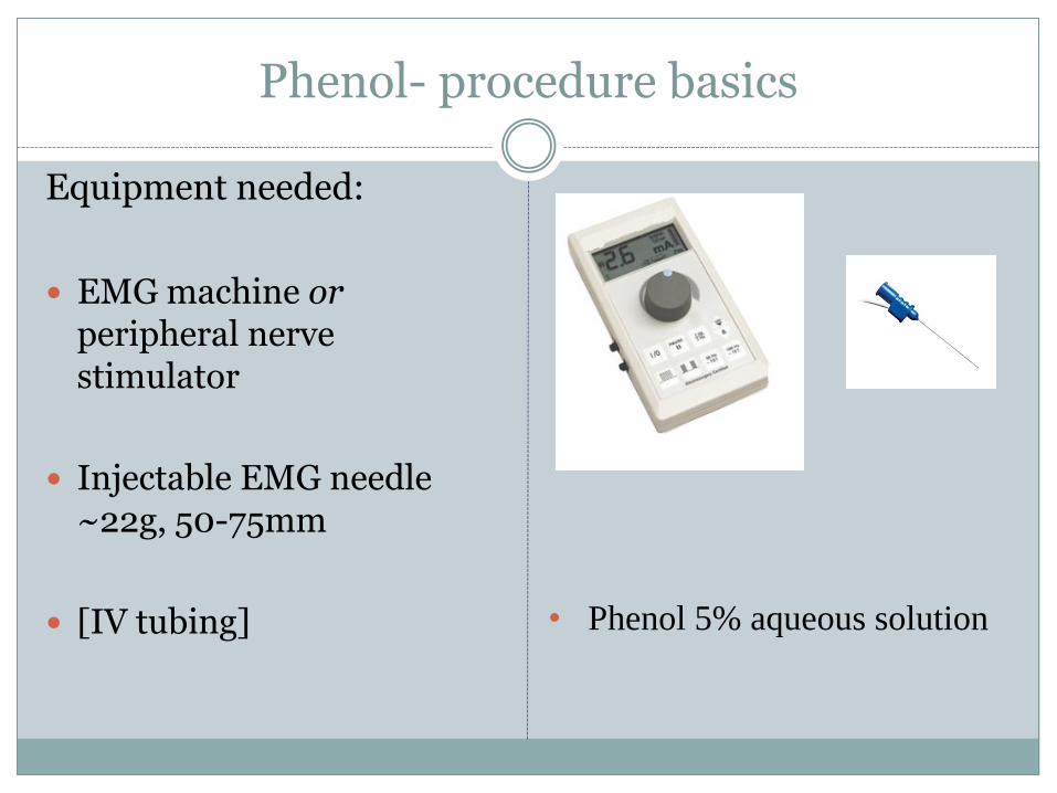
Phenol- procedure basics
Equipment needed:
EMG machine or peripheral nerve stimulator
Injectable EMG needle ~22g, 50-75mm
[IV tubing]
• Phenol 5% aqueous solution
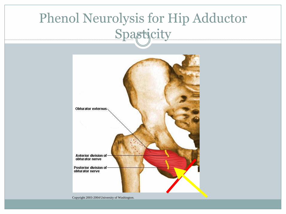
Phenol Neurolysis for Hip Adductor Spasticity
Copyright 2003-2004 University of Washington.
Phenol- summary
Slightly sharper learning curve than botulinum toxin injections
Longer duration of effect than botulinum toxin
Shorter onset of action than botulinum toxin
Less dosing limitation for larger muscle groups
Good option for hip adductor spasticity to facilitate hygiene or seating
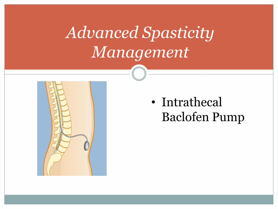
Advanced Spasticity Management
• Intrathecal Baclofen Pump
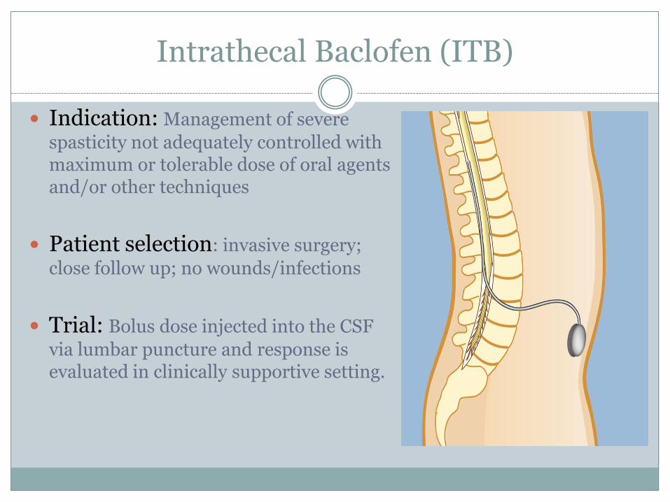
Intrathecal Baclofen (ITB)
Indication: Management of severe
spasticity not adequately controlled with maximum or tolerable dose of oral agents and/or other techniques
Patient selection: invasive surgery;
close follow up; no wounds/infections
Trial: Bolus dose injected into the CSF
via lumbar puncture and response is evaluated in clinically supportive setting.
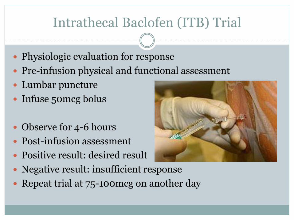
Intrathecal Baclofen (ITB) Trial
Physiologic evaluation for response
Pre-infusion physical and functional assessment
Lumbar puncture
Infuse 50mcg bolus
Observe for 4-6 hours
Post-infusion assessment
Positive result: desired result
Negative result: insufficient response
Repeat trial at 75-100mcg on another day
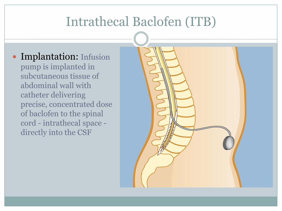
Intrathecal Baclofen (ITB)
Implantation: Infusion
pump is implanted in subcutaneous tissue of abdominal wall with catheter delivering precise, concentrated dose of baclofen to the spinal cord - intrathecal space - directly into the CSF
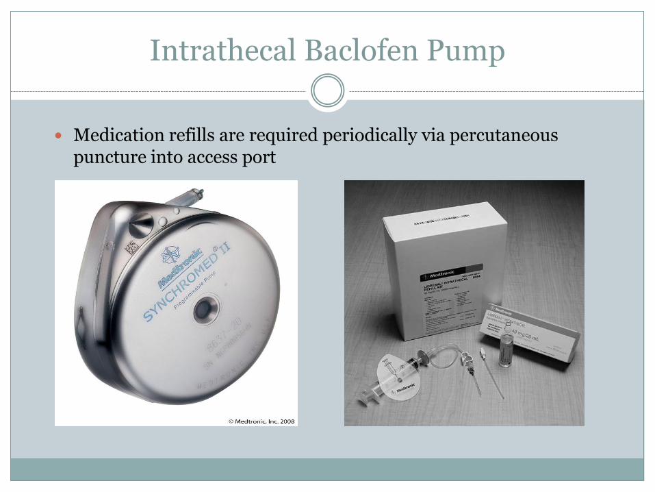
Intrathecal Baclofen Pump
Medication refills are required periodically via percutaneous puncture into access port
Benefits with ITB
Benefits as compared with oral meds:
High concentration of baclofen – 1/100th of oral dose
Reduce likelihood of negative side effects
Precise dose adjusted / customized for optimal effect
Effective in SCI&D: 97% (Penn, 1992) potentially reducing Ashworth Score (Dijkers, 1996)
May also provide improved pain management, decreased bladder hyperactivity, time saving in ADLs and for caregivers
Risks with ITB
Risks: overdose, withdrawal or infection possibly related to:
Catheter or pump moving or eroding through skin
Catheter: disconnection, leak, tear, kink, blockage, dislodgement, migration
Pump empty
Pump failure
Battery depletion
Withdrawal of ITB
Symptoms commonly seen: High fever, sweating
Itching without pruritis
Worsened spasticity and muscle rigidity
Altered mental status – irritability
Add caution for: Patients on high doses
Non-verbal
In care facility
After pump replacement
Patient should have supply/prescription on hand
ITB Clinical Pearls
ITB catheter is most commonly seated in the lumbar spine region for control of lower extremity spasticity, but can be placed higher
Patient, caregivers, providers must be educated on withdrawal symptoms and treatment
Patients/caregivers in remote/rural areas need support
Pump low volume alarm: most common few days after refill
Pump low volume alarm: may not be heard /noticed in loud care setting
Choose concentration of ITB that minimizes refill frequency but no longer than 6 months
MRI compatible but stops then restarts - needs to be checked at 2 hours
Surgery
Surgery
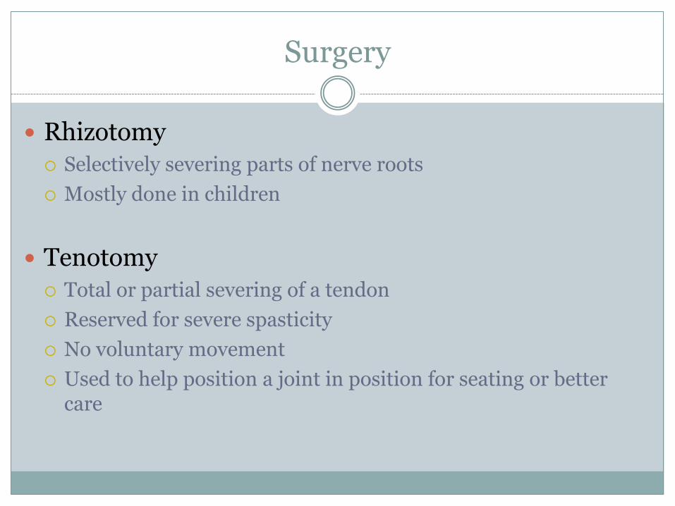
Rhizotomy
Selectively severing parts of nerve roots
Mostly done in children
Tenotomy
Total or partial severing of a tendon
Reserved for severe spasticity
No voluntary movement
Used to help position a joint in position for seating or better care
Algorithm for Spasticity Management
Algorithm – patient considerations
Focal or diffuse?
Upper or lower limbs affected?
Results/adverse reactions from prior treatments
Patient exam
Compliance
Goals of care
Preference
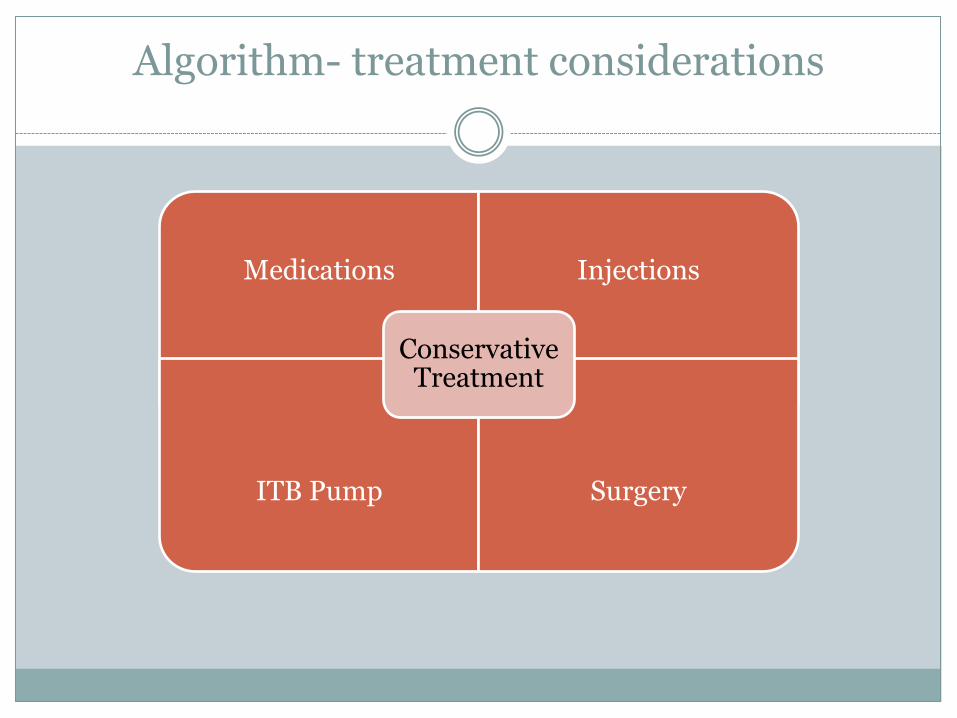
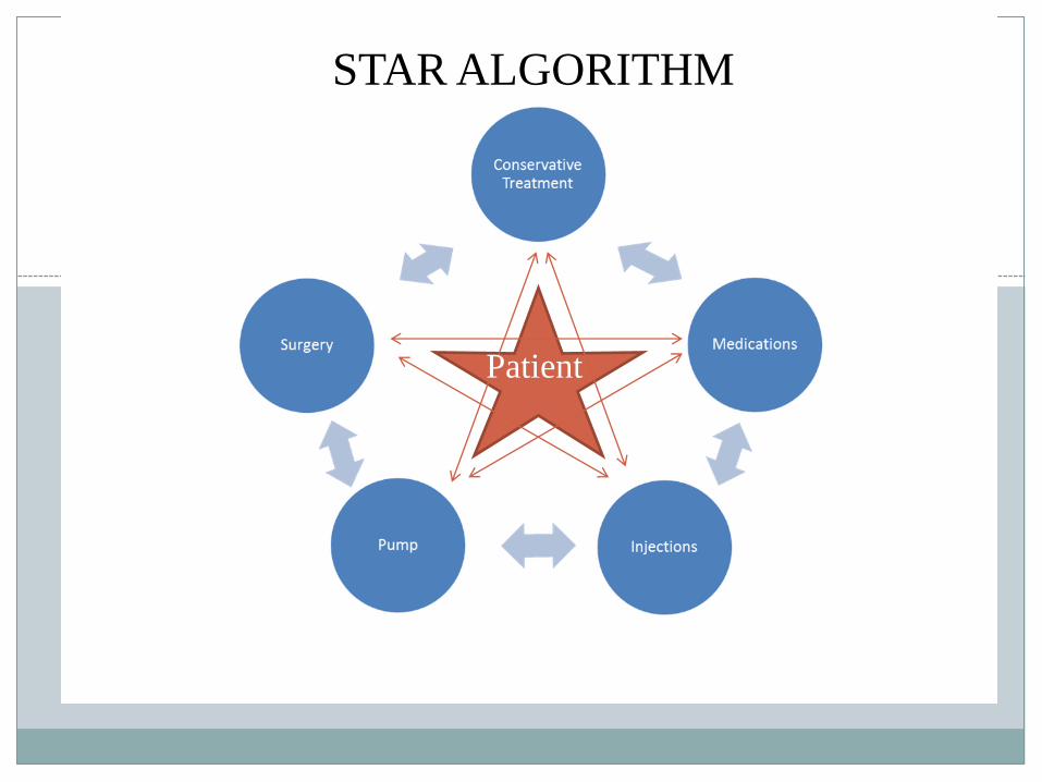
Algorithm- treatment considerations
Medications Injections
ITB Pump Surgery
Conservative Treatment
Factors Contributing to Spasticity
Important to minimize / treat contributing factors prior to treatment
Infection
Wound / pressure ulcer
Syringomyelia (syrinx)
Changes in level of activity, i.e. bed rest
Other noxious stimuli: full bladder/bowels, skin irritation, heat/cold, fracture, tight clothing, binders, discomfort from seating system
Algorithm
Cannot rely on just one treatment option, but a combination
Must constantly reassess patient and situation and adjust
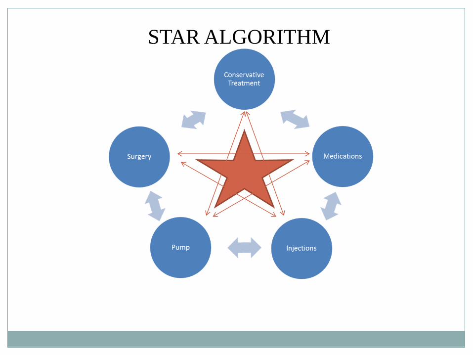
STAR ALGORITHM

Case Examples & Application of the Algorithm
Case 1
Case #1
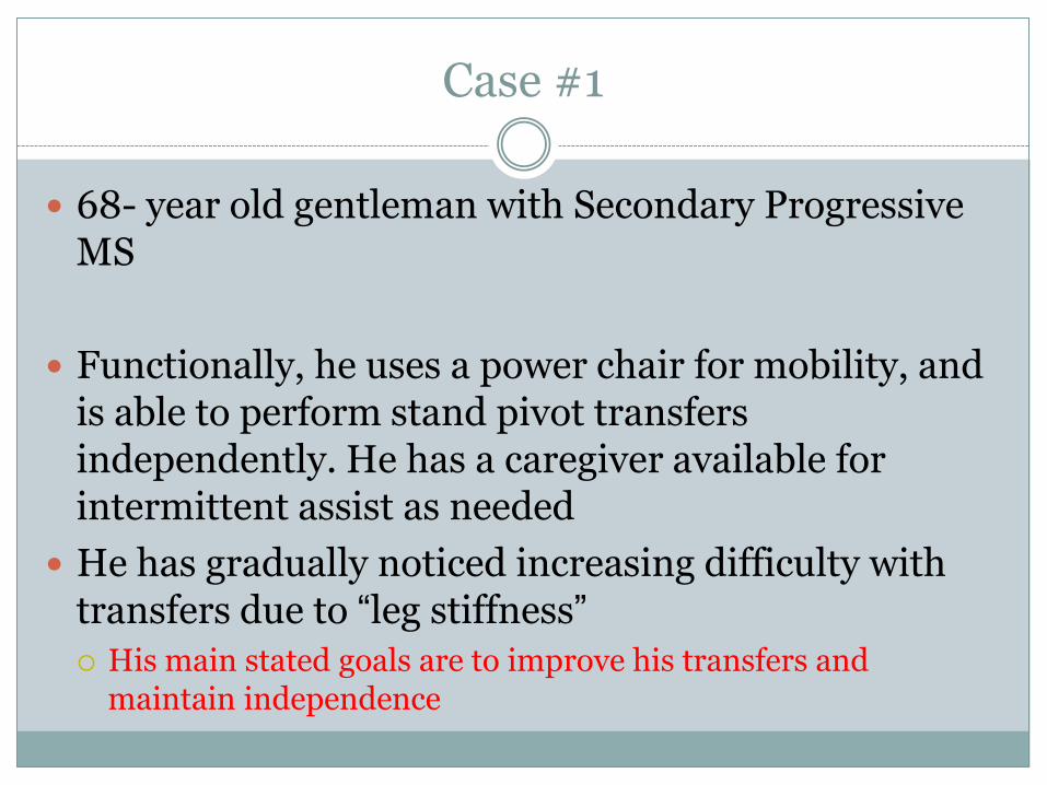
68- year old gentleman with Secondary Progressive MS
Functionally, he uses a power chair for mobility, and is able to perform stand pivot transfers independently. He has a caregiver available for intermittent assist as needed
He has gradually noticed increasing difficulty with transfers due to “leg stiffness”
His main stated goals are to improve his transfers and maintain independence
Case #1, continued
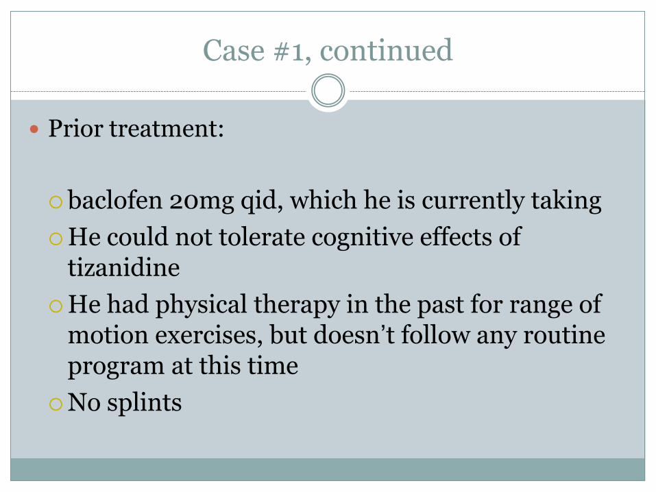
Prior treatment:
baclofen 20mg qid, which he is currently taking
He could not tolerate cognitive effects of tizanidine
He had physical therapy in the past for range of motion exercises, but doesn’t follow any routine program at this time
No splints
Case #1: Physical Exam
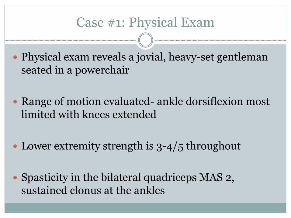
Physical exam reveals a jovial, heavy-set gentleman seated in a powerchair
Range of motion evaluated- ankle dorsiflexion most limited with knees extended
Lower extremity strength is 3-4/5 throughout
Spasticity in the bilateral quadriceps MAS 2, sustained clonus at the ankles
Patient evaluation, cont.
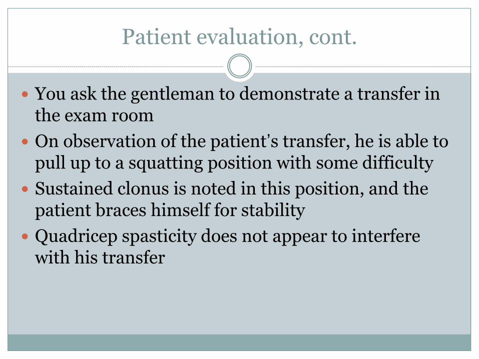
You ask the gentleman to demonstrate a transfer in the exam room
On observation of the patient’s transfer, he is able to pull up to a squatting position with some difficulty
Sustained clonus is noted in this position, and the patient braces himself for stability
Quadricep spasticity does not appear to interfere with his transfer
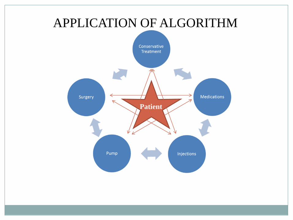
APPLICATION OF ALGORITHM
Patient
Patient treatment
Botulinum toxin injections were performed to the gastrocnemius bilaterally
The patient is referred to physical therapy to reinforce weightbearing stretches and nighttime splinting
Patient follow-up
He follows up in two weeks.
Improved range of motion at the ankles
Improved stability during transfers
He is very satisfied with the result of the treatment
He returns for follow-up at three months and reports effects have begun to wane, therefore he requests repeat injection

Case Examples & Application of the Algorithm
Case 2
Case #2
56 y.o. male Vet with C6 ASIA B tetraplegia; h/o syringomyelia, neurogenic bladder/bowel (SPT), spasticity & increased tone in LE bilaterally
Functionally, he uses a manual chair with power tilt feature for mobility, transfers with ceiling lift with assistance
Complains of muscle spasms resulting in pain, interference with ADLs & hygiene, increased pressure in seating and heel wounds from friction in lower extremities
Case #2, continued
Patient’s goal - better manage spasticity to:
Minimize pain
Decrease effort by him and caregivers for ADLs allowing improved hygiene and skin care
Reduce/eliminate increased pressure on boney prominences especially in lower extremities allowing healing of pressure existing pressure ulcers and decrease risk of further ulcers
Case #2, continued
Prior treatment:
baclofen 20mg qid - higher doses resulted in fatigue and only marginal decrease in symptoms
Physical exam:
thin, knees wind swept, manual w/c with power tilt
Range of motion:
diminished due to increased tone & spasticity in hip flexors, hamstrings, adductors, but reducible
STAR ALGORITHM
Patient
Case #2 - Patient Treatment
Continue conservative measures
Continue treatment with oral medications
Reduce/avoid contributing factors
Treat heel wounds:
osteomyelitis, subtotal calcanectomy with surgical closure
Scheduled for ITB trial
Seating optimized but…
difficult with prominent ITs even with tilt
developed UTI with leaking around catheter
developed pressure ulcer from moisture and pressure
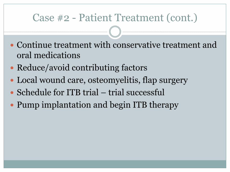
Case #2 - Patient Treatment (cont.)
Continue treatment with conservative treatment and oral medications
Reduce/avoid contributing factors
Local wound care, osteomyelitis, flap surgery
Schedule for ITB trial – trial successful
Pump implantation and begin ITB therapy
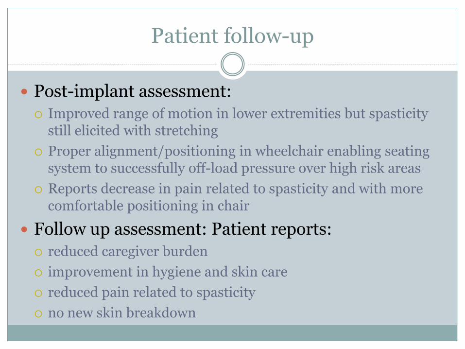
Patient follow-up
Post-implant assessment:
Improved range of motion in lower extremities but spasticity still elicited with stretching
Proper alignment/positioning in wheelchair enabling seating system to successfully off-load pressure over high risk areas
Reports decrease in pain related to spasticity and with more comfortable positioning in chair
Follow up assessment: Patient reports:
reduced caregiver burden
improvement in hygiene and skin care
reduced pain related to spasticity
no new skin breakdown
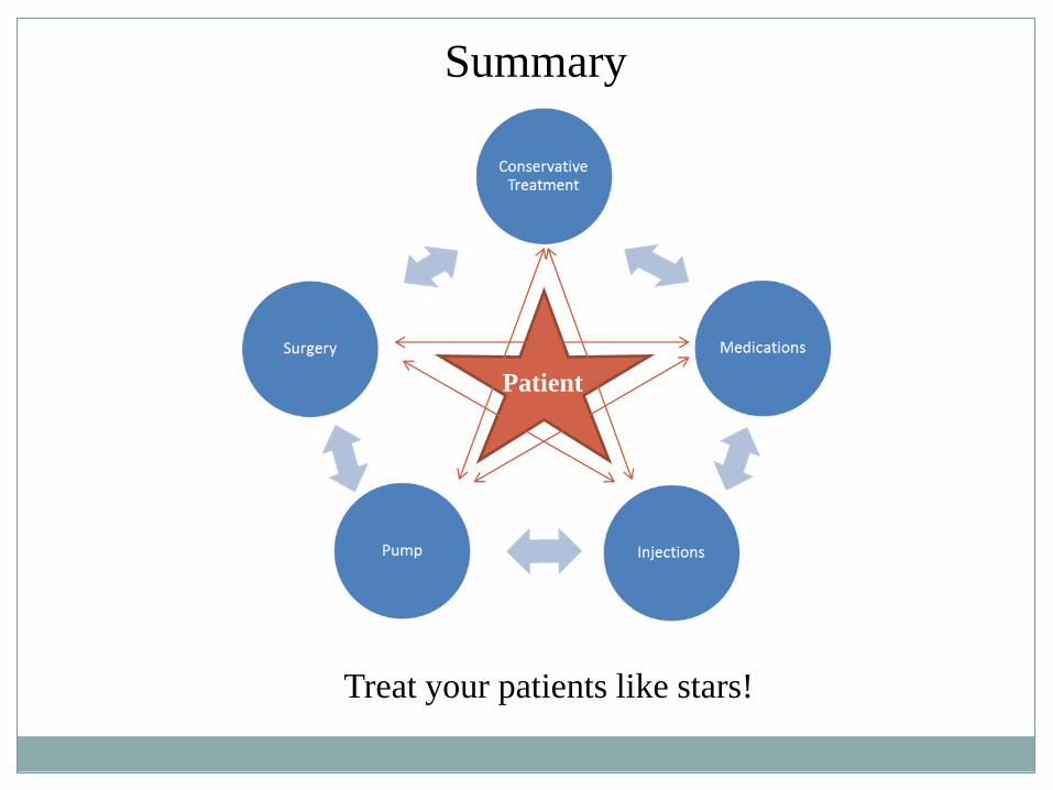
Summary
Patient
Treat your patients like stars!

Thank you!


![AWS Va - Amazon S3€¦ · AWS (DCX) IoT / APN 4 APN / APN 4 APN 18 . AWS 1.0 ... 5.1 AWS APN AWS APN competency-checklist@amazon.com “[APN Partner Name], Retail Competency Technology](https://img.pdfslide.us/doc/110x75/6148a9252918e2056c22d513/aws-va-amazon-s3-aws-dcx-iot-apn-4-apn-apn-4-apn-18-aws-10-51-aws.jpg)