Embed Size (px)
Citation preview
Journal of Molecular and Cellular Cardiology 47 (2009) 171–173
Contents lists available at ScienceDirect
Journal of Molecular and Cellular Cardiology
j ourna l homepage: www.e lsev ie r.com/ locate /y jmcc
Editorial
Keeping the beat: Life without SERCA — Is it possible?
Contraction in heart cells depends on a transient increase incytosolic [Ca2+], with both the extracellular space and intracellularstores serving as sources of Ca2+. In mammalian excitation contrac-tion coupling, the widely held view is that the majority of this Ca2+ isreleased from the sarcoplasmic reticulum (SR) in response to asmaller amount of trigger Ca2+ entering the cell on the L-type Ca2+
current (ICa-L) [1,2]. Ca2+ entry via ICa-L triggers thousands of theelementary units of Ca2+ release, Ca2+ sparks, and these sum toproduce the systolic Ca2+ transient [3–5]. Relaxation of the heart alsodepends critically on an SR Ca2+ transport protein, the SR Ca2+-ATPase (SERCA), and to a far lesser extent on Ca2+ efflux across thesarcolemma by the Na+–Ca2+ exchanger (NCX) and plasmalemmalCa2+-ATPase (PMCA) [6,7].
Thus, SERCA is pivotal to ensuring normal contractility in theworkingmyocardium, and indeedmuch of the contractile dysfunctionobserved in cardiac hypertrophy and heart failure has been attributedto rather modest reductions in SERCA activity [8–10]. Consistent withthis idea, contractile dysfunction in heart failure can be reversed whenSERCA2a (the predominant cardiac isoform) expression is increased infailing myocytes [11]. Ultimately therefore, received wisdom is thatlifewithout SERCAwould be impossible due to a collapse in the heart'sability to contract. It is therefore very surprising that Andersson et al.report in this issue of the Journal of Molecular and Cellular Cardiologythe result that the adult mouse heart can function in the nearcomplete absence of SERCA [12]. The authors utilized a novel inducibleknockout of the Serca2 gene in adult mice and remarkably discoveredthat despite a rapid decline in SERCA2 protein levels to less than 5% ofthe control and a reduction of SR Ca2+ content by approximately 80%,cardiac function is only modestly reduced. Ordinarily such a reductionof SR Ca2+ content would have a catastrophic impact on the systolicCa2+ transient due to the approximately cubic relationship betweenCa2+ transient amplitude and SR Ca2+ content [13,14]. As with someequally surprising recent results [15,16], this appears to be anotherexample of the heart's extraordinary plasticity. Here we discuss theseintriguing findings and address some of the potential compensatorymechanisms employed by the heart to ‘maintain’ a semblance ofnormal contractile function.
1. Excitation contraction coupling and the importance of the SR
Considerable previous work has highlighted the importance of theSR in controlling a number of aspects of cardiac function including: 1)a potential role in sinoatrial pacemaking [17], 2) an established role incontraction [1,2], and 3) an important factor in the generation ofarrhythmias [18,19]. During normal excitation contraction couplingthe SR contributes, depending on the species concerned, between 70and 90% of the Ca2+ required for contraction [2,6,7]. In line with these
0022-2828/$ – see front matter. Crown Copyright © 2009 Published by Elsevier Inc. All rigdoi:10.1016/j.yjmcc.2009.05.005
earlier findings, Andersson et al. note that in the mouse 88% of thesystolic Ca2+ transient amplitude is SR dependent [12] although thisreduces to 30% in the SERCA knockout. However, one of the manyremarkable features of the inducible SERCA2 knockout is that, at ratesconsidered near physiological for the mouse (6 Hz), myocytefractional shortening, is not altered despite the precipitous fall ofboth SR Ca2+ content and the systolic Ca2+ transient amplitude (77%decrease compared with control). The disparity between changes inmyocyte shortening and the amplitude of the systolic Ca2+ transientindicates one of the many coping strategies employed by the SERCAknockout mouse.
2. Life without a key regulator of systolic Ca2+; what's the exitstrategy?
While not addressed in the study by Andersson et al. [12], thedata discussed above are a strong indicator for subtle, yet importantchanges in the properties of the myofilaments and possibly alsocytosolic Ca2+ buffers in order to maintain functionality. There are anumber of additional compensatory mechanisms also employed thatare discussed below. However, it is of note that the SERCA knockoutis not unique in adapting to imposed changes in the Ca2+ handlingtranscriptome. For example, ablation of either NCX [15] or PMCA[20], other proteins involved in cytosolic Ca2+ removal, results inviable mice. With the NCX knockout, the adaptive mechanism ismainly centred on a substantial reduction in Ca2+ entry via ICa-L.This limits the need for Ca2+ efflux pathways to remove the Ca2+
entering by ICa-L in order to maintain Ca2+ flux balance and thusprevents the adverse sequelae associated with intracellular Ca2+
overload.On first appraisal the SERCA knockout should be in an even more
perilous situation than the NCX knockout due to the tenfold greatertranslocation of cytosolic Ca2+ by SERCA compared with NCX. Indeedprevious attempts to generate a homozygous SERCA knockout werefruitless due to embryonic lethality [21]. Andersson et al. [12] haveovercome the issues of embryonic lethality by combining Cre-Loxrecombination of Serca2 with inducible cardiac specific Cre recombi-nase expression in the adult mouse. These mice remain viable forapproximately 7 weeks with less than 20% of control levels of SERCA2present within 1 week of induction.
The inducible SERCA knockout mouse appears to utilize threemajor compensatory mechanisms: i) adaptations to myofilamentproperties as discussed above, ii) an increased reliance on trans-sarcolemmal Ca2+movements and iii) an increase in adrenergic drive.Unlike the NCX knockout, Ca2+ entry via ICa-L is increased and therequirement for Ca2+ flux balance is met by increased NCX function.Surprisingly, though, Andersson et al. find that the decay of the
hts reserved.
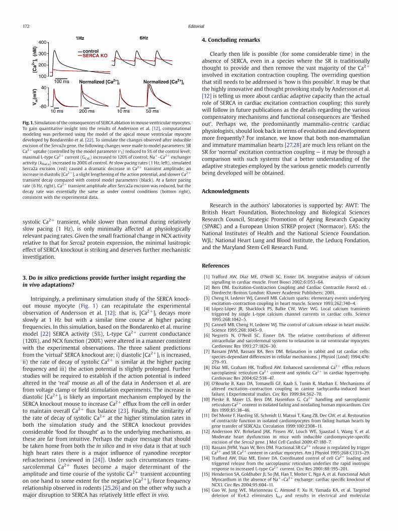
Fig.1. Simulation of the consequences of SERCA ablation inmouse ventricularmyocytes.To gain quantitative insight into the results of Andersson et al. [12], computationalmodeling was performed using the model of the apical mouse ventricular myocytedeveloped by Bondarenko et al. [22]. To simulate the changes observed after inducibleexcision of the Serca2a gene, the following changes were made tomodel parameters: SRCa2+ uptake (controlled by the model parameter ν3) reduced to 5% of the control level;maximal L-type Ca2+ current (GCaL) increased to 120% of control; Na+–Ca2+ exchangeractivity (kNaCa) increased to 200% of control. At slow pacing rates (1 Hz, left), simulatedSerca2a excision (red) caused a dramatic decrease in Ca2+ transient amplitude, anincrease in diastolic [Ca2+], a slight lengthening of the action potential, and slower Ca2+
transient decay compared with control model parameters (black). At a faster pacingrate (6 Hz, right), Ca2+ transient amplitude after Serca2a excision was reduced, but thedecay rate was essentially the same as under control conditions (bottom right),consistent with the experimental data.
172 Editorial
systolic Ca2+ transient, while slower than normal during relativelyslow pacing (1 Hz), is only minimally affected at physiologicallyrelevant pacing rates. Given the small fractional change in NCX activityrelative to that for Serca2 protein expression, the minimal lusitropiceffect of SERCA knockout is striking and deserves further mechanisticinvestigation.
3. Do in silico predictions provide further insight regarding thein vivo adaptations?
Intriguingly, a preliminary simulation study of the SERCA knock-out mouse myocyte (Fig. 1) can recapitulate the experimentalobservation of Andersson et al. [12]; that is, [Ca2+]i decays moreslowly at 1 Hz but with a similar time course at higher pacingfrequencies. In this simulation, based on the Bondarenko et al. murinemodel [22] SERCA activity (5%), L-type Ca2+ current conductance(120%), and NCX function (200%) were altered in a manner consistentwith the experimental observations. The three salient predictionsfrom the ‘virtual’ SERCA knockout are; i) diastolic [Ca2+]i is increased,ii) the rate of decay of systolic Ca2+ is similar at the higher pacingfrequency and iii) the action potential is slightly prolonged. Furtherstudies will be required to establish if the action potential is indeedaltered in the ‘real’ mouse as all of the data in Andersson et al. arefrom voltage clamp or field stimulation experiments. The increase indiastolic [Ca2+]i is likely an important mechanism employed by theSERCA knockout mouse to increase Ca2+ efflux from the cell in orderto maintain overall Ca2+ flux balance [23]. Finally, the similarity ofthe rate of decay of systolic Ca2+ at the higher stimulation rates inboth the simulation study and the SERCA knockout providesconsiderable ‘food for thought’ as to the underlying mechanisms, asthese are far from intuitive. Perhaps the major message that shouldbe taken home from both the in silico and in vivo data is that at suchhigh heart rates there is a major influence of ryanodine receptorrefractoriness (reviewed in [24]). Under such circumstances trans-sarcolemmal Ca2+ fluxes become a major determinant of theamplitude and time course of the systolic Ca2+ transient accountingon one hand to some extent for the negative [Ca2+]i/force frequencyrelationship observed in rodents [25,26] and on the other why such amajor disruption to SERCA has relatively little effect in vivo.
4. Concluding remarks
Clearly then life is possible (for some considerable time) in theabsence of SERCA, even in a species where the SR is traditionallythought to provide and then remove the vast majority of the Ca2+
involved in excitation contraction coupling. The overriding questionthat still needs to be addressed is ‘how is this possible’. It may be thatthe highly innovative and thought provoking study by Andersson et al.[12] is telling us more about cardiac adaptive capacity than the actualrole of SERCA in cardiac excitation contraction coupling; this surelywill follow in future publications as the details regarding the variouscompensatory mechanisms and functional consequences are ‘fleshedout’. Perhaps we, the predominantly mammalio-centric cardiacphysiologists, should look back in terms of evolution and developmentmore frequently? For instance, we know that both non-mammalianand immature mammalian hearts [27,28] are much less reliant on theSR for ‘normal’ excitation contraction coupling — it may be through acomparison with such systems that a better understanding of theadaptive strategies employed by the various genetic models currentlybeing developed will be obtained.
Acknowledgments
Research in the authors' laboratories is supported by: AWT: TheBritish Heart Foundation, Biotechnology and Biological SciencesResearch Council, Strategic Promotion of Ageing Research Capacity(SPARC) and a European Union STREP project (Normacor). EAS: theNational Institutes of Health and the National Science Foundation.WJL: National Heart Lung and Blood Institute, the Leducq Fondation,and the Maryland Stem Cell Research Fund.
References
[1] Trafford AW, Díaz ME, O'Neill SC, Eisner DA. Integrative analysis of calciumsignalling in cardiac muscle. Front Biosci 2002;6:D53–64.
[2] Bers DM. Excitation-Contraction Coupling and Cardiac Contractile Force2 ed. .Dordrecht/Boston/London: Kluwer Academic Publishers; 2001.
[3] Cheng H, Lederer WJ, Cannell MB. Calcium sparks: elementary events underlyingexcitation–contraction coupling in heart muscle. Science 1993;262:740–4.
[4] López-López JR, Shacklock PS, Balke CW, Wier WG. Local calcium transientstriggered by single L-type calcium channel currents in cardiac cells. Science1995;268:1042–5.
[5] Cannell MB, Cheng H, Lederer WJ. The control of calcium release in heart muscle.Science 1995;268:1045–9.
[6] Negretti N, O'Neill SC, Eisner DA. The relative contributions of differentintracellular and sarcolemmal systems to relaxation in rat ventricular myocytes.Cardiovasc Res 1993;27:1826–30.
[7] Bassani JWM, Bassani RA, Bers DM. Relaxation in rabbit and rat cardiac cells:species-dependant differences in cellular mechanisms. J Physiol (Lond) 1994;476:279–93.
[8] Díaz ME, Graham HK, Trafford AW. Enhanced sarcolemmal Ca2+ efflux reducessarcoplasmic reticulum Ca2+ content and systolic Ca2+ in cardiac hypertrophy.Cardiovasc Res 2004;62:538–47.
[9] O'Rourke B, Kass DA, Tomaselli GF, Kaab S, Tunin R, Marban E. Mechanisms ofaltered excitation–contraction coupling in canine tachycardia-induced heartfailure, I Experimental studies. Circ Res 1999;84:562–70.
[10] Pieske B, Maier LS, Bers DM, Hasenfuss G. Ca2+ handling and sarcoplasmicreticulum Ca2+ content in isolated failing and nonfailing human myocardium. CircRes 1999;85:38–46.
[11] Del Monte F, Harding SE, Schmidt U, Matsui T, Kang ZB, Dec GW, et al. Restorationof contractile function in isolated cardiomyocytes from failing human hearts bygene transfer of SERCA2a. Circulation 1999;100:2308–11.
[12] Andersson KV, Birkeland JAK, Finsen AV, Louch WE, Sjaastad I, Wang Y, et al.Moderate heart dysfunction in mice with inducible cardiomyocyte-specificexcision of the Serca2 gene. J Mol Cell Cardiol 2009;47:180–7.
[13] Bassani JWM, Yuan W, Bers DM. Fractional SR Ca2+ release is regulated by triggerCa2+ and SR Ca2+ content in cardiac myocytes. Am J Physiol 1995;268:C1313–29.
[14] Trafford AW, Díaz ME, Eisner DA. Coordinated control of cell Ca2+ loading andtriggered release from the sarcoplasmic reticulum underlies the rapid inotropicresponse to increased L-type Ca2+ current. Circ Res 2001;88:195–201.
[15] Henderson SA, Goldhaber JI, So JM, Han T, Motter C, Ngo A, et al. Functional AdultMyocardium in the absence of Na+–Ca2+ exchange: cardiac specific knockout ofNCX1. Circ Res 2004;95:604–11.
[16] Guo W, Jung WE, Marionneau C, Aimond F, Xu H, Yamada KA, et al. Targeteddeletion of Kv4.2 eliminates Ito,f and results in electrical and molecular
173Editorial
remodeling, with no evidence of ventricular hypertrophy or myocardial dysfunc-tion. Circ Res 2005;97:1342–50.
[17] Lakatta EG, DiFrancesco D. What keeps us ticking: a funny current, a calcium clock,or both? J Mol Cell Cardiol 2009;47:157–70.
[18] Díaz ME, Trafford AW, O'Neill SC, Eisner DA. A measurable reduction of SR Ca2+
content follows spontaneous Ca2+ release in rat ventricular myocytes. PflügersArchiv 1997;434:852–4.
[19] Cheng H, Lederer WJ. Calcium Sparks Physiol Rev 2008;88:1491–545.[20] Oceandy D, Cartwright EJ, Emerson M, Prehar S, Baudoin FM, Zi M, et al. Neuronal
Nitric Oxide Synthase Signaling in the Heart Is Regulated by the SarcolemmalCalcium Pump 4b. Circulation 2007;115:483–92.
[21] Periasamy M, Reed TD, Liu LH, Ji Y, Loukianov E, Paul RJ, et al. Impaired cardiacperformance in heterozygous mice with a null mutation in the sarco(endo)plasmic reticulum Ca2+-ATPase isoform 2 (SERCA2) gene. J Biol Chem 1999;274:2556–62.
[22] Bondarenko VE, Szigeti GP, Bett GC, Kim SJ, Rasmusson RL. Computer model ofaction potential of mouse ventricular myocytes. Am J Physiol Heart Circ Physiol2004;287:H1378–403.
[23] Dibb KM, Eisner DA, Trafford AW. Regulation of systolic [Ca2+]i and cellular Ca2+
flux balance in rat ventricular myocytes by SR Ca2+, L-type Ca2+ current anddiastolic [Ca2+]i. J Physiol (Lond) 2007;585:579–92.
[24] Sobie EA, Song LS, Lederer WJ. Restitution of Ca2+ release and vulnerability toarrhythmias. J Cardiovasc Electrophysiol 2006;17(Suppl 1):S64–70.
[25] Dibb KM, Eisner DA, Trafford AW. Regulation of systolic [Ca2+]i and cellular Ca2+
flux balance in rat ventricular myocytes by SR Ca2+, L-type Ca2+ current anddiastolic [Ca2+]i. J Physiol (Lond) 2007;585:579–92.
[26] Antoons G, Mubagwa K, Nevelsteen I, Sipido KR. Mechanisms underlying thefrequency dependence of contraction and Ca2+ transients in mouse ventricularmyocytes. J Physiol (Lond) 2002;543:889–98.
[27] Snopko RM, Aromolaran AS, Karko KL, Ramos-Franco J, Blatter LA, Mejia-Alvarez R.Cell culture modifies Ca2+ signaling during excitation–contraction coupling inneonate cardiac myocytes. Cell Calcium 2007;41:13–25.
[28] Wang LJ, Sobie EA. Mathematical model of the neonatal mouse ventricular actionpotential. Am J Physiol Heart Circ Physiol 2008;294:H2565–75.
Andrew W. TraffordUnit of Cardiac Physiology, University of Manchester,
3.08 Core Technology Facility, 46 Grafton Street,Manchester, M13 9NT, UK
E-mail address: [email protected] author. Tel.: +44 161 275 7969;
fax: +44 161 275 2703.
W. Jonathan LedererInstitute of Molecular Cardiology,
Medical Biotechnology Center,University of Maryland Biotechnology Institute,
725 W. Lombard Street, Baltimore, MD 21201, USA
Eric A. SobieDepartment of Pharmacology and Systems Therapeutics,Mount Sinai School of Medicine, 1425 Madison Avenue,
New York, NY 10029, USAInstitute of Molecular Cardiology,
Medical Biotechnology Center, USA
8 May 2009


![ROC RyR - Columbia University€¦ · RyR VDAC SERCA SERCA NCE PMCA mM [Ca] 100 nM [Ca] mM [Ca] mM [Ca] 2 Sarcoplasmic Reticulum (SR) / T Tubule System. 3. 4 Twitch Summation Tetanus](https://img.pdfslide.us/doc/110x75/5ec3efeb3ae4ef235843ba2f/roc-ryr-columbia-ryr-vdac-serca-serca-nce-pmca-mm-ca-100-nm-ca-mm-ca-mm.jpg)