Embed Size (px)
Citation preview
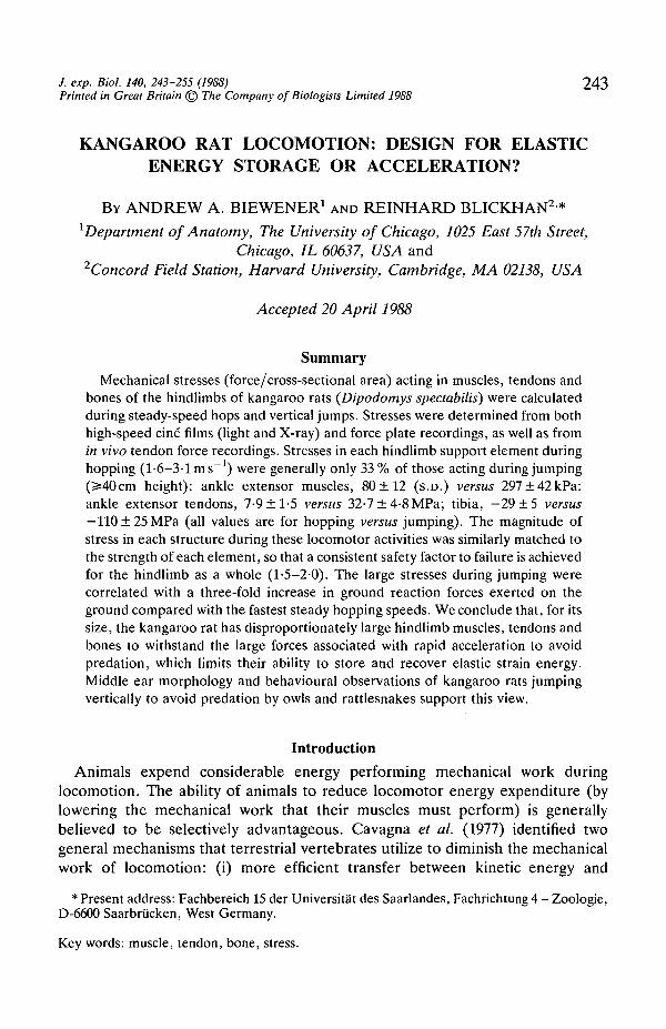
J. exp. Biol. 140, 243-255 (1988) 2 4 3Printed in Great Britain © The Company of Biologists Limited 1988
KANGAROO RAT LOCOMOTION: DESIGN FOR ELASTICENERGY STORAGE OR ACCELERATION?
BY ANDREW A. BIEWENER1 AND REINHARD BLICKHAN2*1 Department of Anatomy, The University of Chicago, 1025 East 57th Street,
Chicago, IL 60637, USA and2Concord Field Station, Harvard University, Cambridge, MA 02138, USA
Accepted 20 April 1988
Summary
Mechanical stresses (force/cross-sectional area) acting in muscles, tendons andbones of the hindlimbs of kangaroo rats {Dipodomys spectabilis) were calculatedduring steady-speed hops and vertical jumps. Stresses were determined from bothhigh-speed cine films (light and X-ray) and force plate recordings, as well as fromin vivo tendon force recordings. Stresses in each hindlimb support element duringhopping (1-6-3-1 m s"1) were generally only 33 % of those acting during jumping(3=40cm height): ankle extensor muscles, 80 ±12 (S.D.) versus 297±42kPa;ankle extensor tendons, 7-9 ±1-5 versus 32-7 ± 4-8 MPa; tibia, —29 ±5 versus— 110±25MPa (all values are for hopping versus jumping). The magnitude ofstress in each structure during these locomotor activities was similarly matched tothe strength of each element, so that a consistent safety factor to failure is achievedfor the hindlimb as a whole (1-5-2-0). The large stresses during jumping werecorrelated with a three-fold increase in ground reaction forces exerted on theground compared with the fastest steady hopping speeds. We conclude that, for itssize, the kangaroo rat has disproportionately large hindlimb muscles, tendons andbones to withstand the large forces associated with rapid acceleration to avoidpredation, which limits their ability to store and recover elastic strain energy.Middle ear morphology and behavioural observations of kangaroo rats jumpingvertically to avoid predation by owls and rattlesnakes support this view.
Introduction
Animals expend considerable energy performing mechanical work duringlocomotion. The ability of animals to reduce locomotor energy expenditure (bylowering the mechanical work that their muscles must perform) is generallybelieved to be selectively advantageous. Cavagna et al. (1977) identified twogeneral mechanisms that terrestrial vertebrates utilize to diminish the mechanicalwork of locomotion: (i) more efficient transfer between kinetic energy and
* Present address: Fachbereich 15 der Universitat des Saarlandes, Fachrichtung 4 - Zoologie,D-6600 Saarbrucken, West Germany.
Key words: muscle, tendon, bone, stress.
244 A. A. BlEWENER AND R. BLICKHAN
potential energy of the body's centre of mass during walking ('inverted pendulum'mechanism), and (ii) elastic energy storage and recovery in elastic elements of thelimb and trunk during trotting, running, hopping and galloping ('spring' mechan-ism). Elastic energy storage involves the transfer of kinetic and potential energy ofthe body when landing into strain energy, primarily by stretching the large tendonsof extensor muscles of the limbs and feet (red kangaroo and wallaby, Alexander &Vernon, 1975; camel, Alexander et al. 1982; man, Ker et al. 1987; horse, A. A.Biewener, unpublished data). The stored strain energy is subsequently recoveredwhen the tendons (and muscles) recoil at take-off. In the red kangaroo, energysavings by elastic energy storage and recovery can account for more than 50 % ofthe total work performed during a hop (Alexander & Vernon, 1975), reflected by alevelling off of the kangaroo's metabolic energy utilization at hopping speedsabove 1-7ms"1 (Dawson & Taylor, 1973).
In an earlier study (Biewener et al. 1981), we compared the ability of a muchsmaller hopper, the kangaroo rat (Dipodomys spectabilis), to store and recoverelastic strain energy during hopping with that of the red kangaroo and wallaby. Wefound only minimal strain energy storage (14% energy recovery) in the ankleextensor tendons and muscles of these small hoppers. This was due to the lowstrains acting in the disproportionately large muscles and thick tendons of thekangaroo rat (for its size) compared with the much larger red kangaroo andwallaby. This finding suggested that these small animals, though highly specializedfor saltatory locomotion, are 'overbuilt' for effective elastic energy savings. If thehindlimbs of the kangaroo rat are not designed well for elastic energy storage andrecovery, what is the functional significance of such disproportionately largehindlimbs?
We propose that a competing factor underlying the locomotor ability of theseanimals is their capacity to accelerate and decelerate rapidly to escape predation.Saltatory movement by its nature is erratic and, hence, well-suited for maximizingchanges in direction through rapid accelerations and decelerations of the animal'sbody. Large accelerations, however, will impose large stresses (force/cross-sectional area) within the skeletal elements of the limb. We believe that such largeaccelerations dictate increased size of hindlimb muscles, tendons and bones in thisspecies, limiting energy savings by elastic energy storage and recovery.
We tested this idea by examining the levels of acceleration achieved bykangaroo rats during vertical jumps compared with those developed during steady-speed hopping (the condition appropriate for elastic energy storage and recovery).In addition, we compared the mechanical stresses developed in hindlimb bones,muscles and tendons during jumping versus hopping.
Materials and methodsAnimals and force platform
The five animals {Dipodomys spectabilis, mean body mass 107 g) and some ofthe data collected in the present study are the same as those reported in our study
Kangaroo rat locomotion 245
examining indirect and direct measurements of locomotor muscle forces(Biewener et al. 1988). In that study we focused on the question: over what rangeof muscle stress does this species operate during its normal range of locomotoractivity? Briefly, the animals were trained to hop down a 4 m runway with a forceplatform, consisting of five contiguous force plates (0-25 m x 0-25 m), locatedmidway along its length. The central (third) force plate was divided along its lengthallowing separate recording of ground reaction forces exerted by each hindlimb.These plates were sensitive to forces acting in the vertical and horizontal (fore-aft)directions with less than 2 % cross-talk between directions. After correcting forcross-talk, the point of application or 'centre of pressure' of the ground reactionforce acting on the foot could be determined to within ±0-5 mm accuracy from thecentral split plate by dividing the output of vertical force recorded at the front endof the plate by the total vertical force recorded. The details of this force platformand runway have been described previously (Heglund, 1981; Biewener etal. 1988).Average hopping speed was determined by the time it took the animals to breaktwo photobeams 0-5 m apart over the force plates. The animals were also trainedto jump when startled by a mild electric shock from small grids located on thesurface of the central, divided force plate. Jump height was measured as the heightchange of the pelvis moving against a calibrated wall. In some cases, the animalcleared the jump at a height of 50 cm.
Vertical and horizontal (fore-aft) forces, together with a synchronization pulsefrom the camera's shutter, photocell outputs to determine velocity and forceoutput from a tendon buckle (see below) were sampled at 1000 Hz by an A/Dconverter and entered into a microcomputer. The force signals were zeroed andfiltered with a 100 Hz digital low-pass filter (Winter, 1979) before further analysis.
Data for ground reaction forces, muscle, tendon and bone stresses not publishedin our earlier study (Biewener et al. 1981) were also calculated from recordings ofsteady-speed hopping over a force plate of similar design and are included in thepresent study.
Kinematic analysis
In the earlier study, we made high-speed X-ray cine" film recordings(150 frames s"1) of the animals passing over the force plate in lateral view. In thepresent study, and for all jumps, the animals were filmed with high-speed light film(200framess"1, Photosonics 1PL camera and Angineaux zoom lens). Whereasjoint locations were identified directly from the X-ray cine films, dark inkmarkings made on the skin after the animals had been shaved were used todetermine joint positions in the light films. The cine films were digitized, and thecoordinate data entered into a microcomputer to calculate joint moments andmuscle, tendon and bone stresses from the force platform recordings. Thereproducibility of joint location ranged from ±0-35 mm (S.D.) at the ankle to±0-94 mm at the knee for the X-ray films, and ±0-42 mm at the ankle to ±0-74 mmat the ischium for the light films. Comparable accuracy was achieved for the lightfilms because the orientation of the tibia could be determined directly from the
246 A. A. BlEWENER AND R. BLICKHAN
position of the fibula, which was visible under the skin after the animals had beenshaved. Based on the tibia's orientation and measured lengths of the animal'stibia, femur and distance from the ischium to the hip, the knee and hip coordinatescould be calculated using plane geometry.
In vivo tendon force measurements
As part of a related study (Biewener et al. 1988), we made in vivo recordings ofthe forces exerted by the gastrocnemius and plantaris muscles from a tendonbuckle force transducer (Loeb et al. 1985; Walmsley et al. 1978) implanted on thetendons of these muscles. This was done to test the reliability of our forceplatform/cine analysis for calculating muscle forces in these muscles. Thetransducer recorded the summed force of the gastrocnemius and plantaris muscles.Because the fibre cross-sectional area of the soleus is less than 2 % of that of theother two muscles, its contribution could be ignored. No significant decrease inweight support was observed in the experimental versus control hindlimb forcerecordings and essentially equivalent results were obtained from the direct andindirect measures of muscle force. Consequently, we report here only the muscleand tendon stresses calculated from the direct recordings of muscle force.
Calculation of bone stress
Calculations of bone stress in the femur and tibia were based on a linked-segment free-body analysis of these two elements, following the approach usedpreviously for chipmunks and ground squirrels (Biewener, 1983) and horses(Biewener et al. 1983a). As we have shown (Biewener et al. 1983a), this approachis sensitive to potentially large errors in stresses calculated to act at the bone'smidshaft (compared with in vivo bone strain data from rosette strain gauges),resulting from relatively small errors in determining the net bending force actingon the bone. However, as direct recordings of bone strain are not feasible for mostanimals smaller than 0-5 kg body mass, the force platform/kinematic approachmust be relied on to estimate skeletal stresses in small animals. In doing so, thespecific stress values computed for each bone should be treated with some caution,and greater emphasis placed on relative comparisons among different locomotoractivities. The greater number of muscles acting across the femur (at the hip andknee) further limits the reliability of the peak stresses computed for this bone. Theextremely good correlation we achieved between direct and indirect measure-ments of muscle force at the ankle, however, indicates that our force analysis ofthe tibia was fairly accurate.
In situ and anatomical measurements
After all experimental recordings had been completed, the animals wereanaesthetized with sodium pentobarbitol to calibrate the tendon force transducerin situ and measure maximal isometric force of the gastrocnemius and plantarismuscles. The animals were then killed and the relevant bones, muscles andtendons of the hindlimb dissected free to measure their masses, moment arms and
Kangaroo rat locomotion 247
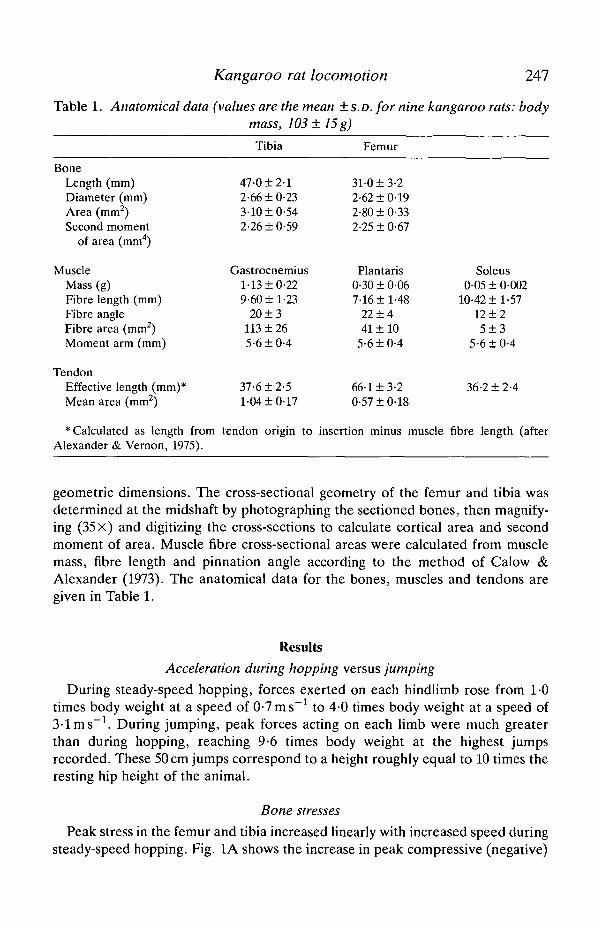
Table 1. Anatomical data (values are the mean ±S.D. for nine kangaroo rats: bodymass, 103±15g)
BoneLength (mm)Diameter (mm)Area (mm2)Second moment
of area (mm4)
MuscleMass (g)Fibre length (mm)Fibre angleFibre area (mm2)Moment arm (mm)
TendonEffective length (mm)*Mean area (mm2)
* Calculated as length fromAlexander & Vernon, 1975).
Tibia
47-0 ±2-12-66 ±0-233-10 ±0-542-26 ±0-59
Gastrocnemius1-13 ±0-229-60 ±1-23
20 ± 3113 ± 265-6 ±0-4
37-6 ±2-51-04 ±0-17
tendon origin to
Femur
31-0 ±3-22-62 ±0-192-80 ±0-332-25 ± 0-67
Plantaris0-30 ± 0-067-16 ±1-48
22 ±441 ±10
5-6 ±0-4
66-1 ±3-20-57 ±048
insertion minus muscle
Soleus0-05 ± 0-002
10-42 ±1-5712 ±25 ± 3
5-6 ±0-4
36-2 ±2-4
fibre length (after
geometric dimensions. The cross-sectional geometry of the femur and tibia wasdetermined at the midshaft by photographing the sectioned bones, then magnify-ing (35 x) and digitizing the cross-sections to calculate cortical area and secondmoment of area. Muscle fibre cross-sectional areas were calculated from musclemass, fibre length and pinnation angle according to the method of Calow &Alexander (1973). The anatomical data for the bones, muscles and tendons aregiven in Table 1.
ResultsAcceleration during hopping versus jumping
During steady-speed hopping, forces exerted on each hindlimb rose from 1-0times body weight at a speed of 0-7 ms" 1 to 4 0 times body weight at a speed of3-lms"1. During jumping, peak forces acting on each limb were much greaterthan during hopping, reaching 9-6 times body weight at the highest jumpsrecorded. These 50 cm jumps correspond to a height roughly equal to 10 times theresting hip height of the animal.
Bone stresses
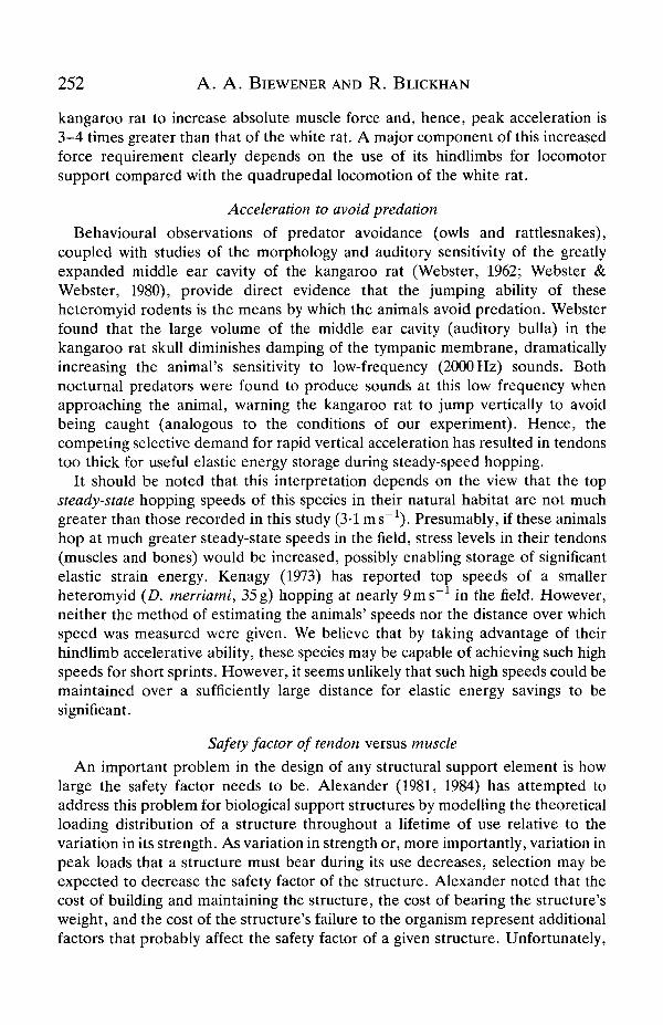
Peak stress in the femur and tibia increased linearly with increased speed duringsteady-speed hopping. Fig. 1A shows the increase in peak compressive (negative)
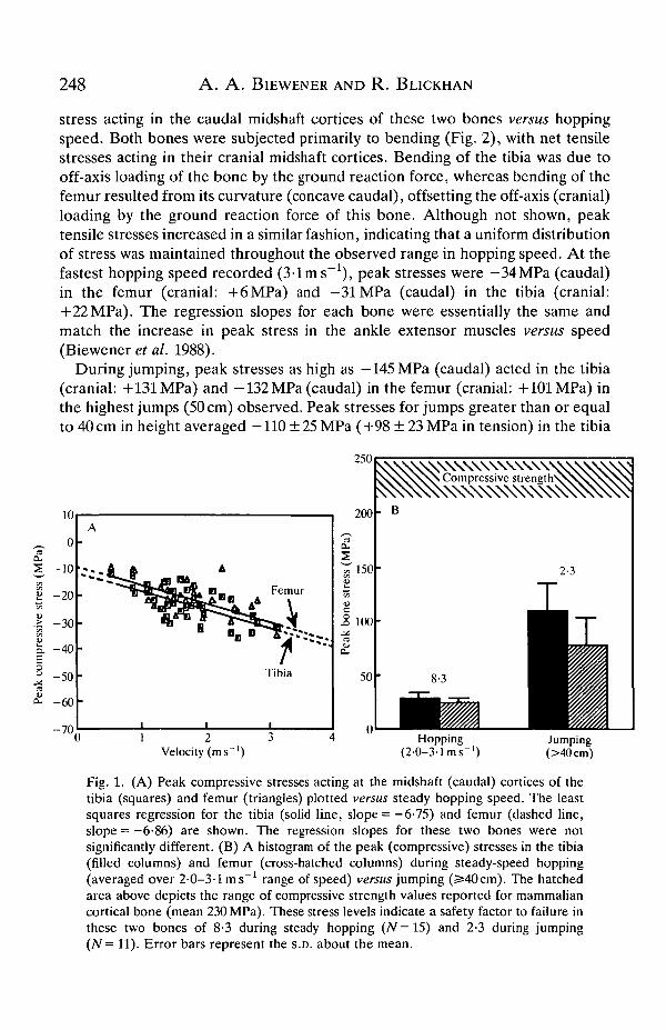
248 A . A . BlEWENER AND R . BLICKHAN
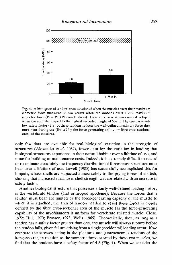
stress acting in the caudal midshaft cortices of these two bones versus hoppingspeed. Both bones were subjected primarily to bending (Fig. 2), with net tensilestresses acting in their cranial midshaft cortices. Bending of the tibia was due tooff-axis loading of the bone by the ground reaction force, whereas bending of thefemur resulted from its curvature (concave caudal), offsetting the off-axis (cranial)loading by the ground reaction force of this bone. Although not shown, peaktensile stresses increased in a similar fashion, indicating that a uniform distributionof stress was maintained throughout the observed range in hopping speed. At thefastest hopping speed recorded (3-1 ms"1), peak stresses were —34MPa (caudal)in the femur (cranial: +6MPa) and —31MPa (caudal) in the tibia (cranial:+22MPa). The regression slopes for each bone were essentially the same andmatch the increase in peak stress in the ankle extensor muscles versus speed(Biewener et al. 1988).
During jumping, peak stresses as high as -145MPa (caudal) acted in the tibia(cranial: +131 MPa) and -132 MPa (caudal) in the femur (cranial: +101 MPa) inthe highest jumps (50 cm) observed. Peak stresses for jumps greater than or equalto 40 cm in height averaged —110 ± 25 MPa (+98 ± 23 MPa in tension) in the tibia
Velocity (ms ')Hopping
(2-0-3-1 ms"')Jumping(>40cm)
Fig. 1. (A) Peak compressive stresses acting at the midshaft (caudal) cortices of thetibia (squares) and femur (triangles) plotted versus steady hopping speed. The leastsquares regression for the tibia (solid line, slope = —6-75) and femur (dashed line,slope = -6-86) are shown. The regression slopes for these two bones were notsignificantly different. (B) A histogram of the peak (compressive) stresses in the tibia(filled columns) and femur (cross-hatched columns) during steady-speed hopping(averaged over 2-0-3-1 ms"1 range of speed) versus jumping (^40cm). The hatchedarea above depicts the range of compressive strength values reported for mammaliancortical bone (mean 230 MPa). These stress levels indicate a safety factor to failure inthese two bones of 8-3 during steady hopping (N=15) and 2-3 during jumping(N= 11). Error bars represent the S.D. about the mean.
249Kangaroo rat locomotion
-120,20 0Film frame
10 12 14 16 18 20
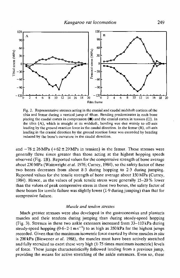
Fig. 2. Representative stresses acting in the cranial and caudal midshaft cortices of thetibia and femur during a vertical jump of 40 cm. Bending predominates in each boneplacing the caudal cortex in compression ( • ) and the cranial cortex in tension ( • ) . Inthe tibia (A), which is straight at its midshaft, bending was due mainly to off-axisloading by the ground reaction force in the caudal direction. In the femur (B), off-axisloading in the cranial direction by the ground reaction force was exceeded by bendinginduced by the bone's curvature in the caudal direction.
and -78±26MPa (+62±29MPa in tension) in the femur. These stresses weregenerally three times greater than those acting at the highest hopping speedsobserved (Fig. IB). Reported values for the compressive strength of bone averageabout 230 MPa (Wainwright et al. 1976; Currey, 1984), so the safety factor of thesetwo bones decreases from about 8-3 during hopping to 2-3 during jumping.Reported values for the tensile strength of bone average about 150 MPa (Currey,1984). Hence, as the values of peak tensile stress were generally 15-20% lowerthan the values of peak compressive stress in these two bones, the safety factor ofthese bones for tensile failure was slightly lower (1-9 during jumping) than that forcompressive failure.
Muscle and tendon stresses
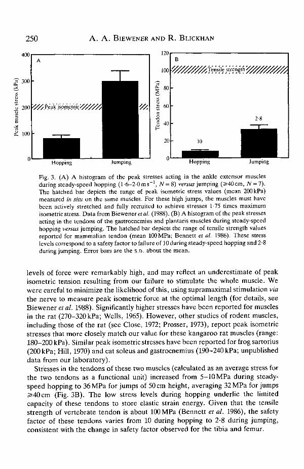
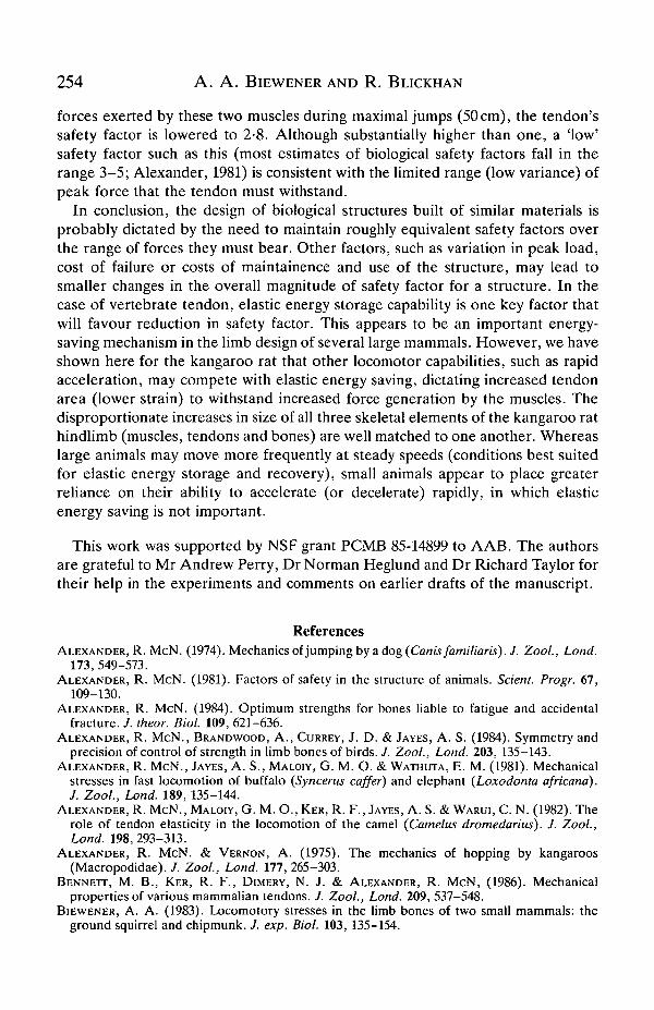
Much greater stresses were also developed in the gastrocnemius and plantarismuscles and their tendons during jumping than during steady-speed hopping(Fig. 3). Stresses in these two ankle extensors increased from 33-110kPa duringsteady-speed hopping (0-6-2-1 ms"1) to as high as 350 kPa for the highest jumpsrecorded. Given that the maximum isometric force exerted by these muscles in situis 200 kPa (Biewener et al. 1988), the muscles must have been actively stretchedand fully recruited to exert these very high (1-75 times maximum isometric) levelsof force. These jumps characteristically followed landing from a previous jump,providing the means for active stretching of the ankle extensors. Even so, these
A . A . BlEWENER AND R . BLICKHAN
120
100
£ 60o
•a40
20
B
Hopping Jumping
10
Hopping Jumping
Fig. 3. (A) A histogram of the peak stresses acting in the ankle extensor musclesduring steady-speed hopping (1-6-2-0 ms"1, N = 8) versus jumping (2540cm, AT = 7).The hatched bar depicts the range of peak isometric stress values (mean 200 kPa)measured in situ on the same muscles. For these high jumps, the muscles must havebeen actively stretched and fully recruited to achieve stresses 1-75 times maximumisometric stress. Data from Biewener et al. (1988). (B) A histogram of the peak stressesacting in the tendons of the gastrocnemius and plantaris muscles during steady-speedhopping versus jumping. The hatched bar depicts the range of tensile strength valuesreported for mammalian tendon (mean 100 MPa; Bennett et al. 1986). These stresslevels correspond to a safety factor to failure of 10 during steady-speed hopping and 2-8during jumping. Error bars are the S.D. about the mean.
levels of force were remarkably high, and may reflect an underestimate of peakisometric tension resulting from our failure to stimulate the whole muscle. Wewere careful to minimize the likelihood of this, using supramaximal stimulation viathe nerve to measure peak isometric force at the optimal length (for details, seeBiewener et al. 1988). Significantly higher stresses have been reported for musclesin the rat (270-320 kPa; Wells, 1965). However, other studies of rodent muscles,including those of the rat (see Close, 1972; Prosser, 1973), report peak isometricstresses that more closely match our value for these kangaroo rat muscles (range:180-200 kPa). Similar peak isometric stresses have been reported for frog sartorius(200 kPa; Hill, 1970) and cat soleus and gastrocnemius (190-240 kPa; unpublisheddata from our laboratory).
Stresses in the tendons of these two muscles (calculated as an average stress forthe two tendons as a functional unit) increased from 5-10 MPa during steady-speed hopping to 36 MPa for jumps of 50 cm height, averaging 32 MPa for jumps2=40cm (Fig. 3B). The low stress levels during hopping underlie the limitedcapacity of these tendons to store elastic strain energy. Given that the tensilestrength of vertebrate tendon is about 100 MPa (Bennett et al. 1986), the safetyfactor of these tendons varies from 10 during hopping to 2-8 during jumping,consistent with the change in safety factor observed for the tibia and femur.
Kangaroo rat locomotion 251
DiscussionAt all levels of locomotor activity that we examined, peak stresses acting in the
three structural components of the kangaroo rat's hindlimb skeleton (muscle,tendon and bone) are closely matched to the strength of each support element,reflecting integrated design of the hindlimb as a whole. The maximum forcesexerted on the ground when kangaroo rats jump to heights as great as 50 cm arethree times greater than those exerted when they hop at their fastest steady speed.These values correspond to ground reaction forces equal to 9-10 times theanimal's own weight (exerted on each hindlimb separately) and jump heightsequal to 10 times the animal's hip height. In association with these large forces, thestresses produced in the ankle extensor muscles, tendons and bones of thehindlimb during maximal recorded jumps are 3-4 times greater than thoseobserved during steady-speed hopping. Safety factors for these elements decreaseto nearly 2, approaching the limit observed for most biological structures that havebeen studied (Alexander, 1981; Biewener, 1983; Biewener et al. 19836; Lowell,1985).
If the ankle extensor tendons of this species were more slender, their ability tosave energy by elastic storage and recovery would be greatly improved (generally,a 50 % reduction in tendon cross-sectional area will result in roughly a four-foldincrease in total strain energy stored for tendons of equivalent length). However,the forces produced to achieve the accelerations we recorded when the animalsjumped would place thinner tendons at great risk of being ruptured. When viewedin the context of maximal acceleration, rather than useful energy savings, thehindlimb of the kangaroo rat is not 'overbuilt'. Indeed, the safety factors for thebones, muscles and tendons of this species are similar to those measured in othermammalian species during strenuous activity (Alexander, 1974; Alexander et al.1981; Alexander & Vernon, 1975; Biewener, 1983; Biewener et al. 1983ft;Biewener & Taylor, 1986; Rubin & Lanyon, 1982).
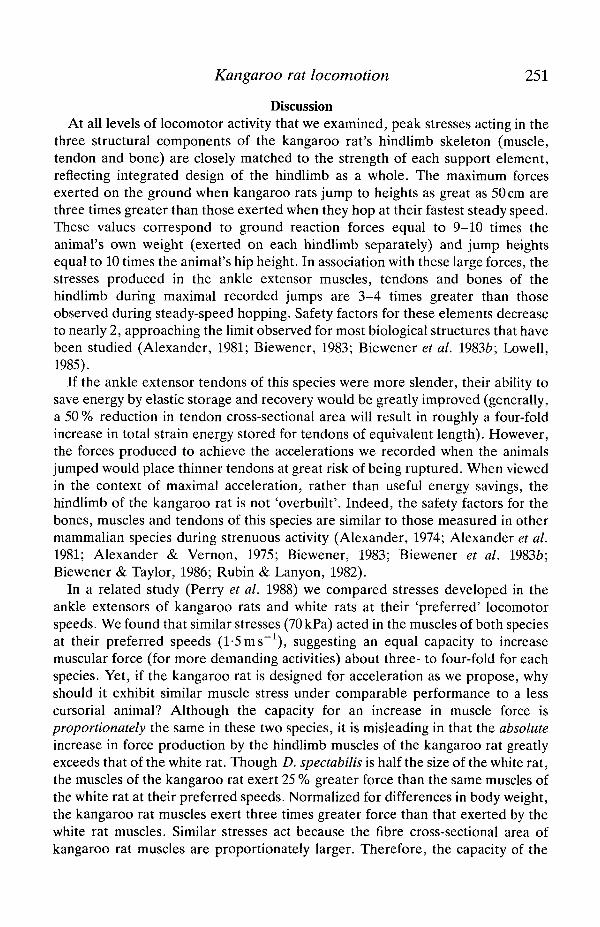
In a related study (Perry et al. 1988) we compared stresses developed in theankle extensors of kangaroo rats and white rats at their 'preferred' locomotorspeeds. We found that similar stresses (70 kPa) acted in the muscles of both speciesat their preferred speeds (1-5 ms"1), suggesting an equal capacity to increasemuscular force (for more demanding activities) about three- to four-fold for eachspecies. Yet, if the kangaroo rat is designed for acceleration as we propose, whyshould it exhibit similar muscle stress under comparable performance to a lesscursorial animal? Although the capacity for an increase in muscle force isproportionately the same in these two species, it is misleading in that the absoluteincrease in force production by the hindlimb muscles of the kangaroo rat greatlyexceeds that of the white rat. Though D. spectabilis is half the size of the white rat,the muscles of the kangaroo rat exert 25 % greater force than the same muscles ofthe white rat at their preferred speeds. Normalized for differences in body weight,the kangaroo rat muscles exert three times greater force than that exerted by thewhite rat muscles. Similar stresses act because the fibre cross-sectional area ofkangaroo rat muscles are proportionately larger. Therefore, the capacity of the
252 A . A . BlEWENER AND R. BLICKHAN
kangaroo rat to increase absolute muscle force and, hence, peak acceleration is3-4 times greater than that of the white rat. A major component of this increasedforce requirement clearly depends on the use of its hindlimbs for locomotorsupport compared with the quadrupedal locomotion of the white rat.
Acceleration to avoid predation
Behavioural observations of predator avoidance (owls and rattlesnakes),coupled with studies of the morphology and auditory sensitivity of the greatlyexpanded middle ear cavity of the kangaroo rat (Webster, 1962; Webster &Webster, 1980), provide direct evidence that the jumping ability of theseheteromyid rodents is the means by which the animals avoid predation. Websterfound that the large volume of the middle ear cavity (auditory bulla) in thekangaroo rat skull diminishes damping of the tympanic membrane, dramaticallyincreasing the animal's sensitivity to low-frequency (2000 Hz) sounds. Bothnocturnal predators were found to produce sounds at this low frequency whenapproaching the animal, warning the kangaroo rat to jump vertically to avoidbeing caught (analogous to the conditions of our experiment). Hence, thecompeting selective demand for rapid vertical acceleration has resulted in tendonstoo thick for useful elastic energy storage during steady-speed hopping.
It should be noted that this interpretation depends on the view that the topsteady-state hopping speeds of this species in their natural habitat are not muchgreater than those recorded in this study (3-lms"1). Presumably, if these animalshop at much greater steady-state speeds in the field, stress levels in their tendons(muscles and bones) would be increased, possibly enabling storage of significantelastic strain energy. Kenagy (1973) has reported top speeds of a smallerheteromyid (D. merriami, 35 g) hopping at nearly 9 ms" 1 in the field. However,neither the method of estimating the animals' speeds nor the distance over whichspeed was measured were given. We believe that by taking advantage of theirhindlimb accelerative ability, these species may be capable of achieving such highspeeds for short sprints. However, it seems unlikely that such high speeds could bemaintained over a sufficiently large distance for elastic energy savings to besignificant.
Safety factor of tendon versus muscle
An important problem in the design of any structural support element is howlarge the safety factor needs to be. Alexander (1981, 1984) has attempted toaddress this problem for biological support structures by modelling the theoreticalloading distribution of a structure throughout a lifetime of use relative to thevariation in its strength. As variation in strength or, more importantly, variation inpeak loads that a structure must bear during its use decreases, selection may beexpected to decrease the safety factor of the structure. Alexander noted that thecost of building and maintaining the structure, the cost of bearing the structure'sweight, and the cost of the structure's failure to the organism represent additionalfactors that probably affect the safety factor of a given structure. Unfortunately,
Kangaroo rat locomotion 253
120
100
80
S 60
40
20
2-8
1-75 xP 0
Muscle force
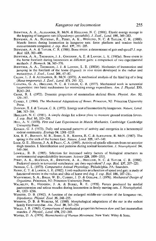
Fig. 4. A histogram of tendon stress developed when the muscles exert their maximumisometric force measured in situ versus when the muscles exert l-75x maximumisometric force (Po = 350 kPa muscle stress). These very large stresses were developedwhen the animals jumped to the highest recorded height of 50 cm. The comparativelylow safety factor (2-8) of these tendons reflects the well-defined maximum force theymust bear during use (limited by the force-generating ability, or fibre cross-sectionalarea, of the muscles).
only few data are available for real biological variation in the strengths ofstructures (Alexander et al. 1984), fewer data for the variation in loading thatbiological structures experience in their natural habitat over a lifetime of use, andnone for building or maintenance costs. Indeed, it is extremely difficult to recordor to estimate accurately the frequency distribution of forces most structures mustbear over a lifetime of use. Lowell (1985) has successfully accomplished this forlimpets, whose shells are subjected almost solely to the prying forces of starfish,showing that increased variance in shell strength was correlated with an increase insafety factor.
Another biological structure that possesses a fairly well-defined loading historyis the vertebrate tendon (and arthropod apodeme). Because the forces that atendon must bear are limited by the force-generating capacity of the muscle towhich it is attached, the area of tendon needed to resist these forces is closelydefined by the fibre cross-sectional area of the muscle (as the force-generatingcapability of the myofilaments is uniform for vertebrate striated muscle; Close,1972; Hill, 1970; Prosser, 1973; Wells, 1965). Theoretically, then, as long as atendon has a safety factor greater than one, the muscle will always rupture beforethe tendon fails, given failure arising from a single (accidental) loading event. If wecompare the stresses acting in the plantaris and gastrocnemius tendons of thekangaroo rat, in relation to the isometric force exerted by these two muscles, wefind that the tendons have a safety factor of 4-8 (Fig. 4). When we consider the
254 A . A . BlEWENER AND R. BLICKHAN
forces exerted by these two muscles during maximal jumps (50 cm), the tendon'ssafety factor is lowered to 2-8. Although substantially higher than one, a 'low'safety factor such as this (most estimates of biological safety factors fall in therange 3-5; Alexander, 1981) is consistent with the limited range (low variance) ofpeak force that the tendon must withstand.
In conclusion, the design of biological structures built of similar materials isprobably dictated by the need to maintain roughly equivalent safety factors overthe range of forces they must bear. Other factors, such as variation in peak load,cost of failure or costs of maintainence and use of the structure, may lead tosmaller changes in the overall magnitude of safety factor for a structure. In thecase of vertebrate tendon, elastic energy storage capability is one key factor thatwill favour reduction in safety factor. This appears to be an important energy-saving mechanism in the limb design of several large mammals. However, we haveshown here for the kangaroo rat that other locomotor capabilities, such as rapidacceleration, may compete with elastic energy saving, dictating increased tendonarea (lower strain) to withstand increased force generation by the muscles. Thedisproportionate increases in size of all three skeletal elements of the kangaroo rathindlimb (muscles, tendons and bones) are well matched to one another. Whereaslarge animals may move more frequently at steady speeds (conditions best suitedfor elastic energy storage and recovery), small animals appear to place greaterreliance on their ability to accelerate (or decelerate) rapidly, in which elasticenergy saving is not important.
This work was supported by NSF grant PCMB 85-14899 to AAB. The authorsare grateful to Mr Andrew Perry, Dr Norman Heglund and Dr Richard Taylor fortheir help in the experiments and comments on earlier drafts of the manuscript.
ReferencesALEXANDER, R. MCN. (1974). Mechanics of jumping by a dog {Canis familiaris). J. Zooi, Lond.
173, 549-573.ALEXANDER, R. MCN. (1981). Factors of safety in the structure of animals. Scient. Progr. 67,
109-130.ALEXANDER, R. MCN. (1984). Optimum strengths for bones liable to fatigue and accidental
fracture. J. theor. Biol. 109, 621-636.ALEXANDER, R. MCN., BRANDWOOD, A., CURREY, J. D. & JAYES, A. S. (1984). Symmetry and
precision of control of strength in limb bones of birds. J. Zooi, Lond. 203, 135-143.ALEXANDER, R. MCN., JAYES, A. S., MALOIY, G. M. O. & WATHUTA, E. M. (1981). Mechanical
stresses in fast locomotion of buffalo (Syncerus caffer) and elephant {Loxodonta africana).J. Zooi., Lond. 189, 135-144.
ALEXANDER, R. MCN., MALOIY, G. M. O., KER, R. F., JAYES, A. S. & WARUI, C. N. (1982). Therole of tendon elasticity in the locomotion of the camel {Camelus dromedarius). J. Zooi.,Lond. 198, 293-313.
ALEXANDER, R. MCN. & VERNON, A. (1975). The mechanics of hopping by kangaroos(Macropodidae). /. Zooi, Lond. 177, 265-303.
BENNETT, M. B., KER, R. F., DIMERY, N. J. & ALEXANDER, R. MCN, (1986). Mechanicalproperties of various mammalian tendons. J. Zooi., Lond. 209, 537-548.
BIEWENER, A. A. (1983). Locomotory stresses in the limb bones of two small mammals: theground squirrel and chipmunk. J. exp. Biol. 103, 135-154.
Kangaroo rat locomotion 255
BIEWENER, A. A., ALEXANDER, R. MCN. & HEGLUND, N. C. (1981). Elastic energy storage inthe hopping of kangaroo rats (Dipodomys spectabilis). J. Zool., Lond. 195, 369-383.
BIEWENER, A. A., BLICKHAN, R., PERRY, A. K., HEGLUND, N. C. & TAYLOR, C. R. (1988).Muscle forces during locomotion in kangaroo rats: force platform and tendon bucklemeasurements compared. /. exp. Biol. 137,191-205.
BIEWENER, A. A. & TAYLOR, C. R. (1986). Bone strain: a determinant of gait and speed? J. exp.Biol. 123, 383-400.
BIEWENER, A. A., THOMASON, J. J., GOODSHIP, A. E. & LANYON, L. E. (1983a). Bone stress inthe horse forelimb during locomotion at different gaits: a comparison of two experimentalmethods. /. Biomech. 16, 565-576.
BIEWENER, A. A., THOMASON, J. J. & LANYON, L. E. (1983b). Mechanics of locomotion andjumping in the forelimb of the horse (Equus): in vivo stress developed in the radius andmetacarpus. 7. Zool., Lond. 201, 67-82.
CALOW, L. J. & ALEXANDER, R. MCN. (1973). A mechanical analysis of the hind leg of a frog(Rana temporaria). J. Zool., Lond. 171, 293-321.
CAVAGNA, G. A., HEGLUND, N. C. & TAYLOR, C. R. (1977). Mechanical work in terrestriallocomotion: two basic mechanisms for minimizing energy expenditure. Am. J. Physiol. 233,R243-261.
CLOSE, R. I. (1972). Dynamic properties of mammalian skeletal fibers. Physiol. Rev. 52,129-197.
CURREY, J. (1984). The Mechanical Adaptations of Bones. Princeton, NJ: Princeton UniversityPress.
DAWSON, T. J. & TAYLOR, C. R. (1973). Energy cost of locomotion by kangaroos. Nature, Lond.246, 313-314.
HEGLUND, N. C. (1981). A simple design for a force plate to measure ground reaction forces.J. exp. Biol. 93, 333-338.
HILL, A. V. (1970). First and Last Experiments in Muscle Mechanics. Cambridge: CambridgeUniversity Press.
KENAGY, G. J. (1973). Daily and seasonal patterns of activity and energetics in a heteromyidrodent community. Ecology 54, 1201-1219.
KER, R. F., BENNETT, M. B., BIBBY, S. R., KESTER, R. C. & ALEXANDER, R. MCN. (1987). Thespring in the arch of the human foot. Nature, Lond. 325, 147-149.
LOEB, G. E., HOFFER, J. A. &PRATT, C. A. (1985). Activity of spindle afferents from cat anteriorthigh muscles. I. Identification and patterns during normal locomotion. J. Neurophysiol. 54,549-564.
LOWELL, R. B. (1985). Selection for increased safety factors of biological structures asenvironmental unpredictability increases. Science 228, 1009-1011.
PERRY, A. K., BLICKHAN, R., BIEWENER, A. A., HEGLUND, N. C. & TAYLOR, C. R. (1988).Preferred speeds in terrestrial vertebrates: are they equivalent? J. exp. Biol. 137, 207-220.
PROSSER, C. L. (1973). Comparative Animal Physiology. Philadelphia, PA: Saunders.RUBIN, C. T. & LANYON, L. E. (1982). Limb mechanics as a function of speed and gait: a study of
functional strains in the radius and tibia of horse and dog. /. exp. Biol. 101, 187-211.WAINWRIGHT, S. A., BIGGS, W. D., CURREY, J. D. & GOSLINE, J. (1976). Mechanical Design of
Organisms. Princeton, NJ: Princeton University Press.WALMSLEY, B., HODGSON, J. A. & BURKE, R. E. (1978). Forces produced by medial
gastrocnemius and soleus muscles during locomotion in freely moving cats. J. Neurophysiol.41, 1203-1216.
WEBSTER, D. B. (1962). A function of the enlarged middle-ear cavities of the kangaroo rat,Dipodomys. Physiol. Zool. 35, 248-255.
WEBSTER, D. B. & WEBSTER, M. (1980). Morphological adaptations of the ear in the rodentfamily Heteromyidae. Am. Zool. 20, 247-254.
WELLS, J. B. (1965). Comparisons of mechanical properties between slow and fast mammalianmuscles. J. Physiol., Lond. 178, 252-269.
WINTER, D. A. (1979). Biomechanics of Human Movement. New York: Wiley & Sons.