Embed Size (px)
Citation preview
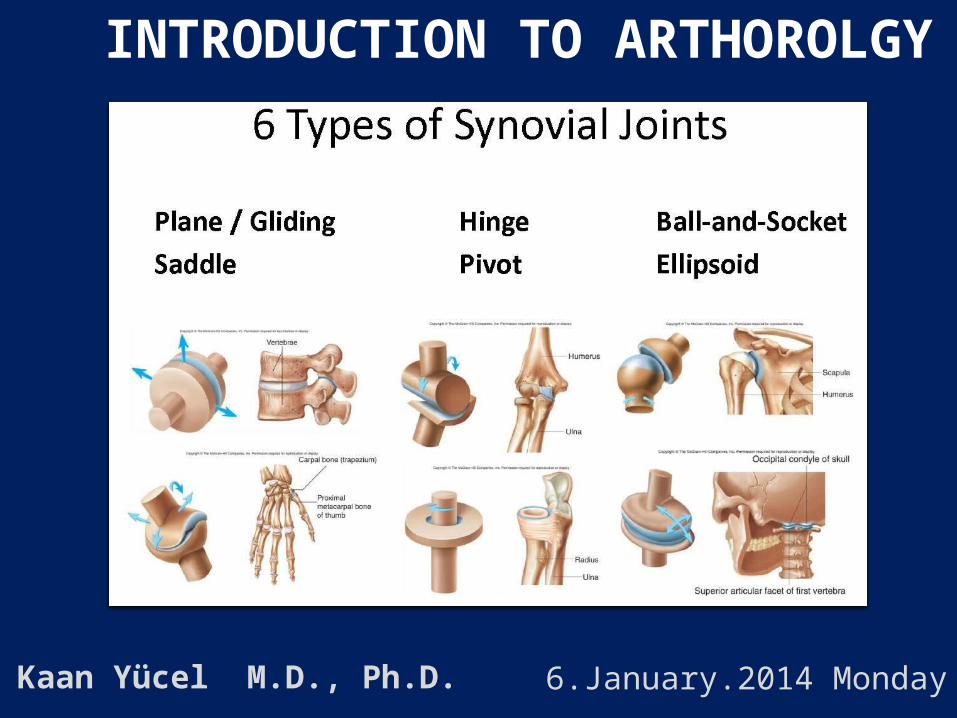
INTRODUCTION TO ARTHOROLGY
Kaan Yücel M.D., Ph.D. 6.January.2014 Monday

2
1.1. CLASSIFICATION OF JOINTS
1.2. STABILITY OF JOINTS
1.3. JOINT VASCULATURE AND INNVERVATION

science concerned with the anatomy, function, dysfunction and treatment of joints.
ARTHROLOGY GREEK A RQRON JOINT –LOGY

according to the tissues that lie between the bones:
1) Fibrous joints
2) Cartilaginous joints
3) Synovial joints
Classification of Joints

Classification of Joints

Fibrous jointsBones are united by fibrous tissue. Sutures of the cranium

Fibrous jointsSyndesmosis type of fibrous joint unites the bones with a sheet of fibrous tissue either a ligament or a fibrous membrane partially movableThe interosseous membrane in the forearm is a sheet of fibrous tissue that joins the radius and ulna in a syndesmosis.

Fibrous jointsDentoalveolar syndesmosis (gomphoses or socket)
a peglike process fits into a socket articulation between the root of the tooth and the alveolar process of the jaw.

Cartilaginous joints
Bones are united by hyaline cartilage or fibrocartilage.

Cartilaginous joints
Pimary cartilaginous joints-synchondroseshyaline cartilage- growth of a bone during early life
Secondary cartilaginous joints-symphyses strong, slightly movable joints united by fibrocartilage

Synovial joints Most common type of joints
Bones united by a joint capsule enclosing an articular cavity.
Provide free movement between the bones they join.
Joint cavity potential space contains lubricating synovial fluid, secreted by the synovial membrane.
Articular cartilagearticular surfaces are covered by hyaline cartilage
Articular capsulesurrounds the joint and formed of two layers.
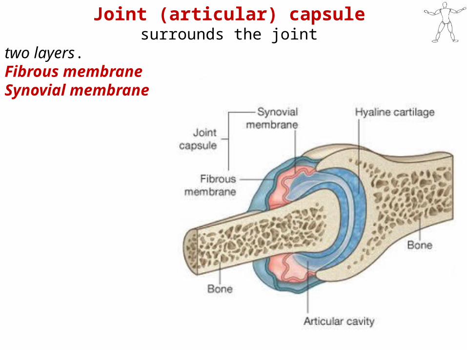
Joint (articular) capsule surrounds the joint
two layers. Fibrous membraneSynovial membrane

Synovial membranelines inner surface of the fibrous membrane. highly vascular produces synovial fluid, and lubricates the articulating surfaces
(helps to minimize the friction by articular surfaces). attaches to the margins of the joint surfaces at the interface between the cartilage and bone and encloses the articular cavity.

Fibrous membrane
Formed by dense connective tissue Surrounds and stabilizes the joint.Parts of it may thicken to form ligaments, further stabilize the joint. Ligaments outside the capsule usually provide additional reinforcement.

Closed sacs of synovial membrane also occur outside joints where they form
synovial bursae or tendon sheaths.

Ligamentsa cord or band of connective tissue uniting two structures.
Articular capsules are usually strengthened by articular ligaments.
Connect the articulating bones to each other.
limit the undesired and/or excessive movements of the joints.

Articular disc: Help to hold the bones together.
Labrum: A fibrocartilaginous ring which deepens the articular surface for one of the bones.

Bursa
Flattened sacs that contain synovial fluid to reduce friction.
Walls are separated by a film of viscous fluid.
Found wherever tendons rub against bones, ligaments, or other tendons.

STABILITY OF JOINTSdepends on four main factors
1. negative pressure within the joint cavity2. shape, size, and arrangement of the articular surfaces3. ligaments4. tone of the muscles around the joint

Joint vasculature and innvervation
Joints receive blood from articular arteries that arise from the vessels around the joint.
Articular veins are communicating veins that accompany arteries (L. venae comitantes) and, like the arteries, are located in the joint capsule, mostly in the synovial membrane.
Joints have a rich nerve supply provided by articular nerves with sensory nerve endings in the joint capsule.

Types of synovial jointsaccording to shape of articulating surfaces- type of
movement they permit
1.Plane joints uniaxial joints- gliding or sliding acromioclavicular joint
2. Hinge joints uniaxial joints- flexion & extensionknee & elbow joints

Types of synovial joints
3. Saddle jointsbiaxial joints- flexion & extension, abduction & adductioncarpometacarpal joint at the base of the 1st digit (thumb)
4. Condyloid (ellipsoid type) biaxial joints- flexion & extension, abduction & adductionmetacarpophalangeal joints (knuckle joints)radiocarpal joint (wrist)

Types of synovial joints
5. Ball and socket joints (spheroidal joints)
multiple axes and planes: flexion and extension, abduction and adduction, medial and lateral rotation, and circumductionhip & shoulder joints

Types of synovial joints
6. Pivot jointsuniaxial joints- rotation around a central axisproximal & distal radioulnar joints