Embed Size (px)
Citation preview
7/28/2019 Jurnal Prognosis 2
http://slidepdf.com/reader/full/jurnal-prognosis-2 1/8
R E S E A R C H Open Access
Prognosis of ampullary cancer based onimmunohistochemical type and expressionof osteopontinXiang-qian Zhao, Jia-hong Dong*, Wen-zhi Zhang and Zhe Liu
Abstract
Background: Ampullary cancer (AC) was classified as pancreatobiliary, intestinal, or other subtype based on the
expression of cytokeratin 7 (CK7) and cytokeratin 20 (CK20). We aimed to explore the association of AC subtype
with patient prognosis.Methods: The relationship of AC subtype and expression of Osteopontin (OPN) with the prognosis of 120 AC
patients after pancreaticoduodenectomy was investigated.
Results: The patients had pancreatobiliary (CK7+ /CK20-, n = 24, 20%), intestinal (CK7- /CK20+, n = 29, 24.2%) or
other (CK7+ /CK20+ or CK7- /CK20-, n = 67, 55.8%) subtypes of AC, and their median survival times were 23 ± 4.2, 38
± 2.8 and 64 ± 16.8 months, respectively. The survival times of 64 OPN - patients (53.3%) and 56 OPN+ patients
(46.7%) were 69 ± 18.4 and 36 ± 1.3 months, respectively. There was no significant effect of AC subtype on survival
of OPN- patients. For OPN+ patients, those with pancreatobiliary AC had a shorter survival time (22 ± 6.6 months)
than those with intestinal AC (37 ± 1.4 months, p = 0.041), and other AC subtype (36 ± 0.9 months, p = 0.010);
intestinal and other AC subtypes had similar survival times.
Conclusions: The prognosis of AC patients can be estimated based on immunohistochemical classification and
OPN status.
Keywords: ampullary cancer, osteopontin, survival analysis, immunohistochemistry, classification, Cytokeratin 20,
Cytokeratin 7
BackgroundAmpullary carcinoma (AC) is a relatively rare tumor of
the hepatopancreatic ampulla that accounts for approxi-
mately 0.2% of gastrointestinal tract malignancies and 7%
of periampullary carcinomas [1]. ACs have different ana-
tomical origins. Kimura et al. initially classified AC as
pancreatobiliary AC if it had papillary projections with
scant fibrous cores and as intestinal AC if it resembled
tubular adenocarcinoma of the stomach or colon [2].Numerous studies have reported that intestinal AC is
associated with a better prognosis than pancreatobiliary
AC [1-5].
AC has also been classified based on immunohistochem-
ical expression of cytokeratin 7 (CK7), Mucins and CDX2
[4,6-8] and HNF4a [9]. However, the clinical significance
and survival rates of AC patients with these different
immunohistochemical subtypes have not been definitely
established.
Histologic classification and immunohistochemical
characterization by cytokeratins are in good agreement
[5]. Fischer et al. reported that the histological subtypes
of AC could be determined by the expression of CK7,
CK20, and MUC2; pancreaticobiliary AC is CK7+
/CK20-
/MUC2-, and intestinal AC is CK7-/CK20+/MUC2+ [10].
Zhou et al. classified CK7-/CK20+ tumors as intestinal
AC, CK7+/CK20- tumors as pancreatobiliary AC, and
tumors that are CK7+/CK20+ or CK7-/CK20- as “other”[3]. However, there was no statistical difference in survi-
val of patients with different CK7/CK20 subtypes [3] or
with different CK20/MUC subtypes [11].* Correspondence: [email protected]
Hospital & Institute of Hepatobiliary Surgery, Chinese PLA General Hospital,
28 Fuxing Road, Beijing 100853, China
Zhao et al . Diagnostic Pathology 2011, 6:98
http://www.diagnosticpathology.org/content/6/1/98
© 2011 Zhao et al; licensee BioMed Central Ltd. This is an Open Access article distributed under the terms of the Creative CommonsAttribution License (http://creativecommons.org/licenses/by/2.0), which permits unrestricted use, distribution, and reproduction inany medium, provided the original work is properly cited.

7/28/2019 Jurnal Prognosis 2
http://slidepdf.com/reader/full/jurnal-prognosis-2 2/8
Osteopontin (OPN) is a secretory calcium-binding phos-
phorylated glycoprotein and plays an important role in
bone metabolism. OPN is widely distributed in the urine,
blood, gastrointestinal tract, pancreas, lungs and else-
where. At the molecular level, OPN plays important roles
in cellular adhesion and migration, tissue repair, and signal
transduction and also in the invasion and metastasis of
several cancers [12]. OPN is significantly associated with
survival rate in several cancers and has value as a marker
of clinical tumor progression [13,14]. In particular, low
OPN levels were significantly associated with a favorable
prognosis in patients with advanced non-small cell lung
cancer [15], laryngeal and hypopharyngeal carcinomas
[16], hepatocellular carcinoma [17], colorectal cancer [18],
idiopathic pulmonary hypertension [19], upper urinary
tract urothelial carcinoma [20], acute myeloid leukemia
[21], oral squamous cell carcinoma [22], and endometrial
cancer [23]. OPN may also be a suitable biomarker foroverall survival and renal outcome of patients who are
critically ill with acute kidney injury [24].
However, few studies have investigated the expression of
OPN in patients with AC. Van Heek et al. reported higher
OPN expression in the sera and tumors of AC patients
than in the sera and duodenal samples of healthy controls
[25]. Bloomston et al. reported that node-negative status
and lack of OPN expression were associated with pro-
longed survival in patients with AC [26]. Hsu et al.
reported that expression of OPN and the presence of
tumor-associated macrophages in bulky AC were asso-
ciated with tumor recurrence, and poorer disease-specific
survival [27].
In the present study, we retrospectively analyzed the
clinical data of 120 patients who were undergoing pan-
creaticoduodenectomy due to AC. We focused on the
association of AC prognosis with the expression of CK7,
CK20, and OPN.
Patients and MethodsPatients
From January 1, 1994 to December 30, 2008, patients
undergoing pancreaticoduodenectomy due to AC were
recruited from the Department of Hepatobiliary Surgery
of the General Hospital of the Peoples Liberation Army (Beijing, China). The exclusion criteria were: (i) duodenal
cancer, cancer of the lower bile duct, or cancer of the pan-
creas or any of these cancers involving the ampulla or
duodenal papilla, based on pathological examination; (ii)
uncertain origin of the cancer; (iii) previous focal resection
of duodenal papillary cancer or AC; (iv) metastasis to
other organs; and (v) presence of concomitant heart dis-
ease, cerebrovascular disease, or pulmonary disease that
made the patient ineligible for surgery. Follow-up exami-
nations were performed at 3 months after surgery, once
every 6 months for 3 years, and then once per year. These
follow-up examinations included routine tests (liver and
kidney function, blood electrolytes, routine blood test),
tests for tumor markers, chest X-ray, and abdominal ima-
ging by ultrasonography, CT, or MRI. The last follow-up
was on January 31, 2010. Tumor stage and lymph node
metastasis were evaluated according to Greene et al [28].
This study was approved by the hospital Institutional
Review Board.
Immunohistochemistry
Carcinoma specimens were embedded in paraffin, cut con-
secutively into sections (4 μm), and the streptavidin-perox-
idase method was used for immunohistochemical
visualization (UltraSensitive™ SP kit, Maximbio. Co. Ltd,
Fuzhou, China). The primary antibodies were mouse anti-
human CK7 or CK20 monoclonal antibodies and rabbit
anti-human OPN polyclonal antibody (Lab Vision & Neo-Markers, USA). The normal serum from non-immunized
goat was used as a negative control of the primary anti-
body, and CK7+/CK20+/OPN+ pancreatic carcinoma was
used as a positive control.
Details for the determination of positive staining were
previously provided[3]. In brief, cells positive for CK7,
CK20, or OPN had brown or yellow-brown granules,
mainly in the cytoplasm. Sections were evaluated by two
independent and blinded pathologists. No staining or
staining in fewer than 10% of cells was considered nega-
tive, and staining of 10% or more of cells was considered
positive.
Statistical analysis
Results are expressed as means or medians with stan-
dard deviations, or counts and percentages. Survival
analysis was analyzed by the Kaplan-Meier method and
the log-rank test. Data were analyzed using SPSS 15.0
(SPSS, Inc., Chicago, IL, USA). All p-values were two-
sided and were considered significant if p was less than
0.05.
ResultsPatient characteristics
A total of 120 patients (84 males, 36 females) met ourinclusion criteria and received follow-up examinations.
The mean age was 55.1 ± 9.8 years and the mean tumor
diameter was 2.4 ± 1.5 cm. The 1-, 3- and 5-year survi-
val rates were 94.8%, 78.7%, and 68.0%, respectively. The
median survival time was 38 ± 11.3 months and the
mean survival time was 53.9 months. A total of 51
patients (42.5%) survived to the end of the follow-up
period (January 31, 2010), 1 patient survived more than
10 years, and 2 patients survived more than 5 years. In
addition to pancreaticoduodenectomy (given to all
Zhao et al . Diagnostic Pathology 2011, 6:98
http://www.diagnosticpathology.org/content/6/1/98
Page 2 of 7
7/28/2019 Jurnal Prognosis 2
http://slidepdf.com/reader/full/jurnal-prognosis-2 3/8
patients), 8 patients received chemotherapy. Among the
69 patients (57.5%) who died in the follow-up period, 21
died within 5 years after surgery and 1 died 11 years
after surgery.
Survival of patients with different subtypes of AC
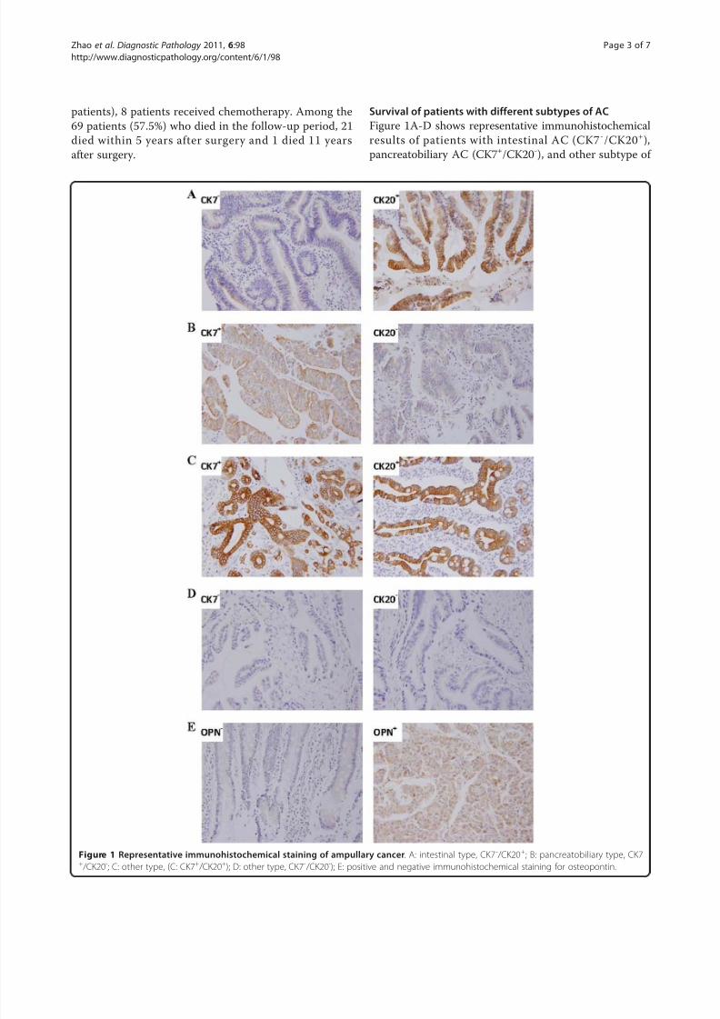
Figure 1A-D shows representative immunohistochemical
results of patients with intestinal AC (CK7 -/CK20+),
pancreatobiliary AC (CK7+/CK20-), and other subtype of
Figure 1 Representative immunohistochemical staining of ampullary cancer. A: intestinal type, CK7 - /CK20+; B: pancreatobiliary type, CK7+ /CK20-; C: other type, (C: CK7+ /CK20+); D: other type, CK7- /CK20-); E: positive and negative immunohistochemical staining for osteopontin.
Zhao et al . Diagnostic Pathology 2011, 6:98
http://www.diagnosticpathology.org/content/6/1/98
Page 3 of 7
7/28/2019 Jurnal Prognosis 2
http://slidepdf.com/reader/full/jurnal-prognosis-2 4/8
AC (CK7+/CK20+ and CK7-/CK20-). Figure 1E shows
representative positive and negative results for OPN
staining.
Table 1 shows the survival times of AC patients strati-
fied by immunohistochemical results, extent of tumor dif-
ferentiation, amount of tumor invasion, and lymph node
metastasis. None of the other associations were significant.
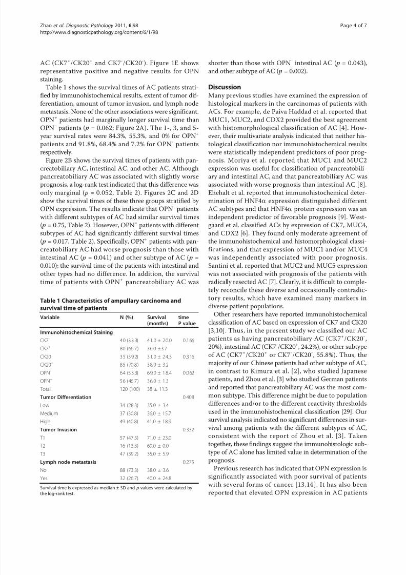
OPN+ patients had marginally longer survival time than
OPN- patients ( p = 0.062; Figure 2A). The 1-, 3, and 5-
year survival rates were 84.3%, 55.3%, and 0% for OPN+
patients and 91.8%, 68.4% and 7.2% for OPN- patients
respectively.
Figure 2B shows the survival times of patients with pan-
creatobiliary AC, intestinal AC, and other AC. Although
pancreatobiliary AC was associated with slightly worse
prognosis, a log-rank test indicated that this difference was
only marginal ( p = 0.052, Table 2). Figures 2C and 2D
show the survival times of these three groups stratified by OPN expression. The results indicate that OPN- patients
with different subtypes of AC had similar survival times
( p = 0.75, Table 2). However, OPN+ patients with different
subtypes of AC had significantly different survival times
( p = 0.017, Table 2). Specifically, OPN+ patients with pan-
creatobiliary AC had worse prognosis than those with
intestinal AC ( p = 0.041) and other subtype of AC ( p =
0.010); the survival time of the patients with intestinal and
other types had no difference. In addition, the survival
time of patients with OPN+ pancreatobiliary AC was
shorter than those with OPN- intestinal AC ( p = 0.043),
and other subtype of AC ( p = 0.002).
DiscussionMany previous studies have examined the expression of
histological markers in the carcinomas of patients with
ACs. For example, de Paiva Haddad et al. reported that
MUC1, MUC2, and CDX2 provided the best agreement
with histomorphological classification of AC [4]. How-
ever, their multivariate analysis indicated that neither his-
tological classification nor immunohistochemical results
were statistically independent predictors of poor prog-
nosis. Moriya et al. reported that MUC1 and MUC2
expression was useful for classification of pancreatobili-
ary and intestinal AC, and that pancreatobiliary AC was
associated with worse prognosis than intestinal AC [8].
Ehehalt et al. reported that immunohistochemical deter-
mination of HNF4a expression distinguished differentAC subtypes and that HNF4a protein expression was an
independent predictor of favorable prognosis [9]. West-
gaard et al. classified ACs by expression of CK7, MUC4,
and CDX2 [6]. They found only moderate agreement of
the immunohistochemical and histomorphological classi-
fications, and that expression of MUC1 and/or MUC4
was independently associated with poor prognosis.
Santini et al. reported that MUC2 and MUC5 expression
was not associated with prognosis of the patients with
radically resected AC [7]. Clearly, it is difficult to comple-
tely reconcile these diverse and occasionally contradic-
tory results, which have examined many markers in
diverse patient populations.
Other researchers have reported immunohistochemical
classification of AC based on expression of CK7 and CK20
[3,10]. Thus, in the present study we classified our AC
patients as having pancreatobiliary AC (CK7+/CK20-,
20%), intestinal AC (CK7-/CK20+, 24.2%), or other subtype
of AC (CK7+/CK20+ or CK7-/CK20-, 55.8%). Thus, the
majority of our Chinese patients had other subtype of AC,
in contrast to Kimura et al. [2], who studied Japanese
patients, and Zhou et al. [3] who studied German patients
and reported that pancreatobiliary AC was the most com-
mon subtype. This difference might be due to population
differences and/or to the different reactivity thresholdsused in the immunohistochemical classification [29]. Our
survival analysis indicated no significant differences in sur-
vival among patients with the different subtypes of AC,
consistent with the report of Zhou et al. [3]. Taken
together, these findings suggest the immunohistologic sub-
type of AC alone has limited value in determination of the
prognosis.
Previous research has indicated that OPN expression is
significantly associated with poor survival of patients
with several forms of cancer [13,14]. It has also been
reported that elevated OPN expression in AC patients
Table 1 Characteristics of ampullary carcinoma andsurvival time of patients
Variable N (%) Survival(months)
timeP value
Immunohistochemical Staining
CK7- 40 (33.3) 41.0 ± 20.0 0 .166
CK7+ 80 (66.7) 36.0 ±3.7
CK20- 35 (39.2) 31.0 ± 24.3 0 .316
CK20+ 85 (70.8) 38.0 ± 3.2
OPN- 64 (53.3) 69.0 ± 18.4 0 .062
OPN+ 56 (46.7) 36.0 ± 1.3
Total 120 (100) 38 ± 11.3
Tumor Differentiation 0.408
Low 34 (28.3) 35.0 ± 3.4
Medium 37 (30.8) 36.0 ± 15.7
High 49 (40.8) 41.0 ± 18.9
Tumor Invasion 0.332
T1 57 (47.5) 71.0 ± 23.0
T2 16 (13.3) 69.0 ± 0.0
T3 47 (39.2) 35.0 ± 5.9
Lymph node metastasis 0.275
No 88 (73.3) 38.0 ± 3.6
Yes 32 (26.7) 40.0 ± 24.8
Survival time is expressed as median ± SD and p-values were calculated by
the log-rank test.
Zhao et al . Diagnostic Pathology 2011, 6:98
http://www.diagnosticpathology.org/content/6/1/98
Page 4 of 7

7/28/2019 Jurnal Prognosis 2
http://slidepdf.com/reader/full/jurnal-prognosis-2 5/8
predicts poor disease-specific survival [25-27]. However,
when we pooled all AC subtypes, we found that survivaltime of OPN- and OPN+ patients had no significant dif-
ference. This is consistent with the results of Matsuzaki
et al., who reported that OPN expression in AC patients
was not associated with survival rate, although OPN
expression in the carcinoma was higher than in adjacent
normal tissues [30].
Our further analysis indicated that the subtype of AC
(intestinal, pancreatobiliary, or other) had no significant
effect on survival of patients with OPN - carcinomas.
However, for patients with OPN+ carcinomas, those with
intestinal AC or other subtype of AC had significantly
Figure 2 Kaplan-Meier survival curves of patients with different immunohistochemical types of ampullary cancer and with positive or
negative expression of osteopontin. *p < 0.05 by log-rank test.
Table 2 Expression of OPN and survival time of patientswith different subtypes of ampullary carcinoma (AC)
Survival time in months (number of patients)
Pancreatobili ary Intestinal O ther p value1
Total 23 ± 4.2 (24) 38 ± 2.8 (29) 64 ± 16.8 (67) 0.052
OPN- 69 ± 39.5 (11) 41 ± 24.8 (16) 69 ± 26.8 (37) 0.750
OPN+ 22 ± 6.6 (13)* 37 ± 1.4 (13) 36 ± 0.9 (30) 0 .017
p value2 0.085 0.509 0.263
Survival time is expressed as median ± SD. 1 p-value for comparison of the 3
types; 2 p-value for comparison of the OPN+ and OPN- groups. * p = 0.041 and
p = 0.010 for comparison with the intestinal type and other type OPN+ AC,
respectively. There was no difference between intestinal AC and other type of
AC ( p = 0.907).
Zhao et al . Diagnostic Pathology 2011, 6:98
http://www.diagnosticpathology.org/content/6/1/98
Page 5 of 7

7/28/2019 Jurnal Prognosis 2
http://slidepdf.com/reader/full/jurnal-prognosis-2 6/8
better survival than those with pancreatobiliary AC.
Thus, OPN expression appears to affect the biological
behavior of AC, and this effect depends on the anatomi-
cal origin of the tumor. These results indicate that deter-
mination of the prognosis of patients with AC should
consider OPN expression.
Previous studies have reported interactions of OPN with
other proteins. For example, in situ proximity ligation ana-
lysis indicated a molecular interaction of OPN and CD44
and that elevated expression of these proteins were asso-
ciated with increased mitosis and significantly enhanced
gastrointestinal stromal tumor cell proliferation in vitro
[31]. Yang et al. reported that OPN combined with CD44
was a promising independent predictor of tumor recur-
rence and survival in patients with hepatocellular carci-
noma [32]. OPN combined with CDX2 appears to predict
survival of advanced gastric cancer patients, and CDX2
may be a transcription factor that modulates the expres-sion of osteopontin [33]. Our results support previous
reports which suggest that OPN has a role in the patho-
genesis of AC [25-27]. However, a limitation of our study
is that patients were enrolled retrospectively, and we did
not include histomorphological classification of patients or
immunochemical determination based on other factors
including CDX2 and mucins. Clearly, the potential inter-
action of OPN, CK7, CDX2, mucins and other factors and
the role of these in the pathogenesis of AC warrant further
studies.
ConclusionsIn conclusion, our results indicate that it is difficult to
determine prognosis of patients with AC based solely on
immunohistochemical classification that considers CK7
and CK20 status. However, the additional consideration of
OPN status allows determination of prognosis. Our results
also suggest that OPN plays a role in the pathogenesis of
AC, but its mechanisms and relationship with CK7 and
CK20 warrant further studies.
Abbreviations
AC: ampullary cancer; OPN: osteopontin; CK7: Cytokeratin 7; CK20:Cytokeratin 20.
Acknowledgements
None.
Authors’ contributions
XQZ carried out the study design, defined the intellectual content,
participated in the literature research and manuscript preparation, analyzed
data, and edited the manuscript. JHD do guarantor of integrity of the entire
study, carried out the study concepts, and reviewed the manuscript. WZZ
carried out the clinical studies, and acquired data. ZL carried out the
experimental studies, and did statistical analysis. All authors read and
approved the final manuscript.
Competing interests
The authors declare that they have no competing interests.
Received: 30 August 2011 Accepted: 13 October 2011
Published: 13 October 2011
References
1. Howe JR, Klimstra DS, Moccia RD, Conlon KC, Brennan MF: Factors
predictive of survival in ampullary carcinoma. Ann Surg 1998, 228:87-94.
2. Kimura W, Futakawa N, Yamagata S, Wada Y, Kuroda A, Muto T, Esaki Y:Different clinicopathologic findings in two histologic types of carcinoma
of papilla of Vater. Jpn J Cancer Res 1994, 85:161-166.
3. Zhou H, Schaefer N, Wolff M, Fischer HP: Carcinoma of the Ampulla of
Vater: Comparative Histologic/Immunohistochemical Classification and
Follow-up. Am J Surg Pathol 2004, 28:875-882.
4. de Paiva Haddad LB, Patzina RA, Penteado S, Montagnini AL, da Cunha JE,
Machado MC, Jukemura J: Lymph node involvement and not thehistophatologic subtype is correlated with outcome after resection of
adenocarcinoma of the ampulla of vater. J Gastrointest Surg 2010,
14:719-728.
5. Le Pessot F, Ranty ML, Hellot MF, Lemoine F, Teniere P, Testart J, Metayer J:Cytokeratins 7 and 20 immunohistochemistry in ampullary carcinomas.
Ann Pathol 2004, 24:312-318.
6. Westgaard A, Schjolberg AR, Cvancarova M, Eide TJ, Clausen OP,
Gladhaug IP: Differentiation markers in pancreatic head
adenocarcinomas: MUC1 and MUC4 expression indicates poor prognosis
in pancreatobiliary differentiated tumours. Histopathology 2009,
54:337-347.
7. Santini D, Baldi A, Vincenzi B, Mellone P, Campioni M, Antinori A,
Borzomati D, Coppola R, Magistrelli P, Tonini G: Mucin 2 (MUC2) and
mucin 5 (MUC5) expression is not associated with prognosis in patients
with radically resected ampullary carcinoma. J Clin Pathol 2007,60:1069-1070.
8. Moriya T, Kimura W, Hirai I, Takasu N, Mizutani M: Expression of MUC1 and
MUC2 in Ampullary Cancer. Int J Surg Pathol 2011, 19:441-447.9. Ehehalt F, Rummele P, Kersting S, Lang-Schwarz C, Ruckert F, Hartmann A,
Dietmaier W, Terracciano L, Aust DE, Jahnke B, Saeger HD, Pilarsky C,
Grutzmann R: Hepatocyte Nuclear Factor (HNF) 4alpha Expression
Distinguishes Ampullary Cancer Subtypes and Prognosis After Resection.
Ann Surg 2011, 254:302-310.
10. Fischer HP, Zhou H: Pathogenesis of carcinoma of the papilla of Vater. J
Hepatobiliary Pancreat Surg 2004, 11:301-309.
11. Kawabata Y, Tanaka T, Nishisaka T, Inao T, Nishi T, Yano S: Cytokeratin 20(CK20) and apomucin 1 (MUC1) expression in ampullary carcinoma:
Correlation with tumor progression and prognosis. Diagn Pathol 2010,
5:75.
12. Goparaju CM, Pass HI, Blasberg JD, Hirsch N, Donington JS: Functional
heterogeneity of osteopontin isoforms in non-small cell lung cancer. J
Thorac Oncol 2010, 5:1516-1523.13. Coppola D, Szabo M, Boulware D, Muraca P, Alsarraj M, Chambers AF,
Yeatman TJ: Correlation of osteopontin protein expression and
pathological stage across a wide variety of tumor histologies. Clin Cancer
Res 2004, 10:184-190.
14. Lorenzen JM, Nickel N, Kramer R, Golpon H, Westerkamp V, Olsson KM,
Haller H, Hoeper MM: Osteopontin in patients with idiopathic pulmonary
hypertension. Chest 2011, 139:1010-1017.
15. Isa S, Kawaguchi T, Teramukai S, Minato K, Ohsaki Y, Shibata K, Yonei T,
Hayashibara K, Fukushima M, Kawahara M, Furuse K, Mack PC: Serumosteopontin levels are highly prognostic for survival in advanced non-
small cell lung cancer: results from JMTO LC 0004. J Thorac Oncol 2009,4:1104-1110.
16. Li Y, Li L, Wang JT, Kan X, Lu JG: Elevated content of osteopontin inplasma and tumor tissues of patients with laryngeal and
hypopharyngeal carcinoma associated with metastasis and prognosis.
Med Oncol 2011, 28:1-6.
17. Sieghart W, Wang X, Schmid K, Pinter M, Konig F, Bodingbauer M, Wrba F,
Rasoul-Rockenschaub S, Peck-Radosavljevic M: Osteopontin expression
predicts overall survival after liver transplantation for hepatocellular
carcinoma in patients beyond the Milan criteria. J Hepatol 2011, 54:89-97.
18. Likui W, Hong W, Shuwen Z: Clinical significance of the upregulated
osteopontin mRNA expression in human colorectal cancer. J Gastrointest
Surg 2010, 14:74-81.
19. Weber GF, Lett GS, Haubein NC: Osteopontin is a marker for cancer
aggressiveness and patient survival. Br J Cancer 2010, 103:861-869.
Zhao et al . Diagnostic Pathology 2011, 6:98
http://www.diagnosticpathology.org/content/6/1/98
Page 6 of 7
7/28/2019 Jurnal Prognosis 2
http://slidepdf.com/reader/full/jurnal-prognosis-2 7/8
20. Ke HL, Chang LL, Yang SF, Lin HH, Li CC, Wu DC, Wu WJ: Osteopontin
overexpression predicts poor prognosis of upper urinary tract urothelial
carcinoma. Urol Oncol 2009.
21. Powell JA, Thomas D, Barry EF, Kok CH, McClure BJ, Tsykin A, To LB,Brown A, Lewis ID, Herbert K, Goodall GJ, Speed TP, Asou N, Jacob B,
Osato M, Haylock DN, Nilsson SK, D’Andrea RJ, Lopez AF, Guthridge MA:
Expression profiling of a hemopoietic cell survival transcriptomeimplicates osteopontin as a functional prognostic factor in AML. Blood
2009, 114:4859-4870.
22. Chien CY, Su CY, Chuang HC, Fang FM, Huang HY, Chen CH, Chen CM,
Huang CC: Comprehensive study on the prognostic role of osteopontin
expression in oral squamous cell carcinoma. Oral Oncol 2009, 45:798-802.
23. Cho H, Kang ES, Kim YT, Kim JH: Diagnostic and prognostic impact of
osteopontin expression in endometrial cancer. Cancer Invest 2009,
27:313-323.
24. Lorenzen JM, Hafer C, Faulhaber-Walter R, Kumpers P, Kielstein JT, Haller H,
Fliser D: Osteopontin predicts survival in critically ill patients with acute
kidney injury. Nephrol Dial Transplant 2011, 26:531-537.
25. Van Heek NT, Maitra A, Koopmann J, Fedarko N, Jain A, Rahman A,
Iacobuzio-Donahue CA, Adsay V, Ashfaq R, Yeo CJ, Cameron JL,
Offerhaus JA, Hruban RH, Berg KD, Goggins M: Gene expression profiling
identifies markers of ampullary adenocarcinoma. Cancer Biol Ther 2004,3:651-656.
26. Bloomston M, Ellison EC, Muscarella P, Al-Saif O, Martin EW, Melvin WS,Frankel WL: Stromal osteonectin overexpression is associated with pooroutcome in patients with ampullary cancer. Ann Surg Oncol 2007,
14:211-217.
27. Hsu HP, Shan YS, Lai MD, Lin PW: Osteopontin-positive infiltrating tumor-
associated macrophages in bulky ampullary cancer predict survival.
Cancer Biol Ther 2010, 10:144-154.
28. Greene FL, Page DL, Fleming ID, Fritz A, Balch CM, Haller DG, Morrow M:
AJCC Cancer Staging Manual (6th Edition). 6 edition. New York, NY: Springer;
2002.
29. Goldstein NS, Bassi D: Cytokeratins 7, 17, and 20 reactivity in pancreatic
and ampulla of vater adenocarcinomas. Percentage of positivity and
distribution is affected by the cut-point threshold. Am J Clin Pathol 2001,115:695-702.
30. Matsuzaki H, Shima K, Muramatsu T, Ro Y, Hashimoto S, Shibahara T,
Shimono M: Osteopontin as biomarker in early invasion by squamous
cell carcinoma in tongue. J Oral Pathol Med 2007, 36:30-34.31. Hsu KH, Tsai HW, Lin PW, Hsu YS, Shan YS, Lu PJ: Osteopontin expression
is an independent adverse prognostic factor in resectable
gastrointestinal stromal tumor and its interaction with CD44 promotes
tumor proliferation. Ann Surg Oncol 2010, 17:3043-3052.
32. Yang GH, Fan J, Xu Y, Qiu SJ, Yang XR, Shi GM, Wu B, Dai Z, Liu YK,
Tang ZY, Zhou J : Osteopontin combined with CD44, a novel prognostic
biomarker for patients with hepatocellular carcinoma undergoing
curative resection. Oncologist 2008, 13:1155-1165.
33. Zhang X, Tsukamoto T, Mizoshita T, Ban H, Suzuki H, Toyoda T,
Tatematsu M: Expression of osteopontin and CDX2: indications of
phenotypes and prognosis in advanced gastric cancer. Oncol Rep 2009,
21:609-613.
doi:10.1186/1746-1596-6-98Cite this article as: Zhao et al .: Prognosis of ampullary cancer based onimmunohistochemical type and expression of osteopontin. Diagnostic
Pathology 2011 6:98.
Submit your next manuscript to BioMed Centraland take full advantage of:
• Convenient online submission
• Thorough peer review
• No space constraints or color figure charges
• Immediate publication on acceptance
• Inclusion in PubMed, CAS, Scopus and Google Scholar
• Research which is freely available for redistribution
Submit your manuscript atwww.biomedcentral.com/submit
Zhao et al . Diagnostic Pathology 2011, 6:98
http://www.diagnosticpathology.org/content/6/1/98
Page 7 of 7

7/28/2019 Jurnal Prognosis 2
http://slidepdf.com/reader/full/jurnal-prognosis-2 8/8
BioMed Central publishes under the Creative Commons Attribution License (CCAL). Under the CCAL, authors
retain copyright to the article but users are allowed to download, reprint, distribute and /or copy articles in
BioMed Central journals, as long as the original work is properly cited.