Embed Size (px)
Citation preview

June 16, 2016
The Office of Evidence Based Practice, 2016
Center of Clinical Effectiveness
1
Children’s Mercy Hospitals and Clinics Evidence Based Practice Clinical Practice Guide
First Nonfebrile Seizure Management Clinical Practice Guideline

June 16, 2016
The Office of Evidence Based Practice, 2016
Center of Clinical Effectiveness
2
Epidemiology: (include definition, how diagnosed, risk factors, patterns, causes,
effects of disease, affected populations as necessary) New-onset seizure activity in an otherwise healthy child without fever is a relatively common
occurrence in the pediatric population. In fact, every year in the US alone, 25,000-40,000 children experience a first-time nonfebrile seizure and 1/10 people will experience a single
seizure in their lifetime. Most of these children are well appearing at the time of presentation with
no lateralizing signs on neurological examination and as such, some providers may not be motivated to perform invasive procedures without clear evidence to support such investigation.
The primary aim of this CPG was to highlight the evidence regarding diagnostic evaluation and management of these children to help avoid unnecessary testing and imaging, particularly
because 75% of these otherwise neuro-developmentally normal children will not experience recurrence. (Hirtz et al., 2003)
Objective of Guideline: The objective of this guideline, besides standardizing care and the benefits associated with care standardization, is to reduce unnecessary testing and assure
appropriate follow-up is obtained.
Target Users: ED providers- physicians, fellows, resident physicians, advance practice nurses,
and direct care nurses.
Guideline Inclusion Criteria: Children 2 to 18-year-old, presenting after a first-time unprovoked, afebrile seizure.
Guideline Exclusion Criteria: Children who have established epilepsy, children with ongoing
seizure activity when they are admitted to the ED, children who were treated with a medication
by emergency medical services (EMS), children with atonic or myoclonic seizures, children who are in status epilepticus. For this CPG, status epilepticus is defined as a series of seizure activity
that grouped together lasts >30 minutes or a single seizure lasting greater than 5 minutes that has not resolved. This definition was attained by consensus of CM providers.
Clinical Questions Answered by Guideline: Questions
1. For the child who presents to the emergency department (ED) after a first nonfebrile seizure should laboratory studies be obtained as part of the acute evaluation?
2. For the child who presents to the ED after a first nonfebrile seizure should a lumbar
puncture to evaluate CSF be obtained as part of the acute evaluation? 3. For the child who presents to the ED after a first nonfebrile seizure should an
electroencephalogram (EEG) be obtained as part of the acute evaluation? 4. For the child who presents to the ED after a first nonfebrile seizure should a computed
tomography (CT) be obtained as part of the acute evaluation? 5. For the child who presents to the ED after a first nonfebrile seizure should a magnetic
resonance imaging (MRI) be obtained as part of the acute evaluation?
6. For the child who presents to the ED after a first nonfebrile seizure should the child be admitted to the hospital?
7. For the child who presents to the ED after a first afebrile seizure what anticipatory guidance should be offered to families?
8. For the child who presents to the ED after a first afebrile seizure what follow-up should
be arranged? Principles of Clinical Management:
In the evaluation of a child who presents after a first-time seizure the primary goal is to determine whether the event was provoked, as this has implications for estimating recurrence
risk. The following elements should be elicited from the history: Is there any history suggestive of a possible electrolyte disturbance?
June 16, 2016
The Office of Evidence Based Practice, 2016
Center of Clinical Effectiveness
3
Examples: vomiting, diarrhea, missed feeds or altered concentrations of feeds,
known underlying condition with associated electrolyte abnormalities (Type I diabetes, congenital adrenal hyperplasia (CAH), kidney disease, etc)
Is there any history suggestive of an ingestion?
Examples: medication overdose, accidental ingestion, new or changed prescriptions, periods of time when a child might have been unattended and around accessible
medications
Is there any history to suggest head trauma?
Examples: fall, non accidental trauma (NAT), etc Is there any underlying history of epilepsy, provoked seizures, or febrile seizures?
The following physical exam findings may also be suggestive of an underlying source of provocation for seizure:
Stigmata of a neurodevelopmental disorder: dysmorphic features, neurocutaneous
markers Obvious signs of trauma (bruising, fractures, bleeding, etc)
* Note: This guideline is not intended for children who have ongoing seizure activity at
the time of presentation or who fail to return to their neurological baseline
The differential diagnosis for seizure includes the following: Syncope, convulsive syncope
Complicated migraine or migraine with aura Gastrointestinal disorders (reflux)
Psychiatric conditions (panic attacks, psychogenic non-epileptic events)
TIA Movement disorders
Breath holding spells Sleep disorders (night terrors, cataplexy)
Stereotypies (hand flapping)
If the child’s history and/or physical examination are suggestive of a provoked seizure, or the
child has been given a medication to stop the seizure, this child goes Off Guideline and work-up will be tailored individually for each clinical scenario. Diagnostic studies could include but are not
limited to serum electrolytes, serum drug levels, urine toxicology screen, coagulation profile, troponins, EKG, plain films, CT scans, etc.
Regarding our own experience at Children’s Mercy Hospital (Zuccarelli & Hall (2014), 133 patients presenting with a first-time nonfebrile seizure, electrolytes were obtained in 13 of 14 (93%)
children with a history suggestive of an underlying abnormality but also in half of children with a reassuring history (62 of 119, 52%). Importantly, no child with an unremarkable history and
exam was found to have electrolyte abnormalities falling below levels most likely to be associated with acute symptomatic seizures (Na <115 mEq/L, glucose <40 mg/dL, Ca <5 mg/dL). Using
even more conservative reference ranges (Na <135 mEq/L, glucose <60 mg/dL and Ca <8.5
mg/dL), 56 of 62 children (90%) with an unremarkable history and exam had normal results, and abnormal results did not change clinical management for any of these children.
Follow-up- While 75% of otherwise healthy, typically developing children with a first-time
nonfebrile seizure will not experience recurrence, 25% of these children may experience another
nonfebrile seizure. As such, we recommend all children be referred for outpatient routine EEG to evaluate for an underlying seizure tendency. If the EEG returns abnormal, the child is at a higher
risk for seizure recurrence and the family should be counseled regarding antiepileptic therapy. The child may follow-up with their pediatrician if the EEG is normal. Because seizures are
common, occurring in 1 in 10 individuals, while epilepsy is much less common, occurring in 1 in
June 16, 2016
The Office of Evidence Based Practice, 2016
Center of Clinical Effectiveness
4
100 individuals, automatic follow-up with a subspecialist, a pediatric neurologist, is not
necessarily warranted, particularly if the EEG is normal.
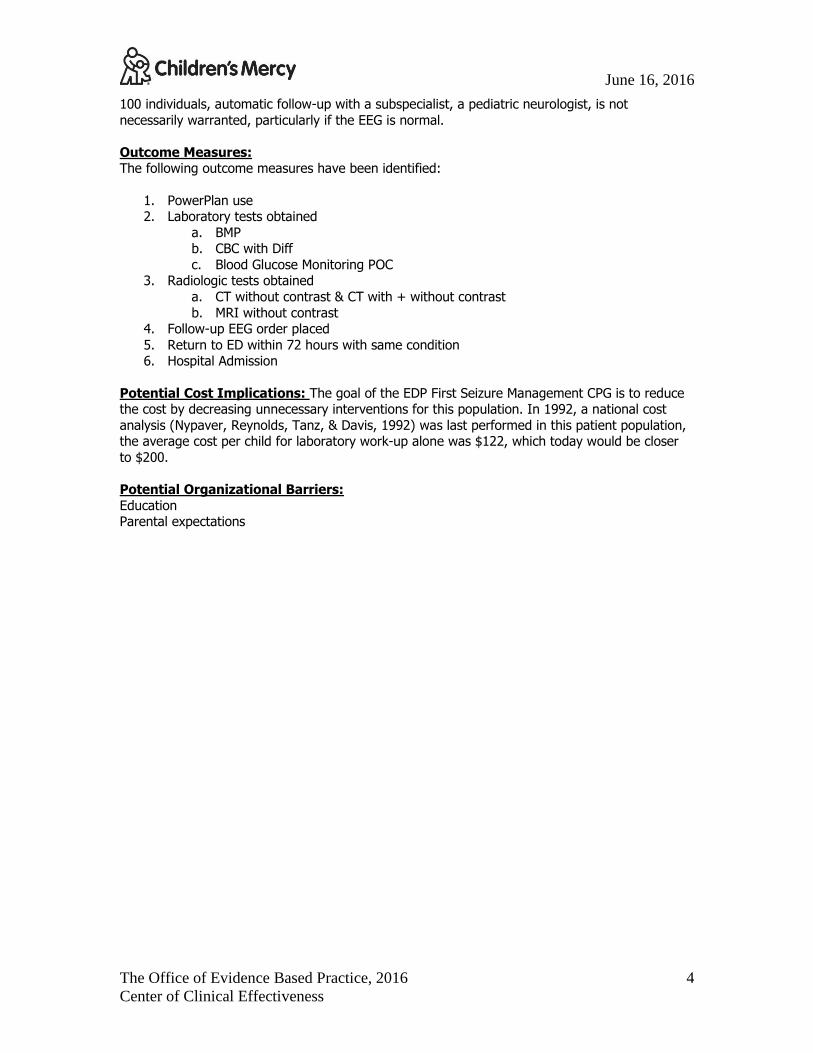
Outcome Measures: The following outcome measures have been identified:
1. PowerPlan use 2. Laboratory tests obtained
a. BMP b. CBC with Diff
c. Blood Glucose Monitoring POC 3. Radiologic tests obtained
a. CT without contrast & CT with + without contrast
b. MRI without contrast 4. Follow-up EEG order placed
5. Return to ED within 72 hours with same condition 6. Hospital Admission
Potential Cost Implications: The goal of the EDP First Seizure Management CPG is to reduce the cost by decreasing unnecessary interventions for this population. In 1992, a national cost
analysis (Nypaver, Reynolds, Tanz, & Davis, 1992) was last performed in this patient population, the average cost per child for laboratory work-up alone was $122, which today would be closer
to $200.
Potential Organizational Barriers:
Education Parental expectations

June 16, 2016
The Office of Evidence Based Practice, 2016
Center of Clinical Effectiveness
5
PowerPlan:
Unique Plan Description: EDP First Nonfebrile Seizure Plan Selection Display: EDP First Nonfebrile Seizure PlanType: ED/UCC Version: 1 Begin Effective Date: 03/11/2016 08:36 End Effective Date: Current Available at all facilities EDP First Nonfebrile Seizure Consults/Therapy
CPG recommendation (NOTE)*
Clinic Referral Neurology Clinic Neurology Clinic, First-time nonfebrile seizure
EEG Request This patient was evaluated for a first-time nonfebrile seizure.
*Report Legend: DEF - This order sentence is the default for the selected order GOAL - This component is a goal IND - This component is an indicator INT - This component is an intervention IVS - This component is an IV Set NOTE - This component is a note Rx - This component is a prescription SUB - This component is a sub phase

June 16, 2016
The Office of Evidence Based Practice, 2016
Center of Clinical Effectiveness
6
Guideline Preparation: This guideline was prepared by The Office of Evidence Based Practice
(EBP) in collaboration with content experts at Children’s Mercy Hospitals and Clinics. Development of this guideline supports the Department of Clinical Effectiveness’s initiative to
promote care standardization that builds a culture of quality and safety that is evidenced by measured outcomes. If a conflict of interest is identified the conflict will be disclosed next to the
team members name.
First Seizure Management in the ED CPG Team Members:
Amy D’Angelo, MD, Emergency Medicine
Andrew Loehr, RN
Brian Burghardt, MD, General Pediatrics
Emily Dague, RN, CPNP Emergency Department and Urgent Care
Marcie Goeden, MD, Pediatric Resident, Neurology
Ara Hall, MD, Epileptologist, Neurology
Jan Wiebe, RN, CPN, SANE-A, SANE-P, Emergency Department Director of Nursing
Amanda Montalbano, MD, MPH, QBS
Kiran Raman, MD
Ibad Siddiqi, PharmD, Emergency Department Pharmacist
Britton Zuccarelli, MD, Neurology Fellow
Evidence Based Practice Scholars: Lynda Bainbridge, RN, MBA
Teresa Bontrager, RN, BSN, MSNed, CPEN
Jamie Cailteux, RN, BSN, CPN
Kate Collum, RN, BSN
Jennifer Foley, RT(R)(N), CNMT
Anne Holmes, RN, MSN, MBA-HCM, CCRC
Patty Lanzer RN, NNP-BC
Andrea Melanson, OTD, OTR/L
Office of EBP Team Members:
Jeffery Michael, DO Medical Director, Office of Evidence Based Practice
Jacqueline Bartlett, PhD, RN Director Office of Evidence Based Practice
Nancy Allen, MS, MLS, RD, LD Evidence Based Practice Research Specialist
Jarrod Dusin, MS, RD, LD, CNSC
Guideline development funded by:
No external funding was obtained in the development of this guideline.
Development Process:
The review summary documents the following steps: 1. Review of existing internal and external guidelines and standards
a. Internal guidelines: None b. External guidelines(Adams & Knowles, 2007) and (Hirtz et al., 2000)
2. Review preparation
a. PICOT questions established b. Team leaders confirmed search terms used by the librarians in the Health Sciences
Libraries. The team leaders also reviewed article titles and abstracts form the searches and identified the articles to be read and synthesized by the Evidence Based Practice
Scholars. 3. Databases searched
a. AHRQ National Guideline Clearinghouse
b. Medline c. Cochrane
4. Critically analyze the evidence

June 16, 2016
The Office of Evidence Based Practice, 2016
Center of Clinical Effectiveness
7
a. Guidelines
i. AGREE criteria were used to analyze published clinical guidelines b. Single studies
i. The EBP Scholars used the Cochrane Collaborative’s electronic software, Review Manager 5 (RevMan), to produce systematic reviews and
meta-analysis of the evidence of the effects of healthcare and delivered these
documents to the team for review. RevMan allowed the EBP Scholars to build the tables of study characteristics, tables of study biases, and analyze study
data in a meta-analysis. . In instances when RevMan could not be used, CASP (Critical Appraisal Skills Programme) tools were utilized to analyze the literature.
c. When a meta-analysis was found in the literature search, or created in RevMan, the GRADE criteria evaluated the literature using the Cochrane Collaborative’s
electronic software known as GRADEprofiler (GRADEpro). GRADEpro assesses
the meta-analysis for: 1. Limitations in study design and execution
2. Inconsistency between studies 3. Indirectness of study outcomes
4. Imprecision
5. Publication bias ii. Table 1 defines how the quality of the evidence is rated and how the
recommendation is established based on the type of evidence.

June 16, 2016
The Office of Evidence Based Practice, 2016
Center of Clinical Effectiveness
8
iii. Table 1. Grading of CPG Recommendations
Grade of
Recommendation
Confidence in
Clarity of Benefits vs.
Harms, Burden, and Cost
Quality of
Supporting Evidence
Implications
Strong recommendation
High quality evidence
Desirable effects
clearly outweigh undesirable effects
or vice versa
Consistent evidence
from well-performed RCTs or exceptionally
strong evidence from unbiased
observational studies
Recommendation can
apply to most patients in most circumstances.
Further research is unlikely to change our
confidence in the
estimate of effect
Strong recommendation
Moderate-quality evidence
Desirable effects
clearly outweigh undesirable effect
or vice versa
Evidence from RCTs
with important limitations
(inconsistent results,
methodological flaws, indirect evidence, or
imprecise results) or unusually strong
evidence from
unbiased observational studies
Recommendation can
apply to most patients in most circumstances.
Further research (if
performed) is likely to have an important
effect on our confidence in the
estimate of effect and
may change the estimate.
Strong recommendation Low-quality evidence
Desirable effects clearly outweigh
undesirable effect
or vice versa
Evidence for at least 1 critical outcome
from observational
studies, from RCTs with serious flaws or
indirect evidence
Recommendation may change when higher-
quality evidence
becomes available. Further research (if
performed) is likely to have an important
influence on our
confidence in the estimate of effect and
is likely to change the estimate.
Strong recommendation
Very-low-quality evidence
(Very rarely applicable)
Desirable effects
clearly outweigh undesirable effect
or vice versa
Evidence for at least
1 of the critical outcomes from
unsystematic clinical observations or very
indirect evidence
Recommendation may
change when higher-quality evidence
becomes available; any estimate of effect,
for at least 1 critical
outcome, is uncertain.
Recommended
High-quality evidence
Desirable effects
closely balanced with undesirable
effects
Consistent evidence
from well-performed RCTs or exceptionally
strong evidence from unbiased
observational studies
The best action may
differ, depending on circumstances or
patients or societal values. Further
research is unlikely to
change our confidence

June 16, 2016
The Office of Evidence Based Practice, 2016
Center of Clinical Effectiveness
9
in the estimate of
effect.
Recommended Moderate-quality
evidence
Desirable effects closely balanced
with undesirable
effects
Evidence from RCTs with important
limitations
(inconsistent results, methodological flaws,
indirect evidence, or imprecise results) or
unusually strong
evidence from unbiased
observational studies
Alternative approaches likely to be better for
some patients under
some circumstances. Further research (if
performed) is likely to have an important
influence on our
confidence in the estimate of effect and
may change the estimate.
Recommended
Low-quality evidence
Desirable effects
closely balanced with undesirable
effects
Evidence for at least
1 critical outcome from observational
studies, from RCTs with serious flaws or
indirect evidence
Other alternatives may
be equally reasonable. Further research is
likely to have an important influence on
our confidence in the
estimate of effect and is likely to change the
estimate.
Recommended
Very-low-quality
evidence
Desirable effects
closely balanced
with undesirable effects
Evidence for at least
1 critical outcome
from unsystematic clinical observations
or very indirect evidence
Other alternatives may
be equally reasonable.
Any estimate of effect, for at least 1 critical
outcome, is uncertain.
Adapted from: Schunemann, H. J., Vist, G. E., Jaeschke, R., Kunz, R., Cook, D. J., & Guyatt, G.
(2002). Advanced topics in moving from evidence to action: Grading recommendations. In Guyatt, G., Rennie, D., Meade, M. O., & Cook, D. J.(Ed.), Users’ guides to the medical literature: A manual for evidence-based clinical practice (pp 679-701). New York, NY:McGraw-Hill.
5. Recommendations for the guideline were developed by a consensus process incorporating
the three principles of EBP (current literature, content experts, and patient and family preference [when possible])
Approval Process: Guidelines are reviewed by Bradley L. Schlaggar, MD, PhD Neurologist-in
Chief, & Adam Ostendorf, MD, Fellow in Pediatric Fellow, both of St Louis Children’s, Content
Expert Team at Children’s Mercy, the Office of EBP, and other appropriate hospital committees as deemed suitable for the guidelines intended use. Guidelines are reviewed and updated as
necessary every 3 years within the Office of EBP at CMH&C. Content expert teams will be involved with every review and update.
Disclaimer: The content experts and the Office of EBP are aware of the controversies surrounding First
Seizure CPG When evidence is lacking or inconclusive, options in care are provided in the guideline and the power plans that accompany the guideline.
These guidelines do not establish a standard of care to be followed in every case. It is recognized
that each case is different and those individuals involved in providing health care are expected to
June 16, 2016
The Office of Evidence Based Practice, 2016
Center of Clinical Effectiveness
10
use their judgment in determining what is in the best interests of the patient based on the
circumstances existing at the time.
It is impossible to anticipate all possible situations that may exist and to prepare guidelines for each. Accordingly, these guidelines should guide care with the understanding that departures
from them may be required at times.

June 16, 2016
The Office of Evidence Based Practice, 2016
Center of Clinical Effectiveness
11
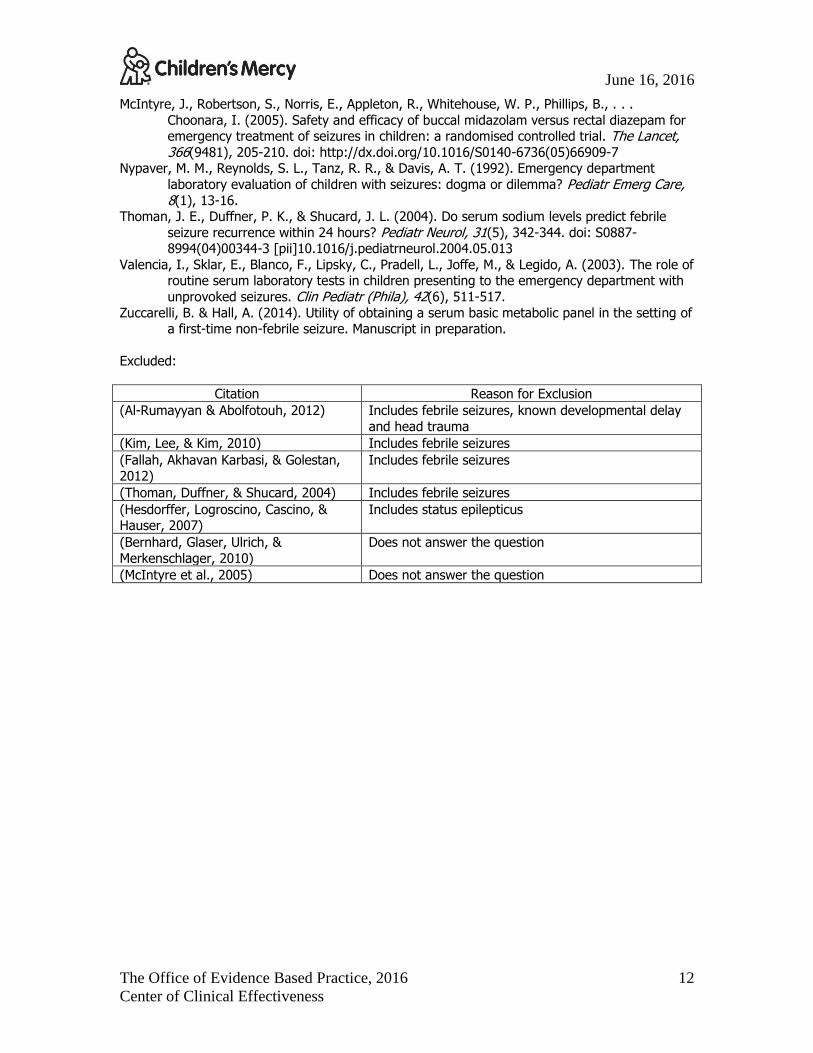
References Adams, S. M., & Knowles, P. D. (2007). Evaluation of a first seizure. Am Fam Physician, 75(9),
1342-1347. Hirtz, D., Ashwal, S., Berg, A., Bettis, D., Camfield, C., Camfield, P., . . . Shinnar, S. (2000).
Practice parameter: evaluating a first nonfebrile seizure in children: report of the quality
standards subcommittee of the American Academy of Neurology, The Child Neurology Society, and The American Epilepsy Society. Neurology, 55(5), 616-623.
Hirtz, D., Berg, A., Bettis, D., Camfield, C., Camfield, P., Crumrine, P., . . . Shinnar, S. (2003). Practice parameter: treatment of the child with a first unprovoked seizure: Report of the
Quality Standards Subcommittee of the American Academy of Neurology and the Practice Committee of the Child Neurology Society. Neurology, 60(2), 166-175.
Al-Rumayyan, A. R., & Abolfotouh, M. A. (2012). Prevalence and prediction of abnormal CT scan
in pediatric patients presenting with a first seizure. Neurosciences (Riyadh), 17(4), 352-356.
Anand, G., Padeniya, A., Jain, R., Hasan, N., Pike, M., Jayawant, S., . . . Zaiwalla, Z. (2012). Video EEG outcome on children referred following a single unprovoked afebrile seizure.
Arch Dis Child, 97(1), 90. doi: 10.1136/archdischild-2011-300960archdischild-2011-
300960 [pii] Arthur, T. M., deGrauw, T. J., Johnson, C. S., Perkins, S. M., Kalnin, A., Austin, J. K., & Dunn, D.
W. (2008). Seizure recurrence risk following a first seizure in neurologically normal children. Epilepsia, 49(11), 1950-1954. doi: 10.1111/j.1528-1167.2008.01775.xEPI1775
[pii] Aydogan, M., Aydogan, A., Kara, B., Basim, B., & Erdogan, S. (2007). Transient peripheral
leukocytosis in children with afebrile seizures. J Child Neurol, 22(1), 77-79.
Bernhard, M. K., Glaser, A., Ulrich, K., & Merkenschlager, A. (2010). Is there a need for ophthalmological examinations after a first seizure in paediatric patients? Eur J Pediatr, 169(1), 31-33. doi: 10.1007/s00431-009-0966-4
Chan, D., Phuah, H. K., Ng, Y. L., Choong, C. T., Lim, K. W., & Goh, W. H. (2010). Pediatric
epilepsy and first afebrile seizure in Singapore: epidemiology and investigation yield at
presentation. J Child Neurol, 25(10), 1216-1222. doi: 10.1177/08830738093589240883073809358924 [pii]
Fallah, R., Akhavan Karbasi, S., & Golestan, M. (2012). Afebrile seizure subsequent to initial febrile seizure. Singapore Med J, 53(5), 349-352.
Hamiwka, L. D., Singh, N., Niosi, J., & Wirrell, E. C. (2007). Diagnostic inaccuracy in children
referred with "first seizure": role for a first seizure clinic. Epilepsia, 48(6), 1062-1066. doi: EPI1018 [pii]10.1111/j.1528-1167.2007.01018.x
Hesdorffer, D. C., Logroscino, G., Cascino, G. D., & Hauser, W. A. (2007). Recurrence of afebrile status epilepticus in a population-based study in Rochester, Minnesota. Neurology, 69(1),
73-78. doi: 69/1/73 [pii]10.1212/01.wnl.0000265056.31752.ff Hsieh, D. T., Chang, T., Tsuchida, T. N., Vezina, L. G., Vanderver, A., Siedel, J., . . . Gaillard, W.
D. (2010). New-onset afebrile seizures in infants: role of neuroimaging. Neurology, 74(2), 150-156. doi: 10.1212/WNL.0b013e3181c9184774/2/150 [pii]
Kim, S. H., Lee, H. Y., & Kim, Y. H. (2010). Subsequent afebrile seizure in children who have a
first seizure with fever after 6 years of age. Pediatr Neurol, 43(2), 122-126. doi: 10.1016/j.pediatrneurol.2010.03.009S0887-8994(10)00141-4 [pii]
Landau, Y. E., Waisman, Y., & Shuper, A. (2010). Management of children with nonfebrile
seizures in the emergency department. Eur J Paediatr Neurol, 14(5), 439-444. doi: 10.1016/j.ejpn.2010.02.006S1090-3798(10)00032-2 [pii]
Lateef, T. M., Tsuchida, T. N., Chang, T., Johnson, J., Gaillard, W. D., & Nelson, K. B. (2008). Diagnostic value of lumbar puncture in afebrile infants with suspected new-onset
seizures. J Pediatr, 153(1), 140-142. doi: 10.1016/j.jpeds.2008.02.030S0022-3476(08)00106-6 [pii]
June 16, 2016
The Office of Evidence Based Practice, 2016
Center of Clinical Effectiveness
12
McIntyre, J., Robertson, S., Norris, E., Appleton, R., Whitehouse, W. P., Phillips, B., . . .
Choonara, I. (2005). Safety and efficacy of buccal midazolam versus rectal diazepam for emergency treatment of seizures in children: a randomised controlled trial. The Lancet, 366(9481), 205-210. doi: http://dx.doi.org/10.1016/S0140-6736(05)66909-7
Nypaver, M. M., Reynolds, S. L., Tanz, R. R., & Davis, A. T. (1992). Emergency department
laboratory evaluation of children with seizures: dogma or dilemma? Pediatr Emerg Care, 8(1), 13-16.
Thoman, J. E., Duffner, P. K., & Shucard, J. L. (2004). Do serum sodium levels predict febrile
seizure recurrence within 24 hours? Pediatr Neurol, 31(5), 342-344. doi: S0887-8994(04)00344-3 [pii]10.1016/j.pediatrneurol.2004.05.013
Valencia, I., Sklar, E., Blanco, F., Lipsky, C., Pradell, L., Joffe, M., & Legido, A. (2003). The role of routine serum laboratory tests in children presenting to the emergency department with
unprovoked seizures. Clin Pediatr (Phila), 42(6), 511-517.
Zuccarelli, B. & Hall, A. (2014). Utility of obtaining a serum basic metabolic panel in the setting of a first-time non-febrile seizure. Manuscript in preparation.
Excluded:
Citation Reason for Exclusion
(Al-Rumayyan & Abolfotouh, 2012) Includes febrile seizures, known developmental delay
and head trauma
(Kim, Lee, & Kim, 2010) Includes febrile seizures
(Fallah, Akhavan Karbasi, & Golestan, 2012)
Includes febrile seizures
(Thoman, Duffner, & Shucard, 2004) Includes febrile seizures
(Hesdorffer, Logroscino, Cascino, & Hauser, 2007)
Includes status epilepticus
(Bernhard, Glaser, Ulrich, &
Merkenschlager, 2010)
Does not answer the question
(McIntyre et al., 2005) Does not answer the question

June 16, 2016
The Office of Evidence Based Practice, 2016
Center of Clinical Effectiveness
13
Author, date, country, and
industry of
funding
Patient Group
Strength of
Evidence (GRADE)
Research design Significant results Limitations
Overview of Diagnostic Work up -AAN Practice Parameter
(Hirtz et al.,
2003)
Children and
adolescents with first
unprovoked seizure
The guideline
was reviewed by two team
members using the AGREE
tool. The
consensus was to accept the
guideline with alterations
Guideline The Practice Parameter addresses the following:
Laboratory studies- may be obtained when history or clinical findings such as vomiting, diarrhea, or
dehydration are present. Lumbar puncture (LP)- should not be obtained
unless meningitis is suspected.
EEG- should be performed as part of the evaluation of first non-febrile seizure. Timing of
the study (within the first 48 hours or later) is not clear.
Neuroimaging- CT scan- should not be obtained.
MRI- should not be obtained for the child with a
first non-febrile seizure who has returned to baseline.
*Concerns with
the AAN Guideline (Hirtz
et al., 2000) include:
The development
group did not include Pediatric
Emergency Medicine,
patient/parent/family
representatives
Methods for formulating the
recommendations, cost
implications and
conflicts of interest are not
reported transparently
Should Laboratory Studies be Obtained?
(Aydogan, Aydogan,
Kara, Basim, &
Erdogan,
Children mean age 4 ±3.6 y
(range 6 m-13 y)
Presenting
Very low Prospective cohort All subjects with
afebrile seizure between Jan
2000 and March
62 subjects were enrolled- 50% male 9 subjects had leukocytosis (14.5%), a second
CBC was obtained and leukocytosis did not persist.
Small number of subjects.
June 16, 2016
The Office of Evidence Based Practice, 2016
Center of Clinical Effectiveness
14
Author, date,
country, and industry of
funding
Patient Group
Strength of
Evidence
(GRADE)
Research design Significant results Limitations
2007)
Turkey
with first afebrile
seizure
2002 were enrolled
Leukocytosis was more prevalent in children with status epilepticus
SE defined as a continuous seizure lasting longer
than 30 minutes or repeating seizures last 30
minutes with recovering consciousness between them.
(Landau,
Waisman, & Shuper,
2010) Israel
85 subjects
average age 7.5 y
(range 0-18 y) who
made 104 visits to the
ED
Excluded febrile
seizure or other
primary
diagnosis
Very Low-
inconsistent includes
subjects that do not apply
to this guideline
Retrospective
chart review
Laboratory tests were obtained in 84% of visits.
Eight percent provided useful information and < 5% were helpful in diagnosis and management.
Only one lumbar puncture was performed. Eight percent of visits had electrocardiography
performed and all were normal Seven percent of visit had electroencephalography
performed and was consistently useful and was
always performed along with a neurology consultation
Mix of children
with first seizure and those already
on medication for seizure. Only 30
(35%) subjects presented with
first seizure.
(Nypaver,
Reynolds, Tanz, &
Davis,
1992) 1 USA
308 ED
charts, 108 febrile
(mean age
2.1 years) 200 non
febrile seizures.
(mean age
5.7 years
Very Low Retrospective
chart review
41 were having their first non febrile seizure. 26
subjects (63%) had at least one laboratory test performed.
No changes in therapy were made as the result of
the laboratory findings. In 1992 US dollars, the mean cost of the
laboratory tests was $122.00 per subject.
Small sample size.
Important changes in newborn
screening since
this study need to be considered,
that is the need for lab studies
may be even
lower.

June 16, 2016
The Office of Evidence Based Practice, 2016
Center of Clinical Effectiveness
15
Author, date,
country, and industry of
funding
Patient Group
Strength of
Evidence
(GRADE)
Research design Significant results Limitations
Included lab tests:
electrolytes Calcium,
magnesium,
ammonia, glucose,
Dextrostix
Scarfone 2000 USA
Infants < or equal to 12
months of age
presenting to the ED of
a tertiary
care children’s
hospital. Serum
chemistry
results were
classified as normal,
outside of range
normal and
clinically significantly
abnormal
Very Low Retrospective chart review
214 patient visits made by infants with febrile and non febrile seizures.
134/214 were non-febrile seizures and 70 of these were a first seizure, or 52% of all
presenting non febrile seizures. 51 of 70 had lab drawn
8/51 (16%) had a clinically significant
abnormality.
Is there a Working Group on Status Epilepticus recommendation that serum chemistries should
be obtained for adults and children with status
epilepticus?
Would expanded newborn
screening change any of this?
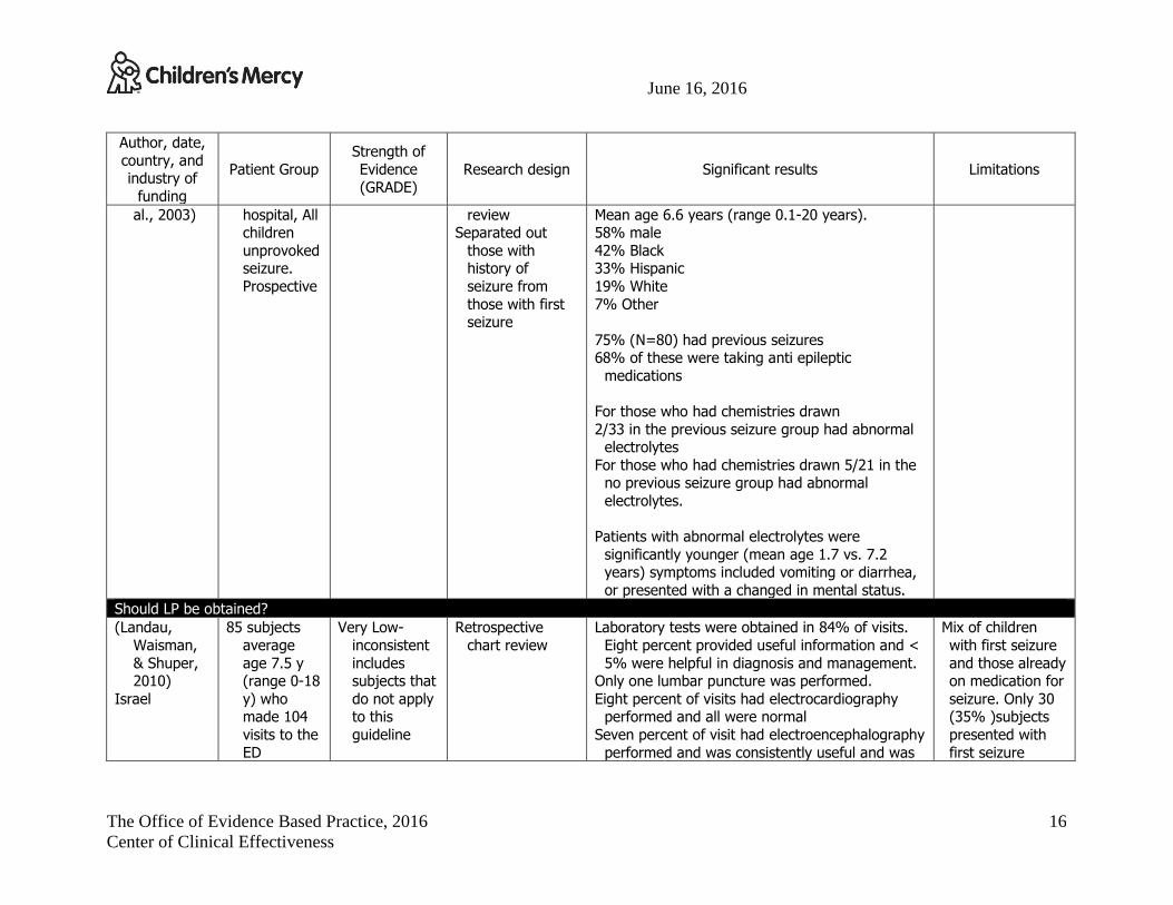
(Valencia et Urban Very Low Prospective chart Total of 107 children met criteria.
June 16, 2016
The Office of Evidence Based Practice, 2016
Center of Clinical Effectiveness
16
Author, date,
country, and industry of
funding
Patient Group
Strength of
Evidence
(GRADE)
Research design Significant results Limitations
al., 2003) hospital, All children
unprovoked seizure.
Prospective
review Separated out
those with history of
seizure from
those with first seizure
Mean age 6.6 years (range 0.1-20 years). 58% male
42% Black 33% Hispanic
19% White
7% Other
75% (N=80) had previous seizures 68% of these were taking anti epileptic
medications
For those who had chemistries drawn
2/33 in the previous seizure group had abnormal electrolytes
For those who had chemistries drawn 5/21 in the no previous seizure group had abnormal
electrolytes.
Patients with abnormal electrolytes were
significantly younger (mean age 1.7 vs. 7.2 years) symptoms included vomiting or diarrhea,
or presented with a changed in mental status.
Should LP be obtained?
(Landau,
Waisman,
& Shuper, 2010)
Israel
85 subjects
average
age 7.5 y (range 0-18
y) who made 104
visits to the
ED
Very Low-
inconsistent
includes subjects that
do not apply to this
guideline
Retrospective
chart review
Laboratory tests were obtained in 84% of visits.
Eight percent provided useful information and <
5% were helpful in diagnosis and management. Only one lumbar puncture was performed.
Eight percent of visits had electrocardiography performed and all were normal
Seven percent of visit had electroencephalography
performed and was consistently useful and was
Mix of children
with first seizure
and those already on medication for
seizure. Only 30 (35% )subjects
presented with
first seizure

June 16, 2016
The Office of Evidence Based Practice, 2016
Center of Clinical Effectiveness
17
Author, date,
country, and industry of
funding
Patient Group
Strength of
Evidence
(GRADE)
Research design Significant results Limitations
Excluded febrile
seizure or other
primary
diagnosis
always performed along with a neurology consultation
(Lateef et al.,
2008)
USA
Children 1- 6
months
with new onset
seizures N= 141
Very Low
Cohort
study, small number of
subjects, not all had
results of HSV or
enteroviral
infection
Prospective cohort Diagnostic standards of infected CSF
WBC > 6 mm3
Protein elevation > 50 mg/dl
Positive bacterial culture
Herpes simplex virus (HSV) PCR
76/141 (54%) underwent LP. Age was the greatest factor in obtaining an LP.
Subjects aged 1-2 mo 70% LP whereas aged 5-6 mo 33 % LP
There was no relationship between presence of CSF abnormalities and the final diagnosis of
seizure. At the time of discharge, 53% of those
who had an abnormal CSF were thought to have a seizure, while the remaining 47% were
thought to have a non-seizure event.
LP is only
performed on
subjects whom the attending
provider deems necessary
Small population
(Chan et al., 2010)
Singapore
Children aged 1 month to
15 years with first
afebrile seizure
108 with ≥ 2
afebrile seizure and
Very Low Population Survey 1st SZ Epilepsy≥ 2
SZ
P value
Develop-
mental exam
(norma
l)
93% 87% P=0.04
6
Population based
study that looked at the
epidemiology of afebrile seizure.
June 16, 2016
The Office of Evidence Based Practice, 2016
Center of Clinical Effectiveness
18
Author, date,
country, and industry of
funding
Patient Group
Strength of
Evidence
(GRADE)
Research design Significant results Limitations
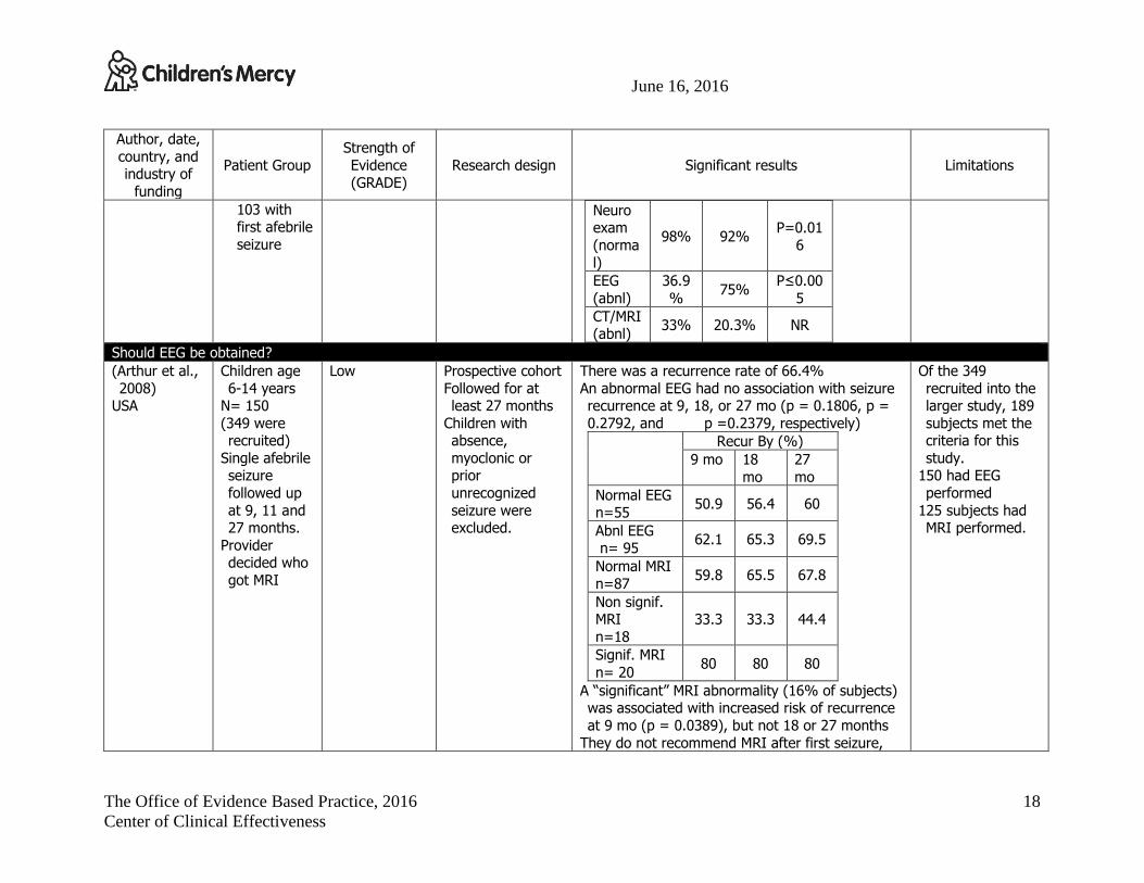
103 with first afebrile
seizure
Neuro exam
(norma
l)
98% 92% P=0.01
6
EEG
(abnl)
36.9
% 75%
P≤0.00
5
CT/MRI (abnl)
33% 20.3% NR
Should EEG be obtained?
(Arthur et al.,
2008) USA
Children age
6-14 years N= 150
(349 were recruited)
Single afebrile seizure
followed up
at 9, 11 and 27 months.
Provider decided who
got MRI
Low Prospective cohort
Followed for at least 27 months
Children with absence,
myoclonic or prior
unrecognized
seizure were excluded.
There was a recurrence rate of 66.4%
An abnormal EEG had no association with seizure recurrence at 9, 18, or 27 mo (p = 0.1806, p =
0.2792, and p =0.2379, respectively)
Recur By (%)
9 mo 18
mo
27
mo
Normal EEG n=55
50.9 56.4 60
Abnl EEG n= 95
62.1 65.3 69.5
Normal MRI
n=87 59.8 65.5 67.8
Non signif. MRI
n=18
33.3 33.3 44.4
Signif. MRI
n= 20 80 80 80
A “significant” MRI abnormality (16% of subjects) was associated with increased risk of recurrence
at 9 mo (p = 0.0389), but not 18 or 27 months
They do not recommend MRI after first seizure,
Of the 349
recruited into the larger study, 189
subjects met the criteria for this
study. 150 had EEG
performed
125 subjects had MRI performed.

June 16, 2016
The Office of Evidence Based Practice, 2016
Center of Clinical Effectiveness
19
Author, date,
country, and industry of
funding
Patient Group
Strength of
Evidence
(GRADE)
Research design Significant results Limitations
because it is not predictive.
(Chan et al.,
2010)
Singapore
Children aged
1 month to
15 years with first
afebrile seizure
108 with ≥ 2
afebrile seizure and
103 with first afebrile
seizure
Very Low Population Survey 1st SZ Epilepsy≥ 2
SZ
P value
Develop-
mental exam
(normal)
93% 87% P=0.04
6
Neuro
exam (norma
l)
98% 92% P=0.01
6
EEG (abnl)
36.9%
75% P≤0.00
5
CT/MRI
(abnl) 33% 20.3% NR
Population based
study that looked at the
epidemiology of afebrile seizure.
(Anand et al., 2012)
United Kingdom
128 children mean age
6.5 years (range 1
month to 17 years.
Very Low Appears to be
an abstract only
Retrospective observational
cohort
Video EEG (vEEG) was normal in 75 subjects (59%)
Non-epileptic events were recorded in 8 subjects (6%)
Idiopathic generalized epilepsy was diagnosed in 14 subjects (11%)
Generalized epilepsy with febrile seizure was
diagnosed in 2 subjects (2%) A focal epilepsy was diagnosed in 29 subjects
(23%) Sensitivity= 100
Specificity = 10
(+) predictive value = 85%
34 subjects had neurodevelopmen
tal problem, 11 subjects had a
family history of epilepsy, and 13
had a history of
febrile seizure.
June 16, 2016
The Office of Evidence Based Practice, 2016
Center of Clinical Effectiveness
20
Author, date,
country, and industry of
funding
Patient Group
Strength of
Evidence
(GRADE)
Research design Significant results Limitations
(-) predictive value= not estimable
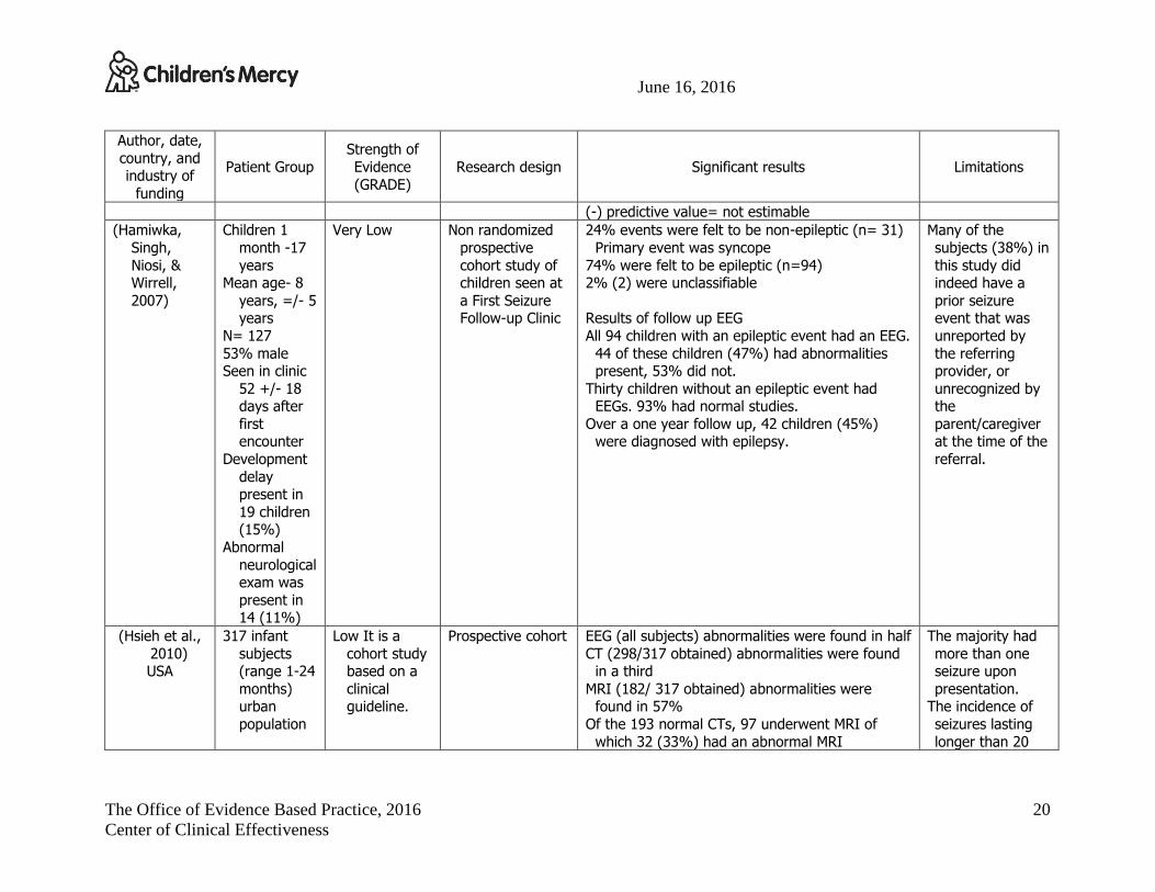
(Hamiwka,
Singh,
Niosi, & Wirrell,
2007)
Children 1
month -17
years Mean age- 8
years, =/- 5 years
N= 127
53% male Seen in clinic
52 +/- 18 days after
first encounter
Development
delay present in
19 children (15%)
Abnormal
neurological exam was
present in 14 (11%)
Very Low Non randomized
prospective
cohort study of children seen at
a First Seizure Follow-up Clinic
24% events were felt to be non-epileptic (n= 31)
Primary event was syncope
74% were felt to be epileptic (n=94) 2% (2) were unclassifiable
Results of follow up EEG
All 94 children with an epileptic event had an EEG.
44 of these children (47%) had abnormalities present, 53% did not.
Thirty children without an epileptic event had EEGs. 93% had normal studies.
Over a one year follow up, 42 children (45%) were diagnosed with epilepsy.
Many of the
subjects (38%) in
this study did indeed have a
prior seizure event that was
unreported by
the referring provider, or
unrecognized by the
parent/caregiver at the time of the
referral.
(Hsieh et al.,
2010) USA
317 infant
subjects (range 1-24
months) urban
population
Low It is a
cohort study based on a
clinical guideline.
Prospective cohort EEG (all subjects) abnormalities were found in half
CT (298/317 obtained) abnormalities were found in a third
MRI (182/ 317 obtained) abnormalities were found in 57%
Of the 193 normal CTs, 97 underwent MRI of
which 32 (33%) had an abnormal MRI
The majority had
more than one seizure upon
presentation. The incidence of
seizures lasting
longer than 20

June 16, 2016
The Office of Evidence Based Practice, 2016
Center of Clinical Effectiveness
21
Author, date,
country, and industry of
funding
Patient Group
Strength of
Evidence
(GRADE)
Research design Significant results Limitations
minutes was 8.5%
30 subjects had a history of
prematurity.
Increased likelihood of
obtaining an MRI in younger
infants.
(Landau,
Waisman, & Shuper,
2010)
Israel
85 subjects
average age 7.5 y
(range 0-18
y) who made 104
visits to the ED
Excluded
febrile seizure or
other primary
diagnosis
Very Low-
inconsistent includes
subjects that
do not apply to this
guideline
Retrospective
chart review
Laboratory tests were obtained in 84% of visits.
Eight percent provided useful information and < 5% were helpful in diagnosis and management.
Only one lumbar puncture was performed.
Eight percent of visits had electrocardiography performed and all were normal
Seven percent of visit had electroencephalography performed and was consistently useful and was
always performed along with a neurology
consultation
Mix of children
with first seizure and those already
on medication for
seizure. Only 30 (35% )subjects
presented with first seizure
Should a CT scan be obtained?
(Hsieh et al.,
2010)
USA
317 infant
subjects
(range 1-24 months)
urban
Low- It is a
cohort study
based on a clinical
guideline.
Prospective cohort EEG (all subjects) abnormalities were found in half
CT (298/317 obtained) abnormalities were found
in a third MRI (182/ 317 obtained) abnormalities were
found in 57%
The majority had
more than one
seizure upon presentation.
The incidence of

June 16, 2016
The Office of Evidence Based Practice, 2016
Center of Clinical Effectiveness
22
Author, date,
country, and industry of
funding
Patient Group
Strength of
Evidence
(GRADE)
Research design Significant results Limitations
population Of the 193 normal CTs, 97 underwent MRI of which 32 (33%) had an abnormal MRI
seizures lasting longer than 20
minutes was 8.5%
30 subjects had a
history of prematurity.
Increased likelihood of
obtaining an MRI
in younger infants.
Kodaphanhad
eh 2006
Iran
125 subjects,
children
mean age 53 ±48
months (range-
1 month-15
years)
Low Retrospective case
series –no control
group. Excluded those with
seizures > 30 minutes or
electrolyte
abnormalities Report on CT scan
and MRI within the first hours of
arrival
Neuro-imaging was obtained in 119 subjects
(95%)
Emergent CT was performed in 108 (91%) and MRI in 11 (9%)
Neuro-imaging was normal in 107 (90%) of subjects.
Clinical significant results were found in 12
subjects (10%) 10 of the 12 subjects with abnormal findings had
abnormal neurological examination.
Study design.
Sharma 2003 USA
500 subjects with new-
onset afebrile
seizure
median age
Low Retrospective Neuro-imaging was performed in 475/500. 25 subjects were not imaged.
Of the subjects who were scanned, CT was performed in 454/475, and MRI was performed
in 21/475.
437/475 had neuro-imaging while in the ED. And
5/6 subjects who fell in the low risk
group by partition analysis had
abnormal findings
on

June 16, 2016
The Office of Evidence Based Practice, 2016
Center of Clinical Effectiveness
23
Author, date,
country, and industry of
funding
Patient Group
Strength of
Evidence
(GRADE)
Research design Significant results Limitations
(16 mo range (0-21
years))
13 had neuro-imaging after the ED visit but within 72 hours of the visit.
Normal imaging results were reported in 395/475 subjects. [83%]
Clinically insignificant results were reported in
42/475 [9%] Clinically significant events were reported in
38/475 subjects [8%] Using Partition analysis, 3 variables partitioned the
subjects into 4 groups
Variables Presence of pre disposing condition, focality of the
seizure and age Groups
Predisposing condition- High risk No predisposing condition
Non-focal seizure- low risk
Focal seizure- age dependent Age > 33 months low risk
Age < 33 months high risk
physical/neurological exam.
One subject subsequently
diagnosed with
grey matter heterotopias had
a normal physical and neurological
exam.
Retrospective design
Should an MRI be obtained?
(Arthur et al.,
2008) USA
Children age
6-14 years N= 150
(349 were
recruited) Single afebrile
seizure followed up
at 9, 11 and
27 months.
Low Prospective cohort
Followed for at least 27 months
Children with
absence, myoclonic or
prior unrecognized
seizure were
excluded.
There was a recurrence rate of 66.4%
An abnormal EEG had no association with seizure recurrence at 9, 18, or 27 mo (p = 0.1806, p =
0.2792, and p =0.2379, respectively)
Recur By (%)
9 mo 18 mo
27 mo
Normal EEG
n=55 50.9 56.4 60
Abnl EEG 62.1 65.3 69.5
Of the 349
recruited into the larger study, 189
subjects met the
criteria for this study.
150 had EEG performed
125 subjects had
MRI performed.

June 16, 2016
The Office of Evidence Based Practice, 2016
Center of Clinical Effectiveness
24
Author, date,
country, and industry of
funding
Patient Group
Strength of
Evidence
(GRADE)
Research design Significant results Limitations
Provider decided who
got MRI
n= 95
Normal MRI n=87
59.8 65.5 67.8
Non signif.
MRI n=18
33.3 33.3 44.4
Signif. MRI n= 20
80 80 80
A “significant” MRI abnormality (16% of subjects)
was associated with increased risk of recurrence at 9 mo (p = 0.0389), but not 18 or 27 months
They do not recommend MRI after first seizure,
because it is not predictive.
(Chan et al.,
2010) Singapore
Children aged
1 month to 15 years
with first
afebrile seizure
108 with ≥ 2 afebrile
seizure and
103 with first
afebrile seizure
Very Low Population Survey
1st SZ Epileps
y≥ 2 SZ
P value
Develo
p- mental
exam (norma
l)
93% 87% P=0.04
6
Neuro exam
(normal)
98% 92% P=0.01
6
EEG
(abnl)
36.9
% 75%
P≤0.00
5
CT/MRI (abnl)
33% 20.3% NR
Population based study that looked
at the
epidemiology of afebrile seizure.

June 16, 2016
The Office of Evidence Based Practice, 2016
Center of Clinical Effectiveness
25
Author, date,
country, and industry of
funding
Patient Group
Strength of
Evidence
(GRADE)
Research design Significant results Limitations
(Hsieh et al., 2010)
USA
317 infant subjects
(range 1-24 months)
urban
population
Low-It is a cohort study
based on a clinical
guideline.
Prospective cohort EEG (all subjects) abnormalities were found in half CT (298/317 obtained) abnormalities were found
in a third MRI (182/ 317 obtained) abnormalities were
found in 57%
Of the 193 normal CTs, 97 underwent MRI of which 32 (33%) had an abnormal MRI
The majority had more than one
seizure upon presentation.
The incidence of
seizures lasting longer than 20
minutes was 8.5%
30 subjects had a
history of prematurity.
Increased likelihood of
obtaining an MRI in younger
infants.
Kodaphanhade
h 2006 Iran
125 subjects,
children
mean age 53 ±48
months (range-
1 month-15 years)
Low Retrospective case
series –no control
group. Excluded those with
seizures > 30 minutes or
electrolyte abnormalities
Report on CT scan
and MRI within the first hours of
arrival
Neuro-imaging was obtained in 119 subjects
(95%)
Emergent CT was performed in 108 (91%) and MRI in 11 (9%)
Neuro-imaging was normal in 107 (90%) of subjects.
Clinical significant results were found in 12 subjects (10%)
10 of the 12 subjects with abnormal findings had
abnormal neurological examination.
Study design.
Rauch 2008 75 children Very Low- Retrospective Below 12 years of age sedated for MRI There was a 53%
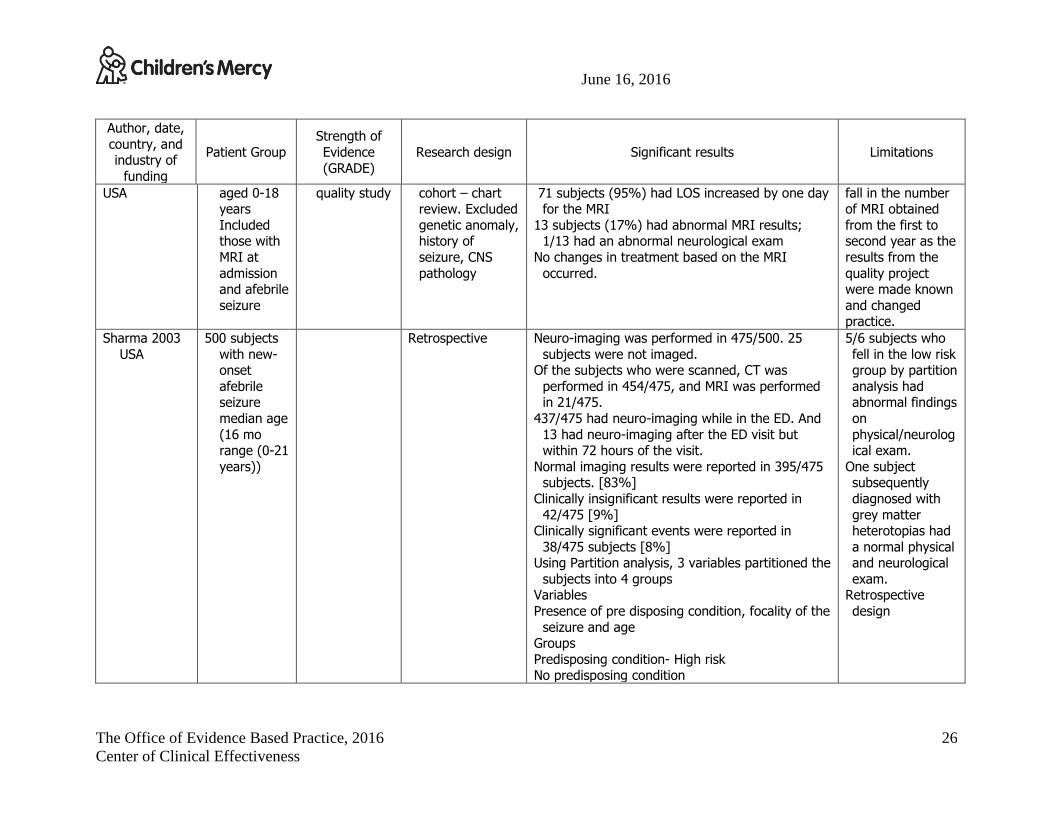
June 16, 2016
The Office of Evidence Based Practice, 2016
Center of Clinical Effectiveness
26
Author, date,
country, and industry of
funding
Patient Group
Strength of
Evidence
(GRADE)
Research design Significant results Limitations
USA aged 0-18 years
Included those with
MRI at
admission and afebrile
seizure
quality study cohort – chart review. Excluded
genetic anomaly, history of
seizure, CNS
pathology
71 subjects (95%) had LOS increased by one day for the MRI
13 subjects (17%) had abnormal MRI results; 1/13 had an abnormal neurological exam
No changes in treatment based on the MRI
occurred.
fall in the number of MRI obtained
from the first to second year as the
results from the
quality project were made known
and changed practice.
Sharma 2003
USA
500 subjects
with new-onset
afebrile seizure
median age
(16 mo range (0-21
years))
Retrospective Neuro-imaging was performed in 475/500. 25
subjects were not imaged. Of the subjects who were scanned, CT was
performed in 454/475, and MRI was performed in 21/475.
437/475 had neuro-imaging while in the ED. And
13 had neuro-imaging after the ED visit but within 72 hours of the visit.
Normal imaging results were reported in 395/475 subjects. [83%]
Clinically insignificant results were reported in
42/475 [9%] Clinically significant events were reported in
38/475 subjects [8%] Using Partition analysis, 3 variables partitioned the
subjects into 4 groups Variables
Presence of pre disposing condition, focality of the
seizure and age Groups
Predisposing condition- High risk No predisposing condition
5/6 subjects who
fell in the low risk group by partition
analysis had abnormal findings
on
physical/neurological exam.
One subject subsequently
diagnosed with
grey matter heterotopias had
a normal physical and neurological
exam. Retrospective
design
June 16, 2016
The Office of Evidence Based Practice, 2016
Center of Clinical Effectiveness
27
Author, date,
country, and industry of
funding
Patient Group
Strength of
Evidence
(GRADE)
Research design Significant results Limitations
Non-focal seizure- low risk Focal seizure- age dependent
Age > 33 months low risk Age < 33 months high risk
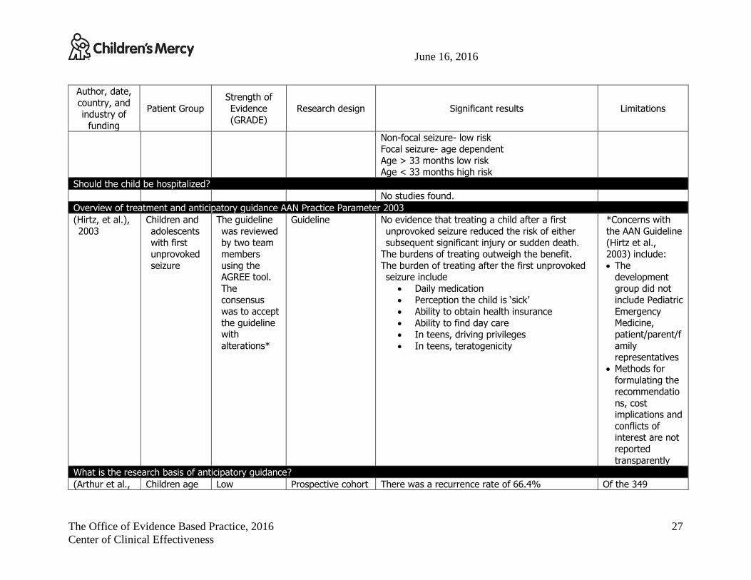
Should the child be hospitalized?
No studies found.
Overview of treatment and anticipatory guidance AAN Practice Parameter 2003
(Hirtz, et al.),
2003
Children and
adolescents
with first unprovoked
seizure
The guideline
was reviewed
by two team members
using the AGREE tool.
The
consensus was to accept
the guideline with
alterations*
Guideline No evidence that treating a child after a first
unprovoked seizure reduced the risk of either
subsequent significant injury or sudden death. The burdens of treating outweigh the benefit.
The burden of treating after the first unprovoked seizure include
Daily medication
Perception the child is ‘sick’
Ability to obtain health insurance
Ability to find day care
In teens, driving privileges
In teens, teratogenicity
*Concerns with
the AAN Guideline
(Hirtz et al., 2003) include:
The
development group did not
include Pediatric
Emergency Medicine,
patient/parent/family
representatives Methods for
formulating the
recommendatio
ns, cost implications and
conflicts of interest are not
reported
transparently
What is the research basis of anticipatory guidance?
(Arthur et al., Children age Low Prospective cohort There was a recurrence rate of 66.4% Of the 349

June 16, 2016
The Office of Evidence Based Practice, 2016
Center of Clinical Effectiveness
28
Author, date,
country, and industry of
funding
Patient Group
Strength of
Evidence
(GRADE)
Research design Significant results Limitations
2008) USA
6-14 years N= 150
(349 were recruited)
Single afebrile
seizure followed up
at 9, 11 and 27 months.
Provider
decided who got MRI
Followed for at least 27 months
Children with absence,
myoclonic or
prior unrecognized
seizure were excluded.
An abnormal EEG had no association with seizure recurrence at 9, 18, or 27 mo (p = 0.1806, p =
0.2792, and p =0.2379, respectively)
Recur By (%)
9 mo 18
mo
27
mo
Normal EEG n=55
50.9 56.4 60
Abnl EEG
n= 95 62.1 65.3 69.5
Normal MRI
n=87 59.8 65.5 67.8
Non signif. MRI
n=18
33.3 33.3 44.4
Signif. MRI n= 20
80 80 80
A “significant” MRI abnormality (16% of subjects)
was associated with increased risk of recurrence at 9 mo (p = 0.0389), but not 18 or 27 months
They do not recommend MRI after first seizure, because it is not predictive.
recruited into the larger study, 189
subjects met the criteria for this
study.
150 had EEG performed
125 subjects had MRI performed.
(Hamiwka,
Singh, Niosi, &
Wirrell, 2007)
Children 1
month -17 years
Mean age- 8 years, =/-
5 years
N= 127 53% male
Very Low Non randomized
prospective cohort study of
children seen at a First Seizure
Follow-up Clinic
24% events were felt to be non-epileptic (n= 31)
Primary event was syncope 74% were felt to be epileptic (n=94)
2% (2) were unclassifiable
Results of follow up EEG
All 94 children with an epileptic event had an EEG. 44 of these children (47%) had abnormalities
present, 53% did not.
Many of the
subjects (38%) in this study did
indeed have a prior seizure
event that was
unreported by the referring
provider, or

June 16, 2016
The Office of Evidence Based Practice, 2016
Center of Clinical Effectiveness
29
Author, date,
country, and industry of
funding
Patient Group
Strength of
Evidence
(GRADE)
Research design Significant results Limitations
Seen in clinic 52 +/- 18
days after first
encounter
Development delay
present in 19 children
(15%)
Abnormal neurologica
l exam was present in
14 (11%)
Thirty children without an epileptic event had EEGs. 93% had normal studies.
Over a one year follow up, 42 children (45%) were diagnosed with epilepsy.
unrecognized by the
parent/caregiver at the time of the
referral.