Embed Size (px)
Citation preview

JRRDJRRD Volume 49, Number 7, 2012
Pages 1083–1100
Vergence in mild traumatic brain injury: A pilot study
Dora Szymanowicz, OD, MS;1 Kenneth J. Ciuffreda, OD, PhD;1* Preethi Thiagarajan, BS Optom, MS;1 Diana P. Ludlam, BS, COVT;1 Wesley Green, MS;1 Neera Kapoor, OD, MS2
Departments of 1Biological and Vision Sciences and 2Clinical Education, State University of New York College of Optometry, New York, NY
Abstract—Vergence dysfunction in individuals with mild traumatic brain injury (mTBI) may have a negative effect on quality of life, functional abilities, and rehabilitative progress. In this study, we used a range of dynamic and static objective and subjective measures of vergence to assess 21 adult patients with mTBI and nearwork symptoms. The results were com-pared with 10 control adult subjects. With respect to dynamic parameters, responses in those with mTBI were slowed, vari-able, and delayed. With respect to static parameters, reduced near point of convergence and restricted near vergence ranges were found in those with mTBI. The present results provide evidence for the substantial adverse effect of mTBI on ver-gence function.
Key words: brain injury, head injury, rehabilitation, TBI, trau-matic brain injury, vergence, vergence dysfunction, vision, vision rehabilitation, visual dysfunction.
INTRODUCTION
Vergence refers to the disjunctive movement of the eyes to track objects varying in depth over the range of one’s binocular visual field [1]. The goal is to rapidly obtain and maintain fusion, or singleness, of the object of interest [2]. Vergence tracking occurs in the horizontal, vertical, and/or cyclorotary dimensions [1,3]. It com-prises a complex and finely tuned interactive oculomotor response to a range of sensory and perceptual stimuli [1–3].Thus, it can be adversely affected by traumatic brain injury (TBI) [4].
The issue of oculomotor-based vision problems in mild TBI (mTBI) is timely with respect to the Department of Veterans Affairs (VA). With the recent military encoun-ters in Iraq and Afghanistan, thousands of returning ser-vicemembers have blast and other types of head injuries, including mTBI, that have been and will continue to be assessed and treated in VA hospitals [5]. Many of these individuals will have binocular vision dysfunctions, including of the vergence system [4]. Thus, the need exists to recognize and diagnose these vergence dysfunctions (e.g., convergence insufficiency [CI]). Early visual inter-vention (e.g., near vision spectacles, prisms, and vision therapy) can then be implemented. This intervention may
Abbreviations: ANOVA = analysis of variance, AP = associ-ated phoria, CI = convergence insufficiency, FD = fixation dis-parity, LED = light-emitting diode, mTBI = mild traumatic brain injury, NPC = near point of convergence, PA = prism adaptation, PD = prism diopter, PFV = positive fusional ver-gence, PRII = Power Refractor II, PRV = positive relative ver-gence, SEM = standard error of the mean, SUNY = State University of New York, TBI = traumatic brain injury, VA = Department of Veterans Affairs.*Address all correspondence to Kenneth J. Ciuffreda, OD, PhD; SUNY College of Optometry, Department of Biologi-cal and Vision Sciences, 33 W 42nd St, New York, NY 10036; 212-938-5765; fax: 212-938-5760. Email: [email protected]://dx.doi.org/10.1682/JRRD.2010.07.0129
1083

1084
JRRD, Volume 49, Number 7, 2012
help the servicemembers attain their vocational and avoca-tional goals in a more timely and efficient manner [6].
Review of Clinical Studies of Vergence in Traumatic Brain Injury
Disturbances in vergence following TBI were first described by Cross [7] and Jaensch [8], both in 1945. Over the past two decades, however, there have been sev-eral clinical studies dealing more quantitatively with visual dysfunction related primarily to mTBI. A variety of oculomotor abnormalities have been reported, includ-ing those in the vergence eye movement system, briefly reviewed next.
One of the earliest studies was by Cohen et al., who investigated vergence abnormalities in two groups of patients: 26 patients with recent-onset TBI (<3 mo postinjury) and 72 patients 3 years after TBI, of the mild variety (mTBI) in most cases [9]. Convergence dysfunc-tion was defined by patients’ near point of convergence (NPC) amplitude. The findings were similar in the two groups: it was abnormal (i.e., significantly receded) in38 percent of the former and in 42 percent of the latter. Ciuffreda et al. found similar results in a retrospective analysis of 160 patients with mTBI [4]. Of the patients, 56.3 percent were diagnosed with a vergence abnormal-ity, with CI being the most common type (36.7%).
Two case series have also been reported. Berne stud-ied three cases of young adult mTBI patients, all of whom exhibited vergence dysfunctions [10]. Each mani-fested a receded NPC, abnormally high exophoria at near, and decreased compensatory fusional vergence ranges at near; thus, they were classified as having CI [4]. More recently, Scheiman and Gallaway presented a case series of nine patients with mTBI [11]. Five (55%) were diag-nosed with CI. The remaining four patients were diag-nosed with other types of vergence abnormalities.
In a hospital-based study of 51 patients with unspeci-fied TBI (mainly mTBI), Schlageter et al. found three vergence abnormalities present as related to the phoria: 38 percent exhibited an abnormal horizontal phoria at near, 18 percent exhibited an abnormal vertical phoria at near, and 26 percent manifested an abnormal horizontal phoria at far [12].
Lastly, Hellerstein et al. studied a range of binocular vision functions in 16 patients with mTBI [13]. These findings were compared with 16 age-matched control subjects, as well as established literature values. Signifi-cant abnormalities were found for the following parame-
ters: near phoria, distance base-in prism break point, NPC break and recovery points, and Randot stereoacuity. Binocular suppression was noted in many: 25 percent at near distance and 37 percent at far, which is consistent with the presence of a vergence anomaly.
Purpose of Present StudyThe purpose of the present study was to assess a wide
range of static and dynamic aspects of vergence in visu-ally symptomatic patients with mTBI.
METHODS
SubjectsWe recruited 21 nonstrabismic patients with mTBI
and near-vision symptoms (e.g., intermittent diplopia, blur) and signs (e.g., receded NPC) from the University Optometric Center at the State University of New York (SUNY) College of Optometry (Table 1). Head trauma with <30 min loss of consciousness, 13 score on the Glasgow Coma Scale, and <24 h posttraumatic amnesiais is defined as mTBI. [14]. It accounts for 70 to 80 percent of TBI in the United States [15–17]. Individuals ranged in age from 24 to 70 years (mean ± standard deviation: 45.7 ± 3.1 yr). There were 15 females and 6 males. All had 20/25 or better corrected visual acuity at distance and near in each eye. Only subjects who had recently completed no more than four sessions of vision rehabilitation in the past 12 months were allowed to participate. Based on our clinical experience, the effect of 4 sessions is minimal in these patients; however, preference was given to those with no previous history of vision rehabilitation, which was true in 16 out of the 21 subjects. Table 2 shows detailed inclusion and exclusion criteria for the group with mTBI.
We recruited 10 control subjects without mTBI from the faculty and student population at the SUNY College of Optometry. They ranged in age from 18 to 67 years (36.7 ± 5.4 yr). There was no significant difference in mean age between the control and mTBI groups (t (29) = 1.57, p = 0.13). There were 7 females and 3 males; thus, the gender ratios were similar to that of the group with mTBI. Table 2 shows detailed inclusion and exclusion criteria for the control group.

1085
SZYMANOWICZ et al. Vergence in mTBI
Table 1. Demographics of patients with mild traumatic brain injury (TBI).
Patient Age (yr)Age at Initial
TBI (yr)No. of TBIs Etiology of TBI Symptoms/Complaints
TBI-V-1 40 27 3 Alcohol and pill overdose, MVA, fall
Intermittent diplopia (near and far), eyestrain, blur, dry eye, light sensitivity, dizziness, impaired memory, balance difficulties, decreasedconcentration.
TBI-V-2 48 41 1 MVA Diplopia, intermittent OS clarity, headache.TBI-V-3 60 49 1 Fall Sleep disturbances, dry eye, mild photosensitivity,
significantly reduced vision in dim illumination, main difficulty with comprehension.
TBI-V-4 70 62 1 Fall Floaters, dry eyes, eyestrain, right eye pain,short-term memory problems.
TBI-V-5 55 25 1 CO poisoning Eye tracking problem, eye-hand coordination problem, difficulty scanning environment, slow thought processes, floaters.
TBI-V-6 52 41 2 Assault (1997), MVA (2007)
Difficulty with near reading and focusing, feels overwhelmed looking at large quantities of text. Balance problems regressed after MVA.
TBI-V-7 26 21 1 MVA Blurry vision OD, headache, dryness, eye fatigue, photosensitivity, reading difficulty, balancedifficulty.
TBI-V-8 55 48 3 Fall (1999), Fall (1999), Fall (2001)
Photosensitivity, headache, near vision blur, short-term memory loss, difficulty multitasking, over-whelmed by visual stimuli, depth perception problem, intermittent diplopia.
TBI-V-9 62 45 2 Fall (1991), MVA (2007)
Blurry vision, intermittent diplopia, dizziness,vertigo, dizzy lying down, memory problem.
TBI-V-10 34 34 1 MVA Headache, slight blur, intermittent diplopia (near and far), trouble focusing at near, dry eye, hyperacusis, photosensitivity, frequent nausea, eyestrain.
TBI-V-11 59 49 1 MVA (struck by van)
Intermittent oblique diplopia, occasional floaters, daily head-tightening infrequently accompanied by vestibular and visual overstimulation, mild dry eye OU.
TBI-V-12 24 23 1 MVA Intermittent blur, headache after nearwork.TBI-V-13 36 34 1 MVA Occasional diplopia, loses place when reading,
sharp occipital headaches, dull general head-aches, nausea, trouble focusing (near), “eyes separate” when reading.
TBI-V-14 27 23 1 Assault Eyestrain with extensive nearwork, occasional dip-lopia, one ocular migraine OS.
TBI-V-15 54 53 1 Struck by swinging book bag
Headaches, loss of place when reading, slower reading speed, dizziness, loss of balance, dis-equilibrium, increased sensitivity to visual motion and light.
TBI-V-16 29 19 1 Fence post dropped on head by excavator
Occasional monocular diplopia OD, floaters OD, uncomfortable feeling OD, tinnitis, dizziness, headache, vestibular migrane, eyestrain with computers, photosensitivity.

1086
JRRD, Volume 49, Number 7, 2012
Instrumentation
DynamicWe objectively obtained dynamic horizontal ver-
gence eye movements using a Power Refractor II (PRII) Plusoptix Inc; Atlanta, Georgia) based on the principle of infrared videography and dynamic retinoscopy. The PRII concurrently measures horizontal and vertical eye posi-tion, refractive state, and pupil diameter in each eye [18]. In the present study, we objectively recorded binocular horizontal position of the eyes and continuously using the PRII with a sampling rate of 12.5 Hz, an effective resolu-tion of 0.9°, and a horizontal linear range of at least ±20°, which were well within the tested range. This sam-pling rate is sufficient to satisfy the Nyquist criterion [1]. Targets comprised the contiguous red and green fixation light-emitting diodes (LEDs, angular size: 0.28°) located on the measuring head of the PRII at 1 m and a white LED (angular size: 0.86°) placed at 0.3 m, both aligned along the midline. The stimulus amplitude was 6.5° for both symmetric convergence and divergence. Since the target LEDs were not in the field of view of the PRII camera, a second small LED was placed at 0.3 m. It was directed toward and visible to the video recorder of the PRII system to depict and record the time of target change on the frame of the video image to obtain a mea-sure of response latency.
We assessed global clinical vergence dynamics using prism flippers positioned in the spectacle plane of the eyes [19]. They were composed of 10 prism diopter (PD) base-out and 3 PD base-in prisms mounted on a handheld holder.
StaticInstrumentation for the static measures included the
phoropter with Risley prisms, the Sheedy disparometer, reduced near Snellen targets, and small pen-tip tracking targets. We employed conventional optometric methods for these [19–20].
ProceduresFigure 1 outlines the sequence of test procedures.
The distance refractive error of each subject was fully corrected with either contact lenses or, more typically, spectacles during all testing.
DynamicThe initial test procedure was vergence facility. We
determined baseline using a standard 1 min test duration [19]. The target of a 20/30 letter on a high contrast (>90%) near Snellen chart. Target luminance was 31 cd/m2. The target was positioned 40 cm from the subject along the midline. We instructed the subject to alternate the prism flipper as rapidly as possible and to keep the target fused and in focus. The task was to achieve as
Patient Age (yr)Age at Initial
TBI (yr)No. of TBIs Etiology of TBI Symptoms/Complaints
TBI-V-17 55 30 Multiple Domestic violence (for 4 yr), lost consciousness twice
Significant increase in fatigue, headaches 3–4 times weekly with prolonged near vision tasks.
TBI-V-18 54 48 1 MVA Sleep disturbances, headaches, worsening hand-writing, short-term memory deficit, decreased concentrating ability.
TBI-V-19 40 36 1 Assault Decreased reading time, dizziness, headaches, photosensitivity, eyestrain, blurry vision, light-headedness with external motion.
TBI-V-20 52 39 1 MVA and CVA (2006)
Visual fatigue, dry eyes, photosensitivity, intermit-tent diplopia.
TBI-V-21 28 27 1 Insulin overdose Visual-spatial deficits, difficulty reading, trouble tracking words on a page, impaired fine motor skills.
Note: Years in parentheses indicate date of TBI.CO = carbon monoxide, CVA = cerebrovascular accident, MVA = motor vehicle accident, OD = right eye, OS = left eye, OU = both eyes, V = vergence.
Table 1. (cont)Demographics of patients with mild traumatic brain injury (TBI).

1087
SZYMANOWICZ et al. Vergence in mTBI
Criteria mTBI Group Control GroupInclusion • At least one medically
documented mTBI.• Visual symptoms
related to nearwork.• Clinical signs consis-tent with nearwork problems.
• Corrected visual acuity of 20/25 or better at distance and at near.
• Less than 4 vision therapy sessions in past year.
• Corrected visual acuity of 20/20 or better at dis-tance and at near.
• Asymptomatic during nearwork.
• No clinical signs suggestive of nearwork problem.
Exclusion • Presence of ocular and/or neurological disease.
• Cognitive and/or psy-chiatric disorders.
• Presence or docu-mented history of stra-bismus.
• Taking drugs or medi-cations that may adversely affect ver-gence and/or attention.
• Presence or history of solely brain tumor, cerebrovascular acci-dent, and/or Lyme dis-ease.
• Reported his-tory of medi-cally documented TBI or any type of acquired brain injury.
• Presence of ocu-lar and/or neuro-logical disease.
• Presence or his-tory of strabis-mus.
• Presence or his-tory of vergence and accommoda-tive disorders.
• Taking medica-tions or drugs that may adversely affect vergence and/or attention.
many prism alterations as possible during the specified1 min test period.
The next test involved objective measurement of symmetric vergence. Subjects placed their chins on the chin rest and their foreheads against the headrest, then gazed straight ahead along the midline at the distant red and green LEDs on the PRII. We recorded binocular hori-zontal position of the eyes objectively and continuously. The only illumination in the room (2.8 lux) was provided by the monitor of the PRII, which faced away from the
subject. We instructed the subjects to binocularly fixate on the red and green distant LEDs, which were illumi-nated at all times. They were instructed to alter their fixa-tion to the near
Figure 1.Sequence of research protocol procedures. PRII = Power Refractor II.
LED target as soon as it was illuminated. There was no verbal warning when the near LED would be illuminated. The timing of the target change was ran-dom to minimize prediction. When the near target was extinguished, subjects were instructed to change their fixa-tion back to the far target as quickly as possible. Subjects altered their binocular fixation between the far and near targets every 3–7 s for total test duration of approxi-mately 1 min. We encouraged subjects to blink minimally during the testing to reduce the chance of either a blink or gaze shift artifact occurring in the response. We recorded several videos, each approximately 1 min long, for each subject. With each subject, we recorded five or six con-vergence and divergence responses. From that, we selected three artifact-free convergence and three artifact-free divergence responses from the middle of the response array for analysis from the right-eye position traces for each subject. In addition, the selected responses had to exhibit stable baselines before and after the target-initiated dynamic response. An exponential decay functionwas fit to the traces [1], and we obtained the response amplitudes and time constants [1]. We derived the peak velocities from first-order differentiation of the exponen-tial equation [1]. We statistically compared the group mean amplitude, latency, time constant, and peak velocityof the responses for both the mTBI and control groups.
At the end of the 1.5 h test session, which included all dynamic and static measures, we reassessed the ver-gence flipper facility. This was immediately followed by a continuous 3 min period of prism alteration in an
Table 2.Inclusion and exclusion criteria for mild traumatic brain injury (mTBI) and control groups.

1088
JRRD, Volume 49, Number 7, 2012
attempt to fatigue the subject visually. Since many patients with mTBI report “visual fatigue” as a primary symptom, we attempted to simulate one possible fatigue component, namely that of the vergence system. This approach was based on our previous study in which a similar paradigm using 3 min accommodative flipper alteration significantly reduced accommodative flipper facility rate, thus demonstrating accommodative system fatigue in individuals with mTBI [21]. We instructed sub-jects to alternate the prism flipper upon examiner com-mand every 10 s. During these 10 s periods, the subject attempted to keep the target fused and in focus at all times. Immediately after the 3 min session, we repeated the 1 min vergence flipper facility test procedure to assess for any fatigue effects, which would be reflected as a reduction in prism flipper facility rate (cycles per minute).
StaticWe assessed the following static vergence parameters
at near: horizontal phoria using both the prism bar and von Graefe techniques, NPC, positive relative vergence (PRV) and negative relative vergence horizontal ranges, horizontal fixation disparity (FD) and associated phoria (AP), and vergence adaptation and prism adaptation (PA), as well as Randot stereoacuity. We used conven-tional optometric methods in the near vergence and stereo-acuity assessments [19–20].
RESULTS
Dynamic
Individual DataFigure 2 presents the best fit exponential for
dynamic convergence and divergence responses in a typi-cal control subject (N-V-10), as well as in a typical sub-ject with mTBI (TBI-V-1), with both exhibiting accurate responses. In the control subject, the time constant for convergence was 201 ms, the convergence peak velocitywas 31 °/s, the time constant for divergence was 239 ms,and the divergence peak velocity was 26 °/s. In contrast, in the subjects with mTBI, these numbers were abnor-mally slowed (based on our statistical analyses described later). The time constant for convergence was 502 ms, the convergence peak velocity was 13 °/s, the time con-stant for divergence was 592 ms, and the divergence peak
velocity was 12 °/s. The goodness of fit (r2) for the expo-nentials was 0.80 for convergence and 0.90 for diver-gence in subject N-V-10, whereas they were 0.89 and 0.75, respectively, in subject TBI-V-1.
Figure 3 presents, with an expanded time scale, the dynamic vergence step responses from a typical control subject (N-V-4) and in two subjects with mTBI (TBI-V-11 and TBI-V-16) manifesting abnormal dynamic pro-files. Subject N-V-4 exhibited little variability with respect to the two mean steady-state levels and for the intervening dynamic response trajectories. In contrast, subject TBI-V-11 exhibited a moderately increased level of overall response variability with respect to subject N-V-4. This was further manifested and increased in subject TBI-V-16. The mean steady-state variability progres-sively increased in these three individuals: 0.5°, 0.6°, and 0.8°, respectively.
Group DataWe compared the mean time constants (±1 standard
error of the mean [SEM]) for the control and mTBI groups for both convergence and divergence (Table 3). The group with mTBI exhibited significantly longer time constants for both convergence (t(26) = 6.709, p < 0.001) and divergence (t(26) = 5.507, p < 0.001) when compared with the control group. The goodness of fit (r2) for the group mean exponentials was 0.90 for convergence and 0.89 for divergence in the control group, whereas they were 0.78 and 0.81, respectively, in the group with mTBI.
We compared the mean peak velocities (±1 SEM) for the control and mTBI groups for both convergence and divergence. The group with mTBI exhibited significantly slower peak velocities for both convergence (t(26) = 10.700, p < 0.001) and divergence (t(26) = 7.363, p < 0.001) when compared with the control group.
We also assessed the mean (±1 SEM) convergence and divergence steady-state response variability for the control and the mTBI groups. The group with mTBI exhibited significantly increased response variability for both convergence (t(26) = 4.440, p < 0.05) and divergence (t(26) = 4.838, p < 0.05) compared with the control group.
We assessed the mean latencies (±1 SEM) for the control and mTBI groups for both convergence and divergence. The group with mTBI exhibited significantly increased response latencies for both convergence (t(26) =2.785, p < 0.05) and divergence (t(26) = 2.528, p < 0.05) compared with the control group.
We also assessed the mean (±1 SEM) initial, prefa-tigue, and postfatigue vergence flipper facility rates for

1089
SZYMANOWICZ et al. Vergence in mTBI
Figure 2.Sample data and best fit exponential for convergence and divergence responses in control subject (N-V-10) and subject with trau-
matic brain injury (TBI-V-1). RE = right eye, V = vergence.
the control and mTBI groups. An unpaired t-test for each facility rate (initial, pre-, and postfatigue) confirmed a significant difference between the control and mTBI groups for the initial vergence facility rate (t(29) = 2.508, p = 0.02) as well as for the prefatigue (t(29) = 2.243, p = 0.03) and postfatigue (t(29) = 2.356, p = 0.03) rates. The group with mTBI exhibited consistently lower flipper rates for all conditions compared with the control group. A repeated-measures analysis of variance (ANOVA) revealed no significant effect for the factor of vergence facility fatigue in either the control group (F(2,9,18) = 0.037, p = 0.96) or group with mTBI (F(2,20,40) = 1.065, p = 0.35). Thus, neither group exhibited a fatigue effect.
We performed a comparison of the key dynamic parameters within the group with mTBI. The results for time constant, peak velocity, and latency revealed nosignificant differences for convergence versus divergence for any of these parameters: latency (t(17) = 1.216, p = 0.24), time constant (t(17) = 1.154, p = 0.26), and peak velocity (t(17) = 0.338, p = 0.74).
Static: Group DataWe compared the mean (±1 SEM) response ampli-
tude for convergence and divergence in the control and mTBI groups. We found no significant difference in response amplitude between the control and mTBI

1090
JRRD, Volume 49, Number 7, 2012
Figure 3.Dynamic vergence step responses with expanded time scale from control subject (N-V-4) and two subjects with mild traumatic brain
injury (TBI-V-11 and TBI-V-16), with mild and moderate abnormal dynamic profiles, respectively. V = vergence
groups for either convergence (t(26) = 0.521, p = 0.61) or divergence (t(26) = 0.075, p = 0.94). Responses were similarly accurate in both groups.
Table 4 presents the mean values (±1 SEM) for the14 static parameters in both the mTBI and control
groups. We performed an unpaired t-test for each param-eter. We found significant differences reflecting response abnormality in five of the parameters in the group with mTBI compared with the control group: NPC break (t(29) = 2.298, p = 0.03) and recovery (t(29) = 2.400, p =

1091
SZYMANOWICZ et al. Vergence in mTBI
Dynamic Parameter TBI GroupControl Group
p-Value
Facility (cpm)
Initial 12 ± 1 16 ± 1 0.02
Prefatigue 12 ± 1 16 ± 2 0.03
Postfatigue 11 ± 1 17 ± 2 0.03
Peak Velocity (/s)
Convergence 14 ± 1 29 ± 1 <0.001
Divergence 15 ± 1 25 ± 1 <0.001
Latency (ms)
Convergence 323 ± 27 216 ± 17 0.01
Divergence 344 ± 22 259 ± 20 0.01
Time Constant (ms)
Convergence 459 ± 26 221 ± 10 <0.001
Divergence 489 ± 27 273 ± 19 <0.001
Steady-State Variability () Convergence 0.80 ± 0.04 0.50 ± 0.03 <0.05
Divergence 0.80 ± 0.05 0.50 ± 0.02 <0.05
Static Parameter TBI Group Control Group p-ValuePredicted Abnormal
DirectionalityCover Test (PD) 5.75 ± 1.00 4.43 ± 0.84 0.39 YesVon Graefe (PD) 7.15 ± 1.40 4.13 ± 1.00 0.15 YesNPC (cm) Break 13.98 ± 2.06 7.03 ± 0.33 0.03 — Recovery 19.46 ± 2.81 9.56 ± 0.46 0.02 —PRV (PD) Break 22.03 ± 2.39 30.10 ± 1.18 0.03 — Recovery 11.30 ± 2.28 18.70 ± 1.48 0.04 —Stereoacuity (sec arc) 38.8 ± 3.8 20.5 ± 0.5 0.01 —NRV (PD) Break 16.40 ± 1.36 17.00 ± 1.83 0.79 No Recovery 10.20 ± 1.30 11.10 ± 1.96 0.69 NoFD (min arc) 2.60 ± 2.45 0.70 ± 1.98 0.58 YesAP (PD) 1.76 ± 2.01 2.70 ± 1.29 0.73 YesPrism Adaptation (PD) 1.45 ± 0.70 2.70 ± 0.89 0.28 YesAmpl () Convergence 6.43 ± 0.28 6.21 ± 0.15 0.61 No Divergence 6.54 ± 0.21 6.57 ± 0.19 0.94 No
Table 3.Between groups comparison of dynamic parameters.
Note: Bold indicates statistically significant (p < 0.05).cpm = cycles per minute.
Table 4. Between groups comparison of static parameters.
Note: Bold indicates statistically significant (p < 0.05). Italic indicates responses in predicted direction.Ampl = response amplitude, AP = associated phoria, FD = fixation disparity, NPC = near point of convergence, NRV = negative relative vergence, PD = prism diopter, PRV = positive relative vergence, TBI = traumatic brain injury.

1092
JRRD, Volume 49, Number 7, 2012
0.02) values, PRV break (t(28) = 2.296, p = 0.03) and recovery (t(28) = 2.168, p = 0.04) values, and stereoacuity(t(29) = 3.229, p = 0.01). We also found five parameters that exhibited predicted directional abnormality in the group with mTBI compared with the control group [22]. These included the von Graefe near phoria test (exo-phoric values only), near cover test (exophoric values only), base-out PA, AP, and FD.
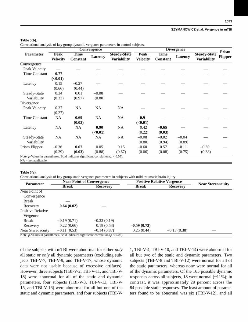
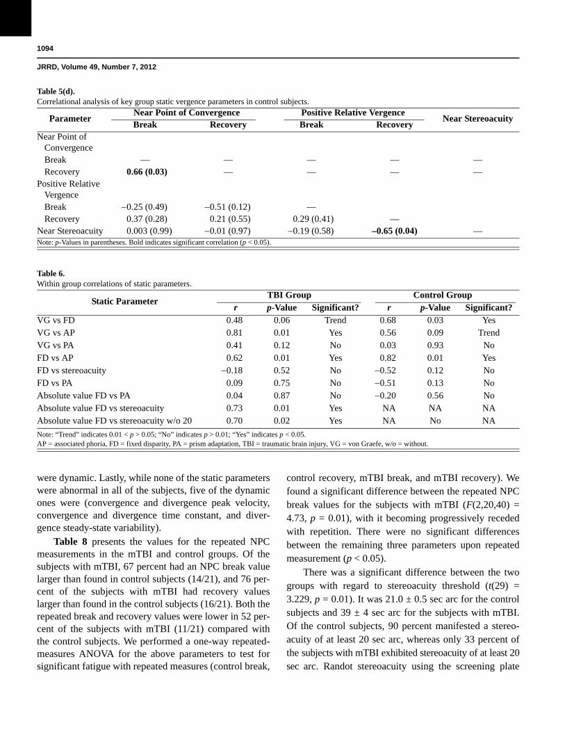
We performed correlations for the static and dynamic vergence parameters that were significantly different between the control and mTBI groups (Table 5). We per-formed these to determine whether poor performance on one parameter was also reflected in any others. For the dynamic parameters, we found 6 significant correlations in the control group and 11 in the group with mTBI. For the static parameters, we found two significant correla-tions in the control group and two in the group with mTBI. Some, but not all, of the significant correlations were the same in both groups.
Table 6 presents the correlations found between the various static parameters that exhibited predicted abnor-mal directional effects, as well as stereoacuity, for the
control and mTBI groups. Both groups demonstrated a significant correlation (or a strong trend) between von Graefe near phoria and near FD, between von Graefe near phoria and near AP, and between FD and near AP. There was a significant correlation in the subjects with mTBI only between the absolute value of FD and stereo-acuity. However, since the stereoacuity test did not extend below 20 sec arc, subjects with a value of 20 sec arc might actually have a lower threshold, and hence be better than indicated. With these five subjects excluded, the correlation was still significant. While there was no correlation between the absolute value of FD and PA, there were two interesting findings in the group with mTBI: some subjects (20%) exhibited either paradoxical negative PA or a large range of FDs with not much evi-dence of PA.
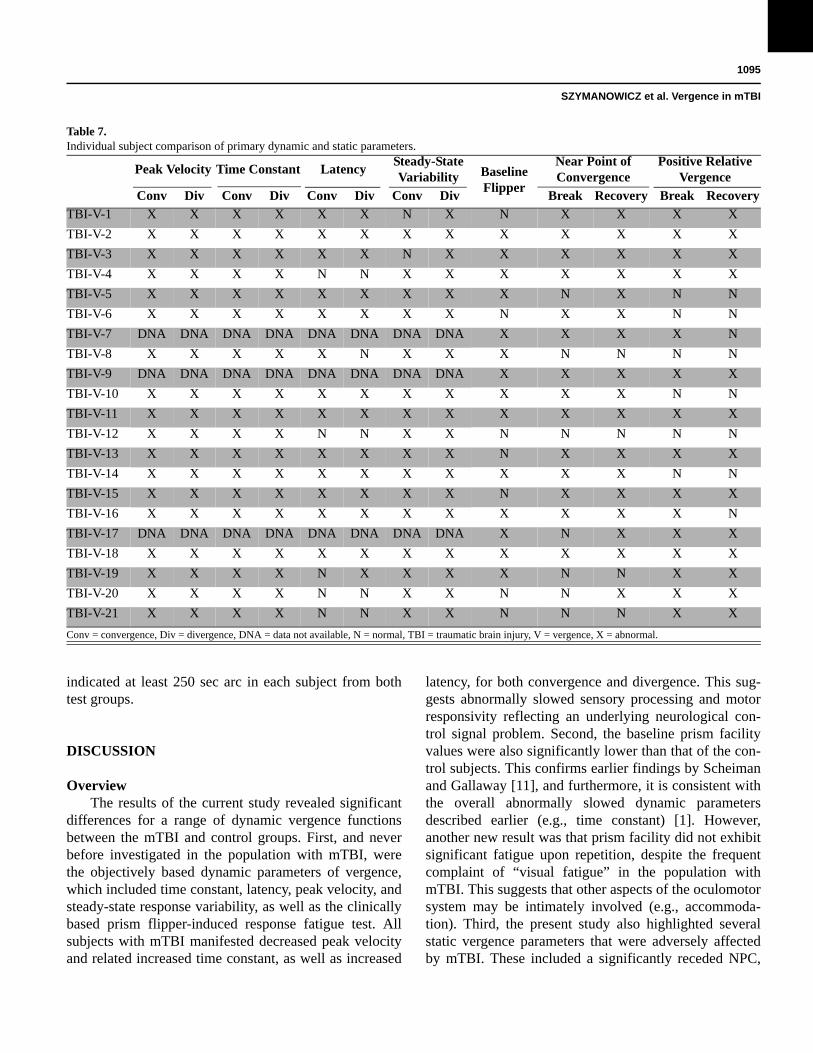
Table 7 presents the individual subjects with mTBI comparative findings for the key 13 static and dynamic parameters. They were dichotomously categorized as being either “normal” or “abnormal” using the following criterion: its value had to exceed the normal group mean by greater than
ParameterConvergence Divergence
Prism Flipper
Peak Velocity
Time Constant
LatencySteady-State Variability
Peak Velocity
Time Constant
LatencySteady-State Variability
Convergence Peak Velocity — — — — — — — — — Time Constant 0.37
(0.13) — — — — — — — —
Latency 0.23(0.33)
0.56 (0.01)
— — — — — — —
Steady-State Variability
0.31(0.2)
0.18(0.46)
0.05 (0.03)
— — — — — —
Divergence Peak Velocity 0.54
(0.01)NA NA NA — — — — —
Time Constant NA 0.49 (0.03)
NA NA –0.54 (0.02)
— — — —
Latency NA NA 0.77 (<0.01)
NA –0.50 (0.03)
–0.50 (0.03)
— — —
Steady-State Variability
NA NA NA 0.4 (0.09)
0.16 (0.51)
0.03 (0.87)
0.03 (0.28)
— —
Prism Flipper 0.53(0.02)
0.48 (0.04)
0.18 (0.45)
0.08 (0.74)
0.25 (0.31)
0.12 (0.61)
0.3 (0.22)
0.12 (0.47)
—
±1 SEM for that specific parameter. None
Table 5(a).Correlational analysis of key group dynamic vergence parameters in subjects with mild traumatic brain injury.
Note: p-Values in parentheses. Bold indicates significant correlation (p < 0.05).NA = not applicable.
1093
SZYMANOWICZ et al. Vergence in mTBI
ParameterConvergence Divergence
Prism Flipper
Peak Velocity
Time Constant
LatencySteady-State Variability
Peak Velocity
Time Constant
LatencySteady-State Variability
Convergence Peak Velocity — — — — — — — — — Time Constant –0.77
(<0.01)— — — — — — — —
Latency 0.15 (0.66)
0.27(0.44)
— — — — — — —
Steady-State Variability
0.34(0.33)
0.01 (0.97)
0.08 (0.80)
— — — — — —
Divergence Peak Velocity 0.37
(0.27)NA NA NA — — — — —
Time Constant NA 0.69(0.02)
NA NA –0.9(<0.01)
— — — —
Latency NA NA 0.90 (<0.01)
NA 0.42(0.22)
–0.65(0.03)
— — —
Steady-State Variability
NA NA NA NA 0.08 (0.80)
0.02 (0.94)
0.04 (0.89)
— —
Prism Flipper 0.36 (0.29)
0.67 (0.03)
0.05(0.88)
0.15(0.67)
0.60 (0.06)
0.57(0.08)
0.11(0.75)
0.30(0.38)
—
ParameterNear Point of Convergence Positive Relative Vergence
Near StereoacuityBreak Recovery Break Recovery
Near Point of ConvergenceBreak —Recovery 0.64 (0.02) —
Positive Relative VergenceBreak 0.19 (0.71) 0.33 (0.19) —Recovery 0.22 (0.66) 0.18 (0.53) –0.59 (0.73) —
Near Stereoacuity 0.11 (0.53) 0.14 (0.87) 0.25 (0.44) 0.13 (0.38) —
of the subjects with mTBI were abnormal for either onlyall static or only all dynamic parameters (excluding sub-jects TBI-V-7, TBI-V-9, and TBI-V-17, whose dynamic data were not usable because of excessive artifacts). However, three subjects (TBI-V-2, TBI-V-11, and TBI-V-18) were abnormal for all of the static and dynamic parameters, four subjects (TBI-V-3, TBI-V-13, TBI-V-15, and TBI-V-16) were abnormal for all but one of the static and dynamic parameters, and four subjects (TBI-V-
1, TBI-V-4, TBI-V-10, and TBI-V-14) were abnormal for all but two of the static and dynamic parameters. Two subjects (TBI-V-8 and TBI-V-12) were normal for all of the static parameters, whereas none were normal for all of the dynamic parameters. Of the 165 possible dynamic responses across all subjects, 18 were normal (~11%); in contrast, it was approximately 29 percent across the84 possible static responses. The least amount of parame-ters found to be abnormal was six (TBI-V-12), and all
Table 5(b).Correlational analysis of key group dynamic vergence parameters in control subjects.
Note: p-Values in parentheses. Bold indicates significant correlation (p < 0.05).NA = not applicable.
Table 5(c).Correlational analysis of key group static vergence parameters in subjects with mild traumatic brain injury.
Note: p-Values in parentheses. Bold indicates significant correlation (p < 0.05).
1094
JRRD, Volume 49, Number 7, 2012
ParameterNear Point of Convergence Positive Relative Vergence
Near StereoacuityBreak Recovery Break Recovery
Near Point of ConvergenceBreak — — — — —Recovery 0.66 (0.03) — — — —
Positive Relative VergenceBreak 0.25 (0.49) 0.51 (0.12) —Recovery 0.37 (0.28) 0.21 (0.55) 0.29 (0.41) —
Near Stereoacuity 0.003 (0.99) 0.01 (0.97) 0.19 (0.58) –0.65 (0.04) —
Static ParameterTBI Group Control Group
r p-Value Significant? r p-Value Significant?VG vs FD 0.48 0.06 Trend 0.68 0.03 YesVG vs AP 0.81 0.01 Yes 0.56 0.09 TrendVG vs PA 0.41 0.12 No 0.03 0.93 NoFD vs AP 0.62 0.01 Yes 0.82 0.01 YesFD vs stereoacuity 0.18 0.52 No 0.52 0.12 NoFD vs PA 0.09 0.75 No 0.51 0.13 NoAbsolute value FD vs PA 0.04 0.87 No 0.20 0.56 NoAbsolute value FD vs stereoacuity 0.73 0.01 Yes NA NA NAAbsolute value FD vs stereoacuity w/o 20 0.70 0.02 Yes NA No NA
were dynamic. Lastly, while none of the static parameters were abnormal in all of the subjects, five of the dynamic ones were (convergence and divergence peak velocity, convergence and divergence time constant, and diver-gence steady-state variability).
Table 8 presents the values for the repeated NPC measurements in the mTBI and control groups. Of the subjects with mTBI, 67 percent had an NPC break value larger than found in control subjects (14/21), and 76 per-cent of the subjects with mTBI had recovery values larger than found in the control subjects (16/21). Both the repeated break and recovery values were lower in 52 per-cent of the subjects with mTBI (11/21) compared with the control subjects. We performed a one-way repeated-measures ANOVA for the above parameters to test for significant fatigue with repeated measures (control break,
control recovery, mTBI break, and mTBI recovery). We found a significant difference between the repeated NPC break values for the subjects with mTBI (F(2,20,40) = 4.73, p = 0.01), with it becoming progressively receded with repetition. There were no significant differences between the remaining three parameters upon repeated measurement (p < 0.05).
There was a significant difference between the two groups with regard to stereoacuity threshold (t(29) = 3.229, p = 0.01). It was 21.0 ± 0.5 sec arc for the control subjects and 39 ± 4 sec arc for the subjects with mTBI. Of the control subjects, 90 percent manifested a stereo-acuity of at least 20 sec arc, whereas only 33 percent of the subjects with mTBI exhibited stereoacuity of at least 20 sec arc. Randot stereoacuity using the screening plate
Table 5(d). Correlational analysis of key group static vergence parameters in control subjects.
Note: p-Values in parentheses. Bold indicates significant correlation (p < 0.05).
Table 6.Within group correlations of static parameters.
Note: “Trend” indicates 0.01 < p > 0.05; “No” indicates p > 0.01; “Yes” indicates p < 0.05.AP = associated phoria, FD = fixed disparity, PA = prism adaptation, TBI = traumatic brain injury, VG = von Graefe, w/o = without.
1095
SZYMANOWICZ et al. Vergence in mTBI
SubjectPeak Velocity Time Constant Latency
Steady-State Variability Baseline
Flipper
Near Point of Convergence
Positive Relative Vergence
Conv Div Conv Div Conv Div Conv Div Break Recovery Break Recovery
TBI-V-1 X X X X X X N X N X X X X
TBI-V-2 X X X X X X X X X X X X X
TBI-V-3 X X X X X X N X X X X X X
TBI-V-4 X X X X N N X X X X X X X
TBI-V-5 X X X X X X X X X N X N N
TBI-V-6 X X X X X X X X N X X N N
TBI-V-7 DNA DNA DNA DNA DNA DNA DNA DNA X X X X N
TBI-V-8 X X X X X N X X X N N N N
TBI-V-9 DNA DNA DNA DNA DNA DNA DNA DNA X X X X X
TBI-V-10 X X X X X X X X X X X N N
TBI-V-11 X X X X X X X X X X X X X
TBI-V-12 X X X X N N X X N N N N N
TBI-V-13 X X X X X X X X N X X X X
TBI-V-14 X X X X X X X X X X X N N
TBI-V-15 X X X X X X X X N X X X X
TBI-V-16 X X X X X X X X X X X X N
TBI-V-17 DNA DNA DNA DNA DNA DNA DNA DNA X N X X X
TBI-V-18 X X X X X X X X X X X X X
TBI-V-19 X X X X N X X X X N N X X
TBI-V-20 X X X X N N X X N N X X X
TBI-V-21 X X X X N N X X N N N X X
indicated at least 250 sec arc in each subject from both test groups.
DISCUSSION
OverviewThe results of the current study revealed significant
differences for a range of dynamic vergence functions between the mTBI and control groups. First, and never before investigated in the population with mTBI, were the objectively based dynamic parameters of vergence, which included time constant, latency, peak velocity, and steady-state response variability, as well as the clinically based prism flipper-induced response fatigue test. All subjects with mTBI manifested decreased peak velocity and related increased time constant, as well as increased
latency, for both convergence and divergence. This sug-gests abnormally slowed sensory processing and motor responsivity reflecting an underlying neurological con-trol signal problem. Second, the baseline prism facility values were also significantly lower than that of the con-trol subjects. This confirms earlier findings by Scheiman and Gallaway [11], and furthermore, it is consistent with the overall abnormally slowed dynamic parameters described earlier (e.g., time constant) [1]. However, another new result was that prism facility did not exhibit significant fatigue upon repetition, despite the frequent complaint of “visual fatigue” in the population with mTBI. This suggests that other aspects of the oculomotor system may be intimately involved (e.g., accommoda-tion). Third, the present study also highlighted several static vergence parameters that were adversely affected by mTBI. These included a significantly receded NPC,
Table 7.Individual subject comparison of primary dynamic and static parameters.
Conv = convergence, Div = divergence, DNA = data not available, N = normal, TBI = traumatic brain injury, V = vergence, X = abnormal.

1096
JRRD, Volume 49, Number 7, 2012
Parameter TBI Group Control Group p-Value Significant?Break 1 12.90 ± 1.13 6.9 ± 0.41 — —Break 2 14.26 ± 1.26 6.75 ± 0.32 — —Break 3 14.76 ± 1.23 7.45 ± 0.47 — —Grand Mean Break 13.98 ± 2.06 7.03 ± 0.33 0.03 YesRecovery 1 17.19 ± 1.25 9.4 ± 0.39 — —Recovery 2 20.86 ± 2.06 9.3 ± 0.60 — —Recovery 3 20.33 ± 1.76 10.05 ± 0.72 — —Grand Mean Recovery 19.46 ± 2.81 9.56 ± 0.46 0.02 Yes
significantly reduced fusional PRV ranges, and abnor-mally large phoria magnitudes, as well as significantly reduced sensory stereoacuity thresholds. These last results confirmed and extended earlier studies involving these static vergence parameters and related aspects [4,9–13]. However, given the relatively small sample size of the present pilot investigation, a definitive conclusion cannot be made. Future studies are needed using a large and perhaps more diverse range of subjects with TBI.
The new finding of increased latency (~100 ms) in the group with mTBI compared with the control group deserves mention. This delay is likely to be too long to be attributed solely to the diffuse axonal damage that occurs in the coup-contrecoup impact. It may be compounded by attentional factors, with increased temporal processing time being common in the group with mTBI [15,23], as well as a more general cognitive impairment [24]. Since none of the subjects were diagnosed with either extraocu-lar muscle paresis or palsy using conventional clinical procedures [4,19], this delay is unlikely to be attributed to gross neural innervational and/or extraocular muscle injury-related delays [25]. However, presence of a subtle, subclinical extraocular muscle disorder may be a partial contributory factor to the overall delayed response.
Correlations within the group with mTBI for the rele-vant static and dynamic parameters demonstrated that in many cases (~50%), if the individual responded poorly for one parameter, they also responded poorly to several others (Tables 5–6). For example, if they responded poorly for convergence, they also responded poorly for divergence and for the parameters of peak velocity, time constant, latency, and steady-state variability. The lack of significant correlations between some of the other param-eters may be expected given the predicted increased vari-ability in their dynamic responsivity.
The individual subject comparisons (Table 7) yield some interesting trends. First, nearly all subjects (20 out of 21) were abnormal on the majority (7) of the 13 static and dynamic parameters. Second, performance was worsefor the dynamic than the static parameters (29% vs 11%, respectively). Third, there were five dynamic but no static parameters that were abnormal across all subjects. There was no apparent relationship between vergence performance and demographic characteristics (e.g., mTBI etiology, age) (Table 1). Thus, there was no appar-ent reason why some subjects with mTBI performed bet-ter than others on the vergence tests. Lastly, these individual subject findings are consistent with the group correlations discussed earlier (Tables 5–6) in which we found more vergence abnormalities for the dynamic than the static parameters.
Relation to Other Human Clinical Studies on Vergence in Traumatic Brain Injury
With regards to static vergence dysfunction, many earlier studies reported significant differences in the near phoria, NPC break and recovery values, and vergence ranges when the population with mTBI was compared with the control group [10–13]. Berne [10] and Scheiman and Gallaway [11] both reported significantly higher exo-phoria in the group with mTBI, which is consistent with the findings of the present study. In addition, the current study also found abnormally high esophoria in 3 of the21 subjects with mTBI (15%). This latter new finding is consistent with the general notion that those with mTBI may present with general abnormal interactive-based bin-ocular vision problems [4,26]. The receded NPC values found in the present study for both the break and recov-ery measures have been reported in numerous studies [10–13]. In the present study, 15 of the 21 subjects (71%)
Table 8. Comparison of repeated near point of convergence break and recovery values between two groups.
Note: “Yes” indicates p 0.05.TBI = traumatic brain injury.

1097
SZYMANOWICZ et al. Vergence in mTBI
revealed significantly receded break values, and 18 of the 21 subjects (86%) demonstrated significantly receded recovery values. Lastly, there was a significant reduction in positive fusional vergence (PFV) ranges at near. This is consistent with the majority of those with mTBI having exophoria at near, thus requiring additional PFV to attain bifoveal fixation. Furthermore, it is consistent with ear-lier studies [10–13].
With regard to dynamic vergence dysfunction in mTBI, the results of the present study provide consider-able new information. The only dynamic vergence test that has been previously performed in the population with mTBI was the baseline clinical vergence prism facility test, a subjective test. In the current study, 11 of the 21 (52%) subjects with mTBI manifested reduced vergence prism facility rates. The new objective dynamic findings of the current study can help explain the reason for the decreased clinical flipper rate; all dynamic param-eters were found to be abnormal in the group with mTBI, thus resulting in slowed and delayed dynamic responsiv-ity. These individual dynamic parameters each contrib-uted to the overall reduced prism facility rate values. That is, the reduced clinical flipper facility rate represents an overall, more global reflection of the vergence dynamic dysfunction, whereas the results of the objectively based vergence dynamics reveal the relative degree of abnor-mality for each of the specific components.
With regard to stereoacuity, the results of the present study are consistent with earlier investigations [12–13]. They too found increased stereoacuity thresholds in the range found in the present study. The average stereoacu-ity found for the population with mTBI was 39 sec arc, with 12 out of 21 patients (60%) having 40 sec arc or worse stereoacuity, which is considered to be clinically abnormal [19]. Slightly reduced stereoacuity is consistent with the large phoria many subjects exhibited, because there is a relationship between the phoria with both the FD direction and magnitude in control subjects [27]; the greater the phoria, the greater the FD, with both being in the same direction. Our new finding was that this relation-ship was also true for the group with mTBI (Table 6).
Effect on Quality of Life and Functional Aspects of Vision
The wide array of both dynamic and static vergence abnormalities reported in individuals with mTBI [23] may have major adverse consequences for a range of vision-related activities, such as reading, visual scanning,
and tracking in depth, as well as in more general activi-ties of daily living [4,6,22,26]. Furthermore, abnormal vergence may also interfere with performance at the workplace (i.e., have an adverse vocational effect), such as performing sustained computer-related activities, which may in return result in loss of income and related employment benefits. Moreover, it can lead to inadequate progress in other rehabilitative services (e.g., cognitive therapy) involving a range of general, as well as specific, visual eye-tracking tasks and demands [24,28].
Clinical ImplicationsThere are several important clinical implications
based on the findings of the present study. First, the objective dynamic results confirm and extend earlier clinical findings. For example, Scheiman and Gallaway [11] found reduced overall vergence facility rates, which were confirmed and extended both objectively and clini-cally in the present study. However, the new objective findings of the present investigation allow us to dissect and assess the individual component’s relative contribu-tion to the overall vergence facility dysfunction. In the present study, each of the dynamic components to vary-ing degrees were found to be abnormal: increased latency, increased time constant and related reduced peak velocity, and increased steady-state response variability. Hence, all components of fast vergence control were impaired, with them encompassing and reflecting slowed visual neurosensory processing, as well as abnormal and/or slowed and more variable motor processing and responsivity. The fact that the vergence response ampli-tude was appropriate and accurate for the stimulus demand suggests a relatively normal amplitude step neu-rological control signal [29], although the increased steady-state vergence response variability suggests increased but subtle neural noise (i.e., variability) in the step signal [30]. However, the concurrent reduced peak velocity suggests a considerable pulse-like neurological control signal deficit (i.e., reduced amplitude and/or duration), which would allow the system to eventually acquire the target, but with a slower overall dynamic time course [30–31]. Thus, both the step and pulse vergence neural controller signal components appear to be adversely affected by the mTBI.
Second, the above dynamic deficits have an impor-tant effect on one’s vision rehabilitation strategy. Namely,all aspects of fast dynamic vergence control were adversely affected, and hence should be targeted using

1098
JRRD, Volume 49, Number 7, 2012
different amounts and directions of disparity step stimuli (e.g., prism flipper step stimuli) [11]. In addition, the treatment implications extend to the static vergence domain. For example, the receded NPC and its visual fatigue with repetition, reduced and restricted fusional PRV ranges, abnormal near phoria magnitude, and increased FD magnitudes all need to be addressed clini-cally. They may be helped by appropriate therapeutic intervention, namely small steps of disparity stimuli, as well as smooth and continuous disparity ramp stimuli (e.g., vectograph ramp stimuli) per models of the ver-gence system [22,31]. Furthermore, the present abnormal static findings suggest, and are consistent with, distur-bance of slow vergence control [32–34] (e.g., ramp stimu-lus for NPC testing). Thus, both fast and slow vergence control appear to be adversely affected by the brain injury.
Third, the abnormal static and dynamic vergence findings are consistent with the symptoms reported by these patients, such as intermittent diplopia, running together and apparent “movement” of lines of print, and vergence-induced blur, to name a few. Thus, reduction in the number of symptoms and their intensity should be correlated with improvement in clinical signs [35] and dynamic responses [36].
Lastly, the results of the present study suggest the following high yield tests: NPC with repetition, baseline prism vergence facility, near horizontal phoria, and fusional PRV ranges. These four tests are also consistent with the diagnosis of CI [19], which is commonly (56.3%) found in the population with mTBI [4]. We have recently developed such “targeted” oculomotor-based diagnostic protocols for assessment in the population with mTBI [37–39].
Study LimitationsThere were three possible limitations to the present
study. First, the binocular sampling rate of the PRII was 12.5 Hz, and thus a discrete sample of dynamic eye posi-tion was obtained every 80 ms, which is sufficient for a relatively slow system such as vergence with its 1 s overall response time [1,40]. However, with a higher sampling rate (e.g., 25 Hz), the estimate of response latency may be slightly improved, and furthermore, the dynamic trajectory may be better resolved and quantified. However, these are likely to be second-order effects. Sec-ond, we did not perform brain imaging. This would have provided important information regarding the precise sites of damage to the brain, especially as related to
vision and, more specifically, to vergence control, as mentioned previously. However, since all were mTBI, it would be expected to be relatively comprehensive in nature, thus having multiple sites of injury per the coup-countercoup aspect [23] frequently found in these patients, and not be as more localized damage as sug-gested by the cerebrovascular accident results in this area [32–34]. Third, because of the relatively small sample size, definitive conclusions cannot be made; thus, further related investigations are warranted.
CONCLUSIONS
The present study uncovered and documented a vari-ety of new static and dynamic vergence dysfunctions in adults with mTBI. These defects suggest damage to vari-ous vergence oculomotor control areas in the brain. Ver-gence abnormalities should be considered in the comprehensive vision examination of these patients, because their presence may adversely affect their quality of life, both vocationally and avocationally. The diagnos-tic and therapeutic aspects and implications are particu-larly relevant to the VA hospitals in the United States with thousands of returning servicemembers having mTBI and in need of related short- and long-term vision care.
ACKNOWLEDGMENTS
Author Contributions:Study concept and design: D. Szymanowicz, K. J. Ciuffreda, P. Thiagarajan, D. P. Ludlam, W. Green, N. Kapoor.Acquisition of data: D. Szymanowicz, K. J. Ciuffreda, W. Green, D. P. Ludlam.Analysis and interpretation of data: D. Szymanowicz, K. J. Ciuffreda, P. Thiagarajan, W. Green.Drafting of manuscript: D. Szymanowicz, K. J. Ciuffreda, P. Thiagarajan, D. P. Ludlam, N. Kapoor.Statistical analysis: D. Szymanowicz, K. J. Ciuffreda, P. Thiagarajan.Obtained funding: K. J. Ciuffreda, D. Szymanowicz.Administrative, technical, or material support: N. Kapoor, P. Thiagarajan, K. J. Ciuffreda.Study supervision: N. Kapoor, K. J. Ciuffreda, D. P. Ludlam.Financial Contributions: The authors have declared that no compet-ing interests exist.Funding/Support: This material was based on work supported by the SUNY Optometry Graduate Program and grant NEI-5T53EY02048103.Institutional Review: We obtained informed consent from each sub-ject after explaining the nature and possible consequences of the study. The research followed the tenets of the Declaration of Helsinki

1099
SZYMANOWICZ et al. Vergence in mTBI
and was approved by SUNY College of Optometry’s internal review board.
Participant Follow-Up: The authors have no plans to notify partici-pants of the publication of this study.
REFERENCES
1. Ciuffreda KJ, Tannen B. Eye movement basics for the cli-nician. St. Louis (MO): Mosby; 1995.
2. Ciuffreda KJ, Kenyon RV. Accommodative vergence and accommodation in normals, amblyopes, and strabismics. In: Schor CM, Ciuffreda KJ, editors. Vergence eye move-ments: Basic and clinical aspects. Boston (MA): Butter-worth; 1983. p. 101–73.
3. Ciuffreda KJ. Components of clinical near vergence test-ing. J Behav Optom. 1992;3:3–13.
4. Ciuffreda KJ, Kapoor N, Rutner D, Suchoff IB, Han ME, Craig S. Occurrence of oculomotor dysfunctions in acquired brain injury: a retrospective analysis. Optometry. 2007;78(4):155–61. [PMID:17400136] http://dx.doi.org/10.1016/j.optm.2006.11.011
5. Warden D. Military TBI during the Iraq and Afghanistan wars. J Head Trauma Rehabil. 2006;21(5):398–402.[PMID:16983225] http://dx.doi.org/10.1097/00001199-200609000-00004
6. Ciuffreda KJ, Ludlam DP, Kapoor N. Clinical oculomotor training in traumatic brain injury. Optom Vis Dev. 2009; 40:16–23.
7. Cross AG. Neuromuscular aspects in ocular sequelae of head injuries. Trans Ophthalmol Soc U K. 1945;65:20–33.
8. Jaensch PA. Fusion sterungen, Horrorfusionis und Konvergen-zspasmen. Klinikal Mbl. Augenheilk. 1945;6(65):142–49. German.
9. Cohen M, Groswasser Z, Barchadski R, Appel A. Conver-gence insufficiency in brain-injured patients. Brain Inj. 1989;3(2):187–91. [PMID:2471568] http://dx.doi.org/10.3109/02699058909004551
10. Berne SA. Visual therapy for the traumatic brain-injured. J Optom Vis Dev. 1990;21:13–16.
11. Scheiman M, Gallaway M. Vision therapy to treat binocularvision disorders after acquired brain injury: Factors affect-ing prognosis. In: Suchoff IB, Cuiffreda KJ, Kapoor N, edi-tors. Visual & vestibular consequences of acquired brain injury. Santa Ana (CA): Optometric Extension Program; 2001. p. 89–113.
12. Schlageter K, Gray B, Hall K, Shaw R, Sammet R. Inci-dence and treatment of visual dysfunction in traumatic brain injury. Brain Inj. 1993;7(5):439–48. [PMID:8401486]http://dx.doi.org/10.3109/02699059309029687
13. Hellerstein LF, Freed S, Maples WC. Vision profile of patients with mild brain injury. J Am Optom Assoc. 1995;66(10):634–39. [PMID:7499718]
14. Kay T, Harrington DE, Adams R, Anderson T, Berrol S, Cicerone K, Dahlberg C, Gerber D, Goka R, Harley P, Hilt J, Horn L, Lehmkuhl D, Malec J. Definition of mild traumatic brain injury. J Head Trauma Rehabil. 1993;8(3):86–87.http://dx.doi.org/10.1097/00001199-199309000-00010
15. Hibbard MR, Gordon WA, Kenner B. The neuropsychologicalevaluation: A pathway to understanding the sequelae of brain injury. In: Suchoff IB, Cuiffreda KJ, Kapoor N, edi-tors. Visual & vestibular consequences of acquired brain injury. Santa Ana (CA): Optometric Extension Program; 2001. p. 32–45.
16. Kraus JF, McArthur DL, Silberman TA. Epidemiology of mild brain injury. Semin Neurol. 1994;14(1):1–7.[PMID:8029555] http://dx.doi.org/10.1055/s-2008-1041052
17. Kushner D. Mild traumatic brain injury: toward under-standing manifestations and treatment. Arch Intern Med. 1998;158(15):1617–24. [PMID:9701095] http://dx.doi.org/10.1001/archinte.158.15.1617
18. Schaeffel F, Wilhelm H, Zrenner E. Inter-individual vari-ability in the dynamics of natural accommodation in humans: relation to age and refractive errors. J Physiol. 1993;461:301–20. [PMID:8350267]
19. Scheiman M, Wick B. Clinical management of binocular vision: Heterophoric, accommodative, and eye movement disorders. 2nd ed. Philadelphia (PA): Lippincott Williams & Wilkins; 2002.
20. Benjamin WJ, editor. Borish’s clinical refraction. 2nd ed. Oxford (England): Butterworth-Heinemann; 2006.
21. Green W, Ciuffreda KJ, Thiagarajan P, Szymanowicz D, Ludlam DP, Kapoor N. Accommodation in mild traumatic brain injury. J Rehabil Res Dev. 2010;47(3):183–99.[PMID:20665345] http://dx.doi.org/10.1682/JRRD.2009.04.0041
22. Kapoor N, Ciuffreda KJ. Vision disturbances following traumatic brain injury. Curr Treat Options Neurol. 2002; 4(4):271–80. [PMID:12036500] http://dx.doi.org/10.1007/s11940-002-0027-z
23. Suchoff IB, Ciuffreda KJ, Kapoor N, editors. Visual & ves-tibular consequences of acquired brain injury. Santa Ana (CA): Optometric Extension Program; 2001.
24. Reding MJ, Potes E. Rehabilitation outcome following ini-tial unilateral hemispheric stroke. Life table analysis approach. Stroke. 1988;19(11):1354–58. [PMID:3188120]http://dx.doi.org/10.1161/01.STR.19.11.1354
25. Leigh RJ, Zee DS. The neurology of eye movements. Oxford (England): Oxford University Press; 2006.
26. Ciuffreda KJ, Rutner D, Kapoor N, Suchoff IB, Craig S, Han ME. Vision therapy for oculomotor dysfunctions in

1100
JRRD, Volume 49, Number 7, 2012
acquired brain injury: a retrospective analysis. Optometry. 2008;79(1):18–22. [PMID:18156092] http://dx.doi.org/10.1016/j.optm.2007.10.004
27. Ogle KN, Martens TG, Dyer JA. Oculomotor imbalance in binocular vision and fixation disparity. Philadelphia (PA): Lea & Febiger; 1967.
28. Groswasser Z, Cohen M, Blankstein E. Polytrauma associ-ated with traumatic brain injury: incidence, nature and impact on rehabilitation outcome. Brain Inj. 1990;4(2): 161–66. [PMID:2331545] http://dx.doi.org/10.3109/02699059009026161
29. Semmlow JL, Alvarez TL, Pedrono C. Dry dissection of disparity divergence eye movements using independent component analysis. Comput Biol Med. 2007;37(7):910–18.[PMID:16867300] http://dx.doi.org/10.1016/j.compbiomed.2006.03.007
30. Yuan W, Semmlow JL, Alvarez TL, Munoz P. Dynamics of the disparity vergence step response: a model-based analy-sis. IEEE Trans Biomed Eng. 1999;46(10):1191–98.[PMID:10513123] http://dx.doi.org/10.1109/10.790495
31. Hung GK, Semmlow JL, Ciuffreda KJ. A dual-mode dynamic model of the vergence eye movement system. IEEE Trans Biomed Eng. 1986;33(11):1021–28.[PMID:3793122] http://dx.doi.org/10.1109/TBME.1986.325868
32. Wiest G, Mallek R, Baumgartner C. Selective loss of ver-gence control secondary to bilateral paramedian thalamic infarction. Neurology. 2000;54(10):1997–99.[PMID:10822443] http://dx.doi.org/10.1212/WNL.54.10.1997
33. Rambold H, Neumann G, Helmchen C. Vergence deficits in pontine lesions. Neurology. 2004;62(10):1850–53.[PMID:15159493] http://dx.doi.org/10.1212/01.WNL.0000125331.95849.62
34. Sander T, Sprenger A, Neumann G, Machner B, Gottschalk S, Rambold H, Helmchen C. Vergence deficits in patients with cerebellar lesions. Brain. 2009;132(Pt 1):103–15.[PMID:19036765] http://dx.doi.org/10.1093/brain/awn306
35. Scheiman M, Mitchell GL, Cotter S, Kulp MT, Cooper J, Rouse M, Borsting E, London R, Wensveen J. A random-ized clinical trial of vision therapy/orthoptics versus pencil pushups for the treatment of convergence insufficiency in young adults. Optom Vis Sci. 2005;82(7):583–95.[PMID:16044063] http://dx.doi.org/10.1097/01.opx.0000171331.36871.2f
36. Grisham JD, Bowman MC, Owyang LA, Chan CL. Ver-gence orthoptics: validity and persistence of the training effect. Optom Vis Sci. 1991;68(6):441–51.[PMID:1891195] http://dx.doi.org/10.1097/00006324-199106000-00005
37. Ciuffreda KJ, Ludlam D. Conceptual model of optometric vision care in mild traumatic brain injury. J Behav Optom. 2011;82:61–63.
38. Ciuffreda KJ, Ludlam D, Thiagarajan P. Oculomotor diag-nostic protocol for the mTBI population. Optometry. 2011;82(2):61–63. [PMID:21276567] http://dx.doi.org/10.1016/j.optm.2010.11.011
39. Ciuffreda KJ, Ludlam DP. Objective diagnostic and inter-ventional vision test protocol for the mild traumatic brain injury population. Optometry. 2011;82(6):337–39.[PMID:21616461] http://dx.doi.org/10.1016/j.optm.2011.03.006
40. Stark L. Neurological control systems; studies in bioengi-neering. New York (NY): Plenum Press; 1968.
Submitted for publication July 9, 2010. Accepted in revisedform July 28, 2011.
This article and any supplementary material should be cited as follows:Szymanowicz D, Ciuffreda KJ, Thiagarajan P, Ludlam DP, Green W, Kapoor N. Vergence in mild traumatic brain injury: A pilot study. J Rehabil Res Dev. 2012; 49(7):1083–1100.http://dx.doi.org/10.1682/JRRD.2010.07.0129