Embed Size (px)
DESCRIPTION
Joy Elizabeth Martindale. Specialist Biomedical Scientist Cellular Pathology. Phospho-histone H3 staining of uveal and choroidal melanomas. Anatomy of the eye. As normal as it gets. Iris. Ciliary Body. Choroid. Anatomy of the eye. Risk Factors. - PowerPoint PPT Presentation
Citation preview
Specialist Biomedical ScientistCellular Pathology
Phospho-histone H3 staining of uveal and choroidal melanomas
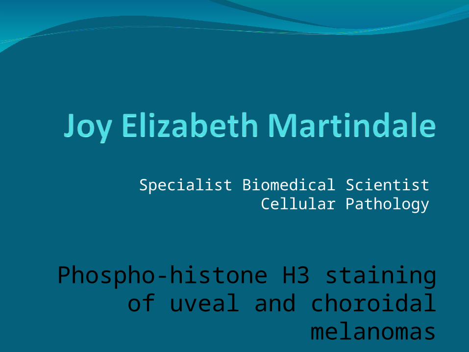
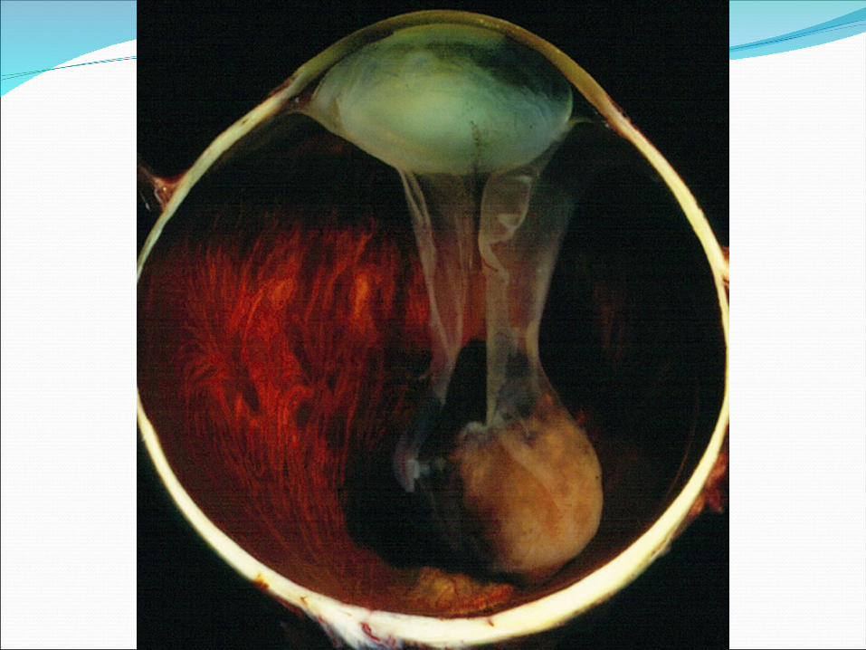
Anatomy of the eyeAnatomy of the eye
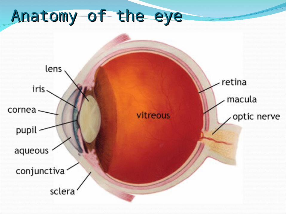
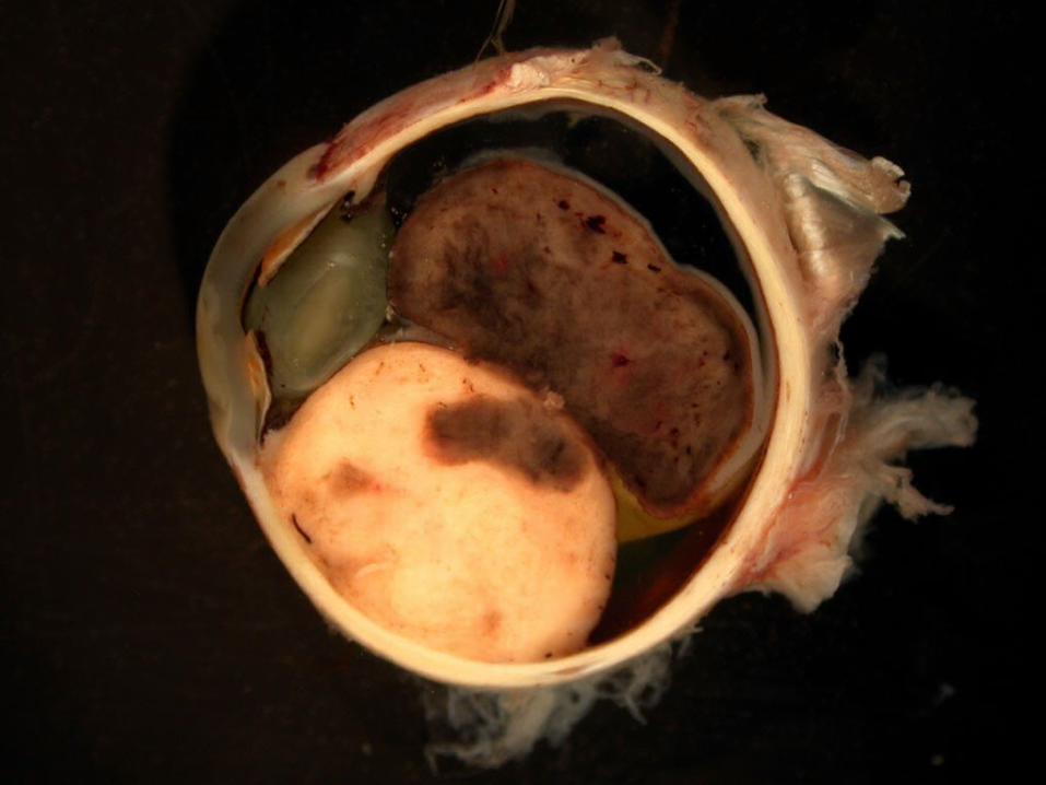
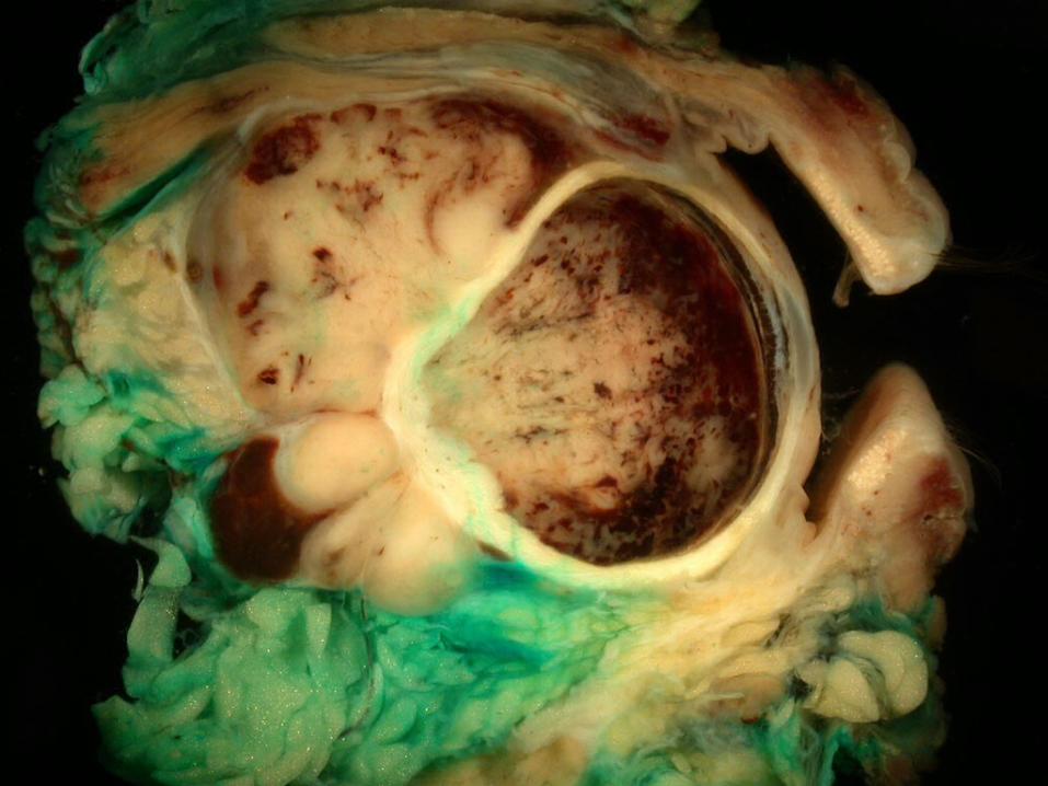
As normal as it gets
Iris Ciliary Body
Choroid
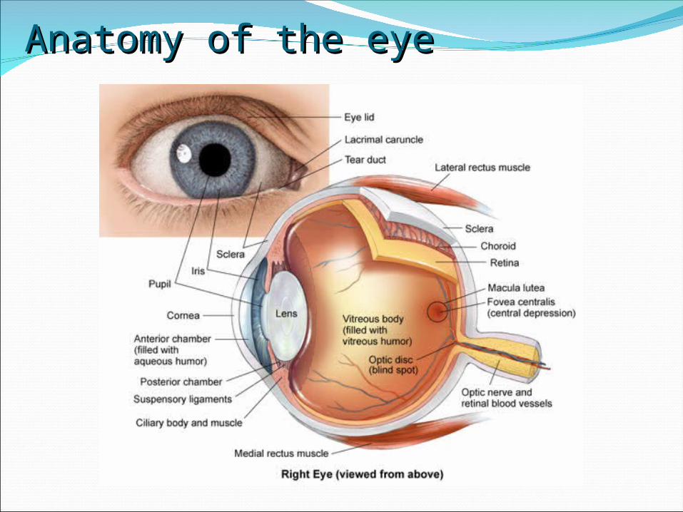
Anatomy of the eyeAnatomy of the eye

Risk FactorsRisk FactorsSimilar risk factors for cutaneous
melanomasIncidence of uveal melanomas is highest
in the white population with between 4 to 10 cases per million
UV light may be a risk factorCongenital ocular melanocytosisFamilial atypical mole and melanoma
syndrome (FAMM)
Prognostic Factors: ClinicalPrognostic Factors: ClinicalAge and Sex
Rare in childhoodRisk increases with age: most melanomas occur in patients
of late middle ageSlight male predominance, but this is not statistically
significantly linked to survivalTumour location
Location of the anterior margin of the tumour is an important predictor of prognosis and survival
Ciliary body tumours carry worst prognosisIris tumours have lowest mortality rateTumours adjacent to the optic disc have worse prognosis
Tumour sizeLarger tumour size indicates worse prognosisSmaller tumours capable of causing death through
metastasis
Prognostic: Cytogenetic and MolecularPrognostic: Cytogenetic and MolecularChromosome aberrations:
Monosomy 3 is predictor of poor prognosisAmplification of 8q is associated with reduced survivalLoss of 1p is associated with increase in mortality from
metastasesGain of 6p is associated with good prognosis
• Loss of tumour suppressor genes• P53
• Rb
• Mutations in tumour promoter genes correlate with prognosis• Poor prognosis:
• DDEF1
• NBS1
• Better prognosis
• C-myc
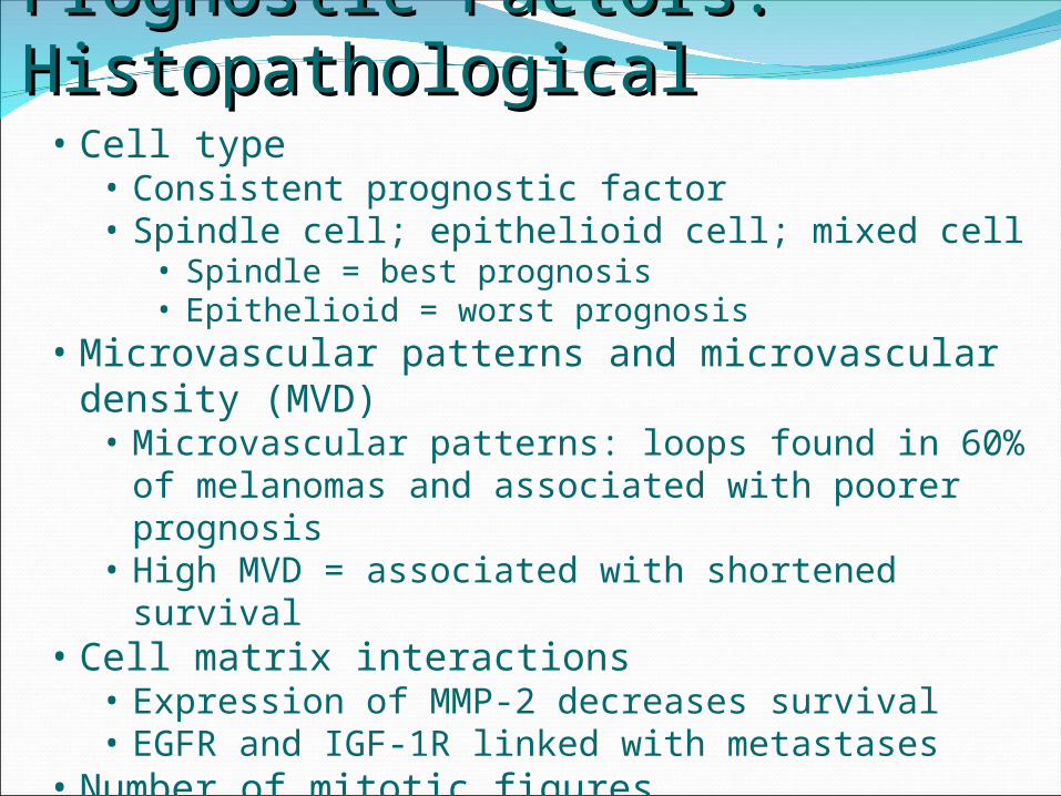
Prognostic Factors: HistopathologicalPrognostic Factors: Histopathological• Cell type
• Consistent prognostic factor• Spindle cell; epithelioid cell; mixed cell
• Spindle = best prognosis• Epithelioid = worst prognosis
• Microvascular patterns and microvascular density (MVD)
• Microvascular patterns: loops found in 60% of melanomas and associated with poorer prognosis
• High MVD = associated with shortened survival• Cell matrix interactions
• Expression of MMP-2 decreases survival• EGFR and IGF-1R linked with metastases
• Number of mitotic figures• Correlates with mortality

PHH3 is a Mitosis-specific MarkerPHH3 is a Mitosis-specific Marker• Core protein Histone H3
• Involved in maintaining the integrity of the DNA double helix within senescent cells
• Is phosphorylated at the serine 10 residue during mitosis
• Phosphorylation only occurs during mitosis and not during apoptosis
• Antibody which detects this phosphorylation event has been developed and used to detect mitotic figures in various different tumours
• Astrocytomas• Meningiomas• Uveal and choroidal melanomas
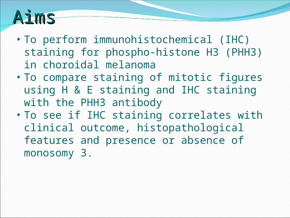
AimsAims• To perform immunohistochemical (IHC)
staining for phospho-histone H3 (PHH3) in choroidal melanoma
• To compare staining of mitotic figures using H & E staining and IHC staining with the PHH3 antibody
• To see if IHC staining correlates with clinical outcome, histopathological features and presence or absence of monosomy 3.
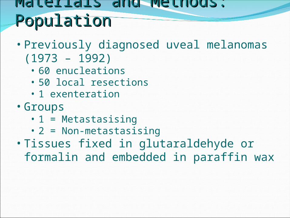
Materials and Methods: PopulationMaterials and Methods: Population• Previously diagnosed uveal melanomas
(1973 – 1992)• 60 enucleations• 50 local resections• 1 exenteration
• Groups• 1 = Metastasising• 2 = Non-metastasising
• Tissues fixed in glutaraldehyde or formalin and embedded in paraffin wax
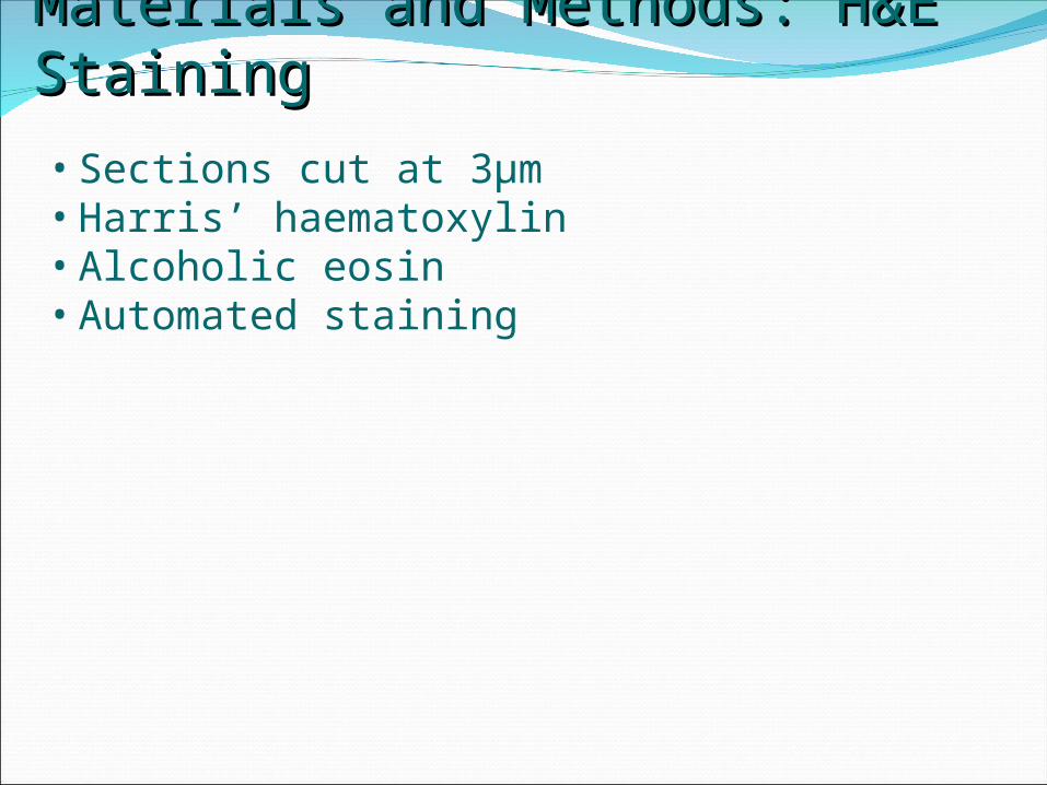
Materials and Methods: H&E StainingMaterials and Methods: H&E Staining• Sections cut at 3µm• Harris’ haematoxylin• Alcoholic eosin• Automated staining
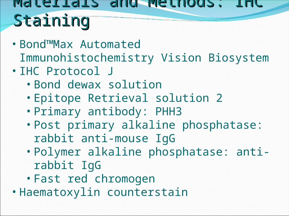
Materials and Methods: IHC StainingMaterials and Methods: IHC Staining• Bond™Max Automated
Immunohistochemistry Vision Biosystem• IHC Protocol J
• Bond dewax solution• Epitope Retrieval solution 2• Primary antibody: PHH3• Post primary alkaline phosphatase: rabbit
anti-mouse IgG• Polymer alkaline phosphatase: anti-rabbit
IgG• Fast red chromogen
• Haematoxylin counterstain
Materials and Methods: Counting MitosesMaterials and Methods: Counting Mitoses
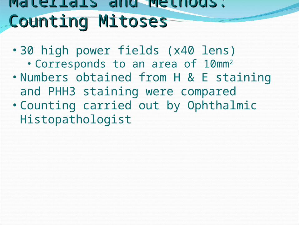
• 30 high power fields (x40 lens)• Corresponds to an area of 10mm2
• Numbers obtained from H & E staining and PHH3 staining were compared
• Counting carried out by Ophthalmic Histopathologist
Materials and Methods: Statistical AnalysesMaterials and Methods: Statistical Analyses
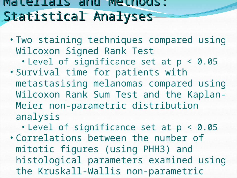
• Two staining techniques compared using Wilcoxon Signed Rank Test
• Level of significance set at p < 0.05• Survival time for patients with metastasising
melanomas compared using Wilcoxon Rank Sum Test and the Kaplan- Meier non-parametric distribution analysis
• Level of significance set at p < 0.05• Correlations between the number of mitotic
figures (using PHH3) and histological parameters examined using the Kruskall-Wallis non-parametric test
• All completed using Minitab software
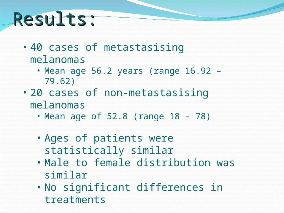
Results: Results: • 40 cases of metastasising melanomas
• Mean age 56.2 years (range 16.92 – 79.62)• 20 cases of non-metastasising
melanomas• Mean age of 52.8 (range 18 – 78)
• Ages of patients were statistically similar
• Male to female distribution was similar
• No significant differences in treatments
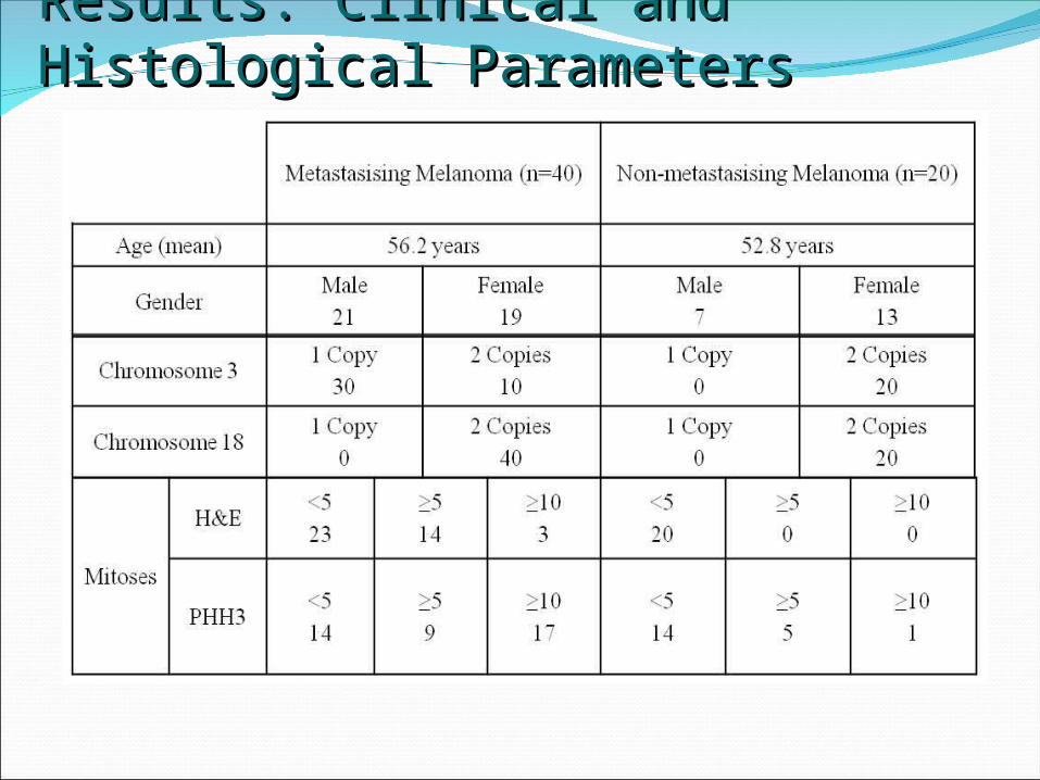
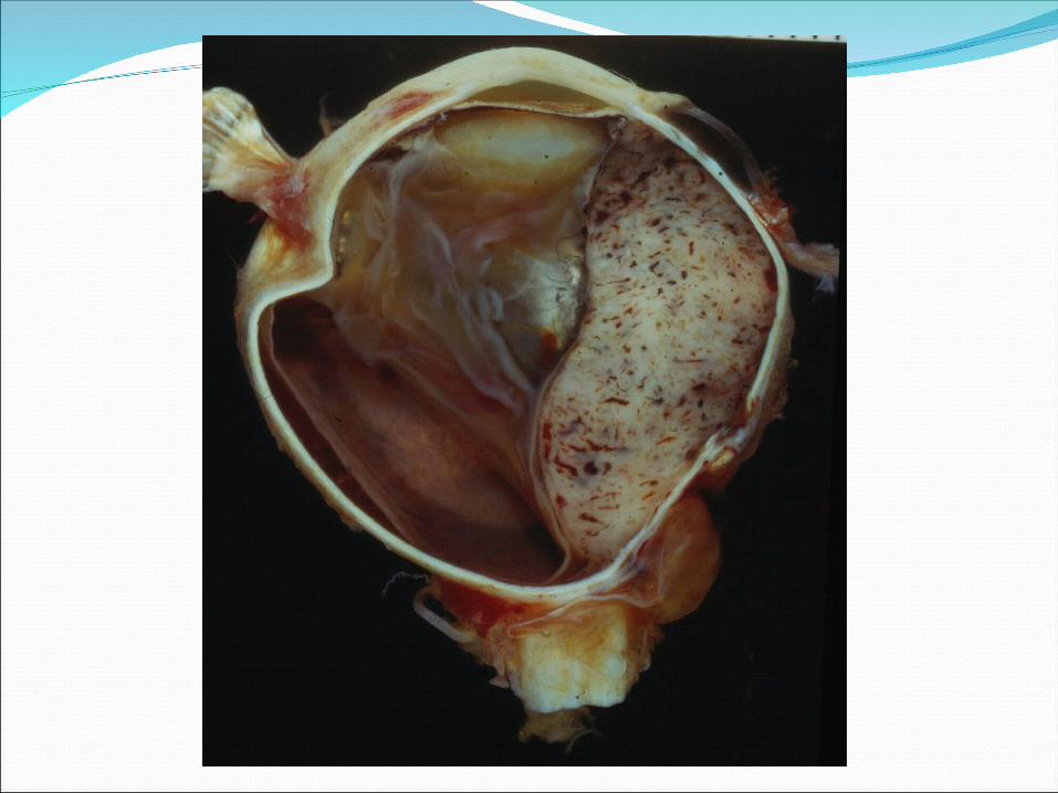
Results: Clinical and Histological Results: Clinical and Histological Parameters Parameters
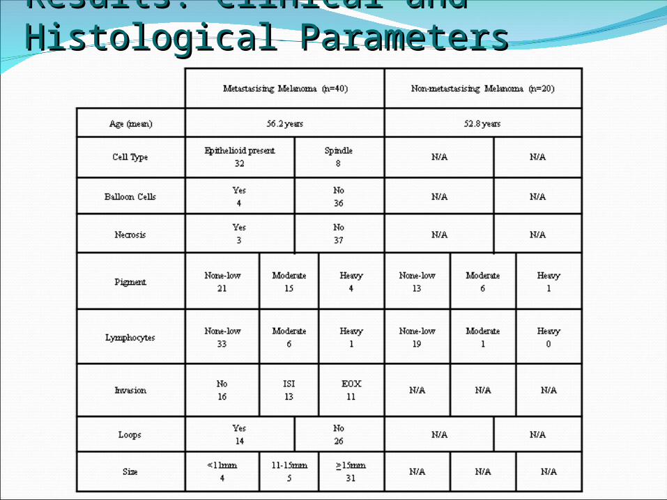
Results: Clinical and Histological Results: Clinical and Histological Parameters Parameters
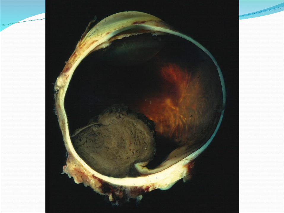
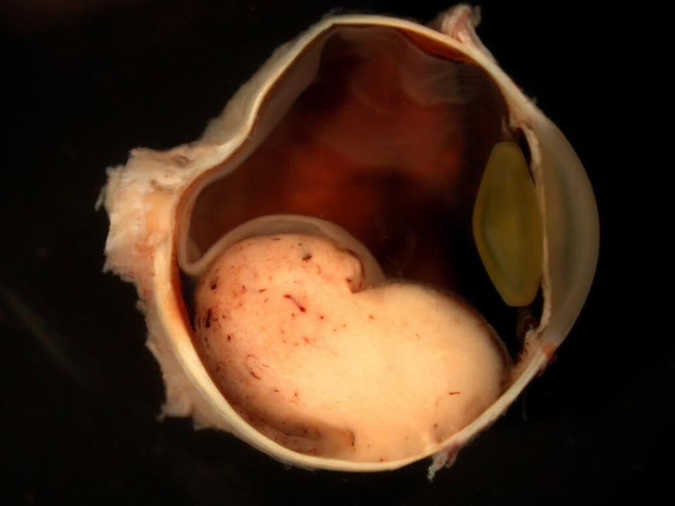
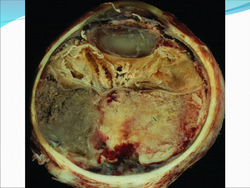
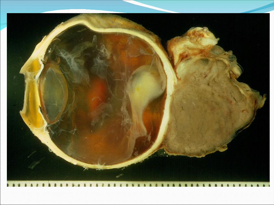
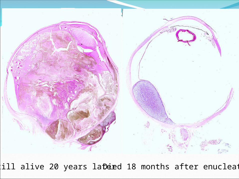
Still alive 20 years later Died 18 months after enucleation
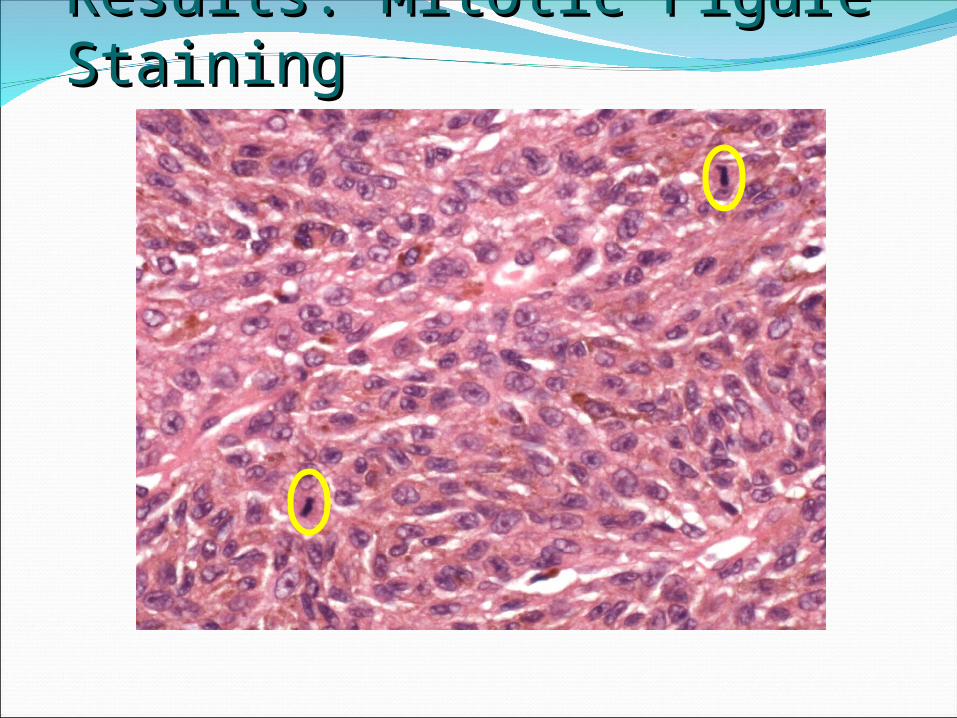
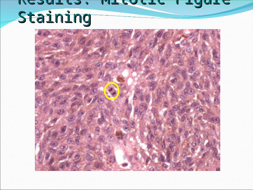
Results: Mitotic Figure StainingResults: Mitotic Figure Staining
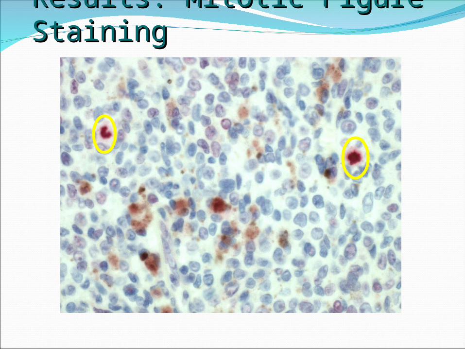
Results: Mitotic Figure StainingResults: Mitotic Figure Staining
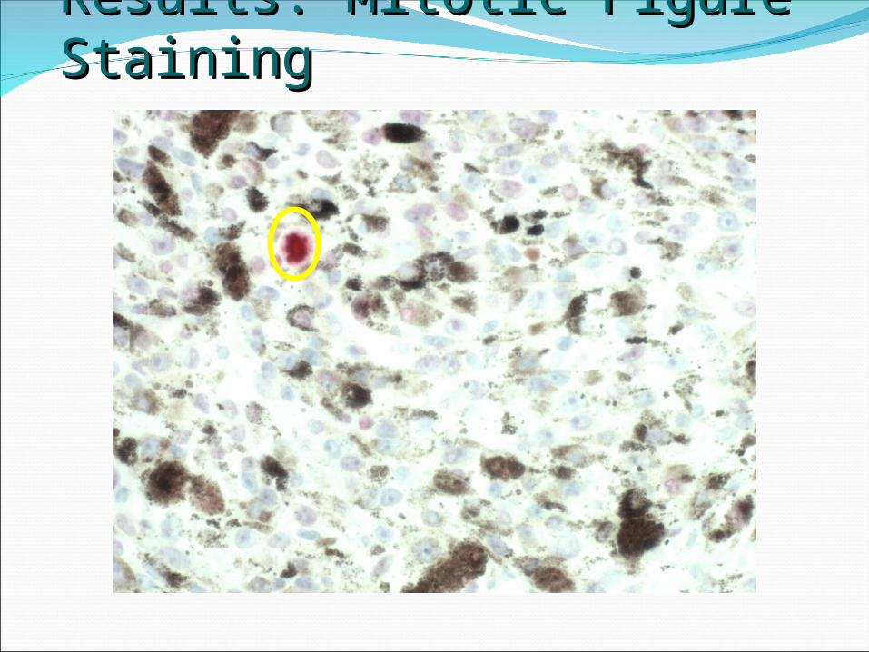
Results: Mitotic Figure StainingResults: Mitotic Figure Staining
Results: Mitotic Figure StainingResults: Mitotic Figure Staining
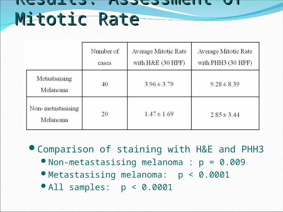
Results: Assessment of Mitotic RateResults: Assessment of Mitotic Rate
Comparison of staining with H&E and PHH3Non-metastasising melanoma : p = 0.009Metastasising melanoma: p < 0.0001All samples: p < 0.0001
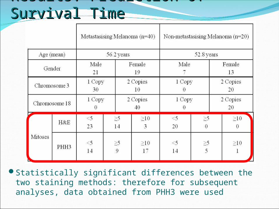
Results: Prediction of Survival TimeResults: Prediction of Survival Time
Statistically significant differences between the two staining methods: therefore for subsequent analyses, data obtained from PHH3 were used
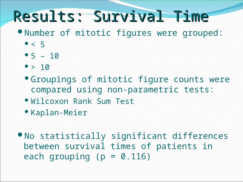
Number of mitotic figures were grouped:< 55 – 10> 10Groupings of mitotic figure counts were
compared using non-parametric tests:Wilcoxon Rank Sum TestKaplan-Meier
No statistically significant differences between survival times of patients in each grouping (p = 0.116)
Results: Survival TimeResults: Survival Time
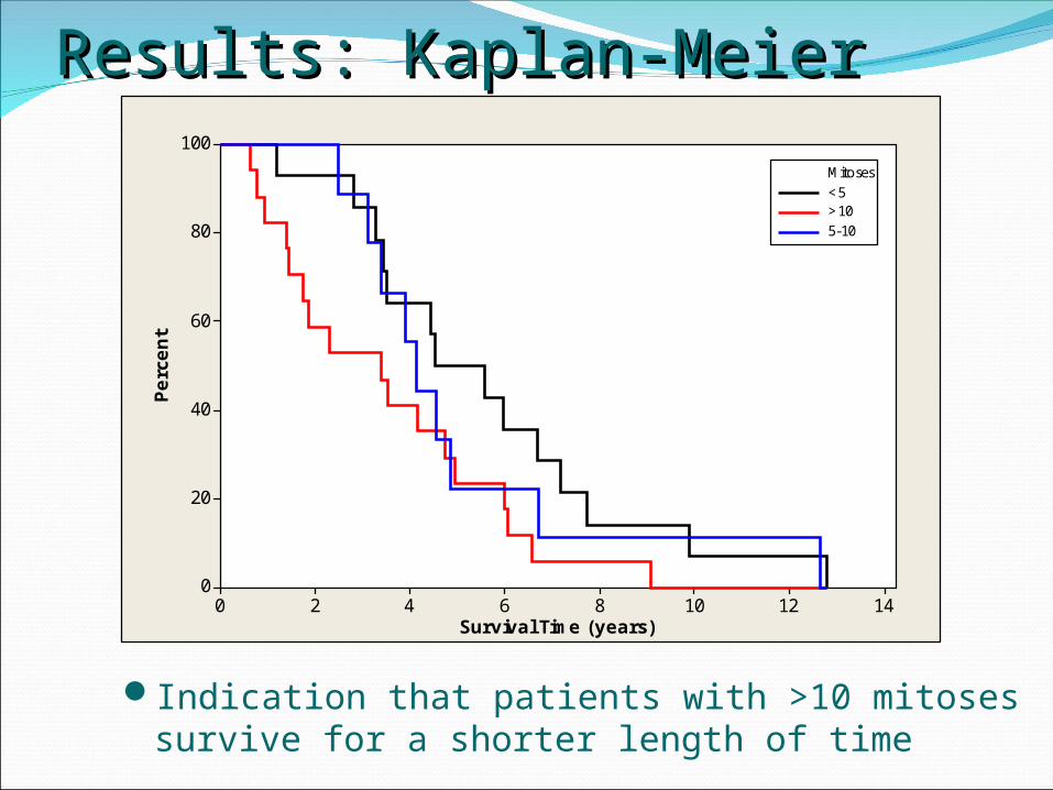
Results: Kaplan-MeierResults: Kaplan-Meier
14121086420
100
80
60
40
20
0
Survival Time (years)
Perc
ent
<5>105-10
Mitoses
Indication that patients with >10 mitoses survive for a shorter length of time
Prediction of Mitoses from Histological Prediction of Mitoses from Histological FeaturesFeatures
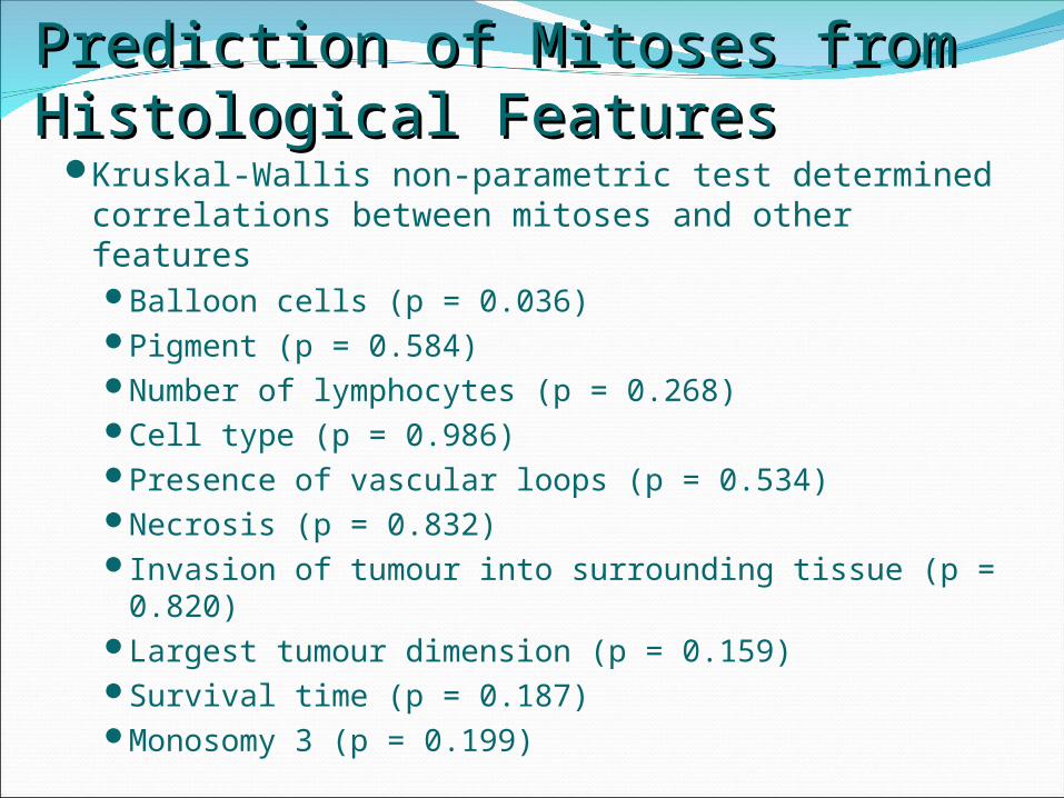
Kruskal-Wallis non-parametric test determined correlations between mitoses and other featuresBalloon cells (p = 0.036)Pigment (p = 0.584)Number of lymphocytes (p = 0.268)Cell type (p = 0.986)Presence of vascular loops (p = 0.534)Necrosis (p = 0.832)Invasion of tumour into surrounding tissue (p =
0.820)Largest tumour dimension (p = 0.159)Survival time (p = 0.187)Monosomy 3 (p = 0.199)
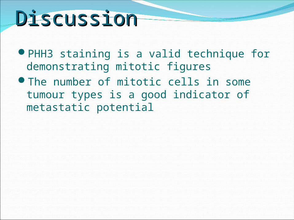
DiscussionDiscussionPHH3 staining is a valid technique for
demonstrating mitotic figuresThe number of mitotic cells in some tumour
types is a good indicator of metastatic potential
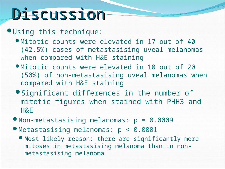
DiscussionDiscussionUsing this technique:
Mitotic counts were elevated in 17 out of 40 (42.5%) cases of metastasising uveal melanomas when compared with H&E staining
Mitotic counts were elevated in 10 out of 20 (50%) of non-metastasising uveal melanomas when compared with H&E staining
Significant differences in the number of mitotic figures when stained with PHH3 and H&E
Non-metastasising melanomas: p = 0.0009Metastasising melanomas: p < 0.0001
Most likely reason: there are significantly more mitoses in metastasising melanoma than in non-metastasising melanoma
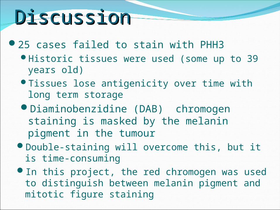
25 cases failed to stain with PHH3Historic tissues were used (some up to 39 years
old)Tissues lose antigenicity over time with long term
storageDiaminobenzidine (DAB) chromogen staining
is masked by the melanin pigment in the tumour
Double-staining will overcome this, but it is time-consuming
In this project, the red chromogen was used to distinguish between melanin pigment and mitotic figure staining
DiscussionDiscussion
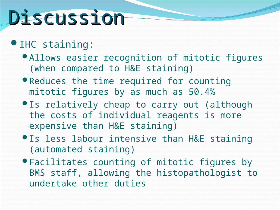
DiscussionDiscussionIHC staining:
Allows easier recognition of mitotic figures (when compared to H&E staining)
Reduces the time required for counting mitotic figures by as much as 50.4%
Is relatively cheap to carry out (although the costs of individual reagents is more expensive than H&E staining)
Is less labour intensive than H&E staining (automated staining)
Facilitates counting of mitotic figures by BMS staff, allowing the histopathologist to undertake other duties
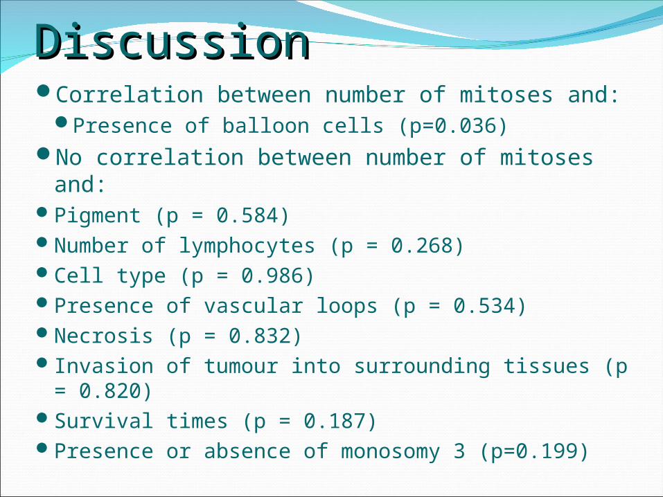
DiscussionDiscussionCorrelation between number of mitoses and:
Presence of balloon cells (p=0.036)No correlation between number of mitoses and:Pigment (p = 0.584)Number of lymphocytes (p = 0.268)Cell type (p = 0.986)Presence of vascular loops (p = 0.534)Necrosis (p = 0.832)Invasion of tumour into surrounding tissues (p =
0.820)Survival times (p = 0.187)Presence or absence of monosomy 3 (p=0.199)
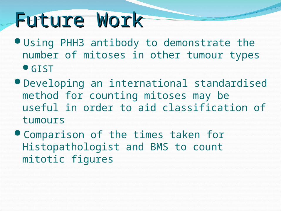
Future WorkFuture WorkUsing PHH3 antibody to demonstrate the
number of mitoses in other tumour typesGIST
Developing an international standardised method for counting mitoses may be useful in order to aid classification of tumours
Comparison of the times taken for Histopathologist and BMS to count mitotic figures


Questions?Questions?