Embed Size (px)
Citation preview

JOURNALOF NEUROPHYSIOLOGY Vol. 65, No. 3, March 1991. Prinrcd In C’.S..-I.
Axes of Eye Rotation and Listing’s Law During Rotations of the Head
J. D. CRAWFORD AND T. VILE Departments of Physiology and Ophthalmology, University of Western Ontario, London, Ontario N6A Xl, Canada .
SUMMARY AND CONCLUSIONS
I. The vestibuloocular reflex (VOR) was examined in four alert monkeys during rotations of the head about torsional, vertical, horizontal, and intermediate axes. Eye positions and axes were recorded in three dimensions (3-D). Visual targets were used to optimize gaze stabilization.
2. Axes of eye rotation during slow phases showed small but systematic deviations from collinearity with the axes of head rota- tion. These noncollinearities apparently resulted from vector sum- mation of torsional, vertical, and horizontal VOR components with different gains.
3. VOR gain was lowest about a head-fixed torsional axis that was correlated with the primary gaze direction, as determined by Listing’s law for saccades. As a result, rotation of the head about a partially torsional axis produced noncollinear slow phases, with axes that tilted toward Listing’s plane.
4. During slow phases, eye position changed not only in the direction of rotation, but also systematically in other directions. Even axes of eye rotation within Listing’s plane caused eye posi- tion to move out of the plane to a torsional position that was then held. Thus Listing’s law for saccades cannot be a product of plant mechanics.
5. VOR slow phases were simulated with the use of a model that incorporated 3-D rotational kinematics into the indirect path and the oculomotor plant. This demonstrated that the observed pattern of position changes is the expected consequence of rotat- ing the eye about a fixed axis and that to hold these positions the indirect path must employ a 3-D velocity-to-position transformation.
6. Quick phases not only corrected the violations of Listing’s law produced by slow phases but anticipated them by directing the eye toward a plane rotated in the direction of head rotation. This was modeled by inputting the vestibular signal to a Listing’s law operator that is shared by the quick phase and saccadic systems.
INTRODUCTION
The vestibuloocular reflex (VOR) prevents slip of the reti- nal image during head rotations. For distant targets, stabili- zation of this image requires that the eye does not rotate relative to space. To achieve this, the slow phase of the VOR must rotate the eye in the direction opposite to the head, but with the same magnitude. To date, most studies have examined the latter, usually expressed as the gain of the VOR (Collewijn et al. 1985; Robinson 1975; Skavenski et al. 1979; Viirre et al. 1986). It is assumed that during normal VOR the eye rotates in the correct direction, i.e., about an axis collinear (parallel) with the axis of head rota- tion. However, there is reason to believe that this ideal state of collinearity may not always be achieved. This is because torsional VOR gain is less than the horizontal and vertical
gains (Collewijn et al. 1985; Ferman et al. 1987a; Leigh et al. 1989; Seidman and Leigh 1989). Rotation of the head about partially torsional axes should result in angular eye velocities with appropriate vertical or horizontal compo- nents but reduced torsional components, i.e., noncollinear. The first goal of this investigation was to evaluate the direc- tion of the VOR by examining collinearity between the axes of the rotating eye and head.
The location of the torsional axis, i.e., the axis of minimal gain, will determine where slow-phase non collinearities oc- cur. Where is this axis? Some have expressed torsion as rotation about the line of sight (Collewijn et al. 1985; von Helmholtz 1925). Others have argued that a head-fixed co- ordinate system is more appropriate, because the vestibular apparatus is fixed in the head and the eye muscles exert torques relative to the head (Fetter et al. 1986; Raphan and Cohen 1986; Robinson 1985; Tweed and Vilis 1987). On the basis of the latter argument, we hypothesized that slow- phase noncollinearities would follow a pattern consistent with gain being lowest about some head-fixed torsional axis.
The axes that the eye rotates about determine the posi- tions that the eye will assume. What positions will be ob- served if the eye rotates about the same axis as the head? During saccades, eye positions conform to Listing’s law. This law states that any arbitrarily chosen (reference) eye position is associated with a particular head-fixed plane, such that the eye only assumes positions that can be reached from this reference position about an axis in that plane. Furthermore, there is one special reference position, primary position, for which the gaze direction is orthogonal to its associated plane. This is called Listing’s plane. By defining ocular torsion as rotation about the head-fixed axis orthogonal to Listing’s plane, one can restate Listing’s law very simply: the eye only assumes positions with a zero torsional component (Ferman et al. 1987b; Nakayama 1983; Tweed and Vilis 1990a). Recently, a surprising aspect of Listing’s law has been confirmed. To keep the torsional component of eye position at zero, saccade axes must have a position-dependent torsional tilt (von Helmholtz 1925; Tweed and Vilis 1990a). Conversely, should the eye always rotate about an axis within Listing’s plane, as in a collinear horizontal VOR, then position-dependent violations of Listing’s law are expected. If the latter situation occurs, Listing’s law must be a product of the neural saccadic sys- tem, contrary to the mechanical hypothesis proposed by some investigators (Ferman et al. 1987b; Sparks and Mays 1990).
OO22-3077/9 1 $1 SO Copyright 0 199 1 The American Physiological Society 407

408 J. D. CRAWFORD AND T. VILIS
The pattern of eye-position changes predicted by rota- tional kinematics should enable discrimination between al- ternative models of the oculomotor position-signal genera- tor (Robinson 1975; Tweed and Vilis 1987). During hori- zontal head rotation, the classical Robinson integrator would convert velocity to position in the horizontal dimen- sion but would be unaware of any torsional changes in eye position that occur when the eye is above or below primary position. The resulting mismatch between motoneuron fir- ing and eye position should result in positional drift. If no such drift occurs, then the brain stem position-signal genera- tor must incorporate the correct laws of rotational kinematics.
The final goal of this study was to examine the action of VOR quick phases on ocular torsion, as compared to sac- cades. Quick phases, which reset eye position after the slow phase, share lower brain stem circuitry with the saccadic system (Chun and Robinson 1978; Ron et al. 1972; Vilis et al. 1989). However, it is not clear at what level these two systems converge. We predicted that, like saccades that obey Listing’s law, quick phases would reset any torsional positions by slow phases to zero; According to the current three dimensional (3-D) model of the saccadic system, this would suggest that rapid eye movements share the same circuitry up to and above the level of the superior colliculus (Tweed and Vilis 1990b).
These questions were examined by the use of a recently developed technique that allowed direct measurement of 3-D axes of eye rotation in the monkey (Tweed et al. 1990). Because our intention was to measure the VOR under opti- mal conditions of gaze stabilization, all experiments were performed in the presence of visual targets. The results sup- ported the above hypotheses and revealed a surprising match between the head-fixed coordinate system of the VOR and that established by Listing’s law for saccades. In addition, quick phases not only reset the torsion produced by slow phases, but actually anticipated it. Some of these results have been reported previously in abstract form (Crawford et al. 1989; Vilis et al. 1989a).
METHODS
Four monkeys, Macaca fasciczdaris, were prepared for chronic behavioral experiments, each undergoing surgery under aseptic conditions and pentobarbital sodium anesthesia. During surgery a skull cap composed of dental acrylic was fastened to the animal’s head, and two enameled copper search coils of 5 mm diam were implanted in one eye for measurement of 3-D eye position. Both coils were positioned nasally, one inferior and one superior. The method used does not require that the coils be aligned orthogo- nally to each other (Tweed et al. 1990). The leads were extended temporally beneath the conjunctiva and then subcutaneously to sockets secured on the cap. In two of the animals, coils were im- planted in both eyes. Coils aligned with the stereotaxic horizontal and sagittal planes were implanted in the skull cap to accurately measure head position. The head of the alert monkey was immo- bilized (by the use of bolts implanted in the skull cap) near the center of three orthogonal magnetic fields. These fields were in phase but operated at different frequencies (250, 125, and 62.5 kHz). Coil signals were digitized by a computer at a sampling frequency of 100 Hz, or occasionally 500 Hz.
The VOR functions optimally when subjects attempt to fixate a visual target (Barr et al. 1976; Ferman et al. 1987a; Skavenski et al.
1979). Under these conditions the VOR is assisted by other visual- oculomotor systems, including the optokinetic reflex (OKN) and smooth pursuit. To optimize gaze stabilization, novel stationary visual stimuli were presented at a distance of I- 1.5 m from the rotating head. These stimuli were placed so as to encourage visual fixation and spontaneous rapid eye movements throughout the oculomotor range.
Angular motion of the head was generated by manually rotating the chair and monkey about an axis parallel to the earth-vertical field direction. A potentiometer detected the angular position of the chair. Because the fields rotated with the monkey, eye coils only detected eye motion relative to the head. The monkey’s eyes were always placed near to the axis of rotation to minimize trans- lations of the eve relative to visual targets. Orientation of the head d within the fields was arbitrary, but Listing’s plane was approxi- mately earth-vertical when the monkey was upright. The monkey could be positioned upright, lying on its back, or lying on either side so that the head was rotated about its vertical, torsional, or horizontal axis. The head was sometimes positioned so that it rotated about axes intermediate between vertical and torsional. At each posture, eye positions were initially recorded with the head immobile for comparison with eye positions during head rotation. During experiments the monkevs were rotated sinusoidally, usually at a frequency of -0.5 Hzyand amplitude of ~60~.
The computer was used to convert coil signals into eye-position quaternions with the use of a method described previously (Tweed et al. 1990). Quaternions were used because, unlike raw coil sig- nals, they provide an accurate and convenient measure of 3-D eye position over a 360° range. Quaternions represent each eye posi- tion as a fixed-axis rotation from a reference position (Westheimer 1957). This reference position was recorded while the monkey looked in the direction of the forward-pointing magnetic field. Quaternions are composed of a scalar part yo, and a vector part q. It is the vector part that is used for representation of data. The vector part has components along torsional, horizontal, and verti- cal axes fixed relative to the head, which in this paper are always parallel to the magnetic field directions. To interpret the data, one need only understand that q is parallel with the axis of eye rotation and its length is proportional to the magnitude of this rotation. To be specific, a quaternion is related to the axis and magnitude of a rotation as follows
y() = cos(oj2) (I)
y = n l sin(tu/2) (2)
The angle CY is the magnitude of the rotation, and n is a 3-D unit vector parallel to the axis of rotation (Tweed and Vilis 1987). At reference position, cy = 0, and so clearly y, = 1 .O and q = 0.
Listing’s law predicts that during saccades and fixation, the qua- ternion vectors of eye positions will align in a plane (because these are simply the axes for rotations that would take the eye directly from reference position to anv other given position). This plane will not necessarily be Listing’splane, unless the reference position happens to be primary position. However, by computing the orien- tation of the plane with respect to the gaze direction at reference position, one can determine primary position and the orientation of Listing’s plane (Tweed et al. 1990).
Quaternions were also used to compute angular velocities of eye rotation. Angular velocity cannot be computed by simply differ- entiating coil signals, quaternions, or any other measure of posi- tion change. The general relation between position quaternions and angular velocity o can be stated as
0 = 24/v (3)
This equation illustrates that rate of change in eve position 4 alone cannot be used to represent eye velocity, because o is also depen- dent on current eye position (Tweed and Vilis 1987). In this paper

AXES AND 3-D POSITIONS OF THE VOR 409
the following method was used to compute the average eye veloc- ity between any two eye positions. If the eye rotates from an initial position q’ to a final position qf, then this rotation can be repre- sented by the quaternion q calculated in Eq. 4.
q = d/q’ (4) The vector part of q gives the axis of the rotation, but q is easilv converted to the more familiar angular velocity. The angle cv of rotation q may be derived by rearranging Eq. 1. This angle is then divided by the time interval between the two eye positions to get the angular speed Cu. The axis n is obtained by entering the vector q and angle CY into Eq. 2. Thus the direction and magnitude of o are specified as
w = &n (-3
The components of w are expressed in the same coordinates as the quaternions that they were derived from. Therefore these coor- dinates are fixed with respect to the head and parallel to the direc- tions of the magnetic fields.
RESULTS
75 I O s t QV
AL - head
slow
- phase
Slow-phase axes
The first step in examining slow-phase direction was to B ~I-!- determine whether the axis of eye rotation remained con- stant during a constant-axis rotation of the head. The con- stant nature of these axes is illustrated in Fig. 1. Each point represents an instantaneous angular velocity vector. Thus a line drawn between zero and any given point would denote the axis of rotation at that time, and the length of this line denotes speed. The right-hand convention is used to indi- cate the direction of rotation about this axis. If the thumb of the right hand is pointed along the axis toward the data
td 1
point (e.g., downward for the eye velocities in Fig. l), then 0 v
i
the fingers curl in the direction of rotation (rightward in this case). As head velocity increased or decreased, so did slow-
J
phase velocity in the opposite direction. The straightness of I
the path followed by these velocity vectors indicates that the ! axis of rotation remained relatively constant.
Figure 1 also suggests that the slow-phase axis was not c precisely co llinear with that of the head . To determine whether this was a consistent phenomenon, the mean veloc- ities of 400 slow phases were examined for each axis of head rotation (Fig. 2, A-C). On average, the slow-phase axes were closely collinear with the head axes. However, two types of non collinearity were evident. The first was a ran- dom variability most prominent during torsional head rota- tions (Fig. 2C). The second was a small systematic tilt, in this case most prominent in the behind view of Fig. 2A. Neither of these appeared to depend on eye speed.
The apparently random variation in noncollinearities was examined for eye-position dependence. Robinson has predicted that, if uncorrected, position-dependent changes in muscle pulling directions should result in axis tilt. For example, a 30° elevation of the eye should result in a 25O vertical tilt of the torsional slow-phase axes (Robinson 1985). There did not appear to be any such pattern in our data. Third-order surfaces were fitted to axis tilt as a func- tion of eye position. The computed position dependence of axis tilt did not follow a consistent pattern between animals and was generally small compared to the standard tions for any one position.
devia-
3
UP
> Right
> Clockwise
> Right
FIG. 1. Instantaneous angular velocities of the eye during a single left- ward rotation of the head (w, velocity points during slow phases). Heavy arrows indicate head velocity during the slow phases. Velocities during quick phases are not shown. A: increasing velocity. Same data are viewed from behind the subject (I&) or from the side (righr), as indicated by the head caricatures. B: decreasing velocity. Axes are labeled according to the direction of rotation (curving arrows) about that axis.
Because the trial-to-trial change in axes appeared to be random, this variation was removed by averaging, leaving only the systematic noncollinearity. Several hundred mean slow-phase axes were averaged for each axis of head rota- tion, in each monkey (Fig. 3). Rotations of the head in opposite directions tended to produce eye rotations about parallel axes. For example, when leftward slow-phase axes tilted back, rightward axes tilted forward (Fig. 3, side view). Axis tilts were most consistent between monkeys from the side view, i.e., vertical axes tilted backward, and torsional axes tilted forward. However, the amount and direction of tilt varied from animal to animal.
410
A
JJ t
. . OH .
3 . WH e , . .
1 0 ' .
l r 0. I
.
l * 1
. b . ,
100% . . l
. .
t t
fJV UT
.
. .
J. D. CRAWFORD AND T. VILIS
WH
I
-- 3
o-
3- w '
C
.
-- 3 0 3-
FIG. 2. Mean velocities of slow phases during fixed-axis head rotations. A: horizontal VOR. B: vertical VOR. C: torsional VOR. Two views of the data are given in each row. Head caricatures indicate the standard behind, above or side views and the axes of head rotation.
How could such systematic noncollinearities arise? One vertical (high gain) and partially torsional (low gain). This possible source of noncollinearity might be differences in test is illustrated in Fig. 4A. The unit torsional and vertical gain between various VOR directions. This hypothesis was head rotation vectors h, and h, have been reversed (multi- tested by rotating the head about an axis that was partially plied by -1) for better comparison with the eye vectors.

411 AXES AND 3-D POSITIONS OF THE VOR
ckwise
FIG. 3. Average torsional, horizontal, and vertical slow-phase axes of all 4 animals. For each axis of head rotation, the axes of negative and positive slow-phase directions are plotted separately. Subject key: solid line, AR; dotted line, F; dashed line, L; interrupted line, CL.
Four hundred average slow-phase velocities were divided by head speed and averaged to get each of the resultant eye vectors e, and e,. These can be thought of as the average VOR output resulting from one unit of head rotation in a given direction, or a 3-D version of gain. Note that the
-h t “+I b-1 I I
I I I I I I
B ae t e * A ----------h+ be V
FIG. 4. A: torsional, vertical, and intermediate axes of head rotation (-: h,, h,, h*) and the resultant axes of eye rotation (---: e,, e,, e*). Torsional and vertical components of the intermediate axis of head rota- tion are indicated as u and b. B: prediction of e* by vector summation. Products ae, and be,, are summed linearly to produce e,. Unlike the other figures, the vertical axis is labeled as such, rather than by the direction of rotation.
length of e, is significantly less than the length of e, and that both are very near to being collinear with the axes of head rotation. The third solid arrow h* illustrates an interme- diate axis of head rotation. The resultant eye rotation e* was always less collinear with the axis of head rotation than the other vectors, and its direction of tilt was toward the vertical axis.
To test whether this noncollinearity was the result of lin- ear vector summation, a 3 x 3 VOR “gain” matrix G was computed for each eye. The components of the torsional eye vector e, constituted the first column of G, those of horizontal vector (not shown) formed the second, and the components of the vertical vector e, formed the third col- umn. Multiplication of h* by this matrix gave the predicted 3-D eye rotation eP. In other words, each component of head rotation was multiplied by the individually computed 3-D “gain” for that direction of VOR, and the resulting three vectors were then summed linearly. Figure 5 is used to illustrate this process qualitatively in two dimensions. Or- thogonal projections were made from h* to the torsional and vertical axes to get the components, a and b (Fig. 4A). These components were then multiplied by the appropriate vectors (e, and e,, i.e., columns 1 and 3 of G) to estimate the eye movements that they would have produced individu- ally (Fig. 4B). Vector summation of these predicted vectors ae, and be, gave the theoretical eye rotation vector e,. The theoretical eye vector eP always predicted the deviations in magnitude and direction of the real eye vector e* from the corresponding head vector ha. This analysis suggested that most of the noncollinearity between h* and e* resulted from low torsional gain. In general, whenever the torsional, vertical, and horizontal gains were not equal, the slow- phase axis tilted toward the axis of highest gain, exactly as predicted by the assumption of linear vector summation.
Although differences in gain appear to produce noncol- linearities during head rotations about intermediate axes,
412 .I. D. CRAWFORD AND T. VILIS
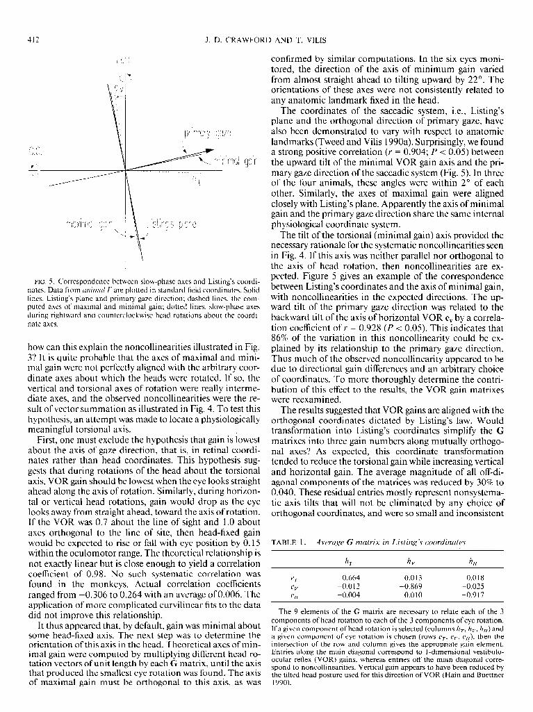
FIG. 5. Correspondence between slow-phase axes and Listing’s coordi- nates. Data from unirnul E’ are plotted in standard field coordinates. Solid lines, Listing’s plane and primary gaze direction: dashed lines, the com- puted axes of maximal and minimal gain; dotted lines, slow-phase axes during rightward and counterclockwise head rotations about the coordi- nate axes.
how can this explain the noncollinearities illustrated in Fig. 37 It is quite probable that the axes of maximal and mini- mal gain were not perfectly aligned with the arbitrary coor- dinate axes about which the heads were rotated. If so, the vertical and torsional axes of rotation were really interme- diate axes, and the observed noncollinearities were the re- sult of vector summation as illustrated in Fig. 4. To test this hypothesis, an attempt was made to locate a physiologically meaningful torsional axis.
First, one must exclude the hypothesis that gain is lowest about the axis of gaze direction, that is, in retinal coordi- nates rather than head coordinates. This hypothesis sug- gests that during rotations of the head about the torsional axis, VOR gain should be lowest when the eye looks straight ahead along the axis of rotation. Similarly, during horizon- tal or vertical head rotations, gain would drop as the eye looks away from straight ahead, toward the axis of rotation. If the VOR was 0.7 about the line of sight and 1 .O about axes orthogonal to the line of site, then head-fixed gain would be expected to rise or fall with eye position by 0.15 within the oculomotor range. The theoretical relationship is not exactly linear but is close enough to yield a correlation coefficient of 0.98. No such systematic correlation was found in the monkeys. Actual correlation coefficients ranged from -0.306 to 0.264 with an average of 0.006. The application of more complicated curvilinear fits to the data did not improve this relationship.
It thus appeared that, by default, gain was minimal about some head-fixed axis. The next step was to determine the orientation of this axis in the head. Theoretical axes of min- imal gain were computed by multiplying different head ro- tation vectors of unit length by each G matrix, until the axis that produced the smallest eye rotation was found. The axis of maximal gain must be orthogonal to this axis, as was
confirmed by similar computations. In the six eyes moni- tored, the direction of the axis of minimum gain varied from almost straight ahead to tilting upward by 22’. The orientations of these axes were not consistently related to any anatomic landmark fixed in the head.
The coordinates of the saccadic system, i.e., Listing’s plane and the orthogonal direction of primary gaze, have also been demonstrated to vary with respect to anatomic landmarks (Tweed and Vilis 1990a). Surprisingly, we found a strong positive correlation (Y = 0.904; P < 0.05) between the upward tilt of the minimal VOR gain axis and the pri- mary gaze direction of the saccadic system (Fig. 5). In three of the four animals, these angles were within 2O of each other. Similarly, the axes of maximal gain were aligned closely with Listing’s plane. Apparently the axis of minimal gain and the primary gaze direction share the same internal physiological coordinate system.
The tilt of the torsional (minimal gain) axis provided the necessary rationale for the systematic noncollinearities seen in Fig. 4. If this axis was neither parallel nor orthogonal to the axis of head rotation, then noncollinearities are ex- pected. Figure 5 gives an example of the correspondence between Listing’s coordinates and the axis of minimal gain, with noncollinearities in the expected directions. The up- ward tilt of the primary gaze direction was related to the backward tilt of the axis of horizontal VOR e, by a correla- tion coefficient of r = 0.928 (P < 0.05). This indicates that 86% of the variation in this noncollinearity could be ex- plained by its relationship to the primary gaze direction. Thus much of the observed noncollinearity appeared to be due to directional gain differences and an arbitrary choice of coordinates. To more thoroughly determine the contri- bution of this effect to the results, the VOR gain matrixes were reexamined.
The results suggested that VOR gains are aligned with the orthogonal coordinates dictated by Listing’s law. Would transformation into Listing’s coordinates simplify the G matrixes into three gain numbers along mutually orthogo- nal axes? As expected, this coordinate transformation tended to reduce the torsional gain while increasing vertical and horizontal gain. The average magnitude of all off-di- agonal components of the matrices was reduced by 30% to 0.040. These residual entries mostly represent nonsystema- tic axis tilts that will not be eliminated by any choice of orthogonal coordinates, and were so small and inconsistent
TABLE 1. Average G matrix in Listing’s coordinates
IIT hv hH
YT -0.664 -0.0 13 -0.0 18 CV -0.0 12 -0.869 -0.025 c;, -0.004 -0.010 -0.917
The 9 elements of the G matrix are necessary to relate each of the 3 components of head rotation to each of the 3 components of eye rotation. If a given component of head rotation is selected (columns hT, hv, hH) and a given component of eye rotation is chosen (rows CT, vv, eH), then the intersection of the row and column gives the appropriate gain element. Entries along the main diagonal correspond to l-dimensional vestibulo- ocular reflex (VOR) gains, whereas entries off the main diagonal corre- spond to noncollinearities. Vertical gain appears to have been reduced by the tilted head posture used for this direction of VOR (Hain and Buettner 1990).
AXES AND 3-D POSITIONS OF THE VOR 413
between animals that they were probably due to measure- ment error. Finally, the standard deviations between corre- sponding elements of the G matrices were reduced from an average of 0.11 to 0.07. Thus transformation into Listing’s coordinates appear to standardize the matrices and increase their diagonality.
Averaging the G matrices of all subjects after transforma- tion into Listing’s coordinates further reduced the off-diag- onal elements (Table 1). One-dimensional ( 1 -D) gain mag- nitudes along the main diagonal of this matrix were 0.664 (torsional), 0.869 (vertical) and 0.9 17 (horizontal). Because
qH T 40”
& Right
qH T
A Upright
the off-diagonal components were very close to zero, the average G matrix was essentially composed of three orthogo- nal columns. Thus it would appear that the overall action of the VOR is best expressed as three gains along the mutually orthogonal axes aligned with Listing’s coordinates.
Slow-phase eye positions
Having determined the axes of slow-phase eye rotation, the resulting changes in eye position were examined. This question is not as trivial as it may seem. The laws of rota-
t
r
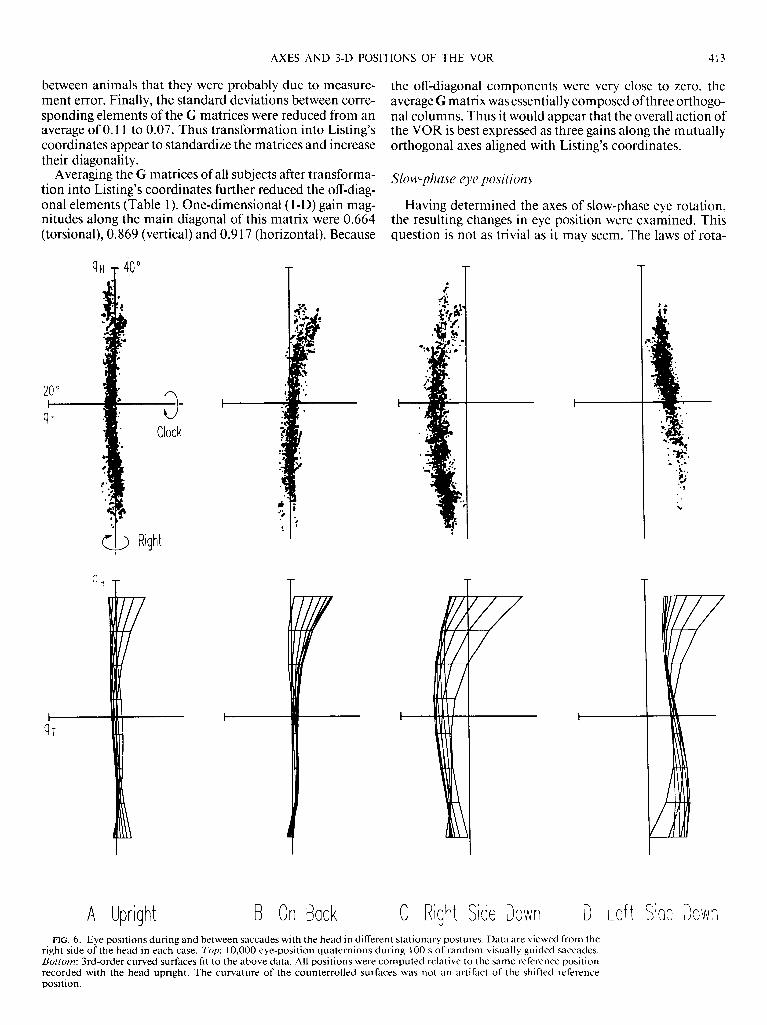
B On Back C Right Side Down D Let t Side Down FIG. 6. Eye positions during and between saccades with the head in different stationary postures. Data are viewed from the
right side of the head in each case. Top: 10,000 eye-position quaternions during 100 s of random visually guided saccades. Bottom: 3rd-order curved surfaces fit to the above data. All positions were computed relative to the same reference position recorded with the head upright. The curvature of the counterrolled surfaces was not an artifact of the shifted reference position.

414 J. D. CRAWFORD AND T. VILIS
tional kinematic require that, when the eye is rotated about To determine whether the slow phases violate Listing’s a fixed vertical axis while gazing upward or downward, its law, it was first necessary to determine Listing’s plane with position must change not only in the horizontal direction, the head stationary. The typical range of eye-position vec- but also vertically and torsionally. The latter change in eye tors during saccades and fixations is illustrated in Fig. 6. As position would be a violation of Listing’s law. On the other reported elsewhere, the range of obtainable eye positions hand, current models of the VOR suggest that only horizon- was distributed within a nearly flat two-dimensional (2-D) tal position will change. surface with a finite torsional width of -3’ (Tweed and
A
B
C
cl5 * I Right
qH T 40”
qT-
0 n .
D Counterclock.
Clockwise
? Right
co. I Rtght
FIG. 7. Slow-phase eye positions during horizontal, vertical, and torsional rotations of the head. Each series of quaternion vectors forms a dotted line representing 1 complete slow phase. Small arrowheads at the final position indicate direction. A: rightward head rotation. B: downward head rotation. Eye positions are shifted torsionally as in Fig. 6C because of the tilted head posture used. C: clockwise head rotation. Head caricatures indicate that data are viewed orthogonal to the axis of rotation (Z@) and down the axis (right).

AXES AND 3-D POSITIONS OF THE VOR 415
Vilis 1990a). Third-order curved surfaces were fitted to the data as illustrated in the lower row of Fig. 6. These show that when the monkey was upright, eye position was con- fined to the highly planar surface defined by Listing’s law.
Was Listing’s plane constant during the other head posi- tions used in this study? Eye positions were also recorded while the animal lay on its back, right, and left sides (Fig. 6, B-D, respectively). With the monkey on its back, Listing’s plane remained intact, with a slight forward tilt. When the monkey was lying on its side, the head was tilted 90’ tor- sionally from the upright position. During such postures the tonic ocular counterroll reflex occurs, resulting in a tor- sional shift of Listing’s plane in the direction opposite to head rotation (Collewijn et al. 1985). These surfaces ap- peared to be thicker than the standard Listing’s plane. How- ever, surface fits revealed that this was partially due to dis- tortion of the plane into a bowl-like surface, i.e., the tor- sional shift was not as great for eccentric gaze directions. The standard deviations of torsional position from the sur- faces of best fit illustrated in Fig. 6 were 0.872” with the monkey upright, 1.246” with the monkey on its back, 1.14 1 O with the monkey right side down, and 1.116” with the monkey left side down. Thus, aside from small shifts and distortions of the plane, eye position during and be- tween saccades remained confined to an essentially planar surface despite tonic head tilt.
Figure 7 illustrates the changes in eye position produced by rotations of the head about fixed vertical, horizontal, and torsional axes. These slow-phase axes were nearly col- linear with the axis of head rotation. As expected, the main change in orbital eye position was in the direction opposite to that of head rotation (Fig. 7, /e$ column). If the right- hand thumb is pointed in the direction of the quaternion vectors, the curl of the fingers indicates that eye positions changed leftward in A, upward in B, and counterclockwise in C. Not surprisingly, the latter drove eye position counter- clockwise out of Listing’s plane.
As required by rotational kinematics, changes in eye-po- sition vectors (the vector part of quaternions as defined in METHODS) did not only occur along the axis of head rota- tion, but also in a systematic manner in the orthogonal directions. In the right column of Fig. 7, data are viewed orthogonal to the axis of rotation, with eye velocities point- ing toward the reader. In the case of horizontal VOR (Fig. 7A), there was a change in torsional position whose direc- tion and magnitude depended on vertical eye position. For example, during leftward slow phases, clockwise position accumulated when the eye looked up, and counterclock- wise position accumulated when the eye looked down. The opposite pattern occurred during rightward slow phases. Vertical slow phases produced a similar pattern of position changes (Fig. 7B), except that in this case the direction and magnitude of torsional position change was dependent on horizontal eye position. Thus rotation of the eye about axes - me,- , l * - 1 , - l-w* . * . l .
Eye (Torsion)
IOmms
50 [ -,j__...-
Head (Horizontal) 1
2o [ 0
in Listings plane produces violations or Listings law, wnen that rotation is not directly toward or away from primary
0 400 ms
position. The DISCUSSION will show that this is not an unex- FIG. 8. Maintenance of accumulated torsional position after horizontal
changes in vertical and horizontal eye position are evident. The change in vertical position depended on initial horizon- tal position, and the change in horizontal position de- pended on initial vertical position. As a result, a circular pattern of position change is observed. The pattern of posi- tion changes can be summarized as follows. If one points the thumb of the right hand in the direction of the eye velocity vector, e.g., off the page in the right coluwzn of Fig. 7, then position vectors changed mostly in this direction. However, the position vectors also circled around reference position in the direction of finger curl. The same circular pattern was followed during horizontal and vertical VOR, but only a slice of the family of circles was present because the onset of quick phases restricted the range of torsional eye positions.
As described in the DISCUSSION below, this pattern of po- sition change was actually a necessary condition for stabili- zation of gaze. It was not a consequence of axis noncollin- earities but rather the expected result of rotating the eye
petted observation. head rotation. The change in eye position was due to the position-depen-
Torsional VOR clarified the pattern of 3-D eye-position dent effect described in the text, not axis noncollinearity. Top: counter-
change that was only partially discernable for the other clockwise changes in eye position during 6 slow phases. Bottom: corre-
VOR directions. When counterclockwise slow-phase posi- sponding rightward changes in head position. The monkey was looking downward in each case. Position traces have been aligned at the time when
tions are viewed down the axis of rotation (Fig. 7C, right), head movement stopped (vertical line).

416 J. D. CRAWFORD AND T. VILE
about a fixed axis, based on the principles of rotational kin- indirect pathway of the VOR to accumulate a horizontal ematics. Are the brain stem circuits of the VOR aware of signal. Vertical and torsional position signals would not be these principles? The conventional view is that during a generated in this example. The current results show that horizontal rotation of the head, it is only necessary for the this would usually result in a mismatch between the posi-
6
A OH T . .
.
. 9’ . .
. . . . ‘.
I #’ . y
. . . * m m . . : l . l
. ‘ , I - . .
. - l
. . l
. - . Z’ . . ’ ’ l *- . . c .
. . . . l
. .
l ..- : l ,- . - , -
.
. l -,: ‘-9 ’ . . . . . l . - ’ :
. 0,. . , . % 8 8 l .‘-,‘. l . ‘. l loo”/s ’ . l ‘.‘-• 1” , ( ,‘. l ,* :
.
.y’ . l - . l l l ** I
I . . r .-4 ( - .
hl . l .
‘9
. : .
l . / . “ f ~ 9” : .
.
, b’..- . . 5
. ’ .
. - . . :
l . . .
l . I
.* ‘. . ‘9’
. l l . ’ l % l
. l #
.
‘.’ m
.
. . : . . . .
. : ‘# . . l . .
. . l .
.
~ . *m* , l
l ‘.
‘I .
.
.*. . l ‘) l . . 9’ l . l
. .
. - . . ) l l .
l ‘ . ( .
. .
l .
.
.
. . . .
C . .
. . . 0.’
. . . ”
. f l
. - • - . *
l .
.’ l
. . . * \ l
1. .’ l ’ l
l .J . . ’ et n l .xm ‘.
9 . .
.
I l -*.-#, . l . . _
1 . -1 .
w
. : ’ .:t”~ . .
ibf : 8 l - • . ,
. .
: . .‘”
: . . .
‘1. ’
l r . .
’ .
. .
WH T
A
FIG. 9. Mean velocities of quick phases during fixed-axis head rotations. Arrowheads indicate the axis of head rotation. A: horizontal head rotation. R: vertical head rotation. C’: torsional head rotation. Head caricatures indicate the viewing perspec- tives and the axes of head rotation.

AXES AND 3-D POSITIONS OF THE VOR 417
tion signals encoded by motoneurons and actual eye posi- tion. Any postrotational torsion should decay exponen- tially in a fraction of a second. However, no such postrota- tional drift was observed in our data. When head rotation stopped, any accumulated torsion held until the next eye movement (Fig. 8). Thus position signals from ocular moto- neurons appear to be perfectly matched to actual eye posi- tion. This suggests that, contrary to the conventional view, the neural circuit of the indirect pathway that converts ve- locity to position incorporates the principles of rotational kinematics.
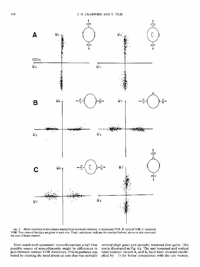
AXES OF ROTATION. As expected, quick phases almost al- ways reversed the direction of eye rotation generated by slow phases and often caused gaze to lead in the direction of head motion. Therefore mean angular velocities of such quick phases had a significant component in the direction of head rotation. However, quick phases also directed gaze to specific visual targets. Because of this, quick-phase axes had almost every combination of vertical and horizontal components (Fig. 9). Therefore, unlike slow-phase axes (Fig. 2) quick-phase axes did not line up with the axes of head rotation. A comparison of these two figures also re- veals that, during horizontal and especially vertical VOR, the quick-phase axes exhibited a larger distribution in the torsional direction than the slow-phase axes. Thus quick- phase axes tilt out of Listing’s plane in a manner similar to saccades, which do so to obey Listing’s law. QUICK PHASES AND LISTING'S LAW. As we have seen, slow phases usually violate Listing’s law. However, large tor- sional components did not accumulate after several slow phases. Therefore the quick phases must have been correct- ing the torsion produced by slow phases. In the case of tor- sional VOR, the slow phases drove eye position almost per- pendicularly out of Listing’s plane. The torsional quick phases not only corrected these violations of Listing’s law, but overshot Listing’s plane by an approximately equal amount (Fig. 10). Thus the eye was directed to a range of positions rotated torsionally in the direction of head rota- tion. This range of positions appears as a plane shift in 3-D plots. The surfaces fit to this data (Fig. 10, bottom) had a bowl shape similar to ocular counterroll surfaces (Fig. 6). The magnitude of this plane shift increased with frequency and speed of head rotation. At the standard frequency of head rotation (0.5 Hz), the torsional quick-phase planes were shifted from Listing’s plane by an average of 5.5O across subjects. As a result of starting and ending at posi- tions with opposite torsional components, the torsional slow phases tended to straddle Listing’s plane.
Is this torsional overshoot by quick phases a violation of Listing’s law, or does the plane of desired eye positions shift to keep slow phases centered on Listing’s plane? Examina- tion of horizontal and vertical VOR revealed that the latter strategy was followed. Quick phases not only corrected vio- lations of Listing’s law but, as in torsional VOR, crossed Listing’s plane (Fig. 1 IA). Recall that leftward quick phases drove the eye counterclockwise when looking down and clockwise when looking up. To reverse this torsion, quick phases had to direct eye position to a plane that was rotated horizontallv. again in the direction of head rotation. Each
B
l m1
. Right
FIG. 10. Final positions of quick phases during torsional VOR. Top YOW: eye positions at the end of counterclockwise (A) and clockwise (I?) quick phases. Bottom r’o~: 3rd-order curved surfaces fit to the above data.
quick phase drove the eye to a plane rotated from Listing’s plane by an angle we called 8. The average rightward and leftward quick-phase plane tilts for these animals are illus- trated in Fig. 1lB. There was a significant difference be- tween the 8s of rightward and leftward quick phases in all animals (P < 0.05). The mean 8 across subjects was 3.8O.
Thus quick phases appear to direct the eye to a plane of positions that anticipated violations of Listing’s law pro- duced by subsequent slow phases. This is most convinc- ingly illustrated by quick phases such as those shown in Fig. 11 C. Take, for example, the case in which the eye initially looked downward, and a leftward slow phase had just driven eye position counterclockwise out of Listing’s plane. Subseauent auick phases that redirected gaze to an upward

418 J. D. CRAWFORD AND T. VILIS
40 0 A +-I qV
qr -5 ( 4) OX
+- @ \ . \ \ \
rotation. A position-dependent pattern of changes is also observed in the other components. Even rotation of the eye about axes in Listing’s plane e.g., vertical and horizontal, produces torsional deviations in eye position, and this tor- sion appears to be held by tonic motoneuron signals. To produce this pattern, the VOR must incorporate the princi- ples of rotational kinematics into the velocity-to-position transformation of the indirect path.
Finally, quick phases appear to not only reset these tor- sional components, but drive the eye to the torsional posi- tion that anticipates the action of the subsequent slow phase. This observation is explained below by a model of the saccade/quick-phase generator, which implements List- ing’s law above the level of convergence with the slow-phase generator.
FIG. 11. Quick-phase eye positions during horizontal VOR. Standard above views of the data are used except that torsional position scaled up 4 times for clarity. A: typical quick phases during rightward head rotation. The angle 0 quantifies the amount that the quick phase overshot the stan- dard Listing’s plane. B: average rotation of quick-phase planes from List- ing’s plane. Solid lines, planes during leftward head rotation in all 4 ani- mals; broken lines, planes during rightward head rotation. C: quick phases that reversed the vertical component of eye position. The dashed line is the average plane of quick-phase final positions from B. Error bars indicate the width of this plane.
direction mai ntained that counterclockwise torsion instead of driving the eye clot kwise. Had these quick phases crossed Listing’s plane, as in Fig. 1 IA, the next slow phase would drive the eye further clockwise, resulting in a large violation of Listing’s law. Thus, by taking eye position to the same side of the plane, such quick phases anticipated the action of the subsequent slow phase. For all VORs, quick phases directed the eye toward a plane of positions rotated in the direction of head rotation, so that the torsional component of the subsequent slow phase straddled Listing’s plane.
DISCUSSION
Three separate but interrelated findings are reported in this paper. First, the data suggest that the slow-phase axis is remarkably collinear with the axis of head rotation, when the latter is either within or orthogonal to Listing’s plane. However, rotation of the head about any other interme- diate axis results in noncollinearity. This appears to be the result of low gain about the head-fixed torsional axis orthog- onal to Listing’s plane. As demonstrated below, these re- sults corroborate Robinson’s diagonal VOR matrix, pro- vided the matrix is expressed in Listing’s coordinates (Rob- inson 1982, 1985).
The second finding is that these slow-phase axes do not only change eye position in the direction opposite to head
Directions ofslow-phase eye movement
The essential feature of slow-phase axes was their close alignment with the axes of head rotation. Thus slow phases rotated in the correct direction to minimize slip of the reti- nal image. Observed errors in direction took the form of small random variations in slow-phase axes and systematic noncollinearities.
The origin of the random variation in slow-phase axis orientation remains uncertain. It has been suggested that the mechanics of the muscles might result in a dependence of the VOR on orbital eye position (Robinson 1985). How- ever, in the present study, deviations from collinearity did not follow the pattern predicted by Robinson for a VOR that did not correct for such a dependence. In general, the data agreed with the previous observation that slow-phase direction (in head-fixed coordinates) is independent of eye position (Fetter et al. 1986). This is consistent with recent anatomic experiments that suggest that muscle pulling di- rections, at least those of the recti, are relatively indepen- dent of horizontal and vertical eye position (Miller and Robins 1987). Alternatively, if a significant pattern of eye- position dependence in muscle pulling directions does ex- ist, our data would suggest that it is compensated for by the VOR.
The combination of two conditions confirmed by this study make systematic noncollinearities inevitable. First, as predicted by Robinson’s matrix model, slow-phase axes can be predicted by applying predetermined vertical, horizon- tal, and torsional gains to the components of head rotation and then summating the resulting vector products (Robin- son 1982, 1985). Second, as observed previously, torsional gain is low (Collewijn et al. 1985). Consequently, rotation of the head about a partially torsional axis will produce a slow-phase axis with proportionately small torsional com- ponents, such that this axis tilts away from the torsional axis.
If slow-phase axes tilt away from the torsional axes, there is practical value in determining the location of this special axis. The intrinsic coordinate systems of the VOR, includ- ing those of the semicircular canals, extraocular muscles, and the position-signal generator, do not encode torsion along a single independent channel (Crawford et al. 1988; Simpson 1983). What is the appropriate coordinate system for the overall action of the VOR, which is the product both of these intrinsic coordinates and the transformations that

AXES AND 3-D POSITIONS OF THE VOR 419
occur between them (Robinson 1982)? The Robinson model arbitrarily puts VOR gain into Cartesian coordinates with the torsional axis pointing straight forward. The main diagonal elements of this matrix were taken from the best available 1 -D measures of gain, and the other elements were set at zero; i.e., the VOR was assumed to be collinear about the head-fixed torsional, vertical, and horizontal axes. Our data confirms that VOR gain is organized in head-fixed orthogonal coordinates and, furthermore, suggests that these coordinates align with those designated by Listing’s law of the saccadic system.
The full implications of this remarkable coincidence be- tween the coordinates of the saccadic system and the VOR are not yet clear. Visual maintenance of VOR gain may be the key. Retinal slip is necessary for calibration of gain, and velocity of slip near the fovea is relatively small during rota- tions of the eye about the line of sight (Gonshor and Melvill Jones 1976; Miles and Fuller 1974; Schultheis and Robin- son 198 1). If the monkey’s preferred range of gaze direc- tions was centered around the primary direction, then cali- bration of gain about this axis might consequently suffer. This effect may be compounded by avoidance of large tor- sional head rotations during visual orientation (Tweed and Vilis 199 1). However, this hypothesis appears to be contra- dicted by the frequent eccentricity of experimentally deter- mined primary positions (Tweed and Vilis 1990a). If pri- mary position does not correspond to the preferred gaze direction, then some other internal factor must be responsi- ble for its significance.
In summary, the gain of the visually assisted VOR ap- pears to be lowest about an axis parallel with the primary gaze direction and highest about axes in Listing’s plane. Systematic noncollinearities arise as follows. Axes of head rotation that were neither in nor orthogonal to Listing’s plane produced axes of eye rotation that tilted toward List- ing’s plane. As the axis of head rotation tilts out of Listing’s plane to a peak of 45”, the contribution of axis noncollin- earity to instability of the retinal image increases. The VOR is apparently most accurate in direction and magnitude when the head is rotated about an axis within Listing’s plane.
Changes in eye position during slow phases
The pattern of position changes observed during slow phases was the consequence of rotating the eye about a sin- gle fixed axis, as predicted by the principles of rotational kinematics. Stated quantitatively, the relationship between rate of eye position change 4, angular velocity O, and current eye position q is
lj = o-y/2 (6)
This equation shows clearly that rate of change in eye posi- tion depends on both the velocity (axis) of rotation and the current eye position. Thus even rotations of the eye about axes within Listing’s plane are expected to violate Listing’s law in a position-dependent manner. The fact that they do shows unequivocally that Listing’s law is not a product of plant mechanics.
Figure 12 provides an intuitive explanation of the changes in eye position that result from rotations about axes fixed in Listing’s plane. The eye is viewed from a fron-
c J D FIG. 12. Effects of fixed-axis rotation on 3-D position of a sphere. A:
initial position. A symbol is painted onto the sphere to indicate rotational position. B: sphere has undergone a 90” leftward rotation from the initial position. Original position of the symbol is outlined for reference. C: sphere is rotated 90” upward about a horizontal axis from the reference position. I>: sphere in C‘ is further rotated 90” to the left. Heavy arrows embedded in the spheres indicate the axes of rotation that would take the eye from initial position A to each of the other positions. Dotted lines indicate the resultant trajectories of points on the surface of the sphere. Axes between the 2 columns indicate that the same rotation occurs in both rows.
tal perspective. Initial position A (gaze directed straight ahead) is chosen as the reference position, and a symbol is superimposed over the pupil. The eye then rotates 90’ hori- zontally about a fixed vertical axis to position B, as it would during an oversized slow phase or saccade. Clearly, eye po- sition relative to reference position has a significant horizon- tal component but no torsional or vertical components. The same overall change in position could be accomplished by several horizontal rotations in any order, illustrating the simple relationship between rotational movement and ro- tational position when motion is restricted to one dimension.
When rotation is neither toward or away from reference position, the relationship between motion and position is not so trivial. This is most easily illustrated by an extreme case. In the bottom TOW of Fig. 12, the eye starts from posi- tion C, which is rotated 90° upward from the initial refer- ence position. A 90’ leftward slow phase rotates the symbol about the line of sight to position D without changing its location. What is the overall change in eye position relative to the head-fixed reference position A? It is that rotation that will take the eye from initial position A to final position D. A purely upward rotation will take the symbol to the correct location but in the wrong orientation. Any combina- tion of upward and leftward rotation without torsion will take the symbol to the wrong location. Thus the axis of rotation from reference position must also tilt forward tor- sionally, as indicated. This is not false torsion; a clockwise
420 J. D. CRAWFORD AND T. VILIS
torque relative to the torque in A must be generated by the extraocular muscles if position D is to be maintained.
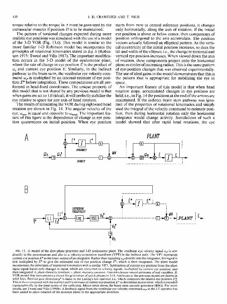
The pattern of torsional changes expected during more realistic eye positions was simulated with the use of a model of the 3-D VOR (Fig. 13A). This model is similar to the more familiar 1-D Robinson model but incorporates the principles of rotational kinematics stated in Eq. 6 (Robin- son 1975; Tweed and Vilis 1987). The important modifica- tion occurs in the 3-D model of the oculomotor plant, where the rate of change in eye position ,?? is the product of O, and current eye position E. Similarly, in the indirect pathway to the brain stem, the vestibular eye velocity com- mand oP is multiplied by an internal estimate of eye posi- tion P before integration. These computations are all per- formed in head-fixed coordinates. The unique property of this model that is not shared by any previous model is that when gains are set to 1 .O (ideal), it will perfectly stabilize the eye relative to space for any axis of head rotation.
The results of simulating the VOR during rightward head rotation are shown in Fig. 14. The angular velocity of the eye, %ye, is equal and opposite to Oh& The important fea- ture of this figure is the dependence of change in eye posi- tion quaternions on initial position. When eye position
starts from zero (a central reference position), it changes only horizontally, along the axis of rotation. If the initial eye position is above or below center, then components of position orthogonal to the axis accumulate. The position vectors actually followed an elliptical pattern. As the verti- cal eccentricity of the initial position increases, so does the tilt and width of the ellipses; i.e., the change in torsional and vertical eye position increases. When viewed down the axis of rotation, these components project onto the horizontal plane as circles of increasing radius. This is the same pattern of eye-position changes that was observed experimentally. The use of ideal gains in the model demonstrates that this is the pattern that is appropriate for stabilizing the eye in space.
An important feature of tins model is that when head rotation stops, accumulated changes in eye position are held; i.e., in Fig. 14 the positions at the end of the arrows are maintained. If the indirect brain stem pathway was igno- rant of the properties of rotational kinematics and simply used the integral of the velocity command to estimate posi- tion, then during horizontal rotation only the horizontal integrator would change activity. Simulations of such a model showed that after rapid head rotations, the eye
A PLANT ---------------~ , I I
I I -
wh I +iJ
w'e' ' E * DlfNCT I
** I . I
B
0 -1
n _ i MN 1 ---------------jg 4 c i E -4, PLANT f- I-----!
I I I , . , r------1
r---- ------
I
+
i” I --1
-- )I/ I
* I L-L-
III- -7
L--
FIG. 13. A: model of the slow-phase generator and 3-D oculomotor plant. The vestibular eye velocity signal wp is sent directly to the motoneurons and also to a velocity-to-position transform (VPT) in the indirect path. The VPT represents current eye position EF as the tonic output of an integrator. Rather than inputting wg directly into the integrator, this signal is first multiplied by E? to give the estimated rate of eye position change @, which is then integrated. The plant model incorporates the principals of rotational kinematics with a similar VPT. Subtraction of current eye position from the plant input signal leaves only changes in input, which are converted to velocity signals, multiplied by current eye position, and then integrated. h-, plant elasticity constant; Y, plant viscosity constant. Asterisks denote neural estimates of real variables. B: VOR model that incorporates a circuit for generation of quick phases in 3-D. Additions to the previous model are drawn in solid lines. Desired gaze direction g* is input to the Listing’s law operator LL, which computes the desired eye position Ed*. This is then compared with the indirect path’s estimate of current eye position p to determine motor error, which is encoded topographically in the deep layers of the colliculus. Motor error drives the brain stem saccade generator (BSG). For more details, see Tweed and Vilis ( 1990b). A feedback signal from the vestibular eye velocity command wp to the LL operator has been added to allow rotation of the position plane in the appropriate direction.

AXES AND 3-D POSITIONS OF THE VOR 421
clock
down
FIG. 14. Simulation of eye positions during rotation of the eye about a vertical axis (horizontal VOR). The eye begins at different vertical posi- tions between 50” down and 50” up. Heavy lines with arrows, realistic paths that eye position would follow during rightward head rotations; dot- ted lines, elliptical paths that eye-position quaternions would follow beyond the oculomotor range; solid circles, projections of eye position onto the plane orthogonal to the axis of rotation.
should show postrotational drift toward a point on the zero torsion plane. No such drift was observed in the experi- ment. Thus the indirect path must make the correct veloc- ity-to-position transformation. This suggests that the brain stem structures involved in generation of the position sig- nal, including the nucleus prepositus hypoglossi and the interstitial nucleus of Cajal, must take current eye position into account before integrating velocity (Cannon and Rob- inson 1987; Crawford et al. 1988). This in turn, requires that these structures be interconnected (Tweed and Vilis 1987).
Quick phases and ListingS law
The function of the quick phase is to reset eye-position changes produced by slow phases of the VOR. This appears to include resetting of torsional deviations produced by slow phases. Furthermore, the results show that quick phases direct the eye to a position that anticipates the effects of the subsequent slow phase. Clearly, this is not accom- plished by simply reversing the slow phase (Fig. 9). Instead, this anticipation appears to be accomplished by directing eye position toward a specific plane that is rotated in the direction of head rotation. Therefore the brain must choose different planes of desired eye position, depending on head velocity.
How are the axes of quick phases selected to accomplish this? One possibility is that they are produced by the same premotor mechanisms that determine saccade axes. In the previous section we have seen that slow-phase axes that do not tilt out of Listing’s plane violate Listing’s law. By the same token, saccade axes must tilt systematically out of the plane to obey Listing’s law (Tweed and Vilis 1990b). This
tilt depends on both the initial and final position of the saccade. A neural circuit that will compute the correct tilt of these axes has recently been proposed (Tweed and Vilis 1990b).
The main elements of this circuit have been incorporated into the previously discussed slow-phase generator (Fig. 13B). The input to this circuit is desired gaze direction rela- tive to the head (g*). This input specifies only two of the three components necessary to define eye position. The brain must select the third component such that the desired eye position Ed lies on Listing’s plane. This process is de- noted by the box labelled LL, the Listing’s law operator. The next step is to select the rotation (initial motor error) that will take the eye from its present position P to the desired position. This is achieved by dividing desired posi- tion by current position. The axis of rotation is thus depen- dent on both these positions. If retinal error is encoded by cells in the superficial layers of the superior colliculus, and motor error is encoded in the deep layers (Schlag-Rey et al. 1989) then the model suggests that Listing’s law is imple- mented by structures efferent to the superficial colliculus and afferent to the deep colliculus. Because there is no fun- damental difference between these computations and those necessary to determine quick-phase axes, we propose that the oculomotor system uses the same circuit for generation of quick phases to visual targets.
To correctly simulate quick phases, the model must be able to direct eye position to a plane rotated in the direction of the head, or more precisely, in the direction opposite to the eye. The vestibular velocity signal to the eye specifies this information and also is a good predictor of subsequent slow-phase magnitude. Therefore a vestibular signal to the LL operator has been added (Figure 13B). This operator treats plane shifts and tilts equally as rotations in the 4-D space of quaternions. The Listing’s law operator then deter- mines the position that is the intersection between this ro- tated plane and the line of correct gaze positions.
This 3-D VOR/quick-phase model suggests that any mechanism involved in stabilization of gaze in space, in- cluding the optokinetic and otolith-ocular reflexes (Morrow and Sharpe 1989; Viirre et al. 1986) will produce violations of Listing’s law identical to those observed in the present study. Furthermore, these violations should be minimized by anticipatory plane shifts or tilts. The model also raises the interesting possibility that some documented examples of tonic ocular torsion, e.g., the ocular-coun- terroll reflex, might be accomplished by a plane shift similar to that described here.
Why does the oculomotor system take such great pains to specifically minimize ocular torsion during head move- ments? At present the answers are speculative. Listing’s law appears to optimize several variables from motor and sen- sory perspectives: 1) maintenance of the extraocular mus- cles at the center of their torsional range of motion confers a mechanical advantage; 2) the eye moves to and from a pre- ferred central position (primary position) along the shortest possible path; 3) interpretation of monocular information is simplified by reducing the degrees of freedom of eye posi- tion with respect to visual space; and 4) binocular vision is facilitated by maintenance of a constant positional relation- ship between the two eyes. For example, the pattern of corresponding points on the two retinas gives rise to a tilted

422 J. D. CRAWFORD AND T. VILIS
vertical horopter (Nakayama 1982), which may disinte- REFERENCES
grate during large conjugate torsion. The violations of List- ing’s law observed during slow phases disrupt these rela- BARR, C. C., SCHULTHEIS, L. W., AND ROBINSON, D. A. Voluntary, non-
tionships, but this effect is minimized by keeping eye posi- visual control of the human vestibulo-ocular reflex. Actu OZ~-Luryngol.
tion centered across Listing’s plane. By minimizing ocular 8 1: 365-375, 1976.
CANNON, S. AND ROBINSON, D. Loss of the neural integrator of the oculo- torsion and retinal slip, while permitting foveation of visual motor system from brainstem lesions in the monkey. J. Neurophysiol. targets, the VOR maintains normal orientating functions 57: 1383-1409, 1987.
despite the challenge of operating from an unstable CHUN, K.-S. AND ROBINSON, D. A. A model of quick phase generation in - a the vestibuloocular reflex. Biol. Ckhern. 28: 209-221. 1978.
platform. In summary, the results of this investigation demonstrate
COLLEWIJN, H., VAN DER STEEN, J.,-FERMAN, L., AND jANSEN, T. C. HU- man ocular counterroll: assessment of static and dynamic properties
three previously untested or unknown properties of the VOR. First, during slow phases, the axis of eye rotation is, for the most part, collinear with that of the head, as would be expected if the function of the VOR is to stabilize the eye in space. Small, systematic deviations from collinearity are apparently due to different gains along mutually orthogo- nal axes and not due to some eye-position dependence. Gain was lowest about an axis fixed in the head. On the basis of our algebraic computations, there is a remarkable coincidence between this axis and the primarv gaze direc-
from electromagnetic scleral coil recordings. E’,up. Brain Res. 59: I85- 196. 1985.
CRAWFORD, D., CADERA, W., AND VILIS, T. The oculomotor velocity to position transformation involves the nucleus of Cajal. Sot:. Ntwwsci. .4bstr. 14: 386.8, 1988.
CRAWFORD, D., VILIS, T., AND CADERA, W. Quick phase planes anticipate
scleral induction coil technique. Vision Res. 27: 8 1 l-828, 1987a.
tion orthogonal to Listing’s plane. Because the-latter is a FERMAN, L., COLLEWIJN, H., AND VAN DEN BERG, A. V. A direct test of
property of saccades, this suggests that the VOR and the Listing’s law. II. Human ocular torsion measured under dynamic condi-
saccadic system share a common coordinate system. It tions. Vision Res. 27: 939-95 1, 1987b.
FETTER, M., HAIN, T. C., AND ZEE, D. S. Influence of eye and head posi- would be important to verify this more directly by rotating tion on the vestibuloocular reflex. kkp. Bruin Res. 64: 208-2 16, 1986.
the head about a number of axes until the axis of minimal GONSHOR, A. AND MELVILL JONES, G. Short-term adaptive changes in the
gain if found. The second finding is that, even for an axis of human vestibuloocular reflex arc. J. Physioi. Lond. 256: 36 l-379, 1976.
rotation restricted to Listing’s plane, the slow phase drives HAIN, T. C. AND BUETTNER, U. W. Static roll and the vestibulo-ocular
reflex. E.w. Brain Rcs. 82: 463-47 1. 1990. the eye out of Listing’s plane and thus violates Listing’s law. This property is a simple consequence of rotational kine- matics. However, the fact that these positions are held at the end of head rotation suggests that the indirect pathway of the VOR performs the correct transformation from angular velocity to angular position.
Finally, maintenance of Listing’s law and stabilization of retinal slip during head rotations are incompatible func- tions. The collinear axes observed during horizontal VOR and the resulting violations of Listing’s law demonstrate clearly that Listing’s law is not implemented by the periph-
VON HELM~OLTZ, H. Trentisc on Physiological Optics (English Trclnslu- tion), translated by J. P. C. Southall. Rochester, NY: Optical Society of America, 1925, vol. 3, p. 44-5 1.
LEIGH, R. J., MAAS, E. F., GROSSMAN, G. E., AND ROBINSON, D. A. Visual cancelation of the torsional vestibulo-ocular reflex in humans. Ex~J. Bruin Res. 75: 22 I-226, 1989.
MILES, F. A. AND FULLER, J. H. Adaptive plasticity of the vestibulo-ocular responses of the rhesus monkey. Brain Rcs. 80: 5 12-5 16, 1974.
MILLER, J. M. AND ROBINS, D. Extraocular muscle sideslip and orbital geometry in monkeys. Vision Res. 27: 38 l-392, 1987.
MORROW, M. J. AND SHARPE, J. A. Torsional optokinetic nystagmus in upright and supine positions in humans (Suppl.). Invest. Ophthalmol. Visual Sci. 30: 5 1, 1989.
era1 plant, contrary to the suggestion of some authors NAKAYAMA, K. Kinematics of normal strabismic eyes. In: Vergence Eye (Sparks and Mays 1990). The neural control system must Movernewts: Busic and Clinical Aspects, edited by C. M. Schor and K. J.
ignore Listing’s law during slow phases of the VOR and Ciuffreda. Boston, MA: Butterworths, 1983. p. 543-564.
then uses quick phases to compensate for the resultant tor- RAPHAN, T. AND COHEN, B. Multidimensional organization of the vesti-
bulo-ocular reflex (VOR). In: Aduptive Procesws in VisuuLI und Oczrlo- sional positions. It does so by directing the eye to a plane of motor Systems, edited by E. L. Keller and D. S. Zee. Oxford, UK: Perga- positions that anticipates the violations of slow phases. In mon, 1986, p. 285-292.
this way, eye position straddles the Listing’s plane observed ROBINSON, D. A. Oculomotor control signals. In: Basic MccXhunisms of
with the head stationary. This supports the hypothesis that Ocular Motility and Their Clinicul Implications, edited by P. Bach-y-
saccades and quick phases share a common neural Listing’s Rita and G. Lennerstrand. Oxford, UK: Pergamon, 1975, p. 337-374. (Wenner-Gren Cent. Int. Symp. Ser.)
law operator, and that maintenance of this law has impor- ROBINSON, D. A. Use of matrices in analyzing the three-dimensional be-
tant functional significance. havior of the vestibulo-ocular reflex. Bioi. Cyhem. 46: 53-66, 1982. ROBINSON, D. A. The coordinates of neurons in the vestibulo-ocular re-
flex. In: Adaptive Muchunisms in Gaze Control. Fucts und Theories,
We thank Dr. D. Tweed for helpful comments and mathematical guid- edited by A. Berhoz and G. Melvill Jones. Amsterdam: Elsevier, 1985, p. 297-311.
ante. We also thank S. Watts and L. Van Cleef for technical assistance and aid in completing figures.
RON, S., ROBINSON, D. A., AND SKAVENSKI, A. Saccades and the quick
This study was supported by the Medical Research Council Grant phase of nystagmus. Vision Res. 12: 20 15-2022, 1972.
NT9335. During the period of this investigation, J. D. Crawford was a SCHLAG-REY, M., SCHLAG, J., AND SHOOK, B. Interactions between natu-
student of the Medical Research Council, and T. Vilis was a Medical Re- ral and electrically evoked saccades. I. Differences between sites carrying
search Council Scientist. retinal error and motor error signals in monkey superior colliculus. E-y!>.
Address for reprint requests: T. Vilis, Dept. of Physiology, University of Bruin. Res. 76: 537-547, 1989.
Western Ontario, London, Ontario N6A JC 1, Canada. SCHULTHEIS, L. W. AND ROBINSON, D. A. Directional plasticity of the
vestibulo-ocular reflex in the cat. In: Vestibulur and Oculomotor PhJ)siol- ogy, edited by B. Cohen. New York: Acad. Sci., 198 1, p. 504-5 12.
Received 25 May 1990; accepted in final form 2 November 1990. SEIDMAN, S. H. AND LEIGH, R. The human torsional vestibulo-ocular re-

AXES AND 3-D POSITIONS OF THE VOR 423
flex during rotation about an earth-vertical axis. Brain. Rex 504: 264- 268, 1989.
SIMPSON, J. I. Transformations of coordinates intrinsic to the vestibulo- ocular reflex. Sot. Neurosci. Abstr. 9: 95.4, 1983.
SKAVENSKI, A. A., HANSEN, R. M., STEINMAN, R. M., AND WINTERSON, B. J. Quality of retinal image stabilization during small natural and artificial body rotations in man. Vision Rex 19: 675-683, 1979.
SPARKS, D. L. AND MAYS, L. E. Signal transformations required for the generation of saccadic eye movements. Annu. Rev. Nezuosci. 13: 309- 336, 1990.
TWEED, D., CADERA, W., AND VILIS, T. Computing three dimensional eye position quaternions and eye velocity from search coil signals. Vision Res. 30: 97-1 10, 1990.
TWEED, D. AND VILIS, T. Implications of rotational kinematics for the oculomotor system in three dimensions. J. Neurophysiol. 58: 832-849, 1987.
TWEED, D. AND VILIS, T. Geometric relations of eye position and velocity vectors during saccades. Vision Res. 30: 1 1 l- 127, 1990a.
TWEED, D. AND VILIS, T. The superior colliculus and spatiotemporal translation in the saccadic system. Neural Networks 3: 75-86, 1990b.
TWEED, D. AND VILIS, T. Listing’s law for gaze-directing head movements. In: The Head-Neck Sensory-Motor System, edited by A. Berthoz, W. Graf, and P. P. Vidal. New York: Wiley. In press, 199 1.
VIIRRE, E., TWEED, D., MILNER, K., AND VILIS, T. A reexamination of the gain of the vestibuloocular reflex. J. Neurophysiol. 56: 439-449, 1986.
VILIS, T., CRAWFORD, D., AND TWEED, D. Simulations of a three-dimen- sional VOR with stable gaze, Sot. Neurosci. Abstr. 15: 2 1 1.4, 1989a.
VILIS, T., HEPP, K., SCHWARZ, U., AND HENN, V. On the generation of vertical and torsional rapid eye movements in the monkey. Exp. Brain Res. 77: l-l 1, 1989b.
WESTHEIMER, G. Kinematics of the eye. J. Opt. Sot. Am. 47: 967-974, 1957.