Embed Size (px)
Citation preview
Volume 20 Issue 1 • DECEMBER 2015
Established in 1989 by Human Anatomy & Physiology Teachers
Journal of the Human Anatomy and Physiology Society
FEATURING: ● A Student Centered Exercise
in Human Physiology
● Team-based Learning in Medical Education
● Learning Muscle Anatomy Using Clay Model Building
● Amyloid beta as a Biomarker for Alzheimer’s Disease
● Comorbidity of Multiple Sclerosis and Depression
● Novel strategies for Overcoming Vancomycin Resistance in S. aureus
● The Mermaid Models of the Bologna Wax Collection
● Multimodality in the Higher Learning Classroom
● Histopathology Case Studies as a Tool for Teaching Undergraduates
● The Human Microbiota
● The Cognitive Roles of Graphic Symbols
● EDu-Snippets

Much more than ateaching platform
Bringing science to life
Trusted by over 10,000 educators to give millions of future doctors, nurses, and researchers active learning experiences in the classroom.
adinstruments.com/education2205 Executive Circle Colorado Springs, CO 80906
C
M
Y
CM
MY
CY
CMY
K
HAPS Educator Ad-LabTutor Learn-USLetter.pdf 1 13/07/15 10:03 am

HAPS EDucator• WINTER 2015/16 HAPS EDUCATOR TABLE OF CONTENTS •
PRESIDENTBetsy [email protected]
PAST-PRESIDENTTom [email protected]
PRESIDENT-ELECTTerry [email protected]
SECRETARYCarol [email protected]
TREASURERKaren [email protected]
REGIONAL DIRECTORSCentral: Steve [email protected]: IA, IL, IN, MI, MN, OH, WI, MOInternational: MB, ON, all other non-Canadian members
Eastern: Leslie [email protected]: CT, DC, DE, MA, MD, NH, NJ, NY, PA, RI, VA, VT, WVInternational: NB, NF, NS, PE, QC
Southern: Rachel [email protected]: AL, AR, FL, GA, KY, LA, MS, NC, OK, SC, TN, TX; Territory: PR
Western: Jon [email protected]: AK, AZ, CA, CO, HI, ID, IS, MY, NE, ND, NM, NV, OR, SD, UT, WA, WYInternational: AB, BC, NU, NT, SK, YT
MESSAGE FROM THE PRESIDENT ELECTLeadership Carries the HAPS Legacy ForwardBy: Terry Thompson, President Elect of HAPS ........................................................................... 4
EDUCATIONAL RESEARCHA Prospective, Controlled, Blinded Study Assessing the Effectiveness of a Student Centered Exercise in Learning Topics of High Importance in Human PhysiologyBy: James R. Cronmiller, MA ...................................................................................................... 5
Does a Team-Based Learning (TBL) Format and Administration Influence Second Year Medical Students’ Attitudes Toward This Teaching Modality? By: Jane Waggoner, MS, Mark W. Braun, MD, Sarah A. Tieman, MD,
Valerie Dean O’Loughlin, PhD ............................................................................................ 10
Learning Muscle Anatomy in First Year: An Assessment of Active, Experiential and Passive Learning Activities.By: Zoë Soon, PhD, Kaitlyn Carlson, BHK, Ngar Nie Neo, and Heather Hurren, Med .................19
CURRENT TOPICS IN ANATOMY AND PHYSIOLOGYAmyloid beta in the Cerebrospinal Fluid and the Blood as a Biomarker for Alzheimer’s DiseaseBy: Brie Paddock, PhD, Kim Canfield, Sarah Cooper, MEd ...................................................... 38
Comorbidity of Multiple Sclerosis and Depression: A PrimerBy: Benjamin Miller, Ph.D. and Maggie Scribner ....................................................................... 44
Humankind vs. the Prokaryotes: Novel and Promising Strategies for Overcoming Vancomycin Resistance in Staphylococcus aureusBy: Sarah Cooper, MEd and Olivia Shaffer .............................................................................. 50
The Mermaid Models of the Bologna Wax CollectionBy: Rossella Gelsi, Alessandro Ruggeri, Kevin Petti, Hutan Ashrafian, Logan Keding, Filippo Fini,
Frank J. Rühli, Francesco M. Galassi ................................................................................. 56
PERSPECTIVES ON TEACHINGMultimodality in the Higher Learning ClassroomBy: Gerald M. McGraw, EdD, MPAS, MBA, PA ........................................................................ 59
Mysteries at the Microscope: Histopathology Case Studies from the HAPS “Histology Challenge” and Their Use as a Tool for Teaching Undergraduate HistologyBy: Nina Zanetti, PhD and William Karkow, MD ........................................................................ 67
The Human Microbiota: The Importance and Relevance of its Incorporation into Anatomy and Physiology CurriculaBy: Lynette Sigola, A. Lucia Fuentes, Rosemary Oh-McGinnis, Jacqueline Vapenik, and
Leonard Millis ..................................................................................................................... 73
The Meaning of the Arrow: Cognitive Roles of Graphic Symbols in Anatomy and PhysiologyBy: Vasiliy Kolchenko, MD/Ph.D.................................................................................................81
EDU-SNIPPETSSnippetsBy Roberta Meehan .................................................................................................................. 88
COVER ART - Courtesy of Nina Zanetti, PhD and William Karkow, MD
A photomicrograph of a portion of the lining of the esophagus from a patient with Barrett’s esophagus, a type of epithelial metaplasia. In response to the stress of chronic exposure to stomach acid (from gastric reflux), the normally stratified squamous epithelium of the esophagus has become transformed into an epithelium more characteristic of the small intestine, complete with simple columnar cells, goblet cells, and Paneth cells. This photo was used in the Histology Challenge #86, and appears here in the article by Zanetti and Karkow (“Mysteries at the Microscope”). Photomicrograph by Bill Karkow.
HAPSBOARD OF DIRECTORS
2015-2016
2 • HAPS EDucator Journal of the Human Anatomy and Physiology Society Volume 20, Issue 1 December 2015
The HAPS-EDucator is the official publication of the Human Anatomy and Physiology Society. As such, the HAPS-EDucator aims to foster the advancement of anatomy and physiology education by facilitating the collaboration of HAPS members through the publication of a biannual journal. Journal articles may include, but are not limited to, those that discuss innovative teaching techniques (eg., the use of technology in classrooms or active learning practices), original lesson plans or lab exercises, reviews of trending topics in anatomy and physiology, and summaries of newsworthy events (eg., seminars or conferences that not all society members can attend). Additionally, an extra issue of HAPS-EDucator will be published after the Annual Conference, highlighting the update speakers, workshops and poster presentations. All submitted articles will undergo a peer-review for educational scholarship. Articles not immediately accepted will be returned to authors with feedback and the opportunity to resubmit.
Submission Guidelines for AuthorsThe complete "Author Submission Packet" is available HERE.
Terms of submissionThe HAPS-EDucator publishes manuscripts consisting of original material that is not currently being consider for publication by another journal, website, or book and has not previously been published. Publication of the manuscript must be approved by all of the authors and have the approval of the appropriate institution(s). Manuscripts are to be submitted electronically to editor-in-chief: Sarah Cooper at [email protected]. Materials for Snippets should be submitted directly to Roberta Meehan at [email protected] .
Formatting Manuscripts are to be submitted in rich text format (rtf.) or .docx, in Arial (10) font with 1” margins on all sides. Accompanying the text, authors should submit an Author Submission Form consisting of a title page that lists the full name, associated institution and address, and email address of each author. A short Abstract of 150 to 200 words that explains the primary thesis of the submission should be included. Photos and illustrations should not be included in the body of the manuscript but they should be submitted, clearly labeled, with the manuscript. They should be submitted in JPEG form or in some other appropriate and usable form.
ReferencesIt is the responsibility of the author to make sure that the information on each reference is complete, accurate and properly formatted. References should be included in the body of the manuscript where appropriate using the following format: Author’s last name and date of publication, (Martini 2011). A list of ‘Literature Cited’ should appear at the end of the paper alphabetically by author’s last name. Example references are available in the complete "Author Submission Packet".
Submissions are accepted at all times and should be sent to [email protected]
Deadlines for specific issues are: • March 15 for the Spring Issue • July 15 for the Conference Issue • November 15 for the Winter IssueYou do not need to be a member of HAPS to publish in the Educator. For more information see the complete submission guidelines using the link above.
Human and animal research subjectsResearch that includes dissection and manipulation of animal tissues and organs must adhere to the Human Anatomy and Physiology Society (HAPS) Position Statement on Animal Use (Adopted July 28, 1995, modified January 2001, Approved April 29, 2012), which states that the use of biological specimens must be in strict compliance with federal legislation and the guidelines of the National Institutes of Health and the United States Department of Agriculture. The use of humans or animals in research must fulfill clearly defined educational objectives.
Experimental animals must be handled in accordance with the author’s institutional guidelines and informed consent must be obtained for studies on humans. It is the responsibility of the author(s) to secure IRB approval for research on humans.
How your submission will be handledThe editor will assign the manuscript to a minimum of 2 and a maximum of 4 members of the HAPS-EDucator editorial board for Educational Scholarship review. The reviewers will evaluate the manuscript for scientific accuracy, appropriateness to the audience, readability and grammar. The reviewers will submit their reports along with a recommendation that the manuscript be (a) published unaltered, (b) published with minor changes, (c) published with major changes or (d) not published at all. The editor will then decide what action will be taken with the manuscript and the author will be notified to prepare and submit a final copy of the manuscript with the changes suggested by the reviewers and agreed upon by the editor. Once the editor is satisfied with the final manuscript, the manuscript can be accepted for publication.
If the editor recommends rejection of the manuscript due to inappropriateness of its subject, lack of quality in its presentation or incorrectness of grammar or style, it will be rejected. If two reviewers recommend rejection of the manuscript made on the basis of inappropriateness of its subject, lack of quality in its presentation or incorrectness of grammar or style, it will be rejected.
The review process is single blinded which means that the reviewers know the identity of the authors of the manuscript but the authors do not have access to information regarding the identity of the reviewers.
PlagiarismAuthors must obtain permission to reproduce any copyright material and the source of this material must be acknowledged in their manuscript.
DisclaimerResponsibility for (1) the accuracy of facts, (2) the expression of opinion and (3) the authenticity of any supporting material presented by the author rests solely with the author. The HAPS-EDucator, its publishers, editors, reviewers and staff, take no responsibility for these things.
CONTACT THE HAPS-EDucator Editor: [email protected]
HAPS, PO Box 2945, LaGrange, GA 30241
The HAPS Educator is published electronically by The Human Anatomy and Physiology Society (HAPS). The written and visual contents of this magazine are protected by copyright. Temporary permission is granted for members of the Human Anatomy and Physiology Society to read it on-line, to print out single copies of it, and to use it unchanged for any non-commercial research and educational purpose, including making copies for classroom use provided the materials are not modified and appropriate acknowledgment is made of the source. All other uses of this material are conditional and require the consent of the editor - and when applicable, the other copyright owners. Requests for permission should be directed to the editor via the contact information stated above.
©2015 All rights reserved.
Editor-in-Chief - Sarah Cooper
Committee Members Kerry Hull - Committee ChairBrad BargerPhillis BrownJackie CarnegieJanet Casagrand
Keely CassidyDavid EvansAnya GoldinaBarbie KleinRichelle Laipply
Alicja Lanfear Roberta MeehanBenjamin MillerJasleen MishraHiranya Roychowdhury
Mary ScottZoe SoonMaria SquireNina Zanetti

“...these systems have successfullytransformed the physiology laboratory.”

4 • HAPS EDucator Journal of the Human Anatomy and Physiology Society Volume 20, Issue 1 December 2015
Leadership Carries the HAPS Legacy Forward
Terry Thompson
President-Elect & 2015-2016 Nominating Committee Chair Professor of Biological Sciences, Wor-Wic Community College Salisbury, MD 21804 [email protected]
The 2014-2019 Strategic Plan captures the mission and spirit of HAPS in the phrase “Learn, Discover, Share”. HAPS’s solid Strategic Plan builds on the organization’s legacy while looking to the future. As HAPS gets ready to hold its 30th annual conference in Atlanta, Georgia, this is a time to celebrate as we look back and look forward. We want to recognize and acknowledge the dedication and great leadership of so many HAPSters that have brought this organization to where it is today. Yet it is also an exciting time of growth and potential as we reach the mid-point of the five-year strategic plan, realizing it will need to be reassessed and adjusted to keep the organization going strong another 30 years.
Building on that, I want to encourage you to seriously consider “sharing” your leadership skills with the organization. One of the responsibilities of the HAPS President-Elect office is to chair the annual Nominating Committee and solicit potential candidates for the upcoming open leadership positions. Working with me this year on the Nominating Committee are Don Kelly, Foundation Oversight Committee Co-chair and past President (2011-2012), Kevin Patton, past President (1997-1998), and Wendy Riggs, Communications Committee Chair.
We are currently accepting nominations for candidates to fill the four HAPS offices with terms that will terminate this year. These offices are: President-Elect, Treasurer, Eastern Regional Director, and Western Regional Director. You may be nominated by a colleague, or you are welcome to submit your own name to the Nominating Committee before January 15, 2016 in order to be considered. Feel free to send any questions or nominations to me at [email protected].
All discussions of potential candidates will remain confidential within the Nominating Committee. The Nominating Committee will review all nominations and verify willingness to serve. A final slate of candidates will be recommended to the Board of Directors for approval in March, with a maximum of two candidates for President-Elect and maximum of three candidates for each of the other offices. The final candidates will be asked to provide a biography and position statement for the April ballot.
All of the elected officers serve on the Board of Directors during their term. All Terms will commence July 1, 2016. The Board meets twice a year in person, once at mid-year during a weekend in October at the location of the next annual conference and again several days prior to the annual conference. The work of the Board is conducted the rest of the year through scheduled monthly e-meetings, synchronous conference calls, and other asynchronous communication as needed.
Descriptions of the roles and responsibilities of each office can be found in the HAPS Governance Documents available on the HAPS website (http://www.hapsweb.org/?page=GovernanceDocuments). Below is a short synopsis of each office that will be filled in the 2016 election:
President-Elect:Election to this office involves a three-year commitment, one year each as President-Elect, then President, and finally Past-President. This provides a year-long training period assisting the President to ensure a smooth transition to the presidency the following year. The President, in consultation with the Board, provides direction and guidance by establishing and managing the policies and affairs of the Society. Following the President’s term, the former president becomes Past-President to provide leadership continuity and help assure strong future officer succession.
Treasurer:The Treasurer is the chief fiscal officer of the Society, one of the official signing officers, and serves on the Executive Committee. The Treasurer oversees all financial transactions, keeps financial records and prepares the annual budget in consultation with the Board of Directors and Steering Committee. The Treasurer’s term of office is for two (2) years, but there is no limit to consecutive terms.
Regional Directors (Eastern & Western Regions – see website for boundaries http://www.hapsweb.org/?page=MapofHAPSRegions&hhSearchTerms=%22regions%22):
Although each Regional Director serves as a representative of one of the four HAPS regions to ensure diverse geographical representation on the Board of Directors, regional directors are elected by the entire membership. They act as a liaison between the region’s constituency and the Board and promote increased involvement of the region’s membership in the activities of the Society, including regional conferences. Each Regional Director’s term of office is for two (2) years. Regional Directors may not serve more than two (2) consecutive terms.
Becoming part of the HAPS leadership team is a great way to give back to the organization and to enhance your personal and professional development with a nationally respected educational organization. If you know you cannot serve right now, but know of a colleague that could, please encourage them and nominate them if they are willing. If you are not ready just yet for the Board, you might want to consider becoming more involved as a member of one of the HAPS committees (http://www.hapsweb.org/?page=SteeringCommittee; http://www.hapsweb.org/?page=abouthapscommittee&terms=%22committees%22). Your time and talent will be put to good use in many different ways asynchronously throughout the year. I assure you it is a rewarding and exciting opportunity to interact with colleagues and help guide HAPS into the future.
■

5 • HAPS EDucator Journal of the Human Anatomy and Physiology Society Volume 20, Issue 1 December 2015
A prospective, Controlled, Blinded Study Assessing the Effectiveness of a Student Centered Exercise in Learning Topics of High Importance in Human Physiology
James R. Cronmiller, MA, Assistant Professor of Biology, Monroe Community College
1000 East Henrietta Road, Rochester, New York 14623
AbstractThe Association of American Colleges and Universities (AACU) states that student centered learning processes are a high-impact teaching practice. These processes help students develop critical thinking skills and reflective judgment and they foster group collaboration, which improves the learning process. The purpose of this study was to assess the effectiveness of a student centered exercise that included a mini case study as a supplement to learning five topics of high importance in Human Physiology. Students in the sections that performed the student centered exercises had higher grades on questions that pertained to these topics than those who did not have access to the exercise. The author found that adding a student centered exercise to the curriculum made a substantial improvement in student understanding of the subject matter and the grades they achieved on questions pertaining to topics in Human Physiology.
IntroductionThe Association of American Colleges and Universities (AACU) advocates student centered learning processes in the classroom, believing them to be a high-impact teaching practice. Case studies are one type of student centered learning process. A case study is an inquiry-based technique that follows the scientific method approach to solving a problem. In a case study exercise, students are provided with results based on a scenario (real-life or fictional) that pertains to a topic taught in class. Students are then asked to answer questions related to the case study by working backwards using information in the textbook, laboratory manual, lecture or other references. This progression is in essence the introduction, concepts, and methods of the scientific method. A mini-case study is a short descriptive scenario consisting of a paragraph or two of written material. Additional student centered teaching techniques can be added to a case study to enhance the learning process. These techniques are designed to foster student collaboration, group interaction, peer review, problem solving, assessment of multiple perspectives, reflective judgment, and critical thinking. (AACU 2007, Brookfield 2012, Crumly 2014, Felder and Brent 2009, Herreid 1994, Herreid 2004, Herreid 2007, Nobitt et al. 2010, Shmaefsky 2007, Weimer 2002, Yadav et al. 2007).
The purpose of this study was to assess the effectiveness of a student centered exercise that included a mini case study as a supplement in learning topics of high importance in Human Physiology.
MethodsThe study was conducted in the Fall semester 2013. This was a prospective blinded cross-over study that was approved by the Monroe Community College Investigational Review Board. The exempt protocol summary form, listing the responsibilities of the principal investigator, was signed by the IRB Committee Chair on July 15, 2013. No IRB number was assigned to this study. Strict confidentiality of student information was adhered to.
The purpose of this study was to assess the effectiveness of a student centered exercise as a supplement in learning five important topics in Human Physiology: homeostasis, membrane transport, urinary system physiology, scientific method and skeletal muscle physiology. Two daytime sections and one night section of Human Physiology, a total of sixty-eight freshmen and sophomore level students were evaluated.
The participating day and night sections were divided into two arms. Arm A comprised one evening section, a total of 24 students, and Arm B comprised the two daytime sections, a total of 44 students. Students in Arm A performed three student centered exercises on Homeostasis, Membrane Transport, and Urinary System Physiology. Students in Arm B performed two student centered exercises on the Scientific Method and Skeletal Muscle Physiology.
This design alternated the experimental and control between day and evening sections based on whether the section was performing an exercise on a particular topic. The design focused on the effectiveness of the exercises while controlling for any possible difference in quality of
continued on next page

6 • HAPS EDucator Journal of the Human Anatomy and Physiology Society Volume 20, Issue 1 December 2015
student in day or evening sections. The student centered exercises were a supplement to lecture and laboratory information on these study topics. Students in all sections received the same information about a study topic using standard lecture and laboratory methodology. Students in the day and evening sections did not interact which could have confounded the results.
Design Scheme:All students in the day and evening sections received information on the five study topics using standard teaching lecture and lab methods (a typical didactic teaching approach).
Arm A (Evening Section) performed a student centered exercise on homeostasis membrane transport and the urinary system
Arm B (Two Day Sections) performed a student centered exercise on scientific method and skeletal muscle
Those who received a student centered exercise were the experimental group and those who did not served as the control.
(Students in Arm A could be an experimental group in one situation and a control in another and the same for students in Arm B)
AssessmentCompared whether receiving a student centered exercise on a particular topic had any effect on student performance on test questions related to that topic.
A mini case study student centered exercise and instructions were presented to students by the professor in the Human Physiology laboratory. A hard copy of the exercise assignment was given to each student. It consisted of a case study scenario pertaining to one of the five topics (homeostasis, membrane transport, urinary system physiology, the scientific method, and skeletal muscle physiology) and one assigned question pertaining to the scenario. Students were placed in groups of four and given roles/duties as follows (Brookfield 2012):
1) an umpire who kept the group focused and maintained civility
2) a recorder who took detailed minutes
3) a detective and an interpreter who summarized discussions, asked questions, and generated suggestions and ideas (Brookfield 2012).
The following are examples of two of our mini-case study exercises. The first one involves homeostasis:
Katie is working in the garden. The air temperature is 97° F and the humidity is high. She is sweating profusely and after an hour feels dizzy and faint. Her daughter, Susan, an RN, notices her mother’s disorientation and leads her into a shaded area. She takes her mom’s blood pressure and it is low. She takes her mom’s pulse and it is slightly elevated. Susan gives her mom a couple of cold glasses of water and in a while her mom feels more alert. Katie’s blood pressure and pulse return to normal and she no longer feels faint or disoriented.
The assigned question related to this mini-case study is: Sweating is a good way to cool down, but what is the negative consequence of excess sweating?
The second mini-case study example involves membrane transport:
Baby Julie has just been diagnosed with a congenital disorder. The brush border cells (simple columnar with microvilli) of her small intestine have non-functional glucose/galactose symporters. Baby Julie is losing weight while breast feeding or receiving infant formula. She also has diarrhea and she is dehydrated.
The assigned question related to this mini-case study is: Why does baby Julie have diarrhea?
Each group was asked to describe the significance of the case study, explain how it related to a topic and answer the assigned question that was associated with their case study. Students were encouraged to contribute thoughts, ask creative questions, prod each other to action, and use suggestions as rungs on a ladder to lift individual students and the group as a whole to a better understanding of the topic. During this period of group interaction, the instructor acted as a facilitator. The time allotted for this activity was approximately 30-45 minutes to complete the first part of the case study exercise.
Each student in the group was required to develop an additional unique and substantiated question related to the case study. This assignment was worth 30 points. Students were given one week to complete the assignment. Students were encouraged to confer with other group members during the week using the Blackboard Course Management System discussion board.
During laboratory the following week, each group chose one of the four questions developed by a colleague to be presented to the class. One individual from each group presented this question to the class. The presenting student was required to discuss the answer to their question, provide supporting evidence for their answer and to explain the relevance of their question to the case study. The class asked questions and discussed the presentation as it related to the case study topic. The time allotted for this activity was approximately 30-45 minutes.
A prospective, Controlled, Blinded Study Assessing the Effectiveness of a Student Centered Exercise in Learning Topics of High Importance in Human Physiology
continued on next page

7 • HAPS EDucator Journal of the Human Anatomy and Physiology Society Volume 20, Issue 1 December 2015
Methods AssessmentThe effectiveness of the student centered exercise as a learning tool was assessed by comparing the percentage of students in the two different groups who correctly answered multiple-choice and short-answer questions on lecture exams that covered the information found in their case study.
Project assignments were graded assessing:
1) the general knowledge that students evidenced about their topic
2) evidence of critical thinking in student explanations
3) the manner in which the students arrived at their conclusions
4) the caliber of the evidence and arguments with which students supported their conclusions
A rubric was used to assess critical thinking on six categories. A grading scale from 1 to 6 was given based on the level of proficiency in a category. The levels were Emerging (1-2), Developing (3-4) and Mastering (5-6). The categories were:
1) student summation of the problem, question or issue
2) clarity of student expression of their own perspective
3) evidence that students were able to formulate an acceptable hypothesis
4) student analyses of their supporting data and evidence
5) student appreciation of the perspectives and positions of others
6) student ability to reach a satisfactory conclusion and communicate the implications and consequences of that conclusion
Students assessed the efficacy of the student centered exercise methodology after each session using a Likert scale and open-ended question. The open-ended question was: What are the strengths and weaknesses of using a student centered learning approach?
Students self-assessed their development and progress in this learning method using a Likert scale and an open-ended questionnaire.
Group dynamics as a learning method was also evaluated. The recorder of each group took notes and discussions were evaluated looking at commonality or differences of thought and approach within and among groups including these key points:
1) Did every student in the group contribute to the discussion?
2) Were multiple view points expressed within the group?
3) Was civility maintained within the group?
4) How did the group decide on an answer to the questions?
5) Were conflicting views defended with sound logic and proof?
6) Were members of the group able to build a consensus?
Statistical AnalysisQualitative data such as the results from a questionnaire or assessment of the student centered exercise methodology were expressed using graphs. Raw data from exams and assignments evaluating the difference in scores achieved by students in the different arms were compared statistically using Chi-square and Microsoft Excel.
ResultsStudents who performed student centered exercises had similar results on Exam I multiple choice questions, related to the case study topic, as those students who did not receive the exercise P > 0.05.
Students who performed student centered exercises did better on multiple-choice questions pertaining to the case study topics on Exams II, III and IV than students who did not receive these exercises. Some of the differences were statistically different P < 0.05.
Graph 1 combines the results of five different short answer questions on four different exams. Each short answer question was worth 0 to 4 points. Students who did the student centered exercises received higher grades on short answer questions than those who did not receive this additional learning exercise P < 0.05.
A prospective, Controlled, Blinded Study Assessing the Effectiveness of a Student Centered Exercise in Learning Topics of High Importance in Human Physiology
continued on next page

8 • HAPS EDucator Journal of the Human Anatomy and Physiology Society Volume 20, Issue 1 December 2015
The number of students who agreed that the use of a student centered exercise is an effective method of teaching and learning increased after each experience with this learning method (Graph 2). The responses by day and evening students to the open-ended question concerning the strength of this learning method were similar. Students felt the exercise reinforced learning and understanding of a topic, provided a practical application for the topic, promoted collaborative learning, taught research and critical thinking skills and fostered an appreciation for different perspectives.
Students generally agreed that the use of a student centered exercise required:
1) adequate preparation and time to perform the exercise
2) clearly stated directions and definitions
3) timely advisement and counseling of groups by the facilitator (instructor) and adjustment within groups when necessary to ensure proper group dynamics
4) on-going, facilitator-led teaching of research techniques including assistance with finding appropriate references and encouraging students to think critically
Students improved with experience in each critical thinking category and by the third exercise most students fell within the developmental level 3-4 with a few students scoring even higher.
Students agreed that their skills in tackling study assignments improved after each experience with this learning method (Graph 3).
Conclusion/DiscussionStudents who performed a student centered exercise received significantly higher grades on short answer questions pertaining to the study topics than those who did not receive this method of learning. Students who received the study exercises appeared to have a more comprehensive understanding of the study topic. They were better able to interpret, integrate and apply information pertaining to the topic. These results support the literature (AACU 2007, Brookfield 2012, Bullard et al. 2008, Crumly 2014, Felder and Brent 2009, Herreid 2007, Weimer 2002).
Students improved in their approach to this process after each exercise/assignment. Students agreed that the student centered method of learning was effective. It encouraged critical thinking, reinforced their understanding of a topic and provided a practical application for the information. Group interaction allowed students to collaborate, gain wider insight into the topic, and learn new material from different perspectives. This data affirms the positive impact student engagement has on learning as put forth by other authors (AACU 2007, Brookfield 2012, Felder and Brent 2009, Herreid 2007).
The author believes that there was a learning curve for the effective use of student centered learning for both students and the instructor. Initially students found this process new and confusing. It was not until the second exercise that students began to feel more comfortable with the process and gain the confidence they needed to manage the process successfully. This greater level of comfort and confidence
A prospective, Controlled, Blinded Study Assessing the Effectiveness of a Student Centered Exercise in Learning Topics of High Importance in Human Physiology
continued on next page

9 • HAPS EDucator Journal of the Human Anatomy and Physiology Society Volume 20, Issue 1 December 2015
was reflected in their grades and attitudes. The role of the instructor is predominantly to act as a facilitator in the process. The instructor/facilitator should be prepared to handle unexpected challenges as they arise, especially at the start of the process. It is very important for the instructor/facilitator to do the following:
1) Provide clear instructions for the students.
2) Set aside adequate class time for the preparation and completion of the study exercises.
3) Provide adequate group oversight to ensure that the work load is shared equally among all of the students in each group and that the group dynamics remain favorable.
4) Provide ongoing supervision and help to ensure that students are able to find appropriate research materials and practice critical thinking skills.
Brookfield (2012) is a proponent of student assessment of a course. This allows for student input concerning appropriate changes to class activities. The author found student assessment helpful as a tool to refine group dynamics, student engagement and student learning.
The author believes that the use of a student centered exercise, as a supplement to instruction of topics of high importance in human physiology, is an effective and fulfilling activity for both the instructor and the students.
Student centered learning has been encouraged and shown to be effective by the AACU and others. Human physiology faculty are often aware of the significance of this technique but they may feel strapped for time to initiate a student centered methodology or they may not know how to approach a student centered process. The author encourages human physiology faculty to try this method. The student centered learning activity presented in this article is easy to implement. It is an activity that progresses quickly, makes efficient use of class time, and has been validated through a controlled experimentation.
References citedAssociation of American Colleges and Universities;
National Leadership Council (U.S.). (2007) College Learning for the New Global Century. Washington, D.C., Association of American Colleges and Universities.
Brookfield BD (2012)Teaching for Critical Thinking. San Francisco, CA: Jossey-Bass.
Bullard L, Felder RM, and Raubenheimer D (2008) Effects of Active Learning on Student Performance and Retention. ASEE Annual Conference Proceedings.
Crumly C (2014) Pedagogies for Student-Centered Learning: Online and On-Ground. Minneapolis: MN, Fortress Press.
Felder RM and Brent R (2009) Active Learning: An Introduction. ASQ Higher Education Brief, 2(4).
Herreid, CF (1994)Case Studies in Science: A Novel Method of Science Education. Journal of College Science Teaching 23: 221-229.
Herreid, CF (2004) Can case Studies be used to teach Critical Thinking? Journal of College Science Teaching 33(6):12-14.
Herreid, CF (2007)Start with a Story: The Case Study Method of Teaching College Science. Arlington, VA:NSTA press, David Beacom, Publisher.
Nobitt, L, DE Vance, and ML DePoy Smith (2010) A Comparison of Case Study and Traditional Teaching Methods for Improvement of Oral Communication and Critical Thinking Skills. Journal of College Science Teaching 39(5):26-32.
Shmaefsky BR (2007) Applied Anatomy and Physiology: A Case Study Approach. St. Paul, MN: EMC/Paradigm.
Weimer M (2002). Learner-centered teaching: Five key changes to practice. San Francisco, CA: Jossey-Bass.
Yadav, A.M, Lundeberg, M, DeSchryver M, Dirkin K, Schiller, K, Maier, K, and CF Herreid (2007) Teaching Science with Case Studies: A National Survey of Faculty Perceptions of the Benefits and Challenges of Using Case Studies. Journal of College Science Teaching 37(1): 34-38.
■
A prospective, Controlled, Blinded Study Assessing the Effectiveness of a Student Centered Exercise in Learning Topics of High Importance in Human Physiology
James Cronmiller is an Assistant Professor of Biology at Monroe Community College in Rochester, New York where he teaches Human Anatomy and Physiology.
James is the Co-Director of undergraduate research at MCC and he is the chairperson of the Institutional Review Board (IRB) at the college and at Rochester General Hospital.
Phone: (585) 292- 2740
e-mail: [email protected]
About the Author

10 • HAPS EDucator Journal of the Human Anatomy and Physiology Society Volume 20, Issue 1 December 2015
Does team-based learning (TBL) format and administration influence second year medical students’ attitudes toward this teaching modality?
Jane Waggoner, MS, Mark W. Braun, MD, Sarah A. Tieman, MD, Valerie Dean O’Loughlin, PhD
Medical Sciences Program, Indiana University School of Medicine – Bloomington
1001 E 3rd St. JH 104, Bloomington, IN 47405
Correspondence to: Valerie O’Loughlin, Medical Sciences Program, Jordan Hall 104, Indiana University School of Medicine, Bloomington, IN 47405
E-mail: [email protected], Phone: 812-855-7723, Fax: 812-855-4436
AbstractPrevious studies have examined team-based learning (TBL) efficacy in medical curricula, yet little research has been done to compare differences in TBL modalities (implementation and design). This study examines student perceptions of differing TBL modalities in two second-year medical courses (pathology and introduction to medicine) at Indiana University School of Medicine, Bloomington (IUSM-Bloomington). The medicine TBLs were traditional, standardized TBLs that use assigned groups and graded individual readiness assurance tests (IRATs) and group readiness assurance tests (GRATs), while the pathology TBLs were non-traditional in their use of self-selected groups and lack of graded IRATs and GRATs. At the end of the academic year, students were invited to complete an anonymous survey comparing and contrasting the two specific TBL designs. The survey contained both quantifiable Likert-scale questions and open-ended (qualitative) questions allowing students to provide feedback. Written comments were examined for common themes. Participants showed no preference for a specific TBL modality but did indicate preferences for particular aspects of each modality. Specifically, students preferred to be assigned to TBL groups, to have a non-graded IRAT/GRAT component, and they found TBLs the most effective when used as a review of material as opposed to a first exposure experience.
Keywords: team-based learning (TBL), health sciences, medical students, pathology, medicine
IntroductionTeam-based learning (TBL) is an instructional strategy first implemented in business education by Larry Michaelsen in the 1970s, and refined over subsequent years (Michaelsen 1983, Parmelee 2008, Fink and Parmelee 2008). While TBL and problem-based learning (PBL) both focus on development of problem-solving skills in a group setting, TBL emphasizes teamwork and the utilization of readiness-assurance tests to gauge the preparation of students, both of which are lacking in the PBL. In recent years TBL has been introduced in the curricula of multiple medical schools in response to administrative calls for reform (Janssen et al. 2008; AAMC Task Force 2015), including implementation of group learning and application through problem solving of clinically relevant concepts. The traditional format of TBLs includes four essential principles: group formation, accountability, feedback, and assignment design (Michaelsen and Sweet 2008). According to Michaelsen and Sweet (2008), successful implementation of TBLs includes some combination of these four principles, but it is up to the discretion of the instructor to design a format for TBLs in their course.
The first principle, group formation, refers to the development of small TBL groups and their management by the instructor. The suggested TBL group organization is to have students of varied levels of expertise in order to achieve a heterogeneous group (Michaelsen and Sweet 2008), and that group formation should be done by the instructor to avoid homogeneity (Bie and Shapiro 1988). Instructors may potentially form groups using criteria such as medical school admissions test (MCAT) scores, undergraduate GPA, and student undergraduate majors/minor. However, a few studies (e.g. Zgheib et al. 2010) reported positive TBL effects even with student self-selected groups. Thus, one aspect of our study was to compare and contrast TBL effectiveness among instructor-formed and student-self-selected groups.
The second principle, accountability for individual and group work, is the assumption that students will develop a sense of ownership if their work is evaluated for quality (Michaelsen and Sweet 2008). The assumption is that utilizing a graded individual readiness assurance test (IRAT) at the beginning of the TBL increases student involvement, because students will invest more time in understanding
continued on next page

11 • HAPS EDucator Journal of the Human Anatomy and Physiology Society Volume 20, Issue 1 December 2015
the material and students will have more to discuss when they come together in groups to take the group readiness assurance test (GRAT) than had they simply answered questions together as a team (Gopalan et al. 2013).
Development of accountability is intimately associated with the third principle, that students have frequent and timely feedback (Michaelsen and Sweet 2008). Typically each individual’s preparation is assessed through a graded IRAT. Students then work in groups to answer the same questions (with no external resources) in a graded GRAT. The GRAT is designed to demonstrate to students the efficacy of working in groups, as each group should be able to come to a correct conclusion when working together.
The general recommendation is that the IRAT and GRAT should be graded to encourage the development of accountability (Fink and Parmelee, 2008; Michaelsen and Sweet, 2008) and several studies have found this to be true for their specific courses (Vasan et al. 2008, Zgheib et al. 2010, Gopalan et al. 2013). However, some instructors choose not to grade the IRAT and GRAT. In one study in which the IRAT and GRAT were not graded, researchers reported that students felt they developed accountability to their team despite a lack of “stakes” when it came to their overall grade (Vasan et al. 2011). Thus, another aspect of this current study addresses utilizing ungraded versus graded IRATs and GRATs to determine whether feedback is necessary for developing a sense of ownership within a group setting.
The fourth and final principle suggests that a majority of the time allotted to TBL be dedicated to a team assignment that should include an application of concepts covered in the course. If utilized, the design of the team assignment should allow for interaction with group members; this is accomplished by requiring teams to come to some conclusion on a topic relating to course concepts (Michaelsen and Sweet 2008). TBLs have the capacity to be an efficient tool for delivering large amounts of information over a short period of time. Vasan et al. (2008) have shown that a majority of students tend to score better when asked questions on exams relating to material covered in a TBL versus material covered in a traditional lecture.
Many studies detailing the use of TBLs in medical school curricula report their efficacy for student learning of health concepts, as evidenced by comparable or improved student performance on unit exams or on comprehensive exams such as Step 1 and Step 2 United States Medical Licensing Examinations (USMLE) (Nider et al. 2005, Thompson et al. 2007a, Conway et al. 2010, Koles et al. 2010, Vasan et al. 2011). In addition, some of these same studies demonstrate that the lower-performing students tend to benefit most from this group learning method (Thompson et al. 2007a, Conway et al. 2010, Koles et al. 2010).
Despite a wealth of information on the benefits of using TBL, only a few studies have been done to examine differences in TBL team structure and student performance. Thompson
et al. (2015) examined TBL team size and cohesiveness with respect to student performance on the National Board Of Medical Examiners (NBME) psychiatry subject test. They found that larger teams and teams formed during later rotations performed better on the subject exam. However, little research has been done to compare and contrast different TBL modalities, specifically their implementation and design. Thus, the goal of our study was to examine differing TBL modalities in use during the second year at IUSM-Bloomington. We use the same student population to compare two courses that utilize different strategies in their implementation of TBL. More specifically, we sought to determine if one modality was superior with respect to learning efficacy, student preference, and ease of use.
MethodsThe second year medical curriculum at IUSM-Bloomington includes five courses—genetics in the fall, introduction to clinical medicine (medicine) in both the fall and spring, pathology (fall and spring), pharmacology (fall and spring), and biostatistics, which only meets in the spring. Team-based learning is used extensively in pathology and introduction to medicine. These two courses differ in organization and format of TBLs as well as the number of TBLs presented in a given semester, as shown in Table 1 and described in detail in the next sections. In general, the medicine course utilized a traditional TBL format while the pathology course utilized a non-traditional TBL format.
Medicine Course – TBL Design & Implementation
Students in the introduction to medicine course are exposed to a more traditional TBL experience. Groups are assigned beforehand based on previous performance in the first year courses along with other criteria, such as MCAT score, undergraduate institution, and undergraduate major/GPA. This is done in an effort to keep the groups comparable and competitive with one another as well as provide a more diverse set of skills and experiences within each group. Students typically have not been previously exposed to the material that is part of the TBL. They usually are assigned readings beforehand and must come prepared to take a graded IRAT. Once in their assigned TBL groups, the students take a graded GRAT and go through a case study with application exercises that pertain to pre-assigned readings. The medicine TBL assignments are a component of the final course grade.
Does TBL format affect student perceptions of learning?
continued on next page

12 • HAPS EDucator Journal of the Human Anatomy and Physiology Society Volume 20, Issue 1 December 2015
Pathology Course – TBL Design & Implementation
The pathology TBL design and implementation have many nontraditional components. First, prior to the TBL session, students attend lectures and read assigned textbook sections that cover topics to be addressed in their TBLs. In this way, pathology TBLs are used more as an all-encompassing review, or capstone exercise, as opposed to a first-exposure experience. In addition, students in the pathology course self-select their groups and do not utilize traditional graded IRAT/GRATs. Instead, each TBL is accompanied by three to five ungraded short answer or discussion style questions pertaining to the case that are to be answered during the group application phase (GAP) of the TBL. Each TBL group is assigned different sets of cases to discuss and evaluate, and by the end of the session each group presents their information to the entire class. The groups work together and discuss the possible diagnoses and make decisions pertaining to lab work and medical imaging methods that would be utilized to formulate a treatment plan. Because the graded IRAT and GRAT are eliminated from this format, points for the TBL activity come from participation, and in this particular course attendance is required in order to receive full points. The goal of this TBL format is to allow students to broaden their understanding of the previously presented material. As with the medicine TBL material, concepts covered during pathology TBLs are assessed on graded unit exams.
TBL Survey Development and Implementation
IUSM-Bloomington second-year medical students (n=36) were invited to complete an anonymous survey in 2012 that addressed their perception of TBLs in the pathology and
medicine courses. The survey began with basic demographic questions and asked about students’ prior exposure to TBLs. Then, using a five-point Likert scale (ranging from “Strongly Agree” to “Strongly Disagree”), participants compared and contrasted their views on the two course’s implementation and utilization of TBL. Following each question, respondents were encouraged to elaborate on that subject.
The Likert-scale survey questions (and their associated written comments) most relevant to this study were:
• My learning of pathology course content and principles is benefited by the use of team-based learning exercises.
• My learning of medicine course content and principles is benefited by the use of team-based learning exercises.
• It is ok with me having my TBL grade be a composite of my personal score of the IRAT plus the group score on the GRAT
In addition, participants were given open-ended questions on the survey to answer about TBLs, and those most pertinent to this study were:
• The medicine and pathology courses run the TBLs differently. Medicine adheres to a more standardized format of IRAT and GRAT. Do you feel there is a clear advantage to your learning with one approach or the other?
• Pathology allowed you to make up your own TBL groups, whereas medicine specified who was in which group. Which approach do you prefer?
Table 1: TBL format comparisons between pathology and medicine courses
TBL FeaturesMedicine course
(traditional TBL format)Pathology course
(non-traditional TBL format)
Number of TBLs (per semester) 3 10
Group selection process Determined by course instructor Student self-selected
Some form of IRAT/GRAT used? Yes Yes
Traditional (graded) IRAT/GRAT used? Yes No
Clinical case presentation During TBL Before TBL as a supplemental reading
Is the TBL content the students’ first exposure to the material? Typically yes No
Does TBL format affect student perceptions of learning?
continued on next page

13 • HAPS EDucator Journal of the Human Anatomy and Physiology Society Volume 20, Issue 1 December 2015
The qualitative data from the survey questions was examined using a grounded theory approach. Grounded theory involves immersing oneself in the qualitative data by reading and rereading the responses. From this immersion, quantifiable themes may appear from the data (Glaser and Strauss 1967, Egan 2002, Bernard 2006, Kennedy and Lingaard 2006). Our approach differed from a traditional grounded theory analysis in that we did not have enough qualitative data to develop a ‘theory’ from our qualitative responses; thus it is more appropriate to say we merely used a grounded theory approach in assessing our data.
ResultsA total of 29 of 31 (94%) IUSM-Bloomington second-year medical students responded to the survey in 2012. Seventeen females and twelve males participated. Approximately 75% of the respondents (21 students) entered medical school directly after finishing their undergraduate education. Only three respondents (10.3%) had extensive experience with a TBL curriculum as undergraduates, while seventeen (58.6%) had minimal prior exposure to TBL and 9 respondents (31%) had no previous exposure to TBLs as undergraduates. Several students commented that their only exposure to TBLs was in their first year of medical school, so it is likely that the ‘minimal prior exposure’ number reflects the 1st year medical school experience versus the undergraduate experience.
Participants were asked to determine if they felt each TBL modality was beneficial to their learning, and overall they felt that both were helpful (Table 2). When asked to elaborate in written comments, participants felt that in both courses the effort put into the TBLs did not necessarily match the yield. One student reported, “I didn’t like needing to do additional work with an already busy schedule when I perceived a low yield from my actions.” Another offered that the TBLs were “ultimately too narrow to be practical for a larger scope.” Still others felt they were useful, but more specifically they felt they were useful as a means to review material. Qualitative analysis of the written comments elucidated three main themes regarding TBL format: group selection, utilization of IRAT/GRAT, and when the related instructional material is first presented to students. Each of these themes is discussed in detail in the following pages:
Group selection preferences
When asked about preferences in TBL group selection in the survey, 34.5% of respondents preferred to have the instructor select the group for them, while 24.1% preferred to select their own groups; the remaining 41.4% were ambivalent. One student opined: “I think being assigned to a group might be more beneficial than choosing your group. By letting us choose our groups, we’re more likely to choose friends we feel comfortable with, rather than people we might come together [with] better academically.” However,
Does TBL format affect student perceptions of learning?
Table 2: Selected TBL survey questions and response rates
Strongly Agree Agree Neutral DisagreeStrongly Disagree
My learning of pathology course content and principles is benefited by the use of team-based learning exercises.
2 (6.9%) 20 (69.0%) 6 (20.7%) 1 (3.5%) 0
My learning of medicine course content and principles is benefited by the use of team-based learning exercises.
2 (6.9%) 20 (69.0%) 5 (17.2%) 0 2 (6.9%)
It is ok with me having my TBL grade be a composite of my personal score of the IRAT plus the group score on the GRAT.
2 (6.9%) 14 (48.3%) 4 (13.8%) 6 (20.69%) 3 (10.3%)
continued on next page

14 • HAPS EDucator Journal of the Human Anatomy and Physiology Society Volume 20, Issue 1 December 2015
some individuals expressed a strong dislike for instructor-selected groups: “Throw in that we were in groups that were randomly assigned, and things were even worse. Not only was there a lot of disagreement, but the unnatural group chemistry often created unnecessary disagreement and discord on the controversial topics.” In the case of the medicine course, students were made aware of the strategy employed when choosing groups, and according to one respondent it “immediately created an atmosphere in the group…. Grades are supposed to be confidential, regardless of whether they are good or bad.” Another student stated: “We are placed in groups with people we do not necessarily interact with regularly and there are varying degrees of preparation…this seems counterintuitive to me.”
IRAT/GRAT utilization preferences
Traditionally, the IRAT and GRAT are both graded components meant to encourage accountability and ownership for ones learning (Michaelsen and Sweet 2008, Parmelee 2008). The IRAT and GRAT used in the medicine course were both graded elements of the exercise, and all were multiple-choice format. In contrast, the pathology course provided open-ended questions that were intended as discussion points that the groups were to answer collectively during the group period of the TBL exercise.
The participants were almost evenly divided about which approach they preferred, with 24.1% preferring the traditional graded multiple-choice IRAT/GRAT format, 37% preferring the nontraditional ‘discussion point’ format, and 34.5% finding both formats equally effective. The majority of written comments spoke in support of the nontraditional discussion point format, such as this student: “A relaxed approach to learning is always better. I like being able to focus on the subject at hand and try to learn what is important, instead of having to get hung up on the minutia of quizzes. We end up spending a huge amount of the class, both in group and class-wide discussions, just arguing about particular questions and how we interpret them.”
In addition, students were equally divided about whether their TBL grade should be a composite of their IRAT and GRAT scores (Table 2). Almost 55% (16 students) agreed it was ok that their TBL grade came from the IRAT and GRAT, while 31% (9 students) disagreed with this concept; the remaining students were neutral.
First exposure vs. review of course material: TBL preferences
TBLs in health professions courses are sometimes used to expose students to new material through clinical case studies1. Pathology used case-based TBLs as a way to review material within a clinical context that had already been presented in lecture and through assigned readings. In contrast, students in medicine were assigned readings from journals or textbooks prior to the class session, and
upon meeting in their groups they were given a clinical case study to work through with their team. The TBL was the only exposure medicine students had to that particular topic. While the survey did not include an explicit question regarding the use of TBL as a review (or capstone experience) versus a first exposure to material, students nevertheless addressed this difference within their written comments. Of those who commented on this, 7 of 9 stated that they prefer TBLs to function in the form of an all-encompassing review of previously covered material.
While survey respondents did not directly state their dislike of using the medicine course TBLs as a first exposure to material, they instead alluded to their preferences for having them as a form of review. For example, one respondent noted: “Sometimes these TBLs cover content we have yet to cover and that makes it challenging since I like to have a foundation or a good source to read before we begin these TBLs.” Another commented: “…when we did the blood disorder TBL in medicine, we had not really covered everything in RBC and WBC disorders in pathology and medicine did not teach anything to do with those topics. The TBL was pretty much spent guessing and researching, and not really understanding.”
General comparison of the medicine (traditional) versus pathology (non-traditional) TBLs
The survey did not prompt respondents to directly compare the traditional (medicine) versus non-traditional (pathology) TBL; rather, participants were asked to evaluate the efficacy of TBLs in both courses individually. An equal percentage of respondents (76%) found both medicine and pathology TBLs as beneficial to learning course content, while 7% (for medicine) and 4% (for pathology) found them unhelpful (the remainder of responses had no stated opinion). The quantitative results suggest that students found the TBLs useful, regardless of the format (traditional – medicine, versus nontraditional – pathology).
However, the open-ended survey responses paint a different picture about student preferences regarding TBL format. Of those who provided written comments, the majority preferred a less structured TBL to be used as a review, yet they also preferred to have their groups selected for them. While responses to the open-ended survey questions indicate a general acceptance of the traditional TBL structure, when respondents were pressed for details several mentioned they found the medicine TBL format drawn out and useful in only a handful of cases.
Does TBL format affect student perceptions of learning?
continued on next page

15 • HAPS EDucator Journal of the Human Anatomy and Physiology Society Volume 20, Issue 1 December 2015
DiscussionMedical education has changed dramatically over the past few decades, with increasing emphasis on problem solving, information gathering, group activities and collaborative leaning (Hanssen et al. 2008, AAMC Task Force 2015). Thus we now realize that one of the important non-academic elements of medical education is to redirect leaning strategies to include group activities and cooperation (Branch 2001, Tucker et al. 2003, Edwards et al. 2004, Michaelsen and Sweet 2008, Vasan et al. 2008).
Problem solving activities such as team-based learning have now become commonplace in the Indiana University School of Medicine curriculum. The second-year medical students at IUSM-Bloomington experience two courses utilizing distinct variations of the TBL format (traditional vs. nontraditional). This provided the opportunity to compare student preferences and perceived learning value of these two versions of team learning. As with other studies (Branch 2001, Tucker et al. 2003, Edwards et al. 2004, Parmelee et al. 2009), the majority of our survey participants appreciated the general learning value of the TBL approach. Yet several additional themes and issues emerged in our study.
Slight Preference for instructor-selected TBL groups
The participants in our study were split as to the method of group membership determination. A slightly greater percentage of students preferred to have the instructor select team members, while a minority preferred to select their own group. One detail stands out from the student survey comments; the criteria for group selection should remain confidential, especially considering how sensitive the issue of grades may be to some students.
Because a predetermined (and not self-selected) group is a more realistic representation of working on a team, many respondents felt this method was advisable, but still offered that group discord posed a potential problem. Students are aware that they will not be able to choose who they work with in a professional setting, but even so felt that the educational process was sufficiently different from the working world and that having no say in group composition was not best. It was suggested that disagreements, discord and variations in preparation often hamper the learning process, especially on new material. This may indeed be a hallmark of teamwork in a health professions setting (Oakley et al. 2004, Thomas and Bowen 2011).
Michaelsen and Sweet (2008) believe having the instructor select the group is best and in most settings this is the way TBLs are run. Thompson et al. (2015) have shown that larger teams tend to perform better than smaller teams, and that team cohesion is a strong predictor for team performance. Our data indicates that a minority of respondents preferred selecting their own groups, as they did in the pathology class, despite their admonition that it may not be the most realistic situation when working in a professional setting.
They felt they were better equipped to communicate with their teammates and despite possible distractions were able to come together as a team and prepare equally for each TBL activity. While it isn’t a guarantee, one would hope that teams would form friendships over time, especially if a sense of trust and camaraderie is developed between teammates (Shellenberger et al 2009). Regardless of how membership in the groups is determined, the variation in preparation among group members should be evened out, in theory, by inspiring accountability to the team.
Preference for a non-traditional IRAT/GRAT format
A greater percentage of students preferred the pathology course’s nonstandard discussion-style IRAT/GRAT format (37.9%) versus the traditional, multiple-choice, graded format in medicine (24.1%). In addition, 10 of the 19 written responses about the IRAT and GRAT specifically stated their preference for the nontraditional format. Some respondents stated that the traditional IRAT/GRAT did not test understanding but rather the ability to pinpoint minute details. In addition, some suggested that the time spent on preparation was not equal to the yield or benefit they received from the in-class portion of the TBL, and a vocal minority did not agree with their TBL grade coming solely from a graded IRAT and GRAT.
Several respondents commented that having the IRAT/GRAT format at each TBL in medicine was repetitive and as such unnecessary. Conceivably, informing students about the TBL design and implementation as well as why the exercises are beneficial may engender a more positive attitude and improved participation (Thompson et al. 2007b, Nagaswami et al. 2009, Reinig et al. 2011). It is interesting to note that students didn’t find the approach of non-graded discussion questions (in the non-traditional TBL format) stressful or repetitive, and many found it was beneficial to their learning. This seems an important observation, given the fact that groups in the pathology course were required to present their case observations and answers to the class as a whole.
Preference of TBL use for contextualizing prior materials (vs. first exposure to content)
Do students feel that TBL is most valuable for learning new material (as is done in a traditional TBL format) or do they prefer the TBL as a review and for contextualizing previously discussed content (non-traditional TBL format)? While the topic of the timing of presentation of the educational material was not explicitly asked in the quantitative portion of the survey, many respondents took it upon themselves to discuss this in their open-ended responses. Based on the comments of those who addressed this topic, participants prefer to use TBLs as a form of review and contextualizing prior information. Having a broader-based foundation before coming together as a team seemed to lend deeper
Does TBL format affect student perceptions of learning?
continued on next page
16 • HAPS EDucator Journal of the Human Anatomy and Physiology Society Volume 20, Issue 1 December 2015
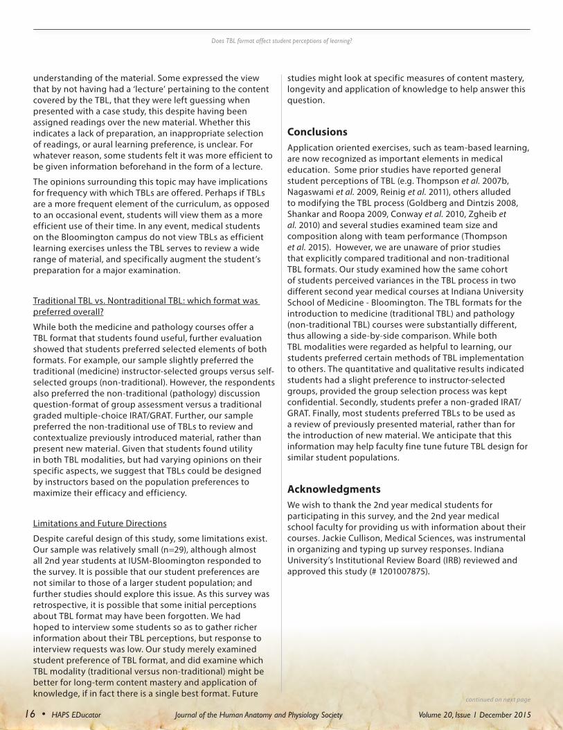
understanding of the material. Some expressed the view that by not having had a ‘lecture’ pertaining to the content covered by the TBL, that they were left guessing when presented with a case study, this despite having been assigned readings over the new material. Whether this indicates a lack of preparation, an inappropriate selection of readings, or aural learning preference, is unclear. For whatever reason, some students felt it was more efficient to be given information beforehand in the form of a lecture.
The opinions surrounding this topic may have implications for frequency with which TBLs are offered. Perhaps if TBLs are a more frequent element of the curriculum, as opposed to an occasional event, students will view them as a more efficient use of their time. In any event, medical students on the Bloomington campus do not view TBLs as efficient learning exercises unless the TBL serves to review a wide range of material, and specifically augment the student’s preparation for a major examination.
Traditional TBL vs. Nontraditional TBL: which format was preferred overall?
While both the medicine and pathology courses offer a TBL format that students found useful, further evaluation showed that students preferred selected elements of both formats. For example, our sample slightly preferred the traditional (medicine) instructor-selected groups versus self-selected groups (non-traditional). However, the respondents also preferred the non-traditional (pathology) discussion question-format of group assessment versus a traditional graded multiple-choice IRAT/GRAT. Further, our sample preferred the non-traditional use of TBLs to review and contextualize previously introduced material, rather than present new material. Given that students found utility in both TBL modalities, but had varying opinions on their specific aspects, we suggest that TBLs could be designed by instructors based on the population preferences to maximize their efficacy and efficiency.
Limitations and Future Directions
Despite careful design of this study, some limitations exist. Our sample was relatively small (n=29), although almost all 2nd year students at IUSM-Bloomington responded to the survey. It is possible that our student preferences are not similar to those of a larger student population; and further studies should explore this issue. As this survey was retrospective, it is possible that some initial perceptions about TBL format may have been forgotten. We had hoped to interview some students so as to gather richer information about their TBL perceptions, but response to interview requests was low. Our study merely examined student preference of TBL format, and did examine which TBL modality (traditional versus non-traditional) might be better for long-term content mastery and application of knowledge, if in fact there is a single best format. Future
studies might look at specific measures of content mastery, longevity and application of knowledge to help answer this question.
Conclusions Application oriented exercises, such as team-based learning, are now recognized as important elements in medical education. Some prior studies have reported general student perceptions of TBL (e.g. Thompson et al. 2007b, Nagaswami et al. 2009, Reinig et al. 2011), others alluded to modifying the TBL process (Goldberg and Dintzis 2008, Shankar and Roopa 2009, Conway et al. 2010, Zgheib et al. 2010) and several studies examined team size and composition along with team performance (Thompson et al. 2015). However, we are unaware of prior studies that explicitly compared traditional and non-traditional TBL formats. Our study examined how the same cohort of students perceived variances in the TBL process in two different second year medical courses at Indiana University School of Medicine - Bloomington. The TBL formats for the introduction to medicine (traditional TBL) and pathology (non-traditional TBL) courses were substantially different, thus allowing a side-by-side comparison. While both TBL modalities were regarded as helpful to learning, our students preferred certain methods of TBL implementation to others. The quantitative and qualitative results indicated students had a slight preference to instructor-selected groups, provided the group selection process was kept confidential. Secondly, students prefer a non-graded IRAT/GRAT. Finally, most students preferred TBLs to be used as a review of previously presented material, rather than for the introduction of new material. We anticipate that this information may help faculty fine tune future TBL design for similar student populations.
AcknowledgmentsWe wish to thank the 2nd year medical students for participating in this survey, and the 2nd year medical school faculty for providing us with information about their courses. Jackie Cullison, Medical Sciences, was instrumental in organizing and typing up survey responses. Indiana University’s Institutional Review Board (IRB) reviewed and approved this study (# 1201007875).
Does TBL format affect student perceptions of learning?
continued on next page

17 • HAPS EDucator Journal of the Human Anatomy and Physiology Society Volume 20, Issue 1 December 2015
References cited:AAMC Task Force on the Clinical Skills Education of
Medical Students. (2015). Recommendations for clinical skills curricula for undergraduate medical education. Association of American Medical Colleges. https://members.aamc.org/eweb/upload/Recommendations%20for%20Clinical%20Skills%20Curricula%202005.pdf [accessed 4 September 2015]
Bernard HR (2006) Research Methods in Anthropology: Qualitative and Quantitative Approaches. 4th Ed. Lanham, MD: Lanham: AltaMira Press, p. 492-515.
Bie RJ, Shapiro DL (1988) Voice and Justification: Their Influence on Procedural Fairness Judgments. Academy of Management Journal, 31(3): 676-685.
Branch WT (2001) Small-group teaching emphasizing reflection can positively influence medical student’s values. Acad Med 76:1171-1173.
Conway SE, Johnson JL, Ripley TL (2010) Integration of team-based learning strategies into a cardiovascular module. Am J Pharm Ed 74(2): p. 35.
Egan TM (2002) Grounded theory research and theory building. Adv Develop Hum Resour 4(3): 277-294.
Glaser BG, Strauss AL (1967) The Discovery of Grounded Theory: Strategies for Qualitative Research. 1st Ed. Piscataway, NJ: Aldine Transaction. 271 p.
Edwards JC, van Walsum K, Sanders CW, Fossum TV, Sadoski M, Bramson R, Wiprud RM (2004) Attitudes of veterinary medical students and medical students toward collaborative learning: An experiment. J Vet Med Educ 31:76-78.
Fink, LD, Parmelee, D (2008) Preface. In: Team-based learning for health professions education: A guide to using small groups for improving learning. 1st Ed (eds. Michaelsen L Parmelee D, McMahon K and Levine RE), pp. xi-xv, Sterling, VA: Stylus Publishing.
Goldberg HR, Dintzis R (2008) The positive impact of team-based virtual microscopy. Adv Physiol Ed 31:261-265.
Gopalan C, Fox DJ, Gaebalein CJ (2013) Effect of an individual readiness assurance test on a team readiness assurance test in the team based learning of physiology. Adv Physiol Ed 37, 61-64.
Janssen, HF, Skeen, NP, Bell, J, Bradshaw, W (2008) Improving Critical Thinking Skills in the Medical Professional with Team-Based Learning. In: Team-based learning for health professions education: A guide to using small groups for improving learning. 1st Ed (eds. Michaelsen L Parmelee D, McMahon K and Levine RE), pp. 61-73, Sterling, VA: Stylus Publishing.
Kennedy TJ, Lingard LA (2006) Making sense of grounded theory in medical education. Med Educ 40:101–108.
Koles PG, Stolfi A, Borges NJ, Nelson S, Parmelee DX (2010) The impact of team-based learning on medical students’ academic performance. Acad Med 85(11): 1739-1745.
Michaelsen, LK (1983). Team-learning in large classes. In: Learning in groups. New Directions in Teaching and Learning Series, No. 14 (eds. Bouton C and Garth RY), pp. 13-22, San Francisco, Jossey-Bass.
Michaelsen, LK and Sweet M (2008) Fundamental Principles and Practices of Team-Based Learning. In: Team-based learning for health professions education: A guide to using small groups for improving learning. 1st Ed (eds. Michaelsen L, Parmelee D, McMahon K and Levine, RE). pp. 9-34, Sterling, VA: Stylus Publishing.
Nider GL, Parmelee DX, Stolfi A, Hudes PD (2005) Team-Based Learning in a Medical Gross Anatomy and Embryology Course. Clin Anat 18: 56-63.
Oakley B, Felder MF, Brent R, Elhajj I (2004) Turning student groups into effective teams. Journal of Student Learning 2:9-34.
Parmelee, D. (2008) Team-Based Learning in Health Professions Education: Why is it a good fit? In: Team-based learning for health professions education: A guide to using small groups for improving learning. 1st Ed (eds. Michaelsen L, Parmelee D, McMahon K and Levine, RE). pp. 3-8, Sterling, VA: Stylus Publishing.
Parmelee D, DeStephen D, Borges NJ (2009) Medical students’ attitudes about team-based learning in a pre-clinical curriculum. Med Educ Online 14:1-7.
Reinig BA, Horowitz I, Whittenburg GE (2011) The effect of team-based learning on student attitudes and satisfaction. Decision Sciences: Journal of Innovative Educ. 9(1): 27-47.
Shankar N, Roopa R (2009) Evaluation of a modified team based learning method for teaching general embryology to 1st year medical graduate students. Indian J of Med Sci 63.1: 4-12.
Shellenberger S, Seale PJ, Harris DL, Johnson JA, Dodrill CL, Velasquez, MM (2009) Applying team-based learning in primary care residency programs to increase patient alcohol screenings and brief intervention. Acad Med 94:340-346.
Thomas PA, Bowen CW (2011) A controlled trial of team-based learning in an ambulatory medicine clerkship for medical students. Teach & Learn Med 23(1): 31-36.
Does TBL format affect student perceptions of learning?
continued on next page

18 • HAPS EDucator Journal of the Human Anatomy and Physiology Society Volume 20, Issue 1 December 2015
Thompson BM, Schneider VF, Haidet P, Levine RE, McMahon KK, Perkowski LC, Richards, BF (2007a) Team-Based Learning at Ten Medical Schools: Two Years Later. Med Educ 41, 250-257.
Thompson BM, Schneider VF, Haidet P, Perkowski LC, Richards BF (2007b) Factors influencing implementation of team-based learning in health sciences education. Acad Med 82: 553-556.
Thompson, BM, Haidet, P, Borges, NJ, Carchedi, LR, Roman, BJB, Townsend, MH, Butler, AP, Swanson, DB, Anderson, MP, Levine, RE (2015) Team cohesiveness, team size and team performance in team-based learning teams. Med Educ 49(4): 379-385.
Tucker K, Wakefield A, Boggis C, Lawson M, Roberts T, Gooch J (2003) Learning together: clinical skills teaching for medical and nursing students. Med Educ 37: 630-637.
Vasan NS, DeFouw DO, Holland BK (2008) Modified use of team-based learning for effective delivery of medical gross anatomy and embryology. Anat Sci Educ 1: 3-9.
Vasan, NS, DeFouw, DO, Compton, S (2009) A Survey of Student Perceptions of Team-Based Learning in Anatomy Curriculum: Favorable Views Unrelated to Grades Anat Sci Educ 2: 150-155.
Vasan NS, DeFoww DO, Compton S (2011) Team-based learning in anatomy: an efficient, effective, and economical strategy. Anat. Sci. Educ 4: 333-339.
Zgheib NK, Simaan JA, Sabra R (2010) Using team-based learning to teach pharmacology to second year medical students improves student performance. Med. Teach 32: 130-135.
Jane Waggoner is a graduate of the Medical Sciences Program at Indiana University Bloomington with an MS in Anatomy. She is currently a consulting analyst for the healthcare strategy team at Huron Consulting Group in Chicago, IL.
Mark Braun received his MD from Indiana University Medical School in 1975. He completed a residency in pathology and is board certified in both anatomical and clinical pathology.. He’s been teaching in the Medical Sciences Program, Indiana University, for more years than he can remember. His research and teaching interests are varied and range from the
study of infectious diseases introduced into Native American populations in the 16th century, to the application of computer assisted teaching in medical education.
Sarah Tieman received her MD from the Indiana University School of Medicine. She is board certified in internal medicine and pediatrics. She is a lecturer of internal medicine and pediatrics in the Medical Sciences Program, Indiana University, where she oversees the 3rd and 4th year medical school curriculum at IU School of Medicine - Bloomington.
Valerie O’Loughlin, Ph.D. is an Associate Professor of Anatomy in the Medical Sciences Program, Indiana University. She teaches anatomy and medical imaging to undergraduate students, gross anatomy to medical students, and mentors anatomy education MS and PhD students. She is president Emeritus of HAPS and currently is co-chair of the HAPS testing committee.
■
Does TBL format affect student perceptions of learning?
About the Authors
19 • HAPS EDucator Journal of the Human Anatomy and Physiology Society Volume 20, Issue 1 December 2015
Learning Muscle Anatomy in First Year: An Assessment of Active, Experiential and Passive Learning Activities
Zoë Soon, PhD1,4
Contributing Authors: Kaitlyn Carlson, B.H.K.1,4, Ngar Nie Neo2,4, and Heather Hurren, M.Ed3,4
1School of Health and Exercise Sciences, Faculty of Health and Social Development 2Psychology Dept., and the 3Centre for Teaching and Learning at the 4University of British Columbia Okanagan, 3333 University Way, Kelowna, BC, Canada, V1V 1V7
Corresponding Author: Zoë Soon, ART166, SHES, FHSD, UBCO, 3333 University Way, Kelowna, BC, Canada, V1V 1V7; Tel. 250-807-9400; Fax. 250-807-9865
Abstract This study compared the effectiveness of learning muscle anatomy through six different lab activities: clay model building, a plastic muscle model scavenger hunt, fill-in-the blank diagrams in a laboratory manual, “selfies”, study hand-outs, and memory charts. The relative effect of each activity on knowledge acquisition, including short-term and long-term memory, was examined. In addition, possible correlations between pre- and post-test scores with respect to student background knowledge in science, individual learning styles (visual, aural, read-write, or kinesthetic), and gender differences, were analyzed. Students were also surveyed for their enjoyment of, and engagement with, each activity. The order of activity completion was evaluated to assess its possible benefits to student learning and student preference. Overall, it was hypothesized that all activities would prove beneficial to all students, regardless of prior knowledge or gender. However, it was anticipated that kinesthetic learners would prefer and receive the most benefit from activities that were more active and experiential such as clay model building and the scavenger hunt. Specifically, these two activities involve partner work, with peer-teaching and learning qualities that might also be valued by aural leaners. It was anticipated that the visual and read/write learners would prefer and receive the most benefit from the independent diagram labeling activity and the rote-memorization required with the “Muscles to Know” chart.
Key words: first-year anatomy and physiology, lab activities, multiple activity instructional design, activity order, Tiny Tim, modeling muscles in clay, plastic model scavenger hunt, “selfies”, skeletal muscle anatomy, background knowledge in science, learning styles, gender differences, sustainability.
IntroductionDuring a Human Anatomy and Physiology Society (HAPS) conference workshop in May 2012, “Feets of Clay” was demonstrated to an appreciative audience by Sandy Zetlan, PhD, and Marsha Segerberg, PhD, both from Estrella Mountain Community College. The workshop presented ideas for using inexpensive clay and plastic Tiny Tim skeletons to learn human skeletal muscles. Very generously, Sandy and Marsha have made all of their resources (ample clay-building pictures and accompanying Tiny Tim clay flashcards) freely available to the public on the Zetlan and Segerberg “Feets of Clay” website. Indeed, clay has been used for several years in teaching various aspects of anatomy because of the potential for hands-on student engagement and learning. As both Monsour and Shipley point out, there are clay kits readily available for purchase that can be used to study not just muscle, but bones, neural pathways, and many other organ systems. These kits are used successfully in high school, college, and university
courses (Monsour 2011, Shipley 2010). With the availability of Tiny Tim skeletons and on-line resources, learning skeletal muscles using clay model building is an attractive, low-budget option for classes of any size.The rationale for implementing a clay model building activity in a first-year anatomy and physiology course at the University of British Columbia (UBCO) hinged on the suspician that UBCO human kinetics students would particularly enjoy hands-on learning and that the learning potential for a clay model building activity would be very high. Furthermore, in the anatomy and physiology laboratory space, which is designed for labs that are environmentally friendly and sustainable, the addition of an inexpensive, non-toxic, non-waste, re-usable clay model building activity would be a good fit. This research study was designed to assess student response to a clay model building activity and the effectiveness of this activity in comparison with methods previously used in this course (plastic muscle models and labeling muscle diagrams in a laboratory manual) to facilitate the learning of human muscle anatomy.
continued on next page

20 • HAPS EDucator Journal of the Human Anatomy and Physiology Society Volume 20, Issue 1 December 2015
Current literature includes several papers that have studied the employment of clay in anatomical labs. Myers et al. (2001) introduced the use of clay to help teach pelvic anatomy to groups of obstetrics and gynecology post-graduate residents. In doing so, Myers et al. (2001) found that clay modeling, in conjunction with lecturing, was more effective than lecture alone for teaching pelvic floor anatomy. Specifically, a two-hour clay modeling session was enough to improve student scores in the short-term as well as in the long-term, 8 weeks later (Myers et al. 2001).
In several more studies, clay modeling has been compared to dissection in the learning of human muscles. Motoike et al. (2009) found that building muscles out of clay was more effective than cat dissection in learning human muscles when assessed by answering lower-order questions. Interestingly, the ability to answer higher-order questions was not affected. Similar higher-order assessment scores were observed from the group of students who had dissected cats and from the group of students who had used human muscle modeling in clay as a basis for study. In discussion of these results, Motoike et al. (2009) point out that the advantages of cat dissection include: first-hand visualization of the fascia wrapped around each muscle and the ability to view anatomical entities such as aponeuroses and the actual and relative thickness of each muscle. This study also mentions several advantages of clay building which include: the ability to see specific muscle attachment points, which are not readily visible in cat dissection, and the ability to focus on human muscles as opposed to cat muscles, some of which are quite different.
At Pennsylvania State University, Waters et al. (2011) studied students who were split into groups with some performing cat muscle dissection and some performing human clay sculpting within the allotted lab time. Waters et al. found that the students who dissected cats in lab performed better on higher-order cat anatomy questions while the students who studied muscles by clay modeling performed better on higher-order human anatomy questions. Waters et al. (2011) theorize that these results are perhaps intuitive since students will naturally perform better on an exam that tests the material with which they have the most familiarity. Waters et al. (2011) also found that in addition to the decreased degree of translational knowledge evidenced by the students taught muscles using clay models on human skeletons, the clay modeling students may have been exposed to a simpler muscle representation, which may have made it easier to learn and/or easier to memorize the muscles (Waters et al. 2011).
Goodwyn and Salm (2007) found that compared to dissection, building clay muscles from deep to superficial gives students a clearer sense of the groupings, synergists and antagonisms of muscles. In addition, DeHoff et al. (2011) found that students who were taught muscles, peripheral nerves, and blood vessels using clay modeling instead of cat dissections rated their lab activity higher in enjoyment
levels, scored the same on higher-order questions, and actually scored higher on low-order questions for peripheral nerves compared to students taught with cat dissections. It is important to note that both lower and higher-order questions in DeHoff’s study related to human anatomy and physiology as opposed to cat dissection. Interestingly, Haspel et al. (2013) found that students also scored higher on human anatomy exam questions when taught with clay as opposed to cat dissection, however, the students did not like the clay activity in the course that was studied. Haspel et al. (2013) speculated that the students in this course were idealizing the cat dissections that they were no longer allowed to do. In another study, Bareither et al. (2013) divided 39 graduate students into three groups (clay activity, worksheet activity and control) and compared their pre-test and post-test scores. They attempted to make correlations to the students’ VARK (Visual, Aural, Read/Write, and Kinesthetic) scores. However, the findings of this study are limited due to: a) the low number of students, and b) the fact that the clay activity group and worksheet activity group would certainly have an advantage over the control group (that had no additional learning tools) in mastering the material for the muscle unit.
In summation, it might be expected that a human muscle activity might better prepare the students for a test on human muscles. Likewise, students prepared with dissection activities tend to do better on dissection style tests. Most studies agree that there are definite advantages to both dissection and clay model building when learning the skeletal muscles of the human body. In particular the group work and hands-on nature of both activities certainly enhance learning by lecture, textbook, lab manual and any type of worksheet activity. As one might expect, several studies have shown that learning, particularly in science and medicine, is heightened by using small collaborative groups of students (Tanner et al. 2003, Theoret et al. 2007).
IntroductionSeveral newly designed activities were created and implemented in the muscle anatomy unit of our first-year anatomy and physiology course in the human kinetics program. The new activities included: clay building on Tiny Tim skeletons, a plastic model scavenger hunt, labeling muscle diagrams in a laboratory manual, the use of student “selfies”, a “Muscles to Know chart” requiring rote memorization, and various study handouts that accompanied all of the other laboratory activities. In incorporating these activities, the following questions were addressed:
1) Which activity is the most effective in enriching our students’ learning of muscle anatomy?
2) Is the use of multiple activities for one topic beneficial? Are six new activities necessary or are six new activities too many?
Learning Muscle Anatomy in First Year: An Assessment of Active, Experiential and Passive Learning Activities
continued on next page

21 • HAPS EDucator Journal of the Human Anatomy and Physiology Society Volume 20, Issue 1 December 2015
3) Does the order in which activities are performed have any effect on how well the topics are learned?
4) Is student understanding and/or working short-term memory better enhanced by one of the activities than another?
5) Are there any other factors that might play a role in the success of the new muscle anatomy activities:
i) Does student background knowledge in science and math correlate with student success on any or all of the new muscle anatomy activities? Does student background knowledge correlate with any of their course exam grades or with the final grade in the course?
ii) Does student primary learning strength, as assessed with the VARK survey, correlate with student success on any of the muscle anatomy activities?
iii) How do first year human kinetics students’ VARK profiles compare to general student and teacher populations? Do students remember or believe in their VARK profile strengths after being incorporated into beginning-of-term discussions regarding metacognition? Will kinesthetic and aural learners benefit more from the hands-on group clay and plastic model activities? Will read/write learners prefer labeling muscle diagrams and studying muscle chart hand-outs? Will visual learners use their cell phones more to create photos of the muscles to study from? Will these muscle model “selfies”, cell phone pictures taken of plastic and clay models, be popular for all students as take-home study tools?
vi) Does student success on any of the new muscle anatomy activities correlate with success on similar questions in the lab exam indicating long-term memory gains?
v) How do students view each of these new activities in terms of: enjoyment, value, importance for learning, difficulty level and usefulness as a study resource?
vi) Are there any gender differences in activity preference?
METHODSCourse Description
This study was approved by the University of British Columbia Behavioral Research Ethics Board and was based on the performance of 162 students enrolled in eight laboratory sections of a first-year anatomy and physiology course (HMKN190) taught during the fall 2013 semester in the School of Health and Exercise Sciences at the
University of British Columbia, Okanagan. The course has no prerequisites, and the students were enrolled in the Human Kinetics program. The vast majority of students were 18-20 years of age. Students enrolled in this courses attend a lecture section that met 3x50min/wk and a laboratory section that met 1x2hr/wk. Individual laboratory sections were capped at 20 students and were taught by graduate student teaching assistants. All teaching assistants (TAs) were supervised by and met weekly with the lab coordinator and the course instructor. The grant sponsor for this study was an Innovations in Teaching and Learning Research Grant. The grant number is F14-00354 and the ethics certification is H12-02144.
Research Method Crossover Design – Term 1, Sept.-Dec. 2013 - Lab Setup Details:
It was desired that all students were given the chance to complete all three main lab activities (clay-building, plastic model scavenger hunt, and lab diagrams with handout charts) in addition to all of the accompanying study hand outs and “selfies” opportunities. To this end, a cross-over design was created which would allow students to complete all of the activities and it would be possible to study the implementation order of each of these methods at the same time. Half of the students completed the clay-building in the first week, and half of the students completed the plastic model scavenger hunt activity in the first week. In the second week, the groups of students switched activities. The supplemental lab manual diagrams and handout charts were available to all students throughout the entire two weeks. In-class time was provided for the diagram labeling activity during the plastic model scavenger hunt activity session. Student learning was monitored through pre- and post-tests of the clay model and plastic model activities. These tests were also used to assess which activity was more effective, or if both were required for complete mastery. If both the clay modeling and the plastic model activities were necessary for complete mastery, we wanted to determine the ideal order of these activities. The specific timetable of these events was as follows:
Wk 1: 166 students were given a chance to fill in consent forms to participate in the study. The subsequent data collected from the 162 consenting students was used for this study.
Wk 2: Students wrote the VARK survey (Fleming 2014) and the “Get Ready for A&P” background science test (Garrett 2012). These tests were not taken for marks but students were highly encouraged to take them and ample time was set aside during lab time for each test. TAs felt that students treated both tests seriously. Lecture time was set aside for a discussion about VARK, metacognition, and a range of study tools and tips for taking the tests.
Learning Muscle Anatomy in First Year: An Assessment of Active, Experiential and Passive Learning Activities
continued on next page

22 • HAPS EDucator Journal of the Human Anatomy and Physiology Society Volume 20, Issue 1 December 2015
Wk 9: Students were given an activity-specific pre-test on muscle names, locations, origins and insertions. Then in four of the eight lab sections, students were given muscle diagrams and muscle chart handouts that detailed muscle name, location, origin and insertion. Students in these four lab sections participated in the plastic models activity, which consisted of finding and exploring each muscle on several plastic models (Somso and 3-B) in a hands-on manner. In the other four lab sections, the students created the same muscles out of clay on small Tiny Tim skeletons. At the end of the lab week nine these students wrote an activity-specific post-test. The activity-specific pre- and post-test for students completing the plastic model activity consisted of fill-in-the-blank labeling of specific muscles highlighted on cadaver pictures that were similar to the plastic models used. The activity-specific pre- and post-test for students completing the Tiny Tim clay building activity consisted of fill-in-the-blank labeling Tiny Tim pictures with specific clay muscles built on. During lab time, cell-phone and iPad picture taking or “selfies” of models were encouraged by TAs. It should be noted that at the end of term, some students regretted not taking “selfies” and asked the course instructor to do this for them and post labelled pictures of the plastic models on to Blackboard Connect.
Wk 10: Labs are cancelled due to Nov. 11 Remembrance Day university closure.
Wk 11: Students switched lab activities performed in week 9, writing the respective pre- and post- test for each activity (clay muscles and plastic muscles).
Wk 12: Students wrote a lab exam that had questions pertaining to all muscle unit activities, and filled out student satisfaction surveys on muscle activities.
Testing and Correlational Analysis
Our goal was to compare the effectiveness of learning major muscles through three different types of lab activities (Tiny Tim clay model building, plastic muscle models, and labeling muscle diagrams in the manual) each of which included a supplemental study hand-out and a “Muscles to Know” chart depicting muscle name, location, origin, insertion and action. Student learning gains for each activity were assessed using a pre-test and a post-test. The pre-test and post-test for these activities consisted of lower-order questions focusing on identifying muscle names, locations, origins, insertions, and actions.
The pre-test and post-test was specifically designed for each activity. For example, the clay building exercise pre-test consisted of 20 pictures of Tiny Tim clay muscles with accompanying questions. This pre-test was given to
the students at the beginning of lab before they did the clay building activity. Each picture in the test had one clay muscle built on a Tiny Tim skeleton. Students were required to fill in the following: a) the muscle name, b) the origin of the muscle, including the specific bone and/or bone marking, c) insertion of muscle, including the specific bone and/or bone marking, and d) the action of muscle using words such as extend, flex, abduct, adduct, medially rotate along with the name of the bone being moved. The test was worth 80 points and partial credit was given for partial answers. The post-test for the clay building exercise was the same test. The students were handed back their pre-test at the end of the clay modeling activity and asked to fill in all the questions they had left blank in the pre-test and now supposedly had learned. Both tests were “closed book” and performed in silence with students given as much time as they needed. In this manner learning gains and short-term retention of the material was measured.
The pre-test for the plastic model and lab muscle diagrams exercise consisted of 20 labeling questions with the same lab manual diagrams, which included both drawings and cadaver pictures. The students were required to fill in the following: a) the muscle name, b) the origin of muscle, including the specific bone and/or bone marking, c) the insertion of muscle, including the specific bone and/or bone marking, and d) the action of muscle using words such as extend, flex, abduct, adduct, medially rotate along with the name of the bone being moved. The test was worth 80 points and partial credit was given for partial answers. The post-test for this exercise was the exact same test. Students were handed back their pre-test and labeling activities at the end of the plastic model scavenger hunt and asked to fill in all the blanks that they had now learned. Both pre- and post-tests were “closed book” and performed in silence with students given as much time as they needed.
As this was a research study, both pre- and post-tests were optional and ungraded but the labTAs were instructed to tell students that the tests would be good practice for the final lab exam. In addition, the TAs announced that prizes (decks of A&P cards) would be given to the highest scoring students. The TAs reported that the vast majority of students participated in all pre- and post-tests.
During data analysis, the following questions were addressed:
1) Which activity is the most effective for our students and does the order of delivery have any effect?
2) Does a student’s science background affect success of any of the muscle anatomy activities in this lab? On course exams? On final grade for the course?
3) Does VARK learning style affect student success on any particular activity in the muscle anatomy unit? Do human kinetics students have different VARK strengths than the general students or teacher populations?
Learning Muscle Anatomy in First Year: An Assessment of Active, Experiential and Passive Learning Activities
continued on next page

23 • HAPS EDucator Journal of the Human Anatomy and Physiology Society Volume 20, Issue 1 December 2015
4) Does student success on any particular muscle anatomy activity correlate with subsequent success on exam questions drawn specifically from that activity?
5) How do students view each of the muscle anatomy activities in terms of enjoyment, value and importance to learning and difficulty level?
Among the anticipated outcomes, it was also hypothesized that students with higher marks on the science background knowledge test would have higher marks in the pre-tests for this unit’s activities as well as the course exams.
RESULTS AND DISCUSSIONAssessment of Students’ Background Knowledge
The background knowledge test consists of 88 questions on high school Math and Science (incl. Anatomy, Cell Biology, & Chemistry) selected from the “Get Ready for A & P” test (Garrett, 2012). 139 participating students took the test scoring an average of 78% within a range 18-94% (Figure 1). As anticipated, it became clear that students coming into the Human Kinetics undergraduate program have a very diverse level of background knowledge in math and science.
Figure 1: Distribution of background science and math test scores for first-year human kinetics students (n=139) on the first day of lab.
Assessment of VARK (Visual, Auditory, Read/Write, Kinesthetic) Learning Styles
The 16 question VARK questionnaire designed by Neil Fleming gives students an idea of their primary learning style, which enables students to tailor their studying habits to capitalize on their strengths (Fleming and Baume 2006). In this course, discussion of VARK has recently been used to stimulate student metacognition and to provide students with ideas, at the beginning of term, for study tools and
strategies. In the VARK survey, students who are designated as visual learners are thought to prefer to draw ideas and learn using symbolic/graphic forms (graphs, charts, logos, diagrams, outlines, and mind maps, but not necessarily videos) (Fleming 2014, Fleming and Baume 2006). Aural learners are thought to master content by listening and speaking. Read/Write learners are believed to prefer visual text and those with a kinesthetic learning style have been found to favor case studies, real-life applications, hands-on materials (including computer work and videos) and role-playing (Fleming 2014, Fleming and Baume 2006). It should be noted that most people are multi-modal to some degree and that in addition to using their primary learning style, students are thought to draw upon all of their strengths and inclinations when learning new material. A comparison found that our cohort of first-year Human Kinetics students were more likely to have kinesthetic or aural primary learning styles than reported previously in teacher and student populations as shown in Figure 1 (Fleming 2014). 16% of our group of students were found to have a visual primary learning style, 24% have a read/write primary learning style, 29% have an auditory primary learning style and 31% have a kinesthetic primary learning style (Figure 2).
Figure 2: VARK questionnaire results of 129 first-year human kinetics students was compared to the general student and teacher population results recorded by Neil Fleming (Fleming, 2013).
Results and Correlational Analysis of Background Knowledge Test and Primary Learning Styles
Results of the “Get Ready for A&P” background science test are shown for the whole class as well as broken down by primary learning style in Figure 3. As one might expect, there was no correlation found between students’ “Get Ready for A&P” score and their individual primary VARK learning style (Table 1). Thus, this result indicates that a student’s learning style has no impact on how well they perform on a background science and math test.
Learning Muscle Anatomy in First Year: An Assessment of Active, Experiential and Passive Learning Activities
continued on next page

24 • HAPS EDucator Journal of the Human Anatomy and Physiology Society Volume 20, Issue 1 December 2015
Figure 3: Primary learning style and average score on science background test for participating first-year human kinetics students (n=112). The data from all consenting students who completed both the test and the VARK survey were used. Please note that some students had “ties” for their top learning style score, in which case they were designated as having two primary learning styles (e.g.visual and auditory). For comparison purposes, the average result for all participating students in the “Getting Ready for A&P test” is shown in the final bar in the graph (n=139).
Table 1. Correlation analysis between “Get Ready for A&P test” scores and students’ primary learning style. No significant correlations were found.
Primary Learning Style Number of Students Pearson Correlation between Learning Style & Science Background test score
Visual 13 r = 0.079, p = 0.407 sig. (2-tailed)
Auditory 25 r = 0.076, p = 0.422 sig. (2-tailed)
Read/Write 22 r = -0.119, p = 0.210 sig. (2-tailed)
Kinesthetic 28 r = 0.131, p = 0.167 sig. (2-tailed)
Visual:Auditory 3 sample size too small
Visual:Read/Write_ 2 sample size too small
Visual:Kinesthetic 6 sample size too small
Auditory:Read/Write 4 sample size too small
Auditory:Kinesthetic 4 sample size too small
Read/Write:Kinesthetic 5 sample size too small
Total: 112 sample size too small
Activity Order and Learning Gains Resulting From Each Activity
Noticeably, the students’ scores kept improving with each activity stage during the two-week muscle unit no matter which way the order of activities was performed. This suggests that the completion of all activities was valuable and pertinent to learning (Figure 4). However, students showed the greatest learning gains when they completed the Tiny Tim clay building in the first week and the plastic model scavenger hunt and lab manual diagrams in the
second week (Figure 4). During the Tiny Tim clay building, Tiny Tim flashcard stations were prepared by placing laminated flashcards on tables around the lab so that the students could move from table to table, building up the muscles on the Tiny Tim. The flashcards were printed from the “Feets of Clay” website provided by Zettan and Segerber (2014) and they are excellent in that on the front side, they portray how to build one muscle and on the back side, they list the specific details of that muscle (e.g. origin, insertion, action).
Learning Muscle Anatomy in First Year: An Assessment of Active, Experiential and Passive Learning Activities
continued on next page

25 • HAPS EDucator Journal of the Human Anatomy and Physiology Society Volume 20, Issue 1 December 2015
Figure 4: Pre-test and post-test activity scores, depicting the learning gains of students as a result of the plastic scavenger hunt, diagram labeling and Tiny Tim clay building activities.
Interestingly, and as one might expect, the data confirms that for the nine students who did not attend and participate in Week 1, their scores were lower on the pre- and post-test for Week 2 ( = 19.18 and = 26.59 respectively compared to the students who attend both weeks ( = 30.41 and = 40.59 for pre- and post-tests respectively). Therefore this data again indicates that attendance and participation in all activities was beneficial and that knowledge gained from the Tiny Tim clay building activity was useful and easily translated to the plastic model scavenger hunt activity.
VARK Learning Style and Correlation to Learning Gains from Each Activity
There were no correlations found between an individual’s VARK learning style and lab activity pre- or post-test performance (Tables 2a and 2b). This indicates that all lab stages (Tiny Tim, plastic muscle models and diagrams) were effective study resources for all types of learners.
Table 2a and b. Pearson correlational analysis (sig. 2-tailed) between students’ VARK learning styles and their learning gains, as a result of each stage of the Tiny Tim clay and plastic muscle model scavenger hunt activities. Learning gains were assessed by activity specific pre-test and post-tests. No significant correlations were found.
a) Results for Lab Sections 1-4. These students completed the plastic model scavenger hunt and muscle diagram labeling activities in Week 1 and the Tiny Tim clay building activity in Week 2:
Stages of muscle unit lab Number of Students
Correlation between students’ learning gains during muscle lab stages and primary learning style
Visual Auditory Read/Write Kinesthetic
1. Before Lab #1 (pre-test score before the Plastic model & diagram activity)
71 r=0.188, p=0.182
r=-0.049, p=0.728
r=-0.141, p=0.317
r=0.021, p=0.885
2. Gain from Lab #1 (Difference between post-test scores and pre-test score)
66 r=0.030, p=0.838
r=-0.096, p=0.511
r=0.083, p=0.569
r=-0.014, p=0.927
3. End of Lab #1 (post-test score) 68 r=0.137, p=0.339
r=-0.097, p=0.497
r=-0.052, p=0.716
r=0.021, p=0.883
4. Before Lab #2 (pre-test score before the Tiny Tim clay building activity)
62 r=0.065, p=0.660
r=0.131, p=0.375
r=-0.046, p=0.754
r=-0.029, p=0.846
5. Gain from Lab #2 (difference between post-test and pre-test scores)
52 r=-0.021, p=0.899
r=0.010, p=0.951
r=0.084, p=0.608
r=-0.069, p=0.674
6. End of Lab #2 (post-test score) 62 r=-0.021, p=0.887
r=0.077, p=0.604
r=-0.074, p=0.620
r=0.014, p=0.926
Learning Muscle Anatomy in First Year: An Assessment of Active, Experiential and Passive Learning Activities
continued on next page

26 • HAPS EDucator Journal of the Human Anatomy and Physiology Society Volume 20, Issue 1 December 2015
b) Results for Lab Sections 5-8. These students completed the Tiny Tim clay building activity in Week 1 and the plastic model scavenger hunt and muscle diagram labeling activities in Week 2:
Stages of muscle unit labNumber
of Students
Correlation between students’ learning gains during muscle lab stages and primary learning style
Visual Auditory Read/Write Kinesthetic
1. Before Lab #1 (pre-test score before the Tiny Tim clay building activity)
65 r=0.160, p=0.256
r=-0.110, p=0.438
r=0.005, p=0.971
r=-0.067, p=0.639
2. Gain from Lab #1 (difference between post-test and pre-test scores)
61 r=-0.028, p=0.849
r=-0.018, p=0.903
r=0.025, p=0.865
r=0.030, p=0.837
3. End of Lab #1 (post-test score) 65 r=0.150, p=0.288
r=-0.048, p=0.734
r=0.000, p=0.997
r=-0.121, p=0.391
4. Before Lab #2 (pre-test score before the plastic models & diagrams)
74 r=0.066, p=0.622
r=-0.200, p=0.133
r=0.128, p=0.340
r=0.027, p=0.842
5. Gain from Lab #2 (difference between the post-test and pre-test scores)
67 r=-0.103, p=0.455
r=0.255, p=0.060
r=-0.235, p=0.084
r=0.065, p=0.639
6. End of Lab #2 (post-test score) 74 r=0.022, p=0.867
r=-0.082, p=0.543
r=0.008, p=0.951
r=0.062, p=0.644
Background Knowledge Test and Correlation to Learning Gains from Each Activity
Background knowledge is another factor which may contribute to student success on pre-test and post-test scores during the muscle anatomy unit in lab. Upon analysis, it was found that there were no correlations between an individual’s “Get Ready for A&P” test performance and their learning gains from each muscle activity in lab for Lab Sections 1-4 (Tables 3a and 4b). This indicates that all of the main activities (Tiny Tim, plastic muscle models and diagrams) were effective study resources for learners with different science and math background knowledge. A single significant correlation did exist within the Lab Sections 5-8 cohort, in which their first pre-test scores did positively correlate with scores on the background science and math test (r=0.371, p=0.003). This data is certainly understandable as this cohort’s previous knowledge could easily be beneficial on a pre-test, in which the material had not yet been taught in the current course. What is striking however, is that background knowledge ceased to give these students an edge as they progressed through the 2 week unit. This suggests that their learning of the material during those 2 weeks and short-term memory contributed more strongly to scores on post-tests than previous knowledge did.
Tables 3a and b. Pearson correlational analysis between background knowledge test scores and learning gains as a result of each stage of the Tiny Tim clay and plastic muscle model activities. Significant correlation is highlighted in blue.
a) Lab Sections 1-4 (L1-4):
Stages of muscle unit lab Number of StudentsCorrelation between Background Science Test scores and scores during Muscle lab Activity Stages
1. Before Lab #1 (plastic models & diagrams) 71 r=0.137, p=0.2832. Gain from Lab #1 66 r=0.215, p=0.0993. End of Lab #1 68 r=0.132, p=0.1954. Before Lab #2 (Tiny Tim) 62 r=0.206, p=0.1325. Gain from Lab #2 52 r=0.024, p=0.8736. End of Lab #2 62 r=0.212, p=0.121
Learning Muscle Anatomy in First Year: An Assessment of Active, Experiential and Passive Learning Activities
continued on next page

27 • HAPS EDucator Journal of the Human Anatomy and Physiology Society Volume 20, Issue 1 December 2015
b) Lab Sections 5-8 (L5-8):
Stages of muscle unit lab Number of StudentsCorrelation between Background Science Test scores and scores during Muscle lab Activity Stages
1. Before Lab #1 (Tiny Tim) 65 r=0.371, p=0.003** sig. at the 0.01 level (2-tailed)
2. Gain from Lab #1 61 r=-0.249, p=0.064
3. End of Lab #1 65 r=0.172, p=0.188
4. Before Lab #2 (plastic models & diagrams) 74 r=0.161, p=0.201
5. Gain from Lab #2 67 r=0.065, p=0.620
6. End of Lab #2 74 r=0.172, p=0.171
Longer-term Retention of Knowledge as a Result of Each of These Three Activities
In terms of longer-term retention as measured by lab exam II (which was 1 week later, due to the proximity of the muscle unit to the end of term), there were few correlations between pre-test, post-test and lab exam scores on Tiny Tim, plastic model, and/or diagram specific questions (Tables 4a, b, c, and d). It is likely that the amount of at-home studying, motivation, and perhaps choice to participate in an optional open lab review on the Friday prior to the lab exam played a larger role in lab exam scores for questions on muscle anatomy. The two correlations that were significant included:
a) a positive correlation of r=0.342 (p=0.020) between students’ learning gains during the Tiny Tim clay building activity and their marks on Tiny Tim muscle-specific questions during the lab exam (in Lab Sections 1-4). Thus, the more the students improved on their muscle anatomy knowledge during the Tiny Tim clay building activity, the better they scored on these types of questions during the exam.
b) a positive correlation of r=0.256 (p=0.048) was found between students’ learning gains during the plastic model scavenger hunt activity and their marks on plastic model muscle-specific questions during the lab exam (in Lab Sections 5-8). So, the more the students improved on their muscle anatomy knowledge during the plastic model activity, the better they scored on these types of questions during the exam.
These two correlations indicate that the final activity for each lab cohort proved to be the most beneficial for answer associated exam questions, perhaps demonstrating the final mastering activity translates to longer or more accurate memories of the topic.
Tables 4a, b, c, and d. Pearson correlational analysis between learning gains as a result of each stage of the muscle activities and subsequent lab exam question scores. Please note that before lab indicates the pre-test score; gain indicates difference between post-test and pre-test; and end of lab indicates post-test score. Significant correlations are highlighted in blue.
a) Lab Sections 1-4:Stages of muscle unit lab Number of Students Correlation between muscle lab stage & Tiny Tim
Q score on Lab Exam II
1. Before Lab #1 (plastic models & diagrams) 71 r=-0.145, p=0.264
2. Gain from Lab #1 66 r=-0.033, p=0.810
3. End of Lab #1 68 r=0.003, p=0.982
4. Before Lab #2 (Tiny Tim clay activity) 62 r=0.058, p=0.676
5. Gain from Lab #2 52 r=0.342, p=0.020* sig. at the 0.05 level (2-tailed)
6. End of Lab #2 62 r=0.247, p=0.071
Learning Muscle Anatomy in First Year: An Assessment of Active, Experiential and Passive Learning Activities
continued on next page

28 • HAPS EDucator Journal of the Human Anatomy and Physiology Society Volume 20, Issue 1 December 2015
b) Lab Sections 5-8:Stages of muscle unit lab Number of Students Correlation between muscle lab stage & Tiny Tim
Q score on Lab Exam II
1. Before Lab #1 (Tiny Tim) 65 r=0.171, p=0.191
2. Gain from Lab #1 61 r=-0.081, p=0.551
3. End of Lab #1 65 r=0.107, p=0.415
4. Before Lab #2 (plastic models & diagrams) 74 r=-0.135, p=0.285
5. Gain from Lab #2 67 r=0.072, p=0.584
6. End of Lab #2 74 r=-0.018, p=0.888
c) Lab Sections 1-4:Stages of muscle unit lab Number of Students Correlation between muscle lab stage & Plastic
model Q score on Lab Exam II
1. Before Lab #1 (plastic models & diagrams) 71 r=0.008, p=0.950
2. Gain from Lab #1 66 r=0.255, p=0.058
3. End of Lab #1 68 r=0.180, p=0.176
4. Before Lab #2 (Tiny Tim) 62 r=0.102, p=0.464
5. Gain from Lab #2 52 r=0.203, p=0.176
6. End of Lab #2 62 r=0.215, p=0.119
d) Lab Sections 5-8:Stages of muscle unit lab Number of Students Correlation between muscle lab stage & Plastic
model Q score on Lab Exam II
1. Before Lab #1 (Tiny Tim) 65 r=0.127, p=0.334
2. Gain from Lab #1 61 r=0.079, p=0.560
3. End of Lab #1 65 r=0.191, p=0.144
4. Before Lab #2 (plastic models & diagrams) 74 r=-0.040, p=0.749
5. Gain from Lab #2 67 r=0.256, p=0.048* sig. at the 0.05 level (2-tailed)
6. End of Lab #2 74 r=0.074, p=0.556
Results and Correlational Analysis of Background Knowledge Test and All Course Exams
After the previous analyses determining whether lab activity order or performance correlated with specific lab exam questions, we wondered whether any other factors might play a role overall. It was therefore of interest to see whether a student’s background knowledge in science and math correlated with any or all of the exam scores within this first year anatomy and physiology term one course. In the analysis, it was apparent that the exams on lecture material (Midterm I, Midterm II, and the Final Exam) as well as the second lab exam did correlate with results scored on “Get Ready for A&P test”. Of note, the muscle unit was tested in Midterm II and Lab Exam II. This data is of course quite limited and it may be that the primary factors of success on these exams are dependent on a students’ background knowledge as well as, or to a greater degree, their study skills, motivation, as well as host of personal factors, possibly in addition to student’s participation and achievements in lab activities, including these muscle unit activities.
Learning Muscle Anatomy in First Year: An Assessment of Active, Experiential and Passive Learning Activities
continued on next page

29 • HAPS EDucator Journal of the Human Anatomy and Physiology Society Volume 20, Issue 1 December 2015
Table 5. Pearson correlational analysis between students’ background knowledge test score and all course exams scores throughout the term, including their final course mark. Significant correlations are highlighted in blue.
Course Exams Number of StudentsCorrelation between Background Science Test scores and scores for all Course Exams and Final Course Mark
1. Midterm I 139 r=0.451, p=0.000** sig. at the 0.01 level (2-tailed)
2. Midterm II 138 r=0.335, p=0.000** sig. at the 0.01 level (2-tailed)
3. Lab Exam I 139 r=0.119, p=0.162
4. Lab Exam II 137 r=0.231, p=0.007** sig. at the 0.01 level (2-tailed)
5. Final Exam 137 r=0.393, p=0.000** sig. at the 0.01 level (2-tailed)
6. Final Course Mark 138 r=0.392, p=0.000** sig. at the 0.01 level (2-tailed)
Student Survey Results
Of the main three activities (muscle diagram labeling, plastic model exploration and Tiny Tim clay building, the survey results indicate that students enjoyed the Tiny Tim clay building activity the most (48% of students in lab sections 1-4 and 59% of students in lab sections 5-8) (Table 6). The plastic model activities was rated the second most enjoyable by the majority of students from all lab sections.
In addition to the survey data on student enjoyment levels, perceived level of detail, value, and difficulty of the three activities were rated (Table 6). Students chose the plastic models and muscle diagrams as being the most valuable and as providing the most detail for studying. This may be due to the fact the diagrams could be taken home and studied, and the students knew that there were more marks on their lab exam for the plastic models stations (16 marks) than the Tiny Tim clay models (4 marks). This likely would have had an impact on their answer.
Table 6. Student Satisfaction Survey Responses after Muscle Anatomy Unit. Results are depicted by Lab Section. Lab Sections 1-4 completed the plastic model scavenger hunt and muscle diagram labelling activities in Week 1. Lab Sections 5-8 completed the Tiny Tim clay building activity in Week 1. Most popular choices are highlighted in blue.
Muscle Diagram Plastic Model Tiny TimLab Section: L1-4 L5-8 L1-4 L5-8 L1-4 L5-8
Total Number of Responders 60 56 60 56 60 56
Activity Order: Week 1 Week 2 Week 1 Week 2 Week 2 Week 1
Most enjoyable activity 10% 11% 42% 30% 48% 59%
2nd most enjoyable activity 33% 29% 35% 55% 32% 16%
Most valuable activity to learning 25% 39% 47% 38% 28% 23%
Least valuable activity to learning 42% 36% 15% 27% 43% 38%
Helped you learn the most detail in this topic 30% 51% 40% 25% 30% 25%
Would like to do as 1st activity 28% 43% 31% 24% 40% 33%
Would like to do as last activity 38% 44% 24% 14% 32% 42%
Would like more lab time for 10% 7% 42% 40% 31% 29%
Easiest Activity 17% 27% 14% 18% 69% 55%
Hardest Activity 56% 50% 31% 27% 14% 23%
Are any of these activities not necessary for learning of this topic 5% 2% 2% 5% 13% 13%
Learning Muscle Anatomy in First Year: An Assessment of Active, Experiential and Passive Learning Activities
continued on next page

30 • HAPS EDucator Journal of the Human Anatomy and Physiology Society Volume 20, Issue 1 December 2015
In terms of which order the students wanted to complete the three main activities, the two sets of lab sections chose the order that they had not been given (lab sections L1-4 chose Tiny Tim clay building and lab sections L5-8 chose muscle diagrams) which is difficult to explain - perhaps with there was a bit of a “grass is always greener” thinking and speculating that the students in other lab sections had a better order of activities. Finally, the vast majority of students found that all activities were necessary for learning (85% of students from lab sections L1-4 and 75% of students from lab sections L5-8). This leads us to believe, that there are true benefits in offering many different instructional strategies for studying muscle anatomy.
Comparisons between survey answers and learning styles were analyzed.
Drilling down into the class data, we were interested to determine whether a student’s learning style played a role in their assessment of the lab activities. Students with strong singular learning styles were analyzed and results are depicted in Tables 7a and 7b alongside data for the whole class (last column). From this analysis, we found that the Tiny Tim clay building was the most enjoyable activity for the majority of students, no matter what their learning style. Also, everyone except 53% of the auditory learners chose the plastic model scavenger hunt as the second most enjoyable activity. The auditory learners, in this instance chose the muscle diagrams labeling activity as the second most enjoyable. For the third question, 58% of the auditory learners chose the muscle diagrams activity as the most valuable, while most of the class chose the plastic model activity. It is not known why the auditory learner may have different opinions on these two topics. However, it definitely was anticipated that Tiny Tim and plastic models would be more enjoyable for most students as these activities are more hands-on and can involve more peer interaction than the diagrams.
In terms of other noted differences in the responses, it was found that visual and kinesthetic learners were less likely to value the muscle diagrams as learning tools, compared to the auditory and read/write leaners. Although we didn’t anticipate that auditory learners would place the most value on the diagram activity, we had hypothesized that the diagrams would be more valued by the read/write learners.
Also, it was found that auditory and kinesthetic learners were more likely to find more details for learning in the plastic model activity which likely reflects their knowledge that most exam questions would come from this activity. In addition, it was found that most students would rather do the Tiny Tim activity first, particularly if they were read/write or kinesthetic learners. This is interesting as it was found
that students who completed the Tiny Tim first actually scored higher on the post-tests at the end of the 2 week unit. So could it be that the students who would chose to do the Tiny Tim clay first, had very accurate metacognition and knew that this order of activities was the most beneficial?
Another point of interest, for the instructors of the course, was that the majority of students would rather have the muscle diagram labeling activity last, which is the opposite of what was anticipated. In retrospect this makes sense as it would make for a good take-home, self-checking study hand-out which definitely doesn’t need to be done during lab time. Other points of interest included, the majority of our visual learners would choose to spend more time on the Tiny Tim clay building activity, while the majority of the class would choose to spend more time with the plastic models. As mentioned previously, plastic models was likely the choice for most students as more exam questions (16) were drawn from this activity. In terms of deciding which activity was hardest and which was easiest, the majority of students agreed, not matter what their learning style, that the Tiny Tim exercise was the easiest and the muscle diagram labeling was the hardest. This is likely because with Tiny Tim clay building, a student focusses at only one muscle at a time.
The results of the “selfies’ question was surprising, in that it was the auditory and read/write learners who took the most pictures and not the visual and kinesthetic learners.
The final question had surprising results as well. The majority of visual learners (as designated by the VARK survey) wrote that they were primarily kinesthetic learners and the majority of read/write learners (as designated by the VARK survey) stated that they were either visual or kinesthetic learners. In addition, there were a high percentage of kinesthetic learners (32%) who wrote that they were visual learners. It is thought that students who stated that their primary learning style was something other than what they had learned in their VARK surveys at the beginning of term, had either decided that the VARK survey results were not accurate, or that they had simply forgotten what their VARK results were. The student satisfaction survey question, did not specifically ask for the students to write down the results of their VARK survey, so students were left to assess themselves. This is quite interesting as it makes one speculate as to the difficulties in coming up with the questions for a VARK survey in the first place and then how a participant may translate their own results going forward.
continued on next page
Learning Muscle Anatomy in First Year: An Assessment of Active, Experiential and Passive Learning Activities

31 • HAPS EDucator Journal of the Human Anatomy and Physiology Society Volume 20, Issue 1 December 2015
Table 7a. Student survey responses (Q1-10) split according to primary learning style with most popular choices highlighted in blue.
Visual Auditory Read/Write Kinesthetic All students
Total Number of Students with Strong Singular Primary Learning Style: 12 21 19 25 116
Muscle diagrams - most enjoyable 0 3 4 0 10%
Plastic models - most enjoyable 4 9 7 6 36%
Tiny Tim clay - most enjoyable 8 (66%) 9 (43%) 8 (42%) 19 (76%) 53%
Muscle diagrams - 2nd most enjoyable 2 7 10 (53%) 4 31%
Plastic models - 2nd most enjoyable 8 (66%) 10 (48%) 5 16 (64%) 45%
Tiny Tim clay the 2nd most enjoyable 2 4 4 5 24%
Muscle diagrams - most valuable to learning 1 5 11 (58%) 7 32%
Plastic models – most valuable to learning 9 (75%) 13 (62%) 4 10 (40%) 42%
Tiny Tim activity – most valuable to learning 2 3 4 8 26%
Muscle diagrams – least valuable to learning 7 (58%) 8 5 11 (44%) 39%
Plastic models – least valuable to learning 1 3 5 6 21%
Tiny Tim activity – least valuable to learning 4 10 (48%) 9 (47%) 8 41%
Muscle diagrams – provided the most detail 4 7 13 (68%) 9 40%
Plastic models – provided the most detail 4 10 (48%) 3 8 (32%) 32%
Tiny Tim activity – provided the most detail 4 4 2 8 28%
Muscle diagrams – would like as 1st activity 5 (42%) 6 5 9 35%
Plastic models – would like as 1st activity 4 10 (48%) 5 5 28%
Tiny Tim activity – would like as 1st activity 3 5 9 (47%) 11 (44%) 36%
Muscle diagrams – would like as last activity 6 (50%) 9 (43%) 7 (37%) 10 41%
Plastic models – would like as last activity 2 3 6 4 21%
Tiny Tim activity – would like as last activity 4 8 6 11 (44%) 38%
Muscle diagrams - not necessary for learning 3 0 0 1 3%
Plastic models - not necessary for learning 0 2 0 1 3%
Tiny Tim activity - not necessary for learning 2 2 5 2 13%
All activities were necessary for learning 7 (58%) 17 (81%) 14 (74%) 21 84%) 80%Muscle diagrams - most valuable Study Hand-Out (SHO) 0 4 1 2 9%
“Muscles to Know” chart - most valuable SHO 1 8 (38%) 8 (42%0 8 (32%) 33%
Cadaver muscle diagrams - most valuable SHO 5 (42%) 4 3 2 18%
Tiny Tim - most valuable SHO 4 3 1 7 21%
Instructor’s labelled plastic model pictures on Connect - most valuable SHO 2 3 6 5 20%
continued on next page
Learning Muscle Anatomy in First Year: An Assessment of Active, Experiential and Passive Learning Activities

32 • HAPS EDucator Journal of the Human Anatomy and Physiology Society Volume 20, Issue 1 December 2015
Table 7b. Student survey responses (Q14-19) split according to primary learning style with most popular choices highlighted in blue and puzzling results highlighted in red.
Visual Auditory Read/Write Kinesthetic All students
Total Number of Students with Strong Singular Primary Learning Style: 12 21 19 25 116
Built 1-5 Tiny Tim clay muscles 4 12 (57%) 12 (63%) 12 (48%) 58%
Built 6-10 Tiny Tim clay muscles 5 (45%) 6 3 9 28%
Built 11-15 Tiny Tim clay muscles 2 2 3 3 12%
Built 16-20 Tiny Tim clay muscles 0 1 0 0 1%
Built over 20 Tiny Tim clay muscles 0 0 1 0 1%
Need for more time with muscle diagrams 1 1 2 1 9%
Need for more time with the plastic models 3 10 (48%) 9 (47%) 10 (40%) 41%
Need for more time with the Tiny Tim 7 (64%) 4 5 7 30%
Had enough time for all activities 0 6 3 6 20%
Easiest activity - muscle diagrams 2 7 4 2 22%
Easiest activity - plastic models 2 5 5 3 16%
Easiest activity - Tiny Tim 8 (66%) 9 (43%) 10 (53%) 19 (79%) 63%
Hardest activity - muscle diagrams 8 (66%) 10 (48%) 9 (47%) 14 (58%) 53%
Hardest activity - plastic models 3 7 6 7 29%
Hardest activity - Tiny Tim 1 4 4 3 18%
Took pictures of Tiny Tim clay models 1 1 1 3 6%
Took pictures of plastic models 2 3 1 2 12%
Took pictures of joint models 2 7 (35%) 10 (53%) 4 27%
Took pictures of both Tiny Tim & plastic models 0 0 0 0 0%
Took pictures of plastic models & joint models 0 3 2 3 10%
Took pictures of all types of models 2 4 0 3 11%
Did not take any pictures 4 (36%) 3 5 9 (38%) 32%
I am primarily a visual learner 2 (20%) 13 9 (47%) 8 (32%) 42%
I am primarily an auditory learner 0 1 (5%) 0 0 2%
I am primarily a kinesthetic learner 6 (60%) 7 (33%) 9 (47%) 13 (52%) 46%
I am primarily a textbook learner 1 0 1 (5%) 1 7%
I am primarily both a visual and kinesthetic learner (circled 2 answers) 1 0 0 2 3%
continued on next page
Learning Muscle Anatomy in First Year: An Assessment of Active, Experiential and Passive Learning Activities

33 • HAPS EDucator Journal of the Human Anatomy and Physiology Society Volume 20, Issue 1 December 2015
Student Engagement and Study Resource Creation Through Picture Taking
During both weeks in lab, the TAs actively encouraged the students to take pictures and selfies with their Tiny Tim clay model creations as well as the plastic muscle models. The students were alerted to the fact that both types of models would be used as stations in the upcoming lab exam at which time the students would be responsible for labeling various muscles citing: muscle name, origin(s), insertion(s) and action(s) on the actual models. In order to study these stations from home, pictures were thought to be an obvious and fun activity to engage in. The survey data found that on average, only 67% of students in total reported taking pictures in lab of one station or another. The break-down for picture taking is shown by lab section in Table 8.
Table 8. Percentages of students in each lab section cohort that took pictures in lab for studying at home. Numbers are shown by lab section cohort (L1-4 and L5-8) as well as the whole class together (last column).
L1-4 L5-8 All students
Number of Students 56 59 115
Took pictures of Tiny Tim clay models 7% 5% 6%
Took pictures of plastic models 10% 15% 12%
Took pictures of joint models 23% 31% 27%
Took pictures of both Tiny Tim & plastic models 0% 0% 0%
Took pictures of both Tiny Tim & joint models 0% 2% 1%
Took pictures of plastic models & joint models 12% 9% 10%
Took pictures of all types of models 2% 22% 11%
Did not take any pictures 47% 16% 32%
It is apparent, that there was a large difference between lab sections in terms of how popular this activity was. 47% of students from L1-4 did not take any pictures and only 16% of students from L5-8 did not take pictures. This is difficult to explain, perhaps this was due to differences in TA instruction or just in motivation levels within each cohort.
Overall, approximately 6% of students took pictures of their Tiny Tim clay models, 12% took pictures of the plastic models, 27% took pictures of the joint models (from a previous unit, which were being displayed at the same time in preparation for the lab exam). Some students took pictures of more than one station. 1% of students took pictures of both their Tiny Tim as well as the plastic models, 10% took pictures of the plastic models and joint models and 11% of students took pictures of all types of models on display. When speculating as to why certain stations were more popular for picture taking than
others, it was noted that the joint models (hip, knee, elbow and shoulder) unlike the muscle stations, did not have a study hand-out provided by the instructor, which might have meant pictures were deemed a more necessary study tool for this station. In addition, the students knew that the “Feets of Clay” website had very accurate pictures of all of the Tiny Tim clay muscles, and this was already available to the students, which might have meant the students had less incentive to take their own study pictures of their Tiny Tims (Zetlan and Segerberg, n. d.). Examples of students’ pictures are shown in Figure 5.
Figure 5:continued on next page
Learning Muscle Anatomy in First Year: An Assessment of Active, Experiential and Passive Learning Activities

34 • HAPS EDucator Journal of the Human Anatomy and Physiology Society Volume 20, Issue 1 December 2015
It should be noted that a few students regretted not taking pictures of the plastic models. The course instructor received email requests for pictures after the labs were over. The course instructor supported this request by taking pictures of the plastic models, labeling them with pop-up sticky notes and posting them to Blackboard Connect. An example of this type of file is shown in Figure 6. These posted pictures turned out to be a really great study guides for the students. Students could view the unlabeled model and then click on the pop-ups to see the labels. In this manner students could effectively test themselves, hiding or un-hiding the labels as they went along.
Figure 6: One of the plastic muscle model pictures taken by the instructor, labeled with yellow pop-up labels, and posted to Blackboard Connect for clickable student study resource. The picture on the left shows sticky notes closed and the picture on the right shows the same sticky notes when opened.
The instructor-posted photos were only assessed through one survey question (reported results in final column, Table 10) but the instructor received positive feedback on this study resource from one student and received no negative feedback. In Table 10 the student responses show how students rated the value of each of the accompanying study hand-outs that were provided during the 2 week muscle anatomy unit in lab including the Adobe activity.
Table 9. Study hand-out (SHO) satisfaction survey responses (n=116). Most popular choices are highlighted in blue.
“Muscles to Know” Chart
(rote memory)
Muscle Diagrams
Cadaver muscle hand-out
Tiny Tim hand-out
Instructor labelled pictures of plastic models on Blackboard Connect
Most valuable SHO 33% 9% 18% 21% 20%2nd most valuable SHO 23% 24% 23% 17% 13%3rd most valuable SHO 18% 25% 21% 25% 11%4th most valuable SHO 14% 30% 25% 16% 15%
continued on next page
Learning Muscle Anatomy in First Year: An Assessment of Active, Experiential and Passive Learning Activities

35 • HAPS EDucator Journal of the Human Anatomy and Physiology Society Volume 20, Issue 1 December 2015
Differences between male and female students during the course of the 2 week muscle unit.
The final aspect of this study, which might play a role in how the various instructional activities were valued or enjoyed during the course of 2 weeks, analyzed the data to determine if gender differences might exist. It is commonly thought that there are benefits to having both male and female study participants and a gender balance throughout to allow for differences in approach to projects. In this two week unit, there were only a few gender differences noted (Figure 7). Females scored slightly lower on the first pre-test (9% for females; 13% for males), but scores evened out by the end of the second week and in the final post-test scores both genders scored the same (47%). This may indicate that males know a little more about muscle anatomy prior to this unit, but that females catch up by the end of it. There was a
difference in the number of “selfies” taken as well. Seventy-three percent of females took pictures and only 60% of males took pictures. More males than females thought that they had enough time for all activities (24% of males, 18% of females). When questioned, more males than females stated that they would have preferred to have spent more time on the Tiny Tim clay building (37% of males and of 26% of females) and a greater percentage of females would have preferred to have spent the time with the plastic models (48% of females and 29% of males). This was surprising as the plastic models made up a greater percentage of the lab exam the study authors anticipated that all of the students would have wanted more time with this activity. Finally, in terms of study handouts, females tended to value the Tiny Tim hand out more than males (28% females and 7% males) and the males valued the “Muscles to Know” chart to a greater extent than females (50% of males, 23% of females).
Figure 7: Gender difference noted in performance during the two week muscle anatomy unit as well as in the student satisfaction survey responses
CONCLUSIONSIn this study, learning gains were monitored to determine the effectiveness of learning muscle anatomy through three different lab activities: Tiny Tim clay building, a plastic muscle model scavenger hunt, and labeling muscle diagrams. All activities included accompanying study hand-outs. The order in which these activities were performed was assessed for effectiveness. In addition, students were asked to evaluate these and other instructional materials including
a “Muscles to Know” study chart (depicting muscle names, locations, origins, insertions and actions), “selfless” and sticky-note pictures that were posted by the instructor on Blackboard Connect. In order to determine possible factors contributing to the effectiveness and perceived value of these activities and materials, students were assessed for their background knowledge levels in math and science as well as their preferred learning style (visual, aural, read/write, and/or kinesthetic).
continued on next page
Learning Muscle Anatomy in First Year: An Assessment of Active, Experiential and Passive Learning Activities

36 • HAPS EDucator Journal of the Human Anatomy and Physiology Society Volume 20, Issue 1 December 2015
As anticipated, it was found that incoming human-kinetics students had a very diverse level of prior knowledge and possessed strong kinesthetic and aural learning skills. Fewer human kinetics students identified with the visual learning category of learning preferences compared with the general student population. This information was useful at the beginning of term to both the students and the instructor in terms of identifying effective personal study skills and lesson plans.
The clay modeling activity and the plastic model activity were both very hands-on and yielded similar learning gains with slightly higher learning gains being observed when the clay building activity was performed first. In correlational analysis, student learning styles did not correlate with background knowledge and pre- and post-activity test performances, indicating that primary learning styles, as assessed by the VARK survey, did not have an impact on test scores.
Background knowledge test scores however did correlate with pre-test scores for one lab section cohort, as well as for four of the five course exam results, including the final grade for the course. This might be expected as high test scores may indicate that having greater background knowledge is advantageous. Other advantageous factors that contribute to high test scores may include: excellent study skills, astute metacognition, high motivation, advanced cognitive abilities and memory and access to study resources such as time.
In the student satisfaction survey, the students reported that they enjoyed the clay building activity the most. They found it to be the easiest activity and preferred to do this activity first. The majority of students found the plastic model scavenger hunt to be the second most enjoyable activity and the most valuable activity. Many students reported that they would have liked to have had more time with this activity. Students reported that the muscle diagram labeling activity was the hardest and that it provided the greatest amount of detail. Many students would have preferred to have done this activity last. The vast majority of students stated that all three activities were necessary for learning. The majority of students said that the rote memory of the study hand-out “Muscles to Know” chart was the most valuable study tool and the rest of the study handouts also rated highly for study value. The instructor-posted sticky-note model photos on Blackboard were also rated highly as a study tool.
The “selfies” turned out to be a popular activity particularly in one cohort (L5-8), where 84% of students took photos. The impromptu sticky-note photos posted by the instructor on Blackboard were rated as most popular study tool or second most popular study tool by 20% of students. Gender differences were evident in preferences for a specific study hand-out and the fact that females took more photos than males. Males resorted that they would have wanted
more time with the Tiny Tim clay building activity and females would have wanted more time with plastic model scavenger hunt.
Finally we were surprised to find that the majority of students identified themselves as having a different primary learning style from the one that has been designated on the VARK survey. The study authors believe this indicates the possibly that students either did not believe the results of their initial assessment, did not understand the results of their initial assessment or had forgotten the results of their initial VARK assessment.
In conclusion, the implementation of all of the instructional activities was successful. Eighty percent of students in a large diverse class reported that all of the instructional activities contributed to the mastery of the material in the muscle unit.
ACKNOWLEDGMENTSThe authors would like to thank the University of British Columbia Okanagan Centre for Teaching and Learning and the UBCO Provost, Dr. Cynthia Mathieson for funding through the Innovation in Teaching and Learning Research Grant. Special thanks to our student photographers: Rosie Aujla, Brianna Baxter, Mohsen Choudhary, Avry Fisher, Juliet Kaczmarek, and Victor Sham. The authors would also like to thank all of UBCO’s 2013-14 HMKN190 and HMKN191 anatomy and physiology students, lab coordinator (Nikki Reiter), and teaching assistants (Sharmin Arefin, Darian Cheng, Afruna Lizu, Katelyn Marsden, and Jonathan Smirl) for their hard work and cooperation in this study.
DISCLOSURESParts of this study were presented as a poster at the Human Anatomy and Physiology Society conference (May, 2013).
REFERENCESBareither, ML, Arbel, V, Growe, M., Muszczynski, E,
Rudd, A, and Marone, JR. (2013) Clay modeling versus written modules as effective interventions in understanding human anatomy. Anat Sci Educ, 6(3), 170-176.
DeHoff, ME, Clark, KL, & Meganathan, K. (2011) Learning outcomes and student-perceived value of clay modeling and cat dissection in undergraduate human anatomy and physiology, Adv Physiol Educ, 35, 68-75.
Fleming, N. (2014) VARK: A guide to learning styles. Christchurch, New Zealand. URL: http://www.vark-learn.com/english/index.asp (accessed 17 May 2014).
continued on next page
Learning Muscle Anatomy in First Year: An Assessment of Active, Experiential and Passive Learning Activities

37 • HAPS EDucator Journal of the Human Anatomy and Physiology Society Volume 20, Issue 1 December 2015
Fleming, N. and Baume, D (2006) Learning Styles Again: VARKing up the right tree! Educational Developments: The Magazine of the Staff and Educational Development Association Ltd (SEDA), 7(4), 4-7.
Garrett, LK (2012) Get Ready for A & P (e/3). Upper Saddle River: Prentice Hall: Benjamin-Cummings Publishing Company.
Goodwyn, L and Salm, S (2007) Modeling muscles. Sci Teach. 74: 49-52.
Haspel, C, Motoike, HK, and Lenchner (2013) The implementation of clay modeling and rat dissection into the human anatomy and physiology curriculum of a large urban community college. Anat Sci Educ.7(1): 38-46.
Monsour, C (2011). Classroom material reviews. Am Biol Teach. 73(3): 188.
Motoike, HK, O’Kane, RL, Lenchner, E. & Haspel, C. (2009) Clay modeling as a method to learn human muscles: A community college study. Anat Sci Educ. 2: 19-23.
Myers, DL, Arya, LA, Verma, A, Polseno, DL, & Buchanan, EM (2001) Graduate Education: Pelvic anatomy for obstetrics and gynecology residents: An experimental study using clay models. Obstet Gynecol 97(2), 321-324.
Shipley, G (2010) Creating clay models of a human torso as an alternative to dissection. Am Biol Teach. 72(3): 146-147.
Tanner, K, Chatman, LS & Allen, D (2003) Approaches to cell biology teaching: Cooperative learning in the science classroom-beyond students working in groups. Cell Biol Educ. 2: 1-5.
Theoret, CL, Eric-Norman, C & Bernier, S (2007) Why dissection videos should not replace cadaver prosections in the gross veterinary anatomy curriculum: Results from a comparative study. J Vet Med Educ. 34: 151-156.
Waters, JR, Van Meter, P, Perrotti, W, Drogo, S and Cyr, RJ (2011) Human clay models versus cat dissection: how the similarity between classroom and the exam affects student performance. Adv Physiol Educ. 35(2): 227-237.
Zetlan, S and Segerberg, M (n. d. “no date”) Feets of Clay. Estrella Mountain Community College. Avondale, AZ. URL: https://feetsofclay.estrellamountain.edu/ (accessed 17 May 2014).
■
About the Author
Zoë Anne Soon, Ph.D. is an instructor in the School of Health and Exercise Sciences in the Faculty of Health and Social Development at the University of British Columbia Okanagan. Kelowna, British Columbia. She teaches anatomy and physiology to first year human kinetics students.

38 • HAPS EDucator Journal of the Human Anatomy and Physiology Society Volume 20, Issue 1 December 2015
Amyloid beta in the Cerebrospinal Fluid and the Blood as a Biomarker for Alzheimer’s Disease
Brie Paddock, PhD1, Kimberly Canfield2, Sarah Cooper, MEd3
1 Assistant Professor, Biology Department, Arcadia University, Glenside, PA 190382 Physician Assistant Graduate Program, Arcadia University, Glenside, PA 190383 Arcadia University Department of Biology, Glenside, PA 19038
AbstractAmyloid beta (Abeta or Aβ) is the biomarker for Alzheimer’s disease that is currently under the most scrutiny. This peptide is regarded as the principal toxic factor in the neuronal degeneration associated with Alzheimer’s disease (Forlenza et al. 2010). It can be detected in both cerebral spinal fluid and blood plasma. Amyloid beta has a toxic effect throughout the brain and plays a role in the breakdown of the blood brain barrier when its production and degradation are not properly balanced. This article examines the role of amyloid beta in Alzheimer’s disease and its potential as a biomarker for this disease. The article also examines other substances that may prove to be useful as biomarkers for the diagnosis of Alzheimer’s disease.
IntroductionAlzheimer’s disease is a complex neurological condition that currently affects over 36 million people worldwide. The number of people with this disorder is expected to increase dramatically, as the number of people surviving into old age increases, to a predicted 115.4 million by the year 2050 (Babic et al. 2014). The pathology of Alzheimer’s disease characteristically starts ten to twenty years before the disease is clinically symptomatic. Having reliable biomarkers for this disease would be extremely helpful, not only for identifying those who are at high risk for the disease but also for facilitating the earliest possible treatment for those who have been diagnosed with the disease. Amyloid beta, one of the primary biomarkers for Alzheimer’s disease, is implicated in the development of the hallmarks of the disease, the presence of amyloid beta plaques and tau tangles in the brain.
Amyloid beta Precursor Protein (APP) processingThe extracellular accumulation of amyloid beta is one of the main pathological hallmarks of Alzheimer’s disease and its presence in the cerebrospinal fluid may indicate a predisposition to Alzheimer’s disease. Amyloid beta is a 39-43 amino acid peptide, generated by the cleavage of
a much larger transmembrane protein known as the Amyloid beta Precursor Protein (APP) (Popp et al. 2010). APP has a physiological role in neuronal growth, synaptogenesis, neuronal protein movement across axons, and transmembrane signaling (Zhang et al. 2011). Once produced, APP is broken down in two possible pathways: an amyloidogenic pathway or a non-amyloidogenic pathway. The initial cleavage of APP is accomplished by either
α-secretase or β-secretase and products of this cleavage are then further broken down by Υ-secretase. The development and toxicity of amyloid beta depends on the pathway by which Amyloid beta Precursor Protein is processed (Zhang et al. 2011).
α-secretase Processing of APP: The Non-Amyloidogenic PathwayWhen APP is processed by α-secretase a soluble molecule known as sAPPα (soluble APPα) is produced. Once produced, sAPPα has a role in neuronal growth, neuronal protection, and stem cell proliferation (De Strooper and Annaert 2000, Zhang et al. 2011). The α-secretase enzyme cleaves APP at the Aβ domain, precluding the production of the neurotoxic amyloid beta peptide. In
continued on next page
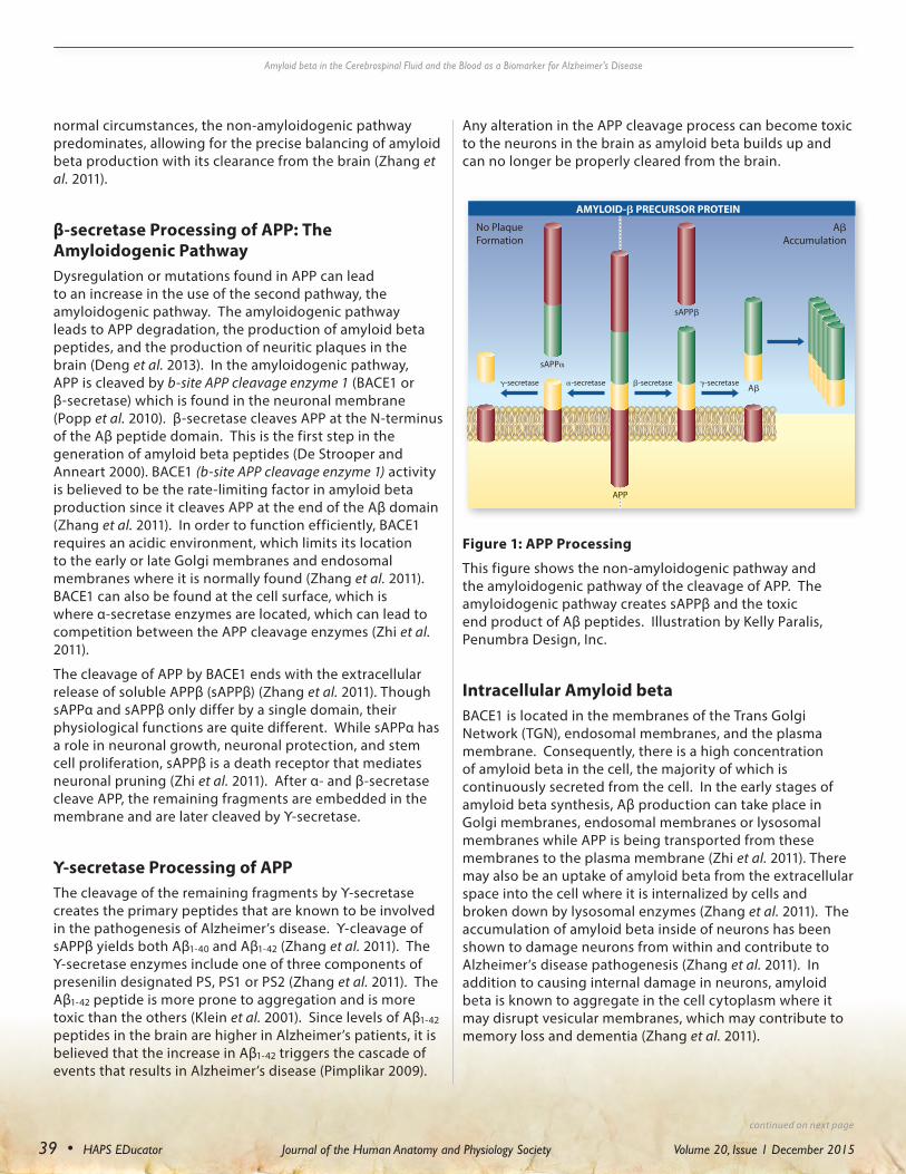
39 • HAPS EDucator Journal of the Human Anatomy and Physiology Society Volume 20, Issue 1 December 2015
normal circumstances, the non-amyloidogenic pathway predominates, allowing for the precise balancing of amyloid beta production with its clearance from the brain (Zhang et al. 2011).
β-secretase Processing of APP: The Amyloidogenic PathwayDysregulation or mutations found in APP can lead to an increase in the use of the second pathway, the amyloidogenic pathway. The amyloidogenic pathway leads to APP degradation, the production of amyloid beta peptides, and the production of neuritic plaques in the brain (Deng et al. 2013). In the amyloidogenic pathway, APP is cleaved by b-site APP cleavage enzyme 1 (BACE1 or β-secretase) which is found in the neuronal membrane (Popp et al. 2010). β-secretase cleaves APP at the N-terminus of the Aβ peptide domain. This is the first step in the generation of amyloid beta peptides (De Strooper and Anneart 2000). BACE1 (b-site APP cleavage enzyme 1) activity is believed to be the rate-limiting factor in amyloid beta production since it cleaves APP at the end of the Aβ domain (Zhang et al. 2011). In order to function efficiently, BACE1 requires an acidic environment, which limits its location to the early or late Golgi membranes and endosomal membranes where it is normally found (Zhang et al. 2011). BACE1 can also be found at the cell surface, which is where α-secretase enzymes are located, which can lead to competition between the APP cleavage enzymes (Zhi et al. 2011).
The cleavage of APP by BACE1 ends with the extracellular release of soluble APPβ (sAPPβ) (Zhang et al. 2011). Though sAPPα and sAPPβ only differ by a single domain, their physiological functions are quite different. While sAPPα has a role in neuronal growth, neuronal protection, and stem cell proliferation, sAPPβ is a death receptor that mediates neuronal pruning (Zhi et al. 2011). After α- and β-secretase cleave APP, the remaining fragments are embedded in the membrane and are later cleaved by Υ-secretase.
Υ-secretase Processing of APP The cleavage of the remaining fragments by Υ-secretase creates the primary peptides that are known to be involved in the pathogenesis of Alzheimer’s disease. Υ-cleavage of sAPPβ yields both Aβ1-40 and Aβ1-42 (Zhang et al. 2011). The Υ-secretase enzymes include one of three components of presenilin designated PS, PS1 or PS2 (Zhang et al. 2011). The Aβ1-42 peptide is more prone to aggregation and is more toxic than the others (Klein et al. 2001). Since levels of Aβ1-42 peptides in the brain are higher in Alzheimer’s patients, it is believed that the increase in Aβ1-42 triggers the cascade of events that results in Alzheimer’s disease (Pimplikar 2009).
Any alteration in the APP cleavage process can become toxic to the neurons in the brain as amyloid beta builds up and can no longer be properly cleared from the brain.
Intracellular Amyloid beta
BACE1 is located in the membranes of the Trans Golgi Network (TGN), endosomal membranes, and the plasma membrane. Consequently, there is a high concentration of amyloid beta in the cell, the majority of which is continuously secreted from the cell. In the early stages of amyloid beta synthesis, Aβ production can take place in Golgi membranes, endosomal membranes or lysosomal membranes while APP is being transported from these membranes to the plasma membrane (Zhi et al. 2011). There may also be an uptake of amyloid beta from the extracellular space into the cell where it is internalized by cells and broken down by lysosomal enzymes (Zhang et al. 2011). The accumulation of amyloid beta inside of neurons has been shown to damage neurons from within and contribute to Alzheimer’s disease pathogenesis (Zhang et al. 2011). In addition to causing internal damage in neurons, amyloid beta is known to aggregate in the cell cytoplasm where it may disrupt vesicular membranes, which may contribute to memory loss and dementia (Zhang et al. 2011).
Figure 1: APP Processing
This figure shows the non-amyloidogenic pathway and the amyloidogenic pathway of the cleavage of APP. The amyloidogenic pathway creates sAPPβ and the toxic end product of Aβ peptides. Illustration by Kelly Paralis, Penumbra Design, Inc.
Amyloid beta in the Cerebrospinal Fluid and the Blood as a Biomarker for Alzheimer’s Disease
continued on next page
No PlaqueFormation
AAccumulation
A
sAPP
sAPP
-secretase -secretase-secretase -secretase
AMYLOID- PRECURSOR PROTEIN
APP

40 • HAPS EDucator Journal of the Human Anatomy and Physiology Society Volume 20, Issue 1 December 2015
Amyloid beta is known to have a function in neuronal cell survival. Neuronal cells die when concentrations of amyloid beta in the brain are too low or when production of amyloid beta ceases. The underlying mechanism of neuronal cell death in the absence of amyloid beta has yet to be determined but it may involve the altered expression of K+ channels, since K+ channel activity is known to govern neuronal excitability (Jack et al. 2010).
Extracellular Amyloid beta Before amyloid beta forms plaques in the brain, it forms soluble oligomers. Amyloid beta oligomers have been shown to impair memory, inhibit long-term potentiation of neurons and lead to cognitive defects (Holtta et al. 2013). The soluble oligomers incorporate mainly Aβ1-42, but also attract Aβ1-40 peptides. Amyloid beta can also form insoluble protofibrils, which aggregate to form fibrils, ultimately leading to plaque formation (Pimplikar 2009). Plaque hinders the communication between neurons causing cell death, cognitive dysfunction, and behavioral abnormalities. The accumulation of plaque also initiates inflammation and the aggregation of tau proteins into neurofibrillary tangles (Prabhulker et al. 2012). Over time, neurons become impaired in their ability to reduce extracellular amyloid beta and to protect synapses in response to synaptic activity (Tampellini et al. 2011). At miniscule concentrations, amyloid beta protects synapses but an overload of amyloid beta, specifically Aβ1-42, alters synapses. Recent studies suggest that amyloid may serve to provide physiological control of synaptic activity by guarding against excessive glutamate release (Jack et al. 2010).
Most of the neurons affected by Alzheimer’s disease are located in the temporal, parietal and cortical regions, areas that control memory and cognitive functions. Synaptic activity increases the levels of secreted, extracellular amyloid beta supporting the hypothesis that brain regions with the highest metabolic rate are the most prone to Alzheimer’s development (Tampallini et al. 2011).
Amyloid beta in the CSFAmyloid beta was originally regarded as a toxic species only present in the brains of aging or demented humans (Zhang et al. 2011). Today, amyloid beta is known to exist in bodily fluids such as cerebrospinal fluid and blood plasma, which may indicate that there is a normal physiological function for amyloid beta (Zhang et al. 2011). A sudden decrease in amyloid beta concentrations in the body may be indicative of a predisposition to Alzheimer’s disease and/or mark the beginning of Aβ1-42 aggregation in the brain. Tapiola et al. found that the concentration of Aβ1-42 in the cerebrospinal fluid of Alzheimer’s patients was lower than that of control groups (Tapiola et al. 2009). This study suggested that the
lower level of Aβ1-42 in cerebrospinal fluid is due to the oligomerization and aggregation of amyloid beta in the brain (Holtta et al. 2013). A multivariate logistic regression analysis performed by Tapiola et al., showed that the number of neuritic plaques in the brain was a significant predictor of decreased cerebrospinal fluid Aβ1-42 levels, predicting the presence of pathological plaques in the brain with an overall accuracy of 90.2% (Tapiola et al. 2009). As a result, amyloid beta concentration in cerebrospinal fluid is being researched as a possible biomarker for an early diagnosis of Alzheimer’s disease.
Amyloid beta in the Blood Plasma Recent studies have begun to show a correlation between Alzheimer’s disease and the amount of amyloid beta in the blood stream and blood plasma levels of amyloid beta have been shown to vary at different stages of Alzheimer’s disease. In early stages of Alzheimer’s, Aβ1-42 levels found in blood plasma are lower compared to patients with moderate to normal cognitive function and they continue to decrease as the disease progresses. The most challenging aspect of the study of amyloid beta levels in blood plasma has been developing a technique by which they can be accurately measured (Song et al. 2011). The amount of amyloid beta in the blood is about ten times less than the amount normally found in the cerebrospinal fluid (Song et al. 2011). The traditional technique for measuring amyloid beta in blood plasma is an ELISA immunoassay. Unfortunately, lipoproteins and Fc-binding proteins interfere with the ELISA immunoassay because soluble amyloid beta interacts with protein binding species found in the plasma (Song et al. 2011), making it difficult to obtain an accurate ELISA reading.
The Effect of Amyloid beta on the Blood Brain BarrierChanges in amyloid beta concentrations in the cerebrospinal fluid and blood plasma are believed to be due to the decreased integrity of the blood brain barrier in Alzheimer’s patients (Perneczky et al. 2013). Dysfunction of the blood brain barrier and the resulting loss of homeostasis may result in widespread neuronal dysfunction (Deli et al. 2010). Even subtle changes to the blood brain barrier can have drastic effects in the brain and lead to severe neurological impairment. It is likely that the accumulation of amyloid beta in the brain affects the permeability of the blood brain barrier, which results in bulk flow of interstitial fluid into cerebrospinal fluid through perivascular spaces, followed by the drainage of interstitial fluid into the blood across the arachnoid villi (Kurz and Pernezky 2011). The disruption in the blood brain barrier ultimately leads to blood vessel regression, hypoperfusion of the brain, and an inflammatory response, the result of which is synaptic and neuronal dysfunction (Deli et al. 2010). The clearance of amyloid beta
Amyloid beta in the Cerebrospinal Fluid and the Blood as a Biomarker for Alzheimer’s Disease
continued on next page

41 • HAPS EDucator Journal of the Human Anatomy and Physiology Society Volume 20, Issue 1 December 2015
across the blood brain barrier is currently an area of intense research (Prabhulker et al. 2013).
Characteristics of BiomarkersBiomarkers are substances that are indicative of the presence of a disease and can be found in biological fluids such as blood, urine and cerebrospinal fluid, or compilations of data collected from computerized body imaging scans. To be accepted for general use, biomarkers must have undergone evaluation in a minimum of two peer-reviewed, cross-sectional clinical studies and their presence must have been confirmed at autopsy with other pathological findings (Babic et al. 2014). Biomarkers need to be both specific and sensitive. A biomarker that is specific for Alzheimer’s disease must be able to differentiate Alzheimer’s patients from healthy control subjects and from patients with other forms of dementia at least 85% of the time. A biomarker that is sensitive for Alzheimer’s disease would have to guarantee a high probability of being able to detect the presence of Alzheimer’s disease (Babic et al. 2014).
A strong biomarker should be assayed with a readily available, cost effective, minimally invasive procedure, since biomarker measurements are often done repeatedly over the course of an illness. Ideally, a biomarker will have levels that correlate with the progression of the disease. Biomarkers that are specific for a particular disease are routinely used for screening purposes, early disease detection, staging the disease, and monitoring patient response to treatment (Babic et al. 2014).
Amyloid beta as a Biomarker for Alzheimer’s diseaseAmyloid beta has been regarded as a late stage biomarker for Alzheimer’s disease for the past few decades (Forlanza et al. 2010). Most of the current research is focused on measuring the presence of amyloid beta in the cerebrospinal fluid since amyloid beta can be measured more accurately in cerebrospinal fluid than in blood plasma. The decreased amount of Aβ1-42 in the cerebrospinal fluid of Alzheimer’s patients is due to the abnormal clearing of amyloid beta from the brain, the accumulation of Aβ1-42 in cells, and the aggregation of Aβ1-42 into plaques (Holtta et al. 2013).
In addition to being helpful in the diagnosis of Alzheimer’s disease, the decreased concentration of amyloid beta in cerebrospinal fluid can be used to monitor the progression of patients with Mild Cognitive Impairment (MCI), the earliest stage of Alzheimer’s disease that effects cognition (Babic et al. 2014).
The reliability of using amyloid beta as a biomarker is still under investigation because a decrease in Aβ1-42 in the cerebrospinal fluid is not unique to Alzheimer’s disease (Babic et al. 2014). Decreased levels of Aβ1-
42 in cerebrospinal fluid are observed in a variety of other disorders including Lewy body disease (LBD), frontotemporal dementia (FTD), vascular dementia (VaD), amyotrophic lateral sclerosis, multiple system atrophy, Creutzfeldt-Jakob disease (CJD), and neuroinflammation. Because of this, the level of Aβ1-42 in cerebrospinal fluid is insufficiently specific to be used by itself in differentiating Alzheimer’s patients from patients with other conditions (Babic et al. 2014). The cut off level for the concentration of Aβ1-42 in cerebrospinal fluid that is indicative of Alzheimer’s disease is yet to be determined, however levels below 500pg/ml are generally only seen in Alzheimer’s disease (Babic et al. 2014).
Understanding the mechanisms of Aβ-induced neurodegeneration will hopefully reveal insights into the pathogenesis of Alzheimer’s disease that may prove useful in the development of an early Alzheimer’s biomarker (Lopez et al. 2010).
Other possible biomarkers for Alzheimer’s disease Recent studies suggest that a panel of biomarkers may have greater specificity in differentiating Alzheimer’s patients from other dementia patients than a single biomarker. Tau protein is one of the substances that can function as a biomarker for Alzheimer’s disease in combination with other substances. Tau protein is normally found inside of cells and functions during microtubule assembly and stabilization. It is released after neuronal cell death (van Harten et al. 2011). When tau protein is excessively phosphorylated (hyperphosphorylation), microtubule assembly is less successful, resulting in a tendency to form neurofibrillary tangles (van Harten et al. 2011). Tau protein in cerebrospinal fluid is known to be a marker for neurodegeneration, while hyperphosphorylated tau (p-tau) indicates the presence of neurofibrillary tangles (van Harten et al. 2011). Like amyloid beta, tau and p-tau can be detected in the cerebrospinal fluid by using an ELISA assay. Using all three proteins; amyloid beta, tau and p-tau proteins, to diagnosis Alzheimer’s disease has been common practice for the past several years. Current studies are now more likely to focus on the use of combinations of biomarkers to increase the specificity of Alzheimer’s detection (van Harten et al. 2011).
In the search for biomarkers for Alzheimer’s disease two other proteins are being studied because their concentration changes in the cerebrospinal fluid of Alzheimer’s patients. The cytoskeleton protein, keratin 9, appears to be found only in cerebrospinal fluid samples collected from Alzheimer’s patients and may prove to be a useful biomarker (Richens et. al. 2014). SPARCL-1 is another potential biomarker, the levels of which are significantly altered in cerebrospinal fluid samples taken from Alzheimer’s patients compared to healthy controls (Richens et al. 2014). SPARCL-1 gene is a tumor suppressor gene,
Amyloid beta in the Cerebrospinal Fluid and the Blood as a Biomarker for Alzheimer’s Disease
continued on next page

42 • HAPS EDucator Journal of the Human Anatomy and Physiology Society Volume 20, Issue 1 December 2015
which is linked to several other diseases including various forms of cancer (Richens et al. 2014).
The level of correlation between the proteins in the cerebrospinal fluid and blood plasma will have to be better understood in order to utilize biomarker diagnosis of Alzheimer’s disease to the fullest. Since the concentration of these proteins is likely linked to functional difficulties involving the integrity of blood brain barrier, the restoration of the integrity of the blood brain barrier is a possible area of study for therapeutics for Alzheimer’s disease patients (Richens et al. 2014).
ConclusionResearch into the pathophysiology of Alzheimer’s disease will continue to attract skilled scientists and clinicians. The need for research in this area is great and the predicted rise in the number of Alzheimer’s patients is daunting. The accurate detection of Alzheimer’s disease in asymptomatic people is still difficult and the search for effective biomarkers for the disease is expensive and time consuming. The least invasive procedures for sampling biomarkers involve blood and urine testing but potential biomarkers from these fluids have not yet shown sufficient specificity or sensitivity to be practical. The use of biomarkers in cerebrospinal fluid is problematic because it requires a lumbar puncture, which is an invasive procedure (Babic et al. 2014). There is great potential for cerebrospinal fluid biomarkers, however, and as they continue to be identified and better understood, it may be possible to decrease the age of detection of Alzheimer’s disease. The ultimate goal of further research into Alzheimer’s disease is to enable treatment of the disease at a pre-clinical stage, ideally before irreversible neurodegeneration has occurred.
Illustration courtesy of Kelly Paralis, Owner, Penumbra Design, Inc. - Studio: 143 North Sylvania Avenue, First Floor, Rockledge, PA 19064 [email protected] tel: 215.379.2832
References cited: Babić, M, Švob Štrac, D, Mück-Šeler D, et al. (2014)
Update on the core and developing cerebrospinal fluid biomarkers for Alzheimer disease. Cerebralspinal fluid physiology and movement 55: 347-365 doi: 10.3325/cmj.2014.55.347.
Deli, MA, Veszelka, S, Csiszar, B, et al. (2010). Protection of the Blood-Brain Barrier by Pentosan Against Amyloid-β-Induced Toxicity. Journal of Alzheimer’s Disease 22: 777-794. DOI 10.3233/JAD-2010-100759.
Deng, Y, Wang, A, Wang, R., Zhang, X, et al. (2013). Amyloid-b protein (Ab) Glu11 is the major b-secretase site of b-site amyloid-b precursor protein-cleaving enzyme 1 (BACE1), and shifting the cleavage site to Ab Asp1 contributes to Alzheimer pathogenesis. European Journal of Neuroscience 37: 1962-1969.
De Strooper, B and Annaert, W (2000). Proteolytic processing and cell biological functions of the amyloid precursor protein. Journal of Cell Science 113: 1857-1870.
Forlenza, OV, Diniz, BS, Gattaz, WF (2010). Diagnosis and biomarkers of predementia in Alzheimer’s disease. BMC Medicine: 8:89. doi:10.1186/1741-7015-8-89.
Hölttä M, Hansson O, Andreasson U, Hertze J, Minthon L, et al. (2013) Evaluating Amyloid-β Oligomers in Cerebrospinal Fluid as a Biomarker for Alzheimer’s Disease. PLoS ONE 8(6): e66381. doi:10.1371/journal.pone.0066381.
Jack, CR, Knopman, DS, Jagust, WJ, et al. (2010). Hypothetical model of dynamic biomarkers of the Alzheimer’s pathological cascade. Lancet Neurology, 9(1), 119. doi:10.1016/S1474-4422(09)70299-6.
Klein, WL, Krafft, GA, Finch, CE (2001). Targeting small Aβ oligomers: the solution to an Alzheimer’s disease conundrum? Trends in Neuroscience. 24(4): 219-224.
Kurz, A and Perneczky, R (2011). Amyloid Clearance As a Treatment Target Against Alzheimer’s Disease. Journal of Alzheimer’s Disease 24: 61-73. DOI 10.3233/JAD-2011-102139.
Lopez,-Toledano, MA, Patel NS, and Wahlestedt, C (2010). Adult Neurogenesis: A Potential Tool for Early Diagnosis in Alzheimer’s Disease? Journal of Alzheimer’s Disease 20: 395-408.
Amyloid beta in the Cerebrospinal Fluid and the Blood as a Biomarker for Alzheimer’s Disease
continued on next page

43 • HAPS EDucator Journal of the Human Anatomy and Physiology Society Volume 20, Issue 1 December 2015
Perneczky, R, Guo, L-H, Kagerbauer, SM, Werle, L, Kurz, A, Martin, J, & Alexopoulos, P (2013). Soluble amyloid precursor protein β as blood-based biomarker of Alzheimer’s disease. Translational Psychiatry, 3(2), e227–. doi:10.1038/tp.2013.11.
Pimplikar, SW (2009). Reassessing the Amyloid Cascade Hypothesis of Alzheimer’s Disease. The International Journal of Biochemistry & Cell Biology,41(6), 1261–1268. doi:10.1016/j.biocel.2008.12.015.
Popp, J, Lewczuk, P, Kolsch, H, et al. (2010). Cholesterol metabolism is associated with soluble amyloid precursor protein production in Alzheimer’s disease. Journal of Neuroscience 123 (2): 310-316. doi: 10.1111/j.1471-4159.2012.07893.x.
Prabhulkar, S, Piatyszek, R, Cirrito, JR, Wu, ZZ, and Li, CZ, (2012). Microbiosensor for Alzheimer’s disease diagnostics: detection of amyloid beta biomarkers. Journal of Neurochemistry 122: 374-381.
Qosa, H, LeVine III, H, Keller, JN, Kaddoumi, A (2014). Mixed oligomers and monomeric amyloid-β disrupts endothelial cells integrity and reduces monomeric amyloid-β transport across hCMEC/D3 cell line as an in vitro blood-brain barrier model. Molecular Basis of Disease 1842(9): 1806-1815.
Richens, J, Vere, K, Light, R, Soria, D, Garibaldi, J, et al. (2014). Practical detection of a definitive biomarker panel for Alzheimer’s disease, comparisons between matched plasma and cerebral spinal fluid. International Journal of Molecular Epidemiology and Genetics 5(2): 53-70.
Song, F, Poljak, A. Valenzuela, M, et al. (2011). Meta-Analysis of Plasma Amyloid-β levels in Alzheimer’s Disease. Journal of Alzheimer’s Disease 26: 365-375.
Tampellini, D, Rahman, N, Lin, MT, Capetillo-Zarate, E, & Gouras, GK (2011). Impaired β-amyloid secretion in Alzheimer’s disease pathogenesis. The Journal of Neuroscience : The Official Journal of the Society for Neuroscience,31(43), 15384–15390. doi:10.1523/JNEUROSCI.2986-11.2011
Tapiola T, Alafuzoff I, Herukka, SK, Parkkinen L, Hartikainen P, Soininen H, Pirttila, T (2009). Cerebrospinal Fluid beta amyloid 42 and tau proteins as biomarkers of Alzheimers-type pathological changes in the brain. Arch Neurol. 66(3):382-9. doi: 10.1001/archneurol.2008.596..
van Harten, A, Kester, M, Visser, P, et al. (2011). Tau and p-tau as CSF biomarkers in dementia: a meta-analysis. Clin Chem Lab Med 49(3): 353-566.
Zhang, Y, Thompson, R, Zhang, H, & Xu, H (2011). APP processing in Alzheimer’s disease. Molecular Brain, 4, 3. doi:10.1186/1756-6606-4-3.
Zhi, P, Chia, C, and Gleeson, PA (2011). Interacellular Trafficking of the β-secretase and Processing of Amyloid Precursor Protein. Life 63(9): 721-729.
Photo credit: http://www.istockphoto.com/
Amyloid beta in the Cerebrospinal Fluid and the Blood as a Biomarker for Alzheimer’s Disease
Kimberly Canfield is currently attending Arcadia University’s Physician Assistant Graduate Program. After graduating from the program, her goal is to work in the Philadelphia area with a desire to be a Physician Assistant in Cardiology.
Brie Paddock, PhD. is an assistant professor of Anatomy and Physiology at Arcadia University. Her research lab investigates the pathogenesis of Alzheimer’s Disease using a Drosophila model.
Sarah Cooper is Editor-in-Chief of the HAPS-EDucator. She has taught human anatomy and general biology at Arcadia University since 1981 and she serves as the pre-nursing adviser and coordinator of the interdisciplinary science program.
Sarah J. CooperInstructor in [email protected]
Sarah Cooper earned a B.S. in Biology from Allegheny College and an M.A. in Education with a concentration in Health Education from Arcadia University.
Cooper teaches in the General Biology (BI101/102) course sequence, which is required of all majors and is an important foundation for upper-level courses. In addition to these courses, Cooper also teaches Human Anatomy (BI205), a course targeted to pre-professional students who are interested in a career in health care. She is the coordinator of the Interdisciplinary Science Program and an instructor in ID101 and ID102. She is the developer and an instructor in a laboratory course for non-science majors titled, Human Biology (BI110) and she frequently teaches a First Year Seminar in Forensic Anthropology. Cooper is the pre-nursing adviser with oversight for the 2+2 pre-nursing program Arcadia maintains with Jefferson University.
Cooper’s professional activities include serving as Editor of the HAPS-EDucator, the international journal of the Human Anatomy and Physiology Society; numerous reviews of texts and lab manuals; and numerous publications including a laboratory manual for the dissection of the cat and articles onthe surgical treatment of pulsatile tinnitus, treatment options in pancreaticobiliary disease,, the use of cosmeceuticals in skin care, and the role of stem cells in non-union bone fractures.
Read about Cooper's scholarship and campus activities in the Bulletin.
Recent Publications
"The Emerging Interface of Entomotoxicology, Forensic Entomology and Decomposition in Modern Crime Scene Investigation". HAPS-EDucator (Journal of the Human Anatomy andPhysiology Society) 8(2): 4-9. Spring 2014.With Allison Gaines, Arcadia '14.
Yoga, Anatomy and the Fitness Explosion on Campus” HAPS-EDucator 18(1): 4-10. Winter 2014. With Susan White Phillips, Jennifer Wood, PhD, and Spencer Lalk, Arcadia '13.
"A Subunit of BOTOX Reduces Pain and Inflammation during Chronic Migraines" HAPS EDucator. 17(2) Spring 2013. With Jessica E. Hartman, Arcadia '13.
"The Use of Stem Cells in Bone Non-Union Fracture Healing" HAPS-EDucator 17(2) Spring2013. With Dylan Marks, Arcadia '13.
"A surgical Approach to the Correction of Pulsatile Tinnitus caused by Sigmoid Sinus Diverticulum"HAPS EDucator. 17(2) Spring 2013. With Shaina John, Arcadia '13.
About the Authors
■
44 • HAPS EDucator Journal of the Human Anatomy and Physiology Society Volume 20, Issue 1 December 2015
Comorbidity of Multiple Sclerosis and Depression: A Primer
Maggie Scribner, [email protected]
Benjamin Miller, Ph.D., [email protected]
Texas Wesleyan University, Department of Biology, 1201 Wesleyan St., Fort Worth, TX 76105
AbstractMultiple Sclerosis (MS) is an autoimmune neurological condition commonly referenced in Human Anatomy and Physiology textbooks. Though patient symptoms vary widely and encompass both physical and cognitive manifestations, educational materials heavily emphasize physical symptomology. This primer will provide general information concerning the clinical presentation of physical symptomology, but will emphasize the prevalence, presentation, and etiology of comorbid depression in multiple sclerosis. Depression is the most common psychiatric disorder associated with MS, and is cited by patients as having a greater impact on their quality of life than other symptomology. While the underlying mechanisms which give rise to MS depression have yet to be fully elucidated, there are similarities between Major Depressive Disorder and MS, which suggest depression may be a primary symptom of multiple sclerosis.
IntroductionMultiple Sclerosis (MS) is the most common disabling neurological condition afflicting young adults (Warren, S and Warren, K 2001), and the second most common disabling nervous system disease overall behind Parkinson’s disease. Over 2 million people worldwide are diagnosed with MS (Opara et al. 2010, 2012). MS is a chronic autoimmune disorder causing overt demylination of oligodendrocytes in the central nervous system (CNS). The CNS is the chief regulator of myriad bodily functions, and as such, patient symptoms may vary. Classic symptoms include numbness or tingling of the extremities, vision disturbances, loss of balance and coordination, fatigue, and muscle spasms (Symptoms & Diagnosis 2014). The Expanded Disability Status Scale (EDSS), the standard for clinical evaluation of MS severity, focuses primarily on these classic symptoms with an emphasis on ambulation. No single clinical feature or diagnostic test can confirm the presence of MS because its specific pathology is complex and poorly understood, but a definitive diagnosis can be achieved through a combination of symptomology, the presence of specific antibodies, and lesion formation, load, and location (McDonald et al. 2001). These aforementioned criteria are collectively known as the McDonald Criteria (McDonald et al. 2001). Lesions are CNS regions of demyelinated neurons which can be evaluated with magnetic resonance imaging (MRI). MS symptoms usually present in a pattern of exacerbations and remissions. For example, an MS exacerbation, characterized by acute onset of specific symptomology, may last as little as 24 hours, but usually last weeks, and can last for months (Symptoms & Diagnosis 2014). The other domain of symptoms, aside
from the physical (or classic), is the domain of cognitive symptoms. Moderate or severe cognitive symptoms, including impaired executive functions, impaired processing speed, and psychiatric manifestations such as behavioral changes or depression, occur in 40-70% of the MS population (Rao et al. 1991; Chiaravalloti and DeLuca 2008). Of these MS-induced cognitive symptoms, depression alone has a lifetime prevalence in MS from 40 to 60% (Wallin 2006) with a 12 month prevalence rate of 15-20% (Sadovnick et al. 1996; Koch et al. 2015). Despite high prevalence, the etiology of comorbid depression in MS remains to be elucidated.
As evidence is becoming available, popular opinion is shifting from viewing MS depression as a maladjustment disorder to a symptom of MS. In fact, depression is the most common psychiatric condition associated with Multiple Sclerosis (Skokou 2012; Goldman Consensus Group 2005; Gunzler 2014; Wallin 2006, Marrie and Handwell 2013, Marrie 2009). While the relationship between MS and depression has been discussed regularly (Whitlock and Siskind 1980; Cottrell and Wilson 1926; Philippopoulos et al. 1958; Feinstein 2011), it is unclear how MS and depressive symptomology interact. Early MS researchers hypothesized that depression may result primarily from maladjustment of living with MS symptomology. However, multiple studies have demonstrated that MS has consistently incurred higher depression rates than other (non-MS) chronic disorders (Sullivan et al. 1995; Devins and Seland 1987; Minden and Schiffer 1990). More importantly, depression is associated with an increased morbidity and mortality of MS patients, and is regarded by those patients as one of the main determinants of their quality of life (Feinstein
continued on next page

45 • HAPS EDucator Journal of the Human Anatomy and Physiology Society Volume 20, Issue 1 December 2015
2011). Visschedijk et al. (2004) went so far as to say that a patient’s subjectively perceived health-related quality of life may also serve as a predictor of objective outcomes such as change in disability status over a substantial period of time (Visschedijk et al. 2004). It must also be recognized that 15% of all MS patient deaths are attributed to suicide, which is 7.5 times greater than that of the general population (Scalfari et al. 2013).
The relevance of depression within the MS framework is undeniable, but our understanding of the mechanistic underpinnings is lacking. Current opinion states: there is no evidence to imply that MS is causative of depression pathologically, but it should still be treated as a symptom of the disease (Rao 1995; Pompili et al. 2012; Opara et al. 2012; Scalfari et al. 2013). However, recent discoveries have revealed many mechanisms of depression in MS. Thus, it is evident that the mechanisms of MS can cause depression which may explain the high prevalence of depression in the MS population. Likewise, it must also be noted that depressive symptoms in MS can exacerbate disease severity. It is the topic of this review to detail current understanding of depression pathology in MS patients, and its relationship to the disease course as a whole. This paper will offer a primer on the possible pathological mechanisms in MS which may directly give rise to depression in MS patients. Here, we also posit that linking abnormal brain regions which MS and Major Depressive Disorder share will provide possible mechanistic insights into depression in MS.
Overview of Depression in MSIn the last 20 years there have been two prevailing theories on why comorbid depression is prevalent in MS. The oldest theory basically points to how difficult it can be to live with MS. It is hypothesized that depression may be a result of maladjustment or malcoping with high stress and anxiety. This is not a large leap to make when considering not only the potential level of disability an MS patient may look forward to, but also the unpredictability of the disease (e.g. fear of an exacerbation, fear of losing one’s job, anxiety of health care costs, or anxiety of burdening loved ones). In fact, J. Kesselring, an MS rehabilitation specialist, believed the disease should have been named MU for Malignant Uncertainty (Kesselring 1989). The second theory observes that, indeed, depression rates in MS are too high for physicians and researchers to consider depression as anything other than an MS symptom. However, due to the paucity of evidence on the pathophysiological mechanisms in depression in MS, it was impossible to speculate on the cause of high comorbid depression rates, or to definitively conclude whether depression is a primary or secondary symptom of MS.
Depression in MS has a different presentation than depression in other chronic disorders. Charcot himself,
the individual who named Multiple Sclerosis, noted that MS patients most often attributed long continued grief or vexation as the cause of their disease during his 1868 lecture of nervous system diseases (Charcot 1868). Since then, numerous studies have indicated depression may commonly occur before disease onset, and even today severe emotional trauma is a risk factor for developing MS (Cottrell and Wilson 1926; Goodstein and Ferrell 1977; Joffe et al. 1987; MeiTal et al. 1970; Philippopoulos et al. 1958; Pratt 1951). For example, Whitlock and Siskind (1980) noted MS patients have more bouts of depression both before and after diagnosis than other neurological disorders causing similar degrees of disability. Moreover, Sullivan et al. (1995) reported that 52% of patients with MS experienced a depressive episode before the onset of MS, compared with 17% of patients with chronic low back pain (CLBP), while 35% of MS patients reported family history of treatment for depression compared to 15% of patients with chronic lower back pain (Sullivan et al. 1995). A 2004 study (Li et al. 2004), demonstrated the bereaved parents, having lost a child under the age of 18, had a 50% increased risk of developing MS compared to parents unexposed to child death. It could be that MS and depression have an intricate relationship because of parallel mechanisms. This would explain why patients frequently experience depression before ever displaying classical MS symptoms. Indeed, Koch et al. (2015), used logistic regression analysis to determine that MS depression is largely chronic, meeting the diagnosis criteria of persistent depressive disorder where an episode of depression lasts for at least two years, compared to major depressive disorder, where 80% of patients recover within 12 months. Therefore, it is now clear that depression is likely associated directly with MS pathophysiology, rather than a maladjustment disease.
Lesion Location and Atrophy in MS and Major Depressive DisorderWhat is well known in MS pathology is that higher lesion load correlates with an increased chance of developing depression, or other psychological symptoms (Berg et al. 2000; Schiffer et al. 1983; Honer et al. 1987). Similar studies have found a correlation between depression, lesion-load/atrophy, and level of disability (Pujol et al. 1997; Zorzon et al. 2001; Mohr et al. 2003). In addition, studies have shown a high degree of correlation between lesion location or atrophy and depression, as well as lesion location or atrophy and depressive severity in several brain regions. The diagnosis of depression is more frequent when atrophy or high lesion load occurs in the right frontal (Zorzon et al. 2001; Feinstein et al. 2010), and right temporal lobes (Zorzon et al. 2001). Similarly, there is a high correlation between depressive severity and lesion load or atrophy in the temporal lobes (Berg et al. 2000; Zorzon et al. 2001; Mohr et al. 2003), but also in the right frontal lobe (Zorzon et al. 2001), the right periventricular region (Mohr
Comorbidity of Multiple Sclerosis and Depression: A Primer
continued on next page

46 • HAPS EDucator Journal of the Human Anatomy and Physiology Society Volume 20, Issue 1 December 2015
et al. 2003), and the right hippocampus (Gold et al. 2014). Interestingly, the location of these lesions and volume reductions which are associated with MS depression coincide with structural abnormalities associated with Major Depressive Disorder (MDD).
Numerous independent studies detailing various structural or functional abnormalities of precise brain regions in MDD overlap extensively with brain regions known to be directly affected by MS pathology (occurring in the majority of MS patients, not just the depressed). Depression and depressive severity in MDD has also been found to be correlated with atrophy of several brain structures, including the hippocampus, basal ganglia, orbitofrontal cortex (OFC), and subgenual prefrontal cortex (Lorenzetti et al. 2009). Similar to MS depression, MDD depression consistently manifests with lesions or atrophy in the frontal regions (Kanner 2004; Videbech and Ravnikilde 2004). Additionally, changes in the temporal region have been reported (Kanner et al. 2004). Furthermore, Samann et al. (2013) found that having larger gray matter volumes of the left hippocampal and bilateral posterior cingulate and lower right temporolateral regions were associated with beneficial treatment response. This may help explain why MS depression is chronic. MS depression includes hippocampal atrophy (Sicotte et al. 2008; Geurts et al. 2007; Aktas and Hartung 2011) and temporal region abnormalities, and MDD appears to be more difficult to treat when atrophy occurs in the temporolateral region.
Abnormalities in the insula (Videbech and Ravnkile 2004; Stratmann et al. 2014), basal ganglia (Kanner et al. 2004; Videbech and Ravnkilde 2004), and hippocampus (MacQueen and Frodl 2011; Videbech and Ravnkile 2004; Samann et al. 2013; Strattmann et al. 2014) are also found frequently in MDD. Furthermore, Stratmann et al. (2014) found that more severe gray matter volume reductions of the hippocampal formation were observed in patients with recurrent depressive episodes compared to patients with a first episode. Recently, Shen et al. (2014) correlated fractional anisotropy abnormalities in the right posterior middle cingulate gyrus, right hippocampus, left hypothalamus, right precentral gyrus, and posterior cingulate with depressive symptom severity in MS. The fact that brain regions subject to lesion or volume reductions which are highly associated with MDD, are also present with abnormalities in MS depression, suggest common pathological mechanisms.
ConclusionThere appears to be many overlapping pathologies in MDD and MS. The brain regions that appear to be consistent with depressive symptoms are the hippocampal region, and the frontal and temporal lobes. One major difference between MDD and MS depression is that MS
depression is more persistent. A possible explanation for this phenomenon is that MS pathology is directly causing depressive symptoms. While this paper focused exclusively on the history of clinical MS depression and brain abnormalities present in both MS and MDD there is other compelling evidence to be considered. For instance there appears to be impaired potassium and glutamate channels within the CNS for both disorders, as well as malregulation of the inflammatory pathways including cytokine and endocrine upregulation of inflammation for extended periods of time.
Although much needs to be elucidated about the causative mechanism of depression in MS, evidence is mounting that seems to suggest that depression may be a direct symptom of MS pathology for some individuals. It is advisable to screen MS patients for depression regularly, and begin the process of developing effective treatments for depressive MS patients which do not risk drug interactions with their current therapies, or add to already cumbersome side-effects of their primary medications, thereby reducing treatment efficacy. The physical manifestations of MS symptomology are easy to identify and quantify when compared to cognitive symptoms, but cognitive impairments and comorbid psychiatric conditions like depression have a greater impact on the patient’s quality of life. Therefore, it is imperative to recognize the cognitive impairments (including depression) and develop better means to evaluate and treat these symptoms in MS patients.
Literature CitedAktas O and Hartung HP. 2011. Beyond axonal
transection: Hippocampal damage in multiple sclerosis. Ann Neurol 69(3):433-6.
Ben Ari Shevil E, Johansson S, Ytterberg C, Bergstrom J, von Koch L. 2014. How are cognitive impairment, fatigue and signs of depression related to participation in daily life among persons with multiple sclerosis? Disabil Rehabil 36(23):2012-8.
Benson C and Kerr BJ. 2014. Pain and cognition in multiple sclerosis. Curr Top Behav Neurosci 20:201-15.
Berg D, Supprian T, Thomae J, Warmuth-Metz M, Horowski A, Zeiler B, Magnus T, Rieckmann P, Becker G. 2000. Lesion pattern in patients with multiple sclerosis and depression. Mult Scler 6(3):156-62.
Bittner S, Ruck T, Fernandez-Orth J, Meuth SG. 2014. TREK-king the blood-brain-barrier. J Neuroimmune Pharmacol 9(3):293-301.
Charcot J. 1868. Comptes rendus des séances et mémoires lus à la société de Biologie.
Comorbidity of Multiple Sclerosis and Depression: A Primer
continued on next page

47 • HAPS EDucator Journal of the Human Anatomy and Physiology Society Volume 20, Issue 1 December 2015
Chiaravalloti ND and DeLuca J. 2008. Cognitive impairment in multiple sclerosis. Lancet Neurol 7(12):1139-51.
Cottrell SS and Wilson SAK. 1926. The affective symptomatology of disseminated sclerosis. J Neuro Psychopath 7: 1–30.
Devins GM and Seland TP. 1987. Emotional impact of multiple sclerosis: Recent findings and suggestions for future research. Psychol Bull 101(3):363-75.
Feinstein A. 2011. Multiple sclerosis and depression. Mult Scler 17(11):1276-81.
Feinstein A, Rector N, Motl R. 2013. Exercising away the blues: Can it help multiple sclerosis-related depression? Mult Scler 19(14):1815-9.
Feinstein A, O’Connor P, Akbar N, Moradzadeh L, Scott CJ, Lobaugh NJ. 2010. Diffusion tensor imaging abnormalities in depressed multiple sclerosis patients. Mult Scler 16(2):189-96.
Fischer A, Otte C, Krieger T, Nicholls RA, Kruger S, Ziegler KJ, Schulz KH, Heesen C, Gold SM. 2012. Decreased hydrocortisone sensitivity of T cell function in multiple sclerosis-associated major depression. Psychoneuroendocrinology 37(10):1712-8.
Foley P, Lawler A, Chandran S, Mead G. 2014. Potential disease-modifying effects of selective serotonin reuptake inhibitors in multiple sclerosis: Systematic review and meta-analysis. J Neurol Neurosurg Psychiatry 85(6):709-10.
Geurts JJ, Bo L, Roosendaal SD, Hazes T, Daniels R, Barkhof F, Witter MP, Huitinga I, van der Valk P. 2007. Extensive hippocampal demyelination in multiple sclerosis. J Neuropathol Exp Neurol 66(9):819-27.
Gold SM, O’Connor MF, Gill R, Kern KC, Shi Y, Henry RG, Pelletier D, Mohr DC, Sicotte NL. 2014. Detection of altered hippocampal morphology in multiple sclerosis-associated depression using automated surface mesh modeling. Hum Brain Mapp 35(1):30-7.
Goldman Consensus Group. 2005. The goldman consensus statement on depression in multiple sclerosis. Mult Scler 11(3):328-37.
Goodstein RK and Ferrell RB. 1977. Multiple sclerosis--presenting as depressive illness. Dis Nerv Syst 38(2):127-31.
Gunzler DD, Perzynski A, Morris N, Bermel R, Lewis S, Miller D. 2014. Disentangling multiple sclerosis and depression: An adjusted depression screening score for patient-centered care. J Behav Med .
Heesen C, Kopke S, Kasper J, Poettgen J, Tallner A, Mohr D, Gold SM. 2012. Behavioral interventions in multiple sclerosis: a biopsychosocial perspective. Expert Review of Neurotherapeutics 12(9): 1089.
Hildebrandt H and Eling P. 2014. A longitudinal study on fatigue, depression, and their relation to neurocognition in multiple sclerosis. J Clin Exp Neuropsychol 36(4):410-7.
Honer WG, Hurwitz T, Li DK, Palmer M, Paty DW. 1987. Temporal lobe involvement in multiple sclerosis patients with psychiatric disorders. Arch Neurol 44(2):187-90.
Joffe RT, Lippert GP, Gray TA, Sawa G, Horvath Z. 1987. Mood disorder and multiple sclerosis. Arch Neurol 44(4):376-8.
Kanner AM. 2004. Structural MRI changes of the brain in depression. Clin EEG Neurosci 35(1):46-52.
Kesselring J. Multiple Sklerose. Kohlhammer, Stuttgart, Koln, Berlin 1989.
Koch MW, Patten S, Berzins S, Zhornitsky S, Greenfield J, Wall W, Metz LM. 2015. Depression in multiple sclerosis: A long-term longitudinal study. Mult Scler 21(1):76-82.
Langdon DW. 2011. Cognition in multiple sclerosis. Curr Opin Neurol 24(3):244-9.
Li J, Johansen C, Bronnum-Hansen H, Stenager E, Koch-Henriksen N, Olsen J. 2004. The risk of multiple sclerosis in bereaved parents: A nationwide cohort study in denmark. Neurology 62(5):726-9.
Lorenzetti V, Allen NB, Fornito A, Yucel M. 2009. Structural brain abnormalities in major depressive disorder: A selective review of recent MRI studies. J Affect Disord 117(1-2):1-17.
MacQueen G and Frodl T. 2011. The hippocampus in major depression: Evidence for the convergence of the bench and bedside in psychiatric research? Mol Psychiatry 16(3):252-64.
Marrie RA and Hanwell H. 2013. General health issues in multiple sclerosis: Comorbidities, secondary conditions, and health behaviors. Continuum (Minneap Minn) 19(4 Multiple Sclerosis):1046-57.
Marrie RA, Horwitz R, Cutter G, Tyry T, Campagnolo D, Vollmer T. 2009. The burden of mental comorbidity in multiple sclerosis: Frequent, underdiagnosed, and undertreated. Mult Scler 15(3):385-92.
Comorbidity of Multiple Sclerosis and Depression: A Primer
continued on next page

48 • HAPS EDucator Journal of the Human Anatomy and Physiology Society Volume 20, Issue 1 December 2015
McDonald WI, Compston A, Edan G, Goodkin D, Hartung HP, Lublin FD, McFarland HF, Paty DW, Polman CH, Reingold SC, et al. 2001. Recommended diagnostic criteria for multiple sclerosis: Guidelines from the international panel on the diagnosis of multiple sclerosis. Ann Neurol 50(1):121-7.
Mei-Tal V, Meyerowitz S, and Engel GL. 1970. The role of psychological process in a somatic disorder: Multiple Sclerosis. Psychosom Med 32: 67-86.
Minden SL and Schiffer RB. 1990. Affective disorders in multiple sclerosis. review and recommendations for clinical research. Arch Neurol 47(1):98-104.
Mohr DC, Epstein L, Luks TL, Goodkin D, Cox D, Goldberg A, Chin C, Nelson S. 2003. Brain lesion volume and neuropsychological function predict efficacy of treatment for depression in multiple sclerosis. J Consult Clin Psychol 71(6):1017-24.
Opara J, Jaracz K, Brola W. 2012. Burden and quality of life in caregivers of persons with multiple sclerosis. Neurol Neurochir Pol 46(5):472-9.
Opara JA, Jaracz K, Brola W. 2010. Quality of life in multiple sclerosis. J Med Life 3(4):352-8.
Philippopoulos GS, Wittkower ED, and Cousineau A. 1958. The etiologic significance of emotional factors in onset and exacerbations of multiple sclerosis. Psychosom Med 20: 458-74.
Pompili M, Forte A, Palermo M, Stefani H, Lamis DA, Serafini G, Amore M, Girardi P. 2012. Suicide risk in multiple sclerosis: A systematic review of current literature. J Psychosom Res 73(6):411-7.
Pratt RT. 1951. An investigation of the psychiatric aspects of disseminated sclerosis. J Neurol Neurosurg Psychiatry 14(4):326-35.
Pujol J, Bello J, Deus J, Marti-Vilalta JL, Capdevila A. 1997. Lesions in the left arcuate fasciculus region and depressive symptoms in multiple sclerosis. Neurology 49(4):1105-10.
Rao SM, Leo GJ, Bernardin L, Unverzagt F. 1991. Cognitive dysfunction in multiple sclerosis. I. frequency, patterns, and prediction. Neurology 41(5):685-91.
Sadovnick AD, Remick RA, Allen J, Swartz E, Yee IM, Eisen K, Farquhar R, Hashimoto SA, Hooge J, Kastrukoff LF, et al. 1996. Depression and multiple sclerosis. Neurology 46(3):628-32.
Samann PG, Hohn D, Chechko N, Kloiber S, Lucae S, Ising M, Holsboer F, Czisch M. 2013. Prediction of antidepressant treatment response from gray matter volume across diagnostic categories. Eur Neuropsychopharmacol 23(11):1503-15.
Scalfari A, Knappertz V, Cutter G, Goodin DS, Ashton R, Ebers GC. 2013. Mortality in patients with multiple sclerosis. Neurology 81(2):184-92.
Schiffer RB, Caine ED, Bamford KA, Levy S. 1983. Depressive episodes in patients with multiple sclerosis. Am J Psychiatry 140(11):1498-500.
Shen Y, Bai L, Gao Y, Cui F, Tan Z, Tao Y, Sun C, Zhou L. 2014. Depressive symptoms in multiple sclerosis from an in vivo study with TBSS. Biomed Res Int 2014:148465.
Sicotte NL, Kern KC, Giesser BS, Arshanapalli A, Schultz A, Montag M, Wang H, Bookheimer SY. 2008. Regional hippocampal atrophy in multiple sclerosis. Brain 131(Pt 4):1134-41.
Sidhom Y, Ben Djebara M, Hizem Y, Abdelkefi I, Kacem I, Gargouri A, Gouider R. 2014. Bipolar disorder and multiple sclerosis: A case series. Behav Neurol 2014:536503.
Skokou M, Soubasi E, Gourzis P. 2012. Depression in multiple sclerosis: A review of assessment and treatment approaches in adult and pediatric populations. ISRN Neurol 2012:427102.
Stratmann M, Konrad C, Kugel H, Krug A, Schoning S, Ohrmann P, Uhlmann C, Postert C, Suslow T, Heindel W, et al. 2014. Insular and hippocampal gray matter volume reductions in patients with major depressive disorder. PLoS One 9(7):e102692.
Sullivan MJ, Weinshenker B, Mikail S, Edgley K. 1995. Depression before and after diagnosis of multiple sclerosis. Mult Scler 1(2):104-8.
Symptoms & Diagnosis [Internet] [cited 2014 June 12]. National Multiple Sclerosis Society. Available from: http://www.nationalmssociety.org/Symptoms-Diagnosis.
Videbech P and Ravnkilde B. 2004. Hippocampal volume and depression: A meta-analysis of MRI studies. Am J Psychiatry 161(11):1957-66.
Visschedijk MA, Uitdehaag BM, Klein M, van der Ploeg E, Collette EH, Vleugels L, Pfennings LE, Hoogervorst EL, van der Ploeg HM, Polman CH. 2004. Value of health-related quality of life to predict disability course in multiple sclerosis. Neurology 63(11):2046-50.
Warren SA and Warren KG.1993. Prevalence, incidence and characteristics of multiple sclerosis in Westlock County, Alberta, Canada. Neurology;43:1760–963.
Whitlock FA and Siskind MM. 1980. Depression as a major symptom of multiple sclerosis. J Neurol Neurosurg Psychiatry 43(10):861-5.
Comorbidity of Multiple Sclerosis and Depression: A Primer
continued on next page

49 • HAPS EDucator Journal of the Human Anatomy and Physiology Society Volume 20, Issue 1 December 2015
Zorzon M, de Masi R, Nasuelli D, Ukmar M, Mucelli RP, Cazzato G, Bratina A, Zivadinov R. 2001. Depression and anxiety in multiple sclerosis. A clinical and MRI study in 95 subjects. J Neurol 248(5):416-21.
Zunszain PA, Anacker C, Cattaneo A, Carvalho LA, Pariante CM. 2011. Glucocorticoids, cytokines and brain abnormalities in depression. Prog Neuropsychopharmacol Biol Psychiatry 35(3):722-9.
Comorbidity of Multiple Sclerosis and Depression: A Primer
Maggie Scribner is a senior biology major at Texas Wesleyan University where she is president of their Beta Beta Beta chapter (Biological Honors Society), works as a science and math tutor, and plays for the basketball team. She was recently accepted to the Texas College of Osteopathic Medicine at the University of North Texas Health Science Center.
Benjamin Miller, Ph.D., is an Assistant Professor of Biology at Texas Wesleyan University where he teaches all the Human Anatomy and Physiology lectures and labs. He received his Ph.D. in Neuroscience and Psychology at Indiana University. His postdoctoral position was at UT Southwestern Medical Center. His research centers on the neurophysiology of neurodegeneration. His current focus is to use the flatworm planaria as a model to explore the neurobehavioral effects of neurotoxicity. The ultimate goal is to discover drugs that ameliorate neuron dysfunction driven by glutamate excitotoxicity.
■
About the Authors
Teaching Central Nervous System Concepts Using Diagnostic Radiology and Case Studies (2 credits)
September 13 - November 2, 2015
Carmen Eilertson
Georgia State UniversityStudents enrolled in college level Anatomy and Physiology courses are required to learn brain and spinal cord anatomy and function. This course will help you develop cases incorporating radiology of the CNS into your teaching portfolio. Students find this method of teaching to be meaningful because they learn how to read radiograms and correlate their analysis with patient symptoms. it is a perfect way to integrate anatomy with physiology and teach valuable diagnostic skills at the same time. Most of the your students will enter the health care field and will benefit from learning clinical problem solving skills early in their academic development. The cases developed as part this course can be used as in class activities, enhancements to your current lecture power points, or as clicker quizzes or exam questions.
Current HAPS-I course offering
REGISTER NOW: Graduate Credit or Professional Development
Courses are continually added, watch our Facebook and Google+ pages for the latest course announcements!

50 • HAPS EDucator Journal of the Human Anatomy and Physiology Society Volume 20, Issue 1 December 2015
Humankind vs. the Prokaryotes: Novel and Promising Strategies for Overcoming Vancomycin Resistance in Staphylococcus aureus
Sarah Cooper, MEd1 and Olivia Shaffer2
1 Department of Biology, Arcadia University, Glenside, PA 190382 Biology/Pre-PT, Arcadia University, Glenside, PA 19038
AbstractBacterial resistance to antibiotics remains a threatening global health issue into the early years of the 21st century. Vancomycin has been one of the most potent combatants against the highly infectious methicillin-resistant Staphylococcus aureus (MRSA) bacteria. However, over a relatively short period of time, vancomycin intermediate (VISA) and vancomycin resistant (VRSA) strains of S. aureus have emerged and the search for effective treatments against bacterial infections is in high gear. The research examined in this article investigates novel and promising strategies for overcoming the resistance of S. aureus to vancomycin.
A world without antibioticsImagine a world without antibiotics.
The year is 1199 and Richard the Lionheart (Coeur de Lion) is at the top of his game. He is the King of England and in France he is Duke of Normandy, Duke of Aquitaine and Poitiers and Count of Anjou. He is young by today’s standards, just 42 years old, and he has been King for ten years. His body is that of a superbly conditioned triathlon athlete, shaped and toned by having spent virtually his entire adult life on horseback in battlefields across England and France. His army has encircled a castle at Chalus in France and a siege is underway. It is not a particularly large or important castle but, sensing victory, Richard cannot wait for the news to be brought to him in his tent. Impulsively he decides to see the action for himself. He leaves his tent quickly, not bothering to put on his armor and protective coverings. He joins two friends who are watching the progress of the siege and decides to stay with them and observe for a little while (Pennman 2014).
A crossbow bolt is shot from one of the battlements and unbelievably, because the distance is great, it slams into his shoulder. The bolt enters Richard’s left shoulder
just superior to the clavicle piercing through tissue and muscle to become tightly wedged in the region between the posterior surfaces of ribs two through seven and
subscapularis. Historians tell us that the force of the impact knocked Richard down but that he got immediately back on his feet. Before his friends can stop him, Richard, accustomed to taking charge of every situation, grabs the shaft of the crossbow arrow and attempts to pull it out. The shaft breaks off at the skin surface and with it goes his only hope of a relatively easy extraction of the bolt. He is immensely strong and he walks under his own power back to his tent between his two friends (Pennman 2014).
A field surgeon is called from the troops. He, like all surgeons of his day, is by reputation only slightly more skilled than the neighborhood butcher. His hands shake as he attempts to dissect out the crossbow bolt, a cylinder of iron approximately the length of the width of a man’s hand. He fears for his life if the surgery is not successful. The surgeon tries repeatedly to dissect down the shaft of the
broken arrow to extract the bolt but his efforts end in failure. Finally, in desperation, he grabs the exposed tip of the arrow with crudely fashioned pliers and yanks the bolt
continued on next page

51 • HAPS EDucator Journal of the Human Anatomy and Physiology Society Volume 20, Issue 1 December 2015
free with brute force. The wound is rinsed with wine and covered with betony (Sarasen’s root), comfrey (Woundwort) and honey (Pennman 2014). There has been no thought of sterile technique and it will be almost 700 years before anesthesia is available. Even today such a horrific wound would present a myriad of clinical problems.
Richard survives the surgery. He has survived much in his life. He has survived the battlefields of the Holy Land, treacherous ocean voyages in a small ship, and captivity in France. He knows, however, as soon as the first red streaks appear around the puncture wound site that he is a dead man. He will not survive this. Within the hour he has started to write his will. In spite of his relative youth, his immense strength, and the keys to kingdoms on two continents, he will not survive the onslaught of prokaryotic life forms that slammed into his body with the crossbow bolt.
It is a fragment of history.
It is a world without antibiotics.
It is a world without hope.
It is a world to which we dare not return.
IntroductionWe are very lucky to live in a world where antibiotics are available but that does not mean that we live in a problem-free world. The fight against prokaryotes continues into the 21st century with no sense that the battle can be definitively won. In fact, our ability to stay a step ahead of these primitive life forms is, as always, in doubt. Staying ahead is a constant struggle.
Although they were originally derived from the fermentation products of microorganisms, modern antibiotics are more likely to be synthetically constructed compounds (Kaufman 2011) that have been designed with a finely tuned understanding of the interactions between drugs and bacteria. Often they are the direct result of the manipulation of specific drug molecules (Kohanski et al. 2010).
Part of the success of antibiotics comes from the ability of the drugs to specifically target bacterial cells, leaving the cells of the host organism relatively unscathed. This concept, known as selective toxicity, is based on the biological and structural differences between prokaryotic bacterial cells and eukaryotic host cells (Kaufman 2011). The development of novel antibiotics takes into consideration this selective toxicity as well as four primary mechanistic targets of the bacteria. The prospective target mechanisms include: bacterial DNA synthesis, RNA synthesis, protein synthesis, and cell wall synthesis (Kohanski et al. 2010, Kaufman 2011). The final classification of an antibiotic is based on the components of the bacterial cell structure that are directly affected by the drug (Aminov 2010, Kohanski et al. 2010, Kaufman 2011).
Common antibiotic actionsAntibiotics known as quinolones prevent the proliferation of bacterial cells by interfering with DNA synthesis. Part of the process of DNA replication requires the use of topoisomerases, the enzymes that cleave DNA to prevent rejoining and supercoiling of the DNA strands during replication. Developed in the 1960s, quinolones primarily target Gram-positive bacteria topoisomerase complexes, which ultimately inhibit DNA replication (Kohanski et al. 2010).
Some antibacterial enzymes target bacterial messenger RNA preventing the transcription and translation that are needed for protein synthesis (Kaufman 2011). Rifamycins are antibiotics that inhibit transcription by binding to RNA polymerase enzymes. They were discovered in the 1950s and have been modified since then to be bactericidal against Gram-positive bacteria and bacteriostatic against Gram-negative bacteria (Kohanski et al. 2010).
Bacterial ribosomes are smaller than human ribosomes and they are an attractive target for antibiotics. Some antibiotic drug therapies can physically block the initiation of protein translation or translocation of bacterial peptidyl-tRNAs, inhibiting peptidyltransferase activity, which results in the production of dysfunctional proteins that are incompatible with bacterial life (Kohanski et al. 2010, Kaufman 2011). Aminoglycosides are the family of antibiotics that hinder the performance of the 50S and 30S ribosomal subunits, which eventually leads to bacterial cell death (Jayaraman 2009, Kohanski et al. 2010).
Unlike bacteria, human cells lack a peptidoglycan cell wall, which makes the bacterial cell wall another good target for drug therapies (Kaufman 2011). Peptidoglycan, a matrix of cross-linked fibers, acts as means of protection and structural stability for the bacterial cell. Beta-lactams and glycopeptides are classes of antibiotics that directly target cell wall synthesis. β-lactams are known to inhibit the formation of peptide bonds between the peptidoglycan subunits by disabling critical enzymes (Kohanski et al. 2010). In general, glycopeptide antibiotics work by binding non-covalently to the peptidoglycan subunits of Gram-positive bacteria and preventing proper cell wall synthesis. Overall, both of these antibiotic attack mechanisms promote cell wall instability, which often leads to lysis of the bacterial cell (Jayaraman 2009, Kohanski e al. 2010).
Mechanisms of bacterial resistance to antibiotics Antibiotics have revolutionized the treatment of bacterial infections but the heavy reliance on these drugs has often resulted in bacterial resistance, which in turn has resulted in the emergence of superbugs (Davies and Davies 2010). Over the past several decades, superbugs have been observed to employ a series of heritable biochemical defense mechanisms resulting in even further resistance.
Humankind vs. the Prokaryotes: Novel and Promising Strategies for Overcoming Vancomycin Resistance in Staphylococcus aureus
continued on next page

52 • HAPS EDucator Journal of the Human Anatomy and Physiology Society Volume 20, Issue 1 December 2015
The most common mechanisms of antibiotic resistance are changes in the Gram-negative outer membrane, the use of efflux pumps, antibiotic deactivation enzymes, and target-altering enzymes (Jayaraman 2011, Debabov 2013).
Bacteria have evolved ways of restricting the influx of antibiotics into bacterial cells (Jayaraman 2011). Many Gram-negative bacteria have proteinaceous channels known as porins in their outer membranes (Debabov, 2013). Some antibiotics are created to act as substrates for these porins. In response, bacteria may reduce the size or number of porins, making it impossible for antibiotics to enter the cell and bestowing resistance on the bacteria (Jayaraman 2011, Debabov 2013).
The use of efflux pumps is another effective means of antibiotic resistance. These transporter proteins pump out the antibiotics that have entered the cell. This
strategy prevents the antibiotic from accumulating to an inhibitory level, rendering the antibiotic useless. Some antibiotic resistant bacterial strains have the ability to pump out more than one antibiotic type, making the bacteria multi-drug resistant (Jayaraman 2011, Debabov 2013). One of the most well studied efflux pump systems is the AcrAB/TolC found in E. coli. This system contains the outer membrane protein Tol C, the periplamic protein Acr A, and the inner membrane protein Acr B. When this system is activated by the expression of the acrR gene, the proteins can form a channel for antibiotics to be pumped out (Jayaraman 2011).
Some antibiotics must be activated once inside the bacterial target in order to take effect. To counter this, certain bacteria have developed a mechanism for disarming and deactivating these antibiotics by the synthesis of β-lactamase enzymes, which cleave β-lactam antibiotics leaving them unable to be activated and unable to incapacitate the bacteria (Jayaraman 2011).
The final defense mechanism, known as antibiotic evasion, involves the alteration of bacterial structure (Jayaraman 2011, Debabov 2013). This process is most often under the control of genetic regulatory systems that can lead to a wide arrange of phenotypic alterations including cell wall thickening and peptide modifications, which prevent the antibiotic from binding to the target (Jayaraman 2011, Debabov 2013). Staphyloccus aureus is one of the most infamous superbugs known to use this antibiotic evasion method of defense (Davies and Davies 2010, Jayaraman 2011, Debabov 2013).
About Staphylococcus aureus Staphylococcus aureus (S. aureus) is the bacteria responsible for a wide variety of infections ranging from mild skin lesions to severe forms of endocarditis, osteomyelitis, and bacteremia (Qureshi et al. 2014, Stryjewski et al. 2014, Kaufman 2011). Resistance of S. aureus to penicillin was first observed in the 1940’s not long after the antibiotic’s
appearance on the market (Coates et al. 2011). This resistance was thought to have been overcome with the introduction of methicillin in 1959. Unfortunately, just one year later, some of the first reports of methicillin-resistant S. aureus (MRSA) were recorded (Davies ad Davies 2010, Coates et al. 2011, Debabov 2013). The glycopeptide antibiotic vancomycin has been used for more than four decades as the primary treatment option for MRSA infections (Fayaz et al. 2010, Coates et al. 2011, Van Hal and Fowler 2013). Vancomycin specifically forms five hydrogen bonds with the N-acyl-D-ala-D-ala dipeptide region of the peptidoglycan layer of the bacterial cell wall (Qureshi et al. 2014, Jayaraman 2011). Once bound, the vancomycin prevents the required peptidoglycan cross-links from forming and as a result, the cell wall becomes greatly impaired, making the cell susceptible to lysis (Jayaraman 2011).
Although it appeared that Staphylococcus aureus was under control, there has been an increase in the number of MRSA isolates with reduced susceptibility to vancomycin, including vancomycin-intermediate S. aureus (VISA), which was discovered in 1996, and the more concerning vancomycin resistant S. aureus (VRSA) (Cameron 2012, Van Hal and Fowler 2013, Qureshi et al. 2014). Whether an S. aureus strain is considered VISA or VRSA depends on the concentration of the antibiotic required to inhibit bacterial activity known as the minimum inhibitory concentration (MIC) (Qureshi et al. 2014). According to the Clinical and Laboratory Standards Institute, the MIC designation for VISA is between 4μg/mL and 8μg/mL, while the MIC of VRSA is anything greater than or equal to 16μg/mL of vancomycin (Rehm and Tice 2010, Qureshi et al. 2014).
It has been noted that VISA arises from a series of spontaneous chromosomal point mutations in cell wall synthesis genes resulting in a thickened cell wall (Cameron et al. 2012, Kos et al. 2012, Qureshi et al. 2014). VRSA has taken a different path to resistance and is believed to have developed resistance by acquiring the vanA operon from enterococci bacteria (Rehm and Tice 2010, Qureshi et al. 2014). The vanA gene encodes for the synthesis of modified peptidoglycan precursors ending with D-lac (Perichon et al. 2009). Further investigation of the VRSA genome lead to the discovery of several important genes responsible for the resistance modifications. These genes include, VanH, VanA, VanR, and VanX, which have been found to alter the peptidoglycan termini used when cross-linking N-acyl-D-ala-D-ala to N-acyl-D-ala-D-lactate (Rehm and Tice 2010, Cameron et al. 2012, Debabov 2013, Qureshi et al. 2014). Using these gene mutations, S. aureus has the ability to change its cell wall structure from the standard D-alanyl-D-alanine dipeptide sequence to D-alanyl-D-lactate making it nearly impossible for vancomycin to bind to the altered peptidoglycan layer of the cell wall (Debabov 2013, Qureshi et al. 2014). These mutated genes have become an area of great interest in the search for a mechanism to combat
Humankind vs. the Prokaryotes: Novel and Promising Strategies for Overcoming Vancomycin Resistance in Staphylococcus aureus
continued on next page

53 • HAPS EDucator Journal of the Human Anatomy and Physiology Society Volume 20, Issue 1 December 2015
this threatening form of bacterial resistance. Without an effective treatment to combat vancomycin resistance, future treatment of bacterial infections appears to be in jeopardy. A practical and efficient therapy is crucial in order to prevent a pandemic. Thankfully, there are at least three emerging novel and promising strategies for overcoming the acquired resistance of Staphylococcus aureus to vancomycin.
Action of VancomycinVancomycin is a glycopeptide antibiotic that inhibits the final stages of peptidoglycan synthesis by attaching to the terminal D-ala-D-ala dipeptide of the S. aureus cell wall. Once attached to the S. aureus cell wall, vancomycin is able to prevent the attachment of the peptidoglycan precursor to the growing polymer. As a result, vancomycin is able to weaken the S. aureus cell wall (Qureshi et al. 2014). Current investigations have demonstrated the importance of isolating the mutated genes responsible for vancomycin resistance in S. aureus. When these genes are experimentally deleted the effect of these mutations in the resistance mechanism can be observed.
Emerging novel and promising strategies for overcoming bacterial resistance1. Inhibition of the VraTSR operon
The first promising strategy for overcoming bacterial resistance concerns the possible inhibition of the VraTSR operon. It appears that a majority of vancomycin intermediate (VISA) and resistant (VRSA) S. aureus strains result from an accumulation of gene mutations in several pathways. Most often these mutations lead to changes in cell wall biosynthesis and the acquisition of a stress response (Cameron et al. 2012). Research done by Qureshi et al. (2014) found that the vanA gene locus and the VraTSR operon are activated under the stress induced by vancomycin. Once activated, these genes initiate changes in the peptidoglycan structure of the S. aureus cell wall from the D-ala-D-ala di-peptide to D-ala-D-lac, making the mutated bacteria resistant to vancomycin (Qureshi et al. 2014). This study was the first to demonstrate that the VraTSR operon is responsible for the vanA gene expression observed in resistant bacteria (Qureshi et al. 2014). These breakthrough discoveries were demonstrated by the need of a functional vraTSR operon for the complete induction of the vanA and vanR genes by vancomycin (Qureshi et al. 2014). Data from the Qureshi study also suggest that part of the stress sensing of the vraTSR operon appears to induce vanA gene expression when cell growth begins to slow. This study showed a significant decrease in vancomycin resistance with the deletion of the vraTSR operon; however, because the MIC required was still in a resistant range, more research will need to be done. It is possible that vraTSR operon inhibitors could be developed into an effective treatment if used in conjunction with vancomycin (Qureshi et al. 2014).
2. Targeting and inhibiting specific S. aureus genes
A second promising new strategy for overcoming bacterial resistance concerns targeting and inhibiting the stp1 gene of S. aureus. Research conducted by Cameron et al. (2012) on various strains of VISA led to the discovery of a new gene mechanism that was found to contribute to reduced susceptibility of S. aureus to vancomycin (Cameron et al., 2012). The stp1 gene was found to encode a serine/threonine phosphatase that is essential for reduced susceptibility and cell wall biosynthesis of the mutated bacterial strain. With stp1 deleted, VISA’s susceptibility to vancomycin was reduced; this was confirmed by the observation of increased MIC levels of vancomycin required to kill the bacteria. The deletion of the stp1 gene also resulted in a thickened cell wall most likely caused the activation of the uppS and sceD genes (Cameron et al. 2012), which contribute to peptidoglycan regulation and activity. This means that when active in the bacterial cell, the stp-1 gene acts as a suppressant of cell wall synthesis. However, removal of the stp1 gene did not result in complete vancomycin resistance suggesting that resistance is caused by the accumulation of multiple mutations (Cameron et al. 2012). The stp1 gene is important for the regulation and expression of virulence genes including hemolysin and phenol-soluble modulin. Removal of the stp1 gene leads to a downregulation of the virulence gene expression. The reduction of VISA virulence due to the removal of stp1 has been observed with testing on animal models. The results of the Cameron et al. (2012) study suggest that with further investigation of the stp1 gene, new drug or treatment options could be developed to target and inhibit the expression of stp1, uppS and sceD genes, making VISA and VRSA susceptible to vancomycin once again (Cameron et al. 2012).
3. Use of gold nanoparticles to restore vancomycin susceptibility
The third promising new strategy for overcoming bacterial resistance in S. aureus involves the use of gold nanoparticles to restore susceptibility to vancomycin in the fight against VRSA (Fayaz et al. 2010). By harnessing metal-based nanoparticles from the fungus Trichoderma viride, researchers were able to overcome antibiotic resistance in VRSA, VSSA, and E. coli samples. Researchers combined these particles with vancomycin, using an incubation/centrifuge, in order to create a hybrid compound referred to as vancomycin-bound-gold-nanoparticles, or VBGNP. Through this research it was found that VBGNP restored vancomycin’s ability to bind to the bacterial cell wall by way of transpepsidases rather than by way of the terminal peptide (Fayaz et al. 2010). This novel mechanism allowed vancomycin to once again disrupt the integrity of the cell wall resulting in bacterial destruction by lysis (Fayaz et al. 2010). Although this is a viable treatment option in need of additional research, it does not appear to be the most fiscally feasible option. However, this opens the doors for
Humankind vs. the Prokaryotes: Novel and Promising Strategies for Overcoming Vancomycin Resistance in Staphylococcus aureus
continued on next page

54 • HAPS EDucator Journal of the Human Anatomy and Physiology Society Volume 20, Issue 1 December 2015
the development of other more cost-effective and practical treatment methods.
Development of new antibiotics to replace vancomycin Some of the more recently developed antibiotics that might replace vancomycin include daptomycin, linezolid, and telavancin (Van Hal and Fowler 2013, Stryjewski et al. 2014). It can take years for a drug to be considered marketable and, unfortunately, the process of developing and marketing a new drug is time consuming and expensive (Coates, 2011). Once a drug is available on the market, there is no guarantee that bacteria will not develop resistance to it over time. Cases of linezolid resistant S. aureus (LRSA) have already been reported (Van Hal and Fowler 2013). Considering the low rate of novel drug development and the increasing rate of bacterial resistance mechanisms, alternative actions such as combination therapy and antibiotic stewardship should be put into effect whenever possible (Coates et al. 2011).
Combination drug therapiesRecent studies have found that the combination of the lipopeptide antibiotic daptomycin and the bactericidal lipoglycopeptide antibiotic telavancin have promising results when used in vitro against MRSA strains that have impaired vancomycin susceptibility (Taglietti et al. 2013, Stryjewski et al. 2014). Taglietti et al. (2013) found that the bacterial cell wall thickening associated with vancomycin exposure did not appear in the presence of the combination of daptomycin and telavancin (Taglietti et al. 2013). Therapies such as this suggest an innovative and potentially effective alternative to vancomycin but more testing still needs to be done.
Antibiotic Stewardship
Antibiotic stewardship must start with a set of clear-cut, internationally accepted guidelines that outline the proper use of antibiotics and the appropriate steps to take in the prevention of bacterial resistance. There are several precautions that can be instituted on a global basis to fight antibiotic resistance.
Bacterial resistance can happen very quickly in hospital settings. Recent studies reported by Rehm and Tice (2010) suggest that a strain of methicillin-susceptible S. aureus (MSSA) can change to MRSA within a 24 to 48 hour period in post-surgical patients (Rehm and Tice 2010). The cause of this rapid rate of mutation is due in part to the overuse of antimicrobial agents, which act as a strong selective pressure leading to the emergence of antibiotic resistant strains of bacteria (Rehm and Tice 2010). In order to minimize the rate of emerging resistant bacterial strains in hospitals, antimicrobial cleaning agents should be rotated constantly and prophylactic antimicrobials
should be used to prevent bacterial colonization of the hospital environment. Proper hygiene practice such as hand washing, which has historically averaged only a 39% compliance rate among health care workers, must become a priority (Rehm and Tice 2010, Pfoh et al. 2013).
It is imperative that proper diagnosis, appropriate prescribing and sufficient dosage of medication be administered and regulated to reduce the spread of bacterial resistance (Kaufman 2011). In most cases, it is better to delay prescribing an antibiotic until it is clear that a patient will benefit from it rather than overprescribing an antibiotic in the hope of preventing further, more severe infections (Kaufman 2011).
In many European countries antibiotics are available without a prescription, which leaves the type and dosage of the antibiotic to the discretion of the patient (Aminov 2010). In these circumstances, antibiotics may be taken to treat viral infections or other non-microbial infections against which they are ineffective (Aminov 2010). Taking unnecessary antibiotics can lead to bacterial resistance in a manner similar to the overuse of antimicrobials in hospitals, by causing selection pressure for resistant colonies of bacteria to emerge.
The timely implementation of specific guidelines for antibiotic use and effective antibiotic stewardship will have great rewards in the years to come.
Conclusion Antibiotic resistance is a global issue that cannot be taken lightly. We have come a long way since the days of Richard the Lionheart but we are still fighting the same battle with prokaryotes. Bacteria are among earth’s most ancient organisms and they have been the most consistent and implacable enemy that humankind has ever faced. Their ancestors were likely the first life forms present on this planet and their track record for survival is exemplary. Pathogenic prokaryotes surround us constantly in our everyday lives. They are prolific life forms and amazingly efficient at developing and passing along resistance to antibiotics. Keeping up with them requires constant vigilance and the judicious use of antibiotics perhaps ultimately through stricter regulations on the prescribing of antibiotics, closer monitoring of unnecessary antibiotic consumption, and appropriate containment and decontamination of people who have been infected with superbugs like VISA and VRSA.
References citedAminov, Rustam I (2010) “A Brief History of the Antibiotic
Era: Lessons Learned and Challenges for the Future.” Frontiers in Microbiology. 134:1-7.
Humankind vs. the Prokaryotes: Novel and Promising Strategies for Overcoming Vancomycin Resistance in Staphylococcus aureus
continued on next page

55 • HAPS EDucator Journal of the Human Anatomy and Physiology Society Volume 20, Issue 1 December 2015
Cameron, David R, Ward, D, Kostoulias, X, Howden, B, Moellering, R, Eliopoulos, G, Peleg, A (2012) “Serine/Threonine Phosphatase Stp1 Contributes to Reduced Susceptibility to Vancomycin and Virulence in Staphylococcus Aureus.” The Journal of Infectious Disease 205:1677-687.
Coates, Anthony RM, Halls, G, Hu, Y (2011) “Novel Classes of Antibiotics.” British Journal of Pharmacology 163:184-94.
Davies, Julian, and Dorothy Davies (2010) “Origins and Evolution of Antibiotic Resistance.” Microbioilogy and Molecular Biology Reviews. 74(3): 417-33.
Debabov, Dmitri (2013) “Antibiotic Resistance: Origins, Mechanisms, Approaches to Counter.” Applied Biochemistry and Microbiology 49(8): 665-71.
Fayaz, A. Mohammed, Girilal, M, Mahdy, S, Somsundar, SS, Venkatesan, R, Kalaichelvan, PT (2010) “Vancomycin Bound Biogenic Gold Nanoparticles: A Different Perspective for Development of Anti VRSA Agents.” Process Biochemistry 46: 636-41.
Jayaraman, R (2011) “Antibiotic Resistance: An Overview of Mechanisms and a Paradigm Shift.” Current Science 96 (11):1475-1484.
Kaufman, G (2011) “Antibiotics: Mode of Action and Mechanisms of Resistance.” Nursing Standard 25(42): 49-55.
Kohanski, Michael A, et al. (2010) “How Antibiotics Kill Bacteria: From Targets to Networks.” Nature Reviews Microbiology 8(6): 423-35.
Kos, Veronica N, et al. (2012) “Comparative Genomics of Vancomycin-Resistant Staphylococcus aureus Strains and Their Positions within the Clade Most Commonly Associated with Methicillin-Resistant S. Aureus Hospital-Acquired Infection in the United States.” American Society of Mircobiology 3(3): 1.
Pennman, Sharon (2014) A King’s Ransom. pp. 744-763. London: PanBooks of Pan Macmillan, a division of Macmillan Publishers Limited.
Perichon, Bruno, et.al. (2009) “VanA-Type Vancomycin-Resistant Staphylococcus Aureus.” Antimicrobial Agents & Chemotherapy 53(11):1.
Pfoh E, Dy S, Engineer C (2013 Mar) Interventions To Improve Hand Hygiene Compliance: Brief Update Review. In: Making Health Care Safer II: An Updated Critical Analysis of the Evidence for Patient Safety Practices. Rockville (MD): Agency for Healthcare Research and Quality (US); (Evidence Reports/Technology Assessments, No. 211.) Chapter 8. Available from: http://www.ncbi.nlm.nih.gov/books/NBK133371/
Qureshi, Nadia K, Shaohui Yin, Susan Boyle-Vavra (2014) “The Role of Staphylococcal VraTSR Regulatory System on Vancomycin Resistance and VanA Operon Expression in Vancomycin-Resistant Staphylococcus Aureus.” PLOS One 9(1): 1-7.
Rehm, Susan J, and Alan Tice. (2010) “Staphylococcus Aureus: Methicillin-Susceptible S. aureus to Methicillin-Resistant S.aureus and Vancomycin-Resistant S. aureus.” Clinical Infectious Diseases 51: 176-82.
Stryjewski, Martin E, Lentek, A, Riordan, W, Pullman, J, Tambyah, P, Miro,P, Fowler, V, Barriere, S, Kitt, M, Corey, GR (2014) “A Randomized Phase 2 Trial of Telavancin versus Standard Therapy in Patients with Uncomplicated Staphylococcus Aureus Bacteremia: The ASSURE Study.” BMC Infectious Diseases 14: 289-306.
Taglietti, F, Principe, L, Bordi, E, D’Arezzo, S. DiBella, S, Falasca, L, Piacentini, M, Stefani, S, Petrosillo, N (2013) “Telavancin and Daptomycin Activity against Methicillin-resistant Staphylococcus Aureus Strains after Vancomycin-resistance Selection in Vitro.” Journal of Medical Microbiology 62:1101-102.
Van Hal, Sebastiaan J and Vance, G. Fowler, Jr. (2013) “Is It Time to Replace Vancomycin in the Treatment of Methicillin-Resistant Staphylococcus Aureus Infections?” Clinical Infectious Diseases 56(12): 1779-788.
Photo credit: http://www.istockphoto.com
Sarah Cooper is Editor-in-Chief of the HAPS-EDucator. She has taught human anatomy and general biology at Arcadia University since 1981 and she serves as the pre-nursing adviser and coordinator of the interdisciplinary science program.
Sarah J. CooperInstructor in [email protected]
Sarah Cooper earned a B.S. in Biology from Allegheny College and an M.A. in Education with a concentration in Health Education from Arcadia University.
Cooper teaches in the General Biology (BI101/102) course sequence, which is required of all majors and is an important foundation for upper-level courses. In addition to these courses, Cooper also teaches Human Anatomy (BI205), a course targeted to pre-professional students who are interested in a career in health care. She is the coordinator of the Interdisciplinary Science Program and an instructor in ID101 and ID102. She is the developer and an instructor in a laboratory course for non-science majors titled, Human Biology (BI110) and she frequently teaches a First Year Seminar in Forensic Anthropology. Cooper is the pre-nursing adviser with oversight for the 2+2 pre-nursing program Arcadia maintains with Jefferson University.
Cooper’s professional activities include serving as Editor of the HAPS-EDucator, the international journal of the Human Anatomy and Physiology Society; numerous reviews of texts and lab manuals; and numerous publications including a laboratory manual for the dissection of the cat and articles onthe surgical treatment of pulsatile tinnitus, treatment options in pancreaticobiliary disease,, the use of cosmeceuticals in skin care, and the role of stem cells in non-union bone fractures.
Read about Cooper's scholarship and campus activities in the Bulletin.
Recent Publications
"The Emerging Interface of Entomotoxicology, Forensic Entomology and Decomposition in Modern Crime Scene Investigation". HAPS-EDucator (Journal of the Human Anatomy andPhysiology Society) 8(2): 4-9. Spring 2014.With Allison Gaines, Arcadia '14.
Yoga, Anatomy and the Fitness Explosion on Campus” HAPS-EDucator 18(1): 4-10. Winter 2014. With Susan White Phillips, Jennifer Wood, PhD, and Spencer Lalk, Arcadia '13.
"A Subunit of BOTOX Reduces Pain and Inflammation during Chronic Migraines" HAPS EDucator. 17(2) Spring 2013. With Jessica E. Hartman, Arcadia '13.
"The Use of Stem Cells in Bone Non-Union Fracture Healing" HAPS-EDucator 17(2) Spring2013. With Dylan Marks, Arcadia '13.
"A surgical Approach to the Correction of Pulsatile Tinnitus caused by Sigmoid Sinus Diverticulum"HAPS EDucator. 17(2) Spring 2013. With Shaina John, Arcadia '13.
About the Authors
Olivia Shaffer is currently completing a major in biology at Arcadia University. Olivia’s interest in the topic of antibiotic resistance began while conducting research for her senior thesis. With completion of her thesis, Olivia continues to advocate antibiotic stewardship and research. After completing her undergraduate studies, she plans to attend graduate school to receive her Doctor of Physical Therapy degree.
Humankind vs. the Prokaryotes: Novel and Promising Strategies for Overcoming Vancomycin Resistance in Staphylococcus aureus
■

56 • HAPS EDucator Journal of the Human Anatomy and Physiology Society Volume 20, Issue 1 December 2015
The Mermaid Models of the Bologna Wax Collection
Rossella Gelsi1*, Alessandro Ruggeri1, Kevin Petti2, Hutan Ashrafian3, Logan Keding4, Filippo Fini5, Frank J. Rühli6, Francesco M. Galassi1
1 Department of Biomedical and Neuromotor Sciences, Via Irnerio 48, 40126, Bologna, School of Medicine, University of Bologna, Italy
2 Departments of Science and Health, San Diego Miramar College, 10440 Black Mountain Road, San Diego, CA USA3 Imperial College London 1089 Queen Elizabeth The Queen Mother Wing (Qeqm), London (UK)4 The University of Winsconsin-La Crosse, USA5 University of Bologna, Bologna, Italy6 University of Zurich, Institute of Evolutionary Medicine, Winterthurerstrasse 190, 8057 Zurich, Switzerland
Correspondence to Ms Rossella Gelsi (MSc) : [email protected]
Abstract
Having recently discussed at length in this journal the role of the University of Bologna anatomical waxes in medical education (Galassi et al. 2015), we are compelled to add to the conversation by discussing one more stunning example of wax sculpture of the utmost importance in the field of teratology and anatomic pathology.
The sculptures in question represent two cases of sirenomelia. The one on the right side (Figure 1) was modeled, most likely in 1824, when the fetus was studied by the renowned wax artist Giuseppe Astorri (1785-1852), who was commissioned for this work by the anatomists Luigi Rodati (1762-1832) and Francesco Mondini (1786-1844). The artist not only deftly demonstrates the physical abnormalities with great precision, but also succeeds in adding an expressiveness that turns it into a rather extraordinary and compelling work of art. In addition, another interesting aspect that makes this wax sculpture unique is its accompanying dry viscera preparation (Rodati 1854). (Figure 2) As was the habit in those days, the waxwork was paired with a Latin commentary published in 1834, where one can still read the original case report as well as admire a meticulous drawing of the abnormal fetus (Rodati 1843). (Figure 3) The wax on the right side represents another case of sirenomelia, probably the one studied and published by Luigi Calori (1807-1896) in 1859 (Calori 1859).
Sirenomelia (from the Greek σιρήν ‘siren’, and μέλος ‘mélos’, ‘limb’), also known as Mermaid Syndrome, is a rare, 1/100,000 live births, (Calori 1859) and severe congenital condition in which the legs are fused together, associated with both visceral and urogenital abnormalities. In most cases this malformation is fatal, as life expectancy is at most one or two days. The exact cause of this condition is unknown, although multiple genetic and environmental factors may be involved in its pathogenesis. Clinical studies have proposed several hypotheses; the most widely
Figure 1Wax models showing sirenomelia from the “Luigi Cattaneo” Museum of Anatomical Waxes of the University of Bologna. Photo from Rossella Gelsi’s private collection.
continued on next page

57 • HAPS EDucator Journal of the Human Anatomy and Physiology Society Volume 20, Issue 1 December 2015
accepted hypothesis postulates a primary vascular defect leaving the caudal part of the embryo hypoperfused (Isik et al. 2015, Garrido-Allepuz et al. 2011).
Throughout history, mermaids have been the objects of a mysterious curiosity and they are found in very early sailors’ tales. The first literary evidence for the myth of the sirens is found in Homer’s Odyssey, which many scholars believe was written in the 8th century BCE. In this epic poem sirens were portrayed with bird bodies and female heads and were endowed with a chant capable of luring sailors to their death (Graves 1963). Thanks to the Danish author Hans Christian Andersen (1805 – 1875) and his famous fairy
tale The Little Mermaid, written in 1837, this idea became increasingly popular with the general public (Romano et al. 2006).
Because of its historical relevance and anatomical precision, even today this wax model proves a very useful tool for medical students approaching the realm of pathology from its morphological foundations. This compelling sculpture also serves as a launching point for a conversation about the connection between anatomy and art, as well as the influence of literature on the language of science and medicine. Such an interdisciplinary exercise enhances medical education in a unique and valuable fashion. Thankfully, the sirenomelia is on display at the University of Bologna Museum of Human Anatomy, encouraging continued dialogue nearly 200 years after it was sculpted.
References citeCalori L (1859) Sopra un sirenomelo. In Memorie
dell’Accademia delle Science dell’Istituto di Bologna. Vol. 10.143-169.
Galassi FM, Ruggeri A, Petti K, Ashrafian H (2015) Marvels of the Bologna Anatomical Wax Museum: their theoretical and clinical importance in the training of 21st century medical students. HAPS-EDucator,19 (2) 4-9.
Garrido-Allepuz G, Haro E, Lamuno Gonzàles D, et al. (2011) A clinical and experimental overview of sirenomelia: insight into the mechanisms of congenital limb malformations, Disease Models & Mechanism. 4: 289-299.
Graves R (1963) I Miti Greci Vol. 2: 674 Longanesi Publishing. Milano, Italy.
Isik Kaygusuz E, Kurek Eken M, Sivrikoz ON, et al. (2015) Sirenomelia: a review of embryogenic theories and discussion of the differences from caudal rdregression syndrome. The Journal of Maternal-Fetal & Nonatal Meidcine, 7: 1-5.
Kallen B, Castilla EE, Lancaster PA, Mutchinick O, Knudsen LB, Martinez-Frias ML; Mastroiacovo P, Robert E (1992). “The cyclops and the mermaid: An epidemiological study of two types of rare malformation”. Journal of Medical Genetics 29 (1): 30–5.
Rodati L (1834). De Fetu Monstruoso. Novi Commentarii Academiae Scientiarum Instituti Bononiensis. Tomus Primus. Bononiae. 32-44.
Romano S, Esposito V, Fonda C, et al. (2006) Beyond the myth: The mermaid syndrome from Homerus to Andersen. A tribute to Hans Christian Andersen’s bicentennial of birth. European Journal of Radiology. 58: 252-259.
Figure 2 Dry viscera preparation from the fetus. “Luigi Cattaneo” Museum of Anatomical Waxes. Photo from Rossella Gelsi’s private collection.
Figure 3 Drawings showing the 1824 case of sirenomelia, from Luigi Rodati’s work (1834). Photo from Rossella Gelsi’s private collection.
continued on next page
The Mermaid Models of the Bologna Wax Collection

58 • HAPS EDucator Journal of the Human Anatomy and Physiology Society Volume 20, Issue 1 December 2015
Acknowledgements
Special thanks to the “Luigi Cattaneo” Museum of Anatomical Waxes and the Sistema Museale d’Ateneo (SMA) of the University of Bologna, to the Mäxi Foundation and to Prof. Paolo Scarani for his precious historical suggestions.
Logan Keding studied biomedical research at the University of Wisconsin-La Crosse and specializes in human anatomy and the history of anatomical representations.
Filippo Fini is about to graduate in medicine from the University of Bologna and is deeply interested in historic anatomic collections.
Prof. Frank J. Rühli is the director and founder of the Institute of Evolutionary Medicine at Zurich University’s Medical Faculty. His research encompasses paleopathology, human morphology and evolution, and molecular biology. He is currently coordinating research on Egyptian canopic jars and mummies in Switzerland and Northern Italy.
Francesco M. Galassi, MD, works as postdoctoral assistant at Zurich University’s Institute of Evolutionary Medicine. His research scope covers paleopathology, history of medicine, pathography and the history of anatomy.
Rossella Gelsi, MSc, has extensive expertise in histology and also worked in the past at the “Luigi Cattaneo” museum. She is particularly interested in the mermaid syndrome from a morphological and genetic perspective.
Alessandro Ruggeri, MD, is Emeritus Professor of Human Anatomy at the University of Bologna, and was the scientific director of the Luigi Cattaneo Museum for decades. He is an expert in the history of waxwork and medical education in Italy.
Kevin Petti, PhD, is Professor at San Diego Miramar College and President Emeritus of HAPS. His main research is in the history of anatomy education in the Italian peninsula. As founder of Anatomia Italiana, he leads HAPS-I and undergraduate study abroad courses to Italy several times each year.
Hutan Ashrafian, PhD, MRCS, is a surgeon and Clinical Lecturer in Surgery at Imperial College London. His research focuses on surgical metabolic modulation, systems biological modelling, robotics, and enchanced patient outcomes. His interests include disease analysis in art and the history of medicine.
About the Authors
■
The Mermaid Models of the Bologna Wax Collection

59 • HAPS EDucator Journal of the Human Anatomy and Physiology Society Volume 20, Issue 1 December 2015
Multimodality in the Higher Learning Classroom
Gerald M. McGraw, EdD, MPAS, MBA, PA
Instructor, Basic Medical Sciences Division, University of South Dakota,
4300 Cheyenne Blvd, Rapid City, SD 57709
AbstractThis article explores the ways in which a multidimensional, multimodal, multimedia classroom creates a more substantial opportunity for students to not only learn the curriculum content but also to learn to apply the curriculum content to future scenarios they may encounter. Although it requires a transformation of the role of the traditional educator in the classroom, results have shown that student outcome in a multimodal classroom are significantly greater in the areas of knowledge retention, student engagement, and student growth in critical thinking skills when compared to many traditional teaching methods.
Key terms: Multimodal classroom, non-traditional classroom, effective classroom practices, multimedia, animations, short videos, interactive exercises
IntroductionThe classroom is no longer limited to boundaries of brick and mortar rectangles with student seats in the rear and the instructor’s desk in the front. Technology has altered not only the physical boundaries of the classroom, but also by extension, the wider living environment of the learner (Karabulut 2012). Student interaction, once limited to the hours of classroom teaching and faculty office hours, has now expanded to encompass the synchronous and asynchronous availability of all of the participating parties. The single-dimensional classroom in which the teacher presents a monologue to students is potentially being replaced by a multidimensional, multimedia, and multimodal environment in which students are able to access huge volumes of real-time information on laptop computers, tablets, and smartphones and make appropriate contributions to the classroom experience.
The world of online education has changed the breadth and width of how classes are taught in a non-traditional classroom. It has also changed the expectations of what actually happens in a face to face classroom. Coined “multimodal” this classroom approach provides the student with an intricate, interwoven pattern of sights, sounds, media, and timing that maximizes student learning (Newman et al. 2004). Instructors may find that even older, nontraditional students expect less one-way dialogue and more interaction inside of the classroom and outside of the classroom.
The majority of students are integrated into the social media aspect of the internet. Such platforms as Twitter© and Facebook© have become a part of the normal day-to-day experiences of all age groups. The integration of texting, social media, and online interactions into all aspects of education has become not so much an option as a requirement.
Background - How Students LearnAmbrose (2010) defines seven main principles that describe how students assimilate information. Within this set of seven principles, Ambrose categorizes student development before, during, and after a classroom experience. Ambrose believes that each student enters the education arena with a different background, unique capabilities, and specific expectations. The content presented must be equally pertinent to all students in the class, taking into consideration the normal variations based on age, gender, background, religion, etc. (Mangurian 2005)
The first principle that Ambrose (2010) sets is that students enter the classroom with a unique set of life experiences as well as a unique set of educational proficiencies. The ability to interpret and understand new information is always influenced by previously learned information. All educational experiences, formal and informal, play a significant part in allowing an individual to supplement previously learned knowledge with new information (Bransford et al. 1999).
continued on next page

60 • HAPS EDucator Journal of the Human Anatomy and Physiology Society Volume 20, Issue 1 December 2015
Ambrose’s (2010) second principle is that each student organizes information and collates knowledge in a unique manner. This variation dictates how students “see’ new information because of how they “file it away” within their neural engrams, or areas of presumed neural encoding. For instance, one student may separate new information into a list of categories while another may simply file it as a single piece of new knowledge. This filtering process results in the new information being labeled as being either congruent or incongruent with past information. When there is a dichotomy between what one has previously learned and new information, a physiological crisis is believed to occur at the level where information is stored in the brain (Ambrose 2010). The new information is either immediately rejected, immediately accepted or eventually changed to fit in with previously processed information (Tisdell 2003).
The third principle that Ambrose (2010) cites is that student success is rooted in student motivation to learn. Students must have the capacity and will to dedicate time and effort to learning new curriculum content. Predictably the greater their motivation the more likely they are to comply with classroom requirements and to engage in the flow of information among themselves, the instructor, and other students. People are innate learners from early childhood, with the capacity and curiosity to view learning in a positive way. The educational system occasionally erodes this process and creates an adversarial relationship with the learning environment (Bransford et al. 1999). Students are often motivated by and mentored by existing role models and predictably, some of these role models will be educators (Feldman 1998). It is therefore imperative that educators make every effort to interact with students in a positive manner.
Ambrose’s (2010) fourth principle is that students must have the opportunity to exercise their new skills in a safe and supportive learning environment so that if they falter, the scenario is treated as a teaching moment. Students quickly learn not to participate when the classroom environment is caustic or antagonistic. Students must be engaged in a way that encourages them to participate in a consistent and substantive dialogue with the instructor and their fellow students (Bligh 2000).
The fifth principle that Amrose (2010) suggests is that the knowledge should be focused on long term, future goals. The information must be presented in a predictable, repetitive, and consistent format so that students can anticipate what will happen next. This predictability creates a “known environment” for the student, generating a strong foundation. Creating a realm of expectation helps to clarify student expectations allowing the student to identify expected standards and the means by which success can be attained. Students must see the relevance of the course content and understand its value in predictable future scenarios (Svinick 2004).
Ambrose’s (2010) sixth principle is that knowledge must
interact with the student’s emotional being. The curriculum content should be compatible with the student’s social strata, network of friends, and family. New information may or may not be consistent with the student’s past knowledge or experiences. Adult learners in particular, expect that any knowledge that is formally acquired should be transportable to all areas of their life. It should ideally be helpful in their career aspirations and, when appropriate, it should help to foster their emotional well-being (Aslanian 2001).
The seventh principle Ambrose (2010) identifies is the importance of teaching students how to be lifelong learners and how to apply knowledge to future scenarios. Students must be encouraged to keep up with technology and new ideas as they apply to their chosen profession.
According to Ambrose (2010) cognitive transference of knowledge through memorization of factual data, identifying patterns and concepts, or synthesizing information, must engage the psychomotor and emotional aspects of the student’s conscious and unconscious mind (Krathwohl et al. 1999).
Finke (2003) coined the term “significant learning experience” to describe the optimal environment in which students can become engaged in the process of learning in an active manner instead of a passive manner, which is characterized by listening. Many students are clamoring for more hands-on, interactive exercises and experiences (Courts and McInerney 1993). Many students would like less fragmentation and more connections made from course to course and they would like educators to be integral to the learning process in a highly networked and socially interactive manner (Fink 2003).
Effective Classroom PracticesHistorically, most classrooms have been conducted with the instructor imparting knowledge to the student learner through a lecture format. This format has recently come under fire as being an antiquated approach that does not maximally create an optimal learning environment (Burgan 2006). However, many people agree with the Thompson et al. (2012) study that suggests that the lecture methodology can be effective if it:
1) provides current research in a field of study
2) utilizes a broad base of resources and observations supported solidly with valid research
3) includes educator modeling and student activities that support the development of critical thinking skills
4) engages students and motivates them to expand their passion for a field of study.
A significant amount of data suggests that a lecture based methodology can be limiting and produce less than positive results. Zoller (1993) suggested that the lecture
Multimodality in the Higher Learning Classroom
continued on next page

61 • HAPS EDucator Journal of the Human Anatomy and Physiology Society Volume 20, Issue 1 December 2015
modality only engages students in their lower cognitive skill areas. Additionally the process creates a “feed and vomit” approach whereby students are fed the material and are then expected to vomit it back up during an exam. The lecture methodology may result in less retention of learned information and it may fail to adequately engage students in their higher cognitive skill areas (Bland et al. 2007).
A third view suggests that teaching methods themselves are neither good or bad, and may have little effect on student outcomes. In this view, both the quality of the teaching and the diversity of the methods used are more important in student success than the specific methods themselves (Prince 2004). Whatever the method, the teaching approach should be student centered (Walker et al. 2008). Students should be engaged through their higher cognitive skills and they should learn critical thinking mechanisms (Crowe et al. 2008). Marton (1992) suggested that the information should be shared and evaluated in such a manner as to “change the way a person experiences, conceptualizes, or understands” the course content. Taylor and Marienau (1997) proposed that “knowledge is neither given nor gotten, but constructed” in the classroom and that “learning and development should be worthy life-long goals.”
Pratt and Malabar (1998) identified seven major principles that facilitate the progression of learning:
1) future knowledge must be built on past knowledge
2) new learning must incorporate past learning
3) the student must be actively involved in the learning process
4) links must be created between past knowledge and the content being learned
5) learning must come in a modality that the student utilizes
6) participative learning has a more profound effect on student success then passive learning
7) the learning mechanism should make the instructor redundant.
The center-point of the Pratt and Malabar application is that students must be able to create a bridge between past experiences and new ideas, and they must be active participants in the process.
Nelson (2010) categorized teaching methodologies into seventeen areas which are here summarized in 6 categories:
1) lecture where the student listens but does not participate
2) interactive lecture where the student participates at given points
3) field work and clinical experiences where the student sees the knowledge put into practical practice
4) a variety of other methods such as simulations, recitation, student/peer feedback, labs, case studies, project based learning, inquiry based learning, problem based learning and just in time teaching
5) classroom assessment, writing or speaking exercises, directed classroom discussion
6) service learning that allows the student to learn while serving the larger community
Nelson identified the various mechanisms that define the types of interactions that students and educators can have in the classroom. It should be noted that Nelson did not address multimodal classrooms per se, but does present the teaching methodologies where multimedia interactions can be inserted.
The Multimodal ClassroomTechnical advancements have projected a plethora of multimedia processes into the face to face classroom. These advances include video, animation, web-based learning sites, and the internet in itself. This expanded capability has created the opportunity for educators to create a true multimodal, multi-media experience for students (Newman et al. 2004).
Scholars have agreed that critical thinking skills should be integrated into the course curriculum (Karabulut 2012). Critical thinking can be taught through the multimodal environment using multiple tangential interactions that are layered to create the ultimate nonlinear multimedia experience (Bezemer and Kress 2008). Multimodal classroom environments can integrate imagination, interpretive analysis, dynamic and interactive experiences, visual participation, audio participation, and mobility, that incorporate the multiplicity of technologies available to the educator (Coiro et al. 2008). The New London Group (2000) suggested that the multimodal arena forces students to apply the knowledge in a multi-scenario approach that more easily integrates it with future experiences. For instance, a student learning basic anatomy and physiology can more easily learn to apply that information to future clinical situations.
Multimodal teaching is believed to result in the development of a sensory/motor complex in the pre-central and post-central gyri of the cerebral cortex (motor and sensory areas) that promotes intra-networking in the brain. This sensory/motor complex results in the creation of multiple neuronal cross bridges in the brain that integrate past knowledge with the present content being that is being learned (Beaudouin-Lafon 2004). Multimodal/multimedia communication is believed to move new knowledge into the higher cognitive areas of the brain where long-term learning is more predictable (Munck and Mayer 2000). Norris (2004), suggested that multimodal teaching, because it triggers
Multimodality in the Higher Learning Classroom
continued on next page

62 • HAPS EDucator Journal of the Human Anatomy and Physiology Society Volume 20, Issue 1 December 2015
many senses at the same time, result not only in assimilation of the primary content, but also in the incorporation of multiple tangential learning bridges that create neuronal connections between the lower and higher cognitive learning centers of the brain.
The multimedia, multimodal environment must be a well-orchestrated, well-coordinated series of interactions, or what should be a harmonic symphony will be transformed into a cacophony of noise. Cicca et al. (2003) describe eight specific elements that can be interwoven into the classroom experience. These elements include:
1) kinesics – the use of body movements, hand gestures, posture, eye gaze, etc.
2) vocalics – the use of volume, rate pitch, silence, or pause
3) physical appearance – hairstyle, cosmetics, clothing, odors
4) haptics – intensity of delivery through visual, auditory, touch or taste
5) proxemics – interpersonal distance, territorialism, or any space-based relationship
6) chronemics – waiting times, time spent in an interaction, punctuality
7) artifacts - surrounding furniture, art, animals, or other elements of the classroom not directly involved with learning.
It should be noted that though no category specifically identifies multimedia per se in their data, the variations of application fit very succinctly within the area of haptics.
Mayer and Moreno (2003) expand on Cicca et al. (2003) particularly in their “haptic” area where they identify eight major principles that should be considered in designing a teaching experience. These include:
1) retention of information accelerates when a combination of words and pictures are included in the content
2) students must see the connection between words and pictures so they can make the necessary connections
3) students retain better when graphics and text are presented simultaneously
4) learning is improved when extraneous elements are removed (words, sounds, pictures)
5) students learn better from animation and narration
6) students learn better when there is no redundancy between teaching modalities
7) design effects have higher importance for high-knowledge learners than for low-knowledge learners, and higher importance for high-spatial learners than for low-spatial learners
8) as the complexity of the content increases, so does the impact of multimodal applications
The goal of multimodal/multimedia classroom is not to inundate the student with layer upon layer of the same information through variations in the mode, but to utilize a specific type of media for a specific type of application.
Elements of the Multimodal/Multimedia/Hybrid ClassroomEducators must consider their responsibility in the multimodal classroom. The instructor must assume the alternate position of researcher, production engineer, and/or classroom facilitator. These job requirements may seem strange to many traditional educators. One way to think about the classroom is to consider it as an entertainment arena where students are not only educated but entertained. The thought is abhorrent to most mature educators but it is nevertheless a possible interpretation of the multimodal classroom experience. The goal is to produce sophisticated classroom experiences that promote higher cognitive skills as well as critical thinking skills (Wright 1995).
When considering the implementation of a multimodal/multimedia classroom, first identify what is possible in the classroom based on its size, logistics and media availability. Does the classroom have an overhead projector with computer input? Does the classroom have a “Smartboard”? Does the classroom have access to the internet? Does the classroom have the ability to add audio content? If the answer is yes to all of those, then you have the perfect area to “produce” your next class.
Wright (1995) has suggested that the more sophisticated and complex the information elements are, the better they are retained by students when they are presented in an animated or video based format. That would suggest that short videos may be the best media to use as the primary teaching element. Videos can be supplemented with other elements (Mayer and Moreno 2003) such as interactive exercises and interactive lectures. Most texts now come with instructor resources that include animations, short videos, and interactive exercises that promote student participation. In addition you will find plenty of information and excellent resources online. For instance, a totally free site that has many very appropriate videos on almost any subject is the Khan Academy (https://www.khanacademy.org ). Believe it or not YouTube has a multitude of very well done short videos (http://www.youtube.com/ ); with the caveat that you will need to review the material for accuracy and validity. Additionally the Higher Learning Commission has other resources (http://www.ncahlc.org/ ). Literally every career track has at least one site and often many more sites where animated or video based information can be obtained. Even Wikipedia ( http://www.wikipedia.org/ ) can be a source but the educator must closely review all the
Multimodality in the Higher Learning Classroom
continued on next page

63 • HAPS EDucator Journal of the Human Anatomy and Physiology Society Volume 20, Issue 1 December 2015
material for accuracy and validity. Wikipedia may also be a good place to locate additional sources of information. In essence the educator has to function as a researcher and scour the internet, institutional libraries and public libraries for potential gems that can be included in the course content.
Historically, lab experimentation in basic science and medicine has been messy, expensive, space intensive and costly. Today, many up-to-date, multimodal textbooks incorporate some type of web-based laboratory experience which might be considered (Straub 2007).
The multimodal classroom can include several other strategies that generally fall in line with narrative learning. Narrative learning actually falls into three main categories: storying, storytelling, and autobiographies.
“Storying” is a mode where the information is presented in a story format that allows the educator and students to interact and the students to interact with each other. An example of this technology is the use if a short vignette that allows the class to view an interaction on a video. The video is then used as the substance of a directed dialogue. When video technology is not available, other alternatives can easily be employed. For instance, students can be assigned to pre-formatted role playing groups or a scenario can be presented to students in a written or verbal format. Whatever the format, the result is that students are given an opportunity to interpret the information, and respond to it, using their past beliefs and experiences. This approach creates not only a substantive element that can be integrated with their past experiences, but can create major changes in those foundational beliefs (Clark and Dirkx 2000).
The second narrative learning approach is “story-telling.” In this format, student learners complete case studies, produce critical reviews of incidents or events, and/or complete exemplars. Case studies can be applied in a myriad of formats. For instance, they can be accomplished within the classroom as a group project (Casotti et al. 2013). The case study is presented to the class, and then the class is broken down into groups to answer a preset set of questions or to identify a set of solutions to the presented problem. Case studies can also be used as an investigative tool for students to do in-depth research on a given subject (Morton 2000). In either case the results can simply be turned into the instructor for evaluation or the results can be used as the content subject for class discussion. Case studies provide a “how to approach” that enables to the student learner to critically evaluate a given situation or event and then respond to that circumstance using past knowledge, experience, and beliefs (Ockjean et al. 2005). This methodology also makes the information easily portable to other future scenarios where the knowledge can be applied (Baumgartner and Merriam 1999).
Other elements of “story-telling” mode include group projects. Students in this format are given a group or
class-wide project with a defined set of “to-do” activities. The students can be allowed to work on their projects within and/or outside of the classroom. “To-do” activities might include: a PowerPoint presentation, a classroom presentation with group work, a non-PowerPoint presentation with a follow-up survey or quiz, and/or a class participation exercise that teaches the rest of the class how to accomplish a given task, or how to react to a given scenario using role playing.
The third and final narrative format is the use of autobiographies. Formats can include journals, logs, a written or verbal presentation, or a blog. The blog format has become very popular in the social media world. Sites like Facebook©, Myspace©, and others have become very much a part of most student life (and is not as age dependent as one might suspect). Educators can create class specific pages whereby students can interact on either prescribed or non-prescribed subjects. The author uses the non-prescribed approach where a Facebook page is created that is focused on a given class. Students past and present are encouraged to “like” the page and thus interact with others. The goal is to create bridges between students in different parts of their career pipeline. The author also “likes” sites within the Facebook page so students can make use of them as resources for cases studies. Similar techniques can be applied to MySpace and may others.
A more controlled form of the blog is the discussion question format where students are assigned either as a class or by groups, a question or series of questions. Students are able to see what their peers have presented, giving them insight into how others might see the content. Discussion questions can also require that students respond to some of their classmates, creating interactions and reactions to what an individual presents (Smith et al. 2011). The discussion question offers a unique, technologically-based format for students to present their views on a given subject to their peers, to observe how their peers interpret the information, and then to reflect on that interaction, sometimes with responses to the responses (Whitehouse 2008). The discussion question allows students to actively explore information, reflect on its short term and long term meaning, and integrate the knowledge into their own past experiences (Du et al. 2011).
The autobiography approach can also be accomplished using a non-technological approach. For instance journaling allows students to document their thoughts regularly so that they can use it for retrospection in the future. One can think of it as an educational diary. Students can be assigned journal homework with the outcome goals of them either retaining the information for their own use, submitting the information to the instructor for comment and feedback, and/or presenting the material to their fellow classmates at a later date. The approach selected will vary with the subject (Clark and Rossiter 2002).
Multimodality in the Higher Learning Classroom
continued on next page

64 • HAPS EDucator Journal of the Human Anatomy and Physiology Society Volume 20, Issue 1 December 2015
The lecture format can be useful as one of the multimodal classroom modalities in a classroom environment so long as it is integrated with others. For instance if an instructor utilizes a PowerPoint embedded with videos, graphics, and animations, and interspaces classroom activities at regular intervals, the lecture transforms from a single direction monologue, into an interactive multimodal presentation.
The final element of the multimodal classroom environment is the “flipped classroom.” In part due to the readily available technology, students can now accomplish what used to be confined to the classroom, in their own homes via the internet (Tucker 2012). In this approach, the student experience in the classroom includes evaluation of scenarios, analysis of problems, and engaging in collaborative learning. The flipped classroom allows the educator to maximally present and develop the more theoretical elements of the content and establishes opportunities for dialogue between students and the instructor as well as between students and their classmates. The lectures are moved out of the classroom and collaborative work, discussions, experimental exercises, debate, and lab work are moved into the classroom. Additionally this approach facilitates the higher cognitive interactions in the classroom as well as perpetuating critical thinking skills (Gerstein 2012).
For the flipped classroom to be successful several elements must be present. First of all the students need to be personally connected with the topic. It must have emotional, social, and future scenario implications that are validated by the student. The interactions in the classroom should be such that the subject is personalized for each student through classroom collaborative participation (Gerstein 2012). Even in a multimodal, multimedia classroom, lectures still have a part in the process, but they should not be instituted until after the student has explored the content on their own. Lecturing after the student has had a chance to explore the content transforms the lecture into an interactive collaborative review of the material. The flipped classroom supports the report by the National Center on Universal Design for Learning (2012) that calls for educators to apply multiple means of student engagement with multiple means of presenting information. This affirmation was made after studies in neurosciences showed that multimodal interactions activated the recognition, strategic, and affective networks, creating an optimal learning experience that facilitated long term memory and critical thinking skill growth so that the knowledge gained could be applied by the learner in future diverse scenarios.
The standard flipped classroom moves the lecture, testing, and non-direct learning elements out of times when the class is assembled. The classroom is used for student interactive activities, group projects, dialogue based experiences, and critical thinking skills opportunities. The basic reading, listening to lectures, and monologue presentations are moved outside of the classroom. Students
are expected to come to class prepared so that they can take full advantage of the higher level of learning. If they do not come prepared, they cannot fully collaborate because the content will not be familiar to them. On the other hand, if they arrive with little preparation, the classroom experience may motivate them not to arrive ill prepared again, and even then may help them better understand the material by observing and interacting with the instructor and their peers (Tucker 2012).
The author has approached the multimodal classroom in several ways. First, all testing is done outside of the classroom. This is in part due to the availability of a testing center at the site where the class is provided. Secondly, labs are conducted outside of the classroom, through video web-based, interactive experiences that students can complete individually or in groups (Nelson 2010). Third, the classroom, though lecture based, includes interactions and student experiences within the content. Also, lectures are integrated with video and audio experiences to better expand on the basic information. Lectures are more focused on the sophisticated and complex concepts of the content rather than on “need to be memorized” information. By focusing in on the higher conceptual information, students are able to see the practical application of the information and its value in future clinical based classes as well as potential clinical scenarios that they might encounter once they graduate.
ConclusionThe multimodal classroom creates a more substantial opportunity for students to not only learn the curriculum content, but also to learn to apply the curriculum content to future scenarios that may vary from the original curriculum material. Although it requires a transformation of the role of the traditional educator in the classroom, results have shown that student outcomes in a multimodal classroom are significantly greater in the areas of knowledge retention, student engagement, and student growth in critical thinking skills. The reason for the success of the multimodal classroom is that it engages not only the lower cognitive skills but also the higher learning skills where students must apply the information in a discriminative manner that not only teaches them the knowledge content, but also teaches them how that knowledge can be applied in real life situations. The result is that the course content knowledge becomes something more than what is contained within the classroom. The content knowledge becomes something that is portable and usable in multiple scenarios in the student learner’s future professional and personal life.
Does the multimodal classroom take educators to unfamiliar paradigm? The answer is absolutely yes. Having said that, isn’t it our duty as educators to be lifelong learners, constantly honing our technical skills as well as our critical thinking skills? Is it up to us to apply every new
Multimodality in the Higher Learning Classroom
continued on next page

65 • HAPS EDucator Journal of the Human Anatomy and Physiology Society Volume 20, Issue 1 December 2015
technological modality that potentially will better prepare our students for their future? I would suggest that the answer to all of these questions is a resounding, “YES!”
References cited:Ambrose, S. (2010). How Learning Works: Seven
Research-Based Principles, San Franciso: Jossey-Bass, pp. 3-5.
Aslanian, C. (2001). Adult students today. New York: College Board.
Baumgartner, L. & Merriam, S. (1999). Adult development and learning: Multicultural stories. Malabar, Fl: Krieger.
Beaudouin-Lafon, M. (2004). Design interaction, not interfaces, AVI’04. Gallipoli, LE, Italy, May pp. 25-28.
Bland, J., Saunders, G., and Kreps-Frish, J. (2007). In defense of the lecture. Journal of College Science Teaching. 37(2): 10-13.
Bligh, D. (2000). Taxonomy of educational objectives: The classification of educational goals. Cognitive Domain. (1) New York: McKay.
Burgan, M. (2006). In defense of lecturing. Change,(38): 30-35.
Bransford, J., Brown, A., & Cocking, R. (1999). How people learn: Brain, mind, experience, and school. Washington, DC: National Academy Press.
Casotti, G., Beneski, J., & Knabb, M. (2013). Teaching Physiology Online: Successful Use of Cases Studies in a Graduate Course. Advanced Physiologic Education, (37): 65-69.
Cicca, A., Step, M., & Turkstra, L. (2003). Show me what you mean: Nonverbal communication theory and application. The ASHA Leader, pp. 4-5, 35.
Clark, M. & Dirkx J. (2000). Moving beyond a unitary self: A reflective dialogue. In A. I. Wilson & E. R. Hays (Eds.), Handbook of adult and continuing education, (pp. 101-116). San Francisco: Jossey-Bass.
Clark, M. & Rossiter, M. (2008). Narrative Learning in Adulthood. New Directions for Adult and Continuing Education, No. 119.
Coiro, J., Knobel, M., Lankshear, C., & Leu, D. (2008). Handbook of research on new literacies. New York: Erlbaum.
Courts P. & McInerney, K. (1993). Assessment in Higher Education: Politics, Pedagogy, and Portfolios. Westport, Conn.: Praeger. pp. 33-38.
Crowe, A., Dirks, C., & Wenderoth, M. (2008). Biology in Bloom: Implementing Bloom’s taxonomy to enhance student learning in biology. CBE Life Science Education 7: 368-381.
Du, J., Yu, C. & Olinizock, A. (2011). Enhancing Collaborative Learning: Impact of Question Prompts Design for Online Discussion. Enhancing Collaborative Learning, Vol. LIII, No. 1, Winter.
Feldman, K. (1998). Effective college teaching from the students’ and faculty’s view: Matched or mismatched priorities? In K. A. Feldman & M. B. Paulson (Eds.) Teaching and learning in the college classroom. e/2. Neeham Heights, MA. pp. 215 - 240.
Fink, L. (2003). Creating significant learning experiences: An integrated approach to designing college courses. San Francisco: Jossey-Bass. pp. 3-10, 22-32, 55-57, 98-106.
Gerstein, J. (2012). The Flipped Classroom: The Full Picture. Creative Commons Attribution-NonCommercial (Kindle Books).
Holton, D. (2012). What’s the Problem With MOOCs? EdTechDev http://edtechdev.wordpress.com/2012/05/04/whats-the-problem-with-moocs/
Karabulut’, U. (2012). How to Tech Critical-thinking in social studies education: An Examination of Three NCSS Journals. Eurasian Journal of Educational Research, Issue 49, Fall 2012. pp. 197-212.
Keegan, R. (2000). What form Transforms? A Constructive-Development Approach to Transformation Learning. IN J. Mezirow and Associates (eds.), Learning as Transformation. San Franciso: Jossey-Bass.
Keen S. & Valley-Fox, A. (1989). Your mythic journey: Finding meaning in your life through writing and storytelling. New York: Tarcher/Putnam.
Krathwohl, D., Bloom, B., & Masia, B. (1999). Taxonomy of educational objectives: Book 2: Affective Domain. Reading, MA: Addison-Wesley.
Lin, S. (2006). Student Characteristics, Sense of Community, and Cognitive Achievement in Web-based and Lab-based Learning Environments. Journal of Research and Technology in Education. 39(2): 205-223.
Marton, F. (1992). Phenomenography and the Art of Teaching All Things to All Men. Qualitative Studies in Education. 5(3): 253-267.
Mayer, R. & Moreno, R. (2003). Nine ways to reduce cognitive load in multimedia learning. In Web-Based Learning: What Do We Know? Where Do We Go? Geenwich, CT: Information Age Publishing. pp. 23-44.
Multimodality in the Higher Learning Classroom
continued on next page
66 • HAPS EDucator Journal of the Human Anatomy and Physiology Society Volume 20, Issue 1 December 2015
Mangurian, L. (2005). Learning and teaching practice: The power of the affective. Paper presented to the annual Lilly Conference on College Teaching South. Greensboro, NC.
Mezirow, J. (2000). Learning to think like an adult. Core concepts of transformational theory. In J. Mezirow & Associates, Learning as transformation: Critical perspectives on a theory in progress (pp. 3-33). San Francisco: Jossey-Bass.
Morton, G. (2000). Combining a Case Study and Original Research in an Extended Writing Assignment. Business Communication Quarterly.63:(2) June 2000.
Munck, A. & Mayer, P. (2000). Designing Human Protocols: A model of human collaboration in the light of technology. http://www.lmunch.Com/articles/HumanComputerProtocls.pdfTucker, B. (2012). The Flipped Classroom. Education Next. Winter 2012.
National Center On Universal Design for Learning (2012). Universal Design for Learning (UDL): Initiatives on the Move. National Center on UDL.
Nelson, L. (2010). Teaching At Its Best. San Franciso: Wiley & Sons, Inc.
Newman, F., Couturier, L. & Seurey, J. (2004). The Future of Higher Education. San Franciso: Jossey-Bass.
Norris, S. (2004). Multimodal discourse analysis: A conceptual framework. In Levine, P. & R. Scollon, editors. Discourse and technology: Multimodal discourse analysis. Washington, DC: Georgetown University Press.
Ockjean, K., Utke, B. & Hupp, S. (2005). Comparing the Use of Case Studies and Application Questions in Preparing Special Education Professionals. Treacher Education and Special Education. 28(2): 104-113.
Pratt D. & Malabar F. (1998). Five Perspectives on Teaching in Adult Higher Education. Malabar, FL: Kreiger Publishing.
Randall, W. (1996). Restorying in life: Adult education and transformative learning. In J. E. Birren, G. M. Kenyon, J. Ruth, J. Schroots, & T. Svensson (Eds.), Aging and biography: Explorations in adult development (pp. 224-247). New York: Springer.
Smith, M, Wood, W., Krauter, K., & Knight, J. (2011). Combining Peer Discussion with Instructor Explanation Increases Student Learning from In-Class Concept Questions. Life Sciences Education, Vol. 10, Spring 2011. pp. 55-63.
Straub, K. (2007). Digital Application of Web-based Laboratory Experiences at Idaho State University. Geological Society of America.
Svinicki, M. (2004). Learning and motivating in the postsecondary classroom. Bolton, MA: Anker.
Taylor, K., & Marienau, C. (1997). Constructive-Development Theory as a Framework for Assessment in Higher Education. Assessment and Evaluation in Higher Education. 22(2): 233-243.
Taylor, K., Marienau, C., and Fidler, M. (2000). Developing Adult Learners: Strategies for Teaching and Trainers. San Franciso: Jossey-Bass.
Thompson, Bolin, G., and Coe, A. (2012). Assessing the Degree of Integrated Learner-Centered Instructions on Student Outcomes in a Large Non-Major Environment Science Course. International Journal for the Scholarship of Teaching and Learning 6(1) January 2012.
Tisdell, E. (2003). Exploring spirituality and culture in adult and higher education. San Franciso: Jossey-Boss
Walker, J., Cotner, H., Baepler, P., and Decker, M. (2008). A delicate balance: Integrating active learning into a large lecture course. Journal of Life Sciences Education. 7(4): 361-367.
Whitehouse, J. (2008). Discussion with a Difference: questions and co-operative learning. Ethos, Term 1.
Wright, I. (1995). Critical-thinking in social studies: Beliefs, commitments, and implementation. Canadian Social Studies. 29(2): 66-68.
Gerald (Jerry) McGraw has worked in the medical arena for over 38 years. He was a medical laboratory technologist in 1972 (trained by the USAF), graduated from Physician Assistant School with the Air Force in 1978, and practiced medicine full time till 1998. He retired from the Air Force as a Major in 1994 after 23 years of honorable service. Throughout his professional career he has taught in several colleges to include University of California, Davis; Texas Tech University, Texas Christian University, The College of Chicago, the University of New Mexico, Arizona State University, and Western Dakota Tech. His degrees include: a bachelor’s degree in medicine (Physician Assistant) from the University of Oklahoma, a masters degree in Physician Assistant Studies from the University of Nebraska; a masters degree in business from Webster University, and a doctorate degree in Education from the University of South Dakota. He has been a full time instructor at the University of South Dakota since August 2010. [email protected]
About the Author
Multimodality in the Higher Learning Classroom
■

67 • HAPS EDucator Journal of the Human Anatomy and Physiology Society Volume 20, Issue 1 December 2015
Mysteries at the Microscope: Histopathology Case Studies from the HAPS “Histology Challenge” and their use as a tool for teaching undergraduate histology
Nina Zanetti, PhD1 and William Karkow, MD2
1Department of Biology, Siena College, 515 Loudon Rd, Loudonville, NY 122112 Department of Natural and Applied Sciences, U. of Dubuque, 1150 N. Algona St., Dubuque, IA 52057
Abstract This paper presents several histopathology-oriented case studies from the archives of the Histology Challenge, a regular feature on the HAPS website. The Histology Challenge presents actual patient cases, in the form of photomicrographs of biopsy or surgical specimens, along with a “live” online discussion. Each case includes a series of questions designed to guide readers through the process of reviewing basic histology, interpreting the photomicrographs, and ultimately diagnosing the disease. The three cases presented here were chosen because of their obvious and easily understood relevance to basic histology, at the level taught in undergraduate anatomy and physiology courses. The discussion will also explores how instructors can use case studies, with photomicrographic images of actual pathology specimens, to improve student engagement and mastery in learning histology.
**Nina Zanetti presented a workshop based on this topic at the HAPS Annual Conference in San Antonio, Texas in May of 2015.
INTRODUCTIONUndergraduates often perceive histology as difficult, dull, and irrelevant. However, students become interested when they see the power of histology as a tool for diagnosing human disease. Remarkably, interpretation of histopathological specimens often hinges on the most basic histology – histology that may well be covered in introductory labs in human anatomy and physiology classes! However, many undergraduate instructors do not have the resources to obtain pathological specimens, nor a local pathologist to help them interpret the slides.
A resource that provides such materials is available to HAPS members in the form of the “Histology Challenge”, a regular feature on the HAPS website. In this article, we present three specific cases from the Histology Challenge, with suggestions on how instructors can use these case studies, with photomicrographic images of actual pathology slides, to improve student engagement in learning basic histology in anatomy and physiology courses.
PART 1: SOME SAMPLE CASES Note: these cases can be found on The Histology Challenge pages, either “archives” (http://www.hapsweb.org/?page=Histology_home , for cases 1 – 100) or “current Histology Challenge” (http://www.hapsweb.org/?page=Histology_home, for cases 101 – present).
CASE #1 (Histology Challenge #86, Barrett’s Esophagus)
Background material:
This specimen was taken from a 57 y/o WM. The two low power images (A and B) show the more normal tissue on
Image A
Image B
continued on next page

68 • HAPS EDucator Journal of the Human Anatomy and Physiology Society Volume 20, Issue 1 December 2015
the right, and abnormal tissue on the left. Image A gives the overview; Image B shows the central surface, and Images C and D show higher magnification of the surface to the left (the abnormal area).
Questions for Discussion:
1. From what organ was this taken?
2. What is the term for the surface tissue on the right (in images A and B; this is the normal lining tissue for this organ)?
3. How would you describe the tissue on the left (in images A and B) and in the close-up images (C and D)? What organ normally has this type of epithelium?
4. We can assume that the abnormal (for this organ) tissue, at the left, was originally identical to that on the right. What is the term for this phenomenon in which one adult tissue changes into a different (but relatively normal-looking) adult tissue?
5. Why might this particular tissue in this place in the body undergo such change?
6. What are the cells with red granules in the lower left corner of Image C? In what organ are these cells normally found? What is the role of these cells?
7. If you look closely at Image D, you’ll see that the epithelium is not quite normal (for any organ). What is abnormal about the growth pattern of the epithelium in this area? What are the long-term implications of such growth?
8. How should this lesion be treated?
Answers to questions:
1. Tissue was taken from the esophagus.
2. Term for surface tissue is stratified squamous epithelium.
3. The other tissue is (a slightly abnormal) simple columnar with goblet cells. It is usually found in the small intestine.
4. The phenomenon in which one adult tissue changes into a different (but relatively normal-looking) adult tissue is called metaplasia
5. Metaplasia is an adaptation that some tissues can undergo in response to sub lethal tissue injury. The esophageal epithelium might have undergone this change in response to the stress of chronic exposure to stomach acid (from gastric reflux).
6. The cells with red granules in Image C are Paneth cells. These cells are normally found in the intestine and produce the anti-bacterial enzyme, lysozyme.
7. The epithelial cells are a bit jumbled and crowded. This pattern would be classified as low grade dysplasia of the epithelium, a condition that could lead to esophageal adenocarcinoma.
8. The recommended treatment is somewhat controversial: surgeons often recommend prophylactic distal esophagectomy whereas gastroenterologists often push for annual endoscopy & multiple biopsies, awaiting high grade dysplasia.
CASE #2 (Histology Challenge #67, Hibernoma)
Background material:
A 46 y/o WF presented with a firm, painless, asymptomatic 1 cm mass anterior to the ear. The tumor was removed, along with a portion (the superficial lobe) of the organ in which it was embedded. Full recovery is expected, with no adverse side effects of the surgery. Image A shows the entire tumor (pale-staining tissue) with dark-staining surrounding organ.
Image C
Image A
Image D
Mysteries at the Microscope: Histopathology Case Studies from the HAPS “Histology Challenge” and their use as a tool for teaching undergraduate histology
continued on next page
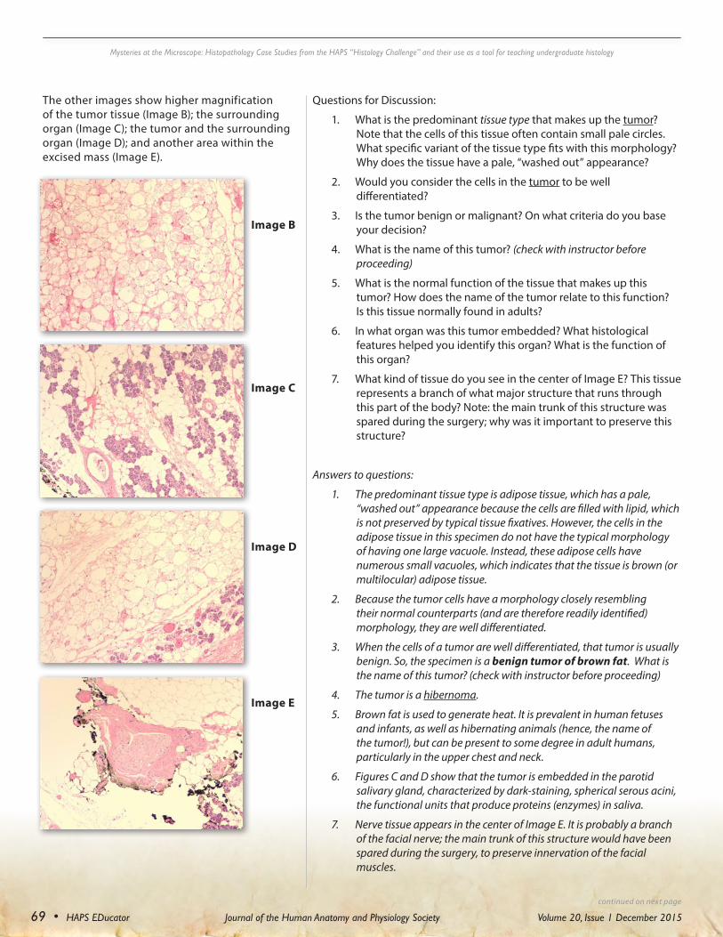
69 • HAPS EDucator Journal of the Human Anatomy and Physiology Society Volume 20, Issue 1 December 2015
The other images show higher magnification of the tumor tissue (Image B); the surrounding organ (Image C); the tumor and the surrounding organ (Image D); and another area within the excised mass (Image E).
Image B
Image C
Image D
Image E
Questions for Discussion:
1. What is the predominant tissue type that makes up the tumor? Note that the cells of this tissue often contain small pale circles. What specific variant of the tissue type fits with this morphology? Why does the tissue have a pale, “washed out” appearance?
2. Would you consider the cells in the tumor to be well differentiated?
3. Is the tumor benign or malignant? On what criteria do you base your decision?
4. What is the name of this tumor? (check with instructor before proceeding)
5. What is the normal function of the tissue that makes up this tumor? How does the name of the tumor relate to this function? Is this tissue normally found in adults?
6. In what organ was this tumor embedded? What histological features helped you identify this organ? What is the function of this organ?
7. What kind of tissue do you see in the center of Image E? This tissue represents a branch of what major structure that runs through this part of the body? Note: the main trunk of this structure was spared during the surgery; why was it important to preserve this structure?
Answers to questions:
1. The predominant tissue type is adipose tissue, which has a pale, “washed out” appearance because the cells are filled with lipid, which is not preserved by typical tissue fixatives. However, the cells in the adipose tissue in this specimen do not have the typical morphology of having one large vacuole. Instead, these adipose cells have numerous small vacuoles, which indicates that the tissue is brown (or multilocular) adipose tissue.
2. Because the tumor cells have a morphology closely resembling their normal counterparts (and are therefore readily identified) morphology, they are well differentiated.
3. When the cells of a tumor are well differentiated, that tumor is usually benign. So, the specimen is a benign tumor of brown fat. What is the name of this tumor? (check with instructor before proceeding)
4. The tumor is a hibernoma.
5. Brown fat is used to generate heat. It is prevalent in human fetuses and infants, as well as hibernating animals (hence, the name of the tumor!), but can be present to some degree in adult humans, particularly in the upper chest and neck.
6. Figures C and D show that the tumor is embedded in the parotid salivary gland, characterized by dark-staining, spherical serous acini, the functional units that produce proteins (enzymes) in saliva.
7. Nerve tissue appears in the center of Image E. It is probably a branch of the facial nerve; the main trunk of this structure would have been spared during the surgery, to preserve innervation of the facial muscles.
Mysteries at the Microscope: Histopathology Case Studies from the HAPS “Histology Challenge” and their use as a tool for teaching undergraduate histology
continued on next page

70 • HAPS EDucator Journal of the Human Anatomy and Physiology Society Volume 20, Issue 1 December 2015
CASE #3 (Histology Challenge #40, Teratoma)
Background material:
A 48 y/o woman came in for a routine pelvic exam for birth control pills. The gynecologist palpated an adnexal mass that she estimated at 4cm diameter, mobile, non-tender, and in the region of the right ovary. She then ordered a pelvic ultrasound that reported imaging a smooth cystic mass with complex heterogeneous echogenicity internally. The patient was scheduled for laparoscopic, possibly open excision, and was warned that she may lose her ovary on this side.
https://iame.com/online/ovary/ovary.html
Image A
Image B
Image C Image H
Image G
Image F
Image E
Image D
Mysteries at the Microscope: Histopathology Case Studies from the HAPS “Histology Challenge” and their use as a tool for teaching undergraduate histology
continued on next page
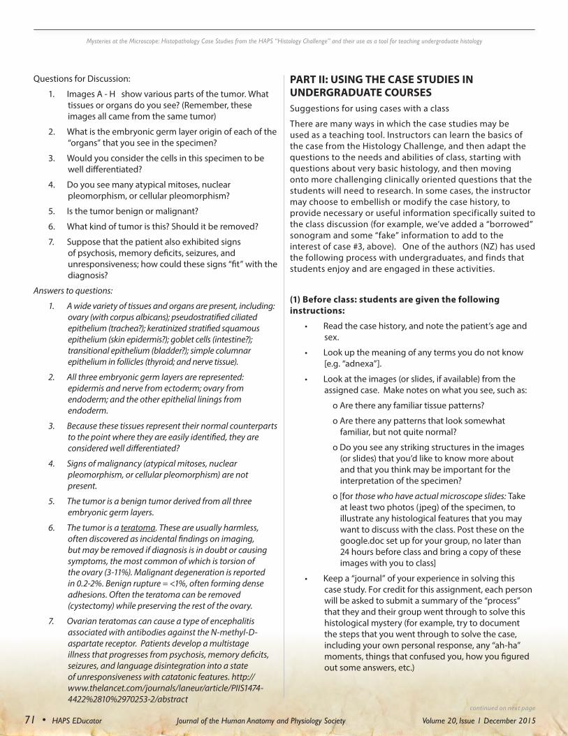
71 • HAPS EDucator Journal of the Human Anatomy and Physiology Society Volume 20, Issue 1 December 2015
Questions for Discussion:
1. Images A - H show various parts of the tumor. What tissues or organs do you see? (Remember, these images all came from the same tumor)
2. What is the embryonic germ layer origin of each of the “organs” that you see in the specimen?
3. Would you consider the cells in this specimen to be well differentiated?
4. Do you see many atypical mitoses, nuclear pleomorphism, or cellular pleomorphism?
5. Is the tumor benign or malignant?
6. What kind of tumor is this? Should it be removed?
7. Suppose that the patient also exhibited signs of psychosis, memory deficits, seizures, and unresponsiveness; how could these signs “fit” with the diagnosis?
Answers to questions:
1. A wide variety of tissues and organs are present, including: ovary (with corpus albicans); pseudostratified ciliated epithelium (trachea?); keratinized stratified squamous epithelium (skin epidermis?); goblet cells (intestine?); transitional epithelium (bladder?); simple columnar epithelium in follicles (thyroid; and nerve tissue).
2. All three embryonic germ layers are represented: epidermis and nerve from ectoderm; ovary from endoderm; and the other epithelial linings from endoderm.
3. Because these tissues represent their normal counterparts to the point where they are easily identified, they are considered well differentiated?
4. Signs of malignancy (atypical mitoses, nuclear pleomorphism, or cellular pleomorphism) are not present.
5. The tumor is a benign tumor derived from all three embryonic germ layers.
6. The tumor is a teratoma. These are usually harmless, often discovered as incidental findings on imaging, but may be removed if diagnosis is in doubt or causing symptoms, the most common of which is torsion of the ovary (3-11%). Malignant degeneration is reported in 0.2-2%. Benign rupture = <1%, often forming dense adhesions. Often the teratoma can be removed (cystectomy) while preserving the rest of the ovary.
7. Ovarian teratomas can cause a type of encephalitis associated with antibodies against the N-methyl-D-aspartate receptor. Patients develop a multistage illness that progresses from psychosis, memory deficits, seizures, and language disintegration into a state of unresponsiveness with catatonic features. http://www.thelancet.com/journals/laneur/article/PIIS1474-4422%2810%2970253-2/abstract
PART II: USING THE CASE STUDIES IN UNDERGRADUATE COURSES Suggestions for using cases with a class
There are many ways in which the case studies may be used as a teaching tool. Instructors can learn the basics of the case from the Histology Challenge, and then adapt the questions to the needs and abilities of class, starting with questions about very basic histology, and then moving onto more challenging clinically oriented questions that the students will need to research. In some cases, the instructor may choose to embellish or modify the case history, to provide necessary or useful information specifically suited to the class discussion (for example, we’ve added a “borrowed” sonogram and some “fake” information to add to the interest of case #3, above). One of the authors (NZ) has used the following process with undergraduates, and finds that students enjoy and are engaged in these activities.
(1) Before class: students are given the following instructions:
• Read the case history, and note the patient’s age and sex.
• Look up the meaning of any terms you do not know [e.g. “adnexa”].
• Look at the images (or slides, if available) from the assigned case. Make notes on what you see, such as:
o Are there any familiar tissue patterns?
o Are there any patterns that look somewhat familiar, but not quite normal?
o Do you see any striking structures in the images (or slides) that you’d like to know more about and that you think may be important for the interpretation of the specimen?
o [for those who have actual microscope slides: Take at least two photos (jpeg) of the specimen, to illustrate any histological features that you may want to discuss with the class. Post these on the google.doc set up for your group, no later than 24 hours before class and bring a copy of these images with you to class]
• Keep a “journal” of your experience in solving this case study. For credit for this assignment, each person will be asked to submit a summary of the “process” that they and their group went through to solve this histological mystery (for example, try to document the steps that you went through to solve the case, including your own personal response, any “ah-ha” moments, things that confused you, how you figured out some answers, etc.)
Mysteries at the Microscope: Histopathology Case Studies from the HAPS “Histology Challenge” and their use as a tool for teaching undergraduate histology
continued on next page

72 • HAPS EDucator Journal of the Human Anatomy and Physiology Society Volume 20, Issue 1 December 2015
(2) During class:
• Students work in groups, to answer the questions. (Ideally, each team of students works on a different case).
• Students look up information, as needed: cell phones, texts, computers are all permitted and encouraged!
• I recommend that, initially, the instructor needs to stay out of the way (to avoid giving away the answer too soon!), until group has a tentative diagnosis. At that point, the instructor can either confirm the answer, or guide the students in the right direction, both for the diagnosis and for additional information.
• Each group works on constructing a summary report, in the form of a PowerPoint (using the images) to present to the rest of the class.
(3) After class:
• Each team submits a summary report and/or the PowerPoint presentation.
• Each student submits their “journal”/ reflection paper.
• Each student submits a peer-evaluation of each member of the team; this evaluation contributes to each team member’s grade.
PART III: THE HISTOLOGY CHALLENGE, A RESOURCE FOR HISTOPATHOLOGICAL CASE STUDIESThe Histology Challenge, a regular feature on the electronic website for (HAPS), provides a series of case studies, each presented with photomicrographic images of actual pathological specimens and with ongoing blog discussions in which participants get to try their hand at diagnosing the disease. We have described the Histology Challenge in detail in a previous publication [(Zanetti, NC and William Karkow, MD (2014). The Histology Challenge: how it came about, how it works now, and thoughts about the future of this dynamic HAPS website feature. HAPS Educator. November 2014.] Current and archived Histology Challenges can be found by going to http://www.hapsweb.org/events/event_list.asp, and then clicking resources, teaching resources, and then either current or archived histology challenge. Any HAPS member can download photomicrographs for use.
Of the nearly 100 Histology Challenges that are posted, some are especially well suited for use in an undergraduate course, because of the obvious correlation of basic histology with the disease or interpretation of the specimen. Some Challenges that we especially recommend, and the basic topics that they illustrate, include:
• Challenge #28: Duodenal celiac sprue (intestinal histology, role of villi)
• Challenge #42: Suture granuloma (connective tissue, inflammation, macrophage)
• Challenge #46: Giardia, small intestine (intestinal histology; parasitology)
• Challenge #53: Leydig cell tumor (histology of testis; role of Leydig cells)
• Challenge #60: Maturing scar (connective tissue, epidermal structures, role of fibroblasts; protein synthesis)
• Challenge #69: Keloid (connective tissue, melanocytes; desmosomes; plasma cells)
• Challenge #86: Barrett’s Esophagus (epithelial types, structure/function relationships)
• Challenge #96: Ameloblastoma (tooth structure and development)
Nina Zanetti’s educational background includes a BS (Biology) from Muhlenberg College; a PhD (Biology) from Syracuse University; and a post-doctoral fellowship (in embryology) from University of Iowa. She has taught in the Department of Biology at Siena College for over thirty years. She began attending HAPS conferences in 2001, and has presented workshops at almost every annual conference since then. When she is not teaching or peering into microscopes, she enjoys playing a variety of musical instruments. [email protected]
About the Authors
■
Dr. William Karkow graduated from the University of Illinois with a BS in Chemistry. He earned his MD from the University of Iowa. Among his many accomplishments, he is the co-author of a book titled, “Atlas of Duplex Scanning”. He spent some of his career practicing in Cincinnati with 8 other vascular surgeons prior to moving to Iowa, where he practiced in rural settings, performing 800-1000 procedures per year. In 2005 he began a second career in teaching anatomy, physiology, and histology at the undergraduate level and taking pre-professional students on yearly short-term Christian medical missions in Mexico. His interests include music (17 piano compositions), canoeing (21 trips to Quetico Provincial Park, Canada), being a family man (10 children, 6 grandchildren), and sharing activities with his RN wife, Louaine, who has worn various hats as a surgical floor nurse, ICU nurse, RN 1st assist, and lately home health care nurse manager. He has been a fellow of the American College of Surgeons, having been Board Certified in both General Surgery and General Vascular Surgery. In 2003-2004 was the President of the Iowa Chapter of the American College of Surgeons. [email protected]
Mysteries at the Microscope: Histopathology Case Studies from the HAPS “Histology Challenge” and their use as a tool for teaching undergraduate histology

73 • HAPS EDucator Journal of the Human Anatomy and Physiology Society Volume 20, Issue 1 December 2015
The Human Microbiota: The Importance and Relevance of its Incorporation into Anatomy and Physiology Curricula
Lynette Sigola1, A. Lucia Fuentes2, Rosemary Oh-McGinnis1, Jacqueline Vapenik3, and Leonard Millis1.
Department of Biology1, Nursing3, Douglas College, 700 Royal Avenue, New Westminster, British Columbia, V3M 5Z5, Canada.2 Natural Sciences Department LaGuardia CC, CUNY, 31-10 Thomson Ave, Long Island City, NY.
AbstractIn recent years, information on the diversity and abundance of microorganisms associated with the human body has significantly expanded. Recent research on the microbiota reveals its importance in the maintenance of normal host physiology and particular dysbioses have been associated with disturbances of gastrointestinal and metabolic health, brain function, and immunological responses. College students in health sciences and related programs, to our knowledge, receive little information regarding the microbiota and its importance in health. Adequate descriptions of the microbiota are only recently starting to appear in undergraduate human anatomy and physiology textbooks and most curricula fail to address the topic of microbiota with the attention that it deserves. Given the growing amount of evidence demonstrating the vital role of the microbiota in body function, it is crucial to educate students about the microbiota in undergraduate human anatomy and physiology courses for health practitioners. In this review, we present examples of recent findings illustrating the importance of the microbiota, and discuss how we may incorporate the microbiota into the teaching of human anatomy and physiology courses.
Introduction Several concepts in biology are essential for an appreciation of the organization and function of the human body. Amongst these concepts are Schleiden and Schwann’s Cell Theory and Claude Bernard’s concept of homeostasis. The cell theory states that organisms are made up of cells that form the simplest structural and functional units of life. The correlation between structure and function is evident in the morphology and physiology of organelles and within specialized cells and organs. Homeostasis, the maintenance of a constant internal environment, provides a critical framework for understanding how the body survives moderate fluctuations in its internal and external environmental conditions.
Beyond these key principles, students taking courses in human anatomy and physiology would benefit from an understanding of the different types of organisms in the biosphere specifically those that can be found on the external and internal body surfaces of multicellular organisms. These organisms constitute the myriad of bacteria (Qin et al. 2010), fungi (Seed 2014), microscopic animals (Marchesi 2010) and non-cellular viruses (Wylie et al. 2014) that are found in and on the human body. The microbes associated with multicellular organisms are collectively known as the microbiota. The microbiota represent multiple cooperative interactions among various organisms that have co-evolved with multicellular organisms for millennia (Ley et al. 2008). The term microbiome refers to
the combined genomes of the microbiota, which is believed to be about a hundred times greater than that of the human host.
The first eukaryotic cells are thought to have formed about 1.5 billion years ago as a result of co-operation between different cell types. The Theory of Endosymbiosis proposes that bacterial cells were likely internalized by a primordial eukaryote, mutualisms were established, and the bacteria eventually became permanent endosymbionts of the larger cell. Prokaryotic endosymbionts are considered to be the ancestors of resident mitochondria and chloroplasts in some eukaryotes.
An increasing body of evidence now shows several critical roles for the microbiota in normal host physiology and in the maintenance of health (Pflughoeft and Versalovic 2012). Over the past decade there has been a concerted effort to identity the microbes on human body surfaces including the mouth, gastrointestinal tract, vagina and skin (The Human Microbiome Project Consortium 2012). Furthermore, when microbial composition and/or functions at these sites become disrupted, the resulting dysbiosis may be associated with disease (Tamboli et al. 2004). The microbiota has been documented to exist and play important roles in many areas of the external and internal surfaces of the human body. Over the past decade there has been a steady increase in the number of research publications indexed in PubMed that report studies defining the composition and function of the microbiota as well as studies that describe dysbioses and
continued on next page

74 • HAPS EDucator Journal of the Human Anatomy and Physiology Society Volume 20, Issue 1 December 2015
its pathophysiological role in the etiology of various human diseases (Figure 1 A and B).
Rather than viewing all microbes as pathogens, or potential pathogens, an alternative view of the human and microbiota interaction is to view it as an ecosystem that is composed of diverse populations of multitudes of different species that occupy particular ecological niches (Bengmark 1998, Dethlefsen et al. 2007, Costello et al. 2012). This ecological model of inter-species cooperation could be viewed as a particular type of “homeostasis” that extends beyond Claude Bernard’s view of the maintenance of specific parameters of the host’s internal environment to include the numerous mutualisms that encompass the entire microbiome/multicellular organism axis. A likely mechanism by which the human microbiota interacts with the host’s internal environment is by producing small molecules that play various roles in regulating physiological activity (Donia and Fischbach 2015).
Based on our personal teaching experiences, we have found that students taking human anatomy and physiology courses often lack sufficient background knowledge of important concepts regarding the microbiota. Instead, students in human anatomy and physiology courses often encounter microbes only as pathogens and learn about their harmful effects to the host. For example, we teach about pathogenic bacteria in the immunology unit in one of our human anatomy and phsyiology courses but we do not describe the flora that exist on and in us that contribute to host immunity in this unit. Overemphasis on the pathogenic nature of some bacteria may lead students to the flawed view that microbes are predominantly deleterious. This view is then often reinforced in courses such as medical microbiology where the predominant focus is on pathogenic organisms with almost no discussion of the beneficial functions of the human microbiota.
One of the only instances in which the human microbiota is typically mentioned in human anatomy and physiology courses is the presence of bacteria in the gastrointestinal tract that aid in production of specific vitamins and contribute to fermentation of non-digestible plant-based polysaccharides. However, this is only briefly mentioned without elaborating on the importance of the gut microbiota for maintenance of health in other systems such as the nervous system and the immune system. When we examined several editions of human anatomy and physiology textbooks at ramdom from a total of five different authors/publishers, we found that the term
“microbiota” did not appear in any of these human anatomy and physiology textbooks. While most textbooks had no mention of the microbiota, a few more recent editions of certain textbooks, notably two editions of “Human Anatomy and Physiology” by Marieb and Hoehn that were published by Pearson in 2010 and 2013, briefly mentioned the presence of bacteria in the gut. We were encouraged to find one recent edition of “Human Anatomy and Physiology” by Amerman, which was published by Pearson in 2016, that contained a short section entitled “bacteria in the large intestine”. We remain hopeful that more human anatomy and physiology textbooks will increase their content on the microbiota in the future. In the interim, we find the overall lack of discussion of the microbiota in most A&P textbooks to be troublesome.
The Human Microbiota: The Importance and Relevance of its Incorporation into Anatomy and Physiology Curricula
continued on next page
A
B
Figure 1. Publications indexed on PubMed related to the human microbiome, physiology and pathology. PubMed was used to determine the number of research publications for each year from 2004 to 2014 using the search terms “human microbiome and physiology” (Figure 1A) and “human microbiome and pathology” (Figure 1B).
0
500
1000
1500
2000
2500
3000
3500
2004 2005 2006 2007 2008 2009 2010 2011 2012 2013 2014
Num
ber o
f pub
licat
ions
Year
Human microbiome and physiology publications PubMed search
0
100
200
300
400
500
600
2004 2005 2006 2007 2008 2009 2010 2011 2012 2013 2014
Num
ber o
f pub
licat
ions
Year
Human microbiome and pathology publications PubMed search

75 • HAPS EDucator Journal of the Human Anatomy and Physiology Society Volume 20, Issue 1 December 2015
Our personal teaching experiences and the scarcity of coverage of the “microbiota” topic in anatomy and physiology textbooks have prompted us to lay out some ideas which may enable other instructors to incorporate the microbiota into their teaching. One of our goals as anatomy and physiology instructors is to provide students with the appropriate context for appreciating the relevance of the microbiota and the essential role it plays in maintaining health. In addition, we hope to increase awareness among our colleagues about the importance of the microbiota. In this review, we provide examples illustrating the critical roles of the human microbiota in the maintenance of gastrointestinal and metabolic health, mental health, and the immune system, We also provide examples where dysbioses are associated with pathology and suggest methods for incorporating microbiota teachings into human anatomy and physiology lectures.
Microbiota and Gut-Brain AxesPopular expressions such as “butterflies in your stomach” or having a “gut feeling” reflect the prevailing intuitive understanding we have regarding the connection between the brain and gut. When exploring digestive system physiology, students learn about this connection by studying modulation of the enteric nervous system by the central nervous system (CNS), as well as some of the feedback that maintains homeostasis of the “gut-brain axis”. Microbes are usually discussed in a targeted manner during anatomy and physiology classes. Students typically encounter gut microbes as they relate to the production of vitamins K and B12 or in the association of H. pylori and peptic ulcers or in the loss of fluids and electrolytes caused by V. cholera. Alternatively, presenting these and other examples in the light of a “bigger” picture in which the human gut microbiota are seen as an essential contributor to homeostasis involving the gut-brain axis would allow for the integration of an immense body of knowledge that has accumulated around the microbiota and normal physiology in recent years.
Between 1013 and 1014 microorganisms inhabit the human gastrointestinal tract (Gill et al. 2006), and the diversity of microbial taxa and genes found in the gut surpasses that of any other body site (The Human Microbiome Project Consortium 2012). Growing evidence suggests that healthy individuals have a balanced composition of different bacterial genera. Although there is great variation between individuals, three distinct “enterotypes”, characterized by differences in relative abundances of certain genera of bacteria, have been found to exist among humans (Arumugam et al. 2011). In spite of individual variations, it appears that maintenance of the overall balance in the composition of the gut microbiota is key to maintaining health including mental health and the health of the brain (Cryan and O’Mahony 2011). The effects of the microbiota
on the central nervous system (CNS) are not unidirectional since the CNS also influences the composition of the microbiota.
The dynamic and interconnected nature of the gut microbiota and CNS can be introduced in an anatomy and physiology course as part of the topics already in the curriculum, such as the parasympathetic and sympathetic, neuroendocrine and enteric nervous system pathways. For example, response to stress, used to illustrate the endocrine-nervous system axis, is a relatively straightforward pathway demonstrating the influence of the nervous system on the microbiota. During stress, secretion of the hormone cortisol is regulated by the hypothalamus-pituitary-adrenal axis and one of the effects of cortisol is the alteration in gut microbiota composition, resulting in dysbiosis (Cryan and Dinan 2012). Gut bacteria affect CNS function in multiple ways, and much of the knowledge in this area has been demonstrated by the effects of alteration in the gut microbiota on stress responses and behaviour (Cryan and Dinan 2012). An interesting example is provided by studies showing an association of fructose malabsorption with depression (Ledochowski et al. 2000). Certain types of bacteria use fructose as their substrate and, in individuals suffering from fructose malabsorption, these bacteria flourish. Eliminating fructose from the diet of depressed individuals alleviated symptoms, and these changes were associated with changes in the patients’ microbiota (Gibson et al. 2007). The gut microbiota also influences the CNS by synthesizing many neurotransmitters and neuromodulators, such as GABA (produced by Lactobacillus and Bifidobacterium spp.), serotonin (produced by Candida, Streptococcus, Escherichia and Enterococcus spp.) and dopamine (produced by Bacillus spp.) (reviewed in Cryan and Dinan 2012).
The influence of the CNS on the environment of gut microbes and the consequential effects on microbe composition is significant in maintaining homeostasis. Gut microbes, on the other hand, influence the brain and behaviour by producing metabolites and activating neural circuits that directly modulate the brain.
Microbiota and ObesityThe importance of understanding the human microbiota may be further illustrated by examining the relationship between gut microbiota and obesity. There is mounting evidence, not only of an association of particular microbiota regimes with obesity, but also of the complex interactions of the microbiota with human cells, that may be a significant causative factor in the development and maintenance of obesity (Harley and Karp 2012). Obesity is widely recognized as a serious public health problem. It is a risk in type 2 diabetes mellitus, coronary artery disease, stroke, hypertension, breast and other cancers, and osteoarthritis.
The Human Microbiota: The Importance and Relevance of its Incorporation into Anatomy and Physiology Curricula
continued on next page

76 • HAPS EDucator Journal of the Human Anatomy and Physiology Society Volume 20, Issue 1 December 2015
The World Health Organization reports an alarming increase in obesity, which has more than doubled in the last 35 years, with 13% of adults being obese and 39% being overweight in 2014 (http://www.who.int/mediacentre/factsheets/fs311/en/). In 2012, more than one third of adults in the United States were found to be obese (Ogden et al. 2014).
Factors such as genetics, diet, level and types of physical activity, as well as other social and environmental elements, have long been considered to contribute to obesity (Friedman 2009). More recently the relationship between obesity and changes in gut microbiota has become a focus of research. The two major classes of bacteria found in the human gut microbiota are the Bacteroides and Firmicutes (Bradlow 2014). Firmicutes predominate in obese individuals and obesity is associated with reduced bacterial diversity. In addition, there is increased production of endotoxins, including deoxycholic acid and lipopolysaccharides, in obese individuals. Together with the thinning of the mucosal lining of the intestine, the presence of certain bacteria and their associated endotoxins contributes to the chronic inflammation associated with obesity (Bradlow 2014).
There is compelling evidence for a role for gut microbiota in the pathogenesis of obesity. For example, when germ-free mice were transplanted with fecal microbiota from human twins discordant for obesity/leanness, it resulted in the following observations: mice that received microbiota from lean humans remained lean, while those that received microbiota from obese individuals became obese. Furthermore, for co-housed mice fed on a low-fat diet, the lean phenotype and their microbiota composition transferred to potentially obese mice, likely through coprophagia, and prevented them from becoming obese (Ridaura et al. 2013). Interestingly, the microbiota transfer appears to be diet dependent, occurring with a low fat, high fruit and vegetable diet, but not occurring with a high fat, low fruit and vegetable diet.
The microbiota is thought to influence the development and maintenance of obesity by increasing the efficiency of energy extraction in the gut of obese individuals (Turnbaugh et al. 2006). The microbiota of obese individuals promote production of triglycerides in the liver and triglyceride storage in adipocytes (Bäckhed et al. 2004). Understanding how the microbiota influence adiposity may have significant implications for preventing and treating obesity and metabolic disorders associated with obesity.
Microbiota and Immunity
The human immune system consists of diverse cellular and humoral mechanisms that together constitute the innate and adaptive arms of the immune response. The immune system protects against microbial invasion of host tissues and also detects altered host cells and transplant tissue. Protective mechanisms are activated
when the host recognizes intruders and/or altered host cells. Microorganisms express microbe associated molecular patterns (MAMPs) (Ausubel 2005). MAMPs are not found in mammals and are thus recognized as foreign by host immune cells through germ line encoded pattern recognition receptors (PRRs) located on host cell surfaces and within intracellular compartments (Medzhitov and Janeway 2002, Akira et al. 2006). It is speculated that MAMP-PRR interactions likely facilitated the commensal and mutualistic interactions between animals and microbes that have evolved over millennia. There is likely a distinction between host responses that promote tolerance to particular symbiotic microbes and others that are activated when mucosal barriers are breached. Danger signals are detected by the host which induce microbicidal inflammatory and immunological responses that limit the spread of invaders and set the stage for tissue repair.
The use of germ-free mice has revealed the significance of gut microbiota in contributing to obesity and to the development of the host immune system (Hooper et al. 2012). Mice maintained from birth in sterile environments have poorly developed gut associated mucosal tissues (Renz et al. 2011, Pollard and Sharon 1970). They have fewer intestinal mucosal goblet cells and thinner protective mucus barriers lining epithelial cell layers compared to conventionally reared mice with intact microbiomes (Deplancke and Gaskins 2001). Therefore, microbes are required for the development of the host immune system. In addition, there is now clear evidence that gut dysbioses predispose to several host disorders including those with an immunopathological basis such as inflammatory bowel disease (Joossens et al. 2011), allergies such as asthma (Arrieta and Finlay 2014) and autoimmunity (Kranich et al. 2011). Some useful examples that might be applied in classrooms to promote student appreciation of the significance of dysbioses include allergic diseases, Clostridium difficile infection and candidiasis.
Perturbations in microbial communities are thought to contribute to the high prevalence of allergic diseases observed in the post-industrial era, with an estimate of 400 million cases of allergic diseases world-wide (Pawankar 2014). The hygiene, microflora and “old friends” hypotheses postulate roles for the microbiota in modulating host immune responses (Brown et al. 2013). These hypotheses propose that excessive cleanliness, lack of exposure to farm animals or pets and early antibiotic treatment alter the focus of the immune response away from exposure to microbes that have co-existed with humans for multiple generations, and towards a heightened responsiveness to allergens that later give rise to allergic airway diseases including allergic rhinitis and asthma.
Specific studies have sought to understand how gut microbes regulate the allergic immune response. Murine studies show that colonization of the gut by organisms such as H. pylori or Clostridium strains modulates host T
The Human Microbiota: The Importance and Relevance of its Incorporation into Anatomy and Physiology Curricula
continued on next page

77 • HAPS EDucator Journal of the Human Anatomy and Physiology Society Volume 20, Issue 1 December 2015
lymphocytes to promote an anti-inflammatory phenotype that suppresses allergic airway responses, including eosinophil infiltration of the airways (Arnold et al. 2011; Atarashi et al. 2011). Discoveries such as this have helped to provide a mechanistic basis to explain previous observations that allergic diseases are less prevalent in children living in larger households (Strachan 1989), those living in rural compared to urban environments (Yemaneberhan et al. 1997), and those who are not exposed to antibiotics early in life (Kozyrskyj et al. 2007).
Further evidence for the importance of the microbiota in host physiology is evident from the adverse effects of antibiotic therapy on some patients, which results in the overgrowth of otherwise non-pathogenic commensal organisms leading to clinical symptoms such as C. difficile infection of the gut and vulvo-vaginal candidiasis. C. difficile causes significant nosocomial infections in patients who have undergone antibiotic treatments for other disorders (Barbut and Petit 2001). Antibiotics are thought to alter the normal gastrointestinal microbial community by reducing the relative abundance of Bacteroides and Firmicutes bacteria. This disruption fosters the overgrowth of C. difficile (Chang et al. 2008). Morbidity and mortality associated with C. difficile infections are high since this bacterium is resistant to many antibiotics and causes debilitating illness that is often particularly severe in the elderly. Fecal transplantation, the transfer of feces from normal donors to patients infected with C. difficile, corrects the dysbiosis by introducing normal microbial organisms into the gastrointestinal tract of sick patients. Several clinical trials have shown the efficacy of this approach, particularly in patients with recurrent C. difficile infections (Gough et al. 2011).
In women of reproductive age, another reported adverse effect of antibiotic use is the development of post-antibiotic vaginal candidiasis (Pirotta et al. 2003, Xu et al. 2008). It has been shown that vaginal Lactobacilli normally maintain an acidic vaginal environment through carbohydrate fermentation. This low pH helps to keep yeast cell numbers in check (Collins and Hardt 1980). Antibiotic use is thought to alter the vaginal microbial community, reducing the number of beneficial bacteria, such as Lactobacilli. This allows yeast cells to proliferate causing local disease. However, one randomized, double blind, placebo-controlled trial of probiotic Lactobacilli concluded that this approach did not prevent the occurrence of post-antibiotic vulvo-candidiasis (Pirotta et al. 2003). In contrast, another prospective study showed prophylaxis with lozenges containing Lactobacilli was effective in reducing oral Candida in elderly patients (Kraft-Bodi et al. 2015).
The results of intensive research endeavors like the Human Microbiome Project are likely to enhance our understanding of human and microbial interactions in the context of both health and disease. Students will increasingly need to integrate this knowledge into biological concepts as they learn human physiology, pathophysiology and
novel therapeutic approaches to managing disorders. For example, current investigations into the efficacy of fecal transplantation to treat C. difficile illustrate how an appreciation of dysbiosis management may impact patient therapies in the future.
DiscussionIn light of the recent findings pointing to the importance of the human microbiota in normal physiology, how can we begin to incorporate this information into our anatomy and physiology courses? One of the main challenges of teaching the microbiota with relative ease lies in the fact that there is limited or no guidance available from anatomy and physiology textbooks. Although instructors may introduce the microbiota in their lectures, we believe a deliberate effort to make it part of the anatomy and physiology curriculum will be the most effective way to ensure that every student is exposed to this growing body of knowledge and has a basic understanding of the human microbiota and its importance in health.
One fairly simple strategy to get students thinking about the microbiota in antomy and physiology courses is the use of case-based and/or problem-based pedagogical approaches (Handelsman et al. 2007). Such strategies are effective teaching tools that can be used in teaching anatomy and physiology courses to help students engage with clinically relevant concepts. C. difficile, among other examples, can easily be discussed in the classroom as a case-based problem.
Other approaches to incorporate microbiota-related concepts in the curriculum are through student activities in the laboratory. Many routine laboratory procedures, such as a buccal cavity scrape to examine epithelial cells, can be used to illustrate the presence of microbiota in the mouth since students often encounter bacteria while viewing methylene blue stained epithelial cells using a light microscope. Tying together the role of the microbiota with the role of human cells can serve to further broaden and strengthen student background knowledge in biology.
There are several informative and helpful websites that aim to describe how to teach the microbiota to students at various levels (http://www.genome.gov/27552808; http://learn.genetics.utah.edu/content/microbiome/; http://www.actionbioscience.org/genomics/the_human_microbiome.html). These websites can be provided to students as resources to view in their own time and may also serve as useful resources for instructors during preparation of lecture material. Instructors may be able to take advantage of recent Microbiology textbooks for guidance and lecture material related to microbiota. We have found several Microbiology textbooks that are exceptional at describing the human microbiota and it is our hope that human anatomy and physiology textbooks will soon follow in the same direction.
The Human Microbiota: The Importance and Relevance of its Incorporation into Anatomy and Physiology Curricula
continued on next page

78 • HAPS EDucator Journal of the Human Anatomy and Physiology Society Volume 20, Issue 1 December 2015
ConclusionIn addition to the concerns we have raised in this article, other groups have similarly noted an absence of microbiota-related teaching in undergraduate premedical programs. This omission may have adverse implications including unnecessary costs in healthcare and treatment of patients (Smith et al. 2015, Taggart and Bergstrom 2014). Although there is currently insufficient microbiota content in human anatomy and physiology textbooks, we urge instructors to consider incorporating microbiota teachings into lectures and laboratories. Inclusion of microbiota-related topics in discussions about human health and disease will provide students with a more thorough and accurate appreciation of the complexity of the human body. We anticipate that this will benefit students in the long-term. We hope that this article will encourage educators to include the microbiota more formally as part of the mainstream curricula in anatomy and physiology courses.
Literature CitedAkira, S., Uematsu, S., and Takeuchi, O. (2006). Pathogen
recognition and innate immunity. Cell 124(4): 783-801. Arnold, I.C., Dehzad, N., Reuter, S., Martin, H., Becher, B.,
Taube, C., and Müller, A. (2011). Helicobacter pylori infection prevents allergic asthma in mouse models through the induction of regulatory T cells. Journal of Clinical Investigation 121(8): 3088-3093.
Arrieta, M.C. and B Finlay, B. (2014). The intestinal microbiota and allergic asthma. Journal of Infection 69(1): S53–S55.
Arumugam et al. (2011). Enterotypes of the human gut microbiome. Nature 473(7346): 174-80.
Atarashi et al. (2011). Induction of colonic regulatory T cells by indigenous Clostridium species. Science 331(6015): 337-341.
Ausubel, F.M. (2005). Are innate immune signaling pathways in plants and animals conserved?” Nature Immunology 6(10): 973-979.
Bäckhed, F., Ding, H., Wang, T., Hooper, L.V., Koh, G.Y., Nagy, A., Semenkovich, C.F., and Gordon, J.I. (2004). The gut microbiota as an environmental factor that regulates fat storage. Proc Natl Acad Sci 101(44): 15718-15723.
Barbut, F. and Petit, J.C. (2001). Epidemiology of Clostridium difficile-associated infections. Clinical Microbiology Infection 7(8): 405-410.
Bengmark, S. (1998). Ecological control of the gastrointestinal tract: the role of probiotic flora. Gut 42(1): 2-7.
Bradlow, H.L. (2014). Obesity and the gut microbiome: pathophysiological aspects. Hormone Molecular Biology Clinical Investigation 17(1): 53-61.
Brown, E.M., Arrieta, M.C. and Finlay, B.B. (2013). A fresh look at the hygiene hypothesis: how intestinal microbial exposure drives immune effector responses in atopic disease. Seminars in Immunology 25(5): 378-387.
Chang, J.Y., Antonopoulos, D.A., Kalra, A., Tonelli, A., Khalife, W.T., Schmidt, T.M., and Young, V.B. (2008). Decreased diversity of the fecal Microbiome in recurrent Clostridium difficile-associated diarrhea. Journal of Infectious Diseases 197(3): 435-438.
Collins, E.B. and Hardt, P. (1980). Inhibition of Candida albicans by Lactobacillus acidophilus. Journal of Dairy Science 63(5): 830-832.
Costello, E.K., Stagaman, K., Dethlefsen, L., Bohannan, B.J.M., and Relman, D.A. (2012). The application of ecological theory towards an understanding of the human microbiome. Science 336(6086): 1255–1262.
Cryan, J.F. and O’Mahony, S.M. (2011). The microbiome-gut-brain axis: from bowel to behavior. Neurogastroenterol Motil 23: 187–192.
Cryan, J.F. and Dinan, T.G. (2012). Mind-altering microorganisms: the impact of the gut microbiota on brain and behaviour. Nature Reviews Neuroscience 13: 701-712.
Deplancke, B and Gaskins, H.R. (2001). Microbial modulation of innate defense: goblet cells and the intestinal mucus layer. American Journal of Clinical Nutrition 73: 1131s-1141s.
Dethlefsen, L., McFall-Ngai, M., Relman, D.A. (2007). An ecological and evolutionary perspective on human-microbe mutualism and disease. Nature 449(7164): 811-818.
Donia, M.S. and Fischbach, M.A. (2015). Small molecules from the human microbiota. Science 349(6246): 1254766.
Friedman, J.M. (2009). Obesity: Causes and control of excess body fat. Nature 459(7245): 340-342.
Gibson, P.R., Newnham, E., Barrett, J.S., Shepherd, S.J., and Muir J.G. (2007). Review article: fructose malabsorption and the bigger picture. Aliment Pharmacol Ther 25(4): 349–363.
Gill, S.R., Pop, M., DeBoy, R.T., Eckburg, P.B., Turnbaugh, P.J., Samuel, B.S., Gordon J.I., Relman, D.A., Fraser-Liggett, C.M., and Nelson, K.E. (2006). Metagenomic Analysis of the Human Distal Gut Microbiome. Science 312(5778): 1355-1359.
Gough, E., Shaikh, H., and Manges, A.R. (2011). Systematic review of intestinal microbiota transplantation (fecal bacteriotherapy) for recurrent Clostridium difficile infection. Clinical Infectious Diseases 53(10): 994-1002.
The Human Microbiota: The Importance and Relevance of its Incorporation into Anatomy and Physiology Curricula
continued on next page
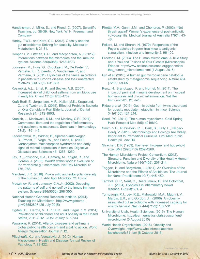
79 • HAPS EDucator Journal of the Human Anatomy and Physiology Society Volume 20, Issue 1 December 2015
Handelsman, J., Miller, S., and Pfund, C. (2007). Scientific Teaching, pp. 38-39. New York: W. H. Freeman and Company.
Harley, T.W.L. and Karp, C.L. (2012). Obesity and the gut microbiome: Striving for causality. Molecular Metabolism 1: 21-31.
Hooper, L.V., Littman, D.R., and Macpherson, A.J. (2012). Interactions between the microbiota and the immune system. Science 336(6086): 1268-1273.
Joossens, M., Huys, G., Cnockaert, M., De Preter, V., Verbeke, K., Rutgeerts, P., Vandamme, P., and Vermeire, S. (2011). Dysbiosis of the faecal microbiota in patients with Crohn’s disease and their unaffected relatives. Gut 60(5): 631-637.
Kozyrskyj, A.L., Ernst, P., and Becker, A.B. (2007). Increased risk of childhood asthma from antibiotic use in early life. Chest 131(6):1753-1759.
Kraft-Bodi, E., Jørgensen, M.R., Keller, M.K., Kragelund, C., and Twetman, S. (2015). Effect of Probiotic Bacteria on Oral Candida in Frail Elderly. Journal of Dental Research 94: 181S-186S.
Kranich, J., Maslowski, K.M., and Mackay, C.R. (2011). Commensal flora and the regulation of inflammatory and autoimmune responses. Seminars in Immunology 23(2): 139–145.
Ledochowski, M., Widner, B., Sperner-Unterweger, B., Propst, T., Vogel, W., and Fuchs, D. (2000). Carbohydrate malabsorption syndromes and early signs of mental depression in females. Digestive Diseases and Sciences 45: 1255–1259.
Ley, R., Lozupone, C.A., Hamady, M., Knight, R., and Gordon, J. (2008). Worlds within worlds: evolution of the vertebrate gut microbiota. Nat Rev Microbiol 6: 776–788.
Marchesi, J.R. (2010). Prokaryotic and eukaryotic diversity of the human gut. Adv Appl Microbiol 72: 43-62.
Medzhitov, R. and Janeway, C.A.Jr. (2002). Decoding the patterns of self and nonself by the innate immune system. Science 296(5566): 298-300.
National Human Genome Research Institute. (2014). Teaching the Microbiome. http://www.genome.gov/27552808 (25 July 2015)
Ogden,C.L., Carroll, M.D., Kit,B.K., and Flegal, K.M. (2014). Prevalence of childhood and adult obesity in the United States, 2011-2012. JAMA 311(8): 806-814.
Pawankar, R. (2014). Allergic diseases and asthma: a global public health concern and a call to action. World Allergy Organization Journal 7: 12.
Pflughoeft, K.J. and Versalovic, J. (2012). Human Microbiome in Health and Disease. Annual Review of Pathology 7: 99-122.
Pirotta, M.V., Gunn, J.M., and Chondros, P. (2003). “Not thrush again!” Women’s experience of post-antibiotic vulvovaginitis. Medical Journal of Australia 179(1): 43-46.
Pollard, M. and Sharon, N. (1970). Responses of the Peyer’s patches in germ-free mice to antigenic stimulation. Infection and Immunity 2: 96-100.
Proctor, L.M. (2013). The Human Microbiome: A True Story about You and Trillions of Your Closest (Microscopic) Friends. http://www.actionbioscience.org/genomics/the_human_microbiome.html (4 August 2015)
Qin et al. (2010). A human gut microbial gene catalogue established by metagenomic sequencing. Nature 464 (7285): 59-65.
Renz, H., Brandtzaeg, P. and Hornef, M. (2011). The impact of perinatal immune development on mucosal homeostasis and chronic inflammation. Nat Rev Immunol 201, 12: 9-23.
Ridaura et al. (2013). Gut microbiota from twins discordant for obesity modulate metabolism in mice. Science 341(6150): 1241214.
Seed, P.C. (2014). The human mycobiome. Cold Spring Harb Perspect Med 5(5): a019810.
Smith, V.H., Rubinstein, R.J., Park, S., Kelly, L., Klepac-Ceraj, V. (2015). Microbiology and Ecology Are Vitally Important to Premedical Curricula. Evol Med Public Health pii: eov014.
Strachan, D.P. (1989). Hay fever, hygiene, and household size. BMJ 299(6710):1259-1260.
The Human Microbiome Project Consortium. (2012). Structure, Function and Diversity of the Healthy Human Microbiome. Nature 486(7402): 207–214.
Taggart, H. and Bergstrom, L. (2014). An Overview of the Microbiome and the Effects of Antibiotics. The Journal for Nurse Practitioners 10(7): 445-450.
Tamboli, C. P., Neut, C., Desreumaux, P., and Colombel, J. F. (2004). Dysbiosis in inflammatory bowel disease. Gut 53(1): 1–4.
Turnbaugh, P.J., Ley, R.E., Mahowald, M.A., Magrini, V., Mardis, E.R., and Gordon, J.I. (2006). An obesity-associated gut microbiome with increased capacity for energy harvest. Nature 444(7122): 1027-31.
University of Utah, Health Sciences. (2015). The Human Microbiome. http://learn.genetics.utah.edu/content/microbiome/ (5 August 2015)
World Health Organization. (2015). Obesity and Overweight. http://www.who.int/mediacentre/factsheets/fs311/en/ (8 October 2015)
The Human Microbiota: The Importance and Relevance of its Incorporation into Anatomy and Physiology Curricula
continued on next page

80 • HAPS EDucator Journal of the Human Anatomy and Physiology Society Volume 20, Issue 1 December 2015
Wylie, K. M., Mihindukulasuriya, K. A., Zhou, Y., Sodergren, E., Storch, G.A., and Weinstock, G.M. (2014). Metagenomic analysis of double-stranded DNA viruses in healthy adults. BMC Biology 12: 71.
Xu, J., Schwartz, K., Bartoces, M., Monsur, J., Severson, R.K., and Sobel, J.D. (2008). Effect of antibiotics on vulvovaginal candidiasis: a MetroNet study. J Am Board Fam Med 21(4): 261-268.
Yemaneberhan, H., Bekele, Z., Venn, A., Lewis, S., Parry, E., and Britton, J. (1997). Prevalence of wheeze and asthma and relation to atopy in urban and rural Ethiopia. Lancet 350(9071): 85-90.
The Human Microbiota: The Importance and Relevance of its Incorporation into Anatomy and Physiology Curricula
About the Authors
Lynette Sigola, MBChB (Hons), Ph.D., is an Instructor in the Biology Department at Douglas College where she teaches Anatomy and Physiology, General Biology and Cell Biology. She is also a part-time Clinical Assistant Professor in the Faculty of Dentistry at the University of British Columbia. Lynette has research interests in neuroendocrine regulation of macrophage functions and
microbial modulation of macrophage phagocytosis.
A. Lucia Fuentes, Ph.D., is an Assistant Professor at LaGuardia, CUNY, where she teaches Anatomy and Physiology, Cell Biology and Microbiology. Before this, she taught at Douglas College where she also collaborated in researching aspects of innate immunity related to polymicrobial infections.
Rosemary Oh-McGinnis, Ph.D., is a Biology Instructor at Douglas College where she teaches Anatomy and Physiology. She has previously worked as an education strategist for the undergraduate Biology degree program at the University of British Columbia.
Jacqueline Vapenik, BSc, MN, NP(F), teaches theory, lab and clinical for the undergraduate nursing program at Douglas College. She also works as a critical care nurse in the Intensive Care Unit at Royal Columbian Hospital, New Westminster, BC, and as Nurse Practitioner in family practice and tropical medicine in a community health clinic.
Leonard Millis, Ph.D., teaches Genetics, Anatomy and Physiology and General Biology in the Biology Department at Douglas College. He also contributes to a collaborative research project involving the use of murine macrophages to study cell receptors involved in modulating phagocytosis.
■

81 • HAPS EDucator Journal of the Human Anatomy and Physiology Society Volume 20, Issue 1 December 2015
The Meaning of the Arrow: Cognitive Roles of Graphic Symbols in Anatomy and Physiology
Vasiliy Kolchenko, M.D./Ph.D., Associate Professor, Department of Biological Sciences,
New York City College of Technology, The City University of New York
P313, 300 Jay St., Brooklyn, NY 11201, tel. 718-260-5954
AbstractAnatomy and physiology are highly visual fields of study and graphic symbols are ubiquitous in learning materials. Arrows, lines, boxes, bars and circles can be extremely helpful cognitive devices, but when they are not used correctly, they can be misleading. This paper investigates the cognitive role of graphic symbols and suggests two ways in which graphic symbols may be made into more effective learning tools: 1) by improving the design of the learning materials and 2) by using these materials in a way that helps utilize and strengthen student interpretive skills. In this study, we analyzed graphic symbols in a set of Infograms for Anatomy and Physiology I, which were developed at New York City College of Technology (Kolchenko 2015). The arrow was the most commonly used symbol and it seemed to have the greatest importance for student understanding of the material. Students who understood the meaning of the arrow symbols showed better comprehension of the concepts they needed to excel in anatomy and physiology. The results of this study suggest that our students may have better interpretive skills than they display in the classroom.
Introduction Rationale Humans have been using graphics for self-expression and knowledge representation since the time of the earliest cave paintings about 30,000 years ago and the first pictogram-based writing systems about 7,000 years ago. Ancient maps, calendars and diagrams are all prototypes of modern educational graphics (Tversky et al. 2000).
Graphics are particularly important in the study of anatomy and physiology. In anatomy, students learn anatomical structures visually and support this knowledge with descriptive text. In macroscopic anatomy, the images are mostly three-dimensional (dissections, photographs, drawings) and in microscopic anatomy, mostly two-dimensional (microscope slides). Other types of images include radiologic images such as x-rays, MRIs, CT scans and sonograms.
In physiology, students learn the description of the functions while supporting the description with animations and diagrams. In anatomy and physiology, various diagrams can convey meaning and make student comprehension most efficient because the diagrams use symbolic graphic representations such as arrows, lines, boxes, bars, circles, crosses, abbreviations and others. These ubiquitous graphics are extremely helpful cognitive tools, but when
they are not used correctly, they can be misleading and counterproductive.
We often say, “A picture is worth a thousand words.” This implies that an image can clarify a complex concept at a glance, much better and faster than a verbal description. As a result, large amounts of information can be internalized and recalled very quickly. The major steps that ensure this cognitive success are: a) optimizing the design of graphics and b) improving students’ graphic interpretive skills and their ability to read visual context.
Background The history of using different kinds of graphic symbols in science has been studied extensively. For example, the male sign was originally known as a sign for iron and the planet Mars and was derived from Greek initial letter of the god’s name, Thouros, which was used for naming the planet. Similarly, the female sign was a sign for copper and the planet Venus. Linnaeus first used the signs for male and female plants in the 18th century. Later, they become conventional symbols for male and female sex and for the characteristics of sex (Stearn 1962).
The analysis and improvement of graphic design for visualizing information was the subject of many studies (Tufte 1983, 1990, 1997 and 2006, Horn 1998). The attempts to categorize graphic symbols and present them as the
continued on next page

82 • HAPS EDucator Journal of the Human Anatomy and Physiology Society Volume 20, Issue 1 December 2015
universal visual language of the new globalized age demonstrated a great diversity of graphics and their tendency to mean different things in different contexts (Dreyfuss 1984, Horn 1998).
Studies of multiple meanings of graphic symbols and the significance of the visuospatial representation showed connections between form and function and between understanding the graphics and cognitive success (Tversky et al. 2000, Tversky 2001, 2005, Heiser and Tversky 2006, Fantini 2006). Many possible meanings of arrows were described by Horn in 1998 (Horn 1998). However, this did not bring greater clarity to our understanding of cognitive problems in using visual abstractions.
Automated interpretation of arrows proved to be a challenge in computer science (Kurata and Egenhofer 2005). From the pedagogical point of view, the importance of understanding the symbolic language of graphics was studied in chemistry (Marais 2000) and in biology (Kragten et al. 2015). The studies showed significant difficulties in symbol comprehension for many students. Eye-tracking and think-aloud studies for presented diagrams with multiple arrows indicated that students who understood the significance of arrows and watched the arrows closely had a better chance for successful learning (Kragten et al. 2015). Marais and Jordaan made suggestions for the deliberate study of the graphic symbolic language in science but their hypothesis was not tested (Marais and Jordaan 2000).
Materials and Methods: Infograms for Better Learning In this study, we analyzed graphic symbols in a set of Infograms for Anatomy and Physiology I, which has been developed and used for a few semesters at New York City College of Technology, an urban federally designated Hispanic Serving Institution with an open access policy. One Infogram condenses the material of a textbook chapter to one page of graphic symbols. (Figure 1) Supplemented by the legend and explanatory text, it provides a valuable cognitive tool for information encoding and decoding, for multiple retrieval and fast review of the material, and for understanding a ‘big
picture’ of the topic and being able to see the connections between the concepts. There was an observed improvement of the learning outcomes in the groups of students who used Infograms vs. the control (Kolchenko 2015).
The Infograms seemed particularly appropriate learning materials for analysis because they were designed by deliberately using symbolic graphics. The objective was to utilize the cognitive advantages of the visual symbolic language to the greatest extent. There was no other choice for students, given the amount of information and the space constraints. The level of graphic abstraction was optimized empirically, based on student feedback.
Arrows were selected as the main focus of the paper for two reasons. First, they were the most common symbols used in the Infograms. Second, they appeared to have the greatest importance for understanding the material. Knowing the meaning of arrows was a good predictor for better learning outcomes. This was understandable as the arrows often express the meaning of the picture, much more so than other visual elements did.
When developing the Infograms, I did not favor any type of symbol, or avoid using any type of symbol, nor was I aware that so many arrows had been used. The selection of graphics for Infogram design was intuitive, based on the need of the narrative. It has been a surprising discovery to
realize the special cognitive role that arrows played in so many of the Infogram units.
An additional question about students’ prior knowledge of arrows was included in a regular classroom quiz. This was as much a learning activity as it was an assessment question. It provided an opportunity for students to reflect on the numerous meanings of arrows, which, in turn, helped them understand the graphics better.
Other ways of learning about the cognitive roles of graphic symbols included: 1) asking individual students about the meaning of arrows in the Infograms and considering their explanations; 2) analyzing student versions of the Infograms that they recreated as a quiz in the lab.
The Meaning of the Arrow: Cognitive Roles of Graphic Symbols in Anatomy and Physiology
continued on next page

83 • HAPS EDucator Journal of the Human Anatomy and Physiology Society Volume 20, Issue 1 December 2015
Results and AnalysisInfograms and Arrows Among all graphic symbols in the Infograms, the most common one was the arrow. In total, it has been used 232 times, about 17 times per Infogram. The numerous arrows on the same page often meant a number of different things. Most of them represented various kinds of direction, in space, time, cause and effect, and others. This made sense because a typical arrow is a line with the arrowhead, which is a line that is not symmetrical and, therefore, can signify directionality. A student may know the usual basic meaning of the symbol but still become confused about its particular scenario and application. Clustering the meanings in a relatively small number of easy-to-understand groups can help prepare students to these scenarios in advance and reduce the confusion. Our preliminary analysis allowed us to identify the following partially overlapping categories of meaning for the arrows.
1. Direction in space either pointing or actual movement (Figure 2 and Figure 3)
2. Direction in time or timelines (Figure 4)
3. Direction of quantitative change, for example: up, more ; down, less (Figure 5)
4. Direction of physical change, for example, arrows in chemical reactions:
2H2 + O2 2H2O (Figure 6)
5. Causal direction: cause effect (Figure 7)
6. Dimensional direction: size, extent: , (Figure 8)
Figure 2. Pointing to the apical (A) and basal (B) surfaces of the epithelial cells
Figure 3. Movement of water through the cell membrane: isotonic solution (top), hypo-tonic solution (middle) and hypertonic solution (bottom)
Figure 4. Timeline for some major developments in biology
Figure 5. Increase and decrease – sympathetic stimulation increases heart rate and contractility; parasympathetic stimulation decreases them
Figure 6. ATP hydrolysis and synthesis; lightning bolt represents energy
Figure 7. Cause and effect – bone fracture and callus formation
Figure 8. Microscope working distance
The Meaning of the Arrow: Cognitive Roles of Graphic Symbols in Anatomy and Physiology
continued on next page
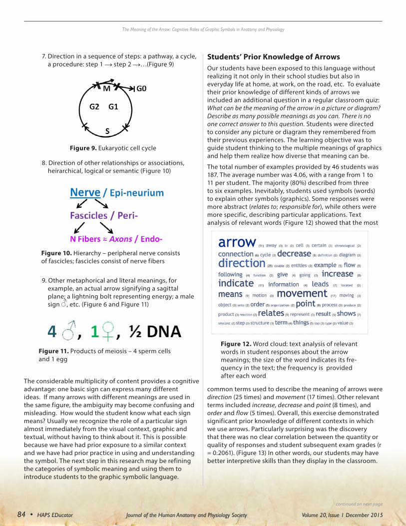
84 • HAPS EDucator Journal of the Human Anatomy and Physiology Society Volume 20, Issue 1 December 2015
7. Direction in a sequence of steps: a pathway, a cycle, a procedure: step 1 step 2 …(Figure 9)
8. Direction of other relationships or associations, heirarchical, logical or semantic (Figure 10)
9. Other metaphorical and literal meanings, for example, an actual arrow signifying a sagittal plane; a lightning bolt representing energy; a male sign , etc. (Figure 6 and Figure 11)
The considerable multiplicity of content provides a cognitive advantage: one basic sign can express many different ideas. If many arrows with different meanings are used in the same figure, the ambiguity may become confusing and misleading. How would the student know what each sign means? Usually we recognize the role of a particular sign almost immediately from the visual context, graphic and textual, without having to think about it. This is possible because we have had prior exposure to a similar context and we have had prior practice in using and understanding the symbol. The next step in this research may be refining the categories of symbolic meaning and using them to introduce students to the graphic symbolic language.
Students’ Prior Knowledge of Arrows Our students have been exposed to this language without realizing it not only in their school studies but also in everyday life at home, at work, on the road, etc. To evaluate their prior knowledge of different kinds of arrows we included an additional question in a regular classroom quiz: What can be the meaning of the arrow in a picture or diagram? Describe as many possible meanings as you can. There is no one correct answer to this question. Students were directed to consider any picture or diagram they remembered from their previous experiences. The learning objective was to guide student thinking to the multiple meanings of graphics and help them realize how diverse that meaning can be.
The total number of examples provided by 46 students was 187. The average number was 4.06, with a range from 1 to 11 per student. The majority (80%) described from three to six examples. Inevitably, students used symbols (words) to explain other symbols (graphics). Some responses were more abstract (relates to; responsible for), while others were more specific, describing particular applications. Text analysis of relevant words (Figure 12) showed that the most
common terms used to describe the meaning of arrows were direction (25 times) and movement (17 times). Other relevant terms included increase, decrease and point (8 times), and order and flow (5 times). Overall, this exercise demonstrated significant prior knowledge of different contexts in which we use arrows. Particularly surprising was the discovery that there was no clear correlation between the quantity or quality of responses and student subsequent exam grades (r = 0.2061). (Figure 13) In other words, our students may have better interpretive skills than they display in the classroom.
The Meaning of the Arrow: Cognitive Roles of Graphic Symbols in Anatomy and Physiology
continued on next page
Figure 9. Eukaryotic cell cycle
Figure 10. Hierarchy – peripheral nerve consists of fascicles; fascicles consist of nerve fibers
Figure 12. Word cloud: text analysis of relevant words in student responses about the arrow meanings; the size of the word indicates its fre-quency in the text; the frequency is provided after each word
Figure 11. Products of meiosis – 4 sperm cells and 1 egg

85 • HAPS EDucator Journal of the Human Anatomy and Physiology Society Volume 20, Issue 1 December 2015
Cognitive Roles of Arrows
Systemic observation of individual students explaining particular visual elements in recently learned Infograms provided a complex picture. The students who knew the meaning of the arrows understood the material. The students who were not sure about the meaning of the visual elements usually did not. A few students who provided high quality examples for the prior knowledge question were at a loss explaining arrows in the specific diagrams. There may be various reasons for this, depending on the individual and the particular situation. One obvious contributor to confusion was using the wrong explanation for the arrow. Not knowing what the true meaning of the arrow was led to one false guess after another.
Analysis of the Infograms recreated by students from memory provided more evidence for the same conclusion. The students who missed or misplaced arrows in the Infograms could not explain the topic well. This group usually represented a small fraction of the class, about 10-20%. The ability to identify individuals who needed additional intervention helped those individuals focus their attention on the connections between the concepts. The cognitive roles of the arrows were like two sides of the same coin. When the arrows helped, they helped a lot. When they confused, they confused a lot. Both roles seemed to be the direct result of the multiple meanings of the symbols.
Discussion and ConclusionSymbols in the Classroom Why do some students understand graphics better and faster than others? The wide-spread confusion on the part of some students may stem, from the inadequate practice of using or, rather, misusing educational graphics. Students develop a mental habit of seeing the image not as a dynamic system of interconnected elements but as a set of static unrelated graphics that must be memorized, recalled during the exam, and then soon forgotten.
This rigidity of mind is often limited to the classroom. The same student may understand graphics very well while driving and reading the road signs, browsing online, or playing a video game. In class, however, the flexibility of the mind is gone, and student cognitive capacity is frozen. What can bring it back? One of the steps may be the introductory conversation about the meaning of arrows and how they bring other graphics together.
As the study led to the better understanding of the cognitive roles of graphic symbols, I started asking students to focus on the connections represented by the arrows in advance. In the process of revising learning materials, I made the usage of arrows more consistent and less ambiguous. I also emphasized various meanings of graphic symbols during the classroom discussions.
Students responded by paying more attention to the connections between the concepts. They became more attuned to the dynamic side of the diagrams, which were often represented by the arrows, and spent less time focusing on memorizing the basic structural elements of the image. The particular cognitive role of the arrows, representing connections and directions of many different kinds, makes them one of the central graphic symbols in anatomy and physiology visuals. Better understanding of this special role may benefit both the instructor and the student and advance student metacognitive skills.
Multiple Meaning: Words vs. Non-verbal Graphics Words, written and spoken, are symbols as well. They may also have more than one meaning. In this case, they are called homonyms (Klein and Murphy 2001). For example, a river bank and a savings bank are homonyms. We usually recognize what the word stands for from the verbal context. Sometimes the ambiguity may lead to misunderstandings, but it is not very likely to become a big cognitive problem. The reasons for this are two-fold. First, although there are many known homonyms, they represent a relatively small fraction of our vocabulary. Second, most homonyms have two or three meanings, not a dozen.
All basic non-verbal graphics like arrows and circles have a large number of meanings. This makes them both economical and robust. This also creates more significant cognitive challenges. The graphics are economical because they are simple shapes or lines, which are easy to draw and recognize. They are robust because they can represent virtually unlimited number of concepts associated with their inherent features (directionality for arrows; roundness for circles).
The Meaning of the Arrow: Cognitive Roles of Graphic Symbols in Anatomy and Physiology
continued on next page
Figure 13. The relationship between the average exam scores of individual students (x) and the number of the arrow meaning examples given by the same student (y), r = 0.2061

86 • HAPS EDucator Journal of the Human Anatomy and Physiology Society Volume 20, Issue 1 December 2015
For example, a plus sign (+) may represent the following:
1. Addition of numbers or quantities (2 + 2)
2. A positive number or variable (+1, +x) vs. a negative number or variable (-1, -x)
3. Interaction of chemicals in a chemical reaction: 2H2 + O2 2H2O
4. Yes (+) vs. no (–) in a table
5. Positive electrical charge (K+)
6. Present (+) vs. absent (–) in the attendance roster
7. Positive feedback mechanism in biology and other fields
8. Increase or gain (+) vs. decrease or loss (–)
9. Bringing together some non-chemical components (sperm + egg zygote)
10. Positive blood types (B+, AB+, etc.; Rh factor is present)
11. Positive test for some infections (HIV+)
12. Other literal or metaphorical meanings – a church or a burial site on the map; good features (positive) vs. bad features (negative), etc.
This impressive multiplicity can be a source of great confusion for students who are not trained enough in using the signs. At the same time, the cognitive challenge is not obvious to instructors who are proficient in this graphic language. Additional research is needed in order to fully understand the scope of the problem and explore the path to more effective visuals in anatomy and physiology learning.
Suggestions for Further Research and Conclusion This study identified and explored an important and promising area of research but barely scratched the surface in an attempt to answer numerous pedagogical questions. The possible directions for further research include:
- Cataloging most common graphic symbols in biology and describing their typical meanings thus creating a graphic symbolic vocabulary for the field
- Designing and introducing new graphic symbols that may be useful in studying different topics of anatomy and physiology
- Developing innovative learning materials and activities for the purposeful improvement of student graphic interpretive skills
- Testing efficacy and effectiveness of these materials and methods in the classroom
- Disseminating the findings in print, online and at HAPS conferences
In conclusion, I would like to share some practical recommendations that resulted from the study and were useful for me both in designing better learning materials and in using those materials more effectively.
- In visuals, use arrows strategically and for a good reason. Do not overuse arrows in the same figure. For pointing, choose lines (leaders) instead of arrows, wherever possible.
- Minimize the use of arrows with different meanings in the close proximity on a single page or slide.
- Vary types of arrows and use those types consistently (for example, use dotted arrows for less likely events).
- Provide a legend as clear as it can be and as close to the image as possible.
- Emphasize the meaning of the arrows; do not assume that it is obvious to students.
- Ask about the meaning of the arrows often. This will provide a reliable feedback about student understanding of the material.
While the ambiguity of the graphic symbols presents the greatest cognitive challenge in symbolic visual thinking, it also brings the greatest benefits. Utilizing the existing interpretive skills more efficiently and developing new visual competencies depend on the meaningful and consistent practice of using symbolic graphics, such as Infograms, and on emphasizing the functional importance of them in the pictures.
References:Dreyfuss, Henry. (1984). Symbol sourcebook: an authoritative
guide to international graphic symbols. John Wiley & Sons.
Fantini, B (2006). Of arrows and flows. Causality, determination, and specificity in the central dogma of molecular biology. History and philosophy of the life sciences, 567-593.
Heiser, J and Tversky, B (2006). Arrows in comprehending and producing mechanical diagrams. Cognitive Science, 30(3), 581-592.
Horn, RE (1998). Visual language: Global communication for the 21st century. Stanford, CA, MacroVU Press.
Klein, D E & Murphy, GL (2001). The representation of polysemous words. Journal of Memory and Language, 45(2), 259-282.
Kolchenko, V (2015). Using Infograms for Graphic Representation in Anatomy and Physiology: Less is More? HAPS Educator 19 (3): 48-51.
Kragten, M, Admiraal, W, and Rijlaarsdam, G (2015). Students’ Learning Activities While Studying Biological Process Diagrams. International Journal of Science Education, 37(12): 1915-1937.
The Meaning of the Arrow: Cognitive Roles of Graphic Symbols in Anatomy and Physiology
continued on next page

87 • HAPS EDucator Journal of the Human Anatomy and Physiology Society Volume 20, Issue 1 December 2015
Kurata, Y, and Egenhofer, MJ (2005). Semantics of Simple Arrow Diagrams. In AAAI Spring Symposium: Reasoning with Mental and External Diagrams: Computational Modeling and Spatial Assistance, pp. 101-104.
Marais, P, Jordaan, F (2000). Are we taking symbolic language for granted? J. Chem. Educ. 77 (10): 1355-1357.
Tversky, B (2005). Functional significance of visuospatial representations. Handbook of higher-level visuospatial thinking, 1-34.
Tversky, B 2001). Spatial schemas in depictions. In Spatial schemas and abstract thought, pp. 79-111.
Tversky, B, Zacks, J, Lee, P and Heiser, J (2000). Lines, blobs, crosses and arrows: Diagrammatic communication with schematic figures. In Theory and application of diagrams (pp. 221-230). Springer Berlin Heidelberg
Tufte, ER (2006). Beautiful evidence. Cheshire, CT: Graphics Press.
Tufte, ER (1997). Visual explanations: images and quantities, evidence and narrative. Cheshire, CT: Graphics Press.
Tufte, ER 1990). Envisioning information. Cheshire, CT: Graphics press.
Tufte, E R (1983). The visual display of quantitative information. Cheshire, CT: Graphics press.
Stearn, WT(1962). The origin of the male and female symbols of biology. Taxon, 11(4): 109-113.
Acknowledgments: This work was supported by the National Science Foundation under Grant #1245655. Tristan Charran, an honors scholar at the CUNY Research Scholars Program, participated in data processing and literature search.
The Meaning of the Arrow: Cognitive Roles of Graphic Symbols in Anatomy and Physiology
Vasiliy Kolchenko is an associate professor of biology at New York City College of Technology, The City University of New York. Vasiliy teaches Anatomy and Physiology and Bioinformatics. His research includes biosensor development and graphic representation in science education. He also writes and performs music. This is his Teaching Science song: https://www.youtube.com/watch?v=CpeI5wHvKE4
About the Author

88 • HAPS EDucator Journal of the Human Anatomy and Physiology Society Volume 20, Issue 1 December 2015
Edu-Snippets
EDU-Snippets:Winter 2015
Connecting, Cellular, and Holiday SnippetsEDU-Snippets – A column that survives
because you - the members - send in your Snippets
Roberta M. MeehanGMU
Phoenix, [email protected]
EDU-Snippets is a column designed to let you, the members of HAPS, share your “ways to make sure your students get it.” Since EDU-Snippets began, our members have been continuously amazed at how many teaching and demonstration ideas pop up and are easily transferred from one instructor to another through Snippets. This edition is no exception. So, our topic for this issue is Connecting, Cellular, and Holiday Snippets. Hopefully you will be able to utilize what our colleagues have submitted. Hopefully, too, some of the ideas presented here will spur you on so that you can either make alterations to fit your own needs or spark your imagination so that you can come up with your own Snippet ideas, which you can then submit for publication.
I. Connective Tissue SnippetConnective tissue can be a bit perplexing for many students. After all, there are so many types and sub-types! Hiranya Sankar Roychowdhury (New Mexico State University, [email protected]) sent in a neat
analogy that ought to help our students understand regular and irregular connective tissue.
For the irregular connective tissue to regular connective tissue confusion for the muscle’s connective tissues and the tendons, I use the following visuals for the students: Imagine a hammock or a trampoline. The bodies of these two contraptions are irregular connective tissue while the cords are regular connective tissue. This is even better illustrated with the hammock analogy where the traditional hammock’s weave (irregular) is made to come together at the ends (regular) to make the strong cords that hang the hammock.
II. Organelle SnippetsA. The Question
On the HAPS-L discussion list recently, there was an interesting discussion on the teaching of cellular organelles. It all started with a question asked by Maria Squire (University of Scranton [email protected]). Here is Maria’s question.
Does anyone have an activity that they do in class for organelles? I find this such a BORING lecture. I’ve done a Jeopardy game before, I’ve had the students MAKE organelles to scale and present them to the class (built a cell), etc. but I’m looking for something new to try. The key parts are:
1. It needs to be an activity that can be done during a 50 minute lecture.
2. It should be appropriate for first year students who have no previous college-level course-work in biology.
3. It should be feasible for a class size of 35.
Any suggestions would be greatly appreciated.
Thank you, Maria
B. Cellular Activity Snippet
Maria’s question prompted a lively discussion. Among the respondents was Karen Groh (Good Samaritan College of Nursing and Health Science, [email protected]) who explained what she does.
Because basic facts like the structure and function of cellular organelles can be easily learned outside of the classroom, I record the organelle portion of the cell biology lecture as a power point with narration (Another option would be to have students read this section in the text.) and require the students to listen to it prior to class. As they listen, they fill in the structure and function of each organelle in a table which they bring to class.
This allows me to use class time for an activity that involves higher level thinking and learning. I give groups of 4 students a large sheet of paper with a list of organelles to draw. As one student draws each organelle, another student describes the structure and function of the organelle, using as a reference the table s/he filled out as homework.
After all of the organelles are drawn, students add additional information to their cell. Examples include:
In or near the mitochondrion, write the equation for the production of energy.
Draw arrows showing the functional relationship between the
continued on next page
89 • HAPS EDucator Journal of the Human Anatomy and Physiology Society Volume 20, Issue 1 December 2015
endoplasmic reticulum, vesicles, Golgi apparatus, and plasma membrane.
Label the centriole with the type of protein it is composed of.
Add an mRNA to 2 different appropriate locations in your cell.
Add some tRNAs to an appropriate location in your cell.
Add anything else necessary to make your cell as complete, beautiful, and accurate as you want it to be.
Take a picture of your cell to hang on your refrigerator!
After they are finished, students view the other groups’ cells, critique them for accuracy, and vote for the best cell. The students enjoy this activity; several of the more perceptive have commented after doing this activity, “It all came together for me!”
(I do something similar for the functions of the regions of the brain.)
C Functional Organelles.
Meanwhile, Julia Schmitz (Piedmont College, [email protected]) came up with another idea for presenting the organelles.
In my intro biology class I divide the students into groups of three or four (depending on the size of the class) and assign them different organelles. Each student in the group is responsible for 3 or 4 organelles, so that each group will have all 16 organelles represented. For homework, the students need to find a common object to represent the function of each organelle they are assigned. I am clear that I don’t want them to buy anything nor do I allow cell phones, parts of cell phones, or water bottles to represent the function. The homework sheet they are given has a place where they need to explain to me why the object represents the function of the organelle. They bring the objects (or pictures of the objects) to the next class and within their group they share why they picked the object
they did to represent the organelle. After they have gone through all the organelles within their group, I rotate the groups around so they can see what others have brought in. When they go to the next group, they are on their own to try to figure out what object represents which organelle, as no one from the previous group is left behind to tell them what represents what. At first, the students are concerned with guessing which object represents which organelle correctly but I stress to the students that there is no correct answer as long as they back up their reasoning for why the object represents the function. I enjoy walking around the room and listening to them debate with each other over which object represents which function. If there is class time, I rotate the groups one more time. After the last rotation, I gather them back together as a class and asks if anyone wants to share their object. The students will also ask of their fellow students, “Which organelle did ____________ object represent and why?” Every year, I am amazed by what students come up with to represent their organelle and this helps them on the exam as they can think of the function of the common household item to help them remember the function of the organelle.
D. Cellular Jello*
(*Jell-O is a registered trademark of Kraft Foods; jello is a colloquial term that can be used for any gelatin-based food.)
Roberta Meehan (GMU, [email protected]) posted a variation on an “oldie-but-goody” that appeared here on EDU-Snippets a few years ago. This is a fun Snippet and not one that provokes deep, higher level thinking skills. This Snippet has another advantage. If you have “older-than-average” students in your class, those students may have kids or grandkids or nieces or nephews in middle school or high school and those young scholars may be studying the cell. This exercise can easily be done by any of them.
Assemble your ingredients before beginning because the gel can gel rather quickly. Start with a baking dish. That is your cell. (If you want to include plant cells in your jello cell, the baking dish can be the plant cell wall.) Line your cell with lettuce or cabbage leaves. Those constitute the cell membrane. Now fill your cell with cytoplasm – clear or yellow jello of some sort. Then add your cell parts. Here are some suggestions:
• Half a plum with pit included for the nucleus and nucleolus (point out the nuclear membrane)
• Lasagna for the endoplasmic reticulum
• Spaghetti for microtubules
• Grapes for lysosomes and other vacuoles
• Raisins for ribosomes
• Peppercorns for inclusions of any sort
• Regular noodles for mRNA
• Elbow macaroni for tRNA
• String beans (broken but not separated) for the centrioles
• Beans (kidney, pinto, navy, whatever) for the mitochondria
• Other fruits, vegetables, and pastas for other cell parts as you see fit
And when you are finished, most of your cell can be eaten! Enjoy!
III. Holiday SnippetsA. Halloween – Rigor Mortis – Snippet
Halloween for 2015 has passed but you might want to hang on to this Snippet for next year. Of course, the Snippet could be used any time – especially if you are teaching muscles! This ghoulish treat was sent in by Andrew J Petto (University of Wisconsin-Milwaukee, [email protected]).
The Science Behind Rigor Mortis
Happy Halloween! Everyone has heard of rigor mortis—the temporary
EDU-Snippets
continued on next page

90 • HAPS EDucator Journal of the Human Anatomy and Physiology Society Volume 20, Issue 1 December 2015
stiffening of the muscles— that occurs several hours after death.
Look at this graph (above) that explores rigor mortis in race horses and answer the questions that follow.
1. Why is sarcomere length relevant to the study of muscle tension in rigor mortis?
2. Based on this graph, how many hours after death does the peak of rigor mortis occur?
(a) At the peak of rigor mortis, what is the value of sarcomere length for the two muscles measured in this study—Longissimus dorsi and semitendinosus?
(b) Explain why these values should represent the peak of rigor mortis.
(c) Based on what you know about muscle contraction, why should death result in rigor mortis?
3. In addition to the time since death, this study examined changes in body temperature (all the carcasses were held in a refrigerated room) and tissue pH.
(a) Which of these (if any) can be used to predict the progression of muscle tension in rigor mortis in the 24 hours after death?
i. Using the values shown on the graph, explain your answer, describing why you think the data show a relationship between body temperature
and/or tissue pH and rigor mortis (or why you think one or both do not).
(b) Of the variables shown in this chart, which is the most reliable for predicting the amount of muscle tension due to rigor mortis?
i. Explain why.
B. Thanksgiving Snippet
Andrew J Petto (University of Wisconsin-Milwaukee, [email protected]) also sent in the following Thanksgiving Snippet – a Snippet that can easily be adapted to any turkey or any other fowl (from chickens to ducks and everything in between). It goes like this:
Based on the appearance of your Thanksgiving turkey, make these observations:
1. What is the overall proportion of muscles with predominantly slow-oxidative fibers compared to those with predominantly fast-glycolytic fibers?
1.a. Explain how you decided this.
2. Based on what you observed in 1, would you predict your turkey would be a good long-distance flyer?
2.a. Explain how you decided this.
3. What predictions can you make about the flight muscles of migratory birds, such as Canada geese?
Dr. Roberta Meehan is a semi-retired science educator presently involved in tutoring and professional writing. Among other literary endeavors, she has written 17 science books and manuals and two non-science books. She has also written for, edited, copy edited, and done various types of analyses for most of the major publishing houses. She has been on the HAPS Editorial Board for 14 years and has been involved with EDU-Snippets for almost that long. Roberta lives with her dachshunds in Phoenix, Arizona.
a. The overall proportions of SO- vs FG-dominant muscles.
b. The likelihood of large, pale flight muscles (“breast” meat)?
c. Explain your answers.
4. How did our domestic fowl---turkeys and chickens---get such big, pale flight muscles?
IV. And We Hope You Will….Keep those cards and letters coming (right to our [email protected] address)! Thank you all for your EDU-Snippet contributions. The influx of Snippets has been good! Please keep it up because more are always needed! Your ideas are tremen-dous! If you have thoughts or ideas, or any other interesting ways – any inspi-rations at all, great or small – to help our students understand anatomy and physiology, EDU-Snippets would love to hear from you! Once again, EDU-Snippets encourages new submit-ters to submit – and regulars to keep on contributing! If you’ve got some Snippets, please share them with us. You will also find a reminder on the HAPS-L list. But, plan ahead. You can even submit your ideas now and maybe next issue you too will see your EDU-Snippet in print! Perhaps you even have a suggestion for a Snippet theme! If that sparks a challenge, send in a Snippet!!
■
EDU-Snippets

91 • HAPS EDucator Journal of the Human Anatomy and Physiology Society Volume 20, Issue 1 December 2015
ANIMAL USERobert Tallitsch, Chair
This committee is charged with developing, reviewing, and recommending policies and position statements on the use of animals in college-level A&P [email protected]
CADAVER USEMelissa Carroll, Chair
This committee is charged with developing, reviewing, and recommending policies and position statements on the use of cadavers for human anatomy and physiology education in colleges, universities and related [email protected]
COMMUNICATIONWendy Riggs, Chair
This committee is tasked with helping HAPS establish its voice in a technological landscape shaped by social media. Committee members work closely with the Marketing Committee to facilitate connections within HAPS as well as recruiting potential members via social media. [email protected]
CONFERENCEEllen Lathrop-Davis, Chair
This committee actively encourages HAPS members to consider hosting an Annual Conference. We provide advice and assistance to members who are considering hosting an annual [email protected]
CURRICULUM AND INSTRUCTIONHiranya Roychowdhury, Chair
This committee develops and catalogs resources that aid in anatomy and physiology course development and [email protected]@hapsconnect.org
EXECUTIVEBetsy Ott, President
Tom Lehman, Past President
Terry Thompson, President Elect
Composed of the HAPS President, President-Elect,
Past President, Treasurer and [email protected]@[email protected]
EXECUTIVE DIRECTORPeter [email protected]
FOUNDATION OVERSIGHTBob Crocker, Co-Chair
Don Kelly, Co-Chair
The HAPS Foundation was established to create a mechanism by which donors could make tax-deductible contributions to projects that support professional development programs for A&P teachers and that enhance the quality of human A&P [email protected]@hapsconnect.org
HAPS EDUCATORKerry Hull, Chair
Sarah Cooper, Editor-in-Chief
This committee is responsible for publishing a quarterly edition of the HAPS-EDucator, the journal of the Human Anatomy and Physiology Society. The committee works closely with the Steering Committee and the President of [email protected]@hapsconnect.org
HAPS INSTITUTEPeter English, Chair
HAPS Institute oversees a program that offers graduate biology credit courses earned through Alverno [email protected]
MEMBERSHIPKyla Ross, Chair
This committee is charged with expanding our membership base to include all Human Anatomy and Physiology educators or those individuals, institutions and corporations crucial to the HAPS mission statement of “Promoting Excellence in the Teaching of Human Anatomy and Physiology.”[email protected]
NOMINATINGTerry Thompson, Chair
This committee recruits nominees for HAPS elected [email protected]
PRESIDENTS EMERITI ADVISORY BOARDValerie O’Loughlin, Chair
This committee consists of an experienced advisory group including all Past Presidents of HAPS. The committee advises and adds a sense of HAPS history to the deliberations of the [email protected]
SAFETYYuli Kainer, Co-Chair
Neal Schmidt, Co-Chair
This committee develops standards for laboratory safety. The committee maintains a variety of safety documents available for [email protected]@hapsconnect.org
STEERINGRon Gerrits, Chair
This committee consists of all committee chairs. It coordinates activities among committees and represents the collective committee activity to the HAPS BOD. [email protected]
TESTINGJennifer Burgoon, Co-Chair
Valerie O’Loughlin, Co-Chair
This committee has completed, tested and approved the HAPS Comprehensive Exam for Human A&P and is developing an on-line version of the [email protected]@hapsconnect.org
2016 CONFERENCE COORDINATORSKyla Ross, Co-Chair
Adam Decker, Co-Chair
The committee chairs invite input from HAPS members and willingly provide information on the activities of their [email protected]@hapsconnect.org
Click here to visit the HAPS committees webpage.
HAPS COMMITTEES AND BOARDS