Embed Size (px)
Citation preview

Journal of Structural Biology 177 (2012) 248–258
Contents lists available at SciVerse ScienceDirect
Journal of Structural Biology
journal homepage: www.elsevier .com/ locate/y jsbi
Biomedical applications of the ESRF synchrotron-based microspectroscopy platform
S. Bohic a,b,c,⇑, M. Cotte b,d, M. Salomé b, B. Fayard b,e, M. Kuehbacher f, P. Cloetens b,G. Martinez-Criado b, R. Tucoulou b, J. Susini b
a Inserm U-836, Team 6, Rayonnement Synchrotron et Recherche Médicales, Grenoble Institut des Neurosciences, Grenoble 38042, Franceb European Synchrotron Radiation Facility (ESRF), BP220, Grenoble F-38043, Francec Université Joseph Fourier 1, Grenoble Institut des Neurosciences, Grenoble 38042, Franced CNRS, UMR 171, Palais du Louvre, 75001 Paris, Francee CNRS, UMR 8502, Laboratoire de Physique des Solides, Université Paris Sud, 91405 Orsay, Francef Hahn-Meitner-Institut, Department of Molecular Trace Element Research in the Life Sciences, Berlin, Germany
a r t i c l e i n f o
Article history:Available online 13 December 2011
Keywords:SynchrotronMetal ionsX-ray spectroscopyX-ray microprobeChemical imagingCell
1047-8477/$ - see front matter � 2011 Elsevier Inc. Adoi:10.1016/j.jsb.2011.12.006
Abbreviations: EDX, X-ray energy-dispersive spenergy loss spectrometry; EFTEM, energy filtered trcopy; FTIR, Fourier transformed infrared spectroscopinductively coupled plasma mass spectrometry; Pemission; SIMS, secondary ion mass spectrometry; SX-ray fluorescence; STEM, scanning transmissionscanning transmission X-ray microscopy; XANES,Spectroscopy; X-PEEM, X-ray photoelectron emission⇑ Corresponding author at: Inserm U-836, Team 6,
Recherches Médicales, Grenoble Institut des NeurFrance.
E-mail address: [email protected] (S. Bohic).
a b s t r a c t
Very little is known about the sub-cellular distribution of metal ions in cells. Some metals such as zinc,copper and iron are essential and play an important role in the cell metabolism. Dysfunctions in this del-icate housekeeping may be at the origin of major diseases. There is also a prevalent use of metals in awide range of diagnostic agents and drugs for the diagnosis or treatment of a variety of disorders. Thisis becoming more and more of a concern in the field of nanomedicine with the increasing developmentand use of nanoparticles, which are suspected of causing adverse effects on cells and organ tissues. Syn-chrotron-based X-ray and Fourier-transformed infrared microspectroscopies are developing into well-suited sub-micrometer analytical tools for addressing new problems when studying the role of metalsin biology. As a complementary tool to optical and electron microscopes, developments and studies havedemonstrated the unique capabilities of multi-keV microscopy: namely, an ultra-low detection limit,large penetration depth, chemical sensitivity and three-dimensional imaging capabilities. More recently,the capabilities have been extended towards sub-100 nm lateral resolutions, thus enabling sub-cellularchemical imaging. Possibilities offered by these techniques in the biomedical field are described throughexamples of applications performed at the ESRF synchrotron-based microspectroscopy platform (ID21and ID22 beamlines).
� 2011 Elsevier Inc. All rights reserved.
1. Introduction
‘‘New Truths become evident when new tools become available’’, afamous quote from Rosalyn Sussman Yalow (Nobel Prize in Physi-ology and Medicine, 1977), is exemplified today by the impressivenumber of techniques which have provided breakthroughs in cel-lular machinery, like far-field fluorescence nanoscopy (Hell,2007) or electron tomography (Steven and Baumeister, 2008).
ll rights reserved.
ectrometry; EELS, electronansmission electron micros-y; LA-ICP-MS, laser ablationIXE, particle induced X-rayR-XRF, synchrotron-radiationelectron microscopy; STXM,X-ray Absorption Near-Edgemicroscopy.
Rayonnement Synchrotron etosciences, Grenoble 38042,
Extending those techniques to the X-ray domain offers uniqueopportunities to probe sub-cellular chemical processes. For exam-ple, soft X-ray microscopes have established capabilities in absorp-tion contrast imaging of thick hydrated biological material in theirnear-native environments at spatial resolutions approaching30 nm, well beyond those achievable with conventional visiblelight microscopy (Carrascosa et al., 2009; Jacobsen, 1999;McDermott et al., 2009; Schneider et al., 2010). In the past decade,there has been a strong tendency in X-ray microscopy to developalternative contrast mechanisms and spectroscopic methods,which can provide both valuable complementary information onthe sample nature and/or a reduction of the necessary radiationdoses (Aitken et al., 2011; Andrews et al., 2011; Bacquart et al.,2007; Chapman, 2010; de Jonge and Vogt, 2010; de Jonge et al.,2008; Heine et al., 2011; Holzner et al., 2010; Jiang et al., 2010;Lombi and Susini, 2009; Schroer et al., 2010). Simultaneously, thedevelopment of high brilliance, high energy synchrotrons, coupledwith advances in manufacturing technologies of focusing optics,has led to significant improvements in sub-micrometer probesfor spectroscopy, diffraction and imaging applications in the
S. Bohic et al. / Journal of Structural Biology 177 (2012) 248–258 249
multi-keV X-ray range (>1 keV termed ‘‘hard’’ X-ray regime). Bothby extrapolation of the experience gained in the soft X-ray regimeand by the development of new techniques, ‘‘hard’’ X-ray micro-scopes now offer a unique analytical tool, which can contributeto a wide range of existing and new applications (Lombi and Susini,2009; Ortega et al., 2009a; Paunesku et al., 2006).
Living systems, for survival, depend on their ability to accumu-late, release and use certain elements, particularly metal ions, todefine a certain composition that is held constant within a givenhomeostatic state. Several essential metal ions participate in thecontrol of numerous metabolic and signaling pathways, but theirrich coordination chemistry and redox properties confer them apropensity to randomly coordinate and catalytically react insidethe cell with protein sites other than those tailored for that pur-pose. Indeed, about one third of all structurally characterized pro-teins are metalloproteins and bound metal ions or co-factors,which play a pivotal role in the structure–function relationshipof proteins and other bio-molecules. In addition, all these cellularessential metals are also potentially toxic. Thus, a number ofsophisticated networks of trafficking pathways are available totightly regulate their uptake, intracellular transport and compart-mentalization, and to avoid their toxic side effects. However, inspite of all the progress made, we are still merely on the brink ofunderstanding these processes.
The synchrotron microspectroscopy techniques as developedtoday contribute to elucidating the distribution, concentrationand chemical state of metals inside tissues and cells at the orga-nelle level. This contribution is not only highly challenging but rep-resents important objectives in modern analytical chemistry andan essential step towards the precise understanding of some cellu-lar physiopathological or toxicological processes. As sketched inFig. 1, multi-keV microspectroscopy can provide insights into thethree key aspects when dealing with metallobiology. Several re-cent reviews have reported on biomedical applications of synchro-tron-based microscopy techniques (McRae et al., 2009; Ortegaet al., 2009a; Qin et al., 2011). The scope of this paper is to presentthe activity of the European Synchrotron Radiation Facility (ESRF)in the field of metallobiology, in particular at the two micro-spec-troscopy beamlines, ID21 and ID22. The two instruments are ded-icated to high spatial resolution quantitative and chemicalimaging, and cover a wide energy range from 2 to 70 keV providingaccess to almost all metal absorption edges. This X-ray microscopyplatform includes the nano-imaging station ID22NI, the ID22microprobe and the ID21 scanning X-ray microscope (SXM). It is
Fig.1. Multi-keV microspectroscopy approaches in metallobiology, namely metalions concentrations (dose), element distribution (co-localization) and chemicalstate (speciation).
completed by a synchrotron-based infrared microscopy end-sta-tion, located at ID21.
The details of the instruments will be presented and the samplepreparations briefly discussed. This review provides some exam-ples of application of the ID21 and ID22 X-ray microscopy platformin the biomedical field with an emphasis on physiological and tox-icological aspects of metals in cells and tissues.
2. Materials and methods
2.1. ID21 scanning X-ray microscope
The ID21 beamline hosts two end-stations: (i) a scanning X-raymicroscope, optimized for submicron X-ray fluorescence (XRF)imaging and X-ray Absorption Near-Edge Spectroscopy (micro-XANES) in the 2–9 keV range (P to Cu K-edges and L- or M-linesof heavier elements) (Cotte et al., 2007); (ii) a FTIR microscopeexploiting the synchrotron emission in the mid-infrared region.The high spatial resolution and sensitivity of the SXM make it par-ticularly suitable for the mapping and speciation of metals in cellsand tissues, while the molecular information brought by the FTIRallows the study of organic matter (Walsh et al., 2008).
The SXM is located on a straight section equipped with twoundulator plus one wiggler sources. A fixed exit double crystalmonochromator offers an energy resolution down to DE/E = 1.5 � 10�4 for spectroscopy. The microscope is located at51 m from the source and housed in a chamber allowing operationin air or in vacuum (10�5 mbar). A load-lock system allows a fastexchange of samples under vacuum, and greatly facilitates opera-tion under cryogenic conditions. The microscope can host two dif-ferent optical focusing configurations: either Fresnel zone plates ora Kirkpatrick–Baez (KB) mirror system, which achieve a typicalspot size of 0.3 (V) � 0.7 (H) lm2 with a flux of 109–1010 pho-tons/s. The advantages of the mirror configuration are its higherefficiency and its achromaticity, which is preferable for micro-XANES.
The SXM can be equipped with a single or a 7-element HpGefluorescence detector (Princeton Gamma-Tech, US), which offersan increased solid angle for an optimized collection of fluorescencephotons, and a large area (80 mm2) XFlash 5100 Bruker SiliconDrift Diode (SDD). A compact X-ray wavelength dispersive spec-trometer, achieving an energy resolution of a few tens of eV, hasalso been implemented for highly selective fluorescence detection.The sample stage can accommodate various sample environmentsdepending on the nature of the samples (solid, liquid or frozen). Avibration-free cryo-stage, passively cooled by a LN2 dewar, allowsthe analysis and preservation of hydrated samples, which is essen-tial for biological applications, in particular for metal speciation innear native state. A visible light video-microscope allows visualiza-tion of the sample, even under vacuum, for precise alignment inthe beam.
2.2. ID21 infrared microscope
This infrared end-station collects the infrared light produced bya bending magnet and a set of mirrors directs the beam toward aFT-IR Nexus Spectrometer coupled to a Nicolet Continulm micro-scope. When using the synchrotron source, the beamsize can beeasily reduced to �6 � 6 lm2, without compromising too muchthe spectral quality and associated acquisition time. The detectionis carried out in transmission or in reflection mode, using a 50 lmMercury Cadmium Telluride detector, in the spectral range 4000–700 cm�1. Biological samples are usually prepared as thin sectionsdeposited onto proper substrates (IR transparent windowsfor transmission, reflecting slides for double-transmission). By
250 S. Bohic et al. / Journal of Structural Biology 177 (2012) 248–258
choosing an adequate substrate, the same sample can be consecu-tively studied with both the X-ray and the FTIR microscopes.
2.3. ID22EH1 hard X-ray microprobe end-station
The beamline ID22 is optimized for high energies and isequipped with two undulators (a U23 in-vacuum undulator anda U19/U32 revolver system) that enable energy tunability and highflux in the 6–70 keV range. The beamline serves two end-stations:the ID22EH1 microprobe and the ID22NI nano-imaging station.The hard X-ray microprobe is located at 41 m from the source. Itdelivers an X-ray focused beam down to 1 (V) � 3.5 (H) lm2 witha photon flux higher than 1011 ph/s in a monochromatic mode(double Si (111) crystals monochromator, DE/E = 1.5 � 10�4). AKB mirror system operating mostly in total reflection geometry isused to focus the beam. This makes the end-station particularlywell adapted for X-ray Absorption Spectroscopy experiments(micro-XANES, micro-EXAFS). Two silicon drift detectors (VortexEX-90, SII Nano-Technology USA Inc.) are used as main detectorsfor XRF. This microprobe can easily accommodate in situ environ-ments like a cryo-stage as well as various combinations of detec-tors for X-ray diffraction or X-ray Excited Optical Luminescence.A parallel beam micro-tomography setup provides full-field 3Dimaging as complementary imaging method.
2.4. ID22NI nano-imaging end-station
The nano-imaging end-station provides a high flux nanoprobefor magnified phase contrast imaging and scanning XRF micros-copy. It operates in an energy range from 17 to 29 keV and is lo-cated at 64 m from the source. The X-ray spot is as small as45 (V) x 60 (V) nm2. The energy bandwidth of the incoming beamcan be tuned from DE/E = 1.6 � 10�2 (flux of 1012 ph/s) to1.5 � 10�4 (flux of 1010 ph/s) to accommodate different techniques(3D imaging and XRF and/or X-ray diffraction).
The X-rays are focused by a compact multilayer coated KBfocusing system. Two identical silicon drift detectors (VortexEX-90, SII Nano-Technology USA Inc.) are used to measure theXRF signal. The end-station is optimized for scanning and full-fieldtomography with a high quality nano-spindle and precise scanningstages.
2.5. Sample preparation
The large penetration depth of hard X-rays (ranging from a fewmicrons to a few mm for a biological matrix depending on the exci-tation energy) offers possibilities for simple preparation proce-dures and more versatile in situ observations in controlledenvironments. With hard X-rays, whole cells can be studied with-out sectioning nor using exogenous dyes; nor do they require highvacuum conditions, and experiments can be performed on frozen-hydrated samples, thus reducing radiation damage and optimizingthe preservation of cell structure. However, the ionizing radiationprevents hydrated biological samples from being analyzed in theirnatural state, except in some particular cases as shown for instancein the study of vanadocytes from ascidians (Fayard et al., 2009).
Sample preparation is a key and often complex procedure ingeneral and more particularly for biological samples and it is farfrom being a routine procedure. Samples can be prepared eitherby chemical fixation methods or by fast cryofixation. Until now,cells are cultured directly onto a plastic target covered with a poly-mer film a few microns thick, such as polycarbonate or onto siliconnitride membranes a few hundred nanometers thick. The protocolmust be optimized for every step of the preparation and for eachcell line. Indeed, some cells grow and differentiate preferentiallywhen specific adhesion molecules are used as coating on the
surface of the target. The culture medium, and any potentiallyadded exogenous compounds, should be removed using an appro-priate buffer of similar pH and osmolarity to preserve cellularintegrity. Generally, polyphosphate buffered saline (PBS) or ammo-nium acetate buffer solution are used. Whenever possible, the cellsare rinsed quickly with ultrapure water to remove all extracellularsalts that would appear as hot spots in the fluorescence maps andthat can saturate the energy dispersive detector. Immediately afterrinsing, the target is carefully blotted and plunged into liquid nitro-gen-chilled isopentane (�160 �C) or ethane (�180 �C). It can thenbe stored in a liquid nitrogen dewar prior to analysis under cryo-genic temperatures (frozen hydrated). However, it is often mostpractical to prepare dried samples using a freeze-drying procedureunder vacuum at low temperature (�60 to �100 �C). Chemicallyfixed cells can also be analyzed although the process can inducesome chemical modifications such as the removal of intracellulardiffusible ions, leaching or redistribution of some labile metal ionsand trace metals contamination if high-grade chemical fixationsolution is not used.(Hackett et al., 2011) Infrared and Ramanmicrospectroscopies have shown that formaldehyde fixation isthe best at preserving the lipids, phosphates and protein compo-nents of cells compared to other chemical fixation (Gazi et al.,2005; Meade et al., 2011). Thus, in some particular cases, suchpreparation can be useful but should be avoided as much as possi-ble when X-ray fluorescence mapping or X-ray absorption micro-spectroscopy are used due to the risk of elemental contaminationand chemical state modification. In the case of tissues, X-ray ana-lytical techniques allow the investigation of conventional sectionsprepared for histology or cryosections from, respectively, chemi-cally fixed or cryofixed tissue biopsies. In some particular cases,it has been demonstrated that combined chemical fixation andhigh-pressure freezing can be of much interest for the ultrastruc-tural preservation of brain tissues (Sosinsky et al., 2008). Impor-tantly, most of the time, human tissue collections rely onarchived tissue samples that are chemically fixed. Although the ef-fects of chemical fixation and long-term fixed tissue storage arestill under debate (Chwiej et al., 2005; Hackett et al., 2011; Jameset al., 2011; Schrag et al., 2010), this can be used when tissues pre-pared and stored in the same way are compared. The use of cryo-immobilized cells through fast freezing methods appears as amethod of choice with the development of X-ray nanochemicalimaging techniques. The vitrification of a piece of biological mate-rial allows to analyze the cells or the tissues in their near-nativehydrated state and provides a high-quality preservation of theintracellular structures. This process is also known to better pre-serve diffusible or displaceable ions. Also, free radicals, bond rup-tured or small molecular fragments produced by ionization tendto remain immobilized at low temperature (Meents et al., 2010).It is very likely that X-ray nanoprobe applications in biology willrequire cryo-environment below 100 K and that biological sampleswill need to be processed as it is routinely done for cryoelectronmicroscopy (Al-Amoudi et al., 2004; Vanhecke et al., 2011).
3. Results
3.1. Metals and cellular physiopathology
Redox and non-redox metals participate in the generation oftoxic reactive oxygen species under either metal ions overload ora perturbation of the metal homeostasis as a result of some geneticor environmental causes. Such imbalance will influence thenetworks of trafficking pathways through which elements are reg-ulated. Their uptake, intracellular transport and compartmentali-zation can be disrupted leading to various diseases. Indeed, thereis evidence that metals influence the misfolding of proteins leading

Fig.2. Mn subcellular distribution in PC-12 cells. (upper panel) The cells were incubated with 100 lM MnCl2 for 24 h. Similar perinuclear Mn distribution was obtained when300 lM MnCl2 was used. (lower panel) Cells were first incubated with 30 lg/ml Brefeldin A for 4 h to collapse the Golgi apparatus and further incubated 24 h with 300 lMMnCl2. The disruption of the Golgi apparatus resulted in a homogeneous redistribution of Mn within the cell (reproduced with permission from ACS Chemical Neuroscience,2010).
S. Bohic et al. / Journal of Structural Biology 177 (2012) 248–258 251
to toxic aggregates involved in pathologies such as some neurode-generative disorders: Parkinson’s disease (PD) and Alzheimer’s(AD). One approach to get a more comprehensive view of the roleof metals in the cellular physiopathology is to study the heteroge-neous distribution of metals quantitatively and their chemical localenvironment from tissular to sub-cellular scale (‘‘mesoscale’’). Thisknowledge, however, is largely ‘‘static’’’ as we still do not haveappropriate, sensitive approaches to follow fluctuations in normalmetal homeostasis that accompany processes of development, dif-ferentiation, senescence and stress responses, etc.
Studies performed at the ESRF on beamlines ID21 and ID22 pro-vide insight into the role of metals in some neurodegenerative dis-eases at cellular or tissular level (Bohic et al., 2008; Carmona et al.,2008; Chwiej et al., 2008; Ortega et al., 2007; Szczerbowska-Boruchowska, 2008) and fully exploit the multimodal capabilitiesoffered by this X-ray microspectroscopy platform. For example,manganese (Mn) is known to be neurotoxic at high concentrations.It has been shown that a combined high intake of iron (Fe) and Mnmay be linked to an increased risk of developing Parkinson’s dis-ease (Powers et al., 2003). Mn intoxication in rhesus monkeys re-sults in Parkinsonian syndrome not responding to L-DOPAtherapy (Olanow et al., 1996 #64174). Recently Mn inhalation tests
on mice was proposed to obtain a novel animal model of Parkin-son’s disease (Ordonez-Librado et al., 2010). The ID22NI nanoprobeprovided insight into the intracellular target of Mn (Carmona et al.,2010) in a dopaminergic cell line. The concomitant use of a fluores-cent organelle specific dyes and XRF mapping at 200 nm spatialresolution suggested a preferential accumulation of Mn withinthe Golgi apparatus (Fig. 2). This specificity is further assessed withthe use of a drug, Brefeldin A, known to cause the collapse of theGolgi apparatus, resulting in the complete redistribution of Mnthroughout the cell (Fig. 2).
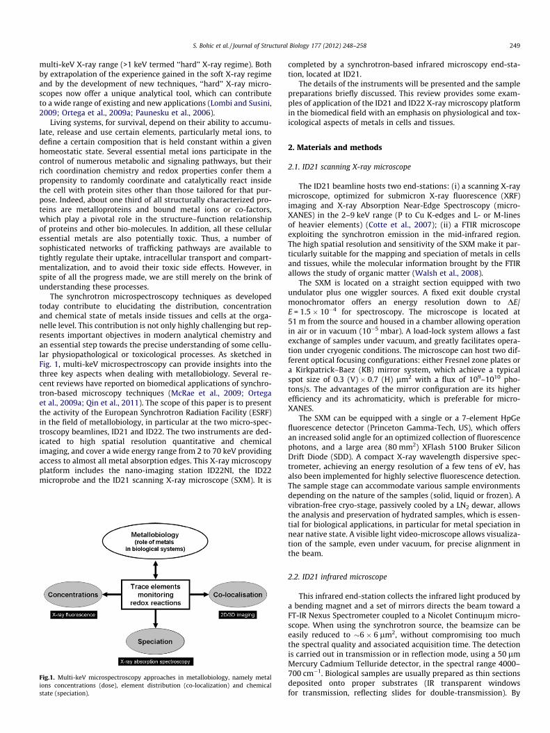
It is not only the spatial distribution of the concentration of ele-ments that matters, their chemical form is also important whenstudying the role of metals in biological systems. Micro-XANES isa rather unique technique since it associates sub-micron spatialresolution with chemical selectivity and sensitivity to local atomicarrangement. Indeed, the transition energies and the shape of theabsorption spectrum contain specific information about the chem-ical bonds. As an example, a study carried out by Szczerbowska-Boruchowska at ID21 reported preliminary results on sulfur inbrain tumor tissues suggesting higher accumulation of sulfide(S2�) in high-grade (IV) glioma compared to controls or grade IIneoplasm (Szczerbowska-Boruchowska, 2008). In a previous work
Fig.3. Sulfur K-edge X-ray Absorption Near-Edge Spectroscopy (XANES) of NM contained in dopaminergic neurons of the substantia nigra pars compacta of control,Parkinsonian, Alzheimer and incidental Lewy Body diseases’ patients. Representative sulfur XANES spectra of NM for the different cases studied (upper left panel). Lower leftpanel shows the sulfur XANES spectra for the set of experimental reference compounds used for linear combination fitting of XANES NM spectra (right panel), with (a) rawspectrum of neuromelanin, (b) L-methionine, (c) S-methyl-L-cysteine, (d) glutathione oxidized, (e) methionine sulfoxide and (f) anthraquinone sulfonic acid.
252 S. Bohic et al. / Journal of Structural Biology 177 (2012) 248–258
we evaluated the proportions and types of various sulfur species inthe intraneuronal pigment neuromelanin (NM) during its differentphases of development (Bohic et al., 2008). Similarly, we recentlyexplored the sulfur chemical environment of NM in neurodegener-ative disorders, particularly Parkinson’s disease. Sulfhydryl com-pounds of NM and alteration of the NM-biosynthetic pathwaywere suggested to play a role in the selective degeneration of mel-anised neuron from the substantia nigra (Spencer et al., 1998). Thetypical sulfur K-edge l-XANES spectra obtained on the intra-cellu-lar pigment found in surviving pigmented dopaminergic neuronsfrom sections of human substantia nigra (SN) are shown in Fig. 3.The spectral signatures for control, PD, AD and incidental LewyBody disease (ILBD) cases were very similar. Three energies ofmaximum absorption were observed at 2473.6, 2476.4 and2481.4 eV that can be assigned to different types of S functionalgroups. The least-square linear combination fits of normalizedstandard spectra of model S compounds shows that the white line(i.e. main absorption peak at 2473.6 eV) has three overlappingcomponents consistent with the presence of thiol, organic mono-sulfide and organic disulfide. The other main peaks at 2476.4 and2481.4 eV were assigned to the presence of sulfoxide and sulfo-nate. The model fits with the minimal set of predictor variables in-cluded S-methyl-L-cysteine, L-methionine, glutathione oxidized,methionine sulfoxide and anthraquinone sulfonic acid as modelcompounds and results in the best match of the experimental data.In dopaminergic neurons of control SN, a proportion of 64% organicmonosulfide, 14% thiol, 13.5% disulfide, 6.4% sulfoxide and 2.1%sulfonate is found. Similar proportions are found for NM of surviv-ing neurons within the substantia nigra of PD, ILBD and AD cases.Similar work that combined the different spectroscopic approaches(various high spatial resolution X-ray imaging modalities and FT-IRmicroscopy) has been recently conducted by Ducic et al. on mye-linated sciatic neurons isolated from wild-type mice (Ducic et al.,
2011). Such work using multi-modal X-ray imaging approacheswill foster future studies in understanding morphological andchemical alterations in myelin that parallel some pathological pro-cesses involved in myelin related neurodegenerative diseases.
The study of the role of metals in the cellular physiopathology isnot limited to brain studies, but can also be applied to metallobiol-ogy in cancer (Collery et al., 2004). Indeed, Farquharson et al.showed a positive correlation between higher zinc levels andestrogen receptor positive tumor cells region in breast cancer tis-sues as compared to surrounding regions (Farquharson et al.,2009). This work highlights the importance of synchrotron XRFin studying the relationships between such biological variablesand the changes in concentration and/or spatial distribution ofmetals in cells and tissues. Also, one of the well accepted hallmarksof cancer is angiogenesis (Hanahan and Weinberg, 2011) and thestudy of Finney et al. using an XRF nanoprobe provided some in-sight into the role of copper regulating angiogenesis (Finneyet al., 2007).
Another example of trace element mapping in cells deals withstudies into the role of trace metals in single spermatozoa develop-ments, which are scarce (Zhang et al., 1996; Kehr et al., 2009).There exists a huge variation in the size of sperm across the animalkingdom. Human sperm cells consist of a head 5 lm by 3 lm and atail 50 lm long. Their development from stem cells into maturespermatozoa is the result of the spermatogenesis. Trace elementsplay an important role within this process. The trace elements sele-nium and zinc are important for sperm motility and therefore areoften associated with infertility. We used the high spatial resolu-tion and the high flux offered by the ID22NI X-ray nanoprobe todemonstrate the capabilities of the technique in this domain. Thisis illustrated in Fig. 4 where the distribution of phosphorus, ironand zinc are well resolved. It clearly demonstrates a compartmen-talization of these elements in different parts of the cell (head

Fig.4. X-ray fluorescence mapping of a single mammalian spermatozoa obtained atthe ID22NI nanoimaging beamline. The cells were deposited on 500 nm thick Si3N4
windows and let dry under inert atmosphere prior to analysis. 2D mosaic X-rayfluorescence mapping of zinc and a RGB-coded XRF map in a single spermatozoa(iron: red; zinc: green; phosphorus: blue).
S. Bohic et al. / Journal of Structural Biology 177 (2012) 248–258 253
where the nucleus with highly condensed DNA is located, the mid-piece and the tail). This clearly provides exciting perspectives tobetter understand how infertility could be linked to certain traceelements, infertility being classified by the World Health Organisa-tion as major health concern.
3.2. Tracking of metal-based compounds and metals-related toxicologyin mammalian cells
Understanding the metabolism of exogenous metals in humancells has always been a key topic relevant to a wide range of healthissues. It is a necessary step for improving the efficiency and tar-geting of metal-based drugs, some medical imaging contrastagents and for evaluating the toxicological effects of metal-basednanoconstructs and environmental metallic complexes. Along withthe recent development of nanotechnologies for nanomedicine orfor industrial and engineering purposes, growing concerns havearisen about their unintentional health and environmental impact.This has brought forward the need for new analytical tools able to
Fig.5. Micro-XRF mapping of cobalt ferrite nano-particles (NPs) in a Balb/3T3 mouse fibrintracellular phosphorus distribution is displayed in magenta and the cobalt fluorescencethe phosphorus distribution. Map size: 22 � 40.5 lm, step size: 0.5 lm, dwell time: 70Research Center, Ispra, Italy.)
characterize their biodistribution at the sub-cellular scale, an issuefor which the emerging X-ray nanoprobes are highly competitive.Although, up to now a very large population of cells cannot bescreened by any existing micro/nanoprobe, recent work showsthe importance of getting simultaneous mapping of endogenouselements (P, S, Ca, K, Cl) as well as exogenous metals in cells.
A direct application of X-ray micro/nano-probes is the mappingof the distribution of metal-based drugs and nano-vectors in cellsand tissues to track their fate. The XRF signal of metals and inor-ganic components forming these nano-materials can be exploitedto localize them. We can cite as an example the mapping of theintracellular distribution of single wall carbon nano-tubes (CNT)in macrophage cells (Bussy et al., 2008). In this case, the XRF signalof the catalyst iron content of these CNT nano-materials was usedas a tracking marker in micro-XRF to localize them. It was also evi-denced that the presence of the nano-tubes modifies the intracel-lular concentration and distribution of endogenous elements, inparticular calcium. It was associated to an increase in cellular cal-cium uptake that could be linked to oxidative stress, inflammationor cytotoxicity. Such quantitative micro-XRF measurements thusalso provide valuable information about the interaction of exoge-nous metals with cell homeostasis. Comparable methodologywas used at the ID22 nanoprobe to screen the intracellular locali-zation of several series of newly developed lanthanide nanoparti-cles used as potential platforms for bio-imaging and bio-deliveryapplications (Lewis et al., 2010). Recently, the ID21 SXM was alsoused to study the uptake and interaction of cobalt ferrite (CoFe2O4)nanoparticles (NPs) in Balb/3T3 mouse fibroblast cells exposed todifferent concentrations of NPs (Fig. 5) (Marmorato et al., 2011).Functionalized quantum dots can be used to label cancer markersor specific proteins. CdSe/ZnS quantum dots are suitable probes fornano-XRF through the XRF signal of their intrinsic Se atoms. Thislabeling approach is very effective at colocalizing specific intracel-lular targets with elements naturally present in the cell and fostersnew possibilities for correlative imaging between confocal fluores-cence microscopy and elemental mapping Corezzi et al., 2009; Le-wis et al., 2010. Such atomic signature is also useful when studyingthe toxicity of imaging contrast agents for Magnetic ResonanceImaging (MRI). Here the XRF signal of the gadolinium (Gd) atomscontained in these contrast agents is exploited. Recently Gd-basedcontrast agents have come under scrutiny by the Food and DrugAdministration due to gadolinium side effects such as nephrogenic
oblast cell exposed to 500 lM solution of NPs for 24 h. On the composite image, thesignal from the NPs in green. The location of the cell nucleus is clearly visible from
0 ms, photon energy: 7.8 keV. (Courtesy of G. Ceccone and P. Marmorato, EC Join
t
254 S. Bohic et al. / Journal of Structural Biology 177 (2012) 248–258
systemic fibrosis. The X-ray fluorescent Gd L-line (6.05 keV) can bedetected using the ID21 SXM and the results obtained by Altissimoet al. on cultured Chang liver cells assessed the intracellular pres-ence of Gd and highlighted a peri-nuclear distribution inside thecells (Fig. 6) (Altissimo et al., 2011). X-ray differential phase con-trast imaging was performed in parallel and provided a depictionof the cell morphology allowing correlation of the Gd distributionwith subcellular structures. Endogenous elements such as phos-phorus, sulfur, calcium and potassium were also mapped and thevariation in their concentration and distribution in exposed andcontrol cells could be compared.
As a natural extension to XRF, micro-XANES is extremely pow-erful for studying the biotransformation of the metals entering bio-logical systems. For example, Ortega et al. collected information onthe carcinogenic mechanism of Cr by imaging the intracellular dis-tribution and speciation of Cr in cells exposed to chromate com-pounds. Such a study was performed on cells exposed to solubleand low-solubility hexavalent chromium compounds (Ortegaet al., 2005), which are carcinogens for the respiratory tract in hu-mans. It was shown that soluble Cr(VI) compounds were fully re-duced to Cr(III) in all cell compartments while low-solubilitychromate compounds were partially reduced to Cr(III) and couldcoexist in the cell environment, as particles in perinuclear
Fig.6. Uptake of Gd in Chang liver cell exposed to 500 lM of Gd containing contrast agentdifferential phase contrast image (c) micro-XRF elemental maps of phosphorus (in red) aphoton energy 7.3 keV (Altissimo et al., 2010).
Fig.7. (a) Reference XANES spectra for Cr(VI) as PbCrO4 and Cr(III) as CrCl3, at the Cr K-edand XRF mapping of potassium (c), total chromium (d) and hexavalent chromium (e) of IGd) were acquired with an exciting X-ray energy of 6.030 keV while map (e) was acquiredonly Cr(VI) forms of chromium are detected. Pixel size is 1 lm � 1 lm. Scale bar is 10 l
structures (Fig. 7). The stronger carcinogenicity of low solubilitychromate with respect to soluble chromate compounds could thusoriginate from a long term exposure to a strong oxidant, Cr(VI)combined with direct genotoxic effects of intracellular Cr(III).
3.3. Metals in ancient medical practices
As discussed above, questions relating to the beneficial use and,conversely, to the potential danger of nanoparticulate materialsseem to appear only at the end of the 20th Century. However, suchmaterials have been in daily use since Antiquity, for examplein inks, paintings and cosmetics (Murr, 2009). For instance,2000 year-old Greco-Roman recipes for dying hair were found torely on the synthesis of black PbS nano-crystals by reaction of leadcompounds on sulfur contained in keratin (Walter et al., 2006). Inthe past, lead was frequently used in cosmetics and pharmaceuti-cal products, which nowadays seems astonishing considering itsknown toxicity. Indeed, there is an apparent contradiction con-cerning the reputation of lead in ancient and modern times. A dec-ade ago, an extensive research programme was initiated to revealancient cosmetic and pharmaceutical practices in Mediterraneancountries. It was based on the analysis of ancient cosmetic powderspreserved in their original containers over many centuries, some of
for 20 h. Cells were fixed and imaged using the ID21 SXM. (a) Visible light image (b)nd gadolinium (in green). Field of view 36 � 40 lm, 0.5 lm step size, 5 s dwell time,
ge, showing the characteristic pre-edge peak of Cr(VI) at 5.9935 keV. Micrograph (b)R-OV1 cells exposed for 24 h to PbCrO4 at a concentration of 1 lg/cm2. Maps (c and
with an exciting X-ray energy of 5.9935 keV corresponding to Cr(VI) pre-edge peak:m.

Fig.8. Study of transdermal diffusion of lead-based compounds, mimicking ancient pharmaceutical products. Top: skin treated with a mixture of lead palmitate/propyleneglycol for 1 day. Elemental mappings are obtained by micro-X-ray fluorescence over the skin cross-section (map size 100 � 100 lm2, step size = 1 � 1 lm2). Bottom: skintreated with a mixture of perdeuterated palmitic acid/propylene glycol for 1 day. Chemical mappings are obtained by micro-FTIR over the skin cross-section (map size36 � 72 lm2, step size = 6 � 6 lm2).
S. Bohic et al. / Journal of Structural Biology 177 (2012) 248–258 255
which are now exhibited in the Louvre museum in Paris. Synchro-tron-based X-ray diffraction was used for the identification of exo-tic inorganic lead hydroxy or carbonate chloride, which wereproved to be synthetic products obtained by ‘wet’ chemistry(Walter et al., 1999). Synchrotron-based FTIR microscopy was em-ployed to identify hybrid compounds, namely lead soaps, obtainedwhen such inorganic compounds were mixed with fat (Cotte et al.,2005). Furthermore, the potential toxicity of such lead-based com-pounds was investigated. Generally, lead transdermal penetrationis assessed by indirect measurements, such as quantification oflead concentration in blood, sweat or urine. Alternatively, synchro-tron-based micro-analytical techniques were used for the directobservation of transdermal lead diffusion into transversal cuts inthe skin. Model lead plasters were synthesised according to ancientrecipes (Cotte et al., 2006) and applied to pig skin, for 24 h, inFrantz-cell transdermal diffusion system. After dismounting, skintransversal sections were obtained and chemical and atomic distri-butions of drug penetration were imaged by synchrotron-basedinfrared micro-spectroscopy and micro-XRF.
Elemental mappings simultaneously reveal distributions ofboth endogenous elements (sulfur and potassium are good mark-ers of the stratum corneum while phosphorous is more abundantin the cells of the epidermis, the corneocytes) and exogenous lead.In the present example (lead introduced as lead palmitate in pro-pylene glycol), the diffusion of lead is stopped in the stratum cor-neum (Fig. 8). FTIR chemical mappings were carried out on thesame skin sections, in particular to compare the penetration ofan acid-based chemical, with its corresponding lead carboxylate,as well as to estimate the effect of the chain length of fatty acidson their transdermal penetration (Cotte et al., 2004). Chemicalmappings reveal the distribution of endogenous components (lip-ids, proteins) as well as exogenous fatty acids (applied as perdeu-terated chains, for a selective identification) (Fig. 8). Aftersaponification of fatty acids with lead, their ability to penetratethe skin is clearly reduced. While fatty acids can be detected inthe epidermis after one day of application, in the same conditions,lead carboxylate diffusion is limited to the superficial part of thestratum corneum. This study shows the potential of synchrotron-based micro-analytical techniques to follow drugs and in particular
penetration of metals into the skin. The combination of micro-XRFwith micro-FTIR offers complementary information, based onatomic and molecular probes, respectively.
4. Discussion
In the arsenal of available analytical tools for the study of met-als in biology, spatially resolved techniques are scarce and mostlyrely on highly sophisticated and complex instrumentation. Table 1compiles the key performance indicators of the most commonlyused metal imaging techniques. All these techniques are comple-mentary and should be selected depending upon the researchobjectives, type of sample, spatial resolution and sensitivity,required throughput and even the availability of the technique.Scanning transmission electron microscopy with energy-dispersiveX-ray analysis (STEM–EDX) or energy-filtered transmission elec-tron microscopy (EFTEM) usually require ultrathin sample sections(30 nm range) which is also a prerequisite to surface sensitivetechniques such as X-ray photoelectron emission microscopy (X-PEEM) and nanoSIMS. Electron microscopy has unsurpassed spatialresolution but the sensitivity, radiation damage and the restrictedfield of view can be limiting factors. NanoSIMS is fully complemen-tary to electron microscopy and provides isotopic analysis in addi-tion to the atomic composition. However, quantification is difficultdue to matrix effects that influence the secondary ion yielddepending on the chemical environment of the surface being sam-pled. The technique allows in-depth analysis but is destructivewith only a limited number of elemental images available, up toseven using recently developed multicollectors.
Other alternative techniques, such as the following, provide ac-cess to much in-depth analysis. Particle-induced X-ray emission(PIXE) technique has a good sensitivity at the micron scale andeven at sub-micron resolution, as recently demonstrated (Barberetet al., 2011). It is a fully quantitative method when coupled toRutherford backscattering spectrometry but suffers from radia-tion-induced effects and a limited sensitivity compared to synchro-tron XRF microscopy. Laser-ablation is a highly efficient, sensitivetechnique at the microscale. Although destructive and with limited

Table 1Major spatially resolved methods for imaging metals in biology.
STEM–EDX STEM–EELS/EFTEM Dynamic SIMS X-PEEM Soft X-raySTXM
SXRF (micro-XANES)
PIXE LA-ICP-MS
Selectivity Atomiccomposition
Atomiccompositionchemical
Atomiccompositionisotopic
Atomic compositionchemical
Chemical Atomiccomposition(chemical)
Atomiccomposition
Atomiccompositionisotopic
Spatialresolution(nm)
1 1 50–100 20 25–50 50–1000 (150–1000)
500–1000 5000–20,000
Analyticaldepth(lm)
0.1–1 <0.1 at 200 keV/<0.5 at 100 keV
<0.1 <0.05 <10 10–1000 10–100 200
Field of view <10 lm2 <10 lm2 <0.1 cm2 10–100 lm2 10 lm2 Sub mm2 tocm2
Sub mm2 Sub cm2 to200 cm2
Detectionlimit (mg/Kg)
>1000 >1000 0.1 100–1000 >1000 <0.1 (10–100) 1–10 <0.1
Quantification Semi-quantitative Semi-quantitative Semi-quantitative Semi-quantitative Semi-quantitative
Quantitative(poor)
Quantitative Quantitative
Samples Thin section (resinembeddedmaterial, frozen-hydrated)
Thin section (resinembeddedmaterial, frozen-hydrated)
Semi-thin or thinsection of resinembeddedmaterial
Thin section of resinembedded material;cryofixed and freeze-dried
Frozenhydrated, driedor embeddedmaterial
Frozenhydrated;dried orembeddedmaterial
Freeze-driedsamples/embeddedmaterial
10–40 lmnativecryosections
Environment High-vacuum High-vacuum Ultra high-vacuum
Ultra high-vacuum High-vacuum In air or high-vacuum
High-vacuum In air
Fig.9. The timeline of the X-ray focus size below and above 2 keV during the last40 years.
256 S. Bohic et al. / Journal of Structural Biology 177 (2012) 248–258
spatial resolution, high quality quantitative images of largehistological sections can be obtained (Becker, 2010; Hare et al.,2010).
Micro-XRF and micro-XANES offer quantitative and chemicalinformation with intermediate spatial resolution in constantimprovement towards the nanoscale. The access to K-absorptionedges and XRF emission lines of most elements, the higher pene-tration depths compared to soft X-rays, ions or electrons allowingimaging of thicker samples or in situ experiments, the favorablewavelengths for coupling with X-ray diffraction studies and therelatively long focal lengths and depths of focus which are advan-tageous for the use of specific sample environments are uniqueattributes to X-ray microspectroscopy (Lombi and Susini, 2009).The full control of both energy tunability and spectral bandwidthof the incoming monochromatic radiation minimizes the radiationdamage without compromising the signal-to-noise and allowsaccurate absolute quantification, within 10–20%. Hard X-raysprobe deeper into samples and relax a number of issues associatedwith attenuation of XRF in the air path and surrounding space. To-day, most microprobes offer absolute detection limits for metalsbelow 10�16 g or 106 atoms. This translates to a relative detectionlimit below 0.01 ppm in three dimensions at high resolution andsensitivity (de Jonge and Vogt, 2010) offering unparalleled insightin biology (De Samber et al., 2010; Lewis et al., 2010). The ID22NIhard X-ray nanoprobe reduces the lower detection limit even fur-ther with attogram sensitivity at sub-100 nm level (unpublishedresults).
None of these methods are straightforward and they are notperformed routinely. The accessibility to all these techniques israther limited with access being restricted to synchrotron facilities.Although they are considered as ‘‘slow’’ methods; still synchrotronX-ray microspectroscopy allows, among the available methods, theshortest time for the analysis of a cell (�1 h) and less than fewminutes for X-ray absorption spectra. The large penetration depthof X-rays allows probing sample thickness of tens of microns andon the other hand X-ray spot of few tens of nanometer started tobe routinely produced making 2D scanning challenging to probethe 3D heterogeneity of biological systems where subcellular com-partments may overlap. However, it is ideally suited for tomo-graphic X-ray microspectroscopy acquisition with the uniqueperspective to provide chemical images at the ultrastructural scale
on whole cells (de Jonge and Vogt, 2010) and avoid the use of thinsectioning.
An important limitation will be the required analytical time toproduce a full 3D image of a sample like a cell. Up to now, few tensof hours are required depending on the sample size and mostly onthe concentration of the elements to be imaged. The radiationdamages become an issue even if the sample is cryopreserved. Toovercome this drawback, much can be gained from the improve-ment of the detection efficiency of the fluorescence emitted bythe sample. Indeed, X-ray fluorescence is emitted over a solid angleof 4p while the actual detectors only cover a few % of it. The adventof new large area and fast detectors such as the MAIA allow to gaina factor 10 in efficiency (Lombi et al., 2011). This will provide abreakthrough in the biomedical application of X-ray nanoprobeto allow fast 2D/3D images of the chemical composition andchanges within cellular and sub-cellular structures.
5. Conclusion and outlook
We have recalled the substantial scientific progress being madein metallobiology studies utilizing multi-keV micro-spectroscopytechniques. The number of applications is growing rapidly andthe availability improves as more synchrotron radiation facilities
S. Bohic et al. / Journal of Structural Biology 177 (2012) 248–258 257
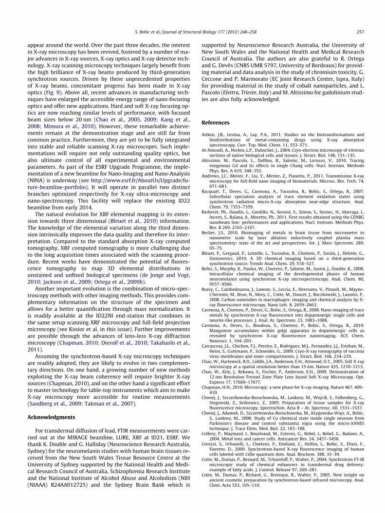
appear around the world. Over the past three decades, the interestin X-ray microscopy has been revived, fostered by a number of ma-jor advances in X-ray sources, X-ray optics and X-ray detector tech-nology. X-ray scanning microscopy techniques largely benefit fromthe high brilliance of X-ray beams produced by third-generationsynchrotron sources. Driven by these unprecedented propertiesof X-ray beams, concomitant progress has been made in X-rayoptics (Fig. 9). Above all, recent advances in manufacturing tech-niques have enlarged the accessible energy range of nano-focusingoptics and offer new applications. Hard and soft X-ray focusing op-tics are now reaching similar levels of performance, with focusedbeam sizes below 20 nm (Chao et al., 2005, 2009; Kang et al.,2008; Mimura et al., 2010). However, these remarkable achieve-ments remain at the demonstration stage and are still far fromcommon practice. Furthermore, they are yet to be fully integratedinto stable and reliable scanning X-ray microscopes. Such imple-mentations will require not only outstanding quality optics, butalso ultimate control of all experimental and environmentalparameters. As part of the ESRF Upgrade Programme, the imple-mentation of a new beamline for Nano-Imaging and Nano-Analysis(NINA) is underway (see http://www.esrf.fr/AboutUs/Upgrade/fu-ture-beamline-portfolio). It will operate in parallel two distinctbranches optimized respectively for X-ray ultra-microscopy andnano-spectroscopy. This facility will replace the existing ID22beamline from early 2014.
The natural evolution for XRF elemental mapping is its exten-sion towards three dimensional (Bleuet et al., 2010) information.The knowledge of the elemental variation along the third dimen-sion intrinsically improves the data quality and therefore its inter-pretation. Compared to the standard absorption X-ray computedtomography, XRF computed tomography is more challenging dueto the long acquisition times associated with the scanning proce-dure. Recent works have demonstrated the potential of fluores-cence tomography to map 3D elemental distributions inunstained and unfixed biological specimens (de Jonge and Vogt,2010; Jackson et al., 2009; Ortega et al., 2009b).
Another important evolution is the combination of micro-spec-troscopy methods with other imaging methods. This provides com-plementary information on the structure of the specimen andallows for a better quantification through mass normalization. Itis readily available at the ID22NI end-station that combines inthe same setup scanning XRF microscopy and full-field projectionmicroscopy (see Kosior et al. in this issue). Further improvementsare possible through the advances of lens-less X-ray diffractionmicroscopy (Chapman, 2010; Dierolf et al., 2010; Takahashi et al.,2011).
Assuming the synchrotron-based X-ray microscopy techniquesare readily adopted, they are likely to evolve in two complemen-tary directions. On one hand, a growing number of new methodsexploiting the X-ray beam coherence will require brighter X-raysources (Chapman, 2010), and on the other hand a significant effortto master technology for table-top instruments which aim to makeX-ray microscopy more accessible for routine measurements(Sandberg et al., 2009; Takman et al., 2007).
Acknowledgments
For transdermal diffusion of lead, FTIR measurements were car-ried out at the MIRAGE beamline, LURE, XRF at ID21, ESRF. Wethank K. Double and G. Halliday (Neuroscience Research Australia,Sydney) for the neuromelanin studies with human brain tissues re-ceived from the New South Wales Tissue Resource Centre at theUniversity of Sydney supported by the National Health and Medi-cal Research Council of Australia, Schizophrenia Research Instituteand the National Institute of Alcohol Abuse and Alcoholism (NIH(NIAAA) R24AA012725) and the Sydney Brain Bank which is
supported by Neuroscience Research Australia, the University ofNew South Wales and the National Health and Medical ResearchCouncil of Australia. The authors are also grateful to R. Ortegaand G. Devès (CNRS UMR 5797, University of Bordeaux) for provid-ing material and data analysis in the study of chromium toxicity. G.Ceccone and P. Marmorato (EC Joint Research Center, Ispra, Italy)for providing material in the study of cobalt nanoparticles, and L.Pascolo (Elettra, Trieste, Italy) and M. Altissimo for gadolinium stud-ies are also fully acknowledged.
References
Aitken, J.B., Levina, A., Lay, P.A., 2011. Studies on the biotransformations andbiodistributions of metal-containing drugs using X-ray absorptionspectroscopy. Curr. Top. Med. Chem. 11, 553–571.
Al-Amoudi, A., Norlen, L.P., Dubochet, J., 2004. Cryo-electron microscopy of vitreoussections of native biological cells and tissues. J. Struct. Biol. 148, 131–135.
Altissimo, M., Pascolo, L., Delfino, R., Salome, M., Lorusso, V., 2010. Tracingexogenous Gd and its effects in single Chang cells. Nucl. Instrum. MethodsPhys. Res. A 619, 348–352.
Andrews, J.C., Meirer, F., Liu, Y., Mester, Z., Pianetta, P., 2011. Transmission X-raymicroscopy for full-field nano imaging of biomaterials. Microsc. Res. Tech. 74,671–681.
Bacquart, T., Deves, G., Carmona, A., Tucoulou, R., Bohic, S., Ortega, R., 2007.Subcellular speciation analysis of trace element oxidation states usingsynchrotron radiation micro-X-ray absorption near-edge structure. Anal.Chem. 79, 7353–7359.
Barberet, Ph., Daudin, L., Gordillo, N., Sorieul, S., Simon, S., Seznec, H., Idarraga, I.,Incerti, S., Balana, A., Moretto, Ph., 2011. First results obtained using the CENBGnanobeam line: performances and applications. Nucl. Instrum. Methods Phys.Res. B 269, 2163–2167.
Becker, J.S., 2010. Bioimaging of metals in brain tissue from micrometre tonanometre scale by laser ablation inductively coupled plasma massspectrometry: state of the art and perspectives. Int. J. Mass Spectrom. 289,65–75.
Bleuet, P., Gergaud, P., Lemelle, L., Tucoulou, R., Cloetens, P., Susini, J., Delette, G.,Simionovici, 2010. A 3D chemical imaging based on a third-generationsynchrotron source. Trends Anal. Chem. 29, 518–527.
Bohic, S., Murphy, K., Paulus, W., Cloetens, P., Salome, M., Susini, J., Double, K., 2008.Intracellular chemical imaging of the developmental phases of humanneuromelanin using synchrotron X-ray microspectroscopy. Anal. Chem. 80,9557–9566.
Bussy, C., Cambedouzou, J., Lanone, S., Leccia, E., Heresanu, V., Pinault, M., Mayne-L’hermite, M., Brun, N., Mory, C., Cotte, M., Doucet, J., Boczkowski, J., Launois, P.,2008. Carbon nanotubes in macrophages: imaging and chemical analysis by X-ray fluorescence microscopy. Nano Lett. 8, 2659–2663.
Carmona, A., Cloetens, P., Deves, G., Bohic, S., Ortega, R., 2008. Nano-imaging of tracemetals by synchrotron X-ray fluorescence into dopaminergic single cells andneurite-like processes. J. Anal. At. Spectrom. 23, 1083–1088.
Carmona, A., Deves, G., Roudeau, S., Cloetens, P., Bohic, S., Ortega, R., 2010.Manganese accumulates within golgi apparatus in dopaminergic cells asrevealed by synchrotron X-ray fluorescence nanoimaging. ACS Chem.Neurosci. 1, 194–203.
Carrascosa, J.L., Chichon, F.J., Pereiro, E., Rodriguez, M.J., Fernandez, J.J., Esteban, M.,Heim, S., Guttmann, P., Schneider, G., 2009. Cryo-X-ray tomography of vacciniavirus membranes and inner compartments. J. Struct. Biol. 168, 234–239.
Chao, W., Harteneck, B.D., Liddle, J.A., Anderson, E.H., Attwood, D.T., 2005. Soft X-raymicroscopy at a spatial resolution better than 15 nm. Nature 435, 1210–1213.
Chao, W., Kim, J., Rekawa, S., Fischer, P., Anderson, E.H., 2009. Demonstration of12 nm Resolution Fresnel Zone Plate Lens based Soft X-ray Microscopy. Opt.Express 17, 17669–17677.
Chapman, H.N., 2010. Microscopy: a new phase for X-ray imaging. Nature 467, 409–410.
Chwiej, J., Szczerbowska-Boruchowska, M., Lankosz, M., Wojcik, S., Falkenberg, G.,Stegowski, Z., Setkowicz, Z., 2005. Preparation of tissue samples for X-rayfluorescence microscopy. Spectrochim. Acta B – At. Spectrosc. 60, 1531–1537.
Chwiej, J., Adamek, D., Szczerbowska-Boruchowska, M., Krygowska-Wajs, A., Bohic,S., Lankosz, M., 2008. Study of Cu chemical state inside single neurons fromParkinson’s disease and control substantia nigra using the micro-XANEStechnique. J. Trace Elem. Med. Biol. 22, 183–188.
Collery, P., Maymard, I., Bourleaud, M., Estevez, G., Rebel, I., Rebel, G., Badawi, A.,2004. Metal ions and cancer cells. Anticancer Res. 24, 3457–3458.
Corezzi, S., Urbanelli, L., Cloetens, P., Emiliani, C., Helfen, L., Bohic, S., Elisei, F.,Fioretto, D., 2009. Synchrotron-based X-ray fluorescence imaging of humancells labeled with CdSe quantum dots. Anal. Biochem. 388, 33–39.
Cotte, M., Dumas, P., Besnard, M., Tchoreloff, P., Walter, P., 2004. Synchrotron FT-IRmicroscopic study of chemical enhancers in transdermal drug delivery:example of fatty acids. J. Control. Release 97, 269–281.
Cotte, M., Dumas, P., Richard, G., Breniaux, R., Walter, P., 2005. New insight onancient cosmetic preparation by synchrotron-based infrared microscopy. Anal.Chim. Acta 553, 105–110.

258 S. Bohic et al. / Journal of Structural Biology 177 (2012) 248–258
Cotte, M., Checroun, E., Susini, J., Dumas, P., Tchoreloff, P., Besnard, M., Walter, P.,2006. Kinetics of oil saponification by lead salts in ancient preparations ofpharmaceutical lead plasters and painting lead mediums. Talanta 70, 1136–1142.
Cotte, M., Welcomme, E., Sole, V.A., Salome, M., Menu, M., Walter, P., Susini, J., 2007.Synchrotron-based X-ray spectromicroscopy used for the study of an atypicalmicrometric pigment in 16th century paintings. Anal. Chem. 79, 6988–6994.
de Jonge, M.D., Vogt, S., 2010. Hard X-ray fluorescence tomography – an emergingtool for structural visualization. Curr. Opin. Struct. Biol. 20, 606–614.
de Jonge, M.D., Hornberger, B., Holzner, C., Legnini, D., Paterson, D., McNulty, I.,Jacobsen, C., Vogt, S., 2008. Quantitative phase imaging with a scanningtransmission X-ray microscope. Phys. Rev. Lett. 100, 163902.
De Samber, B., Silversmit, G., De Schamphelaere, K., Evens, R., Schoonjans, T.,Vekemans, B., Janssen, C., Masschaele, B., Van Hoorebeke, L., Szaloki, I.,Vanhaecke, F., Rickers, K., Falkenberg, G., Vincze, L., 2010. Element-to-tissuecorrelation in biological samples determined by three-dimensional X-rayimaging methods. J. Anal. At. Spectrom. 25, 544–553.
Dierolf, M., Menzel, A., Thibault, P., Schneider, P., Kewish, C.M., Wepf, R., Bunk, O.,Pfeiffer, F., 2010. Ptychographic X-ray computed tomography at the nanoscale.Nature 467, 436–439.
Ducic, T., Quintes, S., Nave, K.A., Susini, J., Rak, M., Tucoulou, R., Alevra, M.,Guttmann, P., Salditt, T., 2011. Structure and composition of myelinated axons:a multimodal synchrotron spectro-microscopy study. J. Struct. Biol. 173, 202–212.
Farquharson, M.J., Al-Ebraheem, A., Geraki, K., Leek, R., Jubb, A., Harris, A.L., 2009.Zinc presence in invasive ductal carcinoma of the breast and its correlation withoestrogen receptor status. Phys. Med. Biol. 54, 4213–4223.
Fayard, B., Salome, M., Takemoto, K., Kihara, H., Susini, J., 2009. Some practicalconsiderations about the effects of radiation damage on hydrated cells imagedby X-ray fluorescence microscopy. J. Electron Spectrosc. Relat. Phenom. 170,19–24.
Finney, L., Mandava, S., Ursos, L., Zhang, W., Rodi, D., Vogt, S., Legnini, D., Maser, J.,Ikpatt, F., Olopade, O.I., Glesne, D., 2007. X-ray fluorescence microscopy revealslarge-scale relocalization and extracellular translocation of cellular copperduring angiogenesis. Proc. Natl. Acad. Sci. USA 104, 2247–2252.
Gazi, E., Dwyer, J., Lockyer, N.P., Miyan, J., Gardner, P., Hart, C., Brown, M., Clarke,N.W., 2005. Fixation protocols for subcellular imaging by synchrotron-basedFourier transform infrared microspectroscopy. Biopolymers 77, 18–30.
Hackett, M.J., McQuillan, J.A., El-Assaad, F., Aitken, J.B., Levina, A., Cohen, D.D.,Siegele, R., Carter, E.A., Grau, G.E., Hunt, N.H., Lay, P.A., 2011. Chemicalalterations to murine brain tissue induced by formalin fixation: implicationsfor biospectroscopic imaging and mapping studies of disease pathogenesis.Analyst 136, 2941–2952.
Hanahan, D., Weinberg, R.A., 2011. Hallmarks of cancer: the next generation. Cell144, 646–674.
Hare, D.J., George, J.L., Grimm, R., Wilkins, S., Adlard, P.A., Cherny, R.A., Bush, A.I.,Finkelstein, D.I., Doble, P., 2010. Three-dimensional elemental bio-imaging ofFe, Zn, Cu, Mn and P in a 6-hydroxydopamine lesioned mouse brain.Metallomics 2, 745–753.
Heine, R., Gorniak, T., Nisius, T., Christophis, C., Pettitt, M.E., Staier, F., Wilhein, T.,Rehbein, S., Grunze, M., Rosenhahn, A., 2011. Digital in-line X-ray holographywith zone plates. Ultramicroscopy 111, 1131–1136.
Hell, S.W., 2007. Far-field optical nanoscopy. Science 316, 1153–1158.Holzner, C., Feser, M., Vogt, S., Hornberger, B., Baines, S.B., Jacobsen, C., 2010. Zernike
phase contrast in scanning microscopy with X-rays. Nat. Phys. 6, 883–887.Jackson, B.P., Pace, H.E., Lanzirotti, A., Smith, R., Ranville, J.F., 2009. Synchrotron X-
ray 2D and 3D elemental imaging of CdSe/ZnS quantum dot nanoparticles inDaphnia magna. Anal. Bioanal. Chem. 394, 911–917.
Jacobsen, C., 1999. Soft X-ray microscopy. Trends Cell Biol. 9, 44–47.James, S.A., Myers, D.E., de Jonge, M.D., Vogt, S., Ryan, C.G., Sexton, B.A., Hoobin, P.,
Paterson, D., Howard, D.L., Mayo, S.C., Altissimo, M., Moorhead, G.F., Wilkins,S.W., 2011. Quantitative comparison of preparation methodologies for X-rayfluorescence microscopy of brain tissue. Anal. Bioanal. Chem. 401, 853–864.
Jiang, H., Song, C., Chen, C.C., Xu, R., Raines, K.S., Fahimian, B.P., Lu, C.H., Lee, T.K.,Nakashima, A., Urano, J., Ishikawa, T., Tamanoi, F., Miao, J., 2010. Quantitative3D imaging of whole, unstained cells by using X-ray diffraction microscopy.Proc. Natl. Acad. Sci. USA 107, 11234–11239.
Kang, H.C., Yan, H.F., Winarski, R.P., Holt, M.V., Maser, J., Liu, C.A., Conley, R., Vogt, S.,Macrander, A.T., Stephenson, G.B., 2008. Focusing of hard X-rays to 16nanometers with a multilayer Laue lens. Appl. Phys. Lett. 92.
Kehr, S., Malinouski, M., Finney, L., Vogt, S., Labunskyy, V.M., Kasaikina, M.V.,Carlson, B.A., Zhou, Y., Hatfield, D.L., Gladyshev, V.N., 2009. X-ray fluorescencemicroscopy reveals the role of selenium in spermatogenesis. J. Mol. Biol. 389,808–818.
Lewis, D.J., Bruce, C., Bohic, S., Cloetens, P., Hammond, S.P., Arbon, D., Blair-Reid, S.,Pikramenou, Z., Kysela, B., 2010. Intracellular synchrotron nanoimaging andDNA damage/genotoxicity screening of novel lanthanide-coated nanovectors.Nanomedicine (Lond) 5, 1547–1557.
Lombi, E., Susini, J., 2009. Synchrotron-based techniques for plant and soil science:opportunities, challenges and future perspectives. Plant Soil 320, 1–35.
Lombi, E., de Jonge, M.D., Donner, E., Ryan, C.G., Paterson, D., 2011. Trends in hard X-ray fluorescence mapping: environmental applications in the age of fastdetectors. Anal. Bioanal. Chem. 400, 1637–1644.
Marmorato, P., Ceccone, G., Gianoncelli, A., Pascolo, L., Ponti, J., Rossi, F., Salomé, M.,Kaulich, B., Kiskinova, M., 2011. Cellular distribution and degradation of cobaltferrite nanoparticles in Balb/3T3 mouse fibroblasts. Toxicol. Lett. 207, 128–136.
McDermott, G., Le Gros, M.A., Knoechel, C.G., Uchida, M., Larabell, C.A., 2009. Soft X-ray tomography and cryogenic light microscopy: the cool combination incellular imaging. Trends Cell Biol. 19, 587–595.
McRae, R., Bagchi, P., Sumalekshmy, S., Fahrni, C.J., 2009. In situ imaging of metals incells and tissues. Chem. Rev. 109, 4780–4827.
Meade, A.D., Clarke, C., Draux, F., Sockalingum, G.D., Manfait, M., Lyng, F.M., Byrne,H.J., 2011. Studies of chemical fixation effects in human cell lines using Ramanmicrospectroscopy. Anal. Bioanal. Chem. 396, 1781–1791.
Meents, A., Gutmann, S., Wagner, A., Schulze-Briese, C., 2010. Origin andtemperature dependence of radiation damage in biological samples atcryogenic temperatures. Proc. Natl. Acad. Sci. USA 107, 1094–1099.
Mimura, H., Handa, S., Kimura, T., Yumoto, H., Yamakawa, D., Yokoyama, H.,Matsuyama, S., Inagaki, K., Yamamura, K., Sano, Y., Tamasaku, K., Nishino, Y.,Yabashi, M., Ishikawa, T., Yamauchi, K., 2010. Breaking the 10 nm barrier inhard-X-ray focusing. Nat. Phys. 6, 122–125.
Murr, L.E., 2009. Nanoparticulate materials in antiquity: the good, the bad and theugly. Mater. Charact. 60, 261–270.
Olanow, C.W., Good, P.F., Shinotoh, H., Hewitt, K.A., Vingerhoets, F., Snow, B.J., Beal,M.F., Calne, D.B., Perl, D.P., 1996. Manganese intoxication in the rhesus monkey:a clinical, imaging, pathologic, and biochemical study. Neurology 46, 492–498.
Ordonez-Librado, J.L., Anaya-Martinez, V., Gutierrez-Valdez, A.L., Montiel-Flores, E.,Corona, D.R., Martinez-Fong, D., Avila-Costa, M.R., 2010. L-DOPA treatmentreverses the motor alterations induced by manganese exposure as a Parkinsondisease experimental model. Neurosci. Lett. 471, 79–82.
Ortega, R., Fayard, B., Salome, M., Deves, G., Susini, J., 2005. Chromium oxidationstate imaging in mammalian cells exposed in vitro to soluble or particulatechromate compounds. Chem. Res. Toxicol. 18, 1512–1519.
Ortega, R., Cloetens, P., Deves, G., Carmona, A., Bohic, S., 2007. Iron storage withindopamine neurovesicles revealed by chemical nano-imaging. PLoS ONE 2, e925.
Ortega, R., Deves, G., Carmona, A., 2009a. Bio-metals imaging and speciation in cellsusing proton and synchrotron radiation X-ray microspectroscopy. J. R. Soc.Interface 6 (Suppl. 5), S649–58.
Ortega, R., Bresson, C., Fraysse, A., Sandre, C., Deves, G., Gombert, C., Tabarant, M.,Bleuet, P., Seznec, H., Simionovici, A., Moretto, P., Moulin, C., 2009b. Cobaltdistribution in keratinocyte cells indicates nuclear and perinuclear accumulationand interaction with magnesium and zinc homeostasis. Toxicol. Lett. 188, 26–32.
Paunesku, T., Vogt, S., Maser, J., Lai, B., Woloschak, G., 2006. X-ray fluorescencemicroprobe imaging in biology and medicine. J. Cell. Biochem. 99, 1489–1502.
Powers, K.M., Smith-Weller, T., Franklin, G.M., Longstreth Jr., W.T., Swanson, P.D.,Checkoway, H., 2003. Parkinson’s disease risks associated with dietary iron,manganese, and other nutrient intakes. Neurology 60, 1761–1766.
Qin, Z., Caruso, J.A., Lai, B., Matusch, A., Becker, J.S., 2011. Trace metal imaging withhigh spatial resolution: applications in biomedicine. Metallomics 3, 28–37.
Sandberg, R.L., Raymondson, D.A., La, O.V.C., Paul, A., Raines, K.S., Miao, J., Murnane,M.M., Kapteyn, H.C., Schlotter, W.F., 2009. Tabletop soft-x-ray Fourier transformholography with 50 nm resolution. Opt. Lett. 34, 1618–1620.
Schneider, G., Guttmann, P., Heim, S., Rehbein, S., Mueller, F., Nagashima, K.,Heymann, J.B., Muller, W.G., McNally, J.G., 2010. Three-dimensional cellularultrastructure resolved by X-ray microscopy. Nat. Methods 7, 985–987.
Schrag, M., Dickson, A., Jiffry, A., Kirsch, D., Vinters, H.V., Kirsch, W., 2010. The effectof formalin fixation on the levels of brain transition metals in archived samples.Biometals 23, 1123–1127.
Schroer, C.G., Boye, P., Feldkamp, J.M., Patommel, J., Schropp, A., Samberg, D.,Stephan, S., Burghammer, M., Schoder, S., Riekel, C., Lengeler, B., Falkenberg, G.,Wellenreuther, G., Kuhlmann, M., Frahm, R., Lutzenkirchen-Hecht, D.,Schroeder, W.H., 2010. Hard X-ray microscopy with elemental, chemical, andstructural contrast. Acta Phys. Pol., A 117, 357–368.
Spencer, J.P., Jenner, P., Daniel, S.E., Lees, A.J., Marsden, D.C., Halliwell, B., 1998.Conjugates of catecholamines with cysteine and GSH in Parkinson’s disease:possible mechanisms of formation involving reactive oxygen species. J.Neurochem. 71, 2112–2122.
Steven, A.C., Baumeister, W., 2008. The future is hybrid. J. Struct. Biol. 163, 186–195.Szczerbowska-Boruchowska, M., 2008. X-ray fluorescence spectrometry, an
analytical tool in neurochemical research. X-Ray Spectrom. 37, 21–31.Takahashi, Y., Suzuki, A., Zettsu, N., Kohmura, Y., Senba, Y., Ohashi, H., Yamauchi, K.,
Ishikawa, T., 2011. Towards high-resolution ptychographic X-ray diffractionmicroscopy. Phys. Rev. B 83, 214109.
Takman, P.A., Stollberg, H., Johansson, G.A., Holmberg, A., Lindblom, M., Hertz, H.M.,2007. High-resolution compact X-ray microscopy. J. Microsc. 226, 175–181.
Vanhecke, D., Asano, S., Kochovski, Z., Fernandez-Busnadiego, R., Schrod, N.,Baumeister, W., Lucic, V., 2011. Cryo-electron tomography: methodology,developments and biological applications. J. Microsc. 242, 221–227.
Walsh, M.J., Fellous, T.G., Hammiche, A., Lin, W.R., Fullwood, N.J., Grude, O., Bahrami,F., Nicholson, J.M., Cotte, M., Susini, J., Pollock, H.M., Brittan, M., Martin-Hirsch,P.L., Alison, M.R., Martin, F.L., 2008. Fourier transform infrared microspectroscopyidentifies symmetric PO(2)(-) modifications as a marker of the putative stem cellregion of human intestinal crypts. Stem Cells 26, 108–118.
Walter, P., Martinetto, P., Tsoucaris, G., Breniaux, R., Lefebvre, M.A., Richard, G.,Talabot, J., Dooryhee, E., 1999. Making make-up in ancient Egypt. Nature 397,483–484.
Walter, P., Welcomme, E., Hallegot, P., Zaluzec, N.J., Deeb, C., Castaing, J., Veyssiere,P., Breniaux, R., Leveque, J.L., Tsoucaris, G., 2006. Early use of PbSnanotechnology for an ancient hair dyeing formula. Nano Lett. 6, 2215–2219.
Zhang, X., Balhorn, R., Mazrimas, J., Kirz, J., 1996. Mapping and measuring DNA toprotein ratios in mammalian sperm head by XANES imaging. J. Struct. Biol. 116,335–344.