Embed Size (px)
Citation preview
Research Article Open Access
Saleem et al., J Plant Pathol Microb 2012, 3:6 DOI: 10.4172/2157-7471.1000141
Volume 3 • Issue 6 • 1000141J Plant Pathol MicrobISSN:2157-7471 JPPM, an open access journal
Keywords: Pathogenicity; Pectinase enzyme; Photosyntheticpigments; Broad bean
IntroductionFungal plant pathogens are a group of microorganisms that show
a very high versatility during their infection cycles [1]. This versatility allows them to infect a wide variety of crops [2]. They employ diverse strategies to infect and colonize the plants, and they also establish a complex interaction between fungus species and their hosts [3-5]. Common strategies of phytopathogenic fungi include forming specialized infection structures (haustoria and appressoria, etc.). In Japan, Rahman et al. [6] studied of the pathogenicity of Alternaria tenuissima isolates, collected from broad bean, on four broad bean cultivars. Two isolates of Alternaria tenuissima failed to produce lesions and two other isolates were less virulent. Gherbawy [7] studied the pathogenicity of Alternaria species collected from several Egyptian crops on tomato fruits. 62% of isolates were pathogenic to wounded tomato fruit. Tuohy et al. [8] examined the pathogenic variation of Drechslera teres f. teres and D. teres f. maculate, the causal agent of net and spot blotch disease of barley. Significant differences were found in overall virulence of net form and spot form isolates. The spot form isolates were responsible for higher levels of disease than net form isolates. Kumar et al. [9] studied the pathogenicity of eleven Alternaria solani isolates, the causal agent of early blight of tomato. Six isolates were found to be virulent, causing severe disease in all tested varieties. The percent disease incidence of virulent isolates ranged between 73.90 and 83.35% while other isolates was categorized under less virulent and avirulent. Recently, Karami et al. [10] reported that, the pathogenicity of Synchytrium psophocarpi on attached and detached winged bean leaves gave positive disease symptoms appearing after 7 and 8 days of inoculations by Petri dish and moist chamber methods respectively. Abdel-Motaal [11] studied the pathogenicity of Cladosporium herbarum on Hyoscyamus muticus plant. The disease symptoms first
appeared after 2 weeks in the form of white spots, which enlarged and turned brown after 3 weeks. Plant-fungi interaction (Pathogenicity) was carried out in many investigations in several parts of the world and several plants [12-18].
Photosynthetic pigments are responsible for absorbing solar energy for the process of photosynthesis in the host plants. Any change in the pigment content would be reflected immediately on the photosynthetic efficacy and subsequently on growth and yield of plant. Plant productivity is quite sensitive to various stress factors of both biotic and abiotic nature [19]. The observed pronounced decrease in growth and pigments at the infected plants in several reports [20-25]. Faheed et al. [26] mentioned that, infected tomato seedlings with Alternaria solani showed highly significant decrease in the contents of chlorophyll a, chlorophyll b, carotenoids and consequently total pigments compared to the control. Elwakil et al. [27] showed significant reductions of photosynthetic pigments in faba bean seedlings infected with Cephalosporium sp., Fusarium solani, F. oxysporium, F. verticillioides, Rhizoctonia solani and Verticillium dahliae pathogens. Recently, Lobato et al. [28] evaluated the infection of Colletotrichum lindemuthianum pathogen on photosynthetic pigments. The total
*Corresponding author: A Saleem, Botany Department, Faculty of Science, South Valley University, 83523 Qena, Egypt, E-mail: [email protected]
Received September 14, 2012; Accepted October 22, 2012; Published October 26, 2012
Citation: Saleem A, El-Said AHM, Maghraby TA, Hussein MA (2012) Pathogenicity and Pectinase Activity of Some Facultative Mycoparasites Isolated from Vicia faba Diseased Leaves in Relation to Photosynthetic Pigments of Plant. J Plant Pathol Microb 3:141. doi:10.4172/2157-7471.1000141
Copyright: © 2012 Saleem A, et al. This is an open-access article distributed under the terms of the Creative Commons Attribution License, which permits unrestricted use, distribution, and reproduction in any medium, provided the original author and source are credited.
AbstractFourteen dematiaceous hyphomycetes fungi related to 9 genera isolated from diseased leaves of broad bean
plant were investigated for their pathogenicity. Eight fungal species (represent 57.15% of total fungi tested) were positive and successfully able to infect broad bean leaves appearing leaf spot symptoms. Among these fungi Alternaria alternata was the most active virulent and produced leaf spots on more than 75% of infected leaves. Six fungal species (42.85%) had negative pathogenicity result and unable to infect the leaves of plant failing to produce any leaf spot symptoms. The photosynthetic pigments (chlorophyll a, chlorophyll b and carotenoids) were significantly decreased as a result of the infection of phytopathogenic fungi. The concentrations of photosynthetic pigments were adversely affected by the degree of pathogenicity. Eight phytopathogenic fungi were screened for their abilities to produce pectinase enzyme using cup-plate method. All isolates tested were pectinase producers, but with variable degrees. Three fungal isolates (37.5% of total isolates) exhibited high pectinase activity and these were: Alternaria citri, A. raphani and A. tenuissima. Three other isolates (37.5%) were found to be moderate pectinase activity and these were: Alternaria alternata, Curvularia lunata and Ulocladium botrytis. Cochliobolus spicifer and Stachybotrys atra var. microspora (25%) were low producers of the enzyme. Maximum production of pectinase produced by A. citri and A. raphani was recorded after 8 days at 30°C and pH 6 in the liquid medium supplemented with citrus pectin and ammonium sulphate as carbon and nitrogen sources respectively.
Pathogenicity and Pectinase Activity of Some Facultative Mycoparasites Isolated from Vicia faba Diseased Leaves in Relation to Photosynthetic Pigments of PlantA Saleem1,2*, AHM El-Said1, TA Maghraby1 and MA Hussein1
1Botany Department, Faculty of Science, South Valley University, 83523 Qena, Egypt 2Biology Department, Faculty of Science, Taibah University, 30002 Al-Madinah Al-Monawarah, Saudi Arabia
Journal ofPlant Pathology & MicrobiologyJo
urna
l of P
lant Pathology &Microbiology
ISSN: 2157-7471
Citation: Saleem A, El-Said AHM, Maghraby TA, Hussein MA (2012) Pathogenicity and Pectinase Activity of Some Facultative Mycoparasites Isolated from Vicia faba Diseased Leaves in Relation to Photosynthetic Pigments of Plant. J Plant Pathol Microb 3:141. doi:10.4172/2157-7471.1000141
Page 2 of 7
Volume 3 • Issue 6 • 1000141J Plant Pathol MicrobISSN:2157-7471 JPPM, an open access journal
chlorophyll in infected plants had progressive reductions of 6.4%, 20.6% and 21.3% on the 4th, 8th and 12th day after infection respectively, when control and inoculated plants were compared.
Pectins are high molecular weight polysaccharides found in higher plants. They form the primary cell wall and the main components of the middle lamella [29]. The hydrolysis of pectin backbone is obtained by the synergistic action of several enzymes [30]. Among the enzymes secreted, polygalacturanase and pectinase are responsible for cell maceration and death of plant tissue [31]. The involvement of pectic enzymes in the degradation of pectic constituents of cell walls and middle lamella of plant tissues has been reported for diverse types of diseases such as soft rot, dry rot, wilts, blights and leaf spots which are caused by pathogenic agents such as fungi, bacteria and nematodes [32]. Enzymes that attack pectic substances in the plant cell wall play a major role in pathogenicity [33]. The role of pectin degrading enzymes in causing cell wall degradation is so important that it determines the virulence of many pathogens [34]. Pectolytic enzymes have been reported to be induced by several substances [35,36]. Osman [37] reported that the optimal temperature for production of pectinase and pectin methylestrase by Aspergillus falvus being 35°C after 7 days of incubation. Yadav et al. [38] found that the optimum pH for pectinase production in culture filtrate of Aspergillus flavus, A. niger, A. phoenicis and A. wentii were 8.0, 7.0, 5.0 and 7.0, respectively. Pedrolli et al. [39] found that polygalacturonase produced by Aspergillus giganteus was maximal on orange bagasse followed by citrus pectin, at pH 6 and 30°C. Recently, Palaniyappan et al. [40] reported that the optimal pH of the medium for pectinase production by Aspergillus niger MTCC 281 was 5.5 with optimal temperature being 30°C. Banu et al. [41] recorded that the production of pectinase by Penicillium chrysogenum was higher at pH 6.5 at 35°C using sucrose and ammonium sulphate as carbon and nitrogen sources. The production of pectolytic enzymes was measured using different sources and physical parameters in several literatures [32,42-51]. This work was carried out to study the pathogenicity of several dematiaceous hyphomycetes on broad bean plant and the ability of these fungi to produce pectinase enzyme which is one of the most important cell wall degrading enzymes for pathogenicity, in addition to the correlation between pathogenicity and formation of photosynthetic pigments of plant.
Materials and MethodsCollection of Vicia faba plant samples
Fifty infected leaves samples of broad bean (Vicia faba L.) plant were collected from different cultivated regions in Qena governorate in Upper Egypt. Each sample was put in a sterile polyethylene bag and transferred to mycological laboratory in Botany Department, Faculty of Science at Qena, South Valley University. Samples were kept in a cool place (5°C) untill fungal analysis.
Mycological analysis of broad bean leaves
Determination of leaf surface fungi: Leaf surface fungi of broad bean plant were isolated from diseased leaves according to the methods described by El-Kholl et al. [52] on different types of media (Glucose-Czapek’s agar, Dichloran-chloramphenicol-malt extract agar, Dichloran-chloramphenicol-peptone agar) to be able to isolate various groups of facultative phytopathogenic fungi.
Broad bean-fungi interaction (Pathogenicity test)
Fourteen fungal species of dematiaceous hyphomycetes, collected from diseased leaves of broad bean plant, were evaluated for their
pathogenicity on broad bean seedlings according to the method described by Berner et al. [52] and Abdel-Motaal [11]. Broad bean plants were grown in a glasshouse throughout the experiment. Pathogenicity test was carried out by using autoclaved sandy clay soil 1:2 (w/w). Three kg of sterilized soil was put in 20 cm diameter pots. Seed surface were sterilized by 0.1% mercuric chloride for 2 minutes, rinsed several times with sterilized water. Three seeds were sowed in each pot and daily irrigated with water until appearance of seedlings. Spore suspensions of selected fungal species were used for inoculation of plants. These fungi were cultivated on potato dextrose agar medium. The pathogenicity of selected species was assayed on 20 days broad bean plants by spraying of leaves, which were grown in a glasshouse for 15 days. Three replicates were used for each tested isolate and control plant was sprayed by sterile distilled water. After 6-10 days of inoculation, spots appeared on the leaf surface of plants.
Photosynthetic pigments of broad bean plant
Extraction of photosynthetic pigments: The photosynthetic pigments (chlorophyll a, chlorophyll b and carotenoids) were determined using the spectrophotometric method recommended by Metzner. The photosynthetic pigments were extracted from a known weight of fresh leaves in 85% aqueous acetone. The extract was centrifuged at 7000 rpm for 15 minutes and the supernatant was decanted and completed to a definite volume by using 85% aqueous acetone to become suitable for spectrophotometric measurements.
Determination of photosynthetic pigments: The pigments extract was measured against a blank of pure 85% acetone at wavelengths of 452.5, 644 and 663 nm. Taking into consideration the dilution factor. The concentration of pigment fractions (Chlorophyll a, Chlorophyll b and carotenoids) was determined (μg/ml) using the following equations. Finally pigment fractions were calculated as mg/g fresh matter.
Chlorophyll a=10.3 E 663 ― 0.918 E 644 Chlorophyll b=19.7 E644 ― 3.87 E663
Carotenoids=4.2 E4525 ― (0.0264 Chl. a + 0.4260 Chl. b)
Pectinolytic activity
Screening of fungi for pectinase production: Seven fungal species representing 5 genera were screened for their abilities to produce extracellular pectinase as described by Osman [37]. Fungi were cultured on Czapek’s agar medium and incubated at 28°C for 5 days. Using a sterile cork borer (10 mm diameter) discs were cut to inoculate 50 ml sterile liquid medium (in 250 ml Erlenmeyer conical flasks) of Hankin et al. [53]. After 7 days of incubation at 28°C, the cultures were filtered and the filtrates were used to detect the activity of pectinase enzyme according to the method described by Ammar et al. [54]. Aliquots of 0.1 ml of culture filtrate were pipetted into 10 mm cavities which were made in plates containing solid medium. After 24 hours of incubation at 28°C, plates were flooded with iodine solution. Clear zone around the cavities indicated the activity of pectinase enzyme. The average diameter of clear zones (in mm) of triplicates for each isolates was recorded.
Factors affecting pectinase production: Alternaria citri and A. raphani were found to be the most active pectinase producers. So they employed to study the effect of different ecological and nutritional factors on pectinase production.
Effect of incubation periods: Flasks containing 50 ml of Hankin et al. [53] medium with pH 7 were inoculated with A. citri and A. raphani and incubated at 28°C for 14 days and harvested at 48 hours intervals.
Citation: Saleem A, El-Said AHM, Maghraby TA, Hussein MA (2012) Pathogenicity and Pectinase Activity of Some Facultative Mycoparasites Isolated from Vicia faba Diseased Leaves in Relation to Photosynthetic Pigments of Plant. J Plant Pathol Microb 3:141. doi:10.4172/2157-7471.1000141
Page 3 of 7
Volume 3 • Issue 6 • 1000141J Plant Pathol MicrobISSN:2157-7471 JPPM, an open access journal
Three replicates were used for each treatment. Filtrates were centrifuged at 4°C for 15 minutes at 15000 rpm and the clear supernatants were assayed for pectinase activity using the method described by Sherwood [55].
Effect of temperature: 50 ml of liquid medium was inoculated separately with A. citri and A. raphani (in 250 ml conical flasks) and incubated at 15, 20, 25, 30, 35 and 40°C for 8 days. Three replicates were made for each treatment. At the end of the incubation period, cultures were filtered, centrifuged at 4°C for 15 minutes at 15000 rpm and the clear supernatants were assayed for pectinase activity.
Effect of pH values: Flasks containing 50 ml medium were adjusted to different pH levels ranging from 2 to 12 using 0.1 N HCl or 0.1 NaOH. Cultures were inoculated with A.citri and A. raphani. The inoculated flasks were incubated at 30°C for 8 days. Three flasks for each pH value were prepared. At the end of the incubation period, cultures were filtered, centrifuged at 4°C for 15 minutes at 15000 rpm and the clear supernatants were assayed for pectinase activity.
Effect of different carbon sources: The medium was supplemented with 0.5% of one of the following carbon sources: citrus pectin, CMC, cellulose powder, starch, glucose, fructose, maltose, lactose and sucrose. Cultures were inoculated with A.citri and A. raphani. The inoculated flasks were incubated at 30°C for 8 days and cultures were filtered. After centrifugation the clear filtrate was used to detect the pectinase activity.
Effect of various nitrogen sources: A. citri and A. raphani were cultured on liquid medium free of ammonium sulphate and adjusted to the best pH for pectinase production. The following nitrogen sources were incorporated separately in the basal medium at a concentration of 0.2% w/v: Ammonium nitrate, potassium nitrate, sodium nitrate, sodium nitrite, calcium nitrate, magnesium nitrate and peptone in addition to ammonium sulphate as a control. After 8 days of incubation at 30°C, cultures were filtered, centrifuged at 4°C for 15 minutes at 15000 rpm and the pectinase activity was determined.
Results and DiscussionPathogenicity test
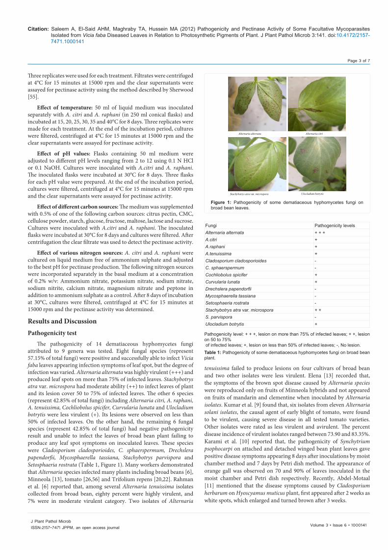
The pathogenicity of 14 dematiaceous hyphomycetes fungi attributed to 9 genera was tested. Eight fungal species (represent 57.15% of total fungi) were positive and successfully able to infect Vicia faba leaves appearing infection symptoms of leaf spot, but the degree of infection was varied. Alternaria alternata was highly virulent (+++) and produced leaf spots on more than 75% of infected leaves. Stachybotrys atra var. microspora had moderate ability (++) to infect leaves of plant and its lesion cover 50 to 75% of infected leaves. The other 6 species (represent 42.85% of total fungi) including Alternaria citri, A. raphani, A. tenuissima, Cochliobolus spicifer, Curvularia lunata and Ulocladium botrytis were less virulent (+). Its lesions were observed on less than 50% of infected leaves. On the other hand, the remaining 6 fungal species (represent 42.85% of total fungi) had negative pathogenicity result and unable to infect the leaves of broad bean plant failing to produce any leaf spot symptoms on inoculated leaves. These species were Cladosporium cladosporioides, C. sphaerspermum, Drechslera papendorfii, Mycosphaerella tassiana, Stachybotrys parvispora and Setosphaeria rostrata (Table 1, Figure 1). Many workers demonstrated that Alternaria species infected many plants including broad beans [6], Minneola [13], tomato [26,56] and Trifolium repens [20,22]. Rahman et al. [6] reported that, among several Alternaria tenuissima isolates collected from broad bean, eighty percent were highly virulent, and 7% were in moderate virulent category. Two isolates of Alternaria
tenuissima failed to produce lesions on four cultivars of broad bean and two other isolates were less virulent. Elena [13] recorded that, the symptoms of the brown spot disease caused by Alternaria species were reproduced only on fruits of Minneola hybrids and not appeared on fruits of mandarin and clementine when inoculated by Alternaria isolates. Kumar et al. [9] found that, six isolates from eleven Alternaria solani isolates, the causal agent of early blight of tomato, were found to be virulent, causing severe disease in all tested tomato varieties. Other isolates were rated as less virulent and avirulent. The percent disease incidence of virulent isolates ranged between 73.90 and 83.35%. Karami et al. [10] reported that, the pathogenicity of Synchytrium psophocarpi on attached and detached winged bean plant leaves gave positive disease symptoms appearing 8 days after inoculations by moist chamber method and 7 days by Petri dish method. The appearance of orange gall was observed on 70 and 90% of leaves inoculated in the moist chamber and Petri dish respectively. Recently, Abdel-Motaal [11] mentioned that the disease symptoms caused by Cladosporium herbarum on Hyoscyamus muticus plant, first appeared after 2 weeks as white spots, which enlarged and turned brown after 3 weeks.
Alternaria alternata Alternaria citri
Stachybotrys atra var. microspora Ulocladium botrytis
Figure 1: Pathogenicity of some dematiaceous hyphomycetes fungi on broad bean leaves.
Fungi Pathogenicity levelsAlternaria alternata + + +A.citri +A.raphani +A.tenuissima +Cladosporium cladosporioides -C. sphaerspermum -Cochliobolus spicifer +Curvularia lunata +Drechslera papendorfii -Mycosphaerella tassiana -Setosphaeria rostrata -Stachybotrys atra var. microspora + +S. parvispora -Ulocladium botrytis +
Pathogenicity level: + + +, lesion on more than 75% of infected leaves; + +, lesion on 50 to 75% of infected leaves; +, lesion on less than 50% of infected leaves; -, No lesion.Table 1: Pathogenicity of some dematiaceous hyphomycetes fungi on broad bean plant.
Citation: Saleem A, El-Said AHM, Maghraby TA, Hussein MA (2012) Pathogenicity and Pectinase Activity of Some Facultative Mycoparasites Isolated from Vicia faba Diseased Leaves in Relation to Photosynthetic Pigments of Plant. J Plant Pathol Microb 3:141. doi:10.4172/2157-7471.1000141
Page 4 of 7
Volume 3 • Issue 6 • 1000141J Plant Pathol MicrobISSN:2157-7471 JPPM, an open access journal
Pectinolytic activity
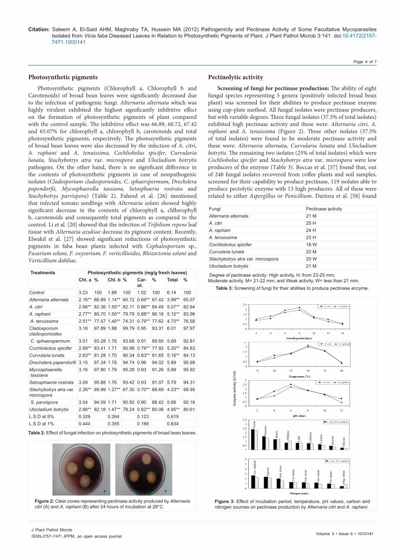
Screening of fungi for pectinase production: The ability of eight fungal species representing 5 genera (positively infected broad bean plant) was screened for their abilities to produce pectinase enzyme using cup-plate method. All fungal isolates were pectinase producers, but with variable degrees. Three fungal isolates (37.5% of total isolates) exhibited high pectinase activity and these were: Alternaria citri, A. raphani and A. tenuissima (Figure 2). Three other isolates (37.5% of total isolates) were found to be moderate pectinase activity and these were: Alternaria alternata, Curvularia lunata and Ulocladium botrytis. The remaining two isolates (25% of total isolates) which were Cochliobolus spicifer and Stachybotrys atra var. microspora were low producers of the enzyme (Table 3). Boccas et al. [57] found that, out of 248 fungal isolates recovered from coffee plants and soil samples, screened for their capability to produce pectinase, 119 isolates able to produce pectolytic enzyme with 13 high producers. All of these were related to either Aspergillus or Penicillium. Dartora et al. [58] found
Photosynthetic pigments
Photosynthetic pigments (Chlorophyll a, Chlorophyll b and Carotenoids) of broad bean leaves were significantly decreased due to the infection of pathogenic fungi. Alternaria alternata which was highly virulent exhibited the highest significantly inhibitive effect on the formation of photosynthetic pigments of plant compared with the control sample. The inhibitive effect was 66.89, 60.72, 67.42 and 65.07% for chlorophyll a, chlorophyll b, carotenoids and total photosynthetic pigments, respectively. The photosynthetic pigments of broad bean leaves were also decreased by the infection of A. citri, A. raphani and A. tenuissima, Cochliobolus spicifer, Curvularia lunata, Stachybotrys atra var. microspora and Ulocladium botrytis pathogens. On the other hand, there is no significant difference in the contents of photosynthetic pigments in case of nonpathogenic isolates (Cladosporium cladosporioides, C. sphaerspermum, Drechslera papendorfii, Mycosphaerella tassiana, Setosphaeria rostrata and Stachybotrys parvispora) (Table 2). Faheed et al. [26] mentioned that infected tomato seedlings with Alternaria solani showed highly significant decrease in the contents of chlorophyll a, chllorophyll b, carotenoids and consequently total pigments as compared to the control. Li et al. [20] showed that the infection of Trifolium repens leaf tissue with Alternaria azukiae decrease its pigment content. Recently, Elwakil et al. [27] showed significant reductions of photosynthetic pigments in faba bean plants infected with Cephalosporium sp., Fusarium solani, F. oxysorium, F. verticillioides, Rhizoctonia solani and Verticillium dahliae.
Treatments Photosynthetic pigments (mg/g fresh leaves)Chl. a % Chl. b % Car-
ot.% Total %
Control 3.23 100 1.88 100 1.02 100 6.14 100Alternaria alternata 2.16** 66.89 1.14** 60.72 0.69** 67.42 3.99** 65.07A. citri 2.66** 82.36 1.55** 82.11 0.86** 84.49 5.07** 82.64A. raphani 2.77** 85.70 1.50** 79.78 0.88** 86.16 5.15** 83.96 A. tenuissima 2.51** 77.57 1.40** 74.31 0.79** 77.62 4.70** 76.58Cladosporium cladosporioides
3.16 97.89 1.88 99.79 0.95 93.31 6.01 97.97
C. sphaerspermum 3.01 93.28 1.76 93.68 0.91 89.60 5.69 92.81Cochliobolus spicifer 2.69** 83.41 1.71 90.98 0.79** 77.92 5.20** 84.83Curvularia lunata 2.63** 81.28 1.70 90.34 0.83** 81.65 5.16** 84.13Drechslera papendorfii 3.15 97.34 1.78 94.74 0.96 94.02 5.89 95.99Mycosphaerella tassiana
3.16 97.80 1.79 95.28 0.93 91.26 5.89 95.92
Setosphaeria rostrata 3.09 95.88 1.76 93.42 0.93 91.07 5.79 94.31Stachybotrys atra var. microspora
2.26** 69.99 1.27** 67.30 0.70** 68.69 4.23** 68.95
S. parvispora 3.04 94.09 1.71 90.92 0.90 88.42 5.66 92.18Ulocladium botrytis 2.66** 82.18 1.47** 78.24 0.82** 80.08 4.95** 80.61L.S.D at 5% 0.329 0.264 0.123 0.619L.S.D at 1% 0.444 0.355 0.166 0.834
Table 2: Effect of fungal infection on photosynthetic pigments of broad bean leaves.
A B
Figure 2: Clear zones representing pectinase activity produced by Alternaria citri (A) and A. raphani (B) after 24 hours of incubation at 28°C.
Fungi Pectinase activityAlternaria alternata 21 MA. citri 25 HA. raphani 24 HA. tenuissima 23 HCochliobolus spicifer 18 WCurvularia lunata 22 MStachybotrys atra var. microspora 20 WUlocladium botrytis 21 M
Degree of pectinase activity: High activity, H: from 23-25 mm;Moderate activity, M= 21-22 mm; and Weak activity, W= less than 21 mm.
Table 3: Screening of fungi for their abilities to produce pectinase enzyme.
00.5
11.5
22.5
3
2 4 6 8 10 12
pH values
A. citri A. raphani
0
0.5
1
1.5
2
2.5
15 20 25 30 35 40
Temperature (oC)
A. citri A. raphani
0
0.5
1
1.5
2
2.5
2 4 6 8 10 12 14
Growth period (days)
A. citri A. raphani
Glu
cose
Sucr
ose
Lact
ose
Cel
lulo
se
CM
C
Mal
tose
Star
chFruc
tosePe
ctin
00.5
11.5
22.5
33.5
Carbon source
A. citri A. raphani
Mag
. nitr
ate
Pot.
nitra
te
Am
. nitr
ate
Sod.
nitr
ite
Cal
. nitr
ate
Sod.
nitr
ate
Pept
one
Am
. sul
phat
e
0
1
2
3
4
5
6
Nitrogen source
A. citri A. raphani
Enzy
me
activ
ity (U
/ml)
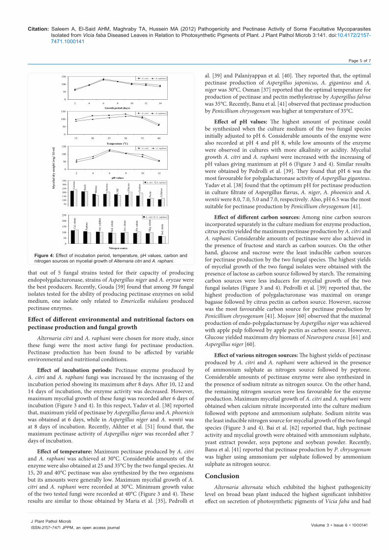
Figure 3: Effect of incubation period, temperature, pH values, carbon and nitrogen sources on pectinase production by Alternaria citri and A. raphani.
Citation: Saleem A, El-Said AHM, Maghraby TA, Hussein MA (2012) Pathogenicity and Pectinase Activity of Some Facultative Mycoparasites Isolated from Vicia faba Diseased Leaves in Relation to Photosynthetic Pigments of Plant. J Plant Pathol Microb 3:141. doi:10.4172/2157-7471.1000141
Page 5 of 7
Volume 3 • Issue 6 • 1000141J Plant Pathol MicrobISSN:2157-7471 JPPM, an open access journal
that out of 5 fungal strains tested for their capacity of producing endopolygalacturonase, strains of Aspergillus niger and A. oryzae were the best producers. Recently, Gouda [59] found that among 39 fungal isolates tested for the ability of producing pectinase enzymes on solid medium, one isolate only related to Emericella nidulans produced pectinase enzymes.
Effect of different environmental and nutritional factors on pectinase production and fungal growth
Alternaria citri and A. raphani were chosen for more study, since these fungi were the most active fungi for pectinase production. Pectinase production has been found to be affected by variable environmental and nutritional conditions.
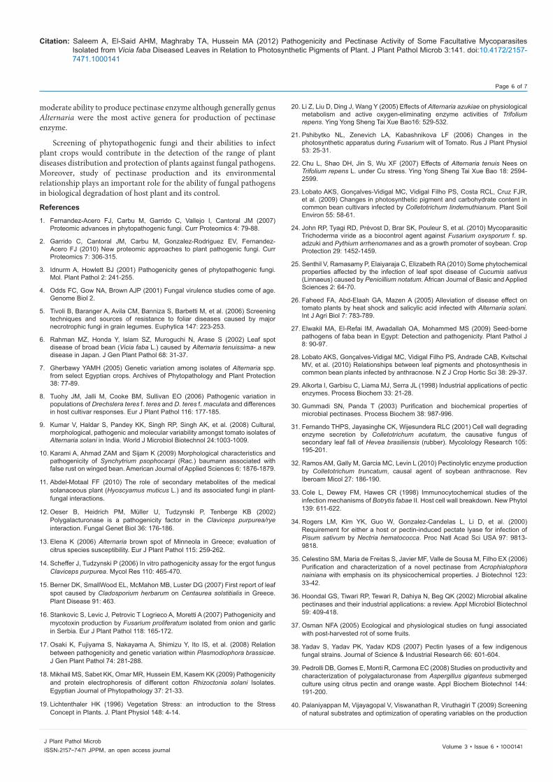
Effect of incubation periods: Pectinase enzyme produced by A. citri and A. raphani fungi was increased by the increasing of the incubation period showing its maximum after 8 days. After 10, 12 and 14 days of incubation, the enzyme activity was decreased. However, maximum mycelial growth of these fungi was recorded after 6 days of incubation (Figure 3 and 4). In this respect, Yadav et al. [38] reported that, maximum yield of pectinase by Aspergillus flavus and A. phoenicis was obtained at 6 days, while in Aspergillus niger and A. wentii was at 8 days of incubation. Recently, Akhter et al. [51] found that, the maximum pectinase activity of Aspergillus niger was recorded after 7 days of incubation.
Effect of temperature: Maximum pectinase produced by A. citri and A. raphani was achieved at 30°C. Considerable amounts of the enzyme were also obtained at 25 and 35°C by the two fungal species. At 15, 20 and 40°C pectinase was also synthesized by the two organisms but its amounts were generally low. Maximum mycelial growth of A. citri and A. raphani were recorded at 30°C. Minimum growth value of the two tested fungi were recorded at 40°C (Figure 3 and 4). These results are similar to those obtained by Maria et al. [35], Pedrolli et
al. [39] and Palaniyappan et al. [40]. They reported that, the optimal pectinase production of Aspergillus japonicus, A. giganteus and A. niger was 30°C. Osman [37] reported that the optimal temperature for production of pectinase and pectin methylestrase by Aspergillus falvus was 35°C. Recently, Banu et al. [41] observed that pectinase production by Penicillium chrysogenum was higher at temperature of 35°C.
Effect of pH values: The highest amount of pectinase could be synthesized when the culture medium of the two fungal species initially adjusted to pH 6. Considerable amounts of the enzyme were also recorded at pH 4 and pH 8, while low amounts of the enzyme were observed in cultures with more alkalinity or acidity. Mycelial growth A. citri and A. raphani were increased with the increasing of pH values giving maximum at pH 6 (Figure 3 and 4). Similar results were obtained by Pedrolli et al. [39]. They found that pH 6 was the most favourable for polygalacturonase activity of Aspergillus giganteus. Yadav et al. [38] found that the optimum pH for pectinase production in culture filtrate of Aspergillus flavus, A. niger, A. phoenicis and A. wentii were 8.0, 7.0, 5.0 and 7.0, respectively. Also, pH 6.5 was the most suitable for pectinase production by Penicillium chrysogenum [41].
Effect of different carbon sources: Among nine carbon sources incorporated separately in the culture medium for enzyme production, citrus pectin yielded the maximum pectinase production by A. citri and A. raphani. Considerable amounts of pectinase were also achieved in the presence of fructose and starch as carbon sources. On the other hand, glucose and sucrose were the least inducible carbon sources for pectinase production by the two fungal species. The highest yields of mycelial growth of the two fungal isolates were obtained with the presence of lactose as carbon source followed by starch. The remaining carbon sources were less inducers for mycelial growth of the two fungal isolates (Figure 3 and 4). Pedrolli et al. [39] reported that, the highest production of polygalacturonase was maximal on orange bagasse followed by citrus pectin as carbon source. However, sucrose was the most favourable carbon source for pectinase production by Penicillium chrysogenum [41]. Mojsov [60] observed that the maximal production of endo-polygalacturnase by Aspergillus niger was achieved with apple pulp followed by apple pectin as carbon source. However, Glucose yielded maximum dry biomass of Neurospora crassa [61] and Aspergillus niger [60].
Effect of various nitrogen sources: The highest yields of pectinase produced by A. citri and A. raphani were achieved in the presence of ammonium sulphate as nitrogen source followed by peptone. Considerable amounts of pectinase enzyme were also synthesized in the presence of sodium nitrate as nitrogen source. On the other hand, the remaining nitrogen sources were less favourable for the enzyme production. Maximum mycelial growth of A. citri and A. raphani were obtained when calcium nitrate incorporated into the culture medium followed with peptone and ammonium sulphate. Sodium nitrite was the least inducible nitrogen source for mycelial growth of the two fungal species (Figure 3 and 4). Bai et al. [62] reported that, high pectinase activity and mycelial growth were obtained with ammonium sulphate, yeast extract powder, soya peptone and soybean powder. Recently, Banu et al. [41] reported that pectinase production by P. chrysogenum was higher using ammonium per sulphate followed by ammonium sulphate as nitrogen source.
ConclusionAlternaria alternata which exhibited the highest pathogenicity
level on broad bean plant induced the highest significant inhibitive effect on secretion of photosynthetic pigments of Vicia faba and had
0
50
100
150
2 4 6 8 10 12 14
Growth period (days)
A. citri A. raphani
0
50
100
150
15 20 25 30 35 40
Temperature (oC)
A. citri A. raphani
0
50
100
150
2 4 6 8 10 12
pH values
A. citri A. raphani
Lacto
se
Star
ch
Malt
ose
Sucr
ose
CMC Glu
cose
Fruc
tose
Pecti
n
Cellu
los e
050
100150200250300350
Carbon source
A. citri A. raphani
Sod.
nitr
ite
Mag
. nitr
ate
Pot.
nitra
te
Am
. nitr
ate
Sod.
nitr
ate
Am
. sul
phat e
Pept
one
Cal.
nitra
te
0
50
100
150
200
250
Nitrogen source
A. citri A. raphani
Myc
elia
l dry
wei
ght (
mg/
50
ml)
Figure 4: Effect of incubation period, temperature, pH values, carbon and nitrogen sources on mycelial growth of Alternaria citri and A. raphani.
Citation: Saleem A, El-Said AHM, Maghraby TA, Hussein MA (2012) Pathogenicity and Pectinase Activity of Some Facultative Mycoparasites Isolated from Vicia faba Diseased Leaves in Relation to Photosynthetic Pigments of Plant. J Plant Pathol Microb 3:141. doi:10.4172/2157-7471.1000141
Page 6 of 7
Volume 3 • Issue 6 • 1000141J Plant Pathol MicrobISSN:2157-7471 JPPM, an open access journal
moderate ability to produce pectinase enzyme although generally genus Alternaria were the most active genera for production of pectinase enzyme.
Screening of phytopathogenic fungi and their abilities to infect plant crops would contribute in the detection of the range of plant diseases distribution and protection of plants against fungal pathogens. Moreover, study of pectinase production and its environmental relationship plays an important role for the ability of fungal pathogens in biological degradation of host plant and its control.
References
1. Fernandez-Acero FJ, Carbu M, Garrido C, Vallejo I, Cantoral JM (2007) Proteomic advances in phytopathogenic fungi. Curr Proteomics 4: 79-88.
2. Garrido C, Cantoral JM, Carbu M, Gonzalez-Rodriguez EV, Fernandez-Acero FJ (2010) New proteomic approaches to plant pathogenic fungi. Curr Proteomics 7: 306-315.
3. Idnurm A, Howlett BJ (2001) Pathogenicity genes of phytopathogenic fungi. Mol. Plant Pathol 2: 241-255.
4. Odds FC, Gow NA, Brown AJP (2001) Fungal virulence studies come of age. Genome Biol 2.
5. Tivoli B, Baranger A, Avila CM, Banniza S, Barbetti M, et al. (2006) Screening techniques and sources of resistance to foliar diseases caused by major necrotrophic fungi in grain legumes. Euphytica 147: 223-253.
6. Rahman MZ, Honda Y, Islam SZ, Muroguchi N, Arase S (2002) Leaf spot disease of broad bean (Vicia faba L.) caused by Alternaria tenuissima- a new disease in Japan. J Gen Plant Pathol 68: 31-37.
7. Gherbawy YAMH (2005) Genetic variation among isolates of Alternaria spp. from select Egyptian crops. Archives of Phytopathology and Plant Protection 38: 77-89.
8. Tuohy JM, Jalli M, Cooke BM, Sullivan EO (2006) Pathogenic variation in populations of Drechslera teres f. teres and D. teres f. maculata and differences in host cultivar responses. Eur J Plant Pathol 116: 177-185.
9. Kumar V, Haldar S, Pandey KK, Singh RP, Singh AK, et al. (2008) Cultural, morphological, pathogenic and molecular variability amongst tomato isolates of Alternaria solani in India. World J Microbiol Biotechnol 24:1003-1009.
10. Karami A, Ahmad ZAM and Sijam K (2009) Morphological characteristics and pathogenicity of Synchytrium psophocarpi (Rac.) baumann associated with false rust on winged bean. American Journal of Applied Sciences 6: 1876-1879.
11. Abdel-Motaal FF (2010) The role of secondary metabolites of the medical solanaceous plant (Hyoscyamus muticus L.) and its associated fungi in plant-fungal interactions.
12. Oeser B, Heidrich PM, Müller U, Tudzynski P, Tenberge KB (2002) Polygalacturonase is a pathogenicity factor in the Claviceps purpurea/rye interaction. Fungal Genet Biol 36: 176-186.
13. Elena K (2006) Alternaria brown spot of Minneola in Greece; evaluation of citrus species susceptibility. Eur J Plant Pathol 115: 259-262.
14. Scheffer J, Tudzynski P (2006) In vitro pathogenicity assay for the ergot fungus Claviceps purpurea. Mycol Res 110: 465-470.
15. Berner DK, SmallWood EL, McMahon MB, Luster DG (2007) First report of leaf spot caused by Cladosporium herbarum on Centaurea solstitialis in Greece. Plant Disease 91: 463.
16. Stankovic S, Levic J, Petrovic T Logrieco A, Moretti A (2007) Pathogenicity and mycotoxin production by Fusarium proliferatum isolated from onion and garlic in Serbia. Eur J Plant Pathol 118: 165-172.
17. Osaki K, Fujiyama S, Nakayama A, Shimizu Y, Ito IS, et al. (2008) Relation between pathogenicity and genetic variation within Plasmodiophora brassicae. J Gen Plant Pathol 74: 281-288.
18. Mikhail MS, Sabet KK, Omar MR, Hussein EM, Kasem KK (2009) Pathogenicity and protein electrophoresis of different cotton Rhizoctonia solani Isolates. Egyptian Journal of Phytopathology 37: 21-33.
19. Lichtenthaler HK (1996) Vegetation Stress: an introduction to the Stress Concept in Plants. J. Plant Physiol 148: 4-14.
20. Li Z, Liu D, Ding J, Wang Y (2005) Effects of Alternaria azukiae on physiological metabolism and active oxygen-eliminating enzyme activities of Trifolium repens. Ying Yong Sheng Tai Xue Bao16: 529-532.
21. Pshibytko NL, Zenevich LA, Kabashnikova LF (2006) Changes in the photosynthetic apparatus during Fusarium wilt of Tomato. Rus J Plant Physiol 53: 25-31.
22. Chu L, Shao DH, Jin S, Wu XF (2007) Effects of Alternaria tenuis Nees on Trifolium repens L. under Cu stress. Ying Yong Sheng Tai Xue Bao 18: 2594-2599.
23. Lobato AKS, Gonçalves-Vidigal MC, Vidigal Filho PS, Costa RCL, Cruz FJR, et al. (2009) Changes in photosynthetic pigment and carbohydrate content in common bean cultivars infected by Colletotrichum lindemuthianum. Plant Soil Environ 55: 58-61.
24. John RP, Tyagi RD, Prévost D, Brar SK, Pouleur S, et al. (2010) Mycoparasitic Trichoderma viride as a biocontrol agent against Fusarium oxysporum f. sp. adzuki and Pythium arrhenomanes and as a growth promoter of soybean. Crop Protection 29: 1452-1459.
25. Senthil V, Ramasamy P, Elaiyaraja C, Elizabeth RA (2010) Some phytochemical properties affected by the infection of leaf spot disease of Cucumis sativus (Linnaeus) caused by Penicillium notatum. African Journal of Basic and Applied Sciences 2: 64-70.
26. Faheed FA, Abd-Elaah GA, Mazen A (2005) Alleviation of disease effect on tomato plants by heat shock and salicylic acid infected with Alternaria solani. Int J Agri Biol 7: 783-789.
27. Elwakil MA, El-Refai IM, Awadallah OA, Mohammed MS (2009) Seed-borne pathogens of faba bean in Egypt: Detection and pathogenicity. Plant Pathol J 8: 90-97.
28. Lobato AKS, Gonçalves-Vidigal MC, Vidigal Filho PS, Andrade CAB, Kvitschal MV, et al. (2010) Relationships between leaf pigments and photosynthesis in common bean plants infected by anthracnose. N Z J Crop Hortic Sci 38: 29-37.
29. Alkorta I, Garbisu C, Liama MJ, Serra JL (1998) Industrial applications of pectic enzymes. Process Biochem 33: 21-28.
30. Gummadi SN, Panda T (2003) Purification and biochemical properties of microbial pectinases. Process Biochem 38: 987-996.
31. Fernando THPS, Jayasinghe CK, Wijesundera RLC (2001) Cell wall degrading enzyme secretion by Colletotrichum acutatum, the causative fungus of secondary leaf fall of Hevea brasiliensis (rubber). Mycolology Research 105: 195-201.
32. Ramos AM, Gally M, Garcia MC, Levin L (2010) Pectinolytic enzyme production by Colletotrichum truncatum, causal agent of soybean anthracnose. Rev Iberoam Micol 27: 186-190.
33. Cole L, Dewey FM, Hawes CR (1998) Immunocytochemical studies of the infection mechanisms of Botrytis fabae II. Host cell wall breakdown. New Phytol 139: 611-622.
34. Rogers LM, Kim YK, Guo W, Gonzalez-Candelas L, Li D, et al. (2000) Requirement for either a host or pectin-induced pectate lyase for infection of Pisum sativum by Nectria hematococca. Proc Natl Acad Sci USA 97: 9813-9818.
35. Celestino SM, Maria de Freitas S, Javier MF, Valle de Sousa M, Filho EX (2006) Purification and characterization of a novel pectinase from Acrophialophora nainiana with emphasis on its physicochemical properties. J Biotechnol 123: 33-42.
36. Hoondal GS, Tiwari RP, Tewari R, Dahiya N, Beg QK (2002) Microbial alkaline pectinases and their industrial applications: a review. Appl Microbiol Biotechnol 59: 409-418.
37. Osman NFA (2005) Ecological and physiological studies on fungi associated with post-harvested rot of some fruits.
38. Yadav S, Yadav PK, Yadav KDS (2007) Pectin lyases of a few indigenous fungal strains. Journal of Science & Industrial Research 66: 601-604.
39. Pedrolli DB, Gomes E, Monti R, Carmona EC (2008) Studies on productivity and characterization of polygalacturonase from Aspergillus giganteus submerged culture using citrus pectin and orange waste. Appl Biochem Biotechnol 144: 191-200.
40. Palaniyappan M, Vijayagopal V, Viswanathan R, Viruthagiri T (2009) Screening of natural substrates and optimization of operating variables on the production
Citation: Saleem A, El-Said AHM, Maghraby TA, Hussein MA (2012) Pathogenicity and Pectinase Activity of Some Facultative Mycoparasites Isolated from Vicia faba Diseased Leaves in Relation to Photosynthetic Pigments of Plant. J Plant Pathol Microb 3:141. doi:10.4172/2157-7471.1000141
Page 7 of 7
Volume 3 • Issue 6 • 1000141J Plant Pathol MicrobISSN:2157-7471 JPPM, an open access journal
of pectinase by submerged fermentation using Aspergillus niger MTCC 281. Afri J Biotech 8: 682-686.
41. Banu AR, Devi MK, Gnanaprabhal GR, Pradeep BV, Palaniswamy M (2010) Production and characterization of pectinase enzyme from Penicillium chrysogenum. Indian J Sci Technol 3: 377-381.
42. Al-Gashgari RMG (2002) Occurrence of fungi and pectolytic activity in fruit juices from Saudi Arabia. Pak J Biolog Sci 5: 609-611.
43. O'Brien PA, Zamani M (2003) Production of pectic enzymes by barepatch isolates of Rhizoctonia solani AG 8. Australas Plant. Pathol 32: 65-72.
44. Jayasinghe CK, Wijayaratne SC, Fernando TH (2004) Characterization of cell wall degrading enzymes of Thanatephorus cucumeris. Mycopathologia 157: 73-79.
45. Phutela U, Dhuna V, Sandhu S, Chadha BS (2005) Pectinase and polygalacturonase production by a thermophilic Aspergillus fumigatus isolated from decomposting orange peels. Braz J Microbiol 36: 63-69.
46. Amadioha AC (2006) Effect of cultural conditions on the production of polygalacturonase by Rhizoctonia bataticola. Archives of Phytopathology and Plant Protection 40: 353-358.
47. Asoufi H, Hameed KM, Mahasneh A (2007) The cellulase and pectinase activities associated with the virulence of indigenous Sclerotinia sclerotiorum isolates in Jordan valley. Plant Pathol J 23: 233-238.
48. Griese EC, Dekker RFH, Barbosa AM (2008) Orange bagasse as substrate for the production of pectinase and laccase by Botryosphaeria rhodina MAMB-05 in submerged and solid state fermentation. Bioresources 3: 335-345.
49. Suresh B, Viruthagiri T, Sasikumar E (2009) Optimization of process variables using response surface methodology (RSM) in the solid-state fermentative production of pectinase by Aspergillus awamori. As J Food Ag-Ind 2: 302-314.
50. Rathod GM, Chavan AM (2010) Extra-cellular pectinase activity of post-harvest fungi from papaya fruits in presence of different influencing factors. Journal of Experimental Sciences 1: 7-11.
51. Akhter N, Morshed MA, Uddin A, Begum F, Sultan T, et al. (2011) Production
of pectinase by Aspergillus niger cultured in solid state media. International Journal of Biosciences 1: 33-42.
52. El-Kholl MM, Ragab MM and Hussein MY (1994) Alternaria spots of sugar beet in Egypt. Egypt J Phytopathol 22: 179-193.
53. Hankin L, Zucker LM, Sands DC (1971) Improved solid medium for the detection and enumeration of pectolytic bacteria. Appl Microbiol 22: 205-209.
54. Ammar MS, Louboudy SS, Azab MS and Afifi MM (1995) A new method for the estimation of fungal pectinase (s) using the pectin clearing zone (P.C.Z.) technique and its application in food industries. Al-Azhar Bull Sci 6: 325-339.
55. Sherwood RT (1966) Pectin lyase and polygalacturonase production by Rhizoctonia solani and other fungi. Phytopathology 56: 279-286.
56. Chaerani R, Roeland EV (2006) Tomato early blight (Alternaria solani): the pathogen, genetics, and breeding for resistance. J Gen Plant Pathol 72: 335-347.
57. Boccas F, Roussos S, Gutierrez M, Serrano L, Viniegra GG (1994) Production of pectinase from coffee pulp in solid state fermentation system: Selection of wild fungal isolate of high potency by a simple three-step screening technique. J Food Sci Technol 31: 22-26.
58. Dartora AB, Bertolin TE, Bilibio D, Silveira MM, Costa JA (2002) Evaluation of filamentous fungi and inducers for the production of endo-polygalacturonase by solid state fermentation. Z. Naturforsch C 57: 666-670.
59. Gouda HAA (2009) Studies on xerophilic, acidiphilic and alkaliphilic fungi in Wadi El-Natrun.
60. Mojsov K (2010) The effects of different carbon sources on biosynthesis of pectinolytic enzymes by Aspergillus niger. Applied Technologies and Innovations 3: 23-29.
61. Polizeli Mde L, Jorge JA, Terenzi HF (1991) Pectinase production by Neurospora crassa: purification and biochemical characterization of extracellular polygalacturonase activity. J General Microbiol 137: 1815-1823.
62. Bai ZH, Zhang HX, Qi HY, Peng XW, Li BJ (2004) Pectinase production by Aspergillus niger using wastewater in solid state fermentation for eliciting plant disease resistance. Bioresour Technol 95: 49-52.