Embed Size (px)
Citation preview
1
MEK inhibition induces therapeutic iodine uptake in a murine model of anaplastic thyroid
cancer.
Oussama ElMokh1, Vincent Taelman2, Piotr Radojewski2, Matthias A. Roelli1,
Amandine Stoss1, Rebecca A. Dumont3, Matthias S. Dettmer4, Wayne A. Phillips5,
Martin A. Walter2, and Roch‐Philippe Charles1*
1Institute for Biochemistry and Molecular Medicine, University of Bern, Switzerland
2Institute for Nuclear Medicine, Geneva University Hospitals, Switzerland
3Department of Radiology, University of California at San Francisco, USA
4Institute for Pathology, University of Bern, Switzerland
5Cancer Biology Laboratory, Peter MacCallum Cancer Centre, Melbourne, Australia
*Corresponding author:
Roch‐Philippe Charles (PhD)
Institute for Biochemistry and Molecular Medicine, University of Bern,
Bühlstrasse 28, CH‐3012 Bern, Switzerland.
Phone: +41 31 631 4344 Email: roch‐[email protected]
First author:
Oussama ElMokh
Central Laboratory of Hematology, University Hospital of Lausanne (CHUV),
27‐Sud, Rue du Bugnon, CH‐1011 Lausanne, Switzerland.
Phone: +41 21 314 4214 Email: oussama.el‐[email protected]
Funding support: Swiss National Science Foundation grant 31003A_149824/1 and Swiss National
Science Foundation grant NCCR‐TransCure.
Conflict of interest: The authors declare no potential conflicts of interest relevant to this article.
Word count (total): 4958
Running title: MEKi induces 131I uptake in advanced thyroid cancer.
Journal of Nuclear Medicine, published on November 21, 2018 as doi:10.2967/jnumed.118.216721by on August 11, 2019. For personal use only. jnm.snmjournals.org Downloaded from
2
ABSTRACT
Rationale: Anaplastic thyroid carcinoma (ATC) is refractory to radioiodine therapy in part due to
impaired iodine metabolism. We targeted the MAPK and PI3’K pathways with the intent to induce
radioiodine uptake for radioiodine treatment of ATC.
Methods: Human ATC cells were used to evaluate the ability of pharmacological inhibition of the
mitogen‐activated protein kinase and phosphatidylinositol 3‐kinase pathways to induce
radioiodine uptake. Thyrocyte‐specific double mutant BRAFV600E PIK3CAH1047R mice were treated
with a MEK inhibitor followed by radioiodine treatment and tumor burden was monitored by
ultrasound imaging.
Results: ATC cell lines showed an increase in sodium‐iodine symporter transcription when treated
with a MEK or BRAFV600E inhibitor alone and in combination with PI3’K inhibitor. This translated
into a dose‐dependent elevation of iodine uptake following treatment with a MEK inhibitor alone
and in combination with a PI3’K inhibitor. In vivo, MEK inhibition but not BRAF nor PI3’K inhibition
upregulated sodium‐iodine symporter transcription. This translated into a stable reduction of
tumor burden when mice were treated with a MEK inhibitor prior to radioiodine administration.
Conclusion: This study confirms the ability of MEK inhibition to induce iodine uptake in in vitro
and in vivo models of ATC. The approach of using a MEK inhibitor before radioiodine treatment
could readily be translated into clinical practice and provide a much‐needed therapeutic option
for patients with ATC.
Key words: Drug resistance, NIS, Targeted therapy, differentiation.
by on August 11, 2019. For personal use only. jnm.snmjournals.org Downloaded from
3
INTRODUCTION
Anaplastic thyroid carcinoma (ATC) is one of the most aggressive human malignancies
(1,2) and is refractory to radioiodine therapy (3).
Impaired iodine metabolism can result from dysregulation of the mitogen‐activated
protein kinase (MAPK) and phosphatidylinositol 3‐kinase (PI3’K) pathways (Figure 1), which
regulate a variety of cellular activities and have been implicated in the development of various
human cancers (4). MAPK mutations are common in ATC, with 25% of tumors harboring KRAS
mutations and depending on the study 27% to 45% are carrying BRAF mutations (5–7). The most
common mutation found in BRAF codes for the protein kinase BRAFV600E, which induces
constitutive activation of the downstream pathway (Figure 1). Several important PI3’K pathway
mutations in ATC include PTEN deletions (10‐20%) and PIK3CA mutations (15‐25%) (8). We have
previously shown that the BRAFV600E mutation in combination with the PIK3CAH1047R mutation
induces tumor progression from PTC to ATC in a mouse model (9).
The link between alterations of the MAPK and PI3’K pathways and impairment of iodine
metabolism is well‐documented, although the underlying mechanisms not fully elucidated. BRAF
mutations are associated with impairment of NIS function and subsequent increased resistance
to radioiodine uptake in (10,11) thyroid cancers. Some studies suggest that BRAF inhibits the
expression of PAX8 and prevents transcription of NIS (12).
The relationship between the MAPK pathway and the ability to store iodine intracellularly
has been further demonstrated in mouse models of papillary thyroid cancer (PTC), where
selective MAPK pathway antagonists have been shown to increase the expression of NIS with
resultant increased uptake of iodine (13). Inhibition of the MAPK pathway via MEK is a promising
strategy for treatment of iodine refractory thyroid cancers and, specifically, ATC. Several studies
by on August 11, 2019. For personal use only. jnm.snmjournals.org Downloaded from
4
suggest that PI3’K inhibition increases radioiodine uptake in thyroid cancer cells (14,15), however
this has not yet been evaluated in vivo. In this study, we aimed to investigate the consequences
of pharmacologically targeting the MAPK and PI3’K pathways separately and in combination to
induce radioiodine uptake in ATC cell lines and in a lethal double mutant ATC (BRAFV600E
PIK3CAH1047R) mouse model. We hypothesized that pharmacological restoration of radioiodine
uptake with subsequent RAI would be an effective means of therapy in a murine model of ATC.
by on August 11, 2019. For personal use only. jnm.snmjournals.org Downloaded from
5
MATERIALS AND METHODS
Small Molecule Kinases Inhibitors
Inhibitors, PD‐325901 (MEK1/2), PLX‐4032/vemurafenib (BRAFV600E), PLX‐4720
(BRAFV600E) and GDC‐0941 (class I PI3’K) were purchased from Abmole Bioscience, Hong‐Kong.
PLX‐4032 was used in vitro as a BRAFV600E inhibitor but due to poor solubility (16), PLX‐4720 was
used for in vivo experiments.
BRAFV600E PIK3CAH1047R Mice
Transgenic mice were bred by combining the following alleles: BrafCA (17), Pik3caLat (18)
and Thyro::CreERT2 (19) as previously described (9). Mutations were induced by 5 consecutive
daily injections of 1 mg tamoxifen IP. Mice were cared for in accordance with Swiss federal
guidelines, housed in isolated ventilated cages and fed ad libitum in a 12/12 h cycle of light and
dark. The experimental protocol was approved by the Bernese cantonal ethical commission for
animal experimentation (License number: BE120/13).
Cell Lines
The human cell lines 8505c, Sw1736 and OCUT‐2 were obtained and maintained as
described previously (20).
In Vitro Evaluation of ATC NIS mRNA Expression
Cells were seeded in 10 cm Petri dishes at 25% confluency overnight then treated with
PD‐325901 at 10 nM, GDC‐0941 at 100 nM, PLX‐4032 at 100 nM, PD‐325901 at 10 nM with GDC‐
0941 at 100 nM or PLX‐4032 at 100 nM with GDC‐0941 at 100 nM at 37 °C for 48 h. Cells were
by on August 11, 2019. For personal use only. jnm.snmjournals.org Downloaded from
6
washed twice with 5 ml cold PBS then collected in 1 ml by scrapping the dish surface. A
centrifugation for 10 min at 3’000 g was performed and the cell pellets were frozen in liquid
nitrogen then stored at ‐80 °C. NIS mRNA levels were evaluated by quantitative PCR see below.
In Vivo Evaluation of Thyroid‐Specific Gene Expression Following Kinase Inhibition
Thyroid tissues from 8 single mutant BRAFV600E mice (BrafCA/+; Thyro::CreERT2) treated for
21 days with 12.5 mg/kg PD‐325901 (N=4) or Hydroxypropyl methylcellulose 0.5%, Tween‐80
0.2% was used as a control (N=4) by oral gavage were fixed in formalin and embedded in paraffin
blocks (21), from which RNA was extracted with High Pure FFPET RNA isolation kit (Roche)
according to the manufacturer’s protocol.
23 BRAFV600E PIK3CAH1047R double mutant mice (BrafCA/+; Pik3caLat/+; Thyro::CreERT2) were
treated 2 months after tumor induction with PD‐325901 at 5 mg/kg (n=4), PLX‐4720 at 30 mg/kg
(n=5) or GDC‐0941 at 50 mg/kg (n=5) as single treatments, or PLX/GDC (n=5) and PD/GDC (n=4)
in combination by oral gavage daily for 10 days. Additionally, thyroid glands from 12 Cre negative
mice (BrafCA/+; Pik3caLat/+) were combined for a total of n=3 and used as non‐tumoral controls.
Hydroxypropyl methylcellulose 0.5%, Tween‐80 0.2% was used as a control. Mice were then
sacrificed, thyroid glands were dissected and snap frozen using liquid nitrogen, and total RNA
was purified with the QIAzol reagent from Qiagen according to the manufacturer's instructions
with a TissueLyser LT using iron beads. Nis, Tshr, Tg, Pax8, Tpo and Ttf1 mRNA levels were
evaluated (see below).
Quantitative PCR
by on August 11, 2019. For personal use only. jnm.snmjournals.org Downloaded from
7
500 ng of RNA underwent reverse transcription using Oligo(dT)12‐18 Primer and the
Super Script II Reverse Transcriptase from Invitrogen following the manufacturer’s suggested
protocol. Samples were run on ViiA™ 7 Real‐Time PCR System (Applied Biosystems, Life
Technologies) using the TaqMan® gene expression Master Mix and primers from Applied
Biosystems. GAPDH was used for normalization using the 2∆∆Ct calculation method. All values
were expressed as percent of the baseline transcription levels found in non‐tumoral Cre negative
thyroid tissues. The following primers were purchased from Applied Biosystems, Life
Technologies: NIS (Hs00166567_m1), Nis (Mm01351811_m1), Tg (Mm01200340_m1), Ttf1
(Mm00657018_m1), Pax8 (Mm00440623_m1), Tpo (Mm00456355_m1), Tshr
(Mm00442027_m1), GAPDH (Hs02758991_g1) and Gapdh (Mm99999915_g1).
In Vitro 123I Uptake Assay
Cells were seeded in Corning® 96‐wells plates at 30% confluency, incubated overnight at 37°C and
5% CO2, and treated for 48 h with various concentrations of PD‐325901 (0, 0.1, 1, 10, 100 and
1000 nM) with or without 200 nM of GDC‐0941. All except for one of the PD‐325901
concentrations used were below the Cmax value (200 nM), which corresponds to the maximal
concentration in human plasma without toxicity recorded (22). Cell were washed with PBS and
cell viability was assessed with the AlamarBlue® Cell Viability Reagent (#765506, Invitrogen) by
measuring fluorescence at 634 nm (SpectraMax M4 plate reader, Molecular Devices, USA). After
removal of AlamarBlue®, cells were washed with PBS and incubated with culture medium
containing 18.5 kBq of 123I (GE Healthcare, Switzerland) for 1 h at 37 °C. Then the medium was
removed and the cells were washed 3 times with PBS, then replaced by 50 µl of scintillation
by on August 11, 2019. For personal use only. jnm.snmjournals.org Downloaded from
8
reagent (MicroScint‐20, PerkinElmer) per well. Activity was measured with a microplate
scintillation counter (Packard Topcount NXT).
In Vivo 125I Uptake Assay
11 BRAFV600E PIK3CAH1047R double mutant mice were treated daily by oral gavage for 2 weeks with
control (n=4), PD‐325901 at 5 mg/kg alone (n=4) or in combination with GDC‐0941 at 30 mg/kg
(n=3). Hydroxypropyl methylcellulose with 0.5% Tween80 was used as a control. Then, mice were
injected via tail vein with 1.11 MBq of 125I (ANAWA, Biomedical Services & Products, Switzerland)
and euthanized 1 h later with CO2. Thyroid glands, salivary glands, liver and blood were harvested
and weighed. Radioactivity was quantified for 30 seconds per sample using a PerkinElmer 2470
automatic gamma counter.
In Vivo Tumor Treatment Experiment
16 BRAFV600E PIK3CAH1047R double mutant mice were treated daily by oral gavage for 10 days with
control (n=8) or PD‐325901 at 5 mg/kg (n=8). Following this, half of the mice in each group were
injected with 18.5 MBq of 131I (PerkinElmer, USA) via tail vein. Tumor volume was assessed weekly
for 9 weeks with the following protocol described previously (20). Tumor burden was determined
by measuring the surface of the largest thyroid cross section normalized to the size of the gland
at the beginning of the study. In this model of thyroid cancer, all thyrocytes are undergoing
molecular changes leading to cancerization of the entire thyroid gland. Therefore, the
assessment of whole thyroid size for tumor burden is a reliable and well‐accepted approximation
(9,21,23).
by on August 11, 2019. For personal use only. jnm.snmjournals.org Downloaded from
9
Statistical Analysis
A two‐way ANOVA test was performed to evaluate the difference in tumor burden measurement
between treatment groups. A one‐way ANOVA test was used to evaluate the difference in NIS
mRNA levels in vitro, radioiodine uptake in vivo, and differences in vivo between treatments on
mRNA levels of different genes (followed by Tukey’s multiple comparisons test). All statistical
analyses were performed using Graphpad Prism. P‐values equal to or less than 0.05 were
considered significant.
RESULTS MEK Inhibition Alone or in Combination with PI3’K Inhibition Enhances NIS mRNA Transcription
and Increases Intracellular Iodine Transport in Vitro
The effect of the MEK inhibitor PD‐325901 and BRAF inhibitor PLX‐4032 (vemurafenib)
alone or combined with the PI3’K inhibitor GDC‐0941 was evaluated on mRNA transcription levels
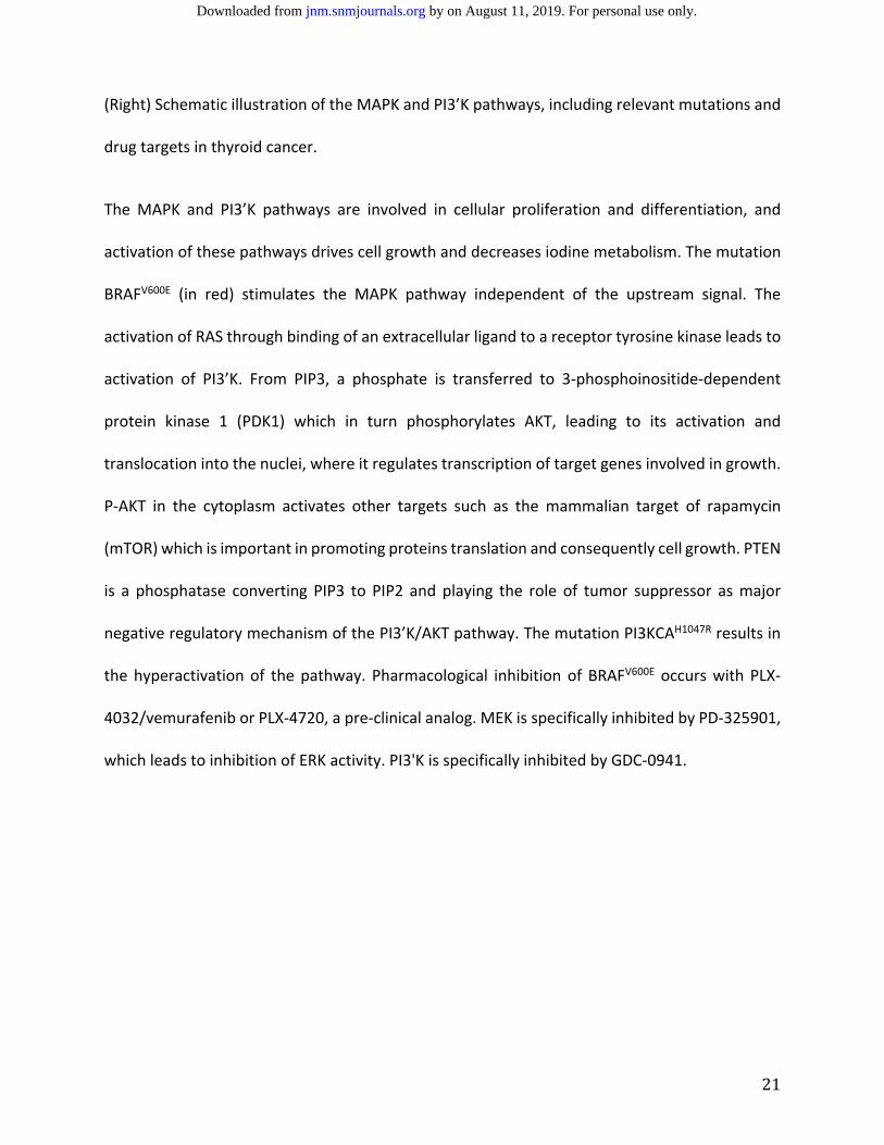
of NIS in 8505c and OCUT‐2, two ATC cell lines harboring a BRAFV600E mutation (Figure 2A). In
8505c, PD‐325901 treatment alone induced a 4‐fold increase in NIS mRNA level after 48 h, the
addition of GDC‐0941 did not further augment NIS mRNA expression. GDC‐0941 alone did not
have an effect on NIS transcription, and PLX‐4032 did not lead to a significant elevation of NIS
mRNA expression when it was applied alone or in combination with GDC‐0941. Treatment of the
OCUT‐2 cell line, which has a PIK3CAH1047R mutation in addition to a BRAFV600E mutation, showed
similar upregulation of NIS mRNA transcription following treatment with PD‐325901, with an
additional response to GDC‐0941 alone (3‐fold increase in NIS mRNA level) and with the
by on August 11, 2019. For personal use only. jnm.snmjournals.org Downloaded from
10
combination of PD‐325901 and GDC‐0941 (9‐fold increase). In OCUT‐2, PLX‐4032 alone or in
combination with GDC‐0941 resulted in a 5‐fold increase in NIS mRNA levels.
Following 48 h treatment with PD‐325901 at varying concentrations ranging from 0 to 1
µM, cellular iodine uptake increased by more than double in OCUT‐2 and 8505c, as well as in
SW1736, a third ATC cell line also harboring a BRAFV600E mutation. Addition of 200 nM GDC‐0941
with PD‐325901 further increased uptake of 123I in the OCUT‐2 and SW1736 cell lines, but not in
8505c (Figure 2B).
MEK Inhibition but not BRAFV600E Inhibition Increases Nis, Tshr, Tg, Pax8, and Tpo mRNA
Transcription in Vivo, Which Correlates with Histological Re‐normalization of Thyroid Follicles.
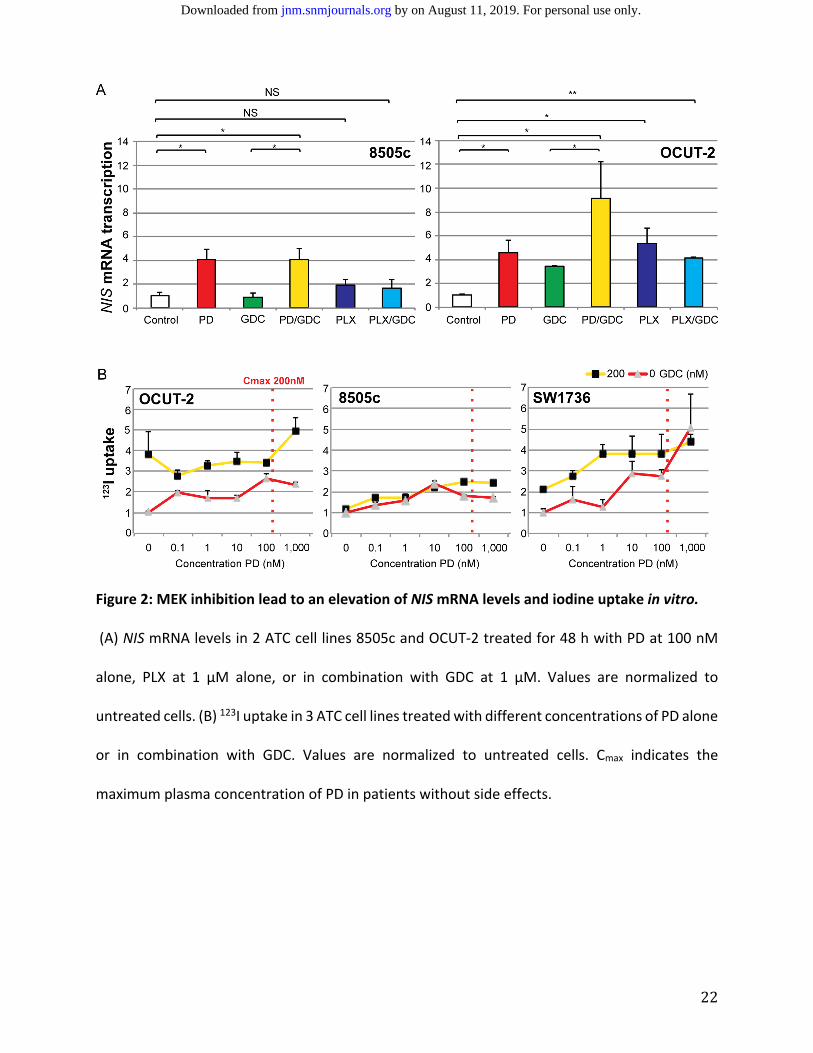
A four‐fold increase in Nis mRNA levels extracted from formalin‐fixed, paraffin‐embedded
tumor sections in BRAFV600E single mutant mice (21) treated with the MEK inhibitor PD‐325091
as compared to control mice (Figure 3A) was observed. Additionally, the treated group showed
restoration of normal thyroid follicular structure (near spheroid follicles containing colloid
substance) on H&E staining (Figure 3B), while the control group displayed a papillary structure
and absence of normal thyroid follicles.
We then switched to a more aggressive murine BRAFV600E PIK3CAH1047R double mutant
model. In animals treated with the MEK inhibitor PD‐325901, there was upregulation of Nis
mRNA by almost 20‐fold. The selective BRAFV600E inhibitor PLX‐4720 did not increase Nis mRNA
transcription, nor did the PI3'K inhibitor GDC‐0941 alone or in combination with PD‐325901 or
PLX‐4720 (Figure 3C).
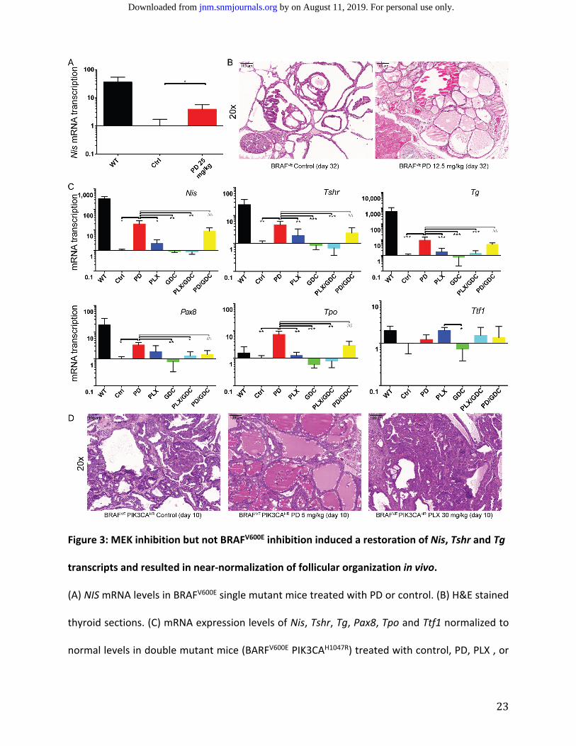
Evaluation of mRNA transcription of additional genes known to be important in thyroid
gland function was performed, including Tshr, Pax8, Tg, Tpo and Ttf1. For all genes except Ttf‐1,
by on August 11, 2019. For personal use only. jnm.snmjournals.org Downloaded from
11
the same pattern as Nis was observed with a strong response to PD‐325901, no further elevation
in combination with GDC‐0941 and little response to any of the other drugs alone (Figure 3C).
Elevation of protein expression could be confirmed in IHC for TG, PAX8 and TTF‐1 (Sup Fig 1).
Increased Nis, Tshr, Tg, Pax8 and Tpo transcription correlated with histological near‐
normalization in the appearance of thyroid follicles after 10 days of PD‐325901 treatment on
H&E, a finding that was not replicated in control mice or mice treated with PLX‐4720, GDC‐0941
(Figure 3D).
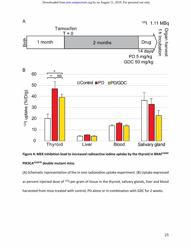
MEK Inhibition Leads to a Significant Increase in Thyroid Radioiodine Uptake in BRAFV600E
PIK3CAH1047R Double Mutant Mice.
ollowing gene expression analyses, a validation of functional changes in in radioiodine
uptake in BRAFV600E PIK3CAH1047R double mutant mice using PD‐325901 alone and in combination
with GDC‐0941 was attempted. After 14 days of daily treatment with PD‐325901 with or without
GDC‐0941, there was a 2.5‐fold increase in 125I thyroid uptake in BRAFV600E PIK3CAH1047R double
mutant mice compared with controls. There was no significant difference in thyroid radioiodine
uptake between mice treated with PD‐325901 or those treated with a combination of PD‐325901
and GDC‐0941. Liver uptake, used to assess background uptake due to passive diffusion through
the apical membrane, remained stable across the different treatment regimens. The level of 125I
in the blood was approximately 15% injected dose per gram and did not significantly change with
the different treatments. The salivary glands, which express NIS but are not able to store iodine
due to lack of TG expression, showed slightly decreased uptake with combination treatment
(Figure 4).
by on August 11, 2019. For personal use only. jnm.snmjournals.org Downloaded from
12
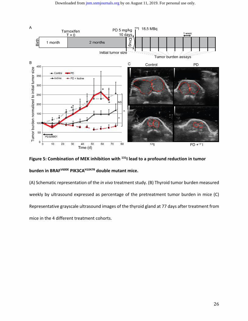
MEK Inhibitor Treatment Prior to 131I Therapy Reduce Tumor Burden in BRAFV600E PIK3CAH1047R
Double Mutant Mice.
An evaluation of the translation from the observed increase in radioiodine uptake in
BRAFV600E PIK3CAH1047R double mutant mice treated with MEK inhibition via PD‐325901 into
reduced tumor burden when injected with therapeutic radioiodine (131I) was performed. The
treatment study design including tumor induction, treatment with PD‐325901 and radioiodine
therapy is depicted in Figure 5A. The control group displayed progressive tumor growth of
approximately 15% each week. Mice receiving a 10‐day course of treatment with PD‐325901 only
experienced an initial reduction in tumor burden, with the therapeutic effect disappearing
immediately after treatment cessation. The group of mice injected with radioiodine alone
demonstrated a stable tumor burden for 6 weeks before tumors eventually increased in size.
Mice treated with a 10‐day course of PD‐325901 followed by injection with 131I displayed a
reduction in the tumor burden by 60% of the initial size 6 weeks after iodine injection and
remained stable for 3 weeks (Figure 5B).
DISCUSSION
Important genes in thyroid hormone synthesis such as NIS, TSHR, TG and TPO are
frequently downregulated in advanced thyroid cancer. The loss of this machinery is progressive,
beginning with NIS then involving TPO, TG and TSHR (24–26).
In the present study, we showed in vitro evidence that MEK inhibition induced robust NIS
mRNA transcription in human ATC cell lines harboring a BRAFV600E mutation, which resulted in
elevated cellular 123I uptake. We also showed that this result could be translated in vivo in a
by on August 11, 2019. For personal use only. jnm.snmjournals.org Downloaded from
13
mouse model of lethally aggressive thyroid cancer by significantly elevating the radioiodine
uptake resulting in stable reduction of tumor burden.
We have previously shown that MEK inhibition in combination with PI3’K inhibition could
lead to profound reduction driven by apoptosis (27). This treatment, while promising, has the
disadvantage of synergistic side effects associated with both drugs. Moreover, after treatment
cessation, a rapid regrowth of tumors occurred. In the present study, a short period of drug
treatment (10 days) enabled a potentially curative treatment with radioiodine.
In vivo, MEK inhibition in a BRAFV600E single mutant mouse model resulted in a Nis 4‐folds
transcription elevation (Figure 3A). This iodine induction in a PTC model is consistent with the
current literature (13,28,29). In a mouse model of more advanced thyroid cancer harboring
BRAFV600E and PIK3CAH1047R mutations (Figure 3C), we demonstrated that a MEK inhibition
induced a strong elevation of mRNA levels of genes important in thyroid hormones synthesis,
such as Nis (6‐folds), Tshr, Tg, Pax8 and Tpo. Interestingly, the combination of MEK inhibition
with PI3’K inhibition did not provide further upregulation of the tested genes. Specific BRAFV600E
inhibition did not induce an increase in any of these transcripts (Figure 3C). This is concordant
with Nagarajah and colleagues, who showed that the more profound MEK inhibition is achieved,
the stronger the NIS re‐expression (29). It was previously demonstrated that BRAF specific
inhibition can induce only transient ERK de‐phosphorylation and the signal is restored with time
through the wildtype allele of BRAF (30). Additionally, our recent findings show that BRAFV600E‐
specific inhibition led to a paradoxical activation of ERK in the double mutant mouse model (20).
Furthermore, there are very few data reporting vemurafenib treatment in ATC and one showed
mitigated results including one progression under BRAFV600E inhibitor (31).
by on August 11, 2019. For personal use only. jnm.snmjournals.org Downloaded from
14
On a functional level, a nearly 2.5‐fold increase in radioiodine uptake in the thyroids of
mice treated with a MEK inhibitor as compared to mice receiving the control treatment was
observed (Figure 4) after only 10 days of PD‐325901 treatment. Increased radioiodine uptake
translated into a stable, profound reduction in tumor burden (Figure 5). Mice treated with
radioiodine showed only an initial stabilization of the tumor burden followed by a rapid tumor
growth. This short‐lasting response to radioiodine could be explained by tumor heterogeneity
with regions of untransformed cells and PTC. As there was no added benefit of using a drug
combination with a PI3’K inhibitor even in tumors harboring a PIK3CAH1047R mutation (Figure 4),
this was not tested in the context of curative radioiodine.
In conclusion, our study shows that MEK inhibition as a single‐drug treatment is the most
efficient regimen to induce the expression of protein machinery required for iodine uptake in
human ATC cell lines and in our aggressive murine model of thyroid cancer. Until now, most
studies have mainly focused on PTC mouse models driven by a single BRAFV600E mutation (13).
Here we provide new evidence that the RAS‐MEK‐ERK axis is essential for NIS expression even in
ATC cell lines and in a clinically relevant mouse model carrying the two mutations BRAFV600E and
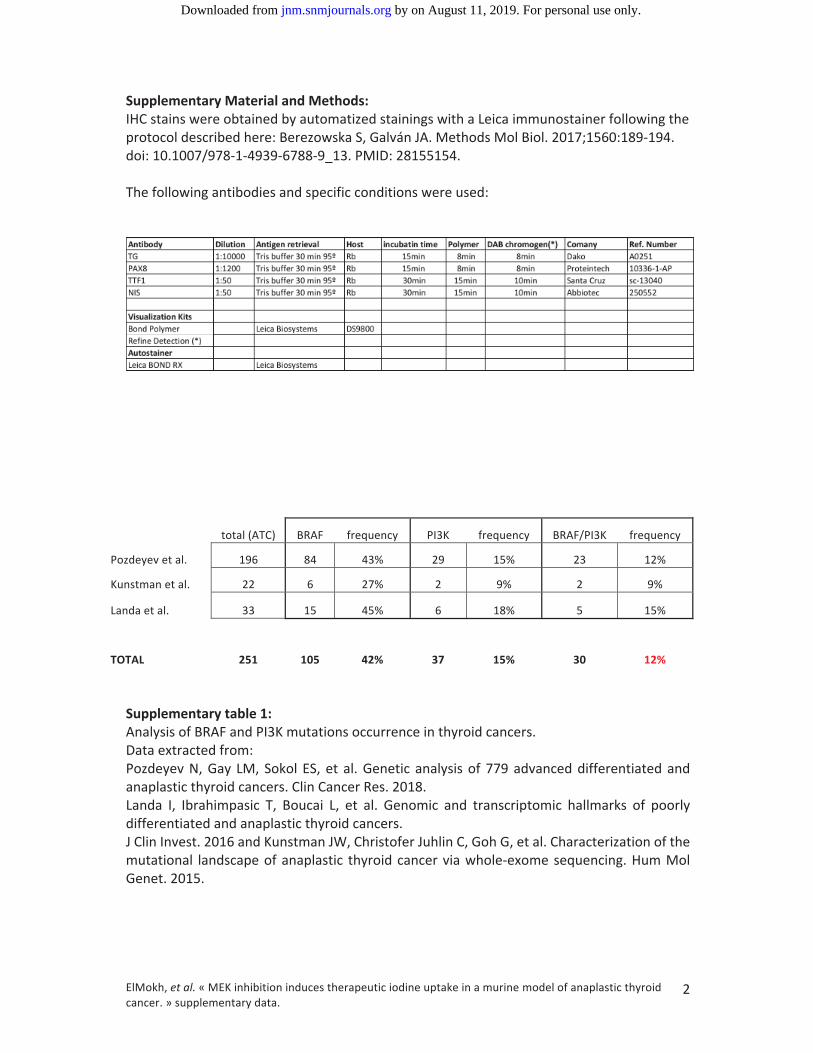
PIK3CAH1047R. We performed a meta‐analysis of the three available studies conducting genetic
profiling in ATC patients, which showed that the occurrence of both BRAF and PIK3CA mutations
is found in 12% of ATC patients (supplementary table and (5–7)). Thus, we believe that the
present results represent an encouraging initial step toward the successful treatment of non‐
radioiodine responsive thyroid cancers.
by on August 11, 2019. For personal use only. jnm.snmjournals.org Downloaded from
15
ACKNOWLEDGEMENTS
We thank Prof. Martin McMahon for his mentorship and for providing the BRAF mutant
mice. We thank Prof. Engelhard, Dr. Deutsch and Dr. Benarafa for permission to use the vivarium
at the Theodor Kocher Institute. We would also like to thank Dr. Colin for his help with Figure 1.
Also, we acknowledge the Microscopy Imaging Center of the University of Bern (MIC) for their
contributions to this study. Oussama ElMokh and Matthias Roelli were enrolled in the Graduate
School for Cellular and Biomedical Research (GCB) of the University of Bern during this study.
by on August 11, 2019. For personal use only. jnm.snmjournals.org Downloaded from
16
REFERENCES
1. Kebebew E, Greenspan FS, Clark OH, Woeber KA, McMillan A. Anaplastic thyroid
carcinoma. Treatment outcome and prognostic factors. Cancer. 2005;103:1330‐1335.
2. Gilliland FD, Hunt WC, Morris DM, Key CR. Prognostic factors for thyroid carcinoma: A
population‐based study of 15,698 cases from the Surveillance, Epidemiology and End
Results (SEER) program 1973‐1991. Cancer. 1997;79:564‐573.
3. Mazzaferri EL. An overview of the management of papillary and follicular thyroid
carcinoma. Thyroid. 1999;9:421‐427.
4. Kim EK, Choi E. Biochimica et Biophysica Acta Pathological roles of MAPK signaling
pathways in human diseases. BBA ‐ Mol Basis Dis. 2010;1802:396‐405.
5. Kunstman JW, Christofer Juhlin C, Goh G, et al. Characterization of the mutational
landscape of anaplastic thyroid cancer via whole‐exome sequencing. Hum Mol Genet.
2015;24:2318‐2329.
6. Pozdeyev N, Gay LM, Sokol ES, et al. Genetic analysis of 779 advanced differentiated and
anaplastic thyroid cancers. Clin Cancer Res. 2018.
7. Landa I, Ibrahimpasic T, Boucai L, et al. Genomic and transcriptomic hallmarks of poorly
differentiated and anaplastic thyroid cancers. J Clin Invest. 2016;126:1052‐1066.
8. Xing M. Molecular pathogenesis and mechanisms of thyroid cancer. Nat Rev Cancer.
2013;13:184‐199.
9. Charles R‐P, Silva J, Iezza G, Phillips WA, McMahon M. Activating BRAF and PIK3CA
Mutations Cooperate to Promote Anaplastic Thyroid Carcinogenesis. Mol Cancer Res.
2014;12:979‐986.
by on August 11, 2019. For personal use only. jnm.snmjournals.org Downloaded from
17
10. Riesco‐Eizaguirre G, Gutiérrez‐Martínez P, García‐Cabezas M, Nistal M, Santisteban P.
The oncogene BRAFV600E is associated with a high risk of recurrence and less
differentiated papillary thyroid carcinoma due to the impairment of Na+/I‐ targeting to
the membrane. Endocr Relat Cancer. 2006;13:257‐269.
11. Romei C, Ciampi R, Faviana P, et al. BRAFV600E mutation, but not RET/PTC
rearrangements, is correlated with a lower expression of both thyroperoxidase and
sodium iodide symporter genes in papillary thyroid cancer. Endocr Relat Cancer.
2008;15:511‐520.
12. Costamagna E, García B, Santisteban P. The Functional Interaction between the Paired
Domain Transcription Factor Pax8 and Smad3 Is Involved in Transforming Growth Factor‐
β Repression of the Sodium/Iodide Symporter Gene. J Biol Chem. 2004;279:3439‐3446.
13. Chakravarty D, Santos E, Ryder M, et al. Small‐molecule MAPK inhibitors restore
radioiodine incorporation in mouse thyroid cancers with conditional BRAF activation. J
Clin Invest. 2011;121.
14. Liu Y‐Y, Zhang X, Ringel MD, Jhiang SM. Modulation of sodium iodide symporter
expression and function by LY294002, Akti‐1/2 and Rapamycin in thyroid cells. Endocr
Relat Cancer. 2012;19:291‐304.
15. Bozorg‐Ghalati F, Hedayati M, Dianatpour M, Azizi F, Mosaffa N, Mehrabani D. Effects of
a Phosphoinositide‐3‐Kinase Inhibitor on Anaplastic Thyroid Cancer Stem Cells. Asian Pac
J Cancer Prev. 2017;18:2287‐2291.
16. Shah N, Iyer RM, Mair H‐JJ, et al. Improved human bioavailability of vemurafenib, a
practically insoluble drug, using an amorphous polymer‐stabilized solid dispersion
by on August 11, 2019. For personal use only. jnm.snmjournals.org Downloaded from
18
prepared by a solvent‐controlled coprecipitation process. J Pharm Sci. 2013;102:967‐981.
17. Dankort D, Filenova E, Collado M, Serrano M, Jones K, McMahon M. A new mouse model
to explore the initiation, progression, and therapy of BRAFV600E‐induced lung tumors.
Genes Dev. 2007;21:379‐384.
18. Kinross KM, Montgomery KG, Kleinschmidt M, et al. An activating Pik3ca mutation
coupled with Pten loss is sufficient to initiate ovarian tumorigenesis in mice. J Clin Invest.
2012;122:553‐557.
19. Undeutsch H, Löf C, Offermanns S, Kero J. A mouse model with tamoxifen‐inducible
thyrocyte‐specific cre recombinase activity. Genesis. 2014;52:333‐340.
20. Roelli MA, Ruffieux‐Daidié D, Stooss A, et al. PIK3CAH1047R‐induced paradoxical ERK
activation results in resistance to BRAFV600E specific inhibitors in BRAFV600E
PIK3CAH1047R double mutant thyroid tumors. Oncotarget. 2017;8:103207‐103222.
21. Charles RP, Iezza G, Amendola E, Dankort D, McMahon M. Mutationally activated
BRAFV600E elicits papillary thyroid cancer in the adult mouse. Cancer Res. 2011;71:3863‐
3871.
22. Haura EB, Ricart AD, Larson TG, et al. A phase II study of PD‐0325901, an oral MEK
inhibitor, in previously treated patients with advanced non‐small cell lung cancer. Clin
Cancer Res. 2010;16:2450‐2457.
23. McFadden DG, Vernon A, Santiago PM, et al. p53 constrains progression to anaplastic
thyroid carcinoma in a Braf‐mutant mouse model of papillary thyroid cancer. Proc Natl
Acad Sci. 2014;111:E1600‐‐E1609.
24. Lazar V, Bidart JM, Caillou B, et al. Expression of the Na+/I‐ symporter gene in human
by on August 11, 2019. For personal use only. jnm.snmjournals.org Downloaded from
19
thyroid tumors: A comparison study with other thyroid‐specific genes. J Clin Endocrinol
Metab. 1999;84:3228‐3234.
25. Gérard AC, Daumerie C, Mestdagh C, et al. Correlation between the Loss of Thyroglobulin
Iodination and the Expression of Thyroid‐Specific Proteins Involved in Iodine Metabolism
in Thyroid Carcinomas. J Clin Endocrinol Metab. 2003;88:4977‐4983.
26. Russo D, Damante G, Puxeddu E, Durante C, Filetti S. Epigenetics of thyroid cancer and
novel therapeutic targets. J Mol Endocrinol. 2011;46.
27. Elmokh O, Ruffieux‐daidié D, Roelli MA, et al. Combined MEK and Pi3 ’ ‐kinase inhibition
reveals synergy in targeting thyroid cancer in vitro and in vivo. Oncotarget.
2017;8:24604‐24620.
28. Ho AL, Grewal RK, Leboeuf R, et al. Selumetinib‐enhanced radioiodine uptake in
advanced thyroid cancer. N Engl J Med. 2013;368:623‐632.
29. Nagarajah J, Le M, Knauf JA, et al. Sustained ERK inhibition maximizes responses of
BrafV600E thyroid cancers to radioiodine. J Clin Invest. 2016;126:4119‐4124.
30. Hatzivassiliou G, Song K, Yen I, et al. RAF inhibitors prime wild‐type RAF to activate the
MAPK pathway and enhance growth. Nature. 2010;464:431‐435.
31. Hyman DM, Puzanov I, Subbiah V, et al. Vemurafenib in Multiple Nonmelanoma Cancers
with BRAF V600 Mutations. N Engl J Med. 2015;373:726‐736.
by on August 11, 2019. For personal use only. jnm.snmjournals.org Downloaded from
20
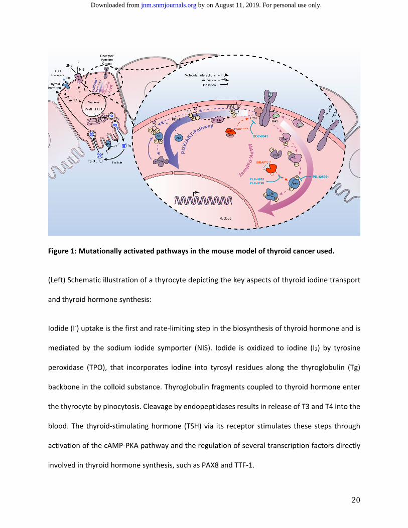
Figure 1: Mutationally activated pathways in the mouse model of thyroid cancer used.
(Left) Schematic illustration of a thyrocyte depicting the key aspects of thyroid iodine transport
and thyroid hormone synthesis:
Iodide (I‐) uptake is the first and rate‐limiting step in the biosynthesis of thyroid hormone and is
mediated by the sodium iodide symporter (NIS). Iodide is oxidized to iodine (I2) by tyrosine
peroxidase (TPO), that incorporates iodine into tyrosyl residues along the thyroglobulin (Tg)
backbone in the colloid substance. Thyroglobulin fragments coupled to thyroid hormone enter
the thyrocyte by pinocytosis. Cleavage by endopeptidases results in release of T3 and T4 into the
blood. The thyroid‐stimulating hormone (TSH) via its receptor stimulates these steps through
activation of the cAMP‐PKA pathway and the regulation of several transcription factors directly
involved in thyroid hormone synthesis, such as PAX8 and TTF‐1.
by on August 11, 2019. For personal use only. jnm.snmjournals.org Downloaded from
21
(Right) Schematic illustration of the MAPK and PI3’K pathways, including relevant mutations and
drug targets in thyroid cancer.
The MAPK and PI3’K pathways are involved in cellular proliferation and differentiation, and
activation of these pathways drives cell growth and decreases iodine metabolism. The mutation
BRAFV600E (in red) stimulates the MAPK pathway independent of the upstream signal. The
activation of RAS through binding of an extracellular ligand to a receptor tyrosine kinase leads to
activation of PI3’K. From PIP3, a phosphate is transferred to 3‐phosphoinositide‐dependent
protein kinase 1 (PDK1) which in turn phosphorylates AKT, leading to its activation and
translocation into the nuclei, where it regulates transcription of target genes involved in growth.
P‐AKT in the cytoplasm activates other targets such as the mammalian target of rapamycin
(mTOR) which is important in promoting proteins translation and consequently cell growth. PTEN
is a phosphatase converting PIP3 to PIP2 and playing the role of tumor suppressor as major
negative regulatory mechanism of the PI3’K/AKT pathway. The mutation PI3KCAH1047R results in
the hyperactivation of the pathway. Pharmacological inhibition of BRAFV600E occurs with PLX‐
4032/vemurafenib or PLX‐4720, a pre‐clinical analog. MEK is specifically inhibited by PD‐325901,
which leads to inhibition of ERK activity. PI3'K is specifically inhibited by GDC‐0941.
by on August 11, 2019. For personal use only. jnm.snmjournals.org Downloaded from
22
Figure 2: MEK inhibition lead to an elevation of NIS mRNA levels and iodine uptake in vitro.
(A) NIS mRNA levels in 2 ATC cell lines 8505c and OCUT‐2 treated for 48 h with PD at 100 nM
alone, PLX at 1 µM alone, or in combination with GDC at 1 µM. Values are normalized to
untreated cells. (B) 123I uptake in 3 ATC cell lines treated with different concentrations of PD alone
or in combination with GDC. Values are normalized to untreated cells. Cmax indicates the
maximum plasma concentration of PD in patients without side effects.
by on August 11, 2019. For personal use only. jnm.snmjournals.org Downloaded from
23
Figure 3: MEK inhibition but not BRAFV600E inhibition induced a restoration of Nis, Tshr and Tg
transcripts and resulted in near‐normalization of follicular organization in vivo.
(A) NIS mRNA levels in BRAFV600E single mutant mice treated with PD or control. (B) H&E stained
thyroid sections. (C) mRNA expression levels of Nis, Tshr, Tg, Pax8, Tpo and Ttf1 normalized to
normal levels in double mutant mice (BARFV600E PIK3CAH1047R) treated with control, PD, PLX , or
by on August 11, 2019. For personal use only. jnm.snmjournals.org Downloaded from

24
GDC, PLX/GDC or PD/GDC for 10 days. (D) H&E stained thyroid sections from mice after
treatments.
by on August 11, 2019. For personal use only. jnm.snmjournals.org Downloaded from
25
Figure 4: MEK inhibition lead to increased radioactive iodine uptake by the thyroid in BRAFV600E
PIK3CAH1047R double mutant mice.
(A) Schematic representation of the in vivo radioiodine uptake experiment. (B) Uptake expressed
as percent injected dose of 125I per gram of tissue in the thyroid, salivary glands, liver and blood
harvested from mice treated with control, PD alone or in combination with GDC for 2 weeks.
by on August 11, 2019. For personal use only. jnm.snmjournals.org Downloaded from
26
Figure 5: Combination of MEK inhibition with 131I lead to a profound reduction in tumor
burden in BRAFV600E PIK3CAH1047R double mutant mice.
(A) Schematic representation of the in vivo treatment study. (B) Thyroid tumor burden measured
weekly by ultrasound expressed as percentage of the pretreatment tumor burden in mice (C)
Representative grayscale ultrasound images of the thyroid gland at 77 days after treatment from
mice in the 4 different treatment cohorts.
by on August 11, 2019. For personal use only. jnm.snmjournals.org Downloaded from
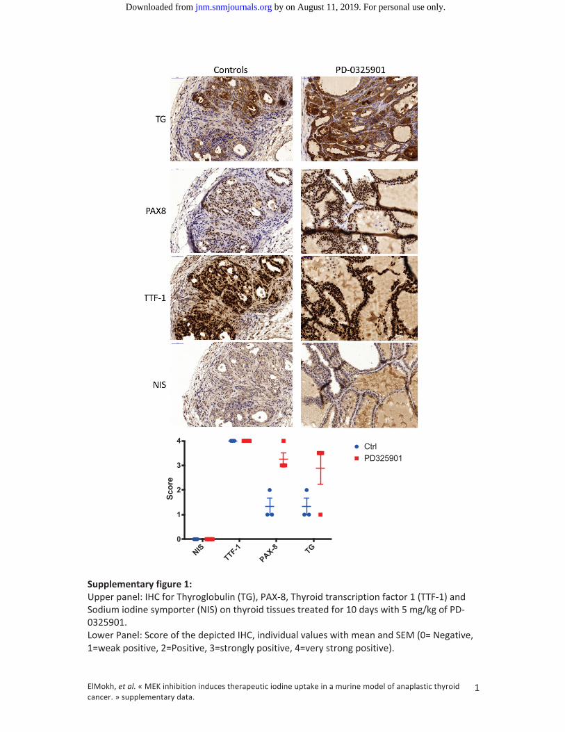
NIS
TTF-1
PAX-8 TG
0
1
2
3
4
Score
CtrlPD325901
by on August 11, 2019. For personal use only. jnm.snmjournals.org Downloaded from
by on August 11, 2019. For personal use only. jnm.snmjournals.org Downloaded from
Doi: 10.2967/jnumed.118.216721Published online: November 21, 2018.J Nucl Med. Matthias Dettmer, Wayne Phillips, Martin Alexander Walter and Roch-Philippe Régis CharlesOussama ElMokh, Vincent Taelmann, Piotr Radojewski, Matthias Andreas Roelli, Amandine Stooss, Rebecca A Dumont, thyroid cancer.MEK inhibition induces therapeutic iodine uptake in a murine model of anaplastic
http://jnm.snmjournals.org/content/early/2018/11/20/jnumed.118.216721This article and updated information are available at:
http://jnm.snmjournals.org/site/subscriptions/online.xhtml
Information about subscriptions to JNM can be found at:
http://jnm.snmjournals.org/site/misc/permission.xhtmlInformation about reproducing figures, tables, or other portions of this article can be found online at:
and the final, published version.proofreading, and author review. This process may lead to differences between the accepted version of the manuscript
ahead of print area, they will be prepared for print and online publication, which includes copyediting, typesetting,JNMcopyedited, nor have they appeared in a print or online issue of the journal. Once the accepted manuscripts appear in the
. They have not beenJNM ahead of print articles have been peer reviewed and accepted for publication in JNM
(Print ISSN: 0161-5505, Online ISSN: 2159-662X)1850 Samuel Morse Drive, Reston, VA 20190.SNMMI | Society of Nuclear Medicine and Molecular Imaging
is published monthly.The Journal of Nuclear Medicine
© Copyright 2018 SNMMI; all rights reserved.
by on August 11, 2019. For personal use only. jnm.snmjournals.org Downloaded from