Embed Size (px)
Citation preview
350 J Nippon Med Sch 2013; 80 (5)
―Original―
Antagonistic Effects of Tetrodotoxin on
Aconitine-induced Cardiac Toxicity
Takiyoshi Ono1, Makiko Hayashida1, Akito Tezuka2,Hideyuki Hayakawa3 and Youkichi Ohno1
1Department of Legal Medicine, Nippon Medical School2Division of Regenerative Medicine and Cardiology, Department of Internal Medicine, Nippon Medical School
3Tsukuba Medical Examiner’s Office
Abstract
Aconitine, well-known for its high cardiotoxicity, causes severe arrhythmias, such asventricular tachycardia and ventricular fibrillation, by opening membrane sodium channels.Tetrodotoxin, a membrane sodium-channel blocker, is thought to antagonize aconitine activity.Tetrodotoxin is a potent blocker of the skeletal muscle sodium-channel isoform Nav1.4 (IC50 10nM), but micromolar concentrations of tetrodotoxin are required to inhibit the primary cardiacisoform Nav1.5. This suggests that substantial concentrations of tetrodotoxin are required toalleviate the cardiac toxicity caused by aconitine. To elucidate the interaction betweenaconitine and tetrodotoxin in the cardiovascular and respiratory systems, mixtures of aconitineand tetrodotoxin were simultaneously administered to mice, and the effects onelectrocardiograms, breathing rates, and arterial oxygen saturation were examined. Comparedwith mice treated with aconitine alone, some mice treated with aconitine-tetrodotoxin mixturesshowed lower mortality rates and delayed appearance of arrhythmia. The decreased breathingrates and arterial oxygen saturation observed in mice receiving aconitine alone were alleviatedin mice that survived after receiving the aconitine-tetrodotoxin mixture; this result suggeststhat tetrodotoxin is antagonistic to aconitine. When the tetrodotoxin dose is greater than thedose that can block tetrodotoxin-sensitive sodium channels, which are excessively activated byaconitine, tetrodotoxin toxicity becomes prominent, and the mortality rate increases because ofthe respiratory effects of tetrodotoxin. In terms of cardiotoxicity, mice receiving the aconitine-tetrodotoxin mixture showed minor and shorter periods of change on electrocardiography.This finding can be explained by the recent discovery of tetrodotoxin-sensitive sodium-channelcardiac isoforms (Nav1.1, 1.2, 1.3, 1.4 and 1.6).(J Nippon Med Sch 2013; 80: 350―361)
Key words: aconitine, tetrodotoxin, arrhythmia, tetrodotoxin-sensitive channel, tetrodotoxin-resistant channel, breathing rate, arterial oxygen saturation
Correspondence to Takiyoshi Ono, Department of Legal Medicine, Nippon Medical School, 1―1―5 Sendagi, Bunkyo-ku,Tokyo 113―8602, JapanE-mail: [email protected] Website (http:��www.nms.ac.jp�jnms�)
TTX’s reaction on ACO toxicity
J Nippon Med Sch 2013; 80 (5) 351
Introduction
Aconitum plants (Ranunculaceae) are distributedworldwide in mountainous regions or cold areas1.The raw root of these plants is highly toxic;however, it has been used in Asian medicine forcenturies. The major toxic compounds of Aconitum
plants are alkaloids, such as aconitine, mesaconitine,hypaconitine, and jesaconitine. The alkaloid contentsin Aconitum plants change quantitatively andqualitatively depending on the species, place oforigin, and time of harvest2. Aconitum alkaloids areneurotoxins that bind to the neurotoxin-bindingreceptor site II on the α subunit of voltage-sensitivesodium channel3,4. This binding results in a sodiumchannel that stays open longer. Aconitine suppressesthe conformational change of the sodium channelfrom the active state to the inactive state. Themembrane stays depolarized owing to the constantsodium influx; therefore, the membrane cannot berepolarized5. Thus, Aconitum alkaloids cause nerveexcitation, followed by paralysis. Concerning thepharmacological or toxic effects of Aconitum
alkaloids, various types of sensory and motormanifestations can be included or elicited. Amongthese manifestations, cardiotoxicity is the mostimportant, causing severe arrhythmia and possibledeath. Because of its high cardiotoxicity, the medianlethal dose (LD50) of aconitine in mice is 1.8 mg�kgwhen administered orally and 0.308 mg�kg whenadministered intraperitoneally. The LD50 of aconitinefor humans is considered to be 1 to 2 mg. Fatalaccidents and murder due to ingestion of Aconitum
plants have been reported. In 1986, a Japanese manwas accused of attempting to murder his wife toreceive a large insurance benefit. However, his wifedeveloped severe vomiting, abdominal pain, andnumbness of the limbs and died 90 minutes after hisdeparture, which was inconsistent with the knownlatency of aconitine intoxication. The puffer fishtoxin, tetrodotoxin, was later found to have been co-administered with aconite alkaloids in this case6.Tetrodotoxin is a selective sodium-channel blocker
that obstructs depolarization of the excitablemembrane. Voltage-gated sodium channels are
responsible for initiating action potentials inexcitable cells. These channels are composed of apore-forming α subnuits and auxiliary β subunits7.Ten genes that encode α subnuits have beenidentified and 9 have been functionally expressed8.These different isoforms have distinct patterns ofdevelopment and localization in the nervous system,skeletal muscle, and cardiac muscle and possessdifferent pharmacological properties. Isoforms thatare preferentially expressed in the central nervoussystem (Nav1.1, 1.2, 1.3, and 1.6) are inhibited bytetrodotoxin at nanomolar concentrations and aresimilar to the isoforms present in adult skeletalmuscles, including those responsible for respiration(Nav1.4; half maximal inhibitory concentration [IC50],10 nM)9,10. In contrast, the primary cardiac isoform(Nav1.5 ; IC50 > 1 μM) requires micromolarconcentrations of tetrodotoxin for inhibition becauseof the presence of a cysteine instead of an aromaticresidue in the pore region of domain I9―13. Thus, thecardiac isoform Nav1.5 is classically calledtetrodotoxin-resistant INa, and the Nav1.1, 1.2, 1.3, 1.4,and 1.6 isoforms are called tetrodotoxin-sensitive INa.Tetrodotoxin blocks sodium conductance and
neuronal transmission in skeletal muscles14. It acts onthe central and peripheral nervous systems (i.e.,autonomic, motor, and sensory nerves). Patients withsevere poisoning may lapse into a coma, and deathmay occur within 4 to 6 hours of ingestion, typicallydue to respiratory failure caused by respiratorymuscle paralysis15. At the membrane sodiumchannel, aconitine and tetrodotoxin counteract eachother. Several studies have found that tetrodotoxinshows anti-arrhythmic activity as a selective sodium-channel blocker16―18. However, in contrast to theskeletal muscle isoform (Nav1.4; IC50, 10 nM), theprimary cardiac isoform (Nav1.5) requires micromolarconcentrations of tetrodotoxin for inhibition,suggesting that it is difficult for tetrodotoxin toalleviate the cardiac toxicity caused by aconitine. In1992, Ohno reported that when aconitine andtetrodotoxin were administered simultaneously, thetime of death due to aconitine was significantlydelayed in proportion to the dose of tetrodotoxinadministered, compared with that when onlyaconitine was administered, and the mortality rate of
T. Ono, et al
352 J Nippon Med Sch 2013; 80 (5)
aconitine was decreased by tetrodotoxin when thedose ratio of the 2 toxins was in a particular range19.This discovery was important, although the reasonfor this interaction has not yet been clarified.The present study was designed to investigate
the effects of simultaneous aconitine andtetrodotoxin administration on theelectrocardiogram (ECG), breathing rates, andarterial oxygen saturation (SpO2) of experimentalanimals and to elucidate the interaction betweenaconitine and tetrodotoxin in the cardiovascular andrespiratory systems. The results are discussed inlight of the recent discovery of tetrodotoxin-sensitivesodium channel cardiac isoforms (Nav1.1, 1.2, 1.3, 1.4,and 1.6).
Materials and Methods
ReagentsAconitine and tetrodotoxin were purchased from
Sigma-Aldrich Corp. (St. Louis, MO, USA). Otherreagents used were of analytical grade.
AnimalsFive-week-old male inbred ICR mice (weight, 30―
35 g) (Charles River Laboratories, Yokohama, Japan)were used in all experiments. The experiments wereperformed according to the guidelines of the EthicalCommittee on Animal Experimentation of NipponMedical School (Tokyo, Japan).
Administration of DrugsThe mice were divided into the following 4
groups: the aconitine group (Group A), thetetrodotoxin group (Group B), the aconitine-tetrodotoxin mixed-toxicity group (Group C), and thecontrol group (Group D). Each group consisted of 50to 60 mice. Aconitine and tetrodotoxin weredissolved in 0.1 M acetate buffer (pH 5) andadministered intraperitoneally to the mice. In GroupA, 0.10, 0.15, 0.3, or 0.4 mg�kg of aconitine wasadministered. In Group B, 5, 10, 15, 20, or 40 μg�kg oftetrodotoxin was administered. In Group C, 0.15 mg�kg of aconitine and 5 or 10 μg�kg of tetrodotoxin, or0.4 mg�kg of aconitine and 15 μg�kg of tetrodotoxinwere administered. In Group D, acetate buffer was
administered.ECGECG was performed with a preamplifier (RMP-
6004, Nihon Kohden, Tokyo, Japan) and a 2-channelpower lab system (model 2�25, Nihon Kohden,Tokyo, Japan). Mice were fastened belly up onto asmall animal restraint,. ECG was performed for 3hours, starting at the time of the administration. TheECG was analyzed with Chart version 5.3, ECGAnalysis Module (AD Instruments, Tokyo, Japan).The appearance and duration of arrhythmias wererecorded. Severe arrhythmias, such as ventriculartachycardia (VT) and ventricular fibrillation (VF),were analyzed.
Breathing Rate and SpO2Breathing rate was analyzed with a force
transducer (AD Instruments). The SpO2 wasdetermined with a portable animal pulse oximeter(Nonin Medical Inc. Minneapolis, MN, USA), andanalyzed with a signal conditioner (Bridge Pod,Nihon Kohden, Tokyo, Japan) and a data acquisitionunit (PowerLab, Nihon Kohden) . An animalanesthesia apparatus (TK-4m, model 8600V, NoninMedical Inc.) was used to analyze the breathing rateand SpO2 of mice under fluothane anesthesia.Fluothane was administered at 2% to 2.5% foranesthetic induction and at 1% for maintenance ofanesthesia.The breathing rates and SpO2 of only the
following groups were analyzed: Group A (0.4 mg�kgof aconitine), Group B (15 μg�kg of tetrodotoxin),Group C (0.4 mg�kg of aconitine and 15 μg�kg oftetrodotoxin), and Group D. Each group consisted of50 to 60 mice.
Statistical AnalysisWelch’s t-test was used to determine statistically
significant differences between the control groupand the other groups and between Group A andGroup C. Differences with p values < 0.05 wereconsidered to be statistically significant. The chi-square test was used to certify mortality.
TTX’s reaction on ACO toxicity
J Nippon Med Sch 2013; 80 (5) 353
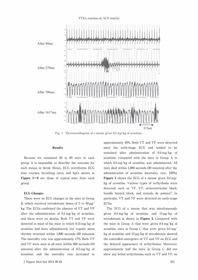
Fig. 1 Electrocardiogram of a mouse given 0.4 mg/kg of aconitine.
Results
Because we examined 50 to 60 mice in eachgroup, it is impossible to describe the outcome foreach mouse in detail. Hence, ECG waveforms, ECGtime courses, breathing rates, and SpO2 shown inFigure 1~8 are those of typical mice from eachgroup.
ECG ChangesThere were no ECG changes in the mice in Group
B, which received tetrodotoxin doses of 5 to 40 μg�kg. The ECGs confirmed the absence of VT and VFafter the administration of 0.1 mg�kg of aconitine,and there were no deaths. Both VT and VF weredetected in most of the mice to which 0.15 mg�kg ofaconitine had been administered, but regular sinusrhythm returned within 1,800 seconds (30 minutes).The mortality rate was approximately 17%. Both VTand VF were seen in all mice within 600 seconds (10minutes) after the administration of 0.3 mg�kg ofaconitine, and the mortality rate increased to
approximately 40%. Both VT and VF were detectedsince the early-stage ECG and tended to besustained after administration of 0.4 mg�kg ofaconitine, compared with the mice in Group A towhich 0.3 mg�kg of aconitine was administered. Allmice died within 1,800 seconds (30 minutes) after theadministration of aconitine (mortality rate, 100%).Figure 1 shows the ECG of a mouse given 0.4 mg�kg of aconitine. Various types of arrhythmia weredetected, such as VF, VT, atrioventricular block,bundle branch block, and torsade de pointes20. Inparticular, VT and VF were detected on early-stageECGs.The ECG of a mouse that was simultaneously
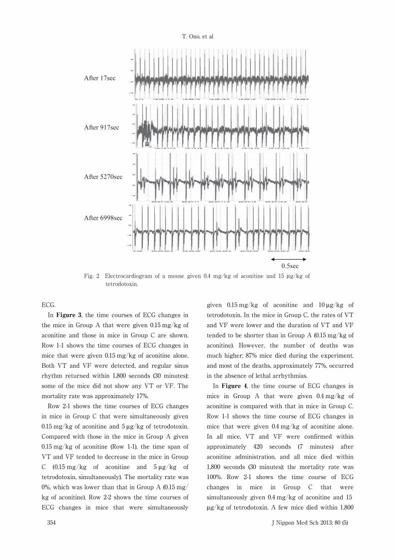
given 0.4 mg�kg of aconitine and 15 μg�kg oftetrodotoxin is shown in Figure 2. Compared withthe mice in Group A that were given 0.4 mg�kg ofaconitine, mice in Group C that were given 0.4 mg�kg of aconitine and 15 μg�kg of tetrodotoxin showedthe controlled emergence of VT and VF on ECG andthe delayed appearance of arrhythmias. Moreover,approximately half the mice in Group C did notshow any lethal arrhythmias, such as VT and VF, on
T. Ono, et al
354 J Nippon Med Sch 2013; 80 (5)
Fig. 2 Electrocardiogram of a mouse given 0.4 mg/kg of aconitine and 15 μg/kg of tetrodotoxin.
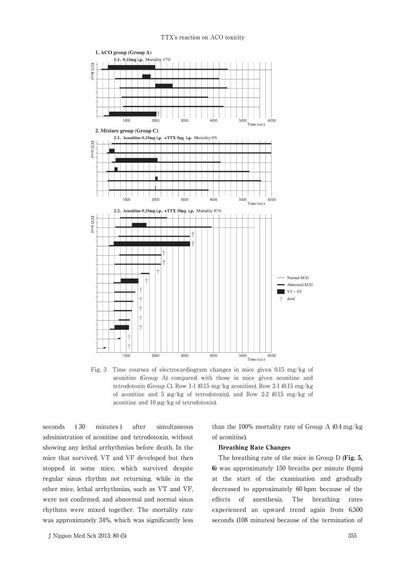
ECG.In Figure 3, the time courses of ECG changes in
the mice in Group A that were given 0.15 mg�kg ofaconitine and those in mice in Group C are shown.Row 1-1 shows the time courses of ECG changes inmice that were given 0.15 mg�kg of aconitine alone.Both VT and VF were detected, and regular sinusrhythm returned within 1,800 seconds (30 minutes);some of the mice did not show any VT or VF. Themortality rate was approximately 17%.Row 2-1 shows the time courses of ECG changes
in mice in Group C that were simultaneously given0.15 mg�kg of aconitine and 5 μg�kg of tetrodotoxin.Compared with those in the mice in Group A given0.15 mg�kg of aconitine (Row 1-1), the time span ofVT and VF tended to decrease in the mice in GroupC (0.15 mg�kg of aconitine and 5 μg�kg oftetrodotoxin, simultaneously). The mortality rate was0%, which was lower than that in Group A (0.15 mg�kg of aconitine). Row 2-2 shows the time courses ofECG changes in mice that were simultaneously
given 0.15 mg�kg of aconitine and 10 μg�kg oftetrodotoxin. In the mice in Group C, the rates of VTand VF were lower and the duration of VT and VFtended to be shorter than in Group A (0.15 mg�kg ofaconitine). However, the number of deaths wasmuch higher; 87% mice died during the experiment,and most of the deaths, approximately 77%, occurredin the absence of lethal arrhythmias.In Figure 4, the time course of ECG changes in
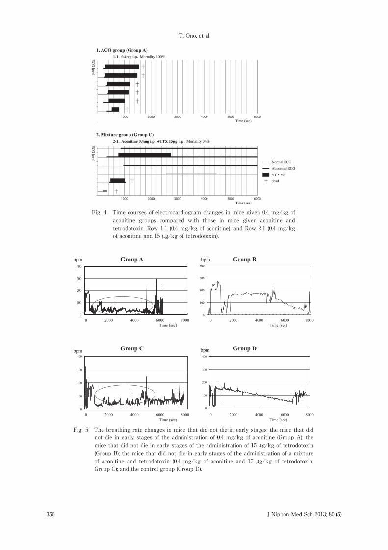
mice in Group A that were given 0.4 mg�kg ofaconitine is compared with that in mice in Group C.Row 1-1 shows the time course of ECG changes inmice that were given 0.4 mg�kg of aconitine alone.In all mice, VT and VF were confirmed withinapproximately 420 seconds (7 minutes) afteraconitine administration, and all mice died within1,800 seconds (30 minutes); the mortality rate was100%. Row 2-1 shows the time course of ECGchanges in mice in Group C that weresimultaneously given 0.4 mg�kg of aconitine and 15μg�kg of tetrodotoxin. A few mice died within 1,800
TTX’s reaction on ACO toxicity
J Nippon Med Sch 2013; 80 (5) 355
Fig. 3 Time courses of electrocardiogram changes in mice given 0.15 mg/kg of aconitine (Group A) compared with those in mice given aconitine and tetrodotoxin (Group C). Row 1-1 (0.15 mg/kg aconitine), Row 2-1 (0.15 mg/kg of aconitine and 5 μg/kg of tetrodotoxin), and Row 2-2 (0.15 mg/kg of aconitine and 10 μg/kg of tetrodotoxin).
seconds ( 30 minutes ) after simultaneousadministration of aconitine and tetrodotoxin, withoutshowing any lethal arrhythmias before death. In themice that survived, VT and VF developed but thenstopped in some mice, which survived despiteregular sinus rhythm not returning, while in theother mice, lethal arrhythmias, such as VT and VF,were not confirmed, and abnormal and normal sinusrhythms were mixed together. The mortality ratewas approximately 34%, which was significantly less
than the 100% mortality rate of Group A (0.4 mg�kgof aconitine).Breathing Rate ChangesThe breathing rate of the mice in Group D (Fig. 5,
6) was approximately 150 breaths per minute (bpm)at the start of the examination and graduallydecreased to approximately 60 bpm because of theeffects of anesthesia. The breathing ratesexperienced an upward trend again from 6,500seconds (108 minutes) because of the termination of
T. Ono, et al
356 J Nippon Med Sch 2013; 80 (5)
Fig. 4 Time courses of electrocardiogram changes in mice given 0.4 mg/kg of aconitine groups compared with those in mice given aconitine and tetrodotoxin. Row 1-1 (0.4 mg/kg of aconitine), and Row 2-1 (0.4 mg/kg of aconitine and 15 μg/kg of tetrodotoxin).
Fig. 5 The breathing rate changes in mice that did not die in early stages; the mice that did not die in early stages of the administration of 0.4 mg/kg of aconitine (Group A); the mice that did not die in early stages of the administration of 15 μg/kg of tetrodotoxin (Group B); the mice that did not die in early stages of the administration of a mixture of aconitine and tetrodotoxin (0.4 mg/kg of aconitine and 15 μg/kg of tetrodotoxin; Group C); and the control group (Group D).
TTX’s reaction on ACO toxicity
J Nippon Med Sch 2013; 80 (5) 357
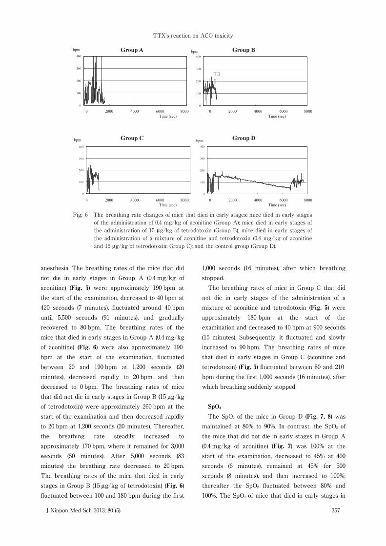
Fig. 6 The breathing rate changes of mice that died in early stages; mice died in early stages of the administration of 0.4 mg/kg of aconitine (Group A); mice died in early stages of the administration of 15 μg/kg of tetrodotoxin (Group B); mice died in early stages of the administration of a mixture of aconitine and tetrodotoxin (0.4 mg/kg of aconitine and 15 μg/kg of tetrodotoxin; Group C); and the control group (Group D).
anesthesia. The breathing rates of the mice that didnot die in early stages in Group A (0.4 mg�kg ofaconitine) (Fig. 5) were approximately 190 bpm atthe start of the examination, decreased to 40 bpm at420 seconds (7 minutes), fluctuated around 40 bpmuntil 5,500 seconds (91 minutes), and graduallyrecovered to 80 bpm. The breathing rates of themice that died in early stages in Group A (0.4 mg�kgof aconitine) (Fig. 6) were also approximately 190bpm at the start of the examination, fluctuatedbetween 20 and 190 bpm at 1,200 seconds (20minutes), decreased rapidly to 20 bpm, and thendecreased to 0 bpm. The breathing rates of micethat did not die in early stages in Group B (15 μg�kgof tetrodotoxin) were approximately 260 bpm at thestart of the examination and then decreased rapidlyto 20 bpm at 1,200 seconds (20 minutes). Thereafter,the breathing rate steadily increased toapproximately 170 bpm, where it remained for 3,000seconds (50 minutes). After 5,000 seconds (83minutes) the breathing rate decreased to 20 bpm.The breathing rates of the mice that died in earlystages in Group B (15 μg�kg of tetrodotoxin) (Fig. 6)fluctuated between 100 and 180 bpm during the first
1,000 seconds (16 minutes), after which breathingstopped.The breathing rates of mice in Group C that did
not die in early stages of the administration of amixture of aconitine and tetrodotoxin (Fig. 5) wereapproximately 180 bpm at the start of theexamination and decreased to 40 bpm at 900 seconds(15 minutes). Subsequently, it fluctuated and slowlyincreased to 90 bpm. The breathing rates of micethat died in early stages in Group C (aconitine andtetrodotoxin) (Fig. 5) fluctuated between 80 and 210bpm during the first 1,000 seconds (16 minutes), afterwhich breathing suddenly stopped.
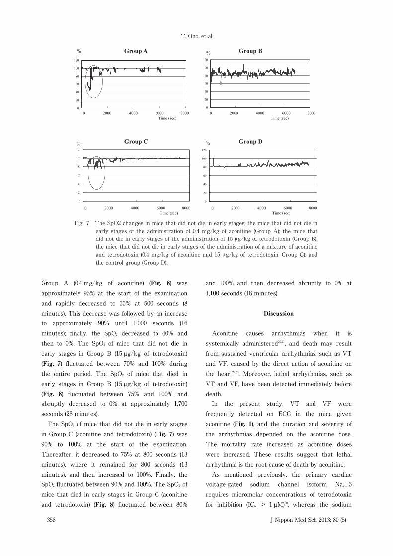
SpO2The SpO2 of the mice in Group D (Fig. 7, 8) was
maintained at 80% to 90%. In contrast, the SpO2 ofthe mice that did not die in early stages in Group A(0.4 mg�kg of aconitine) (Fig. 7) was 100% at thestart of the examination, decreased to 45% at 400seconds (6 minutes), remained at 45% for 500seconds (8 minutes), and then increased to 100%;thereafter the SpO2 fluctuated between 80% and100%. The SpO2 of mice that died in early stages in
T. Ono, et al
358 J Nippon Med Sch 2013; 80 (5)
Fig. 7 The SpO2 changes in mice that did not die in early stages; the mice that did not die in early stages of the administration of 0.4 mg/kg of aconitine (Group A); the mice that did not die in early stages of the administration of 15 μg/kg of tetrodotoxin (Group B); the mice that did not die in early stages of the administration of a mixture of aconitine and tetrodotoxin (0.4 mg/kg of aconitine and 15 μg/kg of tetrodotoxin; Group C); and the control group (Group D).
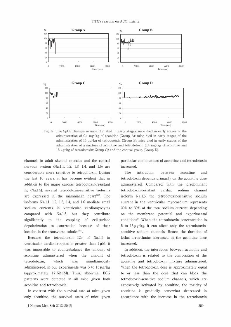
Group A (0.4 mg�kg of aconitine) (Fig. 8) wasapproximately 95% at the start of the examinationand rapidly decreased to 55% at 500 seconds (8minutes). This decrease was followed by an increaseto approximately 90% until 1,000 seconds (16minutes); finally, the SpO2 decreased to 40% andthen to 0%. The SpO2 of mice that did not die inearly stages in Group B (15 μg�kg of tetrodotoxin)(Fig. 7) fluctuated between 70% and 100% duringthe entire period. The SpO2 of mice that died inearly stages in Group B (15 μg�kg of tetrodotoxin)(Fig. 8) fluctuated between 75% and 100% andabruptly decreased to 0% at approximately 1,700seconds (28 minutes).The SpO2 of mice that did not die in early stages
in Group C (aconitine and tetrodotoxin) (Fig. 7) was90% to 100% at the start of the examination.Thereafter, it decreased to 75% at 800 seconds (13minutes), where it remained for 800 seconds (13minutes), and then increased to 100%. Finally, theSpO2 fluctuated between 90% and 100%. The SpO2 ofmice that died in early stages in Group C (aconitineand tetrodotoxin) (Fig. 8) fluctuated between 80%
and 100% and then decreased abruptly to 0% at1,100 seconds (18 minutes).
Discussion
Aconitine causes arrhythmias when it issystemically administered20,21, and death may resultfrom sustained ventricular arrhythmias, such as VTand VF, caused by the direct action of aconitine onthe heart22,23. Moreover, lethal arrhythmias, such asVT and VF, have been detected immediately beforedeath.In the present study, VT and VF were
frequently detected on ECG in the mice givenaconitine (Fig. 1), and the duration and severity ofthe arrhythmias depended on the aconitine dose.The mortality rate increased as aconitine doseswere increased. These results suggest that lethalarrhythmia is the root cause of death by aconitine.As mentioned previously, the primary cardiac
voltage-gated sodium channel isoform Nav1.5requires micromolar concentrations of tetrodotoxinfor inhibition (IC50 > 1 μM)20, whereas the sodium
TTX’s reaction on ACO toxicity
J Nippon Med Sch 2013; 80 (5) 359
Fig. 8 The SpO2 changes in mice that died in early stages; mice died in early stages of the administration of 0.4 mg/kg of aconitine (Group A); mice died in early stages of the administration of 15 μg/kg of tetrodotoxin (Group B); mice died in early stages of the administration of a mixture of aconitine and tetrodotoxin (0.4 mg/kg of aconitine and 15 μg/kg of tetrodotoxin; Group C); and the control group (Group D).
channels in adult skeletal muscles and the centralnervous system (Nav1.1, 1.2, 1.3, 1.4, and 1.6) areconsiderably more sensitive to tetrodotoxin. Duringthe last 10 years, it has become evident that inaddition to the major cardiac tetrodotoxin-resistantINa (Nav1.5), several tetrodotoxin-sensitive isoformsare expressed in the mammalian heart24,25. Theisoforms Nav1.1, 1.2, 1.3, 1.4, and 1.6 mediate smallsodium currents in ventricular cardiomyocytescompared with Nav1.5, but they contributesignificantly to the coupling of cell-surfacedepolarization to contraction because of theirlocation in the transverse tubules26,27.Because the tetrodotoxin IC50 of Nav1.5 in
ventricular cardiomyocytes is greater than 1 μM, itwas impossible to counterbalance the amount ofaconitine administered when the amount oftetrodotoxin, which was simultaneouslyadministered, in our experiments was 5 to 15 μg�kg(approximately 17―52 nM). Thus, abnormal ECGpatterns were detected in all mice given bothaconitine and tetrodotoxin.In contrast with the survival rate of mice given
only aconitine, the survival rates of mice given
particular combinations of aconitine and tetrodotoxinincreased.The interaction between aconitine and
tetrodotoxin depends primarily on the aconitine doseadministered. Compared with the predominanttetrodotoxin-resistant cardiac sodium channelisoform Nav1.5, the tetrodotoxin-sensitive sodiumcurrent in the ventricular myocardium represents20% to 30% of the total sodium current, dependingon the membrane potential and experimentalconditions27. When the tetrodotoxin concentration is5 to 15 μg�kg, it can affect only the tetrodotoxin-sensitive sodium channels. Hence, the duration oflethal arrhythmias increased as the aconitine doseincreased.In addition, the interaction between aconitine and
tetrodotoxin is related to the composition of theaconitine and tetrodotoxin mixture administered.When the tetrodotoxin dose is approximately equalto or less than the dose that can block thetetrodotoxin-sensitive sodium channels, which areexcessively activated by aconitine, the toxicity ofaconitine is gradually somewhat decreased inaccordance with the increase in the tetrodotoxin

T. Ono, et al
360 J Nippon Med Sch 2013; 80 (5)
dose. Therefore, both the frequency of VT and VFand the mortality rate decrease. This point can beconfirmed by the examples of mice (Group C) thatwere given 0.15 mg�kg of aconitine and 5 μg�kg oftetrodotoxin (row 2-1 of Fig. 3) and mice that weregiven 0.4 mg�kg of aconitine and 15 μg�kg oftetrodotoxin (row 2-1 of Fig. 4). Furthermore, in thecase reported by Ohno, the victim developed severevomiting, abdominal pain, and numbness of the limbsbefore death6, which were all typical symptoms ofaconitine poisoning. However, the latency of herdeath (> 90 minutes) was due to the effect oftetrodotoxin.When the dose of tetrodotoxin is greater than the
dose that can block the tetrodotoxin-sensitivesodium channels, which are excessively activated byaconitine, the toxicity of tetrodotoxin becomesprominent. The LD50 of intraperitoneallyadministered tetrodotoxin is 10.7 μg�kg in mice28.Therefore, some mice died of respiratory failurebefore recovery from severe arrhythmias, such asVT and VF. This is the reason for most deaths thatoccurred in the absence of severe arrhythmias inmice that were given 0.15 mg�kg of aconitine and10 μg�kg of tetrodotoxin (row 2-2 of Fig. 3).The results of our investigations of the breathing
rate and SpO2 of mice are consistent with thishypothesis. We were not able to use implantableelectrodes because of the small bodies of the mice.Furthermore, because the probes for measuring thebreathing rate and SpO2 are extremely sensitive,even small movements by the mice may generatenoise. Thus, the breathing rate and SpO2 weremeasured with the mice under anesthesia.We divided the mice into 2 groups, those that died
in early stages (within 30 minutes of theadministration) and those that did not. In mice thatdied in early stages, the breathing rate and SpO2 ofthe mice given both aconitine and tetrodotoxin weremore similar to those of mice given tetrodotoxinalone than those of mice given aconitine alone (Fig.6, 8). This result is consistent with the interpretationthat the deaths of some mice in Group C were morelikely to have been caused by respiratory failure dueto tetrodotoxin. This result is also consistent withthe result that most of these mice died without
showing severe arrhythmias, such as VT and VF, onECG when the dose of tetrodotoxin was greaterthan the dose that can block the tetrodotoxin-sensitive cardiac sodium channels, which areexcessively activated by aconitine (Fig. 3, 4).The breathing rates of the mice that did not die in
early stages of the administration of an aconitine-tetrodotoxin mixture presented waveforms thatwere similar to those of the mice that receivedaconitine alone, but the breathing rates recoveredmore quickly (Fig. 5). In addition, the SpO2 of themice that received an aconitine-tetrodotoxin mixtureshowed a waveform that was similar to that of themice receiving aconitine alone, although thesteepness of the decline phase was alleviated from50% to 80% (Fig. 7). When combined with the resultsof the ECG changes (Fig. 3, 4), these experimentsstrongly suggest that tetrodotoxin antagonizedaconitine and ameliorated the toxicity of aconitine inmice, a result that has been previouslydemonstrated in vitro29,30.
Conclusions
The results of this study lead us to 4 conclusions.1) Mortality and toxicity of aconitine are directly
proportional to the aconitine dose administered.2) Abnormal ECGs were observed in all aconitine-
tetrodotoxin mixed-toxicity groups when 5 to 15 μg�kg of tetrodotoxin was administered simultaneouslywith aconitine.3) Compared with those in mice given aconitine
alone, mortality rates were lower and arrhythmiawas delayed in mice receiving mixtures of aconitineand tetrodotoxin. Furthermore, the decrease inbreathing rates and SpO2 are ameliorated in themice that did not die in early stages of theadministration of the aconitine-tetrodotoxin mixed-toxicity group. The above conclusions suggest thatthe action of tetrodotoxin is antagonistic to theaction of aconitine.4) When the dose of tetrodotoxin is greater than
the dose that can block the tetrodotoxin-sensitivesodium channels, which are excessively activated byaconitine, the toxicity of tetrodotoxin becomesprominent. Under these circumstances, mortality
TTX’s reaction on ACO toxicity
J Nippon Med Sch 2013; 80 (5) 361
increases because of the respiratory effects oftetrodotoxin.
Conflict of Interest: The authors have no financialconflicts of interest regarding the publication of thisarticle.
Acknowledgements: The authors thank Dr. MakotoNihira for his academic advice on toxicologicalexaminations.
References
1.Bisset NG: Arrow poisons in China, Part II.Aconitum-botany, chemistry, and pharmacology. JEthnopharmacol 1981; 4: 247―336.
2.Ito K, Ohyama Y, Hishinuma T, Mizugaki M:Determination of Aconitum alkaloids in the tubers ofAconitum Japonicum using gas chromatography�selected ion monitoring. Planta Med 1996; 62: 57―59.
3.Catterall WA: Neurotoxins that act on voltage-sensitive sodium channels in excitable membranes.Ann Rev Pharmacol Toxicol 1980; 20: 15―43.
4.Gutser UT, Friese J, Heubach JF, et al.: Mode ofantinociceptive and toxic action of alkaloids ofAconitum species. Naunyn Schmiedebergs ArchPharmacol 1998; 357: 39―48.
5.Benoit E: Mechanism of action of neurotoxins actingon the inactivation of voltage-gated sodium channels.C R Seances Soc Biol Fil 1998; 192: 409―436.
6.Ohno Y: The experimental approach to the murdercase of aconite poisoning. J Toxicol-Toxin Rev 1998;17: 1―11.
7.Catterall WA: From ionic currents to molecularmechanisms: the structure and function of voltage-gated sodium channels. Neuron 2000; 26: 13―25.
8.Goldin AL, Barchi RL, Calwell JH, et al.:Nomenclature of voltage-gated sodium channels.Neuron 2000; 28: 365―368.
9.Goldin AL: Resurgence of sodium channel research.Annu Rev Physiol 2001; 63: 871―894.
10.Catterall WA, Goldin AL, Waxman SG: InternationalUnion of Pharmacology. XLVII. Nomenclature andstructure-function relationships of voltage-gatedsodium channels. Pharmacol Rev 2005; 57: 397―409.
11.Fozzard HA, Hanck DA: Structure and function ofvoltage-dependent sodium channels: comparison ofbrain II and cardiac isoforms. Physiol Rev 1996; 76:887―926.
12.Gellens ME, George AL Jr, Chen LQ, et al.: Primarystructure and functional expression of the humancardiac tetrodotoxin-insensitive voltage-dependentsodium channel. Proc Natl Acad Sci USA 1992; 89:554―558.
13.Satin J, Kyle JW, Chen M, Rogart RB, Fozzard HA:The cloned cardiac Na channel α-subunit expressedin Xenopus oocytes show gating and blockingproperties of native channels. J Membr Biol 1992;130: 11―22.
14.Ahasan HM, Chowdhury MA, Azhar MA,Rafiqueuddin AK: Copper sulphate poisoning. Trop
Doct 1994; 24: 52―53.15.Borison HL, McCarthy LE, Clark WG,
Radhakrishnan N: Vomiting, hypothermia, andrespiratory paralysis due to tetrodotoxin (puffer fishpoison) in the cat. Toxicol Appl Pharmacol 1963; 5:350―357.
16.Duff HJ, Sheldon RS, Cannon NJ: Tetrodotoxin:sodium channel specific anti-arrhythmic activity.Cardiovasc Res 1988; 22: 800―807.
17.Abraham S, Beatch GN, MacLeod BA, Walker MJ:Antiarrhythmic properties of tetrodotoxin againstocclusion-induced arrhythmias in the rat: a novelapproach to the study of the antiarrhythmic effectsof ventricular sodium channel blockade. J PharmacolExp Ther 1989; 251: 1166―1173.
18.Zimmer T: Effects of tetrodotoxin on the mammaliancardiovascular system. Mar Drugs 2010; 8: 741―762.
19.Ohno Y, Chiba S, Uchigasaki S, et al.: The influenceof tetrodotoxin on the toxic effects of aconitine invivo. Tohoku J Exp Med 1992; 167: 155―158.
20.Vaughan Williams EM : Classification ofantidysrhythmic drugs. Pharmacol Ther B 1975; 1:115―138.
21.Scherf D: Studies on auricular tachycardia caused byaconitine administration. Proc Soc Exp Biol 1947; 64:233―239.
22.Wada K, Nihira M, Hayakawa H, Tomita Y,Hayashida M, Ohno Y: Effects of long-termadministrations of aconitine on electrocardiogramand tissue concentrations of aconitine and itsmetabolites in mice. Forensic Sci Int 2005; 148: 21―29.
23.Ito K, Tanaka S, Funayama M, Mizugaki M:Distribution of Aconitum alkaloids in body fluids andtissues in a suicidal case of aconite ingestion. J AnalToxicol 2000; 24: 348―353.
24.Haufe V, Camacho JA, Dumaine R, et al.: Expressionpattern of neuronal and skeletal muscle voltage-gated Na+ channels in the developing mouse heart. JPhysiol 2005; 564: 683―696.
25.Haufe V, Cordeiro JM, Zimmer T, et al.: Contributionof neuronal sodium channels to the cardiac fastsodium current INa is greater in dog heart Purkinjefibers than in ventricles. Cardiovasc Res 2005; 65:117―127.
26.Maier SK, Westenbroek RE, Schenkman KA, FeiglEO, Scheuer T, Catterall WA: An unexpected rolefor brain-type sodium channels in coupling of cellsurface depolarization to contraction in the heart.Proc Natl Acad Sci USA 2002; 99: 4073―4078.
27.Kaufmann SG, Westenbroek RE, Zechner C, et al.:Functional protein expression of multiple sodiumchannel α-subunit and β-subunit isoforms in neonatalcardiomyocytes. J Mol Cell Cardiol 2010; 48: 261―269.
28.Xu Q, Huang K, Gao L, Zhang H, Rong K: Toxicity oftetrodotoxin towards mice and rabbits. J Hyg Res2003; 32: 371―374.
29.Huang TF: Effect of tetrodotoxin and manganese ionon the aconitine-induced arrhythmia of the isolatedrabbit atrium and ventricle. Jpn J Physiol 1970; 20:435―443.
30.Huang TF: Antiarrhythmic action of tetrodotoxin invarious animal species. Jpn J Physiol 1973; 23: 39―46.
(Received,(Accepted,
FebruaryMay
4, 2013)7, 2013)