Embed Size (px)
Citation preview

Journal of Neuroimmunology 225 (2010) 100–111
Contents lists available at ScienceDirect
Journal of Neuroimmunology
j ourna l homepage: www.e lsev ie r.com/ locate / jneuro im
Glatiramer acetate reduces Th-17 inflammation and induces regulatory T-cells in theCNS of mice with relapsing–remitting or chronic EAE
Rina Aharoni, Raya Eilam, Ariel Stock, Anya Vainshtein, Elias Shezen, Hilah Gal, Nir Friedman, Ruth Arnon ⁎The Department of Immunology, The Weizmann Institute of Science, Rehovot 76100, Israel
⁎ Corresponding author. Tel.: +972 8 9344017; fax:E-mail address: [email protected] (R. Arno
0165-5728/$ – see front matter © 2010 Elsevier B.V. Adoi:10.1016/j.jneuroim.2010.04.022
a b s t r a c t
a r t i c l e i n f oArticle history:Received 7 March 2010Received in revised form 28 April 2010Accepted 29 April 2010
Keywords:Multiple sclerosisExperimental autoimmune encephalomyelitisInterleukin-17T-regulatory cellsImmunomodulationGlatiramer acetate
The aim of this study was to identify cell populations relevant to pathogenesis and repair within the injuredCNS in mice that recovered from experimental autoimmune encephalomyelitis (EAE). We demonstrate thatin two EAE models, with either relapsing–remitting or chronic course, T-cells and resident activatedmicroglia manifested extensive IL-17 expression, with apparent localization within regions of myelin loss. Inmice treated with glatiramer acetate (GA, Copaxone®), even when treatment started after diseaseexacerbation, CNS inflammation and Th-17 occurrence were drastically reduced, with parallel elevation in T-regulatory cells, indicating the immunomodulatory therapeutic consequences of GA treatment in situ.
+972 8 9469712.n).
ll rights reserved.
© 2010 Elsevier B.V. All rights reserved.
1. Introduction
In multiple sclerosis (MS) and its animal model experimentalautoimmune encephalomyelitis (EAE), the immune system provokesthe pathological process via autoimmune inflammatory mechanisms(Hellings et al., 2002; Hohlfeld and Wekerle, 2004; Behi et al., 2005)leading to the characteristic demyelination and neurological damage(Bjartmar et al., 2003; Hobom et al., 2004). It is largely accepted thatT-cells of the Th1 and Th2 subtypes are involved in the pathology andamelioration of these disease, respectively (Zamvil and Steinman,1990). Recently, accumulating data indicate that Th-17 cells, whichare induced through the transcription factors receptor-related orphanreceptor (ROR)α and RORγt, produce the highly pro-inflammatoryinterleukin (IL)-17, and play an important role in the pathogenesis ofinflammatory and autoimmune diseases (Bettelli et al., 2007; Aranamiand Yamamaura, 2008). At the opposing pathway, the T-regulatorycells (Tregs), with their specific transcription factor forkhead box P3(Foxp3), are potent immunosuppressors that ameliorate wide array ofinflammatory conditions (Vila et al., 2009). Th-17 cells and Tregs havebeen demonstrated to arise from common precursors in a reciprocalmanner, pending on the cytokine milieu, generating opposingoutcomes, namely propagation or suppression of inflammation,respectively (Bettelli et al., 2006; Aranami and Yamamaura, 2008).Notably, recent findings support the notion that in the context ofthe inflammatory stimuli Tregs can convert to the Th17 phenotype,
resulting in excessive inflammation and tissue injury (Afzali et al.,2009; Zhou et al., 2009). These findings point to the importance ofanalyzing these cell populations in the target organ and followingthem in situ as a consequence of disease exacerbation and/or therapy.
Glatiramer acetate (GA, Copaxone®), an approved drug for MStreatment, is effective in the prevention and suppression of EAEinducedby various encephalitogens in several species (Arnon andSela,2003). It has been shown that GA promotes neuroprotection andrepair processes in the CNS, as manifested by the decrease inneurological damage (Gilgum-Sherki et al., 2003; Aharoni et al.,2005a) and demyelination (Aharoni et al., 2008), as well as by theincrease in neurotrophic factors expression (Aharoni et al., 2005b),and neurogenesis (Aharoni et al., 2005a), in EAEmice treated by GA incomparison to untreatedmice. The therapeutic activity of GA has beenattributed to its immunomodulatory effect on various levels of theimmune response. Thus, in the periphery GA was shown to bindpromiscuously tomajor histocompatibility complexmolecules (MHC),acting both as a MHC blocker (Fridkis-Hareli et al., 1994) and a T-cellreceptor antagonist (Aharoni et al., 1999), leading to inhibition ofvarious pathological effector functions. GA affected the properties ofdendritic cells and monocytes, so that they preferentially stimulateTh2-like responses (Farina et al., 2005;Weber et al., 2007). Indeed, GAhas been shown to be a potent inducer of Th2/3 cells that secrete highlevels of anti-inflammatory cytokines such as IL-4, IL-10 andtransforming growth factor-β (TGF-β) (Aharoni et al., 1997). Further-more, adoptively transferred GA-induced Th2/3 cells were detected inthe CNS of recipient mice (Aharoni et al., 2000), expressing IL-10 andTGF-β, indicative of their in situ anti-inflammatory activity (Aharoniet al., 2003). The immunomodulatory effect of GA in the CNS was also
101R. Aharoni et al. / Journal of Neuroimmunology 225 (2010) 100–111
evidenced by the corresponding decrease in IFN-γ, as well as by thebystander expression of Th2/3 cytokine in the resident astrocytepopulation. These cumulative results supported the notion that theeffect of GA is mediated through immunomodulatory shift from thedetrimental Th1 towards the anti-inflammatory Th2/3 response.
Recent studies indicated that the effect of GA on EAE/MSimmunopathogenesis is not restricted to the Th2/3 versus Th1pathways. Thus, it was shown that in vitro exposure of peripheralCD4+ T-cells, from healthy humans or from GA-immunized mice, toGA induced elevated levels of Tregs, through the activation of Foxp3mRNA. Furthermore, in CD4+ T-cells of MS patients, whose Foxp3level was low at baseline, GA treatment led to increased Foxp3expression (Hong et al., 2005). Pretreatment of mice with GA, startingoneweek before the EAE induction resulted in increased expression ofFoxp3 on Tregs during the mild disease which developed subse-quently. Following their isolation from the spleen, these Tregs weremore effective in EAE prevention than those isolated from untreatedmice (Jee et al., 2007). Recently, using the chronic MOG-inducedmodel, it has been demonstrated that GA treatment, starting one dayafter EAE induction, resulted in elevation of Foxp3 and reduction ofTh-17 on the level of mRNA expression (Begum-Haque et al., 2008).
In the current study we attempted to identify cell populations thatare relevant to pathogenesis and repair within the injured CNS. Inparticular we were interested in characterizing in situ the T-cellpopulations that are involved in therapeutic activity of GA whenapplied by the delayed (late) suppression regimen, namely afterdisease exacerbation had already occurred. To attend the issue of themultifaceted nature of MS, two EAE models with relapsing–remittingand chronic courses were utilized. We report herewith that in bothEAE models, T-cells as well as activated microglia manifest extensiveIL-17 expression in the CNS, with apparent localization within regionsof myelin loss. In mice that recovered following GA treatment, CNSinflammation and Th-17 occurrence were drastically reduced withconcomitant elevation in T-regulatory cells that had been generated inthe periphery, but subsequent to disease exacerbation, accumulatedin the inflamed CNS. The ability of the cells induced in the peripheryby GA treatment to accumulate specifically in the injured CNS wasdemonstrated as well.
2. Materials and methods
2.1. Animals
C57BL/6 and (SJL/JxBALB/c)F1 mice were purchased from Harlan(Jerusalem, Israel). Female mice, 8–12 weeks of age, were kept underspecific pathogen free (SPF) environment. All experiments wereapproved by the Institutional Animal Care and Use Committee of theWeizmann Institute.
2.2. EAE
Relapsing–remitting EAE was induced in (SJL/JxBALB/c)F1 mice bythe peptide encompassing amino acids 139–151 of proteolipid protein(PLP), synthesis by Novetide (Haifa Bay, Israel). Chronic EAE wasinduced in C57BL/6 mice by the peptide encompassing amino acids35–55 of myelin oligodendrocyte glycoprotein (MOG), synthesis bySigma (St. Louis, MO). Mice were injected subcutaneously at the flank,with 200 μl emulsion containing 200–300 μg of the encephalitogenicpeptide in incomplete Freund's adjuvant enriched with 3 mg/ml heat-inactivated Mycobacterium tuberculosis (Sigma). Pertussis toxin(Sigma), 200–250 μg/mouse was injected intravenously immediatelyafter the encephalitogenic injection and 48 h later. Mice wereexamined daily. EAE was scored as follows: 0—no disease, 1—limptail, 2—hind limb paralysis, 3—paralysis of all four limbs, 4—moribundcondition, and 5—death.
2.3. Glatiramer acetate (GA, Copaxone, Copolymer 1)
GA consists of acetate salts of synthetic polypeptides containing fouramino acids L-alanine, L-glutamate, L-lysine, and L-tyrosine (Arnon andSela, 2003). GA from batch 242902109, with an average molecularweight of 7400 kDa, obtained from Teva Pharmaceutical Industries(Petah Tiqva, Israel) was used throughout the study. GA treatment wasappliedby consecutive 7–8daily subcutaneous injections (2 mg/mouse)either as early suppression treatment starting one day after diseaseinduction (at day 1), or as late suppression treatment starting after theappearance of clinical manifestations (at days 18 and 13 in the PLP andthe MOG experiments, respectively). A layout of the GA treatmentschedules in characteristic experiments is demonstrated in Fig. 1A.
2.4. Perfusion and organ processing for immunohistochemistry
Animals were deeply anesthetized and perfused transcardiallywith 100 ml of Phosphate Buffered Saline (PBS). Brainswere removed,postfixed in Bouin's solution (Sigma) for 12 h, followed by repeatedwashings with phosphate buffer (PBS). Organs were than cryopro-tected with 15% sucrose solution and sectioned coronally (20 µm) bysliding microtome (Leica) through the entire brain. Free-floatingsections were collected serially in PBS. Spinal cords were removed,snap frozen with isopentan (pre-cooled with liquid nitrogen) andsectioned sagitally (6 µm) by cryostat (Leica) with 50 µm skips.
2.5. Immunohistochemistry
Free-floating brain sections were pre-incubated in PBS solutioncontaining 20% serum and 0.5% triton-x-100 for one hour, and thenincubated overnight at room temperature with primary antibodies.Frozen spinal cord sections were treated with cooled acetone for7 min at −20 °C, dried and incubated with 7% horse serum inantibody diluents (cell Marque) following 1h incubation with thespecific antibody. The following primary antibodies were used: ratanti-CD3 (Serotec) or rabbit anti-CD3 (NeoMarkeres, Fremont, CA),mouse anti-myelin basic protein (Abcam), rat anti-Mac-2 (Cedar-lane,) goat anti-IL-17 (R&D Systems) and rat anti-Foxp3 (eBioscince).IL-17 staining was performed on Bouin's postfixed free-floatingsections. Foxp3 staining was done mainly on frozen acetone treatedsections. The second antibody step was performed by labeling with(donkey) highly cross-absorbed cy2, cy3 or Dylight-conjugatedantibodies to rat, mouse, rabbit, or goat (Jackson ImmunoResearch,West Grove, PA), 1:200 for 20–40 min. Control slides were incubatedwith secondary antibody alone. In some cases in order to enhance thesignal, we used biotinylated secondary antibodies for 90 min,followed by cy2 or cy3 conjugated Streptavidin (Jackson ImmunoR-esearch). Sections were stained with Hoechst 33258 (MolecularProbes) for nuclear counter staining.
2.6. Microscopy
Stained sections were examined and photographed by fluorescencemicroscope (E600, Nikon, Tokyo, Japan), equipped with Plan Fluorobjectives connected to CCD camera (DMX1200F, Nikon), or by confocalmicroscope (Axiovert 100 M, Zeiss, Oberkochen, Germany). Digitalimageswere collected and analyzed using imagepro+software. Imageswere assembled using Adobe Photoshop (Adobe Systems).
2.7. Lymphocyte isolation from spinal cords
Spinal cords were excised and the cervix portions were homog-enized and dissociated to single-cell suspensions. The lymphocytepopulation (pooled from 2 mice for each treatment group) wasisolated by separation on Percoll gradient (Amersham Pharmmacia),collecting the fraction between 30% and 60%.
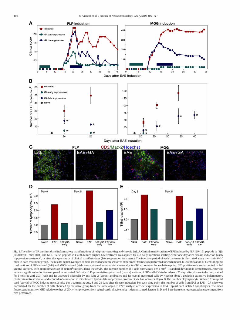
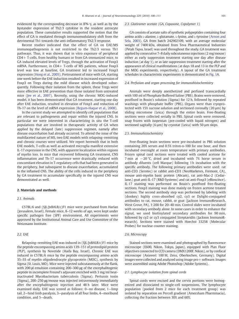
Fig. 1. The effect of GA on clinical and inflammatory manifestations of relapsing–remitting and chronic EAE. A. Clinical manifestations of EAE induced by PLP 139–151 peptide in (SJL/JxBALB/c)F1 mice (left) and MOG 35–55 peptide in C57BL/6 mice (right). GA treatment was applied by 7–8 daily injections starting either one day after disease induction (earlysuppression treatment), or after the appearance of clinical manifestations (late suppression treatment). The injection period of each treatment is illustrated along the x axis, 6–10mice in each treatment group. The results depict averaged clinical score of one representative experiment from 5 to 6 performed for each model. B. Quantification of T-cells in spinalcord sections of PLP-induced (left) and MOG-induced (right) mice, stained immunohistochemically for CD3 expression. For each time point, CD3 positive cells were counted in 2–6sagittal sections, with approximate size of 10 mm2/section, along the cervix. The average number of T-cells normalized per 1 mm2±standard deviation is demonstrated. Asterisksindicate significant reduction compared to untreated EAEmice. C. Representative spinal cord (cervix) sections of PLP andMOG-induced mice 25 days after disease induction, stainedfor T-cells by anti-CD3 (red) and for activated microglia by anti-Mac-2 (green) antibodies and for overall nucleated cells by Hoechst (blue), depicting extensive inflammatoryclusters in untreated mice and reduced inflammation in mice treated by GA - late suppression protocol. Scale bar indicates 50 μm. D. The number of lymphocytes isolated from spinalcord (cervix) of MOG-induced mice, 2 mice per treatment group, 8 and 21 days after disease induction. For each time point the number of cells from EAE or EAE+GA mice wasnormalized for the number of cells obtained by the naïve group from the same organ. E. FACS analysis of T-bet expression in CD4+ spinal cord isolated lymphocytes. The meanfluorescent intensity (MFI) relative to that of CD4+ lymphocytes from spinal cords of naïve mice is demonstrated. Results in D and E are from one representative experiment fromtwo performed.
102 R. Aharoni et al. / Journal of Neuroimmunology 225 (2010) 100–111
103R. Aharoni et al. / Journal of Neuroimmunology 225 (2010) 100–111
2.8. FACS staining and analysis
Cells were stained for surface markers by anti-CD4-Alexa 700 andanti-CD25-PE-Cy7 (eBioscience, San Diego, CA), at the recommendeddilution for 30 min at 4 °C. For intracellular staining, cells were fixedand permeabilized with Foxp3 fixation/permeabilization buffer asrecommended (eBioscience), then stained with anti-mouse Foxp3-APC, GATA3-FITC, RORγt-PE and T-bet-PerCP-Cy5.5 (eBioscience) for30 min at 4 °C. Simultaneous negative-control staining reactions wereperformed with the appropriate isotype controls for each antibody.Stained cells were measured by BD LSR II Flow Cytometer instrument(BD Biosciences, San Jose, CA, USA) and FACS data were analyzed byMatlab (Mathworks, Natick, MA).
2.9. Cytokines secretion assay
Spleen lymphocytes (0.5×10⁶/well) from naïve, EAE or EAE+GAmice were stimulated in vitro by medium alone, GA (50 μg/ml) or byimmobilized anti-CD3 antibodies (5 μg/ml). Culture supernatantswere collected after 3 days and tested for IL-17 and IL-5 concentration(pg/ml) by ELISA using the FlowCytomix Multiplex Kit (Th1/Th210plex; BenderMedSystems, Vienna, Austria).
2.10. Detection of GA-specific T-cells by in vivo imaging system (IVIS)
Spleen lymphocytes from mice that had been immunized with GA(2 mg/mouse, 10 daily subcutaneous injections) were activated invitro by exposure to GA (50 μg/ml) and than labeled by the near-infrared lipophilic carbocyanine dye 1,1′-dioctadecyl-3.3.3′3′-tetra-methyliodotricabocyanine iodine (DiR, Invitrogen), for 45 min in37 °C. The cells were than washed and injected (30×106 cells permouse) into the peritoneum of mice inflicted with EAE (MOG orPLP -induced models), or with inflammatory bowel disease (dextran-induced model ref), or to control naïve mice. Cell injection wasperformed 10 days after EAE or IBD induction. Detection of theinjected cells was performed using the whole body cooled CCDcamera system (IVIS 100 series Imaging System, Xenogen, Alameda,CA).
2.11. Quantification and statistics
Quantitative analysis of Th-17 cells in the brain was performedby counting the double positive CD3+IL-17+ cells from the overallCD3+ T-cells, in 16 coronal sections, along each brain, starting at thelevel of the lateral orbital cortex with intervals of 0.55 mm (Bregma2.3 to −6.5). Quantification of T-cells and T- regulatory cells in thespinal cord was performed by counting all the CD3+ cells andthe double positive CD3+ Foxp3+ cells from the overall CD3+population, in 2–6 sagittal sections with averaged size of 10 mm2 persection, along the cervix. Every counting was performed by scanningindividual fields (0.05 in the brain and 0.13 mm2 in the spinal cord)using analysis software, along the entire section in 2–3 mice fortreatment group for each time point. The results were subjected toone-way analysis of variance (ANOVA), followed by Fishers' LSD post-hoc comparisons where significant differences were found. The levelof significance for all the tests was set at pb0.05.
3. Results
3.1. The effect of GA on clinical and inflammatory manifestations ofrelapsing–remitting and chronic EAE
EAE manifestations as well as the effect of GA treatment wereinvestigated in two MS models: the PLP 139–151 peptide-induced in(SJL/JxBALB/c)F1 mice, which is manifested in a relapsing–remittingcourse and the MOG 35–55 peptide-induced in C57BL/6 mice with a
chronic disease course. GA treatment was applied to the EAE-inducedmice by 7–8 daily injections starting either one day after diseaseinduction (early suppression treatment), or after the appearance ofclinical manifestations (late suppression, namely therapeutic treat-ment). A layout of these systems and the clinical scores observed inone characteristic experiment are demonstrated in Fig. 1A. GAtreatment ameliorated the clinical manifestations in both EAE modelswhen administered at either early or late staged of the disease: Whentreatment was initiated by early suppression, soon after diseaseinduction, its effect was apparent, in the PLPmodel by development ofa much less severe disease with only a single exacerbation, and in theMOG model, by complete prevention of disease development (onlyone from six mice showing grade 1 score). When GA treatment wasapplied after disease symptoms were apparent, it resulted in asubstantial decline of the pre-existing clinical manifestations in bothmodels. The beneficial effect of GA was sustained after treatmenttermination and lasted until the mice were sacrificed.
The in situ effect of GA treatment on CNS inflammation wasmanifested in bothmodels in the numberof T-cells counted in the spinalcord (in entire sagittal cervix sections) stained for CD3 expression invarious time points after disease induction (Fig. 1B). In all the timepoints tested, GA treatment resulted in significant reduction in thenumbers of CD3+ T-cells in comparison to EAE untreated mice.Furthermore, in the mice treated by the early suppression protocolCD3+ cells were rare similar to naïve mice. Representative pictures ofspinal cord sections taken 25 days after EAE induction (Fig. 1C) depictthe extensive widespread inflammatory clusters, containing T-cells(CD3+)andactivatedmicroglia (MAC-2+), in EAEmiceof bothmodels,withmore pronounced T-cell involvement in theMOG-inducedmice. Incontrast, only a few small infiltrations were found in GA-treated miceevenwhen treatment started after disease exacerbation, as described inthe figure. In the mice treated before disease symptoms appearance(early suppression) inflammatory lesions were almost absent.
Correspondingly, the magnitude of the overall lymphocytepopulations isolated from spinal cords (cervix) was elevated by 2.4and 3.4 fold in MOG-induced mice compared with naïve controls,when evaluated either before or after disease appearance, namely8 and 21 days after induction, respectively (Fig. 1D). In contrast, thenumber of spinal cord lymphocytes isolated from GA-treated micewas considerably smaller, 40% reduction from untreated mice by thelate suppression regimen and restoration to the normal level by theearly suppression treatment. Consistent with the CNS inflammationin EAE mice and the anti-inflammatory effect of GA, FACS analysisof spinal cord lymphocytes stained for the Th1 transcription factorT-bet revealed considerable elevation in the mean fluorescentintensity (MFI) of T-bet expression in EAE untreated mice, relativelyto naïve controls, as well as its reduction by GA treatment (Fig. 1E).Treatment started after disease induction induced 30% decrease inT-bet expression from that of EAE untreated mice by day 8, and atotal decline to the normal level by day 21. Even when treatmentwas started after disease appearance it led to a reduction by 30%of T-bet MFI in comparison to untreated mice, at the end of thetreatment (in day 21). These cumulative results are indicative ofthe anti-inflammatory activity of GA on the CNS.
3.2. In situ IL-17 expression in relapsing–remitting and chronic EAE andthe effect of GA on its expression
In view of the increasing evidence for the role of Th-17 in MS, itwas of interest to investigate the involvement of the IL-17 effectorpathway in the CNS pathology of EAE in both the relapsing–remittingand the chronic models, as well as in mice that had recovered fromthese diseases following GA treatment. Th-17 effector cells in the CNSwere detected immunohistochemically by staining brain sections forIL-17 expression, and their T-cell identity was established by doublestaining for the T-subset marker CD3. As demonstrated in Fig. 2A,
104 R. Aharoni et al. / Journal of Neuroimmunology 225 (2010) 100–111
brain section of EAE untreated mice, in both the PLP- and the MOG-induced models, contained multiple infiltrations of T-cells, and aconsiderable proportion of these cells manifested clear cytoplasmatic
Fig. 2. IL-17 expression in PLP- andMOG- induced EAE and the effect of GA treatment. A. Immstained for CD3 (green) and IL-17 (red). Representing images fromwhite matter in the brainGA treated mice the majority of the CD3+ cells are negative for IL-17 expression. Scale bar in50 μm. B. Quantitative analysis of Th-17 cells in the PLP and MOG models, in brains of untrpercentage of Th-17 expressing cells from the overall CD3+ T-cells, counted in 16 coronal sindicate significant reduction from untreated EAE mice. C. FACS analysis of RORγt expressiomice, 2 mice per treatment group, 8 and 21 days after disease induction. MFI of RORγt expeffect of GA on the peripheral secretion of IL-17 (left) and IL-5 (right), in MOG-induced miceCD3 antibodies (5 μg/ml), and their culture supernatants were tested by ELISA. The results
stainingwith anti-IL-17 antibodies. In contrast, in brains of GA-treatedmice we found smaller amounts of T-cells (in accord with the CD3quantification in spinal cords demonstrated in Fig. 1B), and only a
unohistological detection of Th-17 cells in brain sections of PLP andMOG-induced mice,stem area depicting multiple double positive CD3+IL-17+ cells in EAEmice, whereas indicates 100 μm for all rows except the third and the sixth of the PLP (confocal images)—eated and GA-treated mice, by early and late suppression. Each column represents theections, along each brain, 2–3 mice for each time point±standard deviation. Asterisksn, gated on the CD4 marker, in lymphocytes isolated from spinal cords of MOG-inducedression relative to that of CD4+ lymphocytes from naïve mice is demonstrated. D. The. Spleen lymphocytes were stimulated in vitro by GA (50 μg/ml) or by immobilized anti-in C and D are from one representative experiment from two performed.
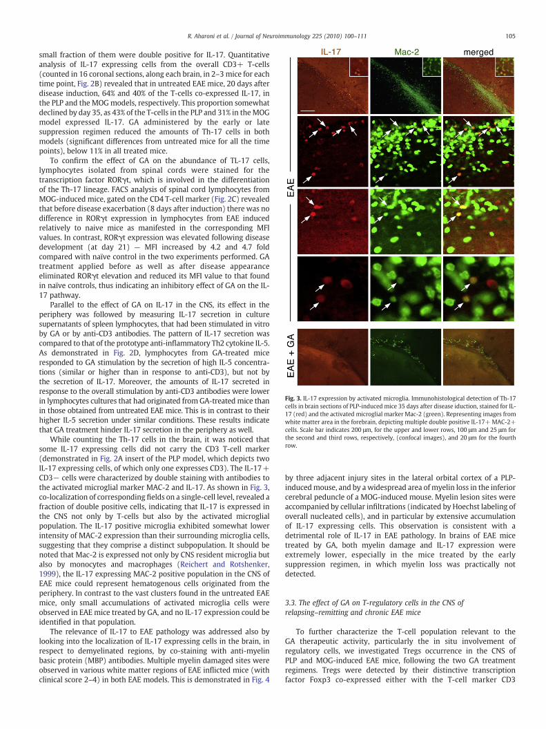
Fig. 3. IL-17 expression by activated microglia. Immunohistological detection of Th-17cells in brain sections of PLP-induced mice 35 days after disease iduction, stained for IL-17 (red) and the activated microglial marker Mac-2 (green). Representing images fromwhite matter area in the forebrain, depicting multiple double positive IL-17+MAC-2+cells. Scale bar indicates 200 μm, for the upper and lower rows, 100 μm and 25 μm forthe second and third rows, respectively, (confocal images), and 20 μm for the fourthrow.
105R. Aharoni et al. / Journal of Neuroimmunology 225 (2010) 100–111
small fraction of them were double positive for IL-17. Quantitativeanalysis of IL-17 expressing cells from the overall CD3+ T-cells(counted in 16 coronal sections, along each brain, in 2–3mice for eachtime point, Fig. 2B) revealed that in untreated EAE mice, 20 days afterdisease induction, 64% and 40% of the T-cells co-expressed IL-17, inthe PLP and the MOGmodels, respectively. This proportion somewhatdeclined by day 35, as 43% of the T-cells in the PLP and 31% in theMOGmodel expressed IL-17. GA administered by the early or latesuppression regimen reduced the amounts of Th-17 cells in bothmodels (significant differences from untreated mice for all the timepoints), below 11% in all treated mice.
To confirm the effect of GA on the abundance of TL-17 cells,lymphocytes isolated from spinal cords were stained for thetranscription factor RORγt, which is involved in the differentiationof the Th-17 lineage. FACS analysis of spinal cord lymphocytes fromMOG-induced mice, gated on the CD4 T-cell marker (Fig. 2C) revealedthat before disease exacerbation (8 days after induction) there was nodifference in RORγt expression in lymphocytes from EAE inducedrelatively to naive mice as manifested in the corresponding MFIvalues. In contrast, RORγt expression was elevated following diseasedevelopment (at day 21) — MFI increased by 4.2 and 4.7 foldcompared with naïve control in the two experiments performed. GAtreatment applied before as well as after disease appearanceeliminated RORγt elevation and reduced its MFI value to that foundin naïve controls, thus indicating an inhibitory effect of GA on the IL-17 pathway.
Parallel to the effect of GA on IL-17 in the CNS, its effect in theperiphery was followed by measuring IL-17 secretion in culturesupernatants of spleen lymphocytes, that had been stimulated in vitroby GA or by anti-CD3 antibodies. The pattern of IL-17 secretion wascompared to that of the prototype anti-inflammatory Th2 cytokine IL-5.As demonstrated in Fig. 2D, lymphocytes from GA-treated miceresponded to GA stimulation by the secretion of high IL-5 concentra-tions (similar or higher than in response to anti-CD3), but not bythe secretion of IL-17. Moreover, the amounts of IL-17 secreted inresponse to the overall stimulation by anti-CD3 antibodies were lowerin lymphocytes cultures that had originated fromGA-treated mice thanin those obtained from untreated EAE mice. This is in contrast to theirhigher IL-5 secretion under similar conditions. These results indicatethat GA treatment hinder IL-17 secretion in the periphery as well.
While counting the Th-17 cells in the brain, it was noticed thatsome IL-17 expressing cells did not carry the CD3 T-cell marker(demonstrated in Fig. 2A insert of the PLP model, which depicts twoIL-17 expressing cells, of which only one expresses CD3). The IL-17+CD3− cells were characterized by double staining with antibodies tothe activated microglial marker MAC-2 and IL-17. As shown in Fig. 3,co-localization of corresponding fields on a single-cell level, revealed afraction of double positive cells, indicating that IL-17 is expressed inthe CNS not only by T-cells but also by the activated microglialpopulation. The IL-17 positive microglia exhibited somewhat lowerintensity of MAC-2 expression than their surrounding microglia cells,suggesting that they comprise a distinct subpopulation. It should benoted that Mac-2 is expressed not only by CNS resident microglia butalso by monocytes and macrophages (Reichert and Rotshenker,1999), the IL-17 expressing MAC-2 positive population in the CNS ofEAE mice could represent hematogenous cells originated from theperiphery. In contrast to the vast clusters found in the untreated EAEmice, only small accumulations of activated microglia cells wereobserved in EAE mice treated by GA, and no IL-17 expression could beidentified in that population.
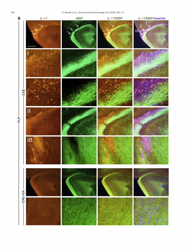
The relevance of IL-17 to EAE pathology was addressed also bylooking into the localization of IL-17 expressing cells in the brain, inrespect to demyelinated regions, by co-staining with anti-myelinbasic protein (MBP) antibodies. Multiple myelin damaged sites wereobserved in various white matter regions of EAE inflicted mice (withclinical score 2–4) in both EAE models. This is demonstrated in Fig. 4
by three adjacent injury sites in the lateral orbital cortex of a PLP-inducedmouse, and by awidespread area of myelin loss in the inferiorcerebral peduncle of a MOG-induced mouse. Myelin lesion sites wereaccompanied by cellular infiltrations (indicated by Hoechst labeling ofoverall nucleated cells), and in particular by extensive accumulationof IL-17 expressing cells. This observation is consistent with adetrimental role of IL-17 in EAE pathology. In brains of EAE micetreated by GA, both myelin damage and IL-17 expression wereextremely lower, especially in the mice treated by the earlysuppression regimen, in which myelin loss was practically notdetected.
3.3. The effect of GA on T-regulatory cells in the CNS ofrelapsing–remitting and chronic EAE mice
To further characterize the T-cell population relevant to theGA therapeutic activity, particularly the in situ involvement ofregulatory cells, we investigated Tregs occurrence in the CNS ofPLP and MOG-induced EAE mice, following the two GA treatmentregimens. Tregs were detected by their distinctive transcriptionfactor Foxp3 co-expressed either with the T-cell marker CD3
106 R. Aharoni et al. / Journal of Neuroimmunology 225 (2010) 100–111
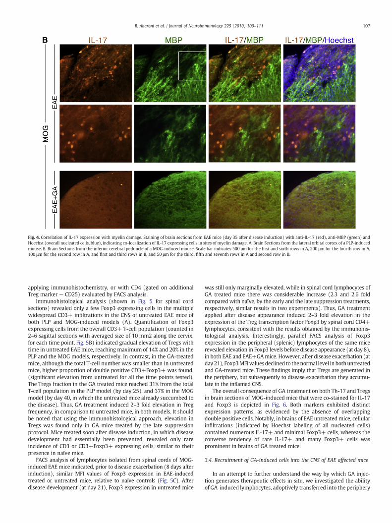
Fig. 4. Correlation of IL-17 expression with myelin damage. Staining of brain sections from EAE mice (day 35 after disease induction) with anti-IL-17 (red), anti-MBP (green) andHoechst (overall nucleated cells, blue), indicating co-localization of IL-17 expressing cells in sites of myelin damage. A. Brain Sections from the lateral orbital cortex of a PLP-inducedmouse. B. Brain Sections from the inferior cerebral peduncle of a MOG-induced mouse. Scale bar indicates 500 μm for the first and sixth rows in A, 200 μm for the fourth row in A,100 μm for the second row in A, and first and third rows in B, and 50 μm for the third, fifth and seventh rows in A and second row in B.
107R. Aharoni et al. / Journal of Neuroimmunology 225 (2010) 100–111
applying immunohistochemistry, or with CD4 (gated on additionalTreg marker — CD25) evaluated by FACS analysis.
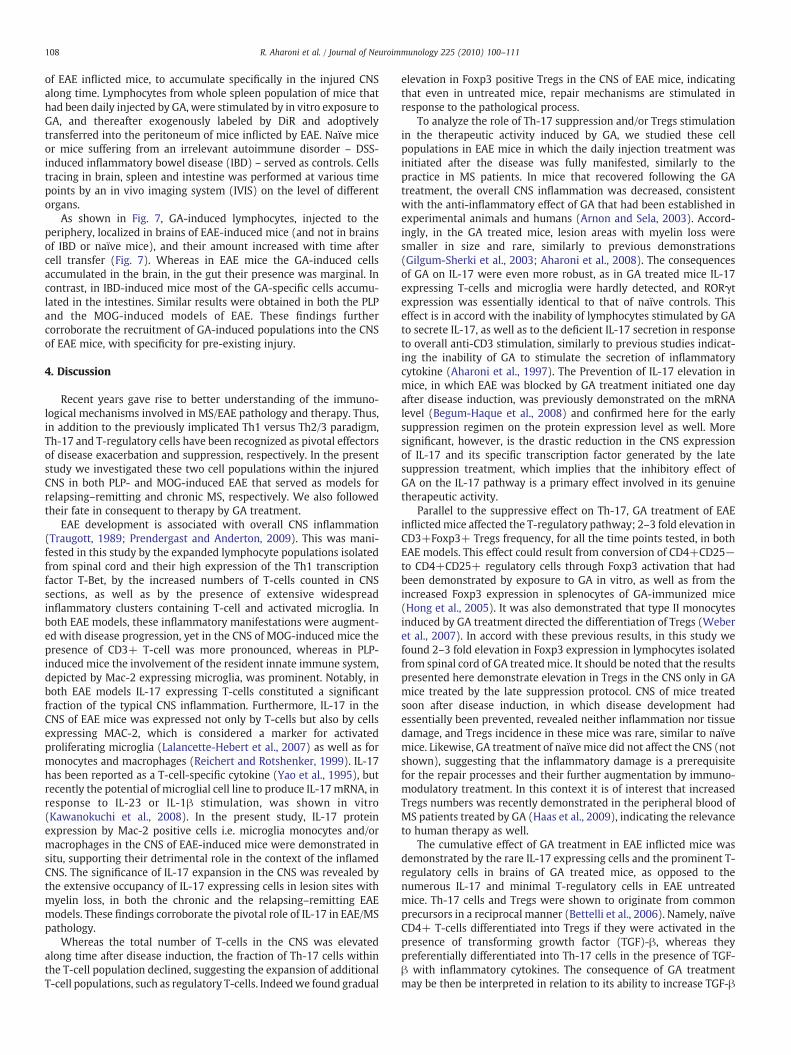
Immunohistological analysis (shown in Fig. 5 for spinal cordsections) revealed only a few Foxp3 expressing cells in the multiplewidespread CD3+ infiltrations in the CNS of untreated EAE mice ofboth PLP and MOG-induced models (A). Quantification of Foxp3expressing cells from the overall CD3+ T-cell population (counted in2–6 sagittal sections with averaged size of 10 mm2 along the cervix,for each time point, Fig. 5B) indicated gradual elevation of Tregs withtime in untreated EAE mice, reaching maximum of 14% and 20% in thePLP and the MOG models, respectively. In contrast, in the GA-treatedmice, although the total T-cell number was smaller than in untreatedmice, higher proportion of double positive CD3+Foxp3+ was found,(significant elevation from untreated for all the time points tested).The Tregs fraction in the GA treated mice reached 31% from the totalT-cell population in the PLP model (by day 25), and 37% in the MOGmodel (by day 40, in which the untreated mice already succumbed tothe disease). Thus, GA treatment induced 2–3 fold elevation in Tregfrequency, in comparison to untreated mice, in both models. It shouldbe noted that using the immunohistological approach, elevation inTregs was found only in GA mice treated by the late suppressionprotocol. Mice treated soon after disease induction, in which diseasedevelopment had essentially been prevented, revealed only rareincidence of CD3 or CD3+Foxp3+ expressing cells, similar to theirpresence in naïve mice.
FACS analysis of lymphocytes isolated from spinal cords of MOG-induced EAE mice indicated, prior to disease exacerbation (8 days afterinduction), similar MFI values of Foxp3 expression in EAE-inducedtreated or untreated mice, relative to naïve controls (Fig. 5C). Afterdisease development (at day 21), Foxp3 expression in untreated mice
was still only marginally elevated, while in spinal cord lymphocytes ofGA treated mice there was considerable increase (2.3 and 2.6 foldcompared with naïve, by the early and the late suppression treatments,respectively, similar results in two experiments). Thus, GA treatmentapplied after disease appearance induced 2–3 fold elevation in theexpression of the Treg transcription factor Foxp3 by spinal cord CD4+lymphocytes, consistent with the results obtained by the immunohis-tological analysis. Interestingly, parallel FACS analysis of Foxp3expression in the peripheral (splenic) lymphocytes of the same micerevealed elevation in Foxp3 levels before disease appearance (at day 8),in both EAE and EAE+GAmice. However, after disease exacerbation (atday 21), Foxp3MFI values declined to the normal level in bothuntreatedand GA-treated mice. These findings imply that Tregs are generated inthe periphery, but subsequently to disease exacerbation they accumu-late in the inflamed CNS.
The overall consequence of GA treatment on both Th-17 and Tregsin brain sections of MOG-induced mice that were co-stained for IL-17and Foxp3 is depicted in Fig. 6. Both markers exhibited distinctexpression patterns, as evidenced by the absence of overlappingdouble positive cells. Notably, in brains of EAE untreatedmice, cellularinfiltrations (indicated by Hoechst labeling of all nucleated cells)contained numerous IL-17+ and minimal Foxp3+ cells, whereas theconverse tendency of rare IL-17+ and many Foxp3+ cells wasprominent in brains of GA treated mice.
3.4. Recruitment of GA-induced cells into the CNS of EAE affected mice
In an attempt to further understand the way by which GA injec-tion generates therapeutic effects in situ, we investigated the abilityof GA-induced lymphocytes, adoptively transferred into the periphery
108 R. Aharoni et al. / Journal of Neuroimmunology 225 (2010) 100–111
of EAE inflicted mice, to accumulate specifically in the injured CNSalong time. Lymphocytes from whole spleen population of mice thathad been daily injected by GA, were stimulated by in vitro exposure toGA, and thereafter exogenously labeled by DiR and adoptivelytransferred into the peritoneum of mice inflicted by EAE. Naïve miceor mice suffering from an irrelevant autoimmune disorder – DSS-induced inflammatory bowel disease (IBD) – served as controls. Cellstracing in brain, spleen and intestine was performed at various timepoints by an in vivo imaging system (IVIS) on the level of differentorgans.
As shown in Fig. 7, GA-induced lymphocytes, injected to theperiphery, localized in brains of EAE-induced mice (and not in brainsof IBD or naïve mice), and their amount increased with time aftercell transfer (Fig. 7). Whereas in EAE mice the GA-induced cellsaccumulated in the brain, in the gut their presence was marginal. Incontrast, in IBD-induced mice most of the GA-specific cells accumu-lated in the intestines. Similar results were obtained in both the PLPand the MOG-induced models of EAE. These findings furthercorroborate the recruitment of GA-induced populations into the CNSof EAE mice, with specificity for pre-existing injury.
4. Discussion
Recent years gave rise to better understanding of the immuno-logical mechanisms involved in MS/EAE pathology and therapy. Thus,in addition to the previously implicated Th1 versus Th2/3 paradigm,Th-17 and T-regulatory cells have been recognized as pivotal effectorsof disease exacerbation and suppression, respectively. In the presentstudy we investigated these two cell populations within the injuredCNS in both PLP- and MOG-induced EAE that served as models forrelapsing–remitting and chronic MS, respectively. We also followedtheir fate in consequent to therapy by GA treatment.
EAE development is associated with overall CNS inflammation(Traugott, 1989; Prendergast and Anderton, 2009). This was mani-fested in this study by the expanded lymphocyte populations isolatedfrom spinal cord and their high expression of the Th1 transcriptionfactor T-Bet, by the increased numbers of T-cells counted in CNSsections, as well as by the presence of extensive widespreadinflammatory clusters containing T-cell and activated microglia. Inboth EAE models, these inflammatory manifestations were augment-ed with disease progression, yet in the CNS of MOG-induced mice thepresence of CD3+ T-cell was more pronounced, whereas in PLP-induced mice the involvement of the resident innate immune system,depicted by Mac-2 expressing microglia, was prominent. Notably, inboth EAE models IL-17 expressing T-cells constituted a significantfraction of the typical CNS inflammation. Furthermore, IL-17 in theCNS of EAE mice was expressed not only by T-cells but also by cellsexpressing MAC-2, which is considered a marker for activatedproliferating microglia (Lalancette-Hebert et al., 2007) as well as formonocytes and macrophages (Reichert and Rotshenker, 1999). IL-17has been reported as a T-cell-specific cytokine (Yao et al., 1995), butrecently the potential of microglial cell line to produce IL-17 mRNA, inresponse to IL-23 or IL-1β stimulation, was shown in vitro(Kawanokuchi et al., 2008). In the present study, IL-17 proteinexpression by Mac-2 positive cells i.e. microglia monocytes and/ormacrophages in the CNS of EAE-induced mice were demonstrated insitu, supporting their detrimental role in the context of the inflamedCNS. The significance of IL-17 expansion in the CNS was revealed bythe extensive occupancy of IL-17 expressing cells in lesion sites withmyelin loss, in both the chronic and the relapsing–remitting EAEmodels. These findings corroborate the pivotal role of IL-17 in EAE/MSpathology.
Whereas the total number of T-cells in the CNS was elevatedalong time after disease induction, the fraction of Th-17 cells withinthe T-cell population declined, suggesting the expansion of additionalT-cell populations, such as regulatory T-cells. Indeedwe found gradual
elevation in Foxp3 positive Tregs in the CNS of EAE mice, indicatingthat even in untreated mice, repair mechanisms are stimulated inresponse to the pathological process.
To analyze the role of Th-17 suppression and/or Tregs stimulationin the therapeutic activity induced by GA, we studied these cellpopulations in EAE mice in which the daily injection treatment wasinitiated after the disease was fully manifested, similarly to thepractice in MS patients. In mice that recovered following the GAtreatment, the overall CNS inflammation was decreased, consistentwith the anti-inflammatory effect of GA that had been established inexperimental animals and humans (Arnon and Sela, 2003). Accord-ingly, in the GA treated mice, lesion areas with myelin loss weresmaller in size and rare, similarly to previous demonstrations(Gilgum-Sherki et al., 2003; Aharoni et al., 2008). The consequencesof GA on IL-17 were even more robust, as in GA treated mice IL-17expressing T-cells and microglia were hardly detected, and RORγtexpression was essentially identical to that of naïve controls. Thiseffect is in accord with the inability of lymphocytes stimulated by GAto secrete IL-17, as well as to the deficient IL-17 secretion in responseto overall anti-CD3 stimulation, similarly to previous studies indicat-ing the inability of GA to stimulate the secretion of inflammatorycytokine (Aharoni et al., 1997). The Prevention of IL-17 elevation inmice, in which EAE was blocked by GA treatment initiated one dayafter disease induction, was previously demonstrated on the mRNAlevel (Begum-Haque et al., 2008) and confirmed here for the earlysuppression regimen on the protein expression level as well. Moresignificant, however, is the drastic reduction in the CNS expressionof IL-17 and its specific transcription factor generated by the latesuppression treatment, which implies that the inhibitory effect ofGA on the IL-17 pathway is a primary effect involved in its genuinetherapeutic activity.
Parallel to the suppressive effect on Th-17, GA treatment of EAEinflictedmice affected the T-regulatory pathway; 2–3 fold elevation inCD3+Foxp3+ Tregs frequency, for all the time points tested, in bothEAE models. This effect could result from conversion of CD4+CD25−to CD4+CD25+ regulatory cells through Foxp3 activation that hadbeen demonstrated by exposure to GA in vitro, as well as from theincreased Foxp3 expression in splenocytes of GA-immunized mice(Hong et al., 2005). It was also demonstrated that type II monocytesinduced by GA treatment directed the differentiation of Tregs (Weberet al., 2007). In accord with these previous results, in this study wefound 2–3 fold elevation in Foxp3 expression in lymphocytes isolatedfrom spinal cord of GA treatedmice. It should be noted that the resultspresented here demonstrate elevation in Tregs in the CNS only in GAmice treated by the late suppression protocol. CNS of mice treatedsoon after disease induction, in which disease development hadessentially been prevented, revealed neither inflammation nor tissuedamage, and Tregs incidence in these mice was rare, similar to naïvemice. Likewise, GA treatment of naïve mice did not affect the CNS (notshown), suggesting that the inflammatory damage is a prerequisitefor the repair processes and their further augmentation by immuno-modulatory treatment. In this context it is of interest that increasedTregs numbers was recently demonstrated in the peripheral blood ofMS patients treated by GA (Haas et al., 2009), indicating the relevanceto human therapy as well.
The cumulative effect of GA treatment in EAE inflicted mice wasdemonstrated by the rare IL-17 expressing cells and the prominent T-regulatory cells in brains of GA treated mice, as opposed to thenumerous IL-17 and minimal T-regulatory cells in EAE untreatedmice. Th-17 cells and Tregs were shown to originate from commonprecursors in a reciprocal manner (Bettelli et al., 2006). Namely, naïveCD4+ T-cells differentiated into Tregs if they were activated in thepresence of transforming growth factor (TGF)-β, whereas theypreferentially differentiated into Th-17 cells in the presence of TGF-β with inflammatory cytokines. The consequence of GA treatmentmay be then be interpreted in relation to its ability to increase TGF-β
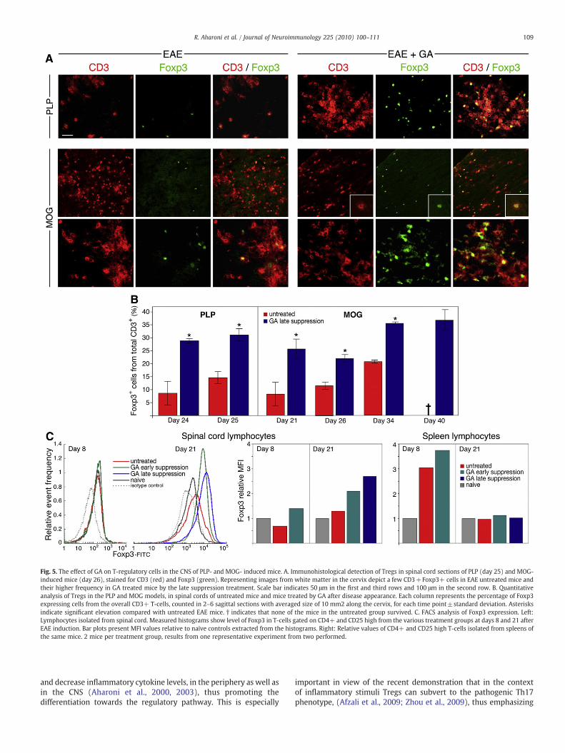
Fig. 5. The effect of GA on T-regulatory cells in the CNS of PLP- and MOG- induced mice. A. Immunohistological detection of Tregs in spinal cord sections of PLP (day 25) and MOG-induced mice (day 26), stained for CD3 (red) and Foxp3 (green). Representing images from white matter in the cervix depict a few CD3+Foxp3+ cells in EAE untreated mice andtheir higher frequency in GA treated mice by the late suppression treatment. Scale bar indicates 50 μm in the first and third rows and 100 μm in the second row. B. Quantitativeanalysis of Tregs in the PLP and MOG models, in spinal cords of untreated mice and mice treated by GA after disease appearance. Each column represents the percentage of Foxp3expressing cells from the overall CD3+ T-cells, counted in 2–6 sagittal sections with averaged size of 10 mm2 along the cervix, for each time point±standard deviation. Asterisksindicate significant elevation compared with untreated EAE mice. † indicates that none of the mice in the untreated group survived. C. FACS analysis of Foxp3 expression. Left:Lymphocytes isolated from spinal cord. Measured histograms show level of Foxp3 in T-cells gated on CD4+ and CD25 high from the various treatment groups at days 8 and 21 afterEAE induction. Bar plots present MFI values relative to naïve controls extracted from the histograms. Right: Relative values of CD4+ and CD25 high T-cells isolated from spleens ofthe same mice. 2 mice per treatment group, results from one representative experiment from two performed.
109R. Aharoni et al. / Journal of Neuroimmunology 225 (2010) 100–111
and decrease inflammatory cytokine levels, in the periphery as well asin the CNS (Aharoni et al., 2000, 2003), thus promoting thedifferentiation towards the regulatory pathway. This is especially
important in view of the recent demonstration that in the contextof inflammatory stimuli Tregs can subvert to the pathogenic Th17phenotype, (Afzali et al., 2009; Zhou et al., 2009), thus emphasizing
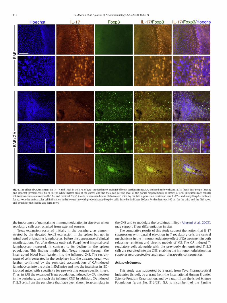
Fig. 6. The effect of GA treatment on Th-17 and Tregs in the CNS of EAE- induced mice. Staining of brain sections from MOG-induced mice with anti-IL-17 (red), anti-Foxp3 (green)and Hoechst (overall cells, blue), in the white matter area of the cortex and the thalamus (at the level of the dorsal hippocampus). In brains of EAE untreated mice cellularinfiltrations contain numerous IL-17+ and minimal Foxp3+ cells, whereas in brains of GA treated mice, by the late suppression treatment, rare IL-17+ and many Foxp3+ cells arefound. Note the perivascular cell infiltration in the lowest raw with predominantly Foxp3+ cells. Scale bar indicates 200 μm for the first row, 100 μm for the third and the fifth rows,and 50 μm for the second and forth rows.
110 R. Aharoni et al. / Journal of Neuroimmunology 225 (2010) 100–111
the importance of maintaining immunomodulation in situ even whenregulatory cells are recruited from external sources.
Tregs expansion occurred initially in the periphery, as demon-strated by the elevated Foxp3 expression in the spleen but not inspinal cord originating lymphocytes, before the appearance of clinicalmanifestations. Yet, after disease outbreak, Foxp3 level in spinal cordlymphocytes increased, in contrast to its decline in the spleenpopulation. This finding implied that Tregs migrate through theinterrupted blood brain barrier, into the inflamed CNS. The recruit-ment of cells generated in the periphery into the diseased organ wasfurther confirmed by the restricted accumulation of GA-inducedlymphocytes into the brain in EAE mice and into the intestines in IBD-induced mice, with specificity for pre-existing organ-specific injury.Thus, in EAE the expanded Tregs population, induced by GA injectionin the periphery, can reach the inflamed CNS. In addition, GA inducedTh2/3 cells from the periphery that have been shown to accumulate in
the CNS and to modulate the cytokines milieu (Aharoni et al., 2003),may support Tregs differentiation in situ.
The cumulative results of this study support the notion that IL-17suppression with parallel elevation in T-regulatory cells are centralmechanisms in the immunomodulatory effect of GA treatment in bothrelapsing–remitting and chronic models of MS. The GA induced T-regulatory cells alongside with the previously demonstrated Th2/3cells are recruited into the CNS, enabling the immunomodulation thatsupports neuroprotective and repair therapeutic consequences.
Acknowledgment
This study was supported by a grant from Teva PharmaceuticalIndustries (Israel), by a grant from the International Human FrontierScience Program Organization, and by a grant from the Israel ScienceFoundation (grant No. 812/08). N.F. is incumbent of the Pauline
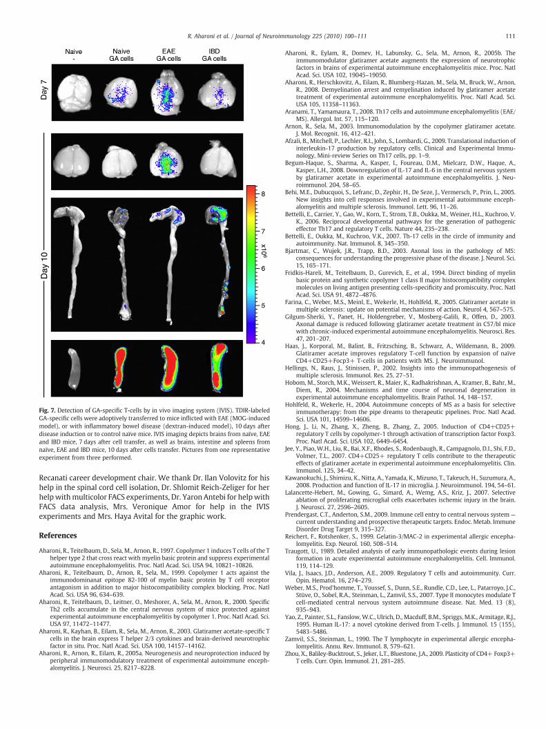
Fig. 7. Detection of GA-specific T-cells by in vivo imaging system (IVIS). TDIR-labeledGA-specific cells were adoptively transferred to mice inflicted with EAE (MOG-inducedmodel), or with inflammatory bowel disease (dextran-induced model), 10 days afterdisease induction or to control naïve mice. IVIS imaging depicts brains from naïve, EAEand IBD mice, 7 days after cell transfer, as well as brains, intestine and spleens fromnaïve, EAE and IBD mice, 10 days after cells transfer. Pictures from one representativeexperiment from three performed.
111R. Aharoni et al. / Journal of Neuroimmunology 225 (2010) 100–111
Recanati career development chair. We thank Dr. Ilan Volovitz for hishelp in the spinal cord cell isolation, Dr. Shlomit Reich-Zeliger for herhelpwithmulticolor FACS experiments, Dr. Yaron Antebi for helpwithFACS data analysis, Mrs. Veronique Amor for help in the IVISexperiments and Mrs. Haya Avital for the graphic work.
References
Aharoni, R., Teitelbaum, D., Sela, M., Arnon, R., 1997. Copolymer 1 induces T cells of the Thelper type 2 that cross react with myelin basic protein and suppress experimentalautoimmune encephalomyelitis. Proc. Natl Acad. Sci. USA 94, 10821–10826.
Aharoni, R., Teitelbaum, D., Arnon, R., Sela, M., 1999. Copolymer 1 acts against theimmunodominanat epitope 82-100 of myelin basic protein by T cell receptorantagonism in addition to major histocompatibility complex blocking. Proc. NatlAcad. Sci. USA 96, 634–639.
Aharoni, R., Teitelbaum, D., Leitner, O., Meshorer, A., Sela, M., Arnon, R., 2000. SpecificTh2 cells accumulate in the central nervous system of mice protected againstexperimental autoimmune encephalomyelitis by copolymer 1. Proc. Natl Acad. Sci.USA 97, 11472–11477.
Aharoni, R., Kayhan, B., Eilam, R., Sela, M., Arnon, R., 2003. Glatiramer acetate-specific Tcells in the brain express T helper 2/3 cytokines and brain-derived neurotrophicfactor in situ. Proc. Natl Acad. Sci. USA 100, 14157–14162.
Aharoni, R., Arnon, R., Eilam, R., 2005a. Neurogenesis and neuroprotection induced byperipheral immunomodulatory treatment of experimental autoimmune enceph-alomyelitis. J. Neurosci. 25, 8217–8228.
Aharoni, R., Eylam, R., Domev, H., Labunsky, G., Sela, M., Arnon, R., 2005b. Theimmunomodulator glatiramer acetate augments the expression of neurotrophicfactors in brains of experimental autoimmune encephalomyelitis mice. Proc. NatlAcad. Sci. USA 102, 19045–19050.
Aharoni, R., Herschkovitz, A., Eilam, R., Blumberg-Hazan, M., Sela, M., Bruck, W., Arnon,R., 2008. Demyelination arrest and remyelination induced by glatiramer acetatetreatment of experimental autoimmune encephalomyelitis. Proc. Natl Acad. Sci.USA 105, 11358–11363.
Aranami, T., Yamamaura, T., 2008. Th17 cells and autoimmune encephalomyelitis (EAE/MS). Allergol. Int. 57, 115–120.
Arnon, R., Sela, M., 2003. Immunomodulation by the copolymer glatiramer acetate.J. Mol. Recognit. 16, 412–421.
Afzali, B., Mitchell, P., Lechler, R.I., John, S., Lombardi, G., 2009. Translational induction ofinterleukin-17 production by regulatory cells. Clinical and Experimental Immu-nology, Mini-review Series on Th17 cells, pp. 1–9.
Begum-Haque, S., Sharma, A., Kasper, I., Foureau, D.M., Mielcarz, D.W., Haque, A.,Kasper, L.H., 2008. Downregulation of IL-17 and IL-6 in the central nervous systemby glatiramer acetate in experimental autoimmune encephalomyelitis. J. Neu-roimmunol. 204, 58–65.
Behi, M.E., Dubucquoi, S., Lefranc, D., Zephir, H., De Seze, J., Vermersch, P., Prin, L., 2005.New insights into cell responses involved in experimental autoimmune enceph-alomyelitis and multiple sclerosis. Immunol. Lett. 96, 11–26.
Bettelli, E., Carrier, Y., Gao, W., Korn, T., Strom, T.B., Oukka, M., Weiner, H.L., Kuchroo, V.K., 2006. Reciprocal developmental pathways for the generation of pathogeniceffector Th17 and regulatory T cells. Nature 44, 235–238.
Bettelli, E., Oukka, M., Kuchroo, V.K., 2007. Th-17 cells in the circle of immunity andautoimmunity. Nat. Immunol. 8, 345–350.
Bjartmar, C., Wujek, J.R., Trapp, B.D., 2003. Axonal loss in the pathology of MS:consequences for understanding the progressive phase of the disease. J. Neurol. Sci.15, 165–171.
Fridkis-Hareli, M., Teitelbaum, D., Gurevich, E., et al., 1994. Direct binding of myelinbasic protein and synthetic copolymer 1 class II major histocompatibility complexmolecules on living antigen presenting cells-specificity and promiscuity. Proc. NatlAcad. Sci. USA 91, 4872–4876.
Farina, C., Weber, M.S., Meinl, E., Wekerle, H., Hohlfeld, R., 2005. Glatiramer acetate inmultiple sclerosis: update on potential mechanisms of action. Neurol 4, 567–575.
Gilgum-Sherki, Y., Panet, H., Holdengreber, V., Mosberg-Galili, R., Offen, D., 2003.Axonal damage is reduced following glatiramer acetate treatment in C57/bl micewith chronic-induced experimental autoimmune encephalomyelitis. Neurosci. Res.47, 201–207.
Haas, J., Korporal, M., Balint, B., Fritzsching, B., Schwarz, A., Wildemann, B., 2009.Glatiramer acetate improves regulatory T-cell function by expansion of naïveCD4+CD25+Focp3+ T-cells in patients with MS. J. Neuroimmunol.
Hellings, N., Raus, J., Stinissen, P., 2002. Insights into the immunopathogenesis ofmultiple sclerosis. Immunol. Res. 25, 27–51.
Hobom, M., Storch, M.K., Weissert, R., Maier, K., Radhakrishnan, A., Kramer, B., Bahr, M.,Diem, R., 2004. Mechanisms and time course of neuronal degeneration inexperimental autoimmune encephalomyelitis. Brain Pathol. 14, 148–157.
Hohlfeld, R., Wekerle, H., 2004. Autoimmune concepts of MS as a basis for selectiveimmunotherapy: from the pipe dreams to therapeutic pipelines. Proc. Natl Acad.Sci. USA 101, 14599–14606.
Hong, J., Li, N., Zhang, X., Zheng, B., Zhang, Z., 2005. Induction of CD4+CD25+regulatory T cells by copolymer-1 through activation of transcription factor Foxp3.Proc. Natl Acad. Sci. USA 102, 6449–6454.
Jee, Y., Piao, W.H., Liu, R., Bai, X.F., Rhodes, S., Rodenbaugh, R., Campagnolo, D.I., Shi, F.D.,Volmer, T.L., 2007. CD4+CD25+ regulatory T cells contribute to the therapeuticeffects of glatiramer acetate in experimental autoimmune encephalomyelitis. Clin.Immunol. 125, 34–42.
Kawanokuchi, J., Shimizu, K., Nitta, A., Yamada, K., Mizuno, T., Takeuch, H., Suzumura, A.,2008. Production and function of IL-17 in microglia. J. Neuroimmunol. 194, 54–61.
Lalancette-Hebert, M., Gowing, G., Simard, A., Wemg, A.S., Kriz, J., 2007. Selectiveablation of proliferating microglial cells exacerbates ischemic injury in the brain.J. Neurosci. 27, 2596–2605.
Prendergast, C.T., Anderton, S.M., 2009. Immune cell entry to central nervous system —current understanding and prospective therapeutic targets. Endoc. Metab. ImmuneDisorder Drug Target 9, 315–327.
Reichert, F., Rotshenker, S., 1999. Gelatin-3/MAC-2 in experimental allergic encepha-lomyelitis. Exp. Neurol. 160, 508–514.
Traugott, U., 1989. Detailed analysis of early immunopathologic events during lesionformation in acute experimental autoimmune encephalomyelitis. Cell. Immunol.119, 114–129.
Vila, J., Isaacs, J.D., Anderson, A.E., 2009. Regulatory T cells and autoimmunity. Curr.Opin. Hematol. 16, 274–279.
Weber, M.S., Prod'homme, T., Youssef, S., Dunn, S.E., Rundle, C.D., Lee, L., Patarroyo, J.C.,Stüve, O., Sobel, R.A., Steinman, L., Zamvil, S.S., 2007. Type II monocytes modulate Tcell-mediated central nervous system autoimmune disease. Nat. Med. 13 (8),935–943.
Yao, Z., Painter, S.L., Fanslow,W.C., Ulrich, D., Macduff, B.M., Spriggs, M.K., Armitage, R.J.,1995. Human IL-17: a novel cytokine derived from T-cells. J. Immunol. 15 (155),5483–5486.
Zamvil, S.S., Steinman, L., 1990. The T lymphocyte in experimental allergic encepha-lomyelitis. Annu. Rev. Immunol. 8, 579–621.
Zhou, X., Baliley-Bucktrout, S., Jeker, L.T., Bluestone, J.A., 2009. Plasticity of CD4+ Foxp3+T cells. Curr. Opin. Immunol. 21, 281–285.


![Chiraldescriptionofmassivegravity - Springer2013)068.pdf · in the Vainshtein mechanism region.1 A non-perturbative formulation of the action was given in [11–14], including its](https://img.pdfslide.us/doc/110x75/5f0c83847e708231d435c816/chiraldescriptionofmassivegravity-springer-2013068pdf-in-the-vainshtein-mechanism.jpg)