Embed Size (px)
Citation preview
BioMed CentralJournal of Medical Case Reports
ss
Open AcceCase reportDistinctive spinal changes in two patients with unusual forms of autosomal dominant endosteal hyperostosis: a case seriesAli Al Kaissi*1,3, Franz Varga1, Shahin Zandieh2, Klaus Klaushofer1 and Franz Grill3Address: 1Ludwig Boltzmann Institute of Osteology at the Hanusch Hospital of WGKK and AUVA Trauma Centre Meidling, 4th Medical Department, Hanusch Hospital, Heinrich Collin Str. 30 A-1140, Vienna, Austria, 2Radiology department, at Hanusch Hospital. Heinrich Collin Strasse. 30 A-1140, Vienna, Austria and 3Orthopaedic Hospital of Speising, Speisinger Strasse. 109, Vienna-1130, Austria
Email: Ali Al Kaissi* - [email protected]; Franz Varga - [email protected]; Shahin Zandieh - [email protected]; Klaus Klaushofer - [email protected]; Franz Grill - [email protected]
* Corresponding author
AbstractEndosteal hyperostosis was encountered in a 26-year-old-man and his 6-month-old daughter. Boththe father and his daughter presented with fractures. Odontoid process hyperplasia, andprogressive sclerosis of the posterior spinal elements, was the other significant features. To thebest of our knowledge, this is the first clinical report describing distinctive spinal changes inassociation with fractures and endosteal hyperostosis.
IntroductionIn 1966, Worth and Wollin described a condition ofhyperostosis corticalis generalisata which was dominantlyinherited [1]. It is radiologically similar to the autosomalrecessive condition called van Buchem syndrome, andsome authors refer to the two conditions as endostealhyperostosis [2]. Facial dysmorphism and diaphysealradiographic changes are present by adolescence and con-sist of an elongation of the mandible and an increasedgonial angle. The forehead becomes flattened and there isa slowly enlarging osseous prominence of the hard palate(torus palatinus). The early radiographic changes includethickening of the endosteum of the long bones and theskull. A progressive increase in the density of the posteriorelements of the spine has been noted in some patients [3-5].
More than 13 kindreds with endosteal hyperostosis havebeen reported. Four families out of 13 had autosomal
dominant inheritance, including male-to-male transmis-sion. Only two patients have been reported in the litera-ture with a history of fractures [6].
We present a father and daughter with endosteal hyperos-tosis. They were unusual in that both had fractures, odon-toid hyperplasia (subclinical basilar invagination), and asimultaneous process of anterior longitudinal spinal scle-rosis along the thoracic vertebrae, associated with progres-sive sclerosis of the posterior elements of the spine. Wefound no previous reports describing this constellation ofspinal abnormalities in association with autosomal dom-inant, endosteal hyperostosis.
Case presentationPatient 1A 6-month-old-girl was referred to the department of pae-diatric orthopaedics because of a fracture of the righthumerus. The child was born at full term, the product of
Published: 22 November 2007
Journal of Medical Case Reports 2007, 1:142 doi:10.1186/1752-1947-1-142
Received: 18 June 2007Accepted: 22 November 2007
This article is available from: http://www.jmedicalcasereports.com/content/1/1/142
© 2007 Al Kaissi et al; licensee BioMed Central Ltd. This is an Open Access article distributed under the terms of the Creative Commons Attribution License (http://creativecommons.org/licenses/by/2.0), which permits unrestricted use, distribution, and reproduction in any medium, provided the original work is properly cited.
Page 1 of 6(page number not for citation purposes)

Journal of Medical Case Reports 2007, 1:142 http://www.jmedicalcasereports.com/content/1/1/142
an uneventful gestation. At birth her weight, length andhead circumference were around the 50th percentile. Hermother was a 25-year-old gravida 1, abortus 0, married toa 26-year-old-unrelated father.
The child's development was normal and there was nohistory of serious illnesses. Clinical examination showedextensive flattening of the posterior aspect of the skull,which was brachycephalic. She had a flat face with mildfrontal bossing, small and deeply set eyes, and low-setears. Musculo-skeletal examination showed normal mus-culature and no associated anomalies. Blood biochemis-try was normal. A lateral skull x-ray showed extensiveflattening across the posterior skull and marked vault andconvolutional sclerosis (figure 1). An AP radiograph ofthe right humerus showed mid-diaphyseal endostealhyperostosis (arrow) and fracture (figure 2). The lateralspine radiograph showed no platyspondyly, but markedsclerosis of the entire vertebral rim circumference andunusual enlargement of the spinous processes (figure 3).
Patient 2A 26-year-old patient, the father of patient I, had a total ofsix fractures from early childhood till the preadolescentperiod, involving the clavicles and humerus, but none ofthe lower limb bones. Thereafter no fractures werereported. Clinical examination showed a man of normalheight and normal phenotype. He had normal sclera,teeth and hearing. He had ligamentous stiffness but no
muscle wasting or myopathic features. Rigidity over thevertebral column, particularly over his kyphotic thoracicspine was notable. His limbs were not bowed.
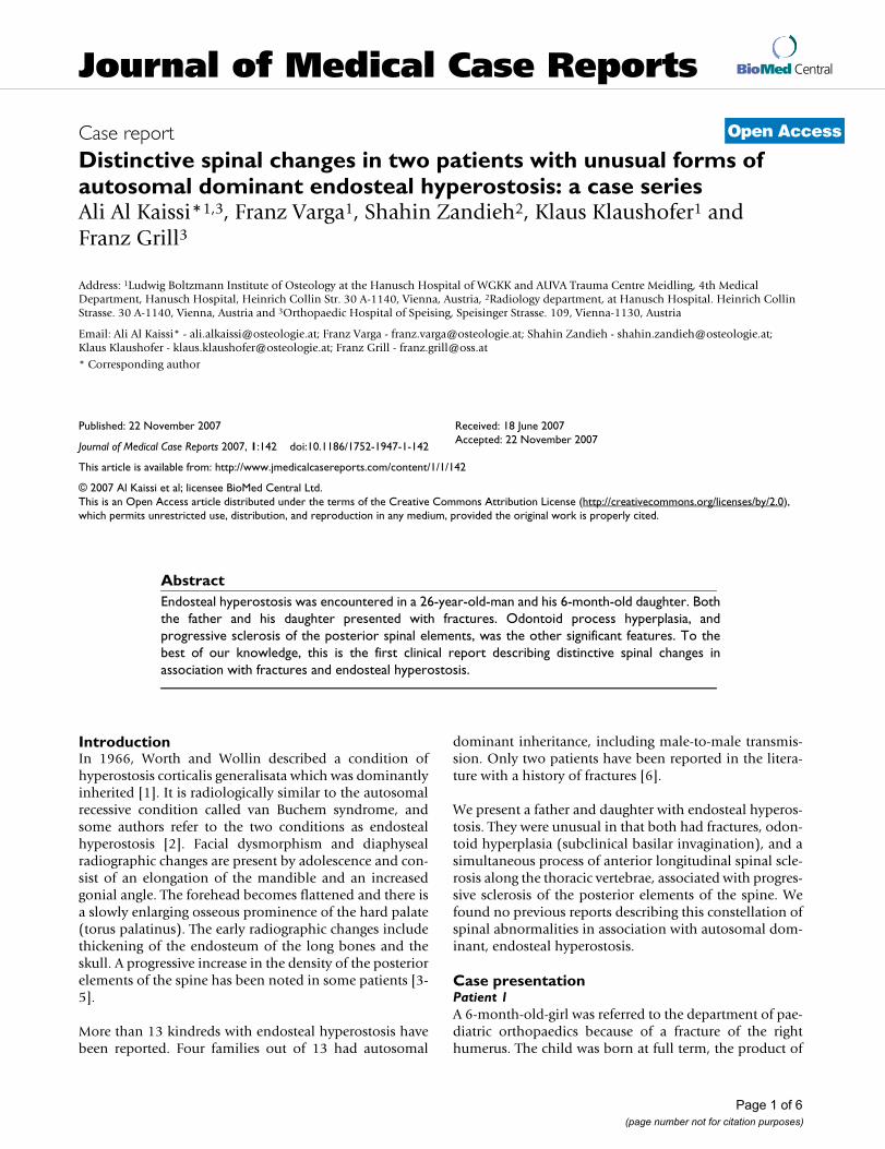
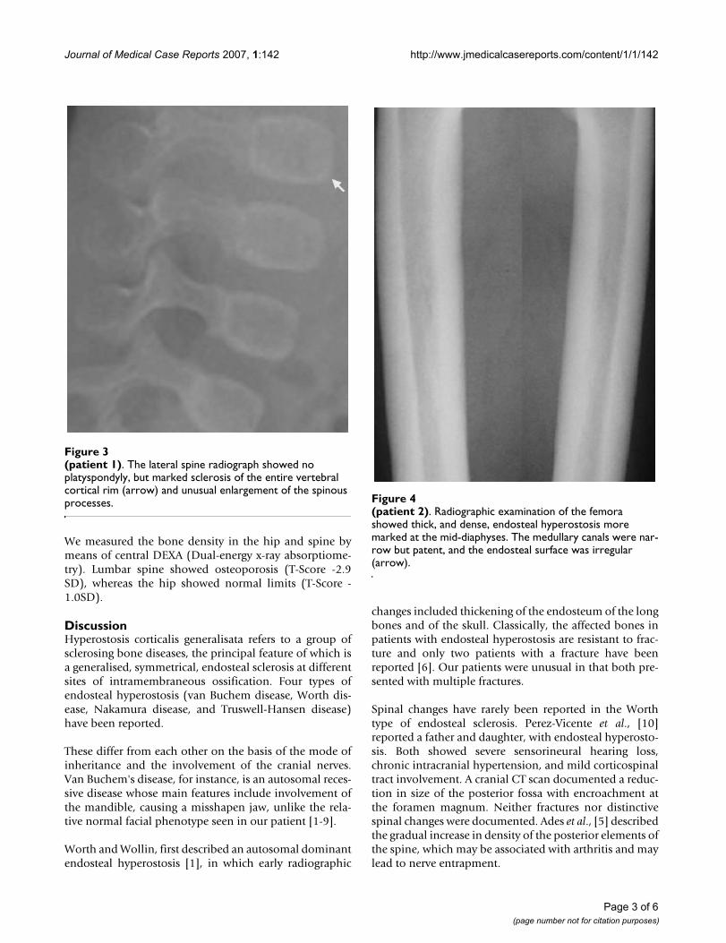
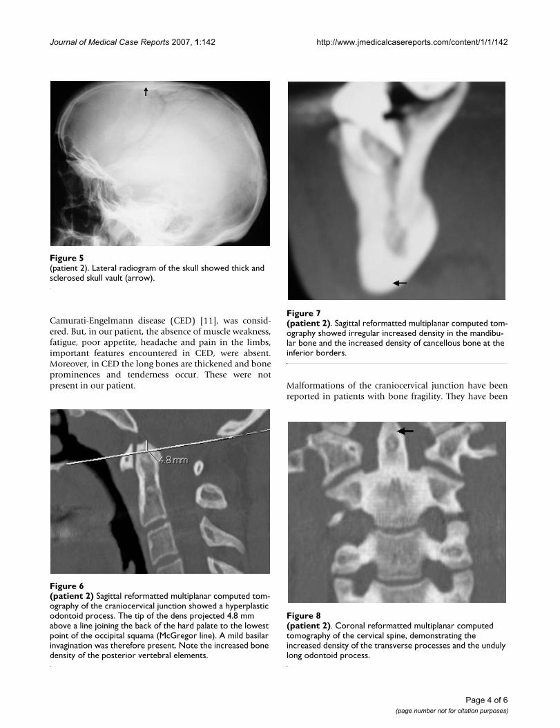
Radiographic examination of the femora showed thick,and dense, endosteal hyperostosis more marked at themid-diaphyses. The medullary canals were narrow butpatent, and the endosteal surface was irregular (figure 4).Lateral radiogram of the skull shows thick and sclerosedskull vault-arrow- (fig 5). Sagittal reformatted multiplanarcomputed tomography of the craniocervical junctionshowed a hyperplastic odontoid process. The tip of thedens projected 4.8 mm above a line joining the back ofthe hard palate to the lowest point of the occipital squama(McGregor line). Subclinical basilar invagination wastherefore present. Note the increased bone density of theposterior vertebral elements (figure 6).
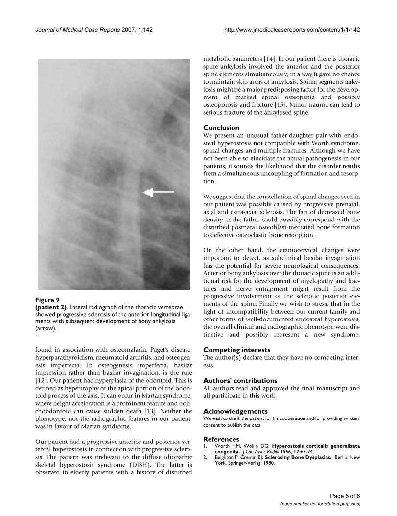
Sagittal reformatted multiplanar computed tomographydemonstrated irregular increased density in the mandibu-lar bone and the increased density of cancellous bone atthe inferior borders (figure 7-arrow). Coronal reformattedmultiplanar computed tomography of the cervical spinedemonstrating the increased density of the transverseprocessed and the unduly long odontoid process-arrow-(fig 8). Lateral radiograph of the thoracic spine showedprogressive sclerosis of the anterior longitudinal ligamentwith subsequent development of bony ankylosis (arrow-fig 9).
(patient 1)Figure 2(patient 1). An anteroposterior radiograph of the right humerus showed mid-diaphyseal fracture and unusual diaphy-seal endosteal hyperostosis (arrow).
(patient1)Figure 1(patient1). A lateral radiogram showed extensive flattening across the posterior skull and marked vault and convolu-tional sclerosis.
Page 2 of 6(page number not for citation purposes)
Journal of Medical Case Reports 2007, 1:142 http://www.jmedicalcasereports.com/content/1/1/142
We measured the bone density in the hip and spine bymeans of central DEXA (Dual-energy x-ray absorptiome-try). Lumbar spine showed osteoporosis (T-Score -2.9SD), whereas the hip showed normal limits (T-Score -1.0SD).
DiscussionHyperostosis corticalis generalisata refers to a group ofsclerosing bone diseases, the principal feature of which isa generalised, symmetrical, endosteal sclerosis at differentsites of intramembraneous ossification. Four types ofendosteal hyperostosis (van Buchem disease, Worth dis-ease, Nakamura disease, and Truswell-Hansen disease)have been reported.
These differ from each other on the basis of the mode ofinheritance and the involvement of the cranial nerves.Van Buchem's disease, for instance, is an autosomal reces-sive disease whose main features include involvement ofthe mandible, causing a misshapen jaw, unlike the rela-tive normal facial phenotype seen in our patient [1-9].
Worth and Wollin, first described an autosomal dominantendosteal hyperostosis [1], in which early radiographic
changes included thickening of the endosteum of the longbones and of the skull. Classically, the affected bones inpatients with endosteal hyperostosis are resistant to frac-ture and only two patients with a fracture have beenreported [6]. Our patients were unusual in that both pre-sented with multiple fractures.
Spinal changes have rarely been reported in the Worthtype of endosteal sclerosis. Perez-Vicente et al., [10]reported a father and daughter, with endosteal hyperosto-sis. Both showed severe sensorineural hearing loss,chronic intracranial hypertension, and mild corticospinaltract involvement. A cranial CT scan documented a reduc-tion in size of the posterior fossa with encroachment atthe foramen magnum. Neither fractures nor distinctivespinal changes were documented. Ades et al., [5] describedthe gradual increase in density of the posterior elements ofthe spine, which may be associated with arthritis and maylead to nerve entrapment.
(patient 2)Figure 4(patient 2). Radiographic examination of the femora showed thick, and dense, endosteal hyperostosis more marked at the mid-diaphyses. The medullary canals were nar-row but patent, and the endosteal surface was irregular (arrow).
(patient 1)Figure 3(patient 1). The lateral spine radiograph showed no platyspondyly, but marked sclerosis of the entire vertebral cortical rim (arrow) and unusual enlargement of the spinous processes.
Page 3 of 6(page number not for citation purposes)
Journal of Medical Case Reports 2007, 1:142 http://www.jmedicalcasereports.com/content/1/1/142
Camurati-Engelmann disease (CED) [11], was consid-ered. But, in our patient, the absence of muscle weakness,fatigue, poor appetite, headache and pain in the limbs,important features encountered in CED, were absent.Moreover, in CED the long bones are thickened and boneprominences and tenderness occur. These were notpresent in our patient. Malformations of the craniocervical junction have been
reported in patients with bone fragility. They have been
(patient 2)Figure 8(patient 2). Coronal reformatted multiplanar computed tomography of the cervical spine, demonstrating the increased density of the transverse processes and the unduly long odontoid process.
(patient 2) Sagittal reformatted multiplanar computed tom-ography of the craniocervical junction showed a hyperplastic odontoid processFigure 6(patient 2) Sagittal reformatted multiplanar computed tom-ography of the craniocervical junction showed a hyperplastic odontoid process. The tip of the dens projected 4.8 mm above a line joining the back of the hard palate to the lowest point of the occipital squama (McGregor line). A mild basilar invagination was therefore present. Note the increased bone density of the posterior vertebral elements.
(patient 2)Figure 5(patient 2). Lateral radiogram of the skull showed thick and sclerosed skull vault (arrow).
(patient 2)Figure 7(patient 2). Sagittal reformatted multiplanar computed tom-ography showed irregular increased density in the mandibu-lar bone and the increased density of cancellous bone at the inferior borders.
Page 4 of 6(page number not for citation purposes)
Journal of Medical Case Reports 2007, 1:142 http://www.jmedicalcasereports.com/content/1/1/142
found in association with osteomalacia, Paget's disease,hyperparathyroidism, rheumatoid arthritis, and osteogen-esis imperfecta. In osteogenesis imperfecta, basilarimpression rather than basilar invagination, is the rule[12]. Our patient had hyperplasia of the odontoid. This isdefined as hypertrophy of the apical portion of the odon-toid process of the axis. It can occur in Marfan syndrome,where height acceleration is a prominent feature and doli-choodontoid can cause sudden death [13]. Neither thephenotype, nor the radiographic features in our patient,was in favour of Marfan syndrome.
Our patient had a progressive anterior and posterior ver-tebral hyperostosis in connection with progressive sclero-sis. The pattern was irrelevant to the diffuse idiopathicskeletal hyperostosis syndrome (DISH). The latter isobserved in elderly patients with a history of disturbed
metabolic parameters [14]. In our patient there is thoracicspine ankylosis involved the anterior and the posteriorspine elements simultaneously; in a way it gave no chanceto maintain skip areas of ankylosis. Spinal segments anky-losis might be a major predisposing factor for the develop-ment of marked spinal osteopenia and possiblyosteoporosis and fracture [15]. Minor trauma can lead toserious fracture of the ankylosed spine.
ConclusionWe present an unusual father-daughter pair with endo-steal hyperostosis not compatible with Worth syndrome,spinal changes and multiple fractures. Although we havenot been able to elucidate the actual pathogenesis in ourpatients, it sounds the likelihood that the disorder resultsfrom a simultaneous uncoupling of formation and resorp-tion.
We suggest that the constellation of spinal changes seen inour patient was possibly caused by progressive prenatal,axial and extra-axial sclerosis. The fact of decreased bonedensity in the father could possibly correspond with thedisturbed postnatal osteoblast-mediated bone formationto defective osteoclastic bone resorption.
On the other hand, the craniocervical changes wereimportant to detect, as subclinical basilar invaginationhas the potential for severe neurological consequences.Anterior bony ankylosis over the thoracic spine is an addi-tional risk for the development of myelopathy and frac-tures and nerve entrapment might result from theprogressive involvement of the sclerotic posterior ele-ments of the spine. Finally we wish to stress, that in thelight of incompatibility between our current family andother forms of well-documented endosteal hyperostosis,the overall clinical and radiographic phenotype were dis-tinctive and possibly represent a new syndrome.
Competing interestsThe author(s) declare that they have no competing inter-ests.
Authors' contributionsAll authors read and approved the final manuscript andall participate in this work
AcknowledgementsWe wish to thank the patient for his cooperation and for providing written consent to publish the data.
References1. Worth HM, Wollin DG: Hyperostosis corticalis generalisata
congenita. J Can Assoc Radiol 1966, 17:67-74.2. Beighton P, Cremin BJ: Sclerosing Bone Dysplasias. Berlin, New
York, Springer-Verlag; 1980.
(patient 2)Figure 9(patient 2). Lateral radiograph of the thoracic vertebrae showed progressive sclerosis of the anterior longitudinal liga-ments with subsequent development of bony ankylosis (arrow).
Page 5 of 6(page number not for citation purposes)
Journal of Medical Case Reports 2007, 1:142 http://www.jmedicalcasereports.com/content/1/1/142
Publish with BioMed Central and every scientist can read your work free of charge
"BioMed Central will be the most significant development for disseminating the results of biomedical research in our lifetime."
Sir Paul Nurse, Cancer Research UK
Your research papers will be:
available free of charge to the entire biomedical community
peer reviewed and published immediately upon acceptance
cited in PubMed and archived on PubMed Central
yours — you keep the copyright
Submit your manuscript here:http://www.biomedcentral.com/info/publishing_adv.asp
BioMedcentral
3. Maroteaux P, Fontaine G, Scharfman W, Farriaux JP: L'hyperostosecorticale generalisee a transmission dominante (typeWorth). Arch Fr Pediatr 1971, 28:685-698.
4. Beals RK, McLoughlin SW, Teed RL, McDonald C: Dominant endo-steal hyperostosis. Skeletal characteristics and review of theliterature. J Bone Joint Surg Am 2001, 83-A(11):1643-9.
5. Ades LC, Morris LL, Burns R, Haan EA: Neurological involvementin Worth type endosteal hyperostosis: report of a family. AmJ Med Genet 1994, 51:46-50.
6. Irie T, Takahashi M, Kaneko M: Case report 546: Endostealhyperostosis (Worth type). Skeletal Radiol 1989, 18:310-3.
7. Greenspan A: Sclerosing bone dysplasias: a target-siteapproach. Skeletal Radiol 1991, 20:561-583.
8. Van Buchem FSP, Hadders HN, Ubbens R: Hyperostosis corticalisgeneralisata familiaris. Acta Radiol (Diagn) 1955, 44:109-114.
9. Beals RK: Endosteal hyperostosis. J Bone Joint Surg Am 1976,58:1172-3.
10. Perez-Vicente JA, Rodriguez De Castro E, Lafuente J, Mateo MMD,Gimenez-Roldan S: Autosomal dominant endosteal hyperosto-sis: report of a Spanish family with neurological involvement.Clin Genet 1987, 31:161-169.
11. Camurati M: Di uno raro caso di osteite simmetrica ereditariadegli arti inferiori. Chir Organi Mov 1922, 6:662-665.
12. Hayes M, Parker G, Ell J, Sillence D: Basilar impression complicat-ing osteogenesis imperfecta type IV: the clinical and neuro-logical findings in four cases. J Neurosurg Psychiatry 1999,66:357-346.
13. MacKenzie JM, Rankin R: Sudden death due to atlantoaxial sub-luxation in marfan syndrome. Am J Forensic Med Pathol 2003,24(4):369-70.
14. Forestier J, Lagier R: Ankylosing hyperostosis of the spine. ClinOrthop 1971, 74:65-83.
15. Hendrix R, Melany M, Miller F, Rogers L: Fracture of the spine inpatients with Ankylosis: Clinical and Imaging Findings. Am JRadiol 1994, 162:899-904.
Page 6 of 6(page number not for citation purposes)