Embed Size (px)
Citation preview

Journal of Invertebrate Pathology 144 (2017) 65–73
Contents lists available at ScienceDirect
Journal of Invertebrate Pathology
journal homepage: www.elsevier .com/ locate/ j ip
Characterization of a novel member of genus Iflavirus in Helicoverpaarmigera
http://dx.doi.org/10.1016/j.jip.2017.01.0110022-2011/� 2017 Elsevier Inc. All rights reserved.
⇑ Corresponding author.E-mail address: [email protected] (K. Wu).
1 These authors contributed equally to this work.
He Yuan a,1, Pengjun Xu a,b,1, Xianming Yang a, Robert I. Graham c, Kenneth Wilson d, Kongming Wu a,⇑a State Key Laboratory for Biology of Plant Diseases and Insect Pests, Institute of Plant Protection, Chinese Academy of Agricultural Sciences, Beijing 100193, PR Chinab Tobacco Research Institute, Chinese Academy of Agricultural Sciences, Qingdao, PR ChinacCrop and Environment Sciences, Harper Adams University, Edgmond, Shropshire TF10 8NB, UKd Lancaster Environment Centre, Lancaster University, Lancaster LA1 4YQ, UK
a r t i c l e i n f o a b s t r a c t
Article history:Received 1 September 2016Revised 16 January 2017Accepted 19 January 2017Available online 3 February 2017
Keywords:Helicoverpa armigeraIflavirusRNA virusHorizontal transmissionVertical transmissionTissue distribution
The cotton bollworm, Helicoverpa armigera, is one of the most important agricultural pests of many eco-nomic crops worldwide. Herein, we found a novel single-strand RNA virus by RNA-Seq and PolymeraseChain Reaction (PCR) method in H. armigera named Helicoverpa armigera iflavirus (HaIV), which possesseda genome with 10,017 nucleotides in length and contained a single large open reading frame (ORF)encoding a putative polyprotein of 3021 amino acids with a predicted molecular mass of 344.16 kDaand a theoretical isoelectric point (pI) of 6.45. The deduced amino acid sequence showed highest similar-ity (61.0%) with the protein of Lymantria dispar Iflavirus 1. Phylogenetic analysis with putative RdRpamino acid sequences indicated that the virus clustered with members of the genus Iflavirus. The viruswas mainly distributed in the fat body of its host and was found to be capable of both horizontal and ver-tical transmission. The efficiency of perorally horizontal transmission was dose dependent (100% infec-tion rate with a viral dose of 108 copies/ll) while vertical transmission efficiency was found to berelatively low (<28.57%). These results suggest that we have found a novel member of genus Iflavirusin H. armigera.
� 2017 Elsevier Inc. All rights reserved.
1. Introduction
Insects are susceptible to a variety of pathogens, which canresult in chronic or lethal infections (Burden et al., 2003). Histori-cally, viruses have been isolated and subsequently studied after theobservation of overt disease symptoms in the host. However, thereare also covert infections found within hosts displaying no obvioussigns of disease, which could remain undetected (Burden et al.,2003; Kemp et al., 2011; Murillo et al., 2011). These covert persis-tent viral infections, such as baculoviruses, are almost ubiquitousin many lepidopteran insect species and their discovery is drivingfurther research into the dynamics and behavior of covert infec-tions and their role in the ecology of host populations, especiallythose of economic and agricultural importance (Graham et al.,2015).
The cotton bollworm, Helicoverpa armigera, is one of the mostimportant agricultural pests of cotton and other important eco-nomic crops worldwide. The adult moth is highly migratory, and
populations have been reported in Australia, Asia, Africa, Europe(Feng et al., 2007; Wu et al., 1997) and most recently from SouthAmerica (Tay et al., 2013). Since the introduction of Bt-cotton intoChina in the 1990s, the H. armigera population has declined dra-matically. However, several studies have reported that Bt-resistance has evolved in the field (Gunning et al., 2005; Zhanget al., 2012). Thus, other forms of biological pest control, includingthe use of host-specific viral pesticides, derived from baculovirus(Allaway and Payne, 1984; Chen et al., 2001; Fuxa, 2004; Sunet al., 2002, 2004), small RNA viruses (Christian et al., 2005) anddensoviruses (El-Far et al., 2012), has attracted more attentionfrom researchers. Moreover, high-through-put analytical methodssuch as metagenomics and RNA sequencing provide sensitive andeffective methods for the discovery of novel viruses and asymp-tomatic disease agents that may be useful as biological controlproducts (Diatchenko et al., 1996; Ge et al., 2012; Marguerat andBähler, 2009; Mokili et al., 2012; Radford et al., 2012; Roossincket al., 2015), or conversely, may negatively or positively impactupon the biopesticide products being used. For example, the recentdiscovery of a novel densovirus (HaDV2) from healthy migratorycotton bollworms revealed that HaDV2 infection significantlyincreased host resistance to the host-specific baculovirus HaNPV
66 H. Yuan et al. / Journal of Invertebrate Pathology 144 (2017) 65–73
and to the generalist biopesticide Bt toxin (Xu et al., 2014).Thedevelopment of molecular tools and next generation sequencingtechnology paves the way for a greater understanding as to howwe may manipulate the host-pathogen system, with the aim ofreducing pest outbreaks and economic crop damage.
In this study, a novel virus infecting H. armigera named Helicov-erpa armigera iflavirus (HaIV), was discovered by RNA-Seq. Align-ment and phylogenetic analysis revealed that the virus showed ahigh sequence identity with members of the Iflavirus, which isthe only genus within the family Iflaviridae. Members of this genuspossess a single positive-strand RNA genome and share severalcommon features, including: (1) non-enveloped icosahedral parti-cles measuring 30 nm in diameter; (2) genome translation into apolyprotein; (3) the viral coat proteins containing three jelly-rolldomains; (4) a three-domain containing a superfamily III helicase,a (cysteine) proteinase with a chymotrypsin-like fold and an RNA-dependent RNA polymerase (RdRp) (Le Gall et al., 2008). The gen-ome of Iflavirus is monocistronic with one single large open readingframe (ORF) encoding a single large polyprotein. To date, only ninespecies of iflaviruses have been recognized by The InternationalCommittee on Taxonomy of Viruses (ICTV), including deformedwing virus (Lanzi et al., 2006), Ectropis obliqua virus (Wang et al.,2004), Infectious flacherie virus (Isawa et al., 1998), Lygus lineolarisvirus 1 (Perera et al., 2012), Nilaparvata lugens honeydew virus 1(Murakami et al., 2013), Perina nuda virus (Wu et al., 2002), Sac-brood virus (Ghosh et al., 1999), Slow bee paralysis virus (deMiranda et al., 2010), and Varroa destructor virus 1 (Ongus et al.,2004), although other iflaviruses have been reported (Silva et al.,2015; Suzuki et al., 2015). Herein, we report the nucleotidesequence, genome organization, phylogeny, transmission andtissue distribution of HaIV.
2. Materials and methods
2.1. Insect culture
A laboratory colony of H. armigera was originally captured in2005 from Langfang (Hebei province, China). H. armigera larvaewere reared on an artificial diet (Liang et al., 2008) and adult mothswere cultured with a 10% sugar and 2% vitamin mix (Liang et al.,1999) at 25 ± 1 �C with a 14:10, light: dark photoperiod.
2.2. Transcriptome analysis and annotation
For transcriptome analysis, Illumina RNA-sequencing was con-ducted by Novogene (Beijing, China). Four 5th instar larvae (oneday post-ecdysis) were individually collected and total RNA iso-lated using the TRIzol kit (Invitrogen, Carlsbad, CA, USA), accordingto the manufacturer’s instructions. RNA samples were dissolved inRNase-free water and used to construct the cDNA library of H.armigera with suitable fragments (about 200 bp). = Paired-endtranscriptome sequencing was subsequently performed using anIllumina HiSeqTM 2000. Adaptor sequences and low-quality readswere trimmed and clean reads were used for de novo assemblyusing Trinity (Grabherr et al., 2011). The assembled contigs wereannotated using BLASTx and BLASTn against the NCBI non-redundant nucleic acid database (NT) and the NCBI non-redundant protein database (NR), using a cut-off E-value of 10�5.
2.3. Cloning the iflavirus genome of H. armigera
Total RNA was extracted from individual adult moths reared inlaboratory using the TRIzol reagent kit as described above. Single-stranded cDNA was synthesized using the FastQuant RT Kit (Tian-gen, Beijing, China), according to the manufacturer’s instructions.
Based on the assembled sequence from RNA-seq, nine pairs of pri-mers were subsequently designed (Table S1). The genome of theiflavirus isolated from H. armigera was amplified and sequencedusing cDNA as template, using the following PCR program: 4 minat 94 �C; 30 s at 94 �C, 30 s at 55 �C, and 2 min at 72 �C for 40cycles. The PCR product was purified, inserted into the pEASY-Tcloning vector (TransGen, Beijing, China), and sequenced.
2.4. Sequence and phylogenetic analysis
The open reading frame (ORF) of the viral genome waspredicted using ORF finder at NCBI (http://www.ncbi.nlm.nih.gov/gorf/gorf.html). The amino acid sequences encoding non-structural proteins were compared with members of the orderPicornavirales using Clustal W (Thompson et al., 1994). The com-plete nucleotide sequence of the virus described in this studywas submitted to GenBank under accession number KX228231.The deduced RdRp amino acid sequence of the new virus, togetherwith that of members of the family Iflaviridae, was used in phylo-genetic analysis. Acyrthosiphon pisum virus and Spodoptera exiguavirus AKJ-2014were used as outgroup. The phylogenetic trees wereconstructed using the maximum likelihood method with a boot-strap of 1000 replicates in MEGA6 (Tamura et al., 2013). Gaps wereregarded as a complete deletion unless specifically noted.
2.5. Virus detection and quantification
A partial sequence exhibiting high similarities with known ifla-viruses was identified from the RNA-seq data. For the detection ofthe novel virus, a pair of specific primers, VPF1/VPR1 (Table S1)were designed to amplify a PCR product of 593 bp, according tothe assembled sequence of the virus in H. armigera. The PCR pro-gram used was as follows: 30 s at 94 �C, 30 s at 57 �C, and 30 s at72 �C for 40 cycles. To assess the detection threshold of the virusassay, a 10-fold dilution series of cDNA (3.37 � 109 to 3.37 � 101 -copies/ll) containing the virus was made and tested using theVPF1/VPR1 primers. 10 ll of each PCR product was analyzed byagarose gel electrophoresis.
Copy numbers of the novel virus were quantified using a stan-dard curve by an absolute quantification qPCR method (Wongand Medrano, 2005). For quantification, the primers and probesof the virus (VPF/VPR, Table S1) were designed. A fragment wasamplified using the primers and cloned into the pEASY-T CloningVector (TransGen, Beijing, China) and sequenced. The PCR programwas as follows: 30 s at 94 �C, 30 s at 54 �C, and 30 s at 72 �C for 40cycles. The plasmid was used for the construction of the standardcurve. Virus quantification was conducted with TaqMan in 20 mlreaction agent, which comprised 1 ml of template DNA,2 � SuperReal PreMix (Probe, Tiangen, Beijing, China), 50 � ROXReference Dye, 0.2 mM of each primer and 0.4 mM probe. The ther-mal cycling conditions on a 7500 Real-time PCR System (AppliedBiosystems) were as follows: 40 cycles of 95 �C for 3 s, 60 �C for30 s. The standard curve equation of y = �0.9990x + 41.6662(y = the logarithm of plasmid copy number to base 2, x = Ct value,R2 = 0.9998) was used to calculate the copy number of the virus(Fig. S1).
2.6. Transmission of the virus
Filtered liquid containing an unpurified form of the iflaviruswas prepared (Xu et al., 2014). RNA was isolated and the concen-tration of the virus was quantified. A NONINF strain was estab-lished from a single breeding pair that was not infected with theiflavirus, according to the method described by Xu et al. (2014).An infected line (INF strain) of H. armigera was established byorally infecting NONINF strain larvae with the filtered liquid.

H. Yuan et al. / Journal of Invertebrate Pathology 144 (2017) 65–73 67
Fifth-instar larvae were randomly selected to detect the infectionrate of the virus.
For quantification of vertical transmission rates, four pair-types($+/#+, $+/#�, $�/#+, and $�/#�) were mated. Positive and nega-tive individuals were from INF and NONINF culture strains respec-tively. The infection rate of the virus in offspring was detected from3rd instar offspring larvae.
For quantification of horizontal transmission rates, non-infectedNONINF strain neonates were provided artificial diet with differentconcentrations of the virus: 108, 107, 106, 105, 104, 0 (virus-freecontrol) copies/ll for 2 days, then transferred to a 24-well plateand reared in glass tubes until eclosion. Newly eclosed adult mothswere sampled to determine the infection rate of the virus underdifferent titer regimes. This diet contamination assay (mimickinga similar mechanism in wild populations) was conducted accord-ing to the method described by Xu et al. (2014). Infection ratewas detected by PCR and larval frass from the INF strain was usedto quantify the virus copy number by qPCR.
2.7. Quantification of the virus in eggs and different tissues
To test whether vertical transmission was transovum or transo-varial, we quantified the copy number of the virus in three groups ofboth non-treated (control) and sodium hypochlorite treated eggs(n = 50 eggs per group) ofH. armigera. The eggswere from INF strainbreeding pairs, that is both females and males were infected withthe virus and treated according to the method described by Xuet al. (2014). Third instar larvae originating from these eggs werealso sampled to quantify the copy number of the virus.
To examine the virus infection in different body tissues of thehost, infected fifth-instar larvae were dissected into foregut, mid-gut, hindgut, hemolymph, malpighian tubules and fat body, usinga fresh scalpel for every cut to avoid virus cross contamination.In addition, infected females and males were dissected into brain,muscle, wing, malpighian tubule, fat body, ovary/testis and gut asdescribed above. Total RNA was extracted from the different bodyparts (both larval and adult stages) and cDNA was used as a tem-plate to quantify the copy numbers of the virus by qPCR. The cDNAsample of each body part was replicated three times. The copynumber of the virus in each tissue was calculated, and these weresummed to determine total copy numbers of the virus in each indi-vidual. The percentage of virus in each tissue in the same individ-ual was statistically analyzed (larvae: n = 9; adult males: n = 6;adult females: n = 9) (Xu et al., 2014).
To examine virus replication in different developmental stagesof the host, larvae 24 h to 240 h post-infection (1st-5th instar)and newly eclosed adults were sampled. Absolute quantificationqPCR and the comparative 2�44Ct method (Livak and Schmittgen,2001) were used, respectively. b-actin (GenBank EU527017) wasused as the reference gene to normalize the virus expression. Thereaction was conducted in 20 ll reaction mixtures, containing10 ll of SuperReal PreMix Plus (TIANGEN, Beijing, China), 0.6 llof primers (10 lM), 1 ll of sample cDNA, 0.5 ll of Rox ReferenceDye and 8.3 ll of RNase-free ddH2O. The cycling parameters were:95 �C for 15 min, followed by 40 cycles of 95 �C for 10 s and 62 �Cfor 32 s. To ensure reproducibility, each sample was carried out inthree biological replicates and in three technical replicates.
2.8. Electron microscopy
Adult moths from the INF culture strain were collected. Thenthe virus were isolated and purified using the method of SucroseDensity Gradient Centrifugation according to the methoddescribed by La Fauce et al. (2007). Purified particles were nega-tively stained with 2% sodium phosphotungstate at pH 6.8 andobserved with a transmission electron microscope.
3. Results
3.1. Transcriptome analysis and annotation
Using RNA-Seq we sequenced the transcriptome ofH. armigera, obtaining about 5 gigabases (in-depth) for each sam-ple, and a total of 106,785 assembled contigs. Using BLASTx andBLASTn, 45,609 genes (42.71% of transcripts) and 34,383 genes(32.20% of transcripts), respectively, were obtained throughBLAST hits, using the E-value cutoff. Because of the relativelyshort length of transcripts (mean size of 758.65 bp) and lack ofgenome reference, most of the assembled sequences could notbe matched to any known genes. The E-value distribution of thebest hit in the nr database showed that 57% of the mappedsequences had strong homology (smaller than 1.0E�50), whereas43% of the homologous sequences ranged from 1.0E�5 to1.0E�50 (Fig. S2A). Homologous genes came from several species,with 66% of the unigenes having the highest homology to genesfrom Danaus plexippus, followed by Bombyx mori (5%), Triboliumcastaneum (3%), and Helicoverpa armigera (2%) (Fig. S2B). TheRNA-seq original datasets generated in this study are availablein the NCBI GEO database (accession number: GSE86914)(https://www.ncbi.nlm.nih.gov/geo/).
3.2. The genome sequence of HaIV
The assembled contig of 1495 nucleotides (nt) in length from103,935 reads encoded actin of H. armigera, and one contig of10,008 nt in length assembled using 41,719 reads showed highidentity with Lymantria dispar iflavirus 1. According to the refer-ence sequence, we designed specific primers to amplify the viralgenome sequence of HaIV containing the whole ORF, which was10,017 nt in length, containing a single large ORF (between nt733 and 9798) encoding a polyprotein of 3021 amino acids. Ithas a predicted molecular mass of 344.16 kDa and a theoretical iso-electric point (pI) of 6.45. The coding sequence was flanked by a732 bp 50 untranslated region (UTR) and a 219 bp 30 UTR. Thenucleotide base composition of the genome was 30.6%A, 35.6%U,13.6%C and 20.2%G. The total A + U and G + C content were 66.2%and 33.8% respectively. Alignment analysis with putative aminoacid sequences showed high identities with the members of genusIflavirus, in which the highest identity was 61.0% with L. dispar ifla-virus 1 (Table S2). It contained all three conserved domains, includ-ing the three conserved domains in helicase sequences (Fig. 1A),the GXCG and GXHXXG conserved motifs in the proteasesequences (Fig. 1B) and the eight conserved domains in the RdRpamino acid sequences (Fig. 1C). The conservation of HaIV polypro-tein and the various segments were compared with those ofHeliconius erato virus (HeIV) (Smith et al., 2014) and the resultsare summarized in Table 1.
3.3. Phylogenetic analysis
Neighbor-joining trees with Poisson model were constructedfor the putative RdRp amino acid sequences (Fig. 2) of the familyIflaviridae using Acyrthosiphon pisum virus and Spodoptera exiguavirus AKJ-2014 as the outgroup. The result indicated that the HaIVclustered with members of the genus Iflavirus.
3.4. Sensitivity of detection
An amplification product could be visualized by ethidium bro-mide staining when as little as 3.37 � 103 copies/ll of HaIV incDNA were used as template (Fig. S3).

Fig. 1. Protein domain alignments. Shown are alignments of the helicase (A), protease (B) and RdRp sequences (C) domains of the novel virus (HaIV) with those of the otherIflaviridae (ApIV, DWV, EoV, HeIV, IFV, LyLV-1, PnV, SBV and VDV-1), some Dicistroviridae (ABPV, BQCV, CrPV and DCV) and one Picornaviridae (EMCV). Conserved regionscorresponding to those recognized by Koonin et al. (1993) are indicated by bars above the protein alignment. Black shading indicates 100% sequence identity and otherresidues that are also conserved among these sequences are shaded in gray. The full names and the accession numbers of the virus are given in Table S3.
68 H. Yuan et al. / Journal of Invertebrate Pathology 144 (2017) 65–73
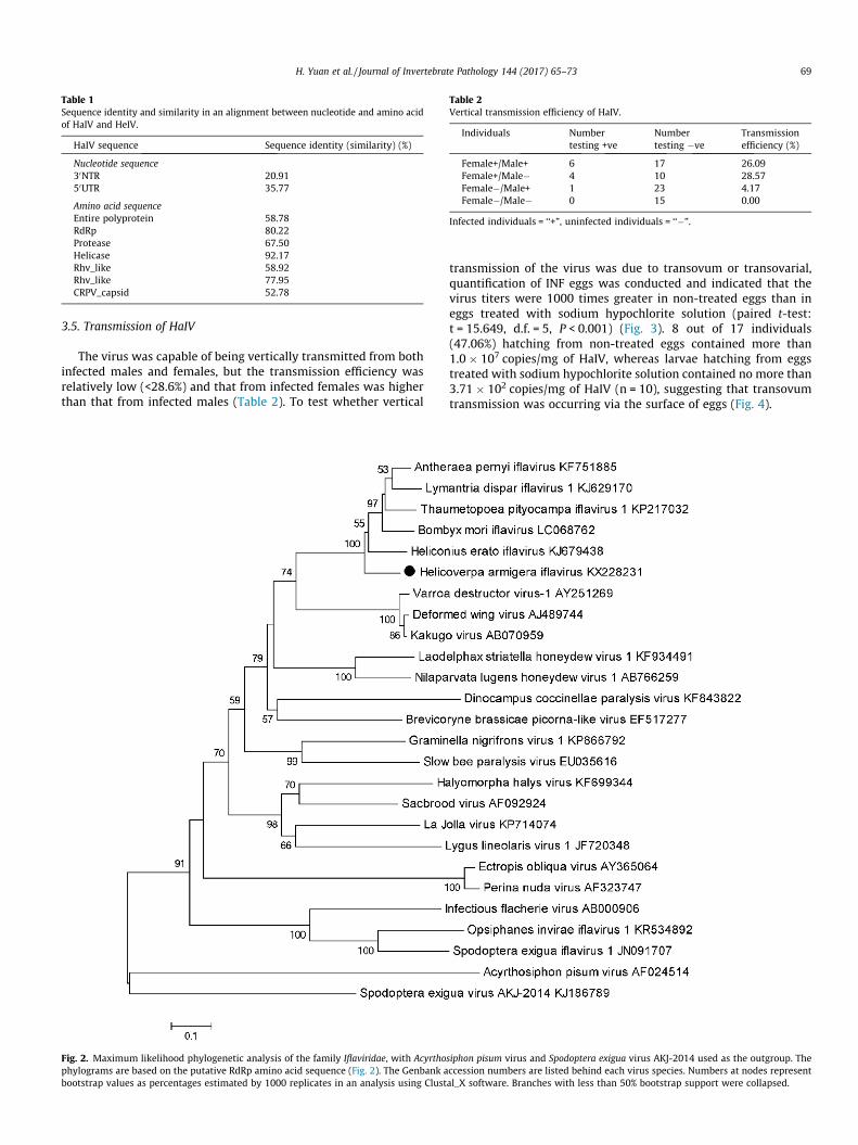
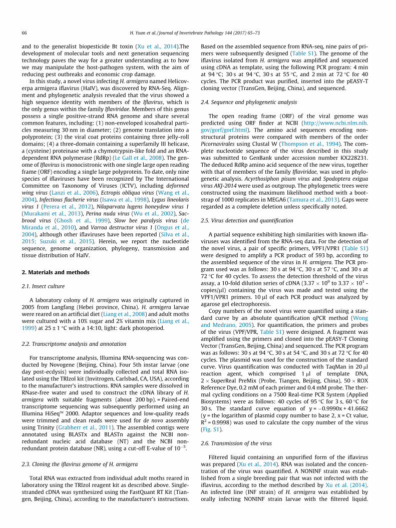
Table 1Sequence identity and similarity in an alignment between nucleotide and amino acidof HaIV and HeIV.
HaIV sequence Sequence identity (similarity) (%)
Nucleotide sequence30NTR 20.9150UTR 35.77
Amino acid sequenceEntire polyprotein 58.78RdRp 80.22Protease 67.50Helicase 92.17Rhv_like 58.92Rhv_like 77.95CRPV_capsid 52.78
Table 2Vertical transmission efficiency of HaIV.
Individuals Numbertesting +ve
Numbertesting �ve
Transmissionefficiency (%)
Female+/Male+ 6 17 26.09Female+/Male� 4 10 28.57Female�/Male+ 1 23 4.17Female�/Male� 0 15 0.00
Infected individuals = ‘‘+”, uninfected individuals = ‘‘�”.
H. Yuan et al. / Journal of Invertebrate Pathology 144 (2017) 65–73 69
3.5. Transmission of HaIV
The virus was capable of being vertically transmitted from bothinfected males and females, but the transmission efficiency wasrelatively low (<28.6%) and that from infected females was higherthan that from infected males (Table 2). To test whether vertical
Fig. 2. Maximum likelihood phylogenetic analysis of the family Iflaviridae, with Acyrthophylograms are based on the putative RdRp amino acid sequence (Fig. 2). The Genbank abootstrap values as percentages estimated by 1000 replicates in an analysis using Clust
transmission of the virus was due to transovum or transovarial,quantification of INF eggs was conducted and indicated that thevirus titers were 1000 times greater in non-treated eggs than ineggs treated with sodium hypochlorite solution (paired t-test:t = 15.649, d.f. = 5, P < 0.001) (Fig. 3). 8 out of 17 individuals(47.06%) hatching from non-treated eggs contained more than1.0 � 107 copies/mg of HaIV, whereas larvae hatching from eggstreated with sodium hypochlorite solution contained no more than3.71 � 102 copies/mg of HaIV (n = 10), suggesting that transovumtransmission was occurring via the surface of eggs (Fig. 4).
siphon pisum virus and Spodoptera exigua virus AKJ-2014 used as the outgroup. Theccession numbers are listed behind each virus species. Numbers at nodes represental_X software. Branches with less than 50% bootstrap support were collapsed.

Fig. 3. Viral load of HaIV in cotton bollworm eggs. Absolute quantification of HaIVcopy number per lg of host RNA in eggs washed or non-washed in 1% sodiumhypochlorite (n = 6).
Table 3Detection of HaIV infecting larvae dosed at a range of concentrations.
Concentrations (copynumber/ll)
Numbertesting +ve
Numbertesting �ve
Infectionrate (%)
108 11 0 100.00107 10 3 76.92106 9 3 75.00105 8 4 66.67104 4 8 33.330 0 12 0
Infected individuals = ‘‘+ve”, uninfected individuals = ‘‘�ve”.
Fig. 5. Absolute quantification of HaIV copy number per mg of frass of larvae. LF:larval frass.
70 H. Yuan et al. / Journal of Invertebrate Pathology 144 (2017) 65–73
To examine horizontal transmission, NONINF strain neonateswere exposed to different concentrations of virus. The resultsshowed that the infection rate of NONINF strain larvae was dose-dependent, with 100% infection rates at a dose of 108 copies/ll(Table 3). To examine the transmission efficiency through frass oflarvae, we placed NONINF strain neonates in diet cells which hadpreviously housed infected insects (n = 12) and quantified the copynumber of the virus in frass of larvae. The results showed that thefrass contained no more than 106 copies/mg (Fig. 5). However, 75%(9 of 12 samples) of NONINF strain individuals could be infectedvia horizontal transmission by frass (Fig. 6).
Fig. 6. The detection of HaIV in sample used in diet contamination assay. (a) PCRdetection of HaIV in larvae which were reared on contaminated diet for 6 days; (b)PCR detection of HaIV in frass of larvae; (c) b-actin was used as internal referencegene to test the integrity of each cDNA templates.
3.6. Host tissue distribution
The copy number of the virus in different body tissues of H.armigera was quantified by qPCR. In both larvae and adults, thevirus titers in the fat body were significantly higher than in othertissues: larvae: F = 11.32, df = 5,12, P < 0.001 (Fig. 7A); adultfemales: F = 11.57, df = 6,56, P < 0.001 (Fig. 7B); adult males:F = 2.89, df = 6,21, P = 0.033 (Fig. 7C). The virus was also detectedin female ovaries and male testes, but at lower titers than in fatbody.
3.7. Total levels of HaIV
Absolute quantification qPCR results showed that the copynumber of HaIV increased over time, reaching the highest infection
Fig. 4. Viral load of HaIV in larvae hatching from non-treated eggs (C1-
load (1.36 � 107 copies/mg) at 144 h post infection and remainingstable after this time (Fig. 8A). In undertaking the relative quantifi-cation assay, all of the expression levels of HaIV were comparedwith those at 24 h. The expression levels of HaIV increased overtime and reached 10,000 fold at 240 h, with an addition slightincrease in expression during the adult stage (Fig. 8B).
C17) and eggs treated with sodium hypochlorite solution (T1-T10).

Fig. 7. Tissue distribution of the HaIV in (A) larvae, (B) adult females and (C) adult male cotton bollworms. Within each figure, significant differences ascribed using Tukeytests are shown using different letters. Percentage (%) = the ratio of HaIV in different tissues (per mg), as described by Xu et al. (2014) (larvae: n = 9; adult males: n = 6; adultfemales: n = 9). Means ± SE.
Fig. 8. HaIV levels in different developmental stages of host were tested using twomethods. (A) Absolute quantification qPCR method. (B) The comparative 2�44Ct
method.
Fig. 9. Electron microscopy image showing HaIV particles purified from an extractfrom INF strain (100000�).
H. Yuan et al. / Journal of Invertebrate Pathology 144 (2017) 65–73 71
3.8. Virus morphology
The virus particles purified from H. armigera were observed byusing an electron microscope. They had an isometric appearanceand an approximate diameter of 30 nm (Fig. 9).
4. Discussion
Recently, next-generation sequencing technology has provideda rapid approach to high-throughput sequence determinationand allowed a wide diversity of novel viruses to be discovered(Ansorge, 2009; Mokili et al., 2012; Roossinck et al., 2015). Herein,a novel virus named H. armigera iflavirus (HaIV) was detected inlarvae of H. armigera by RNA-seq method. The genome organiza-tion of HaIV has the common features of other members within
the family Iflaviridae. Briefly, the virus is monocistronic, with asingle-stranded RNA genome, and contains a single large ORFencoding both structural and non-structural proteins. It also pos-sesses the functional motifs of RNA helicase, protease and RdRpconserved in members of the order Picornavirales (van Oers,2010). As members of the genus Iflavirus, the genomic sequenceof HaIV is A/U rich (>60% A/U). Based on the genomic nucleotidesequences and the amino acid sequences, phylogenetic analysisindicated HaIV clustered with the members of Iflavirus, suggestingthat HaIV was a novel member of genus Iflavirus (Carrillo-Trippet al., 2014; Silva et al., 2015; van Oers, 2010).
Viruses distribution in host tissues were diverse according todifferent virus species, such as HaDV2, a beneficial densoviruswhich could accelerate the growth rate of its insect host, wasmainly distributed in fat body of H. armigera (Xu et al., 2014) andHaNPV, a lethal baculovirus, viral loads of which were higher inthe head, legs and wings than in the abdomen and thorax(Graham et al., 2015). Previously, some members of Iflavirus don’tappear to be lethal to their hosts, such as Kakugo virus (Fujiyukiet al., 2009) and Brevicoryne brassicae virus (Ryabov, 2007). And,some members of Iflavirus are known to be harmful to their hostsby lead to developmental problems and host mortality (van Oers,2010). For example, sacbrood virus (SBV) is mainly found in larvalfat body cells of the honeybee, which may impact metabolic func-tion of these cells, resulting in failure to pupate and ultimatelycausing death (Bitondi et al., 2006; Park et al., 2016). Interestingly,HaIV was also mainly distributed in the fat body both in larvae andadults while with much lower titers in both the ovary and testis,suggesting a similar functionwith othermembers of genus Iflavirus.

72 H. Yuan et al. / Journal of Invertebrate Pathology 144 (2017) 65–73
As beneficial virus, HaDV2 could be high efficiently transmittedby both the horizontal and vertical mode (Xu et al., 2014). How-ever, the harmful virus (eg. HaNPV) could infect host through highhorizontal transmission rate (86.2% mortality at a dose of 107 OBs/ml) whereas quite low vertical transmission rate (12.6% ± 2.0%)(Zhou et al., 2005). Members of Iflavirus can transmit horizontallyand vertically. For example, DWV could vertically transmit fromqueens to both worker and drone offspring (Yue and Genersch,2005), and horizontally transmit through Varroa mites (Bowen-Walker et al., 1999; Wilfert et al., 2016). Transovarial and horizon-tal transmission of SBV have also been found to occur (Shen et al.,2005). Herein, we also found that HaIV could be both horizontallyand vertically transmitted, which the horizontally transmitted effi-ciency was dose-dependent and the vertically transmitted effi-ciency was quite low (<28.57%). To examine the possibility ofhorizontal transmission on natural conditions, we performed dietcontamination assay and the results suggested that although thedose of HaIV in frass was no more than 106/mg, the infection ratewas 75%. The copy number of HaIV associated with eggs was sig-nificantly decreased by washing with sodium hypochlorite solu-tion, suggesting that transovum transmission was occurring viathe surface of eggs.
In conclusion, we report a novel virus isolated from the hostlepidopteran H. armigera named H. armigera iflavirus. Molecularcharacterization and phylogenetic analysis indicated that HaIVwas a novel member of the genus Iflavirus. HaIV was found to bemainly distributed in the fat body of its host, and could be bothhorizontally and vertically transmitted but with low efficiency,suggesting a harmful factor to its host.
Acknowledgments
This work was supported by Science Fund for Creative ResearchGroups of the National Natural Science Foundation of China (No.31321004) and the National Natural Science Foundation of China(Grant No. 31401752). KW was supported by a Biotechnologyand Biological Sciences Research Council UK-China PartneringAward (ref: BB/L026821/1).
Appendix A. Supplementary material
Supplementary data associated with this article can be found, inthe online version, at http://dx.doi.org/10.1016/j.jip.2017.01.011.
References
Allaway, G.P., Payne, C.C., 1984. Host range and virulence of five baculoviruses fromlepidopterous hosts. Ann. Appl. Biol. 105, 29–37.
Ansorge, W.J., 2009. Next-generation DNA sequencing techniques. N. Biotechnol. 25,195–203.
Bitondi, M.M., Nascimento, A.M., Cunha, A.D., Guidugli, K.R., Nunes, F.M., Simões, Z.L., 2006. Characterization and expression of the Hex 110 gene encoding aglutamine-rich hexamerin in the honey bee, Apis mellifera. Arch. InsectBiochem. 63, 57–72.
Bowen-Walker, P., Martin, S., Gunn, A., 1999. The transmission of deformed wingvirus between honeybees (Apis mellifera L.) by the ectoparasitic miteVarroajacobsoni Oud. J. Invertebr. Pathol. 73, 101–106.
Burden, J.P., Nixon, C.P., Hodgkinson, A.E., Possee, R.D., Sait, S.M., King, L.A., Hails, R.S., 2003. Covert infections as a mechanism for long-term persistence ofbaculoviruses. Ecol. Lett. 6, 524–531.
Carrillo-Tripp, J., Krueger, E.N., Harrison, R.L., Toth, A.L., Miller, W.A., Bonning, B.C.,2014. Lymantria dispar iflavirus 1 (LdIV1), a new model to study iflaviralpersistence in lepidopterans. J. Gen. Virol. 95, 2285–2296.
Chen, X., IJkel, W.F., Tarchini, R., Sun, X., Sandbrink, H., Wang, H., Peters, S., Zuidema,D., Lankhorst, R.K., Vlak, J.M., 2001. The sequence of the Helicoverpa armigerasingle nucleocapsid nucleopolyhedrovirus genome. J. Gen. Virol. 82, 241–257.
Christian, P.D., Murray, D., Powell, R., Hopkinson, J., Gibb, N.N., Hanzlik, T.N., 2005.Effective control of a field population of Helicoverpa armigera by using the smallRNA virus Helicoverpa armigera stunt virus (Tetraviridae: Omegatetravirus). J.Econ. Ent. 98, 1839–1847.
de Miranda, J.R., Dainat, B., Locke, B., Cordoni, G., Berthoud, H., Gauthier, L.,Neumann, P., Budge, G.E., Ball, B.V., Stoltz, D.B., 2010. Genetic characterization
of slow bee paralysis virus of the honeybee (Apis mellifera L.). J. Gen. Virol. 91,2524–2530.
Diatchenko, L., Lau, Y., Campbell, A.P., Chenchik, A., Moqadam, F., Huang, B.,Lukyanov, S., Lukyanov, K., Gurskaya, N., Sverdlov, E.D., 1996. Suppressionsubtractive hybridization: a method for generating differentially regulated ortissue-specific cDNA probes and libraries. Proc. Natl. Acad. Sci. USA 93, 6025–6030.
El-Far, M., Szelei, J., Yu, Q., Fediere, G., Bergoin, M., Tijssen, P., 2012. Organization ofthe ambisense genome of the Helicoverpa armigera densovirus. J. Virol. 86, 7024.
Feng, H., Wu, K., Ni, Y.X., Cheng, D., Guo, Y., 2007. Return migration of Helicoverpaarmigera (Lepidoptera: Noctuidae) during autumn in northern China. B.Entomol. Res. 95, 361–370.
Fujiyuki, T., Matsuzaka, E., Nakaoka, T., Takeuchi, H., Wakamoto, A., Ohka, S.,Sekimizu, K., Nomoto, A., Kubo, T., 2009. Distribution of Kakugo virus and itseffects on the gene expression profile in the brain of the worker honeybee Apismellifera L. J. Virol. 83, 11560–11568.
Fuxa, J.R., 2004. Ecology of insect nucleopolyhedroviruses. Agric. Ecosyst. Environ.103, 27–43.
Ge, X., Li, Y., Yang, X., Zhang, H., Zhou, P., Zhang, Y., Shi, Z., 2012. Metagenomicanalysis of viruses from bat fecal samples reveals many novel viruses ininsectivorous bats in China. J. Virol. 86, 4620–4630.
Ghosh, R., Ball, B., Willcocks, M., Carter, M., 1999. The nucleotide sequence ofsacbrood virus of the honey bee: an insect picorna-like virus. J. Gen. Virol. 80,1541–1549.
Grabherr, M.G., Haas, B.J., Yassour, M., Levin, J.Z., Thompson, D.A., Amit, I., Adiconis,X., Fan, L., Raychowdhury, R., Zeng, Q., 2011. Full-length transcriptomeassembly from RNA-Seq data without a reference genome. Nat. Biotechnol.29, 644–652.
Graham, R.I., Tummala, Y., Rhodes, G., Cory, J.S., Shirras, A., Grzywacz, D., Wilson, K.,2015. Development of a real-time qPCR assay for quantification of covertbaculovirus infections in a major African crop pest. Insects 6, 746–759.
Gunning, R.V., Dang, H.T., Kemp, F.C., Nicholson, I.C., Moores, G.D., 2005. Newresistance mechanism in Helicoverpa armigera threatens transgenic cropsexpressing Bacillus thuringiensis Cry1Ac toxin. Appl. Environ. Microb. 71,2558–2563.
Isawa, H., Asano, S., Sahara, K., Iizuka, T., Bando, H., 1998. Analysis of geneticinformation of an insect picorna-like virus, infectious flacherie virus ofsilkworm: evidence for evolutionary relationships among insect, mammalianand plant picorna (-like) viruses. Arch. Virol. 143, 127–143.
Kemp, E.M., Woodward, D.T., Cory, J.S., 2011. Detection of single and mixed covertbaculovirus infections in eastern spruce budworm, Choristoneura fumiferanapopulations. J. Invertebr. Pathol. 107, 202–205.
Koonin, E.V., Dolja, V.V., Morris, T.J., 1993. Evolution and taxonomy of positive-strand RNA viruses: implications of comparative analysis of amino acidsequences. Crit. Rev. Biochem. Mol. Biol. 28, 375–430.
La Fauce, K.A., Elliman, J., Owens, L., 2007. Molecular characterisation ofhepatopancreatic parvovirus (PmergDNV) from Australian Penaeusmerguiensis. Virology 362, 397–403.
Lanzi, G., de Miranda, J.R., Boniotti, M.B., Cameron, C.E., Lavazza, A., Capucci, L.,Camazine, S.M., Rossi, C., 2006. Molecular and biological characterization ofdeformed wing virus of honeybees (Apis mellifera L.). J. Virol. 80, 4998–5009.
Le Gall, O., Christian, P., Fauquet, C.M., King, A.M., Knowles, N.J., Nakashima, N.,Stanway, G., Gorbalenya, A.E., 2008. Picornavirales, a proposed order ofpositive-sense single-stranded RNA viruses with a pseudo-T = 3 virionarchitecture. Arch. Virol. 153, 715–727.
Liang, G., Tan, W., Guo, Y., 1999. An improvement in the technique of artificialrearing cotton bollworm. Plant Protec. 25, 15–17.
Liang, G., Wu, K., Yu, H., Li, K., Feng, X., Guo, Y., 2008. Changes of inheritance modeand fitness in Helicoverpa armigera (Hubner) (Lepidoptera: Noctuidae) alongwith its resistance evolution to Cry1Ac toxin. J. Invertebr. Pathol. 97, 142–149.
Livak, K.J., Schmittgen, T.D., 2001. Analysis of relative gene expression data usingreal-time quantitative PCR and the 2 (-Delta Delta C(T)) method. Methods 25,402–408.
Marguerat, S., Bähler, J., 2009. RNA-seq: from technology to biology. Cell. Mol. LifeSci. 67, 569–579.
Mokili, J.L., Rohwer, F., Dutilh, B.E., 2012. Metagenomics and future perspectives invirus discovery. Curr. Opin. Virol. 2, 63–77.
Murakami, R., Suetsugu, Y., Kobayashi, T., Nakashima, N., 2013. The genomesequence and transmission of an iflavirus from the brown planthopper,Nilaparvata lugens. Virus Res. 176, 179–187.
Murillo, R., Hussey, M.S., Possee, R.D., 2011. Evidence for covert baculovirusinfections in a Spodoptera exigua laboratory culture. J. Gen. Virol. 92, 1061–1070.
Ongus, J.R., Peters, D., Bonmatin, J.M., Bengsch, E., Vlak, J.M., van Oers, M.M., 2004.Complete sequence of a picorna-like virus of the genus Iflavirus replicating inthe mite Varroa destructor. J. Gen. Virol. 85, 3747–3755.
Park, C., Kang, H., Jeong, J., Kang, I., Choi, K., Yoo, M.S., Kim, Y.H., Kang, S.W., Lim, H.Y., Yoon, B.S., 2016. In-situ hybridization for the detection of sacbrood virus ininfected larvae of the honey bee (Apis cerana). J. Comp. Pathol. 154, 258–262.
Perera, O.P., Snodgrass, G.L., Allen, K.C., Jackson, R.E., Becnel, J.J., O’Leary, P.F.,Luttrell, R.G., 2012. The complete genome sequence of a single-stranded RNAvirus from the tarnished plant bug, Lygus lineolaris (Palisot de Beauvois). J.Invertebr. Pathol. 109, 11–19.
Radford, A.D., Chapman, D., Dixon, L., Chantrey, J., Darby, A.C., Hall, N., 2012.Application of next-generation sequencing technologies in virology. J. Gen.Virol. 93, 1853–1868.

H. Yuan et al. / Journal of Invertebrate Pathology 144 (2017) 65–73 73
Roossinck, M.J., Martin, D.P., Roumagnac, P., 2015. Plant virus metagenomics:advances in virus discovery. Phytopathology 105, 716–727.
Ryabov, E.V., 2007. A novel virus isolated from the aphid Brevicoryne brassicae withsimilarity to Hymenoptera picorna-like viruses. J. Gen. Virol. 88, 2590–2595.
Shen, M., Cui, L., Ostiguy, N., Cox-Foster, D., 2005. Intricate transmission routes andinteractions between picorna-like viruses (Kashmir bee virus and sacbroodvirus) with the honeybee host and the parasitic varroa mite. J. Gen. Virol. 86,2281–2289.
Silva, L.A., Ardisson-Araujo, D.M., Tinoco, R.S., Fernandes, O.A., Melo, F.L., Ribeiro, B.M., 2015. Complete genome sequence and structural characterization of a noveliflavirus isolated from Opsiphanes invirae (Lepidoptera: Nymphalidae). J.Invertebr. Pathol. 130, 136–140.
Smith, G., Macias-Muñoz, A., Briscoe, A.D., 2014. Genome sequence of a noveliflavirus from mRNA sequencing of the butterfly Heliconius erato. GenomeAnnounc. 2, e00398-14.
Sun, X., Chen, X., Zhang, Z., Wang, H., Bianchi, F.J., Peng, H., Vlak, J.M., Hu, Z., 2002.Bollworm responses to release of genetically modified Helicoverpa armigeranucleopolyhedroviruses in cotton. J. Invertebr. Pathol. 81, 63–69.
Sun, X., Wang, H., Sun, X., Chen, X., Peng, C., Pan, D., Jehle, J.A., van der Werf, W.,Vlak, J.M., Hu, Z., 2004. Biological activity and field efficacy of a geneticallymodified Helicoverpa armigera single-nucleocapsid nucleopolyhedrovirusexpressing an insect-selective toxin from a chimeric promoter. Biol. Control29, 124–137.
Suzuki, T., Takeshima, Y., Mikamoto, T., Saeki, J.D., Kato, T., Park, E.Y., Kawagishi, H.,Dohra, H., 2015. Genome sequence of a novel iflavirus from mRNA sequencingof the pupa of Bombyx mori inoculated with Cordyceps militaris. GenomeAnnounc. 3, e01039-15.
Tamura, K., Stecher, G., Peterson, D., Filipski, A., Kumar, S., 2013. MEGA6: molecularevolutionary genetics analysis version 6.0. Mol. Biol. Evol. 30, 2725–2729.
Tay, W.T., Soria, M.F., Walsh, T., Thomazoni, D., Silvie, P., Behere, G.T., Anderson, C.,Downes, S., 2013. A brave newworld for an old world pest: Helicoverpa armigera(Lepidoptera: Noctuidae) in Brazil. PLoS One 8, e80134.
Thompson, J.D., Higgins, D.G., Gibson, T.J., 1994. CLUSTAL W: improving thesensitivity of progressive multiple sequence alignment through sequenceweighting, position-specific gap penalties and weight matrix choice. Nucl.Acids Res. 22, 4673–4680.
van Oers, M.M., 2010. Genomics and Biology of Iflaviruses. Insect Virology. CaisterAcademic Press, Norfolk, pp. 231–250.
Wang, X., Zhang, J., Lu, J., Yi, F., Liu, C., Hu, Y., 2004. Sequence analysis and genomicorganization of a new insect picorna-like virus, Ectropis obliqua picorna-likevirus, isolated from Ectropis obliqua. J. Gen. Virol. 85, 1145–1151.
Wilfert, L., Long, G., Leggett, H., Schmid-Hempel, P., Butlin, R., Martin, S., Boots, M.,2016. Deformed wing virus is a recent global epidemic in honeybees driven byVarroa mites. Science 351, 594–597.
Wong, M.L., Medrano, J.F., 2005. Real-time PCR for mRNA quantitation.Biotechniques 39, 75–85.
Wu, C., Yang, H., Lo, C., Wang, C., 2002. A Perina nuda cell line (NTU-Pn-HF) frompupal ovary that is persistently infected with a picorna-like virus (PnPV). Appl.Entomol. Zool. 37, 171–179.
Wu, K., Xu, G., Guo, Y., 1997. Observations on migratory activity of cotton bollwormmoths across the Bohai Gulf in China. Acta Phytophys. Sin. 25, 337–340.
Xu, P., Liu, Y., Graham, R.I., Wilson, K., Wu, K., 2014. Densovirus is a mutualisticsymbiont of a global crop pest (Helicoverpa armigera) and protects against abaculovirus and Bt biopesticide. PLoS Pathog. 10, e1004490.
Yue, C., Genersch, E., 2005. RT-PCR analysis of deformed wing virus in honeybees(Apis mellifera) and mites (Varroa destructor). J. Gen. Virol. 86, 3419–3424.
Zhang, H., Tian, W., Zhao, J., Jin, L., Yang, J., Liu, C., Yang, Y., Wu, S., Wu, K., Cui, J.,Tabashnik, B.E., Wu, Y., 2012. Diverse genetic basis of field-evolved resistance toBt cotton in cotton bollworm from China. Proc. Natl. Acad. Sci. USA 109, 10275–10280.
Zhou, M., Sun, X., Sun, X., Vlak, J.M., Hu, Z., van der Werf, W., 2005. Horizontal andvertical transmission of wild-type and recombinant Helicoverpa armigerasingle-nucleocapsid nucleopolyhedrovirus. J. Invertebr. Pathol. 89, 165–175.