Embed Size (px)
Citation preview
Contents lists available at ScienceDirect
Journal of Immunological Methods
journal homepage: www.elsevier.com/locate/jim
Research paper
Genetic engineering in primary human B cells with CRISPR-Cas9ribonucleoproteins
Chung-An M. Wua,1, Theodore L. Rothb,1, Yuriy Baglaenkoh,i,1,2, Dario M. Ferrih,i,Patrick Braueri,j, Juan Carlos Zuniga-Pfluckeri,j, Kristina W. Rosbef, Joan E. Witherh,i,k,⁎⁎⁎,Alexander Marsonb,c,d,e,⁎⁎, Christopher D.C. Allena,g,⁎
a Cardiovascular Research Institute, Sandler Asthma Basic Research Center, 555 Mission Bay Blvd S, University of California, San Francisco, San Francisco, CA 94143,USAbDepartment of Microbiology and Immunology, 513 Parnassus Ave, University of California, San Francisco, CA 94143, USAc Department of Medicine, Diabetes Center, Helen Diller Family Comprehensive Cancer Center, 513 Parnassus Ave, University of California, San Francisco, CA 94143, USAd Innovative Genomics Institute, 2151 Berkeley Way, University of California, Berkeley, Berkeley, CA 94720, USAe Chan Zuckerberg Biohub, 499 Illinois St, San Francisco, CA 94158, USAfDepartment of Otolaryngology, 550 16th St, University of California, San Francisco, San Francisco, CA 94143, USAg Department of Anatomy, 555 Mission Bay Blvd S, University of California, San Francisco, San Francisco, CA 94143, USAh Krembil Research Institute, 60 Leonard Ave, University Health Network, Toronto, Ontario, Canadai Department of Immunology, 60 Leonard Ave, University of Toronto, Toronto, Ontario, Canadaj Sunnybrook Research Institute, 2075 Bayview Ave, University of Toronto, Toronto, Ontario, Canadak Department of Medicine, 60 Leonard Ave, University of Toronto, Toronto, Ontario, Canada
A R T I C L E I N F O
Keywords:CRISPR-Cas9Cas9 ribonucleoproteinPrimary human B cellsGenome engineering
A B S T R A C T
Genome editing in human cells with targeted nucleases now enables diverse experimental and therapeuticgenome engineering applications, but extension to primary human B cells remains limited. Here we report amethod for targeted genetic engineering in primary human B cells, utilizing electroporation of CRISPR-Cas9ribonucleoproteins (RNPs) to introduce gene knockout mutations at protein-coding loci with high efficienciesthat in some cases exceeded 80%. Further, we demonstrate knock-in editing of targeted nucleotides with effi-ciency exceeding 10% through co-delivery of oligonucleotide templates for homology directed repair. We de-livered Cas9 RNPs in two distinct in vitro culture systems to achieve editing in both undifferentiated B cells andactivated B cells undergoing differentiation, reflecting utility in diverse experimental conditions. In summary,we demonstrate a powerful and scalable research tool for functional genetic studies of human B cell biology thatmay have further applications in engineered B cell therapeutics.
1. Introduction
The ability to genetically manipulate human cells provides immenseopportunity for research and therapeutic applications (Lombardo andNaldini, 2014). The engineered nuclease CRISPR-Cas9 has re-volutionized the ability to generate targetable knockout and knock-ingenomic edits, facilitating mechanistic genetic studies directly in pri-mary human cells, which is critical for understanding medically
relevant biology that may not be conserved in model organisms(Barrangou and Doudna, 2016). Recent studies also provide pre-clinicalevidence for the potential of CRISPR in therapeutic applications, suchas disruption of the hepatitis B virus (Zhen et al., 2015), prevention ofmuscular dystrophy via germline DNA editing in a mouse model (Longet al., 2014), correction of a CFTR gene defect in intestinal stem cellorganoids cultured from cystic fibrosis patients (Schwank et al., 2013),and skin transplantation of human epidermal progenitor cells
https://doi.org/10.1016/j.jim.2018.03.009Received 10 January 2018; Received in revised form 24 March 2018; Accepted 26 March 2018
⁎ Correspondence to: Christopher D.C. Allen, Cardiovascular Research Institute, Sandler Asthma Basic Research Center, and Department of Anatomy, University of California, SanFrancisco, 555 Mission Bay Blvd S, San Francisco, CA 94143, USA.
⁎⁎ Correspondence to: Alexander Marson, Department of Microbiology and Immunology, Department of Medicine, Diabetes Center, and Helen Diller Family Comprehensive CancerCenter, University of California, San Francisco, 513 Parnassus Ave, San Francisco, CA 94143, USA.
⁎⁎⁎ Correspondence to: Joan E. Wither, Krembil Research Institute, University Health Network, 60 Leonard Ave, Toronto, Ontario M5T 2S8, Canada.
1 Authors contributed equally.2 Present address: Building for Transformative Medicine, 60 Fenwood Rd., Brigham and Women's Hospital, Boston, MA 02115, USA.
E-mail addresses: [email protected] (J.E. Wither), [email protected] (A. Marson), [email protected] (C.D.C. Allen).
Journal of Immunological Methods 457 (2018) 33–40
Available online 31 March 20180022-1759/ © 2018 Elsevier B.V. All rights reserved.
T
engineered to secrete GLP-1 as a treatment for obesity in mice (Yueet al., 2017).
The components of CRISPR-Cas9 can be delivered in multiple ways,including viral transduction. Electroporation of Cas9 ribonucleopro-teins (RNPs), comprised of synthetic guide RNA (gRNA) and Cas9protein, has emerged as a method for high efficiency editing in primaryhuman T cells (Schumann et al., 2015). RNP assembly does not requiremolecular cloning, which allows this approach to be readily scaled intoa high-throughout, arrayed platform (Hultquist et al., 2016). In addi-tion, electroporation obviates the need for viral production and stablegenomic integration of CRISPR components, thereby simplifying ex-perimentation and offering potential safety benefits for eventual clin-ical applications.
B cells present an attractive platform for genetic editing given theirinvolvement in numerous autoimmune and infectious diseases(Granholm and Cavallo, 1992). One report described targeting of theimmunoglobulin heavy chain locus in order to enforce class switchingin mouse B cells and immortalized human-derived B cell lines (Cheonget al., 2016), while another used targeted gene knockouts to study V(D)J recombination in mouse pro-B cell lines (Lenden Hasse et al., 2017).Other studies have demonstrated the ability to generate high-efficiencygene knockouts in primary mouse B cells expressing a Cas9 transgene(Chu et al., 2016, 2017). Extension of these CRISPR-based editingtechniques to primary human B cells has clear applications. While mostresearch studies of B cells have been conducted in model systems or celllines, use of CRISPR could enable detailed molecular and mechanisticstudies of primary human B cells, providing valuable new insights intomolecular function that may be relevant to human disease. B cells havealso received minimal attention as a platform for therapeutic geneticmanipulation, in contrast to T cells, of which engineered cell therapiesare already clinically approved (Bach et al., 2017; Neelapu et al., 2017).Given the critical role of the B cell in humoral immunity, the vast rangeof potential peptide and non-peptide specificities conferred by the B cellreceptor, and its ability to act at a distance via secretion of solubleimmunoglobulin (LeBien and Tedder, 2008), engineered B cell thera-pies would have broad potential applications.
To achieve genetic manipulation of primary human B cells, we de-veloped a methodology to deliver CRISPR-Cas9 RNPs by electropora-tion to B cells isolated from human peripheral blood or tonsils. Wedemonstrated genetic editing in experimental conditions reflecting awide range of biological B cell states via application to two distinct invitro culture systems, one which retained B cells in an undifferentiatedstate via co-culture with feeder cell lines, and another which permittedanalysis of differentiating B cells that had been activated with solublefactors. We ablated single or even multiple genes at once by deliveringappropriately targeted RNPs, and we additionally confirmed efficientediting at both genomic and protein expression levels. Finally, we de-monstrated knock-in editing of a targeted gene by introducing a single-stranded DNA oligonucleotide (ssODN) template for homology directedrepair (HDR) (Lin et al., 2014). Taken together, our findings establish amethodology for CRISPR-Cas9-based editing of primary human B cells,which will allow for experimental and therapeutic genomic editing ofthe humoral immune system.
2. Materials and methods
2.1. Stromal cell lines
A stable BAFF/CD40L-expressing OP9 cell line (OP9-BAFF/CD40L)was generated by a retroviral transduction approach using a CD40L-P2A-BAFF containing plasmid in a similar fashion to the CD40L-ex-pressing OP9 cell line described previously (Oppermann et al., 2016).The pGEM-T plasmid containing human CD40L cDNA (NM_000074.2)and the pMD18-T plasmid containing human BAFF cDNA(NM_006573.3) were purchased from Sino Biological Inc. The CD40Lcoding region (excluding the stop codon) was amplified via PCR, with
the addition of a 5′ BglII restriction site and a partial P2A linker at the 3′site, using the following primers: forward 5′-cg gaa ttc AGA TCT ATGATC GAA ACA TAC AAC CAA ACT TC-3′; reverse 5′- C CTC CAC GTCTCC AGC CTG CTT CAG CAG GCT GAA GTT AGT AGC TCC GCT TCCGAG TTT GAG TAA GCC AAA GG-3′. The BAFF coding region wasamplified using the partial P2A linker sequence at the 5′ site and aBamHI site at the 3′ site using the following primers: forward 5′-CTGCTG AAG CAG GCT GGA GAC GTG GAG GAG AAC CCT GGA CCT ATGGAT GAC TCC ACA GAA AGG-3′; reverse 5′-gcg tcg GGA TCC TCA CAGCAG TTT CAA TGC AC-3′. Both products were purified and joined in aPCR reaction using the flanking primers. The CD40L-P2A-BAFF productand the pMIY2 retroviral expression construct with an IRES EYFP re-porter downstream of the cloning site were digested using BglII andBamHI and then ligated, and the correct sequence was verified. Ret-rovirus was generated by transfection of 293 T cells with Effectene(Qiagen) and a mixture of pMIY2-CD40L-P2A-BAFF and the GAG/Poland VSV packaging plasmids. The medium was replaced at 24 h andcollected at 48, 72, and 96 h. The virus-containing medium was filteredand then used to transduce low-passage OP9 bone marrow stromal cells(ATCC-CRL2749). OP9 cells that were positive for YFP and thereforeinfected with the retrovirus were stained with an anti-CD40L-PE anti-body (Clone 24–31, Biolegend) were sorted using a FACS Aria II cellsorter (BD). OP9 or OP9-BAFF/CD40L cells were cultured in Alpha-MEM media (Invitrogen) supplemented with 20% fetal bovine serum(FBS, Wisent).
2.2. Human B cell isolation and culture
2.2.1. Peripheral blood B cell isolation and cultureHealthy human subjects between the ages of 18 and 40 years with
no family history of autoimmune disease were recruited with approvalby the Research Ethics Board of the University Health Network andinformed consent of all subjects for collection of human peripheralblood samples. Peripheral blood mononuclear cells (PBMCs) were iso-lated via Ficoll-Paque Plus (GE Healthcare) gradient centrifugationfollowing vendor instructions, and residual RBCs were lysed (RBC lysisbuffer, Biolegend). Cells were washed once with 10% RPMI (Sigma) atroom temperature. Primary B cells were negatively isolated fromPBMCs using a Human B Cell Isolation Kit II (Miltenyi) followingvendor instructions. Isolated B cells were rested in complete RPMIconsisting of RPMI 1640 (Sigma) supplemented with 10% FBS (Wisent),L-glutamine (Gibco), non-essential amino acids (Gibco), and penicillin-streptomycin (Gibco) for up to 1 h at room temperature while RNPswere prepared (see below). Following electroporation, B cells wereplated onto the OP9 or OP9-BAFF/CD40L stroma.
2.2.2. Tonsil B cell isolation and cultureHuman tonsil samples were obtained from donors undergoing rou-
tine tonsillectomies through a protocol approved by the UCSF HumanResearch Protection Program and Institutional Review Board. Sampleswere deidentified upon collection and subsequent studies were con-sidered not human subjects research according to the guidelines fromthe UCSF Institutional Review Board.
Tonsillar tissue was mechanically dissociated using scissors inphosphate-buffered saline (PBS, Gibco) containing 0.5 μg/ml ampho-tericin B (Sigma-Aldrich), then passed over a 40 μm nylon cell strainer.From this suspension, mononuclear cells were enriched by Ficoll gra-dient centrifugation in accordance with vendor-supplied instructions.Briefly, 15ml of Ficoll-Paque PLUS (GE Life Sciences) media was addedto a SepMate-50 conical tube (Stemcell Technologies), and 30ml oftonsillar cell suspension were carefully layered on top. SepMate tubeswere centrifuged at 1200×g at room temperature for 10min, and thetop layer was poured into a new 50ml conical tube. Leftover cells abovethe SepMate insert were scraped off using a sterile pipette tip andcollected as well. Cells were washed three times with PBS, resuspendedin a 9:1 by volume mixture of FBS (Gibco) and dimethyl sulfoxide
C.-A.M. Wu et al. Journal of Immunological Methods 457 (2018) 33–40
34
(DMSO) at a cell density of 50 million cells per ml for cryopreservation.B cells were subsequently isolated from cryopreserved aliquots
using the Dynabeads Untouched Human B cells kit (Thermo FisherScientific) following vendor instructions, then resuspended in completeIscove's modified Dulbecco's medium (IMDM) consisting of IMDM withGlutaMAX (Gibco) supplemented with 10% FBS (Gibco), 1× penicillin-streptomycin (UCSF CCF), 0.25 μg/ml amphotericin B (Sigma-Aldrich),100 U/ml Nystatin (Sigma-Aldrich), 50 μM 2-mercaptoethanol (ThermoFisher Scientific), and 1× Insulin-Transferrin-Selenium (Gibco). Cellswere cultured in 96 well U-bottom plates (Corning) at a density of100,000 cells in 200 μl volume per well and stimulated with 100 ng/mlanti-CD40 antibody (Clone G28.5, Bio-X-Cell) and 20 ng/ml re-combinant human IL-4 (Peprotech) for 48 to 72 h prior to electro-poration.
2.3. Assembly of ribonucleoprotein (RNP) complexes
Assembly of RNPs was performed as previously described(Schumann et al., 2015). Briefly, for experiments depicted in Fig. 1, pre-annealed complexes of CRISPR-targeting RNA (crRNA) and trans-acti-vating crRNA (tracrRNA) were obtained at 40 μM from Synthego andmixed with 40 μM recombinant Streptococcus pyogenes Cas9 (UC Ber-keley QB3 Macrolab) to form RNPs at 20 μM. In experiments depicted inFigs. 2 and 3, chemically synthesized crRNA and tracrRNA (IDT andDharmacon) at concentrations of 160 μM were mixed and incubated at37 °C for 30min to form guide RNA (gRNA) at a concentration of80 μM. Next, this gRNA was mixed in a 1:1 ratio by volume with 40 μMCas9 and incubated at 37 °C for 15min to form RNPs at a concentrationof 20 μM. The selection of target sequences to maximize on-target andminimize off-target binding was aided by the web applications Deskgenand Benchling.
OP9BAFF/CD40L
CD22
–
(%ofIgD+cells)
IgD+ cells
PrimaryB cells
HumanPBMCs
Magneticselection
Electroporation
Culturetime (d) 0 1 7-1
OP9 or BAFF/CD40Lstroma plated sequencing
A
B
D E
CDay 1 Day 3 Day 7
OP9 BAFF/CD40L
OP9 BAFF/CD40LOP9
OP9
CD27 CD86
IgMIg
D
IgD
OP9BAFF/CD40L
CD22
–
(%ofIgD–cells)
IgD– cells
CD22
IgD–
IgD+
Protein Gene
62.3%
64.3% 63.2%60.4% 12.0% 5.0%6.1%
58.4%58.4%7.2% 5.7% 3.3%
0
20
40
60
80
14.9%
12.1% 13.1%9.6% 18.0% 18.7%17.5%
16.2% 25.6% 27.0%19.6%15.6%
44.1%
16.5%
55.9%
40.4%
33.7%
25.4%
47.6%
Cas9I I I I I I I I I I I
I I I II I I I I
I I I I
guide RNA
I I I I I
I I I I
Cas9 RNP
CD22 gRNA
Control gRNA
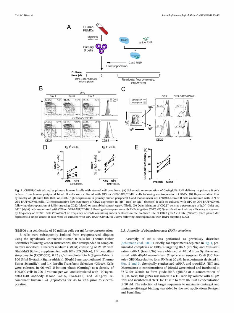
Fig. 1. CRISPR-Cas9 editing in primary human B cells with stromal cell co-culture. (A) Schematic representation of Cas9-gRNA RNP delivery to primary B cellsisolated from human peripheral blood. B cells were cultured with OP9 or OP9-BAFF/CD40L cells following electroporation of RNPs. (B) Representative flowcytometry of IgD and CD27 (left) or CD86 (right) expression in primary human peripheral blood mononuclear cell (PBMC)-derived B cells co-cultured with OP9 orOP9-BAFF/CD40L cells. (C) Representative flow cytometry of CD22 expression in IgD+ (top) or IgD− (bottom) B cells co-cultured with OP9 or OP9-BAFF/CD40Lfollowing electroporation of RNPs targeting CD22 (black) or scrambled control (grey, filled). (D) Quantification of CD22− cells as a percentage of IgD+ (left) andIgD− (right) cells co-cultured with OP9 or OP9-BAFF/CD40L following electroporation with RNPs targeting CD22. (E) Quantification of editing efficiency as assessedby frequency of CD22− cells (“Protein”) or frequency of reads containing indels centered on the predicted site of CD22 gRNA cut site (“Gene”). Each paired dotrepresents a single donor. B cells were co-cultured with OP9-BAFF/CD40L for 7 days following electroporation with RNPs targeting CD22.
C.-A.M. Wu et al. Journal of Immunological Methods 457 (2018) 33–40
35
2.4. Primary B cell electroporation
B cells were pooled and resuspended in P3 buffer (Lonza) and addedto 16-well Nucleocuvette Strips (Lonza). All experiments depicted inthis study utilized cell densities ranging from 1 to 3× 10⁶ cells in avolume of 20 μl per reaction, which was found to be optimal, thoughediting was achieved at a lower bound of 3×10⁵ cells/reaction. Toeach reaction, 2 to 5 μl of RNPs at 20 μM were added, with or without1 μl of 100 μMHDR template (Table S1). For all experiments depicted inFig. 2, a control HDR template with no homology to the human genome(Table S1) was added to increase Cas9 editing efficiency (Richardsonet al., 2016). Nucleocuvette Strips were loaded into an Amaxa 4D Nu-cleofector (Lonza) and electroporated using the EH-115 (for Fig. 1) orEH-140 (for Figs. 2 and 3) pulse codes.
2.5. Post-electroporation B cell culture
For experiments depicted in Fig. 1, B cells were resuspended incomplete RPMI with 20% FBS for 30min prior to plating onto 24-wellplates that had been pre-seeded with OP9 or OP9-BAFF/CD40L stromalcells overnight at a density of 50,000 cells per well in complete RPMI.For experiments depicted in Figs. 2 and 3, B cells were resuspended incomplete IMDM prior to plating onto 96-well plates. Each reaction wassplit into 6 wells and resuspended in a final volume of 200 μl per wellcontaining 100 ng/ml anti-CD40 and 20 ng/ml IL-4.
2.6. Flow cytometry
For experiments depicted in Fig. 1: Cell suspensions were stainedwith antibodies (Table S3) in PBS containing 2% FBS and 0.1% NaN3
0 103 104 105 0 103 104 105 0 103 104 105
Donor 1
Donor 2
BA
CD19 CD20
CD23(FCER2)
CD20(MS4A1)
gRNA includedCD19(CD19)
CD23
%of
max
C
0 103 104 105 0 103 104 105 0 103 104 105
D E
0 103 104 105
pY705-STAT3
STAT3Scramble
Scramble
Guide IL-21
-
+
+
0 103 104 105
0102
103
104
105
CD27
CD
38
0 103 104 105
0102
103
104
105
0 103 104 105
0102
103
104
1054.1%
ScrambleNo IL-21
ScrambleIL-21 (20 ng/ml)
STAT3 gRNAIL-21 (20 ng/ml)
PrimaryB cells
Humantonsil
Magneticselection
Electroporation
Culturetime (d) 0 2-3 7
Readouts:
sequencing
CD19 CD22 CD23
5.6%
11.0%
47.5%
17.9%
16.1%
15.1%
12.6%
19.0%
11.6%
8.1%
CD23(FCER2)
CD19(CD19)
CD22(CD22)
70.6% 71.5%
59.0%
44.6%
9.9%
7.5% 69.1%
58.1%
10.6%
87.2%
88.2%
83.9%
22.0% 6.3%
67.7%
58.5%
75.8%
85.6%
54.4%
%of
max
Cas9I I I I I I I I I I I
I I I II I I I I
I I I I
guide RNA
I I I I I
I I I I
Cas9 RNP
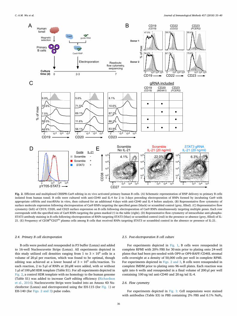
Fig. 2. Efficient and multiplexed CRISPR-Cas9 editing in ex vivo activated primary human B cells. (A) Schematic representation of RNP delivery to primary B cellsisolated from human tonsil. B cells were cultured with anti-CD40 and IL-4 for 2 to 3 days preceding electroporation of RNPs formed by incubating Cas9 withappropriate crRNAs and tracrRNAs in vitro, then cultured for an additional 4 days with anti-CD40 and IL-4 before analysis. (B) Representative flow cytometry ofsurface molecule expression following electroporation of Cas9 RNPs targeting the specified genes (black) or scrambled control (grey, filled). (C) Representative flowcytometry (left) of CD19, CD20, and CD23 surface expression on B cells following electroporation of Cas9 RNPs simultaneously targeting multiple genes. Each rowcorresponds with the specified mix of Cas9 RNPs targeting the genes marked (•) in the table (right). (D) Representative flow cytometry of intracellular anti-phospho-STAT3 antibody staining in B cells following electroporation of RNPs targeting STAT3 (blue) or scrambled control (red) in the presence or absence (grey, filled) of IL-21. (E) Frequency of CD38hiCD27hi plasma cells among B cells that received RNPs targeting STAT3 or scrambled control in the absence or presence of IL-21.
C.-A.M. Wu et al. Journal of Immunological Methods 457 (2018) 33–40
36
for 30min on ice. Propidium iodide was used to exclude nonviablecells. Flow cytometry data were collected on a FACS Canto II (BD) andanalyzed with FlowJo v9.
For experiments depicted in Figs. 2 and 3: Cell suspensions werestained with antibodies (Table S3) diluted in flow buffer consisting ofPBS with 2% FBS, 1mM EDTA, and 0.1% NaN3 for 20min on ice.Nonviable cells were excluded by labeling with fixable viability dyeeFluor780 (eBioscience) during surface staining as described (Yanget al., 2012). To detect intracellular phosphorylated STAT3, cells werefixed in a final concentration of 2% (v/v) paraformaldehyde at roomtemperature for 10min, then permeabilized in ice cold 90% methanoladded dropwise, before staining with anti-pY705 STAT3-Alexa Fluor647 (BD Bioscience) at room temperature for 30min. Cells were washedtwice with flow buffer between all steps. Flow cytometry data werecollected on an LSR Fortessa (BD) and analyzed with FlowJo v10. Allsamples were gated on FSC-A versus SSC-A over a broad range of FSC-A
to include blasting lymphocytes, followed by FSC-W versus FSC-H andthen SSC-W versus SSC-H gates to exclude doublets.
2.7. IL-21 stimulation
To assay plasma cell differentiation, post-electroporation B cellswere cultured in the presence of 100 ng/ml anti-CD40, 20 ng/ml IL-4,and 20 ng/ml recombinant human IL-21 (Peprotech) for 5 days. Toassay STAT3 phosphorylation, B cells that had been cultured in thepresence of anti-CD40 and IL-4 for 3 to 4 days following electroporationwere washed and resuspended in PBS containing fixable viability dyeeFluor780 (eBioscience) at a 1/600 dilution and incubated for 10min at37 °C to label nonviable cells. The cells were then washed and re-suspended in 100 μl of complete IMDM with or without 20 ng/ml IL-21and incubated for 30min at 37 °C before harvesting for flow cytometricanalysis as described above.
A
5' -GAGGGAATCTAAGACTTTGGGGG- 3'3' -CTCCCTTAGATTCTGAAACCCCC- 5'
R E S K T L G
Guide sequence PAM
GACTTTGTAAGT L *
5' -GAGGGAATCTAAGACTTTGGGGG- 3'3' -CTCCCTTAGATTCTGAAACCCCC- 5'
R E S K T L G
Guide sequence PAM
GACTTTGGGTGT L G
CD20 nonsense mutation(Nonsense mutation HDRT)
CD20 silent mutation(Silent mutation HDRT)
5' -GAGGGAATCTAAGACTTTGGGGG- 3'3' -CTCCCTTAGATTCTGAAACCCCC- 5'
R E S K T L G
Guide sequence PAM
CD20 no mutation(Off-target HDRT)
Nonsensemutation HDRT Silent mutation HDRTOff-target HDRT
47 53
B
C
TotalB cells
CD20–B cells
CD20+B cells
HDRTmatch(%ofreads)
0
10
20
30D
TotalB cells
CD20–B cells
CD20+B cells
0
20
40
60
Indels(%ofreads)
Off-target
Silent mutationNonsense mutation
Donor 1
Donor 2
CD200 103–103 104 105
Control gRNA
60.4%
63.3%
48.7%
60.9%
34.2%
45.8%
4.8%
13.8%
CD20 (MS4A1) gRNA
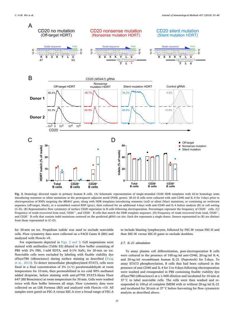
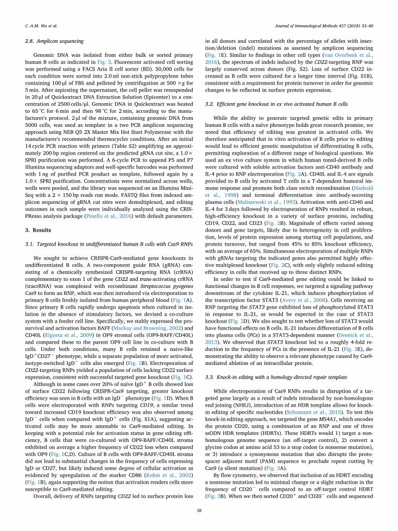
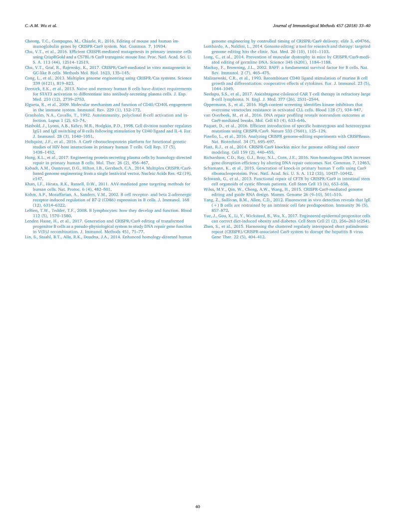
Fig. 3. Homology directed repair in primary human B cells. (A) Schematic representation of single-stranded CD20 HDR templates with 62 nt homology armsintroducing nonsense or silent mutations at the protospacer adjacent motif (PAM, green). (B–D) B cells were cultured with anti-CD40 and IL-4 for 3 days prior toelectroporation of RNPs targeting the MS4A1 gene, along with HDR templates introducing nonsense (red) or silent (blue) mutations, or containing an irrelevantsequence (off-target, black), or a scrambled control RNP (grey), then cultured for an additional 4 days with anti-CD40 and IL-4 before analysis (B) or cell sorting(C–D). (B) Representative flow cytometry of surface CD20 expression in B cells following electroporation. Percentages represent the frequency of CD20− cells. (C)Frequency of reads recovered from total, CD20+, and CD20− B cells that match the HDR template sequence. (D) Frequency of reads recovered from total, CD20+,and CD20− B cells that contain indel mutations centered on the predicted gRNA cut site. Each dot represents a single donor. Donors represented in (B) are distinctfrom those represented in (C–D).
C.-A.M. Wu et al. Journal of Immunological Methods 457 (2018) 33–40
37
2.8. Amplicon sequencing
Genomic DNA was isolated from either bulk or sorted primaryhuman B cells as indicated in Fig. 3. Fluorescent activated cell sortingwas performed using a FACS Aria II cell sorter (BD). 50,000 cells foreach condition were sorted into 2.0ml non-stick polypropylene tubescontaining 100 μl of FBS and pelleted by centrifugation at 500×g for5min. After aspirating the supernatant, the cell pellet was resuspendedin 20 μl of Quickextract DNA Extraction Solution (Epicenter) to a con-centration of 2500 cells/μl. Genomic DNA in Quickextract was heatedto 65 °C for 6min and then 98 °C for 2min, according to the manu-facturer's protocol. 2 μl of the mixture, containing genomic DNA from5000 cells, was used as template in a two PCR amplicon sequencingapproach using NEB Q5 2X Master Mix Hot Start Polymerase with themanufacturer's recommended thermocycler conditions. After an initial14 cycle PCR reaction with primers (Table S2) amplifying an approxi-mately 200 bp region centered on the predicted gRNA cut site, a 1.0×SPRI purification was performed. A 6-cycle PCR to append P5 and P7Illumina sequencing adaptors and well-specific barcodes was performedwith 1 ng of purified PCR product as template, followed again by a1.0× SPRI purification. Concentrations were normalized across wells,wells were pooled, and the library was sequenced on an Illumina Mini-Seq with a 2× 150 bp reads run mode. FASTQ files from indexed am-plicon sequencing of gRNA cut sites were demultiplexed, and editingoutcomes in each sample were individually analyzed using the CRIS-PResso analysis package (Pinello et al., 2016) with default parameters.
3. Results
3.1. Targeted knockout in undifferentiated human B cells with Cas9 RNPs
We sought to achieve CRISPR-Cas9-mediated gene knockouts inundifferentiated B cells. A two-component guide RNA (gRNA) con-sisting of a chemically synthesized CRISPR-targeting RNA (crRNA)complementary to exon 1 of the gene CD22 and trans-activating crRNA(tracrRNA) was complexed with recombinant Streptococcus pyogenesCas9 to form an RNP, which was then introduced via electroporation toprimary B cells freshly isolated from human peripheral blood (Fig. 1A).Since primary B cells rapidly undergo apoptosis when cultured in iso-lation in the absence of stimulatory factors, we devised a co-culturesystem with a feeder cell line. Specifically, we stably expressed the pro-survival and activation factors BAFF (Mackay and Browning, 2002) andCD40L (Elgueta et al., 2009) in OP9 stromal cells (OP9-BAFF/CD40L)and compared these to the parent OP9 cell line in co-culture with Bcells. Under both conditions, many B cells retained a naive-likeIgD+CD27− phenotype, while a separate population of more activated,isotype-switched IgD− cells also emerged (Fig. 1B). Electroporation ofCD22-targeting RNPs yielded a population of cells lacking CD22 surfaceexpression, consistent with successful targeted gene knockout (Fig. 1C).
Although in some cases over 20% of naïve IgD+ B cells showed lossof surface CD22 following CRISPR-Cas9 targeting, greater knockoutefficiency was seen in B cells with an IgD− phenotype (Fig. 1D). When Bcells were electroporated with RNPs targeting CD19, a similar trendtoward increased CD19 knockout efficiency was also observed amongIgD− cells when compared with IgD+ cells (Fig. S1A), suggesting ac-tivated cells may be more amenable to Cas9-mediated editing. Inkeeping with a potential role for activation status in gene editing effi-ciency, B cells that were co-cultured with OP9-BAFF/CD40L stromaexhibited on average a higher frequency of CD22 loss when comparedwith OP9 (Fig. 1C,D). Culture of B cells with OP9-BAFF/CD40L stromadid not lead to substantial changes in the frequency of cells expressingIgD or CD27, but likely induced some degree of cellular activation asevidenced by upregulation of the marker CD86 (Kohm et al., 2002)(Fig. 1B), again supporting the notion that activation renders cells moresusceptible to Cas9-mediated editing.
Overall, delivery of RNPs targeting CD22 led to surface protein loss
in all donors and correlated with the percentage of alleles with inser-tion/deletion (indel) mutations as assessed by amplicon sequencing(Fig. 1E). Similar to findings in other cell types (van Overbeek et al.,2016), the spectrum of indels induced by the CD22-targeting RNP waslargely conserved across donors (Fig. S2). Loss of surface CD22 in-creased as B cells were cultured for a longer time interval (Fig. S1B),consistent with a requirement for protein turnover in order for genomicchanges to be reflected in surface protein expression.
3.2. Efficient gene knockout in ex vivo activated human B cells
While the ability to generate targeted genetic edits in primaryhuman B cells with a naïve phenotype holds great research promise, wenoted that efficiency of editing was greatest in activated cells. Wetherefore anticipated that in vitro activation of B cells prior to editingwould lead to efficient genetic manipulation of differentiating B cells,permitting exploration of a different range of biological questions. Weused an ex vivo culture system in which human tonsil-derived B cellswere cultured with soluble activation factors anti-CD40 antibody andIL-4 prior to RNP electroporation (Fig. 2A). CD40L and IL-4 are signalsprovided to B cells by activated T cells in a T-dependent humoral im-mune response and promote both class switch recombination (Hasboldet al., 1998) and terminal differentiation into antibody-secretingplasma cells (Maliszewski et al., 1993). Activation with anti-CD40 andIL-4 for 3 days followed by electroporation of RNPs resulted in robust,high-efficiency knockout in a variety of surface proteins, includingCD19, CD22, and CD23 (Fig. 2B). Magnitude of effects varied amongdonors and gene targets, likely due to heterogeneity in cell prolifera-tion, levels of protein expression among starting cell populations, andprotein turnover, but ranged from 45% to 85% knockout efficiency,with an average of 65%. Simultaneous electroporation of multiple RNPswith gRNAs targeting the indicated genes also permitted highly effec-tive multiplexed knockout (Fig. 2C), with only slightly reduced editingefficiency in cells that received up to three distinct RNPs.
In order to test if Cas9-mediated gene editing could be linked tofunctional changes in B cell responses, we targeted a signaling pathwaydownstream of the cytokine IL-21, which induces phosphorylation ofthe transcription factor STAT3 (Avery et al., 2008). Cells receiving anRNP targeting the STAT3 gene exhibited loss of phosphorylated STAT3in response to IL-21, as would be expected in the case of STAT3knockout (Fig. 2D). We also sought to test whether loss of STAT3 wouldhave functional effects on B cells. IL-21 induces differentiation of B cellsinto plasma cells (PCs) in a STAT3-dependent manner (Deenick et al.,2013). We observed that STAT3 knockout led to a roughly 4-fold re-duction in the frequency of PCs in the presence of IL-21 (Fig. 2E), de-monstrating the ability to observe a relevant phenotype caused by Cas9-mediated ablation of an intracellular protein.
3.3. Knock-in editing with a homology-directed repair template
While electroporation of Cas9 RNPs results in disruption of a tar-geted gene largely as a result of indels introduced by non-homologousend joining (NHEJ), introduction of an HDR template allows for knock-in editing of specific nucleotides (Schumann et al., 2015). To test thisknock-in editing approach, we targeted the geneMS4A1, which encodesthe protein CD20, using a combination of an RNP and one of threessODN HDR templates (HDRTs). These HDRTs would 1) target a non-homologous genome sequence (an off-target control), 2) convert aglycine codon at amino acid 53 to a stop codon (a nonsense mutation),or 3) introduce a synonymous mutation that also disrupts the proto-spacer adjacent motif (PAM) sequence to preclude repeat cutting byCas9 (a silent mutation) (Fig. 3A).
By flow cytometry, we observed that inclusion of an HDRT encodinga nonsense mutation led to minimal change or a slight reduction in thefrequency of CD20− cells compared to an off-target control HDRT(Fig. 3B). When we then sorted CD20+ and CD20− cells and sequenced
C.-A.M. Wu et al. Journal of Immunological Methods 457 (2018) 33–40
38
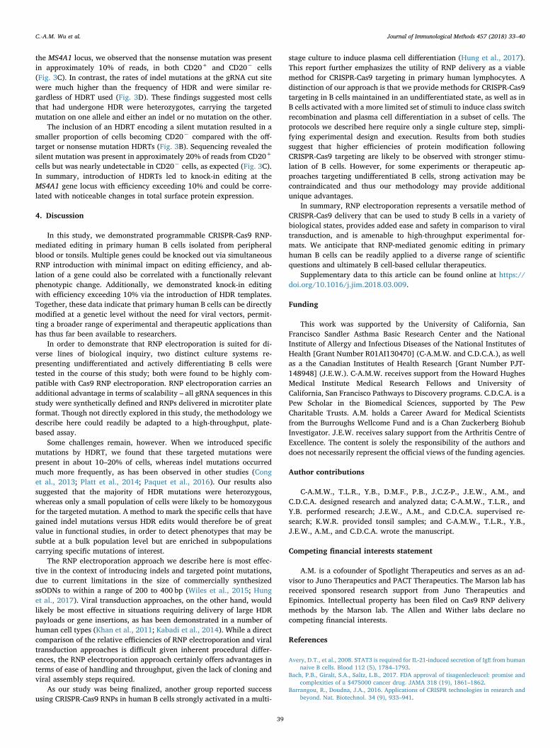
the MS4A1 locus, we observed that the nonsense mutation was presentin approximately 10% of reads, in both CD20+ and CD20− cells(Fig. 3C). In contrast, the rates of indel mutations at the gRNA cut sitewere much higher than the frequency of HDR and were similar re-gardless of HDRT used (Fig. 3D). These findings suggested most cellsthat had undergone HDR were heterozygotes, carrying the targetedmutation on one allele and either an indel or no mutation on the other.
The inclusion of an HDRT encoding a silent mutation resulted in asmaller proportion of cells becoming CD20− compared with the off-target or nonsense mutation HDRTs (Fig. 3B). Sequencing revealed thesilent mutation was present in approximately 20% of reads from CD20+
cells but was nearly undetectable in CD20− cells, as expected (Fig. 3C).In summary, introduction of HDRTs led to knock-in editing at theMS4A1 gene locus with efficiency exceeding 10% and could be corre-lated with noticeable changes in total surface protein expression.
4. Discussion
In this study, we demonstrated programmable CRISPR-Cas9 RNP-mediated editing in primary human B cells isolated from peripheralblood or tonsils. Multiple genes could be knocked out via simultaneousRNP introduction with minimal impact on editing efficiency, and ab-lation of a gene could also be correlated with a functionally relevantphenotypic change. Additionally, we demonstrated knock-in editingwith efficiency exceeding 10% via the introduction of HDR templates.Together, these data indicate that primary human B cells can be directlymodified at a genetic level without the need for viral vectors, permit-ting a broader range of experimental and therapeutic applications thanhas thus far been available to researchers.
In order to demonstrate that RNP electroporation is suited for di-verse lines of biological inquiry, two distinct culture systems re-presenting undifferentiated and actively differentiating B cells weretested in the course of this study; both were found to be highly com-patible with Cas9 RNP electroporation. RNP electroporation carries anadditional advantage in terms of scalability – all gRNA sequences in thisstudy were synthetically defined and RNPs delivered in microtiter plateformat. Though not directly explored in this study, the methodology wedescribe here could readily be adapted to a high-throughput, plate-based assay.
Some challenges remain, however. When we introduced specificmutations by HDRT, we found that these targeted mutations werepresent in about 10–20% of cells, whereas indel mutations occurredmuch more frequently, as has been observed in other studies (Conget al., 2013; Platt et al., 2014; Paquet et al., 2016). Our results alsosuggested that the majority of HDR mutations were heterozygous,whereas only a small population of cells were likely to be homozygousfor the targeted mutation. A method to mark the specific cells that havegained indel mutations versus HDR edits would therefore be of greatvalue in functional studies, in order to detect phenotypes that may besubtle at a bulk population level but are enriched in subpopulationscarrying specific mutations of interest.
The RNP electroporation approach we describe here is most effec-tive in the context of introducing indels and targeted point mutations,due to current limitations in the size of commercially synthesizedssODNs to within a range of 200 to 400 bp (Wiles et al., 2015; Hunget al., 2017). Viral transduction approaches, on the other hand, wouldlikely be most effective in situations requiring delivery of large HDRpayloads or gene insertions, as has been demonstrated in a number ofhuman cell types (Khan et al., 2011; Kabadi et al., 2014). While a directcomparison of the relative efficiencies of RNP electroporation and viraltransduction approaches is difficult given inherent procedural differ-ences, the RNP electroporation approach certainly offers advantages interms of ease of handling and throughput, given the lack of cloning andviral assembly steps required.
As our study was being finalized, another group reported successusing CRISPR-Cas9 RNPs in human B cells strongly activated in a multi-
stage culture to induce plasma cell differentiation (Hung et al., 2017).This report further emphasizes the utility of RNP delivery as a viablemethod for CRISPR-Cas9 targeting in primary human lymphocytes. Adistinction of our approach is that we provide methods for CRISPR-Cas9targeting in B cells maintained in an undifferentiated state, as well as inB cells activated with a more limited set of stimuli to induce class switchrecombination and plasma cell differentiation in a subset of cells. Theprotocols we described here require only a single culture step, simpli-fying experimental design and execution. Results from both studiessuggest that higher efficiencies of protein modification followingCRISPR-Cas9 targeting are likely to be observed with stronger stimu-lation of B cells. However, for some experiments or therapeutic ap-proaches targeting undifferentiated B cells, strong activation may becontraindicated and thus our methodology may provide additionalunique advantages.
In summary, RNP electroporation represents a versatile method ofCRISPR-Cas9 delivery that can be used to study B cells in a variety ofbiological states, provides added ease and safety in comparison to viraltransduction, and is amenable to high-throughput experimental for-mats. We anticipate that RNP-mediated genomic editing in primaryhuman B cells can be readily applied to a diverse range of scientificquestions and ultimately B cell-based cellular therapeutics.
Supplementary data to this article can be found online at https://doi.org/10.1016/j.jim.2018.03.009.
Funding
This work was supported by the University of California, SanFrancisco Sandler Asthma Basic Research Center and the NationalInstitute of Allergy and Infectious Diseases of the National Institutes ofHealth [Grant Number R01AI130470] (C-A.M.W. and C.D.C.A.), as wellas a the Canadian Institutes of Health Research [Grant Number PJT-148948] (J.E.W.). C-A.M.W. receives support from the Howard HughesMedical Institute Medical Research Fellows and University ofCalifornia, San Francisco Pathways to Discovery programs. C.D.C.A. is aPew Scholar in the Biomedical Sciences, supported by The PewCharitable Trusts. A.M. holds a Career Award for Medical Scientistsfrom the Burroughs Wellcome Fund and is a Chan Zuckerberg BiohubInvestigator. J.E.W. receives salary support from the Arthritis Centre ofExcellence. The content is solely the responsibility of the authors anddoes not necessarily represent the official views of the funding agencies.
Author contributions
C-A.M.W., T.L.R., Y.B., D.M.F., P.B., J.C.Z-P., J.E.W., A.M., andC.D.C.A. designed research and analyzed data; C-A.M.W., T.L.R., andY.B. performed research; J.E.W., A.M., and C.D.C.A. supervised re-search; K.W.R. provided tonsil samples; and C-A.M.W., T.L.R., Y.B.,J.E.W., A.M., and C.D.C.A. wrote the manuscript.
Competing financial interests statement
A.M. is a cofounder of Spotlight Therapeutics and serves as an ad-visor to Juno Therapeutics and PACT Therapeutics. The Marson lab hasreceived sponsored research support from Juno Therapeutics andEpinomics. Intellectual property has been filed on Cas9 RNP deliverymethods by the Marson lab. The Allen and Wither labs declare nocompeting financial interests.
References
Avery, D.T., et al., 2008. STAT3 is required for IL-21-induced secretion of IgE from humannaive B cells. Blood 112 (5), 1784–1793.
Bach, P.B., Giralt, S.A., Saltz, L.B., 2017. FDA approval of tisagenlecleucel: promise andcomplexities of a $475000 cancer drug. JAMA 318 (19), 1861–1862.
Barrangou, R., Doudna, J.A., 2016. Applications of CRISPR technologies in research andbeyond. Nat. Biotechnol. 34 (9), 933–941.
C.-A.M. Wu et al. Journal of Immunological Methods 457 (2018) 33–40
39
Cheong, T.C., Compagno, M., Chiarle, R., 2016. Editing of mouse and human im-munoglobulin genes by CRISPR-Cas9 system. Nat. Commun. 7, 10934.
Chu, V.T., et al., 2016. Efficient CRISPR-mediated mutagenesis in primary immune cellsusing CrispRGold and a C57BL/6 Cas9 transgenic mouse line. Proc. Natl. Acad. Sci. U.S. A. 113 (44), 12514–12519.
Chu, V.T., Graf, R., Rajewsky, K., 2017. CRISPR/Cas9-mediated in vitro mutagenesis inGC-like B cells. Methods Mol. Biol. 1623, 135–145.
Cong, L., et al., 2013. Multiplex genome engineering using CRISPR/Cas systems. Science339 (6121), 819–823.
Deenick, E.K., et al., 2013. Naive and memory human B cells have distinct requirementsfor STAT3 activation to differentiate into antibody-secreting plasma cells. J. Exp.Med. 210 (12), 2739–2753.
Elgueta, R., et al., 2009. Molecular mechanism and function of CD40/CD40L engagementin the immune system. Immunol. Rev. 229 (1), 152–172.
Granholm, N.A., Cavallo, T., 1992. Autoimmunity, polyclonal B-cell activation and in-fection. Lupus 1 (2), 63–74.
Hasbold, J., Lyons, A.B., Kehry, M.R., Hodgkin, P.D., 1998. Cell division number regulatesIgG1 and IgE switching of B cells following stimulation by CD40 ligand and IL-4. Eur.J. Immunol. 28 (3), 1040–1051.
Hultquist, J.F., et al., 2016. A Cas9 ribonucleoprotein platform for functional geneticstudies of HIV-host interactions in primary human T cells. Cell Rep. 17 (5),1438–1452.
Hung, K.L., et al., 2017. Engineering protein-secreting plasma cells by homology-directedrepair in primary human B cells. Mol. Ther. 26 (2), 456–467.
Kabadi, A.M., Ousterout, D.G., Hilton, I.B., Gersbach, C.A., 2014. Multiplex CRISPR/Cas9-based genome engineering from a single lentiviral vector. Nucleic Acids Res. 42 (19),e147.
Khan, I.F., Hirata, R.K., Russell, D.W., 2011. AAV-mediated gene targeting methods forhuman cells. Nat. Protoc. 6 (4), 482–501.
Kohm, A.P., Mozaffarian, A., Sanders, V.M., 2002. B cell receptor- and beta 2-adrenergicreceptor-induced regulation of B7-2 (CD86) expression in B cells. J. Immunol. 168(12), 6314–6322.
LeBien, T.W., Tedder, T.F., 2008. B lymphocytes: how they develop and function. Blood112 (5), 1570–1580.
Lenden Hasse, H., et al., 2017. Generation and CRISPR/Cas9 editing of transformedprogenitor B cells as a pseudo-physiological system to study DNA repair gene functionin V(D)J recombination. J. Immunol. Methods 451, 71–77.
Lin, S., Staahl, B.T., Alla, R.K., Doudna, J.A., 2014. Enhanced homology-directed human
genome engineering by controlled timing of CRISPR/Cas9 delivery. elife 3, e04766.Lombardo, A., Naldini, L., 2014. Genome editing: a tool for research and therapy: targeted
genome editing hits the clinic. Nat. Med. 20 (10), 1101–1103.Long, C., et al., 2014. Prevention of muscular dystrophy in mice by CRISPR/Cas9-medi-
ated editing of germline DNA. Science 345 (6201), 1184–1188.Mackay, F., Browning, J.L., 2002. BAFF: a fundamental survival factor for B cells. Nat.
Rev. Immunol. 2 (7), 465–475.Maliszewski, C.R., et al., 1993. Recombinant CD40 ligand stimulation of murine B cell
growth and differentiation: cooperative effects of cytokines. Eur. J. Immunol. 23 (5),1044–1049.
Neelapu, S.S., et al., 2017. Axicabtagene ciloleucel CAR T-cell therapy in refractory largeB-cell lymphoma. N. Engl. J. Med. 377 (26), 2531–2544.
Oppermann, S., et al., 2016. High-content screening identifies kinase inhibitors thatovercome venetoclax resistance in activated CLL cells. Blood 128 (7), 934–947.
van Overbeek, M., et al., 2016. DNA repair profiling reveals nonrandom outcomes atCas9-mediated breaks. Mol. Cell 63 (4), 633–646.
Paquet, D., et al., 2016. Efficient introduction of specific homozygous and heterozygousmutations using CRISPR/Cas9. Nature 533 (7601), 125–129.
Pinello, L., et al., 2016. Analyzing CRISPR genome-editing experiments with CRISPResso.Nat. Biotechnol. 34 (7), 695–697.
Platt, R.J., et al., 2014. CRISPR-Cas9 knockin mice for genome editing and cancermodeling. Cell 159 (2), 440–455.
Richardson, C.D., Ray, G.J., Bray, N.L., Corn, J.E., 2016. Non-homologous DNA increasesgene disruption efficiency by altering DNA repair outcomes. Nat. Commun. 7, 12463.
Schumann, K., et al., 2015. Generation of knock-in primary human T cells using Cas9ribonucleoproteins. Proc. Natl. Acad. Sci. U. S. A. 112 (33), 10437–10442.
Schwank, G., et al., 2013. Functional repair of CFTR by CRISPR/Cas9 in intestinal stemcell organoids of cystic fibrosis patients. Cell Stem Cell 13 (6), 653–658.
Wiles, M.V., Qin, W., Cheng, A.W., Wang, H., 2015. CRISPR-Cas9-mediated genomeediting and guide RNA design. Mamm. Genome 26 (9–10), 501–510.
Yang, Z., Sullivan, B.M., Allen, C.D., 2012. Fluorescent in vivo detection reveals that IgE(+) B cells are restrained by an intrinsic cell fate predisposition. Immunity 36 (5),857–872.
Yue, J., Gou, X., Li, Y., Wicksteed, B., Wu, X., 2017. Engineered epidermal progenitor cellscan correct diet-induced obesity and diabetes. Cell Stem Cell 21 (2), 256–263 (e254).
Zhen, S., et al., 2015. Harnessing the clustered regularly interspaced short palindromicrepeat (CRISPR)/CRISPR-associated Cas9 system to disrupt the hepatitis B virus.Gene Ther. 22 (5), 404–412.
C.-A.M. Wu et al. Journal of Immunological Methods 457 (2018) 33–40
40