Embed Size (px)
Citation preview

ATP directed agent, 8-chloro-adenosine, inducesAMP activated protein kinase activity, leading toautophagic cell death in breast cancer cellsStellrecht et al.
JOURNAL OF HEMATOLOGY& ONCOLOGY
Stellrecht et al. Journal of Hematology & Oncology 2014, 7:23http://www.jhoonline.org/content/7/1/23

JOURNAL OF HEMATOLOGY& ONCOLOGY
Stellrecht et al. Journal of Hematology & Oncology 2014, 7:23http://www.jhoonline.org/content/7/1/23
RESEARCH Open Access
ATP directed agent, 8-chloro-adenosine, inducesAMP activated protein kinase activity, leading toautophagic cell death in breast cancer cellsChristine M Stellrecht1,2*, Hima V Vangapandu1,2, Xiao-Feng Le1, Weiqun Mao1 and Shujun Shentu1
Abstract
Background: 8-chloro-adenosine (8-Cl-Ado) is a unique ribonucleoside analog which is currently in a phase I clinicaltrial for hematological malignancies. Previously, we demonstrated in breast cancer cells that a 3-day treatment with10 μM 8-Cl-Ado causes a 90% loss of clonogenic survival. In contrast, there was only a modest induction of apoptosisunder these conditions, suggesting an alternative mechanism for the tumoricidal activity of 8-Cl-Ado.
Methods: Cellular metabolism, AMP-activated protein kinase (AMPK) and mammalian target of rapamycin (mTOR)pathway signaling, as well as autophagy induction was evaluated in breast cancer cell lines treated with 8-Cl-Ado. Theeffects of knocking down essential autophagy factors with small interfering RNA on 8-Cl-Ado-inhibited cell survival wasassessed in breast cancer cells by examining apoptosis induction and clonogenic survival. In vivo efficacy of8-Cl-Ado was measured in two breast cancer orthotopic model systems.
Results: We demonstrate that in breast cancer cell lines, the metabolism of 8-Cl-Ado results in depletion of endogenousATP that subsequently induces the phosphorylation and activation of the energy sensor, AMPK. This was associated withan attenuation of mTOR signaling and an induction of the phosphorylation of the autophagy factor, Unc51-like kinase 1on Ser555. 8-Cl-Ado-mediated induction of autophagy was evident by increased aggregates of microtubule-associatedprotein 1 light chain 3B (LC3B) which was associated with its conversion to its lipidated form, LC3B-II, p62 degradativeflux, and increased formation of acidic vesicular organelles. Additionally, transfection of MCF-7 cells with siRNA to ATG7or beclin 1 provided partial protection of the cells to 8-Cl-Ado cytotoxicity as measured by clonogenicity. In vivo,8-Cl-Ado inhibited growth of both MCF-7 and BT-474 xenograft tumors. Moreover, in 9 of 22 BT-474 tumors treated with100 mg/kg/day 3 times a week, there was an absence of macroscopically detectable tumor after 3 weeks of treatment.
Conclusions: Our data demonstrates that 8-Cl-Ado treatment activates the AMPK pathway leading to autophagyinduction of in breast cancer cells, eliciting, in part, its tumoricidal effects. Additionally, 8-Cl-Ado effectively inhibitedin vivo tumor growth in mice. Based on this biological activity, we are planning to test 8-Cl-Ado in the clinic for patientswith breast cancer.
Keywords: Autophagy, AMPK, mTOR, ATP, 8-chloro-adenosine
BackgroundBreast cancer is the most prevalent cancer in women inindustrialized countries. Each year more than a half millionnew cases of breast cancer are diagnosed in the US andEurope. Although in recent years, advances in therapieshave contributed to decreased mortality; worldwide nearly
* Correspondence: [email protected] of Experimental Therapeutics, University of Texas MD AndersonCancer Center, Unit 1950, P.O. Box 301429, Houston, TX 77230-1429, USA2Graduate School of Biomedical Sciences, The University of Texas HealthScience Center, Houston, TX, USA
© 2014 Stellrecht et al.; licensee BioMed CentrCommons Attribution License (http://creativecreproduction in any medium, provided the orDedication waiver (http://creativecommons.orunless otherwise stated.
500,000 deaths are attributed to breast cancer, annually. Inaddition, toxicity associated with current treatments andthe risk of developing secondary cancers [1] highlight theurgent need for new therapies against the disease.8-chloro-adenosine (8-Cl-Ado) is a unique ribonucleoside
analog that is currently in a phase I clinical trial at the UTMD Anderson Cancer Center for the treatment of chroniclymphocytic leukemia (CLL). Previously, we demonstratedthis analog is tumoricidal to breast cancer cells [2]. In
al Ltd. This is an Open Access article distributed under the terms of the Creativeommons.org/licenses/by/4.0), which permits unrestricted use, distribution, andiginal work is properly credited. The Creative Commons Public Domaing/publicdomain/zero/1.0/) applies to the data made available in this article,

Stellrecht et al. Journal of Hematology & Oncology 2014, 7:23 Page 2 of 12http://www.jhoonline.org/content/7/1/23
addition, our group and others have shown that 8-Cl-Adoinhibits the growth and survival of numerous otherhematological and solid tumor model systems [3-19] butnot normal lymphocytes [12] or non-transformed mam-mary epithelial cells (unpublished data).Much of our prior work on the cytotoxic effects of
8-Cl-Ado focused on the accumulation of 8-Cl-ATP[2,8-10,12,20] and its incorporation into mRNA [10]preceding an inhibition of transcription [2,8-10,12,19] andpoly(A) polymerase [21,22]. The net effects of these eventslead to an induction of apoptosis [2,8-10,12]. In breastcancer cells, 8-Cl-Ado-induced cytotoxicity is only par-tially attributed to apoptosis. In addition to the impact ontranscription, 8-Cl-Ado-induces a reduction of the intra-cellular ATP pool [2,8-10,12] and ATP depletion has beenassociated with apoptosis-independent cell death [23,24].Within the cell, ATP levels are tightly regulated by
AMP-activated protein kinase (AMPK), which functionsas a cellular energy sensor [25]. AMP and ATP directlybind and regulate AMPK such that when ATP levelsdecrease, AMP is allowed to bind and allosterically ac-tivate AMPK. Additionally, upstream kinases can thenphosphorylate and further activate AMP bound AMPK.Once activated, AMPK promotes events that lead to meta-bolic switches to restore cellular bioenergy. Significantenergy regulating effects of AMPK pertaining to cancertherapeutics is its ability to attenuate growth and proteinsynthesis by regulating mammalian target of rapamycin(mTOR) pathway and induce autophagy.AMPK inhibition of mTOR signaling occurs through
two pathways. First, AMPK directly phosphorylates andactivate tuberous sclerosis protein 2 which is involved inmTOR inhibitory signaling. Second, AMPK has beenshown to phosphorylate and inactivate raptor, a memberof mTORC1. Both mTORC1 and AMPK regulate autoph-agy through the phosphorylation of Unc51-like kinase 1(ULK1). [For a review see ref. [26]]. ULK1 is a mammalianhomolog of a yeast autophagy initiating factor, Atg1.Although there has been some discrepancy regardingthe specific AMPK phosphorylation sites on ULK1, it isclear that AMPK activates ULK1 while the phosphorylationby mTORC1 inhibits ULK1 activity. AMPK also regulatesautophagy through modulation of components of Vsp34complexes [27].The events above delineate the mode by which di-
minished ATP levels can alter cellular processes. Since8-Cl-Ado treatment diminishes the ATP pool [2,8-10,12],we hypothesize that 8-Cl-Ado treatment will educe AMPKactivation and mTORC1 attenuation which will initiateautophagy. Our current investigation further evaluated thecellular metabolism of 8-Cl-Ado and its metabolic effectson bioenergy. Moreover, we examined the mechanismof action of the bioenergy depletion that leads to a non-apoptotic cell death in breast cancer cells.
Results8-Cl-Ado accumulation/elimination and effects on energyproductionPreviously, our group demonstrated that 8-Cl-Ado(Figure 1A) is tumoricidal to primary CLL lymphocytes[12], breast cancer [2], myeloma [17], and mantle celllymphoma (MCL) [8] cell lines and this cytotoxicity wasdependent on the analog’s metabolism to its monopho-sphate form by adenosine kinase [9] followed by triphos-phate accumulation. In the breast cancer cell lines, weshowed that a 3-day treatment with 10 μM 8-Cl-Adoinhibited over 90% clonogenic survival [2] (Figure 1B).Paradoxically, we demonstrated that such treatments wereassociated with only a ~30% induction of apoptosis in bothcell lines as measured by annexin V and PI staining [19](Figure 1C and Additional file 1: Figure S1).We established that 8-Cl-Ado treatment results in a
rapid depletion of ATP within 12-hours which was as-sociated with 8-Cl-ATP accumulation (Additional file 1:Figure S2 and [2]). To examine the impact of 8-Cl-Adotreatment on energy producing metabolic pathways, weanalyzed both glycolysis and mitochondrial respiration.Extracellular acidification rate (ECAR), as a measure ofglycolysis, and oxygen consumption rate (OCR), for mito-chondrial respiratory function, were assessed in both MCF-7 and BT-474 cells treated with and without 8-Cl-Ado for18 hr using a Seahorse XF96 analyzer. Our results indicatedthat, both basal mitochondrial respiration (Figure 2A,B, and C) and glycolysis (Figure 2D) were perturbed by8-Cl-Ado treatment. The finding of decreased O2 con-sumption is in keeping with previous data which indicated8-Cl-Ado may be an inhibitor of mitochondrial complexV, ATP synthase [28]. The alteration in glycolysis maypossibly be associated with decreased glucose uptake,which was seen in myeloma cells treated with an 8-Cl-Adocongener compound, 8-amino-adenosine [29]. Interestingly,further assessment of cellular respiratory chain with XFCell Mito Stress Test assay possibly revealed additionalterations in cellular respiration induced by 8-Cl-Adotreatment, as OCR was still attenuated even after theaddition of an uncoupler, FCCP, to alleviate the depend-ency of complex I-IV on complex V’s transport of electronsacross the mitochondrial membrane (Figure 2B and C).Because tumor cells are known to be sensitive to ATPdepletion, we decided to assess the effects of 8-Cl-Ado onATP levels throughout the 3-day treatment. Our resultsshowed that in BT-474 cells the ATP levels continued todiminish over a 3-day treatment with 10 μM 8-Cl-Ado(Figure 2E) while in MCF-7 cells the levels remainedbelow control levels but did show evidence of some recoveryby 24-hours. Additionally, we examined the accumulationof the analog’s cytotoxic metabolite, 8-Cl-ATP, over72-hours. The accumulation of the analog triphosphateinversely paralleled the ATP depletion as it peaked at

Figure 1 Effect of 8-Cl-Ado on the survival of breast cancer cells. (A) Structure of 8-Cl-Ado. (B) The effects of 8-Cl-Ado on the clonogenicgrowth of MCF-7 cells. Cells were treated with 10 μM 8-Cl-Ado for 3-days, washed with PBS, and cultured in fresh medium for 14-days. Coloniesof >50 cells were counted under a dissecting microscope. (C) Flow cytometery analysis of annexin V and PI staining in MCF-7, hatched bars, andBT-474 cells, black bars, treated with 10 μM 8-Cl-Ado for the indicated times.
Stellrecht et al. Journal of Hematology & Oncology 2014, 7:23 Page 3 of 12http://www.jhoonline.org/content/7/1/23
24- and 72-hours in MCF-7 and BT-474 cells, respectively(Figure 2F). Since the ratio of 8-Cl-ATP to ATP hasbeen shown to be a determinant of the cytotoxic effectof 8-Cl-Ado [2,8], the kinetics of this relationship wasalso examined (Figure 2G). We demonstrated that theratio of the analog triphosphate to normal ATP was high-est in MCF-7 cells which rapidly peaked at 12-hours whilein BT-474 cells the ratio continually increased until itreached a plateau by 24-hours. The elimination kineticsof 8-Cl-ATP was also measured by treating the cellswith 10 μM 8-Cl-Ado for 3-days, washing drug off andcontinued culturing the cells in drug-free medium foranother 3-days (Figure 2H). In both lines there appearedto be an initial, more rapid elimination of 8-Cl-ATP afterdrug removal. If a biphasic elimination kinetics is consid-ered, the 8-Cl-ATP half-lives in MCF-7 cells were 3.8- and25.5-hours while in BT-474 it was 6.4-hours and >7-days.An assessment of the kinetics as a monophasic elimin-ation yields the 8-Cl-ATP half-lives as 5.8 and 11.4-hoursfor MCF-7 and BT-474, respectively.
8-Cl-Ado induces AMPK activity in breast cancer cellsThe 8-Cl-Ado-induced ATP depletion is expected toincrease the AMP to ATP ratio which would lead toactivation of AMPK. To determine if 8-Cl-Ado is able toinduce AMPK activity, we treated MCF-7 and BT-474cells with 10 μM 8-Cl-Ado for various times and assessedchanges in p-AMPK (Thr172) levels by immunoblotanalysis. The results demonstrated that while total AMPKprotein levels were unchanged by 8-Cl-Ado treatment,phosphorylation of AMPK (Thr172) was induced in atime-dependent manner, being readily detected within7- to 12-hours (Figure 3A). To further assess AMPK ac-tivity, we examined the cellular phosphorylation of one
of its critical downstream substrates, ACC on Ser79 andshowed that AMPK activity is induced within 4-hours of8-Cl-Ado treatment (Figure 3B). These events occurred inboth MCF-7 and BT-474 cells which indicate they do notrequire p53 as MCF-7 cells have a wild type p53 genotypewhile BT-474 cells harbor a mutant p53 [30].
Inhibition of mTOR by 8-Cl-AdoA significant energy regulating effect of AMPK per-taining to cancer therapeutics is the ability of AMPK toinhibit the mTOR pathway. To determine if the 8-Cl-Ado-dependent AMPK induction altered the activity ofthe mTOR pathway, we assessed the effects of 8-Cl-Adoon Ser792 residue of raptor; an AMPK phosphorylationsite on this mTORC1 protein. We determined that 10 μM8-Cl-Ado-treatment readily induced the phosphorylationof raptor Ser792 (Figure 4A) in MCF-7 cells. This wasassociated with diminished mTOR autophosphorylationon Ser2481 (data not shown). To further assess mTORC1activity, we examined the phosphorylation status of themTORC1 target, 4E-BP1, and found 8-Cl-Ado treat-ment diminished the level of p-4E-BP1 (Ser65), a rapa-mycin sensitive phosphorylation site [31] and reducedthe levels of the slower migrating hyperphosphorylated4E-BP1 bands (Figure 4B). Overall, these results demon-strate 8-Cl-Ado treatment attenuated mTOR activity inbreast cancer cells.
8-Cl-Ado-induced activation of ULK1AMPK and mTOR are both able to regulate autophagythrough direct phosphorylation of Unc51-like kinase 1(ULK1), which is the human counterpart of the yeastautophagy initiating factor, Atg1. Although there has beensome discrepancy as to the ULK1 phosphorylation sites,

Figure 2 Accumulation and elimination of 8-Cl-ATP and effects on ATP production. (A) 10 μM 8-Cl-Ado treatment, hatched bars, perturbsbasal OCR as compared to untreated, solid bars, in MCF-7, gray bars, and BT-474 cells, black bars. Effects of altering ETC complexes with the agents,oligomycin, FCCP, antimycin, and rotenone on OCR in (B) MCF-7 and (C) BT-474 cells treated with, black circles, and without, gray circles, 10 μM8-Cl-Ado. (D) 10 μM 8-Cl-Ado treatment, hatched bars, reduces basal glycolysis as demonstrated by decreased ECAR in MCF-7, gray bars, and BT-474cells, black bars. (E) Time dependent depletion of the endogenous ATP pool, (F) the accumulation of 8-Cl-ATP, and (G) changes in the 8-Cl-ATP/ATPratio in MCF-7, ○, and BT-474 cells, ●. Cells were treated with 10 μM 8-Cl-Ado for the indicated times and acid extracts were analyzed by HPLCto measure nucleotide levels. (H) The elimination of 8-Cl-ATP in MCF-7, ○, and BT-474 cells, ●. Cells were treated with 10 μM 8-Cl-Ado for 3 days,washed with PBS, and cultured in fresh medium. At the indicated times, acid extracts were analyzed as above.
Stellrecht et al. Journal of Hematology & Oncology 2014, 7:23 Page 4 of 12http://www.jhoonline.org/content/7/1/23
there have been several different reports which indicateAMPK directly phosphorylates ULK on Ser555 [32-35].We examined the effects of 8-Cl-Ado on the AMPK
phosphorylation of this site and determined that 10 μM8-Cl-Ado-treatment induces ULK1 phosphorylation onSer555 in MCF-7 and BT-474 cells (Figure 5A).
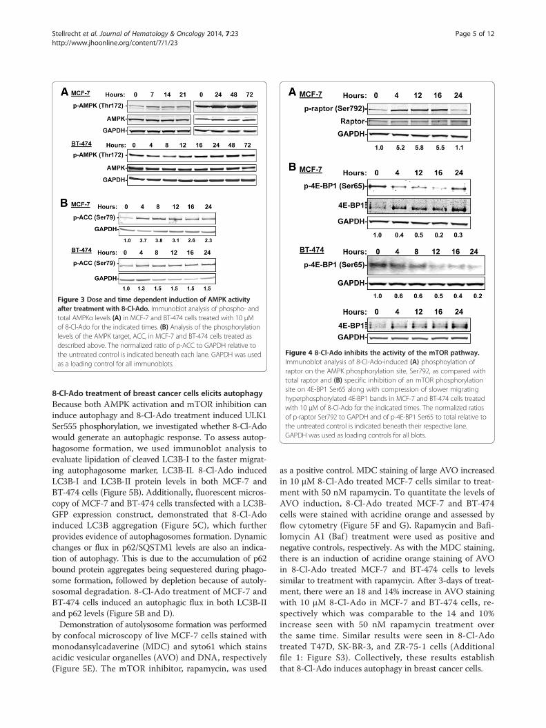
Figure 3 Dose and time dependent induction of AMPK activityafter treatment with 8-Cl-Ado. Immunoblot analysis of phospho- andtotal AMPKα levels (A) in MCF-7 and BT-474 cells treated with 10 μMof 8-Cl-Ado for the indicated times. (B) Analysis of the phosphorylationlevels of the AMPK target, ACC, in MCF-7 and BT-474 cells treated asdescribed above. The normalized ratio of p-ACC to GAPDH relative tothe untreated control is indicated beneath each lane. GAPDH was usedas a loading control for all immunoblots.
Figure 4 8-Cl-Ado inhibits the activity of the mTOR pathway.Immunoblot analysis of 8-Cl-Ado-induced (A) phosphoylation ofraptor on the AMPK phosphorylation site, Ser792, as compared withtotal raptor and (B) specific inhibition of an mTOR phosphorylationsite on 4E-BP1 Ser65 along with compression of slower migratinghyperphosphorylated 4E-BP1 bands in MCF-7 and BT-474 cells treatedwith 10 μM of 8-Cl-Ado for the indicated times. The normalized ratiosof p-raptor Ser792 to GAPDH and of p-4E-BP1 Ser65 to total relative tothe untreated control is indicated beneath their respective lane.GAPDH was used as loading controls for all blots.
Stellrecht et al. Journal of Hematology & Oncology 2014, 7:23 Page 5 of 12http://www.jhoonline.org/content/7/1/23
8-Cl-Ado treatment of breast cancer cells elicits autophagyBecause both AMPK activation and mTOR inhibition caninduce autophagy and 8-Cl-Ado treatment induced ULK1Ser555 phosphorylation, we investigated whether 8-Cl-Adowould generate an autophagic response. To assess autop-hagosome formation, we used immunoblot analysis toevaluate lipidation of cleaved LC3B-I to the faster migrat-ing autophagosome marker, LC3B-II. 8-Cl-Ado inducedLC3B-I and LC3B-II protein levels in both MCF-7 andBT-474 cells (Figure 5B). Additionally, fluorescent micros-copy of MCF-7 and BT-474 cells transfected with a LC3B-GFP expression construct, demonstrated that 8-Cl-Adoinduced LC3B aggregation (Figure 5C), which furtherprovides evidence of autophagosomes formation. Dynamicchanges or flux in p62/SQSTM1 levels are also an indica-tion of autophagy. This is due to the accumulation of p62bound protein aggregates being sequestered during phago-some formation, followed by depletion because of autoly-sosomal degradation. 8-Cl-Ado treatment of MCF-7 andBT-474 cells induced an autophagic flux in both LC3B-IIand p62 levels (Figure 5B and D).Demonstration of autolysosome formation was performed
by confocal microscopy of live MCF-7 cells stained withmonodansylcadaverine (MDC) and syto61 which stainsacidic vesicular organelles (AVO) and DNA, respectively(Figure 5E). The mTOR inhibitor, rapamycin, was used
as a positive control. MDC staining of large AVO increasedin 10 μM 8-Cl-Ado treated MCF-7 cells similar to treat-ment with 50 nM rapamycin. To quantitate the levels ofAVO induction, 8-Cl-Ado treated MCF-7 and BT-474cells were stained with acridine orange and assessed byflow cytometry (Figure 5F and G). Rapamycin and Bafi-lomycin A1 (Baf ) treatment were used as positive andnegative controls, respectively. As with the MDC staining,there is an induction of acridine orange staining of AVOin 8-Cl-Ado treated MCF-7 and BT-474 cells to levelssimilar to treatment with rapamycin. After 3-days of treat-ment, there were an 18 and 14% increase in AVO stainingwith 10 μM 8-Cl-Ado in MCF-7 and BT-474 cells, re-spectively which was comparable to the 14 and 10%increase seen with 50 nM rapamycin treatment overthe same time. Similar results were seen in 8-Cl-Adotreated T47D, SK-BR-3, and ZR-75-1 cells (Additionalfile 1: Figure S3). Collectively, these results establishthat 8-Cl-Ado induces autophagy in breast cancer cells.

Figure 5 8-Cl-Ado-induces autophagy. Immunoblot analysis of (A) phospho-ULK (Ser555) levels and (B) LC3B-I lipidation to form LC3B-II inMCF-7 and BT-474 cells treated with 10 μM of 8-Cl-Ado for the indicated times. (C) Fluorescent microscopy of MCF-7 and BT-474 cells transientlytransfected with GFP-LC3B and treated with 10 μM of 8-Cl-Ado for 12-hours to assess aggregation of LC3B. (D) Immunoblot analysis of p62 levelsin MCF-7 and BT-474 cells treated with 10 μM of 8-Cl-Ado for the indicated times. The normalized ratio of p62 to GAPDH relative to the untreatedcontrol is indicated beneath each lane. (E) Fluorescent microscopic imaging of autolysosomes. MCF-7 cells were untreated or treated for 2-dayswith 8-Cl-Ado or rapamycin. Cells were stained with MDC, blue, to visualize AVO and Syto 61 (DNA), red, for nuclei counterstaining. (F) Representativeflow cytometery histograms of AVO stained with acridine orange in MCF-7 cells untreated or treated for 3 days with 50 nM rapamycin or 10 μM 8-Cl-Ado.Baf was added before 30 min prior to staining to neutralize AVO staining. (G) Quantification triplicate experiments of MCF-7 and BT-474 cells treatedand analyzed as in E.
Stellrecht et al. Journal of Hematology & Oncology 2014, 7:23 Page 6 of 12http://www.jhoonline.org/content/7/1/23
Depletion of autophagy proteins diminished 8-Cl-Adoinduced cytotoxicityPreviously, we had noted that with a 3-day treatment of10 μM 8-Cl-Ado there was a ~90% loss of clonogenicsurvival while the amount of apoptosis induction onlyreached ~30% [2] (Figure 1B and C), suggesting thereis another mechanism of cell killing occurring in the8-Cl-Ado treated breast cancer cells. One possibility isthrough autophagic cell death. To test this hypothesis,we transfected MCF-7 cells with small interfering RNA(siRNA) directed against ATG7, or BECN1, which are
required autophagy factors, and compared 8-Cl-Adotreatment of these cells to MCF-7 cells transfected withsiCONT. The targeting siRNAs effectively depleted theirrespective targets during the 3 days of 8-Cl-Ado treat-ment and blocked autophagy induction (Figure 6A andC). Interestingly, although siBECN1 and siATG7 didnot alter the extent of 8-Cl-Ado-induced apoptosis(Figure 6A and B), they did increase clonogenic survival(Figure 6D and E). These results indicate that 8-Cl-Ado cytotoxicity is mediated in part by autophagic celldeath.
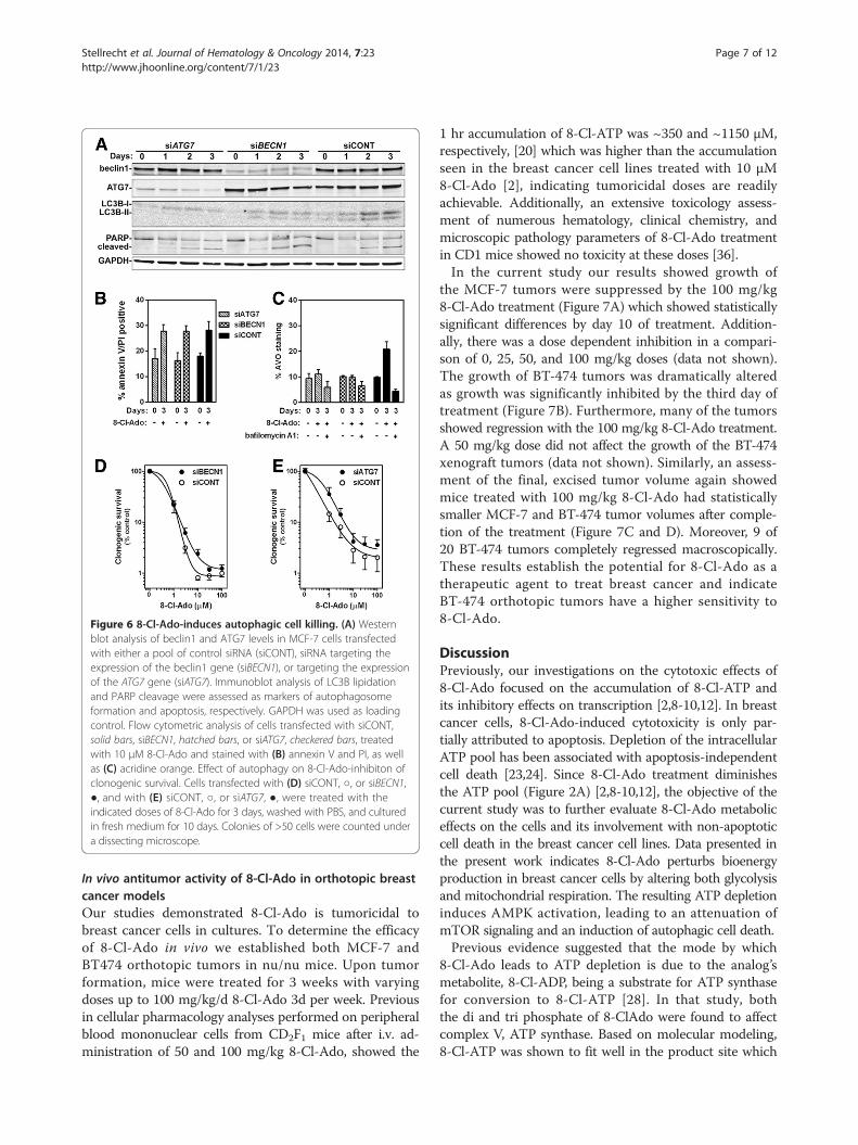
Figure 6 8-Cl-Ado-induces autophagic cell killing. (A) Westernblot analysis of beclin1 and ATG7 levels in MCF-7 cells transfectedwith either a pool of control siRNA (siCONT), siRNA targeting theexpression of the beclin1 gene (siBECN1), or targeting the expressionof the ATG7 gene (siATG7). Immunoblot analysis of LC3B lipidationand PARP cleavage were assessed as markers of autophagosomeformation and apoptosis, respectively. GAPDH was used as loadingcontrol. Flow cytometric analysis of cells transfected with siCONT,solid bars, siBECN1, hatched bars, or siATG7, checkered bars, treatedwith 10 μM 8-Cl-Ado and stained with (B) annexin V and PI, as wellas (C) acridine orange. Effect of autophagy on 8-Cl-Ado-inhibiton ofclonogenic survival. Cells transfected with (D) siCONT, ○, or siBECN1,●, and with (E) siCONT, ○, or siATG7, ●, were treated with theindicated doses of 8-Cl-Ado for 3 days, washed with PBS, and culturedin fresh medium for 10 days. Colonies of >50 cells were counted undera dissecting microscope.
Stellrecht et al. Journal of Hematology & Oncology 2014, 7:23 Page 7 of 12http://www.jhoonline.org/content/7/1/23
In vivo antitumor activity of 8-Cl-Ado in orthotopic breastcancer modelsOur studies demonstrated 8-Cl-Ado is tumoricidal tobreast cancer cells in cultures. To determine the efficacyof 8-Cl-Ado in vivo we established both MCF-7 andBT474 orthotopic tumors in nu/nu mice. Upon tumorformation, mice were treated for 3 weeks with varyingdoses up to 100 mg/kg/d 8-Cl-Ado 3d per week. Previousin cellular pharmacology analyses performed on peripheralblood mononuclear cells from CD2F1 mice after i.v. ad-ministration of 50 and 100 mg/kg 8-Cl-Ado, showed the
1 hr accumulation of 8-Cl-ATP was ~350 and ~1150 μM,respectively, [20] which was higher than the accumulationseen in the breast cancer cell lines treated with 10 μM8-Cl-Ado [2], indicating tumoricidal doses are readilyachievable. Additionally, an extensive toxicology assess-ment of numerous hematology, clinical chemistry, andmicroscopic pathology parameters of 8-Cl-Ado treatmentin CD1 mice showed no toxicity at these doses [36].In the current study our results showed growth of
the MCF-7 tumors were suppressed by the 100 mg/kg8-Cl-Ado treatment (Figure 7A) which showed statisticallysignificant differences by day 10 of treatment. Addition-ally, there was a dose dependent inhibition in a compari-son of 0, 25, 50, and 100 mg/kg doses (data not shown).The growth of BT-474 tumors was dramatically alteredas growth was significantly inhibited by the third day oftreatment (Figure 7B). Furthermore, many of the tumorsshowed regression with the 100 mg/kg 8-Cl-Ado treatment.A 50 mg/kg dose did not affect the growth of the BT-474xenograft tumors (data not shown). Similarly, an assess-ment of the final, excised tumor volume again showedmice treated with 100 mg/kg 8-Cl-Ado had statisticallysmaller MCF-7 and BT-474 tumor volumes after comple-tion of the treatment (Figure 7C and D). Moreover, 9 of20 BT-474 tumors completely regressed macroscopically.These results establish the potential for 8-Cl-Ado as atherapeutic agent to treat breast cancer and indicateBT-474 orthotopic tumors have a higher sensitivity to8-Cl-Ado.
DiscussionPreviously, our investigations on the cytotoxic effects of8-Cl-Ado focused on the accumulation of 8-Cl-ATP andits inhibitory effects on transcription [2,8-10,12]. In breastcancer cells, 8-Cl-Ado-induced cytotoxicity is only par-tially attributed to apoptosis. Depletion of the intracellularATP pool has been associated with apoptosis-independentcell death [23,24]. Since 8-Cl-Ado treatment diminishesthe ATP pool (Figure 2A) [2,8-10,12], the objective of thecurrent study was to further evaluate 8-Cl-Ado metaboliceffects on the cells and its involvement with non-apoptoticcell death in the breast cancer cell lines. Data presented inthe present work indicates 8-Cl-Ado perturbs bioenergyproduction in breast cancer cells by altering both glycolysisand mitochondrial respiration. The resulting ATP depletioninduces AMPK activation, leading to an attenuation ofmTOR signaling and an induction of autophagic cell death.Previous evidence suggested that the mode by which
8-Cl-Ado leads to ATP depletion is due to the analog’smetabolite, 8-Cl-ADP, being a substrate for ATP synthasefor conversion to 8-Cl-ATP [28]. In that study, boththe di and tri phosphate of 8-ClAdo were found to affectcomplex V, ATP synthase. Based on molecular modeling,8-Cl-ATP was shown to fit well in the product site which

Figure 7 Efficacy of 8-Cl-Ado in breast cancer xenograft models. MCF-7 and BT474 xenografts in nude mice were established as described inMaterials and Methods. Mice were treated with control PBS (0 mg/kg) or 8-Cl-Ado (100 mg/kg) three times a week for 3 weeks. MCF-7 (A) andBT-474 (B) tumor growth during 8-Cl-Ado treatment were assessed by measuring maximum tumor diameter each day of treatment. Final MCF-7(C) and BT-474 (D) tumor volumes of tumors excised within 3 days of the final treatment. Statistical significance was determined using an unpairedt-test was used to, *P < 0.05, **P < 0.01, ***P < 0.001.
Stellrecht et al. Journal of Hematology & Oncology 2014, 7:23 Page 8 of 12http://www.jhoonline.org/content/7/1/23
is the “loose” binding conformation of FoF1-ATP synthasecatalytic site, thus inhibiting catalysis of its naturalsubstrate ADP to synthesize ATP. Our evaluation of basalmitochondrial respiration in breast cancer cells treatedwith 8-Cl-Ado is in agreement with this study’s finding on8-Cl-Ado treatment inhibiting complex V activity.Additionally, our data from the Mito Stress Test suggests
8-Cl-Ado also alters a component(s) of complex I-V of theelectron transport chain complex (ETC) as well. This is incontrast to the prior study [28] which concluded 8-Cl-Adotreatment did not affect complex I-IV. Their premise wasbased on similar O2 consumption rate in cells treated with8-Cl-Ado versus adenosine. While our analysis did notevaluate the effects of adenosine on these cells, ourconclusions are based on the use of an uncoupler, FCCP,which allows complex I-V to work at full function evenwhen complex V is inhibited. Under these conditions,the 8-Cl-Ado treated breast cancer cells still showeddepressed rates of O2 consumption. One possible mech-anism by which 8-Cl-Ado may be affecting the ETC isthrough disturbing the citric acid cycle. In MCL cells,8-Cl-AMP was found to be metabolized to both 8-Cl-ATP
and to succinyl-8-Cl-Ado [37]. The high metabolism toa succinylated moiety in MCL cells depleted fumarate,a component of the citric acid cycle. In MCL cells, theaccumulation of succinyl-8-Cl-Ado was greater than 8-Cl-ATP. While high accumulation of succinyl-8-Cl-AMP wasnot seen in MCF-7 or BT-474 cells as the levels ofsuccinyl-8-Cl-Ado reached less than 10% of the 8-Cl-ATPlevels (unpublished data), it does suggests 8-Cl-Ado mayhave some degree of an effect on the citric acid cycle inbreast cancer cells. Additional studies are ongoing todetermine what component(s) of the ETC is affected by8-Cl-Ado treatment.Besides metabolism to succinyl-8-Cl-Ado, the analog
may be indirectly affecting ATP production through alter-ing glycolysis. An 8-Cl-Ado congener compound, 8-amino-adenosine has been shown to alter the localization andexpression of glucose transporters, and reduce glucoseconsumption, in myeloma cells [29]. Thus, it probablethat 8-Cl-Ado may also be altering glycolysis in breastcancer cells through similar mechanisms.In the breast cancer cells, we found the 8-Cl-Ado-me-
diated depletion of the endogenous cellular ATP pool

Stellrecht et al. Journal of Hematology & Oncology 2014, 7:23 Page 9 of 12http://www.jhoonline.org/content/7/1/23
was associated with an induction of AMPK phosphor-ylation and activity as measured by phosphorylation ofAMPK target proteins, ACC and raptor. In agreementwith our work, both Han et al. [38] and Lucchi et al.[39] showed that 8-chloro-cyclic AMP (8-Cl-cAMP) alsoactivates AMPK. Our group and others have demonstratedthat 8-Cl-cAMP serves as prodrug for 8-Cl-Ado, as thecyclic analog is converted extracellularly in plasma or inmedium to 8-Cl-Ado via the enzymatic actions of serumphosphodiesterase and 5′-nucleotidase [6,7,17,40]. Inconcert, studies using an adenosine kinase deficient cellline demonstrated that 8-Cl-cAMP needs to be convertedto 8-Cl-Ado and metabolized to 8-Cl-AMP intracellularlyto induce its cytotoxic effects [9]. In both studies by Hanet al. [38] and Lucchi et al. [39], the authors wenton to further examine the growth inhibitory effects of8-Cl-cAMP-induced AMPK activity on p38 mitogen-activated protein kinase to induce apoptosis. Both groupsconclusively demonstrated that 8-Cl-cAMP mediatedAMPK and p38 activation promoted the growth inhibitoryand apoptotic effects of 8-Cl-cAMP, as combinations withthe p38 inhibitors attenuated these events.It is also interesting to note that while the induction
of AMPK and autophagy occurred in both MCF-7 andBT-474 cells, the induction was not as robust in theBT-474 cells. For example, phosphorylation of ACCreached 3.8 fold increase in MCF-7 and 1.5 fold in BT-474; flux in p62 levels in MCF-7 increased to 1.70 foldand then decreased down to 0.67 fold while in BT-474increased to 1.13 fold and then decreased down to 0.77fold change. MCF-7 and BT-474 cells are p53 wild typeand mutant [30], respectively; thus, the occurrence ofAMPK and autophagy induction in both cell lines indi-cates these events do not require p53. However, sincethere was less induction in BT-474 cells, this may suggestthere might be some dependency on p53 to achieve thesame level of induction as seen in MCF-7 cells. Thoughthis notion is intriguing, additional studies would beneeded to explore the possibility of p53 status playinga role in the differential induction levels. A study of 8-amino-adenosine in breast cancer cell lines MCF-7 andMDA-MB-231 (p53 mutant) cell lines also showed thisanalog was able to induce apoptosis and autophagy inboth cell lines, though the mechanism for the autophagyinduction was not examined [41]. Moreover, in agreementwith our work, this study showed that inhibition of au-tophagy did not affect the level of apoptosis. Becauseclonogenic survival was not examined in this study, itis unclear if the inhibition of autophagy affected thissurvival.Agents with AMPK agonist activity have generated con-
siderable interest for use in cancer therapeutics [reviewedin [25]]. The diabetes drug, metformin, is believed to bean oxidative phosphorylation inhibitor that induces
AMPK through its ability to decrease ATP levels. Metfor-min has been shown to suppress spontaneous tumor for-mation in various animal models as well as suppressin vitro and in vivo tumor growth. Moreover, several stud-ies have shown metformin reduces cancer risks in diabeticpatients as well as improved therapeutic response in thosewith breast cancer. Interestingly, in vivo studies in mousemodel systems indicate both p53 deficient [42] and HER2over expressing tumor cells [43] have an increased sen-sitivity to metformin treatment. Similarly, we demon-strated 8-Cl-Ado had the highest efficacy in the BT-474xenograft tumors which are both p53 deficient andHER2 over expressing. While 8-Cl-Ado inhibited thegrowth of both MCF-7 and BT-474 xenograft tumors, 45%of the BT-474 tumors were no longer detectable macro-scopically after a 3 week treatment with 100 mg/kg dose.A study by Cheong et al. demonstrated that metformin in
combination with the glycolysis inhibitor, 2-deoxyglucose,activated AMPK, inhibited mTORC1 and induced autoph-agy [23]. Furthermore, the authors stated that in 13 of15 cancer cell lines tested, this combination was morecytotoxic than either agent alone. Moreover, the increasedsensitivity of this couplet correlated with ATP depletionand was associated with the down regulation of the expres-sion of key components of the ETC complex I. In contrast,combination of AICAR and 2-deoxyglucose was also ableto activate AMPK and inhibit mTORC1 but was not cyto-toxic and did not deplete ATP. This was attributed to in-creased expression of numerous genes in ETC I, II, III, IVand FoF1-ATP synthase complexes, which would promoteATP production. Although Cheong et al. did not evaluateautophagy in the AICAR treated cells, it is importantto point out there is controversy as to whether AICARpromotes or inhibits autophagy [44-46]. Their resultsdo indicate a superior therapeutic effect is achieved with adual inhibition of energy pathways for targeting tumorbioenergetics. Based on our findings that 8-Cl-Ado is ableto perturb glycolysis as well as deplete ATP it is interest-ing to speculate this ribonucleoside analog, as a singleagent, may achieve the dual effects seen with metforminand 2-deoxy-glucose combination.
ConclusionIn summary, we show that 8-Cl-Ado is cytotoxic to breastcancer cells and this cytotoxicity is mediated by bothapoptosis and autophagy. The 8-Cl-Ado-induced depletionof ATP elicits the autophagic response through activationof AMPK and inhibition of mTORC1; with the endpoint ofthese events being the activation of an autophagy initiationfactor, ULK1, leading to the induction of autophagy.This unique nucleoside analog is in a phase I clinicaltrial for hematological malignancies. Preliminary analysisof the cells from patients in the trial indicates ATP deple-tion, AMPK activation, and induction of autophagy occurs

Stellrecht et al. Journal of Hematology & Oncology 2014, 7:23 Page 10 of 12http://www.jhoonline.org/content/7/1/23
while the patients are undergoing treatment with 8-Cl-Ado(unpublished data). Taken together our results indicatethat targeting the bioenergy production of breast cancercells would be an effective strategy for treating this disease,which can be readily achieved with 8-Cl-Ado.
MethodsMaterials8-Cl-Ado was obtained from Dr. V. Rao at the DrugDevelopment Branch of the National Cancer Instituteand was dissolved in water. Rapamycin (LC Laboratories,Woburn, MA) and bafilomycin A1 (Baf) (Sigma-Aldrich,St. Louis, MO) were dissolved in DMSO.
Cell culture, clonogenic assays, and transfectionMCF-7 and BT-474 cell lines were obtained from ATCC(Manassas, VA) and maintained in DMEM:F12 (Media-tech, Manassas, VA) supplemented with 10% fetal bovineserum in the presence of 5% CO2 at 37°C. Cells wereroutinely tested for Mycoplasma infection and wereauthenticated by short tandem repeat analysis by UTMD Anderson Cancer Center’s Characterized Cell LineCore facility. Colony formation assays were performedas described [2].siRNA transfections were performed with ON-TARGET-
plus siATG7, siBECN1 SMARTpool or ON-TARGETplussiCONTROL (siCONT) (Dharmacon, Lafayette, CO).Dharmafect 4 was used to transfect 1.5 × 10⁶ MCF-7 cellswith 600 pmoles siRNA as per the manufacturer’s proto-col (Dharmacon). Expression knockdown was allowed toproceed for ~2-days before reseeding for treatment ana-lysis. GFP-LC3 expression construct was obtained fromDr. Gordon Mills in the Department of Systems Biology,UT MD Anderson Cancer Center. Using NucleofectorKit V, programs P-20 and T-20, respectively, (AmaxaBiosystems, Koeln, Germany), 2 × 10⁶ MCF-7 and BT-474cells were transfected with 2 μg DNA. After transfection,cells were cultured for 1-day prior to selection with500 μg/ml G418 > 4 weeks to obtain pools of stablytransfected GFP-LC3 expressing cells.
Measurement of glycolysis, oxygen consumption, andintracellular NTPsOCR was measured using XF 96 Extracellular Analyzerinstrument (Seahorse Bioscience Inc., Chicopee, MA).MCF7 and BT474 cells were plated at 30,000 cells perwell on XF96 cell culture microplate in 100 μl of culturemedia. Cells were treated with 10 μM 8-Cl-Ado for 18 hrs.Media was then replaced with fresh XF assay medium(Seahorse Bioscience Inc.), supplemented with 17.5 mMGlucose and 2 mM Sodium Pyruvate, (175 μl/well) and isincubated in a CO2 free chamber of XF Prep station for1 hr. XF Cell Mito Stress Test assay was performed as perthe manufacturer’s instructions using the following final
concentrations; 1.25 μM oligomycin, 1 μM FCCP, 0.75 μMantimycin and 1.25 μM rotenone (Seahorse BioscienceInc.). Each assay was repeated at least twice. For ECAR,the culture media was replaced with glycolysis stress testmedia (prepared in accordance with Seahorse Glycolysisstress kit) supplemented with 2 mM of fresh L-Glutamine.For the glycolysis assay, all ports were injected with 25 μlof drugs for the following final concentrations; PortA-10 mM glucose, Port B −1.25 oligomycin, Port C–100 mM 2-deoxyglucose.Perchloric acid was used to extract NTPs from MCF-7
and BT-474 cells treated with 10 μM 8-Cl-Ado and neu-tralized extracts were analyzed by HPLC (Waters 600ESystem Controller; Waters Corp., Milford, MA, USA) asdescribed [2].
Flow cytometryAnalysis of annexin V and PI labeling was performed asdescribed [2]. To detect and quantify the developmentof 8-Cl-Ado-induced AVO, treated and untreated MCF-7and BT-474 cells were stained directly in culture with1 μg/ml acridine orange (Invitrogen) for 15 minutes at 37°Cessentially as described [47]. Thirty minutes prior tostaining, 0.1 μg/ml Baf was added to a duplicate cultureof 8-Cl-Ado treated cells as a control for negative AVOstaining. Cells treated with 50 nM rapamycin were usedas a positive control. Cells were removed from the platewith Accumax (Fisher Scientific) and combined withpellets of cells detached during treatment, then analyzedusing a Becton Dickinson FACSCalibur flow cytometerand CellQuest software (San Jose, CA, USA).
Immunoblot analysisExponentially growing cells were treated with 10 μM8-Cl-Ado for various amounts of time and protein lysateswere isolated and analyzed using an Odyssey InfraredImaging System (LI-COR Biosciences) as described [19].Primary antibodies were rabbit polyclonal antibodies againstp-AMPKα (Thr172), AMPKa, p-acetyl-coA carboxylase(ACC) (Ser79), p-raptor (Ser792), p-mTOR (Ser2481), p4E-BP1 (Ser65) (Cell Signaling Technology), LC3B, beclin1 (Novus Biologicals, Inc, Littleton, CO), p62 (Enzo LifeSciences, Farmingdale, NY); rabbit monoclonal antibodiesagainst p-ULK1 (Ser555) clone D1H4, raptor clone 24C12,mTOR clone 7C10 (Cell Signaling Technology), ATG7(Novus Biologicals, Inc, Littleton, CO); mouse monoclonalantibodies GAPDH clone 6C6 (Abcam, Inc, Cambridge,MA); and goat polyclonal antibody 4E-BP1 (Santa CruzBiotechnology, Inc, Santa Cruz, CA).
Microscopic labeling of autophagic vesiclesMCF-7 cells were seeded overnight into 4 well chamberslides at a density of 1.5 × 10⁴ cells/chamber followedby a 72-hours treatment with or without either 10 μM

Stellrecht et al. Journal of Hematology & Oncology 2014, 7:23 Page 11 of 12http://www.jhoonline.org/content/7/1/23
8-Cl-Ado or 50 nM rapamycin. AVO were stained byincubating cells in 50 mM MDC (Sigma-Aldrich) in PBSat 37°C for 10 minutes and then washed three times withPBS. Nuclei were counter stained with 5 μM SYTO 61(Invitrogen) in Tris-buffered saline (25 mM Tris, 150 mMNaCl, pH 7.5) at 37°C for 10 minutes and then washedtwice with PBS and immediately analyzed at UT MDAnderson Cancer Center’s Flow Cytometry and CellularImaging Core Facility by fluorescence microscopy usingan inverted microscope (Olympus 1X71, Melville, NY)equipped with a filter system (excitation filter: 350/50 nm,emission filter: 528/38 nm). Images were obtained with aHamamatsu Orca II ER camera (Hamamatsu, Japan) andprocessed using the program Slidebook (3I, Denver, CO).
Xenograft studiesMCF-7 and BT474 xenografts were established in 4- 6-week-old nu/nu female athymic nude mice (Departmentof Experimental Radiation Oncology Animal Facility, TheUniversity of Texas MD Anderson Cancer Center) supple-mented with 0.72 mg 60-day release 17β-estrogen pellets(Innovative Research of America, Sarasota, FL) by inocu-lating 5 × 10⁶ MCF-7 cells or 1 × 10⁷ BT474 cells into twosites of the mammary fat pads [48,49]. When maximumtumor diameters reached ~3 mm, the animals wererandomly allocated (8–10 mice per group) for a 3 weeksi.p. treatment with 8-Cl-Ado dissolved in PBS (0 or100 mg/kg, 3 days/week). Body weight and tumor growthwere assessed 3 days/week and maximum tumor diame-ters were recorded. Shortly after the end of the treatment(1–3 days), the mice were sacrificed, tumors were col-lected and tumor volumes were calculated in mm3 usingthe formula length × width × height × π/6. All experimentsinvolving animals were performed in accordance withthe guidelines of the Institutional Animal Care and UseCommittee.
Statistical analysisAll graphing, statistical, and regression analysis wasperformed using Prism software (GraphPad Software,San Diego, CA). The elimination of 8-Cl-ATP was examinedby one phase and two phase decay nonlinear regressionanalysis.
Additional file
Additional file 1: Figure S1. Effect of 8-Cl-Ado on the survival of breastcancer cells. Figure S2: Accumulation of 8-Cl-ATP and effects on ATPproduction. Figure S3: 8-Cl-Ado-induces autophagy.
Abbreviations8-Cl-Ado: 8-chloro-adenosine; 8-Cl-cAMP: 8-Cl-cyclic AMP; AMPK: AMP-activatedprotein kinase; AVO: Acidic vesicular organelles; Baf: Bafilomycin A1;CLL: Chronic lymphocytic leukemia; ECAR: Extracellular acidification rate;ETC: Electron transport chain complex; FCCP: Trifluorocarbonylcyanide
phenylhydrazone; GFP: Green fluorescent protein; LC3B: Microtubule-associated protein 1 light chain 3B; MCL: Mantle cell lymphoma;MDC: Monodansylcadaverine; mTOR: Mammalian target of rapamycin;OCR: Oxygen consumption rate; p: Phospho; PI: Propidium iodide;siRNA: Small interfering RNA; Ulk1: Unc51-like kinase 1.
Competing interestThe authors declare that they have no competing interest.
Authors’ contributionsCMS wrote the manuscript, performed transfections experiments,immunoblot, HPLC, mouse xenograft, flow cytometry and microscopicanalyses, analyzed and designed all experiments; HVP performed andanalyzed glycolysis and aerobic respiration analysis; XFL and WM providedassistance and guidance in mouse xenograft studies; SS performedimmunoblot and mouse xenograft analysis and prepared samples for HPLCand flow cytometry. All authors read and approved the final manuscript.
AcknowledgmentsThis work was supported by a Career Catalyst Award, KG080366, from SusanG. Komen for the Cure and by a Cancer Center Support Grant, P50 CA16672,from the National Cancer Institute, Department of Health and HumanServices.The authors thank Drs. Lisa S. Chen and Varsha Gandhi for critically reviewingthis manuscript, Dr. Gordon Mills for generously providing the GFP-LC3expression construct, and Dr. Jared K. Burks for advice on staining forconfocal microscopy studies.
Received: 17 February 2014 Accepted: 11 March 2014Published: 14 March 2014
References1. Schaapveld M, Visser O, Louwman MJ, de Vries EG, Willemse PH, Otter R,
van der Graaf WT, Coebergh JW, van Leeuwen FE: Risk of new primarynonbreast cancers after breast cancer treatment: a Dutch population-basedstudy. J Clin Oncol 2008, 26:1239–1246.
2. Stellrecht CM, Ayres M, Arya R, Gandhi V: A unique RNA-directed nucleosideanalog is cytotoxic to breast cancer cells and depletes cyclin E levels. BreastCancer Res Treat 2010, 121:355–364.
3. Lange-Carter CA, Vuillequez JJ, Malkinson AM: 8-Chloroadenosine mediates8-chloro-cyclic AMP-induced down-regulation of cyclic AMP-dependentprotein kinase in normal and neoplastic mouse lung epithelial cells by acyclic AMP-independent mechanism. Cancer Res 1993, 53:393–400.
4. Zhang HY, Gu YY, Li ZG, Jia YH, Yuan L, Li SY, An GS, Ni JH, Jia HT: Exposureof human lung cancer cells to 8-chloro-adenosine induces G2/M arrestand mitotic catastrophe. Neoplasia 2004, 6:802–812.
5. Zhu B, Zhang LH, Zhao YM, Cui JR, Strada SJ: 8-chloroadenosine inducedHL-60 cell growth inhibition, differentiation, and G(0)/G(1) arrest involvesattenuated cyclin D1 and telomerase and up-regulated p21(WAF1/CIP1).J Cell Biochem 2006, 97:166–177.
6. Taylor CW, Yeoman LC: Inhibition of colon tumor cell growth by8-chloro-cAMP is dependent upon its conversion to 8-chloro-adenosine.Anticancer Drugs 1992, 3:485–491.
7. Van Lookeren Campagne MM, Villalba Diaz F, Jastorff B, Kessin RH:8-Chloroadenosine 3′,5′-monophosphate inhibits the growth of Chinesehamster ovary and Molt-4 cells through its adenosine metabolite. CancerRes 1991, 51:1600–1605.
8. Dennison JB, Balakrishnan K, Gandhi V: Preclinical activity of 8-chloroadenosinewith mantle cell lymphoma: roles of energy depletion and inhibition of DNAand RNA synthesis. Br J Haematol 2009, 147:297–307.
9. Gandhi V, Ayres M, Halgren RG, Krett NL, Newman RA, Rosen ST: 8-chloro-cAMPand 8-chloro-adenosine act by the same mechanism in multiple myelomacells. Cancer Res 2001, 61:5474–5479.
10. Stellrecht CM, Rodriguez CO Jr, Ayres M, Gandhi V: RNA-directed actionsof 8-chloro-adenosine in multiple myeloma cells. Cancer Res 2003,63:7968–7974.
11. Ghias K, Ma C, Gandhi V, Platanias LC, Krett NL, Rosen ST: 8-Amino-adenosineinduces loss of phosphorylation of p38 mitogen-activated protein kinase,extracellular signal-regulated kinase 1/2, and Akt kinase: role in inductionof apoptosis in multiple myeloma. Mol Cancer Ther 2005, 4:569–577.

Stellrecht et al. Journal of Hematology & Oncology 2014, 7:23 Page 12 of 12http://www.jhoonline.org/content/7/1/23
12. Balakrishnan K, Stellrecht CM, Genini D, Ayres M, Wierda WG, Keating MJ, Leoni LM,Gandhi V: Cell death of bioenergetically compromised and transcriptionallychallenged CLL lymphocytes by chlorinated ATP. Blood 2005, 105:4455–4462.
13. Langeveld CH, Jongenelen CA, Theeuwes JW, Baak JP, Heimans JJ, Stoof JC,Peters GJ: The antiproliferative effect of 8-chloro-adenosine, an activemetabolite of 8-chloro-cyclic adenosine monophosphate, and disturbancesin nucleic acid synthesis and cell cycle kinetics. Biochem Pharmacol 1997,53:141–148.
14. Langeveld CH, Jongenelen CA, Heimans JJ, Stoof JC: 8-Chloro-cyclicadenosine monophosphate, a novel cyclic AMP analog that inhibitshuman glioma cell growth in concentrations that do not inducedifferentiation. Exp Neurol 1992, 117:196–203.
15. Langeveld CH, Jongenelen CA, Heimans JJ, Stoof JC: Growth inhibition ofhuman glioma cells induced by 8-chloroadenosine, an active metaboliteof 8-chloro cyclic adenosine 3′:5′-monophosphate. Cancer Res 1992,52:3994–3999.
16. Krett NL, Zell JL, Halgren RG, Pillay S, Traynor AE, Rosen ST: Cyclicadenosine-3′,5′-monophosphate-mediated cytotoxicity in steroidsensitive and resistant myeloma. Clin Cancer Res 1997, 3:1781–1787.
17. Halgren RG, Traynor AE, Pillay S, Zell JL, Heller KF, Krett NL, Rosen ST: 8Cl-cAMPcytotoxicity in both steroid sensitive and insensitive multiple myeloma celllines is mediated by 8Cl-adenosine. Blood 1998, 92:2893–2898.
18. Carlson CC, Chinery R, Burnham LL, Dransfield DT: 8-Cl-adenosine-inducedinhibition of colorectal cancer growth in vitro and in vivo. Neoplasia 2000,2:441–448.
19. Stellrecht CM, Phillip CJ, Cervantes-Gomez F, Gandhi V: Multiple myelomacell killing by depletion of the MET receptor tyrosine kinase. Cancer Res2007, 67:9913–9920.
20. Gandhi V, Chen W, Ayres M, Rhie JK, Madden TL, Newman RA: Plasma andcellular pharmacology of 8-chloro-adenosine in mice and rats. CancerChemother Pharmacol 2002, 50:85–94.
21. Chen LS, Du-Cuny L, Vethantham V, Hawke DH, Manley JL, Zhang S, GandhiV: Chain termination and inhibition of mammalian poly(A) polymeraseby modified ATP analogues. Biochem Pharmacol 2010, 79:669–677.
22. Chen LS, Sheppard TL: Chain termination and inhibition ofSaccharomyces cerevisiae poly(A) polymerase by C-8-modified ATP ana-logs. J Biol Chem 2004, 279:40405–40411.
23. Cheong J-H, Park ES, Liang J, Dennison JB, Tsavachidou D, Nguyen-CharlesC, Wa Cheng K, Hall H, Zhang D, Lu Y, Ravoori M, Kundra V, Ajani J, Lee J-S,Ki Hong W, Mills GB: Dual inhibition of tumor energy pathway by 2-deoxyglucose and metformin is effective against a broad spectrum ofpreclinical cancer models. Mol Cancer Ther 2011, 10:2350–2362.
24. Schafer ZT, Grassian AR, Song L, Jiang Z, Gerhart-Hines Z, Irie HY, Gao S,Puigserver P, Brugge JS: Antioxidant and oncogene rescue of metabolicdefects caused by loss of matrix attachment. Nature 2009, 461:109–113.
25. Shackelford DB, Shaw RJ: The LKB1-AMPK pathway: metabolism andgrowth control in tumour suppression. Nat Rev Cancer 2009, 9:563–575.
26. Mihaylova MM, Shaw RJ: The AMPK signalling pathway coordinates cellgrowth, autophagy and metabolism. Nat Cell Biol 2011, 13:1016–1023.
27. Kim J, Kim YC, Fang C, Russell RC, Kim JH, Fan W, Liu R, Zhong Q, Guan KL:Differential regulation of distinct Vps34 complexes by AMPK in nutrientstress and autophagy. Cell 2013, 152:290–303.
28. Chen LS, Nowak BJ, Ayres ML, Krett NL, Rosen ST, Zhang S, Gandhi V:Inhibition of ATP synthase by chlorinated adenosine analogue. BiochemPharmacol 2009, 78:583–591.
29. Shanmugam M, McBrayer SK, Qian J, Raikoff K, Avram MJ, Singhal S, GandhiV, Schumacker PT, Krett NL, Rosen ST: Targeting glucose consumptionand autophagy in myeloma with the novel nucleoside analogue8-aminoadenosine. J Biol Chem 2009, 284:26816–26830.
30. Bartek J, Iggo R, Gannon J, Lane DP: Genetic and immunochemical analysisof mutant p53 in human breast cancer cell lines. Oncogene 1990, 5:893–899.
31. Sun S-Y, Rosenberg LM, Wang X, Zhou Z, Yue P, Fu H, Khuri FR: Activationof Akt and eIF4E survival pathways by rapamycin-mediated mammaliantarget of rapamycin inhibition. Cancer Res 2005, 65:7052–7058.
32. Bach M, Larance M, James DE, Ramm G: The serine/threonine kinase ULK1 isa target of multiple phosphorylation events. Biochem J 2011, 440:283–291.
33. Egan DF, Shackelford DB, Mihaylova MM, Gelino S, Kohnz RA, Mair W,Vasquez DS, Joshi A, Gwinn DM, Taylor R, Asara JM, Fitzpatrick J, Dillin A,Viollet B, Kundu M, Hansen M, Shaw RJ: Phosphorylation of ULK1 (hATG1)by AMP-activated protein kinase connects energy sensing to mitophagy.Science 2011, 331:456–461.
34. Tripathi DN, Chowdhury R, Trudel LJ, Tee AR, Slack RS, Walker CL, WoganGN: Reactive nitrogen species regulate autophagy through ATM-AMPK-TSC2-mediated suppression of mTORC1. Proc Natl Acad Sci U S A 2013,110:E2950–E2957.
35. Wirth M, Joachim J, Tooze SA: Autophagosome formation-the role ofULK1 and Beclin1-PI3KC3 complexes in setting the stage. Semin CancerBiol 2013, 23:301–309.
36. Stellrecht CM, Shentu S, Gandhi V: In vivo activity of the ribonucleosideanalog, 8-chloro-adenosine, in breast cancer mouse models. ProceedingsAACR 2013, 52:5506.
37. Dennison JB, Ayres ML, Kaluarachchi K, Plunkett W, Gandhi V: Intracellularsuccinylation of 8-chloroadenosine and its effect on fumarate levels.J Biol Chem 2010, 285:8022–8030.
38. Han JH, Ahn YH, Choi KY, Hong SH: Involvement of AMP-activated proteinkinase and p38 mitogen-activated protein kinase in 8-Cl-cAMP-inducedgrowth inhibition. J Cell Physiol 2009, 218:104–112.
39. Lucchi S, Calebiro D, de Filippis T, Grassi ES, Borghi MO, Persani L: 8-Chloro-cyclicAMP and protein kinase A I-selective cyclic AMP analogs inhibit cancer cellgrowth through different mechanisms. PloS one 2011, 6:e20785.
40. Stellrecht CM, Krett N, Ayres M, Rosen S, Gandhi V: 8-chloro-cAMP serves as aprodrug for the RNA directed nucleoside analog, 8-chloro-adenosine. InAcute leukemia IX: Basic Research, Experimental Approaches and Novel Therapies.Volume 9. Edited by Hiddemann W, Büchner T, Ritter J, Unterhalt M, HaferlachT. Heidelberg, Germany: Springer; 2003:193–199.
41. Polotskaia A, Hoffman S, Krett NL, Shanmugam M, Rosen ST, Bargonetti J:8-Amino-adenosine activates p53-independent cell death of metastaticbreast cancers. Mol Cancer Ther 2012, 11:2495–2504.
42. Buzzai M, Jones RG, Amaravadi RK, Lum JJ, DeBerardinis RJ, Zhao F, Viollet B,Thompson CB: Systemic treatment with the antidiabetic drug metforminselectively impairs p53-deficient tumor cell growth. Cancer Res 2007,67:6745–6752.
43. Zhu P, Davis M, Blackwelder A, Bachman N, Liu B, Edgerton S, Williams LL,Thor AD, Yang X: Metformin selectively targets tumor initiating cells inerbB-2 overexpressing breast cancer models. Cancer Prevention Research2014, 7:199–210.
44. Lee JW, Park S, Takahashi Y, Wang H-G: The association of AMPK withULK1 regulates autophagy. PloS one 2010, 5:e15394.
45. Liang J, Shao SH, Xu ZX, Hennessy B, Ding Z, Larrea M, Kondo S, DumontDJ, Gutterman JU, Walker CL, Slingerland JM, Mills GB: The energy sensingLKB1-AMPK pathway regulates p27(kip1) phosphorylation mediating thedecision to enter autophagy or apoptosis. Nat Cell Biol 2007, 9:218–224.
46. Meley D, Bauvy C, Houben-Weerts JH, Dubbelhuis PF, Helmond MT,Codogno P, Meijer AJ: AMP-activated protein kinase and the regulationof autophagic proteolysis. J Biol Chem 2006, 281:34870–34879.
47. Kanzawa T, Kondo Y, Ito H, Kondo S, Germano I: Induction of autophagiccell death in malignant glioma cells by arsenic trioxide. Cancer Res 2003,63:2103–2108.
48. Wen XF, Yang G, Mao W, Thornton A, Liu J, Bast RC Jr, Le XF: HER2signaling modulates the equilibrium between pro- and antiangiogenicfactors via distinct pathways: implications for HER2-targeted antibodytherapy. Oncogene 2006, 25:6986–6996.
49. Le XF, Vallian S, Mu ZM, Hung MC, Chang KS: Recombinant PMLadenovirus suppresses growth and tumorigenicity of human breastcancer cells by inducing G1 cell cycle arrest and apoptosis. Oncogene1998, 16:1839–1849.
doi:10.1186/1756-8722-7-23Cite this article as: Stellrecht et al.: ATP directed agent, 8-chloro-adenosine,induces AMP activated protein kinase activity, leading to autophagiccell death in breast cancer cells. Journal of Hematology & Oncology 2014 7:23.