Embed Size (px)
Citation preview
BioMed Central
Journal of Hematology & Oncology
ss
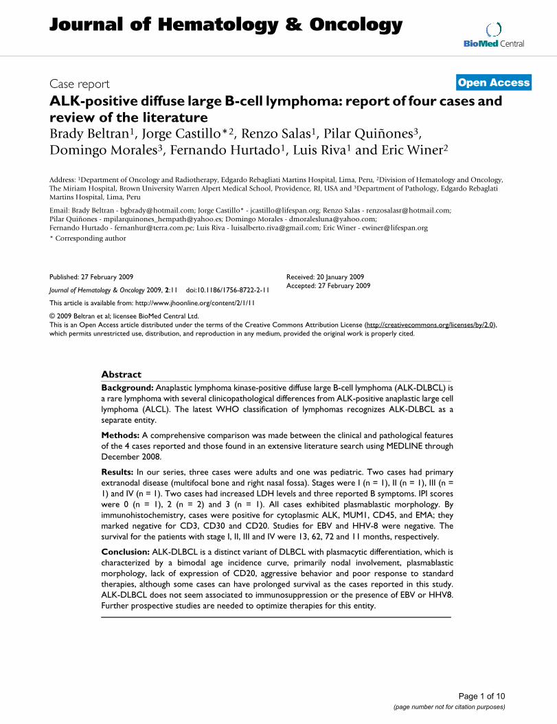
Open AcceCase reportALK-positive diffuse large B-cell lymphoma: report of four cases and review of the literatureBrady Beltran1, Jorge Castillo*2, Renzo Salas1, Pilar Quiñones3, Domingo Morales3, Fernando Hurtado1, Luis Riva1 and Eric Winer2Address: 1Department of Oncology and Radiotherapy, Edgardo Rebagliati Martins Hospital, Lima, Peru, 2Division of Hematology and Oncology, The Miriam Hospital, Brown University Warren Alpert Medical School, Providence, RI, USA and 3Department of Pathology, Edgardo Rebaglati Martins Hospital, Lima, Peru
Email: Brady Beltran - [email protected]; Jorge Castillo* - [email protected]; Renzo Salas - [email protected]; Pilar Quiñones - [email protected]; Domingo Morales - [email protected]; Fernando Hurtado - [email protected]; Luis Riva - [email protected]; Eric Winer - [email protected]
* Corresponding author
AbstractBackground: Anaplastic lymphoma kinase-positive diffuse large B-cell lymphoma (ALK-DLBCL) isa rare lymphoma with several clinicopathological differences from ALK-positive anaplastic large celllymphoma (ALCL). The latest WHO classification of lymphomas recognizes ALK-DLBCL as aseparate entity.
Methods: A comprehensive comparison was made between the clinical and pathological featuresof the 4 cases reported and those found in an extensive literature search using MEDLINE throughDecember 2008.
Results: In our series, three cases were adults and one was pediatric. Two cases had primaryextranodal disease (multifocal bone and right nasal fossa). Stages were I (n = 1), II (n = 1), III (n =1) and IV (n = 1). Two cases had increased LDH levels and three reported B symptoms. IPI scoreswere 0 (n = 1), 2 (n = 2) and 3 (n = 1). All cases exhibited plasmablastic morphology. Byimmunohistochemistry, cases were positive for cytoplasmic ALK, MUM1, CD45, and EMA; theymarked negative for CD3, CD30 and CD20. Studies for EBV and HHV-8 were negative. Thesurvival for the patients with stage I, II, III and IV were 13, 62, 72 and 11 months, respectively.
Conclusion: ALK-DLBCL is a distinct variant of DLBCL with plasmacytic differentiation, which ischaracterized by a bimodal age incidence curve, primarily nodal involvement, plasmablasticmorphology, lack of expression of CD20, aggressive behavior and poor response to standardtherapies, although some cases can have prolonged survival as the cases reported in this study.ALK-DLBCL does not seem associated to immunosuppression or the presence of EBV or HHV8.Further prospective studies are needed to optimize therapies for this entity.
Published: 27 February 2009
Journal of Hematology & Oncology 2009, 2:11 doi:10.1186/1756-8722-2-11
Received: 20 January 2009Accepted: 27 February 2009
This article is available from: http://www.jhoonline.org/content/2/1/11
© 2009 Beltran et al; licensee BioMed Central Ltd. This is an Open Access article distributed under the terms of the Creative Commons Attribution License (http://creativecommons.org/licenses/by/2.0), which permits unrestricted use, distribution, and reproduction in any medium, provided the original work is properly cited.
Page 1 of 10(page number not for citation purposes)
Journal of Hematology & Oncology 2009, 2:11 http://www.jhoonline.org/content/2/1/11
BackgroundDLBCL is the most common histological variant of NHL.It encompasses multiple subtypes and has heterogeneousclinical and pathological features. In 1997, Delsol andcolleagues reported seven cases of a distinct variant ofDLBCL expressing rearrangements of the ALK gene [1].The plasmablastic appearance and CD20-negativity ofALK-DLBCL makes this entity a potentially diagnosticchallenge with a broad differential diagnosis. Clinically,ALK-DLBCL shows very aggressive behavior, high relapserate and lack of response to standard regimens.
Although in the initial report by Delsol and colleagues theclassic ALK gene rearrangement observed in ALCL couldnot be shown [1], modern techniques have been able toprove recurrent chromosomal abnormalities in ALK-DLBCL. The most commonly observed cytogenetic abnor-mality is t(2;17)(p23;q23) or clathrin/ALK [2-10]. Theclassic ALCL-related t(2;5)(p23;q35) or nucleophosmin/ALK has also been described [11-13]. Other rare cytoge-netic abnormalities have been reported [14,15].
The main objective of this study was to describe the clin-icopathological characteristics of four additional cases ofALK-DLBCL and compare them with those of 46 litera-ture-reported cases.
Materials and methodsFour cases of ALK-DLBCL were identified from the Hema-tology and Medical Oncology consultation files at theEdgardo Rebagliati Martins Hospital in Lima, Perubetween January 1, 1997 and June 30, 2008. Clinical andlaboratory information for each of the four patients wasobtained through physician interview and medical chartreview, after approval of this study by the IRB. Routinehematoxylin and eosin-stained sections were preparedfrom formalin-fixed and/or B5-fixed paraffin blocks.Immunohistochemical analysis included a broad panel ofantibodies against ALK1 (Dako, Carpinteria, CA; dilution1:50), CD45 (Dako; dilution 1:400), CD4 (Novocastra,Newcastle upon Tyne, UK; dilution 1:20), CD56 (Sanbio,Uden, The Netherlands; 1:200), CD20 (Dako; dilution1:100), CD79a (Dako; dilution 1:25) and light chains ofimmunoglobulin. The samples were also stained forCD30 (Novocastra; dilution 1:100) and EMA (Dako; dilu-tion 1:50), which are usually expressed by ALCL cells.
Immunohistochemical studies for Epstein Barr virus(EBV) and human herpesvirus 8 (HHV-8) were performedat the Department of Pathology of the Rhode Island Hos-pital in Providence, RI. EBV clone was CS1-4 (Dako; dilu-tion 1:500) obtained through heat retrieval pretreatmentwith Target Retrieval solution (Dako) for 25 minutes.HHV8 clone was 13B10 (Vector Laboratories, Burlin-game, CA; dilution 1:50) obtained through heat retrieval
pretreatment with Target Retrieval solution (Dako) for 25minutes. Cytogenetic studies by FISH looking for ALKgene rearrangement were performed at the Department ofCytogenetics of the Tufts Medical Center in Boston, MA.The immunohistochemical analysis for HHV-8 andcytogenetic studies were performed in only two of thepresent cases. Further studies could not be attempted onthe other two cases due to lack of available remainingspecimen.
For the review, we performed a literature search usingPubmed/MEDLINE looking for articles reporting clinico-pathological data in patients with ALK-DLBCL throughDecember 2008. Eighteen articles were considered for thisreview. Data were gathered on age, sex, pattern of ALKexpression, ALK gene rearrangement variety, expression ofCD30, CD45, plasma cell, B-cell, T-cell and NK-cell mark-ers, EMA and light chain, heavy chain gene and T-cellreceptor gene rearrangements, presence of EBV, site of pri-mary disease, clinical stage, LDH levels, IPI score, therapyat presentation and at relapse, outcome, survival inmonths and cause of death. Survival analyses wereattempted using Kaplan-Meier estimates for age, sex, T-cellmarker expression, primary site of presentation, clinicalstage, LDH levels and IPI score. All reported p-values aretwo-sided.
ResultsCase ReportsA summary of the clinical features of the four patients isprovided in Table 1.
Case 1A 27-year-old male patient presented with multifocalbone lesions detected with bone scintigraphy. Patient alsoreported the presence of B symptoms. LDH levels wereelevated. Serum protein electrophoresis (SPEP) did notshow a monoclonal spike. A computed tomography (CT)scan of the thorax and abdomen showed no mass lesionsor additional lymphadenopathy. An incisional biopsy ofbone was performed, which showed a diffuse lymphomaof plasmablastic appearance. A staging bone marrow aspi-ration and biopsy was positive for involvement by lym-phoma. Patient was staged as IVB and underwent sixcycles of EPOCH (cyclophosphamide, vincristine, doxo-rubicin, etoposide and prednisone) with persistent bonemarrow infiltration at the end of the initial therapy. He iscurrently receiving hyperCVAD (hyperfractionated cyclo-phosphamide, vincristine, doxorubicin and dexametha-sone alternating with cytarabine and methotrexate). At 11months, he was alive with persistent disease.
Case 2A 41-year-old male patient presented with history of nasalobstruction for one month. He was otherwise asympto-
Page 2 of 10(page number not for citation purposes)

Journal of Hematology & Oncology 2009, 2:11 http://www.jhoonline.org/content/2/1/11
matic with an excellent performance status and had nosignificant past medical history. Hematologic, basic meta-bolic, liver function studies and LDH levels were withinnormal limits. SPEP did not show monoclonal spike. CTscan of the head, neck, chest, abdomen and pelvisrevealed only a mass in the right nasal fossa. Biopsy oftumor was performed revealing a tumor with plasmablas-tic morphology. Staging bone marrow was negative. Dueto an initial diagnosis of solitary plasmacytoma, patientreceived involved field radiation therapy. At 13 months,he was alive and free of disease.
Case 3A 13-year-old female patient presented with a rapidlyenlarging left neck mass and B symptoms. Physical exam-ination and radiological studies showed axillary andmediastinal lymph nodes and costal bone involvement. Abiopsy of the cervical mass was performed and revealed anaggressive lymphoma with plasmablastic features. Bonemarrow biopsy was negative for lymphoma. SPEP was notperformed. She received the regimen LNH96-2002, whichis based on induction with vincristine, prednisone, cyclo-phosphamide, daunorubicin, L-asparaginase and meth-otrexate; followed by consolidation based oncyclophosphamide, cytarabine, methotrexate then inten-sification with vincristine and doxorubicin and mainte-nance based on methotrexate and mercaptopurine. Shehad a complete response to the induction phase and thenreceived consolidation and maintenance. She has 62months alive and free from recurrence.
Case 4A 70-year-old male patient presented with cervical, axil-lary and inguinal lymphadenopathy without B symp-toms. Bone marrow was not involved. He had aperformance status of 2. Cervical lymph node biopsy wasdone showing a diffuse lymphoma with plasmablasticappearance. LDH levels were within normal limits. SPEP
was not performed. Patient was considered stage IIIB. IPIscore was 3 out of 5. He received CHOP-21 regimen for sixcycles and achieved a complete response. He is alive with72 months free from recurrence.
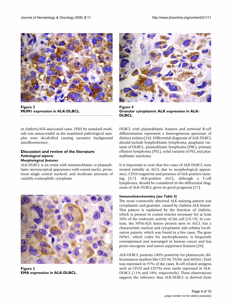
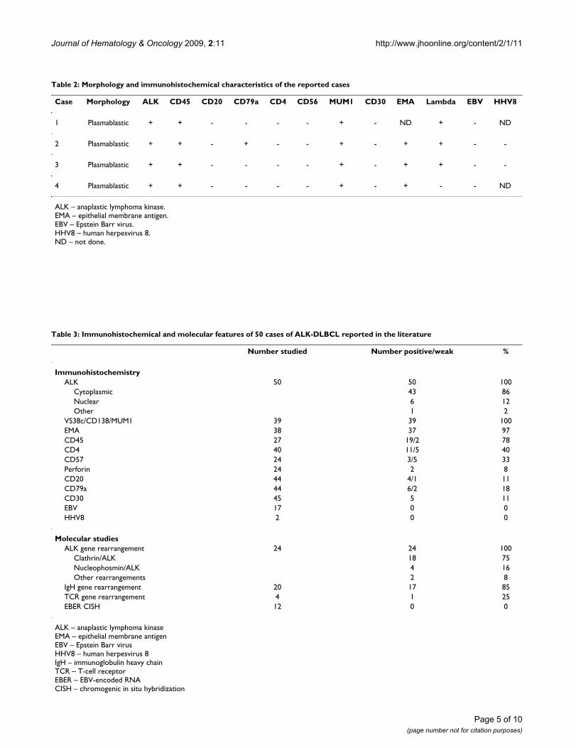
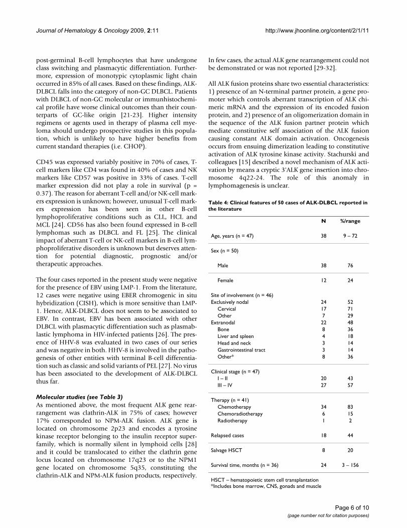
Pathological aspects of the reported casesAll four cases showed plasmablastic morphologic featureswith effacement of the normal architecture by sheets oftumor cells. The neoplastic cells in all cases were largewith round, regular, with centrally located nuclei, dis-persed chromatin, single central, prominent nucleolus,and moderate eosinophilic or amphophilic cytoplasm.Table 2 provides a summary of the immunohistochemicalcharacteristics in the four reported cases. All tested caseswere positive for CD45, MUM1 (Figure 1), and EMA (Fig-ure 2), and were negative for CD4, CD20 (Figure 3) andCD30. All cases were positive for ALK in a granular cyto-plasmic distribution (Figure 4), which has been described
Table 1: Clinical characteristics of the reported cases
Case Age Sex Primary site Bone marrow involvement Stage IPI Therapy Survival (Months) Outcome
1 27 M Bone Yes IVB 3 HyperCVAD 11 Alive, with disease
2 41 F Nasal fossa No IA 0 Radiotherapy 13 Alive, NED
3 13 F Cervical LN No IIB 2 LNH96-2002 62 Alive, NED
4 70 M Cervical LN No IIIB 3 CHOP 72 Alive, NED
IPI – International Prognostic Index.NED – no evidence of disease.LN – lymph node.HyperCVAD – hyperfractionated cyclophosphamide, vincristine, doxorubicin and dexamethasone alternating with cytarabine and methotrexate.CHOP – cyclophosphamide, doxorubicin, vincristine, prednisone.
Negative CD20 expression in ALK-DLBCLFigure 1Negative CD20 expression in ALK-DLBCL.
Page 3 of 10(page number not for citation purposes)
Journal of Hematology & Oncology 2009, 2:11 http://www.jhoonline.org/content/2/1/11
in clathrin/ALK-associated cases. FISH by standard meth-ods was unsuccessful as the examined pathological sam-ples were decalcified causing excessive backgroundautofluorescence.
Discussion and review of the literaturePathological aspectsMorphological featuresALK-DLBCL is an entity with immunoblastic or plasmab-lastic microscopical appearance with round nuclei, prom-inent single central nucleoli, and moderate amounts ofvariably eosinophilic cytoplasm.
DLBCL with plasmablastic features and terminal B-celldifferentiation represents a heterogeneous spectrum ofdistinct entities [16]. Differential diagnosis of ALK-DLBCLshould include lymphoblastic lymphoma, anaplastic var-iants of DLBCL, plasmablastic lymphoma (PBL), primaryeffusion lymphoma (PEL), solid variants of PEL and plas-mablastic myeloma.
It is important to note that few cases of ALK-DLBCL weretreated initially as ALCL due to morphological appear-ance, CD20-negativity and presence of ALK-positive stain-ing [5,7]. ALK-positive ALCL, although a T-celllymphoma, should be considered in the differential diag-nosis of ALK-DLBCL given its good prognosis [17].
Immunohistochemistry (see Table 3)The most commonly observed ALK staining pattern wascytoplasmic and granular, caused by clathrin-ALK fusion.This pattern is explained by the function of clathrin,which is present in coated vesicles necessary for at least50% of the endocytic activity of the cell [18,19]. In con-trast, the NPM-ALK fusion protein seen in ALCL has acharacteristic nuclear and cytoplasmic sub-cellular locali-zation pattern, which was found in a few cases. The geneNPM1, which codes for nucleophosmin, is frequentlyoverexpressed and rearranged in human cancer and hasproto-oncogenic and tumor suppressor features [20].
ALK-DLBCL presents 100% positivity for plasmacytic dif-ferentiation markers like CD138, VS38c and MUM1; EMAwas expressed in 97% of the cases. B-cell related antigenssuch as CD20 and CD79a were rarely expressed in ALK-DLBCL (11% and 18%, respectively). These observationssupport the inference that ALK-DLBCL is derived from
MUM1 expression in ALK-DLBCLFigure 2MUM1 expression in ALK-DLBCL.
EMA expression in ALK-DLBCLFigure 3EMA expression in ALK-DLBCL.
Granular cytoplasmic ALK expression in ALK-DLBCLFigure 4Granular cytoplasmic ALK expression in ALK-DLBCL.
Page 4 of 10(page number not for citation purposes)
Journal of Hematology & Oncology 2009, 2:11 http://www.jhoonline.org/content/2/1/11
Page 5 of 10(page number not for citation purposes)
Table 2: Morphology and immunohistochemical characteristics of the reported cases
Case Morphology ALK CD45 CD20 CD79a CD4 CD56 MUM1 CD30 EMA Lambda EBV HHV8
1 Plasmablastic + + - - - - + - ND + - ND
2 Plasmablastic + + - + - - + - + + - -
3 Plasmablastic + + - - - - + - + + - -
4 Plasmablastic + + - - - - + - + - - ND
ALK – anaplastic lymphoma kinase.EMA – epithelial membrane antigen.EBV – Epstein Barr virus.HHV8 – human herpesvirus 8.ND – not done.
Table 3: Immunohistochemical and molecular features of 50 cases of ALK-DLBCL reported in the literature
Number studied Number positive/weak %
ImmunohistochemistryALK 50 50 100
Cytoplasmic 43 86Nuclear 6 12Other 1 2
VS38c/CD138/MUM1 39 39 100EMA 38 37 97CD45 27 19/2 78CD4 40 11/5 40CD57 24 3/5 33Perforin 24 2 8CD20 44 4/1 11CD79a 44 6/2 18CD30 45 5 11EBV 17 0 0HHV8 2 0 0
Molecular studiesALK gene rearrangement 24 24 100
Clathrin/ALK 18 75Nucleophosmin/ALK 4 16Other rearrangements 2 8
IgH gene rearrangement 20 17 85TCR gene rearrangement 4 1 25EBER CISH 12 0 0
ALK – anaplastic lymphoma kinaseEMA – epithelial membrane antigenEBV – Epstein Barr virusHHV8 – human herpesvirus 8IgH – immunoglobulin heavy chainTCR – T-cell receptorEBER – EBV-encoded RNACISH – chromogenic in situ hybridization
Journal of Hematology & Oncology 2009, 2:11 http://www.jhoonline.org/content/2/1/11
post-germinal B-cell lymphocytes that have undergoneclass switching and plasmacytic differentiation. Further-more, expression of monotypic cytoplasmic light chainoccurred in 85% of all cases. Based on these findings, ALK-DLBCL falls into the category of non-GC DLBCL. Patientswith DLBCL of non-GC molecular or immunhistochemi-cal profile have worse clinical outcomes than their coun-terparts of GC-like origin [21-23]. Higher intensityregimens or agents used in therapy of plasma cell mye-loma should undergo prospective studies in this popula-tion, which is unlikely to have higher benefits fromcurrent standard therapies (i.e. CHOP).
CD45 was expressed variably positive in 70% of cases, T-cell markers like CD4 was found in 40% of cases and NKmarkers like CD57 was positive in 33% of cases. T-cellmarker expression did not play a role in survival (p =0.37). The reason for aberrant T-cell and/or NK-cell mark-ers expression is unknown; however, unusual T-cell mark-ers expression has been seen in other B-celllymphoproliferative conditions such as CLL, HCL andMCL [24]. CD56 has also been found expressed in B-celllymphomas such as DLBCL and FL [25]. The clinicalimpact of aberrant T-cell or NK-cell markers in B-cell lym-phoproliferative disorders is unknown but deserves atten-tion for potential diagnostic, prognostic and/ortherapeutic approaches.
The four cases reported in the present study were negativefor the presence of EBV using LMP-1. From the literature,12 cases were negative using EBER chromogenic in situhybridization (CISH), which is more sensitive than LMP-1. Hence, ALK-DLBCL does not seem to be associated toEBV. In contrast, EBV has been associated with otherDLBCL with plasmacytic differentiation such as plasmab-lastic lymphoma in HIV-infected patients [26]. The pres-ence of HHV-8 was evaluated in two cases of our seriesand was negative in both. HHV-8 is involved in the patho-genesis of other entities with terminal B-cell differentia-tion such as classic and solid variants of PEL [27]. No virushas been associated to the development of ALK-DLBCLthus far.
Molecular studies (see Table 3)As mentioned above, the most frequent ALK gene rear-rangement was clathrin-ALK in 75% of cases; however17% corresponded to NPM-ALK fusion. ALK gene islocated on chromosome 2p23 and encodes a tyrosinekinase receptor belonging to the insulin receptor super-family, which is normally silent in lymphoid cells [28]and it could be translocated to either the clathrin genelocus located on chromosome 17q23 or to the NPM1gene located on chromosome 5q35, constituting theclathrin-ALK and NPM-ALK fusion products, respectively.
In few cases, the actual ALK gene rearrangement could notbe demonstrated or was not reported [29-32].
All ALK fusion proteins share two essential characteristics:1) presence of an N-terminal partner protein, a gene pro-moter which controls aberrant transcription of ALK chi-meric mRNA and the expression of its encoded fusionprotein, and 2) presence of an oligomerization domain inthe sequence of the ALK fusion partner protein whichmediate constitutive self association of the ALK fusioncausing constant ALK domain activation. Oncogenesisoccurs from ensuing dimerization leading to constitutiveactivation of ALK tyrosine kinase activity. Stachurski andcolleagues [15] described a novel mechanism of ALK acti-vation by means a cryptic 3'ALK gene insertion into chro-mosome 4q22-24. The role of this anomaly inlymphomagenesis is unclear.
Table 4: Clinical features of 50 cases of ALK-DLBCL reported in the literature
N %/range
Age, years (n = 47) 38 9 – 72
Sex (n = 50)
Male 38 76
Female 12 24
Site of involvement (n = 46)Exclusively nodal 24 52
Cervical 17 71Other 7 29
Extranodal 22 48Bone 8 36Liver and spleen 4 18Head and neck 3 14Gastrointestinal tract 3 14Other* 8 36
Clinical stage (n = 47)I – II 20 43III – IV 27 57
Therapy (n = 41)Chemotherapy 34 83Chemoradiotherapy 6 15Radiotherapy 1 2
Relapsed cases 18 44
Salvage HSCT 8 20
Survival time, months (n = 36) 24 3 – 156
HSCT – hematopoietic stem cell transplantation*Includes bone marrow, CNS, gonads and muscle
Page 6 of 10(page number not for citation purposes)

Journal of Hematology & Oncology 2009, 2:11 http://www.jhoonline.org/content/2/1/11
Immunoglobulin heavy chain gene rearrangements weredetected by PCR analysis in 17 of 20 studied cases (85%).The previous finding, along with the expression of mono-typic cytoplasmic immunoglobulin light chain, confirmsthe B-cell lineage of this disorder.
Clinical aspects (see Table 4)Age and sex distributionForty-seven cases of ALK-DLBCL reported age of presenta-tion. The average age of presentation was 38 years, rangingfrom 9 to 72 years of age. Despite the small amount ofcases, we can already observe a bimodal age distribution.Eleven cases of ALK-DLBCL have been reported in pediat-ric population [2,5,7,8,12], accounting for 24% of thetotal number of cases. In patients younger than 18 years,the average age of presentation was 12.4 years and inadults it was 43.4 years. There was no difference in sur-vival between pediatric and adult cases (p = 0.97; Figure5), despite more intensive therapies in pediatric popula-tion.
In regards of sex distribution, the male to female ratio was3:1; female cases accounted for 23% of the cases. In pedi-atric cases, the male to female ratio was 1.8:1 and in adults4.3:1. There was no statistical difference between the over-all survival of men compared to women (p = 0.45).
Sites of involvementData on primary sites of presentation were available in 46cases. Twenty-four cases (52%) were exclusively nodal inorigin. The most commonly affected areas were cervicaland mediastinal. Few cases presented with generalizedlymphadenopathy. The remaining cases (48%) had someextranodal component and from these, only 6 were exclu-sively extranodal. Most common extranodal sites of dis-
ease were bone, liver, spleen, gastrointestinal tract and thehead and neck region.
ALK-DLBCL differs somewhat from other subtypes ofDLBCL with plasmacytic differentiation. Plasmablasticlymphoma (PBL), a CD20-negative DLBCL associated toHIV and EBV coinfection, tends to present with extranodalinvolvement, usually in oral and gastrointestinal sites;nodal presentation in PBL has been reported in only 6%of the cases [26]. In a similar fashion, PEL, another CD20-negative DLBCL seen exclusively in association with HHV-8, tends to present in body cavities such as pleura and per-itoneum [27]. Although nodal PEL has been described[33], available data on extracavitary or solid variants ofPEL is very limited. In the survival analysis, there was nostatistical difference between nodal and extranodal sites ofinvolvement (p = 0.58).
Clinical stage and IPI scoreFrom 47 ALK-DLBCL cases of the literature, advancedstage (i.e. III and IV) was reported in 57% of the cases; theremainder 43% presented with stages I or II. As observedin other malignant lymphoproliferative disorders, clinicalstage had a strong correlation with survival (p = 0.0055;Figure 6).
The IPI score has been accepted as the standard methodfor risk stratification in patients with DLBCL [34]. Unfor-tunately, only 8 of the 50 reported cases (17%) had avail-able data on IPI scores, including the 4 cases reported inthis study. Furthermore, the gathered data did not allowthe authors to calculate IPI scores as serum LDH levels andperformance status were seldom reported. Survival analy-ses using IPI scores or LDH levels were not performed.
Kaplan-Meier survival estimates according to age in 50 ALK-DLBCL cases from the literatureFigure 5Kaplan-Meier survival estimates according to age in 50 ALK-DLBCL cases from the literature.
Kaplan-Meier survival estimates according to clinical stage in 50 ALK-DLBCL cases from the literatureFigure 6Kaplan-Meier survival estimates according to clinical stage in 50 ALK-DLBCL cases from the literature.
Page 7 of 10(page number not for citation purposes)

Journal of Hematology & Oncology 2009, 2:11 http://www.jhoonline.org/content/2/1/11
Therapy and relapsesData on therapy was available in 41 ALK-DLBCL cases. Ofthese 32 cases (83%) received combination chemother-apy, 6 cases (15%) received chemoradiotherapy and onecase (2%) received radiotherapy. Only one case [6]received immunotherapy with rituximab despite theCD20-negative nature of ALK-DLBCL; this patient died oflymphoma 6 months after diagnosis. From the 34 casestreated with chemotherapy, 12 cases (38%) were treatedwith CHOP and the remaining 20 cases (62%) weretreated with more intensive regimens. From the 12 casestreated with CHOP, 6 cases (50%) needed more therapydue to relapsing disease and 4 cases (33%) died of pro-gressive lymphoma.
In the 11 ALK-DLBCL pediatric cases, all regimens usedwere highly intensive (i.e. BFM90 [35], LMB89 [36],LMB96 [37], POG8719 [38]). Most of these regimens areused successfully to treat children with lymphoblastic,Burkitt and aggressive B-cell lymphomas. Of note, someof the patients were enrolled in randomized clinical trialsthat are still undergoing recruitment (i.e. ALCL99 [39]). Incontrast with the reported efficacy of these regimens inother types of aggressive NHL, the success in ALK-DLBCLwas rather moderate with 6 patients (55%) alive at thetime of report.
In total, 18 ALK-DLBCL cases (44%) experienced refrac-tory or relapsing disease. Most salvage regimens werebased on platinum-containing compounds (i.e. ESHAP,DHAP, ICE). Hematopoietic stem cell transplantation(HSCT) was performed in 8 of the refractory or relapsingcases (44%) [1,5,7,10,11,14,30]. Four patients receivedautologous HSCT, one patient underwent allogeneicHSCT and 3 patients were treated with non-specifiedHSCT. All but one of the cases died after transplantation;the range of survival in transplanted cases was between 3and 44 months after diagnosis. The case treated with allo-geneic HSCT died of thrombotic thrombocytopenic pur-pura (TTP) 7 months after transplantation [30].
From the 50 cases of the literature, the authors couldobserve that standard CHOP regimen seems inadequateto treat ALK-DLBCL given evidence of progressive diseaseand multiple recurrences. The lack of expression of CD20antigen in most cases of ALK-DLBCL makes the therapeu-tic role of rituximab rather unclear. Nonetheless, rituxi-mab should be used in the few CD20-expressing ALK-DLBCL cases [32].
Outcome and survivalALK-DLBCL was fatal in 56% of the cases. The most com-mon cause of death was progressive lymphoma, observedin 90% of the reported cases. Other causes of deathincluded TTP and infectious complications. The average
survival for the 36 cases in which survival times werereported was 24 months. Multiple clinical factors such asage, sex and nodal primary sites do not seem to correlatewith survival in ALK-DLBCL. The strongest factor associ-ated to survival in ALK-DLBCL cases from the literaturewas clinical stage at presentation; patients with advancedstages had a median survival of 18 months while patientswith earlier stages have not reached their median survival(Figure 6).
ConclusionALK-DLBCL is a distinct subtype of DLBCL with plasma-cytic differentiation that affects pediatric and adultpatients. It has characteristic genetic abnormalities andcorresponding specific ALK-staining patterns with a prog-nosis that depends largely on clinical stage. The clinicalcourse of ALK-DLBCL is aggressive with primary refractorydisease and high relapse rates. The classical CHOP regi-men appears insufficient to treat this condition andnewer, more intensive therapies are needed. Given itsCD20-negativity, the role of rituximab in the treatment ofALK-DLBCL is unclear. It would be of interest to try agentsborrowed from plasma cell myeloma regimens or agentsactive in novel pathways in combination with chemother-apy given ALK-DLBCL plasmacytic nature. Despite thisaggressiveness, some cases, even in advanced stages, couldhave prolonged survival times as the authors describe inthe present article. Further basic and clinical research isnecessary to improve our understanding of the biology ofthe different subtypes of DLBCL with plasmacytic differ-entiation in order to identify patients with a better prog-nosis and to develop newer therapeutic techniques.
ConsentWritten informed consent was obtained directly from 3patients and from the parents of 1 patient for publicationof this case report and any accompanying images.
Competing interestsThe authors declare that they have no competing interests.
Authors' contributionsBB, RS, FH and LR referred the patients for this report. PQand DM carried out pathology studies in Peru. JC per-formed the survival statistical analyses. JC and EW coordi-nated pathology studies in the U.S. BB, JC and EWprepared the manuscript. All authors read and approvedthe final manuscript.
AcknowledgementsThe authors would like to thank Dr. Ronald DeLellis, Chief of the Depart-ment of Pathology at Rhode Island Hospital in Providence, RI and Dr. Janet Cowan, Director of the Department of Cytogenetics at Tufts Medical Center in Boston, MA for their support in this study.
Page 8 of 10(page number not for citation purposes)

Journal of Hematology & Oncology 2009, 2:11 http://www.jhoonline.org/content/2/1/11
References1. Delsol G, Lamant L, Mariame B, Pulford K, Dastugue N, Brousset P,
Rigal-Huguet F, al Saati T, Cerretti DP, Morris SW, Mason DY: Anew subtype of large B-cell lymphoma expressing the ALKkinase and lacking the 2; 5 translocation. Blood 1997,89(5):1483-1490.
2. Bubala H, Maldyk J, Wlodarska I, Sonta-Jakimczyk D, Szczepanski T:ALK-positive diffuse large B-cell lymphoma. Pediatr Blood Can-cer 2006, 46(5):649-653.
3. Chikatsu N, Kojima H, Suzukawa K, Shinagawa A, Nagasawa T, OzawaH, Yamashita Y, Mori N: ALK+, CD30-, CD20- large B-cell lym-phoma containing anaplastic lymphoma kinase (ALK) fusedto clathrin heavy chain gene (CLTC). Mod Pathol 2003,16(8):828-832.
4. Choung HS, Kim HJ, Kim WS, Kim K, Kim SH: [Cytomorphologyand molecular characterization of CLTC-ALK rearrange-ment in 2 cases of ALK-positive diffuse large B-cell lym-phoma with extensive bone marrow involvement]. Korean JLab Med 2008, 28(2):89-94.
5. De Paepe P, Baens M, van Krieken H, Verhasselt B, Stul M, Simons A,Poppe B, Laureys G, Brons P, Vandenberghe P, Speleman F, Praet M,De Wolf-Peeters C, Marynen P, Wlodarska I: ALK activation bythe CLTC-ALK fusion is a recurrent event in large B-celllymphoma. Blood 2003, 102(7):2638-2641.
6. Gascoyne RD, Lamant L, Martin-Subero JI, Lestou VS, Harris NL,Muller-Hermelink HK, Seymour JF, Campbell LJ, Horsman DE,Auvigne I, Espinos E, Siebert R, Delsol G: ALK-positive diffuselarge B-cell lymphoma is associated with Clathrin-ALK rear-rangements: report of 6 cases. Blood 2003, 102(7):2568-2573.
7. Gesk S, Gascoyne RD, Schnitzer B, Bakshi N, Janssen D, Klapper W,Martin-Subero JI, Parwaresch R, Siebert R: ALK-positive diffuselarge B-cell lymphoma with ALK-Clathrin fusion belongs tothe spectrum of pediatric lymphomas. Leukemia 2005,19(10):1839-1840.
8. Isimbaldi G, Bandiera L, d'Amore ES, Conter V, Milani M, Mussolin L,Rosolen A: ALK-positive plasmablastic B-cell lymphoma withthe clathrin-ALK gene rearrangement. Pediatr Blood Cancer2006, 46(3):390-391.
9. McManus DT, Catherwood MA, Carey PD, Cuthbert RJ, AlexanderHD: ALK-positive diffuse large B-cell lymphoma of the stom-ach associated with a clathrin-ALK rearrangement. HumPathol 2004, 35(10):1285-1288.
10. Momose S, Tamaru J, Kishi H, Mikata I, Mori M, Toyozumi Y, ItoyamaS: Hyperactivated STAT3 in ALK-positive diffuse large B-celllymphoma with clathrin-ALK fusion. Hum Pathol 2009,40(1):75-82.
11. Adam P, Katzenberger T, Seeberger H, Gattenlohner S, Wolf J, Stein-lein C, Schmid M, Muller-Hermelink HK, Ott G: A case of a diffuselarge B-cell lymphoma of plasmablastic type associated withthe t(2;5)(p23;q35) chromosome translocation. Am J SurgPathol 2003, 27(11):1473-1476.
12. Onciu M, Behm FG, Downing JR, Shurtleff SA, Raimondi SC, Ma Z,Morris SW, Kennedy W, Jones SC, Sandlund JT: ALK-positive plas-mablastic B-cell lymphoma with expression of the NPM-ALKfusion transcript: report of 2 cases. Blood 2003,102(7):2642-2644.
13. Rudzki Z, Rucinska M, Jurczak W, Skotnicki AB, Maramorosz-Kurian-owicz M, Mruk A, Pirog K, Utych G, Bodzioch P, Srebro-Stariczyk M,Wlodarska I, Stachura J: ALK-positive diffuse large B-cell lym-phoma: two more cases and a brief literature review. Pol JPathol 2005, 56(1):37-45.
14. Lee HW, Kim K, Kim W, Ko YH: ALK-positive diffuse large B-cell lymphoma: report of three cases. Hematol Oncol 2008,26(2):108-113.
15. Stachurski D, Miron PM, Al-Homsi S, Hutchinson L, Harris NL, WodaB, Wang SA: Anaplastic lymphoma kinase-positive diffuselarge B-cell lymphoma with a complex karyotype and cryptic3' ALK gene insertion to chromosome 4 q22-24. Hum Pathol2007, 38(6):940-945.
16. Teruya-Feldstein J: Diffuse large B-cell lymphomas with plas-mablastic differentiation. Curr Oncol Rep 2005, 7(5):357-363.
17. Gascoyne RD, Aoun P, Wu D, Chhanabhai M, Skinnider BF, GreinerTC, Morris SW, Connors JM, Vose JM, Viswanatha DS, Coldman A,Weisenburger DD: Prognostic significance of anaplastic lym-phoma kinase (ALK) protein expression in adults with ana-plastic large cell lymphoma. Blood 1999, 93(11):3913-3921.
18. Gong Q, Huntsman C, Ma D: Clathrin-independent internaliza-tion and recycling. J Cell Mol Med 2008, 12(1):126-144.
19. Rappoport JZ: Focusing on clathrin-mediated endocytosis. Bio-chem J 2008, 412(3):415-423.
20. Grisendi S, Mecucci C, Falini B, Pandolfi PP: Nucleophosmin andcancer. Nat Rev Cancer 2006, 6(7):493-505.
21. Alizadeh AA, Eisen MB, Davis RE, et al.: Distinct types of diffuselarge B-cell lymphoma identified by gene expression profil-ing. Nature 2000, 403(6769):503-511.
22. Hans CP, Weisenburger DD, Greiner TC, Gascoyne RD, Delabie J,Ott G, Muller-Hermelink HK, Campo E, Braziel RM, Jaffe ES, Pan Z,Farinha P, Smith LM, Falini B, Banham AH, Rosenwald A, Staudt LM,Connors JM, Armitage JO, Chan WC: Confirmation of the molec-ular classification of diffuse large B-cell lymphoma by immu-nohistochemistry using a tissue microarray. Blood 2004,103(1):275-282.
23. Rosenwald A, Wright G, Chan WC, et al.: The use of molecularprofiling to predict survival after chemotherapy for diffuselarge-B-cell lymphoma. N Engl J Med 2002, 346(25):1937-1947.
24. Kaleem Z, White G, Zutter MM: Aberrant expression of T-cell-associated antigens on B-cell non-Hodgkin lymphomas. Am JClin Pathol 2001, 115(3):396-403.
25. Isobe Y, Sugimoto K, Takeuchi K, Ando J, Masuda A, Mori T, OshimiK: Neural cell adhesion molecule (CD56)-positive B-cell lym-phoma. Eur J Haematol 2007, 79(2):166-169.
26. Castillo J, Pantanowitz L, Dezube BJ: HIV-associated plasmablas-tic lymphoma: lessons learned from 112 published cases. AmJ Hematol 2008, 83(10):804-809.
27. Brimo F, Michel RP, Khetani K, Auger M: Primary effusion lym-phoma: a series of 4 cases and review of the literature withemphasis on cytomorphologic and immunocytochemical dif-ferential diagnosis. Cancer 2007, 111(4):224-233.
28. Morris SW, Kirstein MN, Valentine MB, Dittmer KG, Shapiro DN,Saltman DL, Look AT: Fusion of a kinase gene, ALK, to a nucle-olar protein gene, NPM, in non-Hodgkin's lymphoma. Science1994, 263(5151):1281-1284.
29. Colomo L, Loong F, Rives S, Pittaluga S, Martinez A, Lopez-GuillermoA, Ojanguren J, Romagosa V, Jaffe ES, Campo E: Diffuse large B-celllymphomas with plasmablastic differentiation represent aheterogeneous group of disease entities. Am J Surg Pathol 2004,28(6):736-747.
30. Ishii K, Yamamoto Y, Nomura S: [CD30-negative diffuse large B-cell lymphoma expressing ALK]. Rinsho Ketsueki 2005,46(7):501-506.
31. Reichard KK, McKenna RW, Kroft SH: ALK-positive diffuse largeB-cell lymphoma: report of four cases and review of the lit-erature. Mod Pathol 2007, 20(3):310-319.
32. Wang WY, Ma ZG, Li GD, Liu WP, Zhong L, Wang Y, Li JM, Li L, JiangW, Tang Y, Liao DY: [Diffuse large B-cell lymphoma withexpression of anaplastic lymphoma kinase protein: clinico-pathologic and immunohistochemical study of 5 cases].Zhonghua Bing Li Xue Za Zhi 2006, 35(9):529-534.
33. Carbone A, Gloghini A, Vaccher E, Cerri M, Gaidano G, Dalla-FaveraR, Tirelli U: Kaposi's sarcoma-associated herpesvirus/humanherpesvirus type 8-positive solid lymphomas: a tissue-basedvariant of primary effusion lymphoma. J Mol Diagn 2005,7(1):17-27.
34. A predictive model for aggressive non-Hodgkin's lymphoma.The International Non-Hodgkin's Lymphoma PrognosticFactors Project. N Engl J Med 1993, 329(14):987-994.
35. von Stackelberg A, Hartmann R, Buhrer C, Fengler R, Janka-Schaub G,Reiter A, Mann G, Schmiegelow K, Ratei R, Klingebiel T, Ritter J,Henze G: High-dose compared with intermediate-dose meth-otrexate in children with a first relapse of acute lymphoblas-tic leukemia. Blood 2008, 111(5):2573-2580.
36. Patte C, Auperin A, Michon J, Behrendt H, Leverger G, Frappaz D,Lutz P, Coze C, Perel Y, Raphael M, Terrier-Lacombe MJ: The Soci-ete Francaise d'Oncologie Pediatrique LMB89 protocol:highly effective multiagent chemotherapy tailored to thetumor burden and initial response in 561 unselected childrenwith B-cell lymphomas and L3 leukemia. Blood 2001,97(11):3370-3379.
37. Patte C, Auperin A, Gerrard M, Michon J, Pinkerton R, Sposto R,Weston C, Raphael M, Perkins SL, McCarthy K, Cairo MS: Results ofthe randomized international FAB/LMB96 trial for interme-diate risk B-cell non-Hodgkin lymphoma in children and ado-
Page 9 of 10(page number not for citation purposes)

Journal of Hematology & Oncology 2009, 2:11 http://www.jhoonline.org/content/2/1/11
Publish with BioMed Central and every scientist can read your work free of charge
"BioMed Central will be the most significant development for disseminating the results of biomedical research in our lifetime."
Sir Paul Nurse, Cancer Research UK
Your research papers will be:
available free of charge to the entire biomedical community
peer reviewed and published immediately upon acceptance
cited in PubMed and archived on PubMed Central
yours — you keep the copyright
Submit your manuscript here:http://www.biomedcentral.com/info/publishing_adv.asp
BioMedcentral
lescents: it is possible to reduce treatment for the earlyresponding patients. Blood 2007, 109(7):2773-2780.
38. Link MP, Shuster JJ, Donaldson SS, Berard CW, Murphy SB: Treat-ment of children and young adults with early-stage non-Hodgkin's lymphoma. N Engl J Med 1997, 337(18):1259-1266.
39. ClinicalTrials.gov: A Service of the U.S. National Institute ofHealth [http://www.clinicaltrials.gov]
Page 10 of 10(page number not for citation purposes)