Embed Size (px)
Citation preview
Research Paper
The Gardenia jasminoides extract and its constituent, geniposide, elicitanti-allergic effects on atopic dermatitis by inhibiting histamine in vitroand in vivo
Yoon-Young Sung, A Yeong Lee, Ho Kyoung Kim n
Herbal Medicine Resources Group, Korea Institute of Oriental Medicine, 1672 Yuseong-daero, Yuseong-gu, Daejeon 305-811, Republic of Korea
a r t i c l e i n f o
Article history:Received 14 April 2014Received in revised form23 July 2014Accepted 25 July 2014Available online 19 August 2014
Keywords:Anti-allergicAnti-inflammatoryGardenia jasminoidesGeniposideHistamineMast cells
a b s t r a c t
Ethnopharmacological relevance: Gardenia jasminoides Ellis has been used in traditional medicine fortreatment of inflammation, edema, and dermaitis. The aim of this study was to investigate themechanism by which Gardenia jasminoides extract (GJE) elicits anti-allergic effects in mast cells and inmice with atopic dermatitis (AD).Materials and methods: We investigated the effects of GJE and its fractions on compound 48/80-inducedhistamine release from MC/9 cells and Dermatophagoides farinae-exposed NC/Nga mice. The effects of itsconstituents on histamine release from MC/9 cells were also investigated.Results: GJE and its ethyl acetate fraction (GJE-EA) inhibited compound 48/80-induced histamine releasefrom MC/9 mast cells. The topical application of GJE or GJE-EA to Dermatophagoides farinae-exposed NC/Nga mice reduced the symptoms of AD, inhibited the infiltration of inflammatory cells, and lowered theserum levels of immunoglobulin E and histamine. Both GJE and GJE-EA reduced the expression ofcytokines (interleukin [IL]-4, IL-6, and tumor necrosis factor-alpha) and adhesion molecules (intercellularadhesion molecule-1 and vascular cell adhesion molecule-1) in ear lesions. In addition, the quantitativeanalysis of GJE and GJE-EA by high-performance liquid chromatography revealed the presence of crocinand geniposide. Geniposide, but not crocin, inhibited the release of histamine frommast cells, which maycontribute to the anti-allergic effect of GJE and GJE-EA.Conclusions: These results suggest that GJE and GJE-EA can suppress mast cell degranulation-inducedhistamine release, and geniposide may be potential therapeutic candidates for AD.
& 2014 Elsevier Ireland Ltd. All rights reserved.
1. Introduction
Gardenia jasminoides Ellis (Fructus gardenia), which is listed in theChinese, Korean, and Japanese pharmacopoeias, is widely used due toits homeostatic, antiphlogistic, analgesic, anti-inflammatory, andantipyretic effects (Sheng et al., 2006; Jung et al., 2008). The fruitof Gardenia jasminoides has been included in traditional medicineformulations for the treatment of inflammation, headache, edema,fever, hepatic disorders, and hypertension (Koo et al., 2006). Huang-Lian-Jie-Du-Tang is a prescription formulation that contains Coptix
chinensis, Scutellaria baicalensis, Phellodendron amurense, and Gar-denia jasminoides, and it is used to treat various inflammatorydiseases, such as gastritis, dermatitis, and aphthous stomatitis (Maet al., 2005). Importantly, Gardenia jasminoides inhibits tumor necro-sis factor-alpha (TNF-α)-induced vascular inflammation in humanumbilical vein endothelial cells (Hwang et al., 2010). The majorconstituents of Gardenia jasminoides are iridoid glycosides, such asgeniposide, gardenoside, shanzhiside, scandoside methyl ester,deacetyl-asperulosidic acid methyl ester, and genipin-1-β-D-gentio-bioside (Zhou et al., 2007). The anti-asthmatic effects of geniposide ina mouse model of ovalbumin-induced allergic airway inflammationhave been recently reported (Deng et al., 2013). Although studies onthe physiological functions of Gardenia jasminoides, including anti-inflammatory and anti-allergic effects, have been performed, theeffectiveness of Gardenia jasminoides and its constituents in reducingallergic skin inflammatory reactions and improving atopic dermatitis(AD) symptoms have not been evaluated.
AD is a chronic, relapsing, and inflammatory skin disease thatoccurs most often in infants and children, and its prevalence isincreasing (Spergel and Paller, 2003). It is characterized by elevated
Contents lists available at ScienceDirect
journal homepage: www.elsevier.com/locate/jep
Journal of Ethnopharmacology
http://dx.doi.org/10.1016/j.jep.2014.07.0600378-8741/& 2014 Elsevier Ireland Ltd. All rights reserved.
Abbreviations: AD, atopic dermatitis; GJE, Gardenia jasminoides extract;GJE-BuOH, GJE n-butanol-soluble fraction; GJE-EA, ethyl acetate-soluble fraction ofGJE; GJE-H2O, water-soluble fraction of GJE; GJE-Hx, GJE n-hexane-soluble fraction;GJE-MC, GJE methylene chloride-soluble fraction; HPLC, high-performance liquidchromatography; ICAM-1, intercellular adhesion molecule-1; IgE, immunoglobulinE; IL, interleukin; Th, T-helper cell; TNF-α, tumor necrosis factor-alpha; VCAM-1,vascular cell adhesion molecule-1
n Corresponding author. Tel.: þ82 42 868 9502; fax: þ82 42 863 9434.E-mail address: [email protected] (H.K. Kim).
Journal of Ethnopharmacology 156 (2014) 33–40

serum immunoglobulin E (IgE) levels, peripheral eosinophilia, andpruritic and relapsing eczematous skin lesions, which have beeninfiltrated by inflammatory cells, such as T lymphocytes, macro-phages, and mast cells (Leung and Bieber, 2003). In response to thecross-linking of the IgE receptor by antigens, mast cells degran-ulate and release various inflammatory mediators, including hista-mine, proteases, chemokines, and cytokines (Galli et al., 1991). Thepathogenesis of AD is complex and involves genetic, environmental,and immunological factors (Leung et al., 2004). Although thepathology of AD is not fully understood, recent studies have shownthat the typical symptoms of AD involve increased levels of T-helpercell type-2 (Th2)-mediated cytokines and a deficiency in T-helpercell type-1 (Th1)-mediated cytokines (Sawada et al., 2012). Th2 cellsproduce interleukin (IL)-4, IL-5, and IL-13, which are important inthe onset and development of AD in its acute phase. Th1 cellsproduce interferon (IFN)-γ, which contributes to pathogenesisduring the chronic phase of AD (Leung et al., 2004).
The NC/Nga mouse is an inbred strain of Japanese fancy mice(Nishiki-Nezumi) that was the first mouse model of AD-like skindisease to be reported (Matsuda et al., 1997). These mice sponta-neously develop AD-like skin lesions when placed in conventionalareas, not in specific pathogen-free conditions (Choi et al., 2013).NC/Nga mice exhibit frequent scratching, elevated serum IgElevels, and the infiltration of inflammatory cells into skin lesions(Vestergaard et al., 1999). The topical application of an extract ofDermatophagoides farinae (DfE), which is a major species of housedust mite, increases IgE production and AD-like skin lesions, aswell as Th2 responses, in NC/Nga mice (Yamamoto et al., 2007).
To evaluate the anti-allergic effects of Gardenia jasminoides, weexamined several fractions of Gardenia jasminoides extract (GJE)and demonstrate that its constituents could modulate the allergicimmune response through the inhibition of histamine release andcytokines in mast cells and AD mice.
2. Materials and methods
2.1. Preparation of ethanolic extract and various fractions fromGardenia jasminoides fruits
The dried fruits of Gardenia jasminoides were purchased fromOmniherb Co. (Yeoungcheon, Korea) and authenticated based onthe macroscopic characteristics provided by the Classification andIdentification Committee of the Korea Institute of Oriental Medi-cine (KIOM). A voucher specimen (No. 000599) was deposited inthe herbarium of the Department of Herbal Resources Research atthe KIOM. The dried fruits of Gardenia jasminoides (250.54 g) weresoaked three times in 3.0 L of 70% ethanol in an ultrasonic bath(8510 model, 25 CW, 44 kHz; Branson Co., Danbury, CT, USA) for90 min. This residue was filtered and evaporated in vacuo to yieldthe total, 70%-ethanol extraction (59.82 g). The extract was thensuspended in water, followed by several partitioning processeswith various extraction solvents, including n-hexane, methylenechloride, ethyl acetate, and n-butanol. The resulting fractions wereGJE-Hx (the n-hexane-soluble fraction), GJE-MC (the methylenechloride-soluble fraction), GJE-EA (the ethyl acetate-soluble frac-tion), GJE-BuOH (the n-butanol-soluble fraction), and GJE-H2O (thewater-soluble fraction).
2.2. Reagents and cell culture
Compound 48/80, L-glutamine, and 2-mercaptoethanol werepurchased from Sigma Chemical Co. (St. Louis, MO, USA). The MC/9mouse mast cell line was purchased from the American TypeCulture Collection (Manassas, VA, USA). The cells were maintainedin Dulbecco's Modified Eagle's Medium, which was supplemented
with 2 mM L-glutamine, 0.05 mM 2-mercaptoethanol, 10% ratT-STIM (Becton Dickinson, Franklin Lakes, NJ, USA), and 10% fetalbovine serum (FBS), at 37 1C in a 5%-CO2 incubator.
2.3. Histamine assay in mast cells
Histamine levels were measured using an enzyme-linkedimmunosorbent assay (ELISA) kit (Oxford Biomedical ResearchInc., Oxford, MI, USA), according to the manufacturer's instruc-tions. MC/9 mast cells were incubated with GJE or its fractions(0–800 mg/ml) for 30 min at 37 1C, after which compound 48/80(25 mg/ml) was added and incubated for an additional 30 min. Theoptical density of each well was measured at 650 nm using aBenchmark-plus microplate spectrophotometer (BioRad, Hercules,CA, USA).
2.4. Animals
Male NC/Nga mice (8 weeks old) were purchased from SLC, Inc.(Hamamatsu, Japan) and housed in an air-conditioned animalroom with a 12-h light/dark cycle at 2271 1C and 50710%humidity. Mice were provided access to a standard laboratory dietand water ad libitum. All animal experiments were conducted inaccordance with the Guide for the Care and Use of LaboratoryAnimals of the National Institutes of Health (NIH publication No.85-23, revised 1996) and were approved by the InstitutionalAnimal Care and Use Committee of the KIOM.
2.5. Induction of AD in NC/Nga mice
Induction of AD-like skin lesions using mite antigen wasperformed as described previously (Sung et al., 2011). The sche-matic of the experimental procedure is shown in Fig. 1A. Micewere anesthetized with ether, and hair on the upper back of eachmouse was removed with a clipper and a shaver 1 day before theexperiments. Barrier disruption was achieved using 4% sodiumdodecyl sulfate (150 ml) on the shaved dorsal skin and bothsurfaces of each ear. After 3 h, the skin was treated with 100 mgointment that was prepared from a crude extract of DfE (Biostir-AD, Biostir, Kobe, Japan). Control mice were treated with theointment base (hydrophilic petrolatum). Sodium dodecyl sulfatewas applied to the skin twice a week for 3 weeks, and then 100 mgDfE ointment or hydrophilic petrolatum was reapplied.
Mice were randomly divided into six groups: (1) normal con-trols that did not receive DfE application, (2) DfE-treated controlgroup (100 mg/mouse), (3) DfE-treated mice that received 400 mgGJE, (4) DfE-treated mice that received 400 mg GJE-EA, (5) DfE-treated mice that received 400 mg GJE-H2O, and (6) DfE-treatedmice that received 100 mg Protopic ointment containing 0.1%tacrolimus (Astellas Pharma Inc., Deerfield, IL, USA) as a positivecontrol. The powder of GJE or it fractions was dissolved in thevehicle (acetone: distilled water¼3:1, v/v) solution. The samevolume of vehicle was applied to the normal and DfE-treatedcontrol groups without GJE or its fractions. After the second DfEapplication on day 7, GJE or its fractions were applied daily for 14days. On each day of DfE application, GJE or its fractions wereapplied 1 h before the addition of the antigen. Ear thickness wasmeasured twice a week using a micrometer (Mitutoyo Corpora-tion, Kanagawa, Japan). Mice received the last GJE treatment onday 21 and were sacrificed on day 22 to evaluate immunologicaland histological changes. The mice were anesthetized with etherand blood samples were obtained from the inferior vena cava ofeach mouse.
Y.-Y. Sung et al. / Journal of Ethnopharmacology 156 (2014) 33–4034
2.6. Evaluation of dermatitis severity
The severity of dermatitis on the ear and dorsal regions wasevaluated twice a week. The development of erythema/hemorrhage,scarring/dryness, edema, and excoriation/erosion was scored as 0(none), 1 (mild), 2 (moderate), or 3 (severe). The sum of the individualscores comprised the dermatitis severity score (Matsuda et al., 1997).
2.7. Measurement of serum IgE, histamine, and IL-4 levels
The blood samples were centrifuged at 2000g for 20 min at4 1C, then the serumwas collected and stored at �70 1C for furtherinvestigations. Serum total IgE, histamine, and IL-4 levels weremeasured using ELISA kits from Shibayagi (Gunma, Japan), OxfordBiomedical Research Inc., and R&D Systems (Minneapolis, MN,USA), respectively, according to the manufacturers' instructions.
2.8. Histopathological examination
Tissue specimens from the ear and dorsal skin of NC/Nga micewere removed, fixed in 10% formalin, embedded in paraffin, andserially sectioned at 2–3 mm. Tissue sections were then stainedwith hematoxylin and eosin or toluidine blue. Histopathologicalchanges were examined by light microscopy (Olympus CX31/BX51,Olympus Optical Co., Tokyo, Japan) and photographed (OlympusDP70, Olympus Optical Co.).
2.9. Real-time reverse transcription-polymerase chain reaction(RT-PCR)
Total RNA was extracted from mouse ear tissues using an easy-BLUE total RNA extraction kit (Intron, Seoul, Korea). Reversetranscription was carried out in a 20-ml reaction with 2 mgtotal RNA, Maxime RT premix (Intron), oligo-dT primers, anddiethylpyrocarbonate-treated water, and the solution was incu-bated at 45 1C for 60 min. The reaction was terminated by heatinactivation at 95 1C for 5 min. Gene expression was quantified byreal-time PCR with SYBR green master mix (Qiagen, Tokyo, Japan).Amplification was carried out using the Rotor-gene 3000 (CorbettResearch, Sydney, Australia), according to the following protocol:94 1C for 2 min, followed by 35 cycles of 94 1C for 20 s, 60 1C for20 s, and 72 1C for 30 s. The fold change in target gene expressionrelative to control mice was normalized to the internal control,glyceraldehyde 3-phosphate dehydrogenase (GAPDH), using the2�ΔΔCt method. The primers for target genes were designed usingthe Primer 3 software (Table 1).
2.10. Quantitative high-performance liquid chromatography (HPLC)analysis
Quantitative analyses were performed using an Alliance 2695model coupled with a photodiode array detector (Waters Corpora-tion, Milford, MA, USA). Data processing was performed with
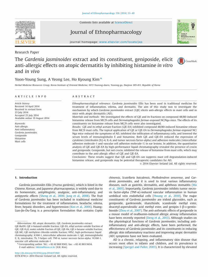
Fig. 1. Effects of Gardenia jasminoides extract (GJE) and its fractions on histamine release from mast cells and Dermatophagoides farinae (DfE)-induced atopic dermatitis inNC/Nga mice. (A) Experimental induction of atopic dermatitis-like skin lesions. (B) Release of histamine from MC/9 mast cells. MC/9 cells (2�105 cells/ml) were pretreatedwith GJE or its fractions (200 mg/ml) at 37 1C for 30 min prior to the addition of compound 48/80 (25 mg/ml). (C) Representative photographs of clinical features on day 21.(D) Dermatitis severity score. (E) Ear thickness. Results are expressed as mean7SD (n¼6). Normal, untreated group; control, DfE-treated group; tacrolimus, tacrolimus-treated group; GJE, GJE-treated group; GJE-EA, GJE-ethyl acetate fraction-treated group. ♯po0.05, ♯♯po0.01, ♯♯♯po0.001 vs. normal; npo0.05, nnpo0.01, nnnpo0.001 vs.control.
Y.-Y. Sung et al. / Journal of Ethnopharmacology 156 (2014) 33–40 35
Empower 2 (Waters Corporation, Milford, MA, USA), and thechromatographic separation was carried out on a Xselect™ HSST3 C18 column (4.6�250 mm, 5-mm particle size, Waters Corpora-tion, Milford, MS, USA) at ambient temperature. The mobile phaseconsisted of distilled water (A) and methanol (B), and the gradientwas 0–50 min with 10–100% B. The ultraviolet (UV) wavelengthwas detected from 200 to 500 nm. Geniposide and crocin weremonitored at 238 nm and 440 nm, respectively. The flow rate was0.80 ml/min, and the injection volume was 10.0 ml. Sample peakswere compared to the retention time of standard compounds andUV spectra in the chromatogram. Analytical-grade crocin andgeniposide (495.0%) were obtained from Sigma-Aldrich Co. (St.Louis, MO, USA) and Wako Pure Chemical Industries Ltd. (Osaka,Japan), respectively. Preparative and HPLC-grade chromatographicsolvents were purchased from Daejung Chemicals & Metals Co.,Ltd. (Gyeonggi-do, Korea) and J. T. Baker Inc. (Phillipsburg, NJ,USA), respectively.
2.11. Statistics
Results are expressed as mean7standard error of the mean(S.E.M.). Statistical significance was determined using the one-wayanalysis of variance, followed by Dunnett's test. A value of po0.05was used to indicate significant differences.
3. Results
3.1. Effects of GJE and it fractions on histamine release from MC/9mast cells
Using the release of histamine by cell degranulation as anindicator of anti-allergic activity, we evaluated the effect of GJEand its fractions on compound 48/80-induced histamine releasefrom MC/9 cells (Fig. 1B). GJE significantly inhibited histaminerelease. In addition, the ethyl acetate fraction of GJE, GJE-EA,significantly decreased histamine release from mast cells. Thewater fraction of GJE inhibited histamine release in a similarmanner, although it was not statistically significant. Furthermore,GJE and its fractions did not affect cell viability and were not toxicto MC/9 cells (data not shown).
3.2. Effects of GJE and GJE-EA on NC/Nga mice that were exposed todust mite allergens
Next, we investigated whether GJE and the GJE-EA fractioncould ameliorate AD in mice. To establish a dust mite allergen-induced AD model, DfE was applied twice per week to the dorsalskin of NC/Nga mice. Skin conditions were evaluated twice perweek using the dermatitis severity score. The clinical features, earthickness, and dermatitis severity scores of the treatment groupsare shown in Fig. 1C, D, and E. The repeated application of DfEinduced skin dryness, followed by erythema, hemorrhage, edema,scarring, erosion, and excoriation (Fig. 1C). However, GJE and
GJE-EA inhibited these symptoms of AD. Ear swelling increasedgradually in DfE-treated control mice, and DfE-induced ear swel-ling was significantly reduced by GJE or GJE-EA, but not GJE-H2O(Fig. 1D). Dermatitis severity scores increased rapidly in DfE-treated control mice and were significantly higher than those ofnormal controls after 3 days. However, the severity of dermatitiswas significantly lower in GJE- and GJE-EA-treated mice comparedwith DfE-treated controls (Fig. 1E). GJE-H2O also inhibited thedermatitis score, but this was not statistically significant. Amongthe experimental groups, the dermatitis severity score and earthickness were most effectively inhibited by GJE-EA, and theseinhibitory effects were significantly higher than those of GJE mice.These results demonstrate that GJE and GJE-EA suppressed DfE-induced AD in NC/Nga mice.
3.3. Effects of GJE and GJE-EA on serum IgE, IL-4, and histaminelevels
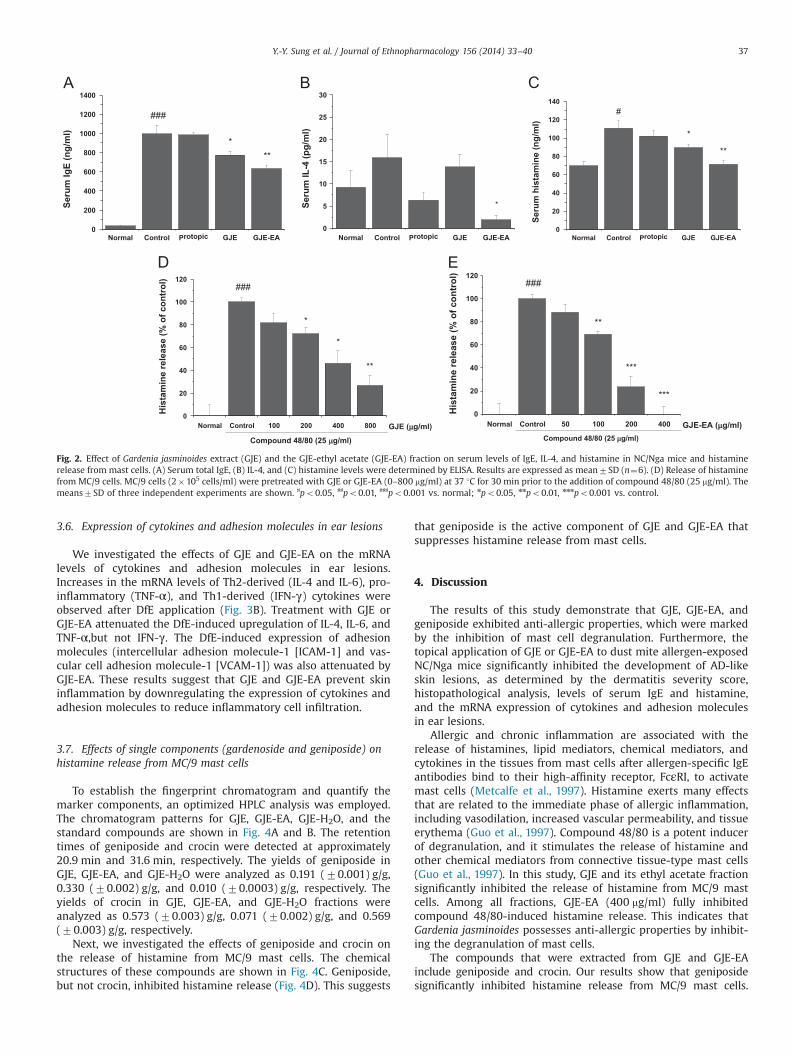
Because an elevated serum level of IgE is a main feature of AD,we tested whether GJE and GJE-EA could affect the serum levels ofIgE in DfE-treated NC/Nga mice. On day 22, the total IgE level innormal controls was 34.5874.03 ng/ml. DfE treatment eliciteda 29-fold increase in IgE levels (995.34787.58 ng/ml). Treatmentwith GJE or GJE-EA decreased the DfE-induced increase in total IgElevels (Fig. 2A). In addition, GJE-EA decreased DfE-induced serumIL-4 levels (Fig. 2B). Histamine release from mast cell granules wassignificantly higher in DfE-treated controls than in normal mice,but treatments with GJE or GJE-EA significantly reduced DfE-induced increases in histamine levels (Fig. 2C).
3.4. Dose-dependent effects of GJE and GJE-EA on histamine releasefrom MC/9 mast cells
Both GJE and GJE-EA inhibited the release of histamine fromMC/9mast cells in a dose-dependent manner (Fig. 2D and E). The IC50values of GJE and GJE-EA were 400.44 mg/ml and 126.33 mg/ml,respectively, and the inhibitory effect on histamine release was 3.2-fold higher in GJE-EA-treated mice than in GJE-treated mice.
3.5. Histopathological features
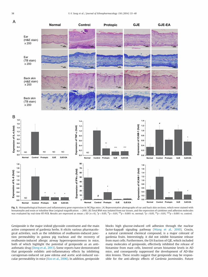
The histopathological features of the ear and dorsal skin lesionsare shown in Fig. 3A. Epidermal thickening by squamous cellhyperplasia (acanthosis) and inflammatory cell infiltration of thedermis were observed in DfE-treated control mice. However,treatment with GJE or GJE-EA inhibited these pathologicalchanges. To evaluate DfE-induced mast cell infiltration, ear andskin sections were stained with toluidine blue. The number ofmast cells in the dermis increased markedly in DfE-treated controlmice compared with normal mice, but this increase was reducedby treatment with GJE or GJE-EA.
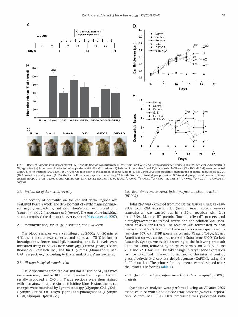
Table 1Real-time RT-PCR primer sequences.
Genes Forward Reverse Accession number Length (bp)
IL-4 TCAACCCCCAGCTAGTTGTCA CATCGAAAAGCCCGAAAGAG NM_021283 313IL-6 CCGGAGAGGAGACTTCACAG TCCAGTTTGGTAGCATCCATC NM_031168 220TNF-α CCTGTAGCCCACGTCGTAGC TTGACCTCAGCGCTGAGTTG NM_013693 373IFN-γ GCTACACACTGCATCTTGGCTTTG CACTCGGATGAGCTCATTGAATGC NM_008337 404ICAM-1 CCTCTGCTCCTGGCCCTGGT CGGACTGCTGTCCTCCCCGA NM_010492 237VCAM-1 TCGCGGTCTTGGGAGCCTCA TCGCGGTCTTGGGAGCCTCA NM_011693 213GAPDH AAGCTGTGGCGTGATGGCCG TGGGCCCTCAGATGCCTGCT NM_008084 228
Y.-Y. Sung et al. / Journal of Ethnopharmacology 156 (2014) 33–4036
3.6. Expression of cytokines and adhesion molecules in ear lesions
We investigated the effects of GJE and GJE-EA on the mRNAlevels of cytokines and adhesion molecules in ear lesions.Increases in the mRNA levels of Th2-derived (IL-4 and IL-6), pro-inflammatory (TNF-α), and Th1-derived (IFN-γ) cytokines wereobserved after DfE application (Fig. 3B). Treatment with GJE orGJE-EA attenuated the DfE-induced upregulation of IL-4, IL-6, andTNF-α,but not IFN-γ. The DfE-induced expression of adhesionmolecules (intercellular adhesion molecule-1 [ICAM-1] and vas-cular cell adhesion molecule-1 [VCAM-1]) was also attenuated byGJE-EA. These results suggest that GJE and GJE-EA prevent skininflammation by downregulating the expression of cytokines andadhesion molecules to reduce inflammatory cell infiltration.
3.7. Effects of single components (gardenoside and geniposide) onhistamine release from MC/9 mast cells
To establish the fingerprint chromatogram and quantify themarker components, an optimized HPLC analysis was employed.The chromatogram patterns for GJE, GJE-EA, GJE-H2O, and thestandard compounds are shown in Fig. 4A and B. The retentiontimes of geniposide and crocin were detected at approximately20.9 min and 31.6 min, respectively. The yields of geniposide inGJE, GJE-EA, and GJE-H2O were analyzed as 0.191 (70.001) g/g,0.330 (70.002) g/g, and 0.010 (70.0003) g/g, respectively. Theyields of crocin in GJE, GJE-EA, and GJE-H2O fractions wereanalyzed as 0.573 (70.003) g/g, 0.071 (70.002) g/g, and 0.569(70.003) g/g, respectively.
Next, we investigated the effects of geniposide and crocin onthe release of histamine from MC/9 mast cells. The chemicalstructures of these compounds are shown in Fig. 4C. Geniposide,but not crocin, inhibited histamine release (Fig. 4D). This suggests
that geniposide is the active component of GJE and GJE-EA thatsuppresses histamine release from mast cells.
4. Discussion
The results of this study demonstrate that GJE, GJE-EA, andgeniposide exhibited anti-allergic properties, which were markedby the inhibition of mast cell degranulation. Furthermore, thetopical application of GJE or GJE-EA to dust mite allergen-exposedNC/Nga mice significantly inhibited the development of AD-likeskin lesions, as determined by the dermatitis severity score,histopathological analysis, levels of serum IgE and histamine,and the mRNA expression of cytokines and adhesion moleculesin ear lesions.
Allergic and chronic inflammation are associated with therelease of histamines, lipid mediators, chemical mediators, andcytokines in the tissues from mast cells after allergen-specific IgEantibodies bind to their high-affinity receptor, FcεRI, to activatemast cells (Metcalfe et al., 1997). Histamine exerts many effectsthat are related to the immediate phase of allergic inflammation,including vasodilation, increased vascular permeability, and tissueerythema (Guo et al., 1997). Compound 48/80 is a potent inducerof degranulation, and it stimulates the release of histamine andother chemical mediators from connective tissue-type mast cells(Guo et al., 1997). In this study, GJE and its ethyl acetate fractionsignificantly inhibited the release of histamine from MC/9 mastcells. Among all fractions, GJE-EA (400 mg/ml) fully inhibitedcompound 48/80-induced histamine release. This indicates thatGardenia jasminoides possesses anti-allergic properties by inhibit-ing the degranulation of mast cells.
The compounds that were extracted from GJE and GJE-EAinclude geniposide and crocin. Our results show that geniposidesignificantly inhibited histamine release from MC/9 mast cells.
Fig. 2. Effect of Gardenia jasminoides extract (GJE) and the GJE-ethyl acetate (GJE-EA) fraction on serum levels of IgE, IL-4, and histamine in NC/Nga mice and histaminerelease from mast cells. (A) Serum total IgE, (B) IL-4, and (C) histamine levels were determined by ELISA. Results are expressed as mean7SD (n¼6). (D) Release of histaminefrom MC/9 cells. MC/9 cells (2�105 cells/ml) were pretreated with GJE or GJE-EA (0–800 mg/ml) at 37 1C for 30 min prior to the addition of compound 48/80 (25 mg/ml). Themeans7SD of three independent experiments are shown. ♯po0.05, ♯♯po0.01, ♯♯♯po0.001 vs. normal; npo0.05, nnpo0.01, nnnpo0.001 vs. control.
Y.-Y. Sung et al. / Journal of Ethnopharmacology 156 (2014) 33–40 37
Geniposide is the major iridoid glycoside constituent and the mainactive component of gardenia herbs. It elicits various pharmacolo-gical activities, such as the inhibition of ovalbumin-induced junc-tion permeability in guinea pig tracheas and the recovery ofovalbumin-induced allergic airway hyperresponsiveness in mice,both of which highlight the potential of geniposide as an anti-asthmatic drug (Deng et al., 2013). Some reports have demonstratedthat geniposide exhibits anti-inflammatory effects by inhibitingcarrageenan-induced rat paw edema and acetic acid-induced vas-cular permeability in mice (Koo et al., 2006). In addition, geniposide
blocks high glucose-induced cell adhesion through the nuclearfactor-kappaB signaling pathway (Wang et al., 2010). Crocin,a natural carotenoid chemical compound, is a major colorant ofgardenia fruits. Interestingly, it did not inhibit histamine releasefrommast cells. Furthermore, the EA fraction of GJE, which includedmany molecules of geniposide, effectively inhibited the release ofhistamine from mast cells, lowered serum histamine levels in ADmice, and consequently suppressed the development of AD-likeskin lesions. These results suggest that geniposide may be respon-sible for the anti-allergic effects of Gardenia jasminoides. Future
Fig. 3. Histopathological features and inflammatory gene expression in NC/Nga mice. (A) Representative photographs of ear and back skin sections, which were stained withhematoxylin and eosin or toluidine blue (original magnification: �200). (B) Total RNA was isolated from ear tissues, and the expression of cytokines and adhesion moleculeswas evaluated by real-time RT-PCR. Results are expressed as mean7SD (n¼6). ♯po0.05, ♯♯po0.01, ♯♯♯po0.001 vs. normal; npo0.05, nnpo0.01, nnnpo0.001 vs. control.
Y.-Y. Sung et al. / Journal of Ethnopharmacology 156 (2014) 33–4038
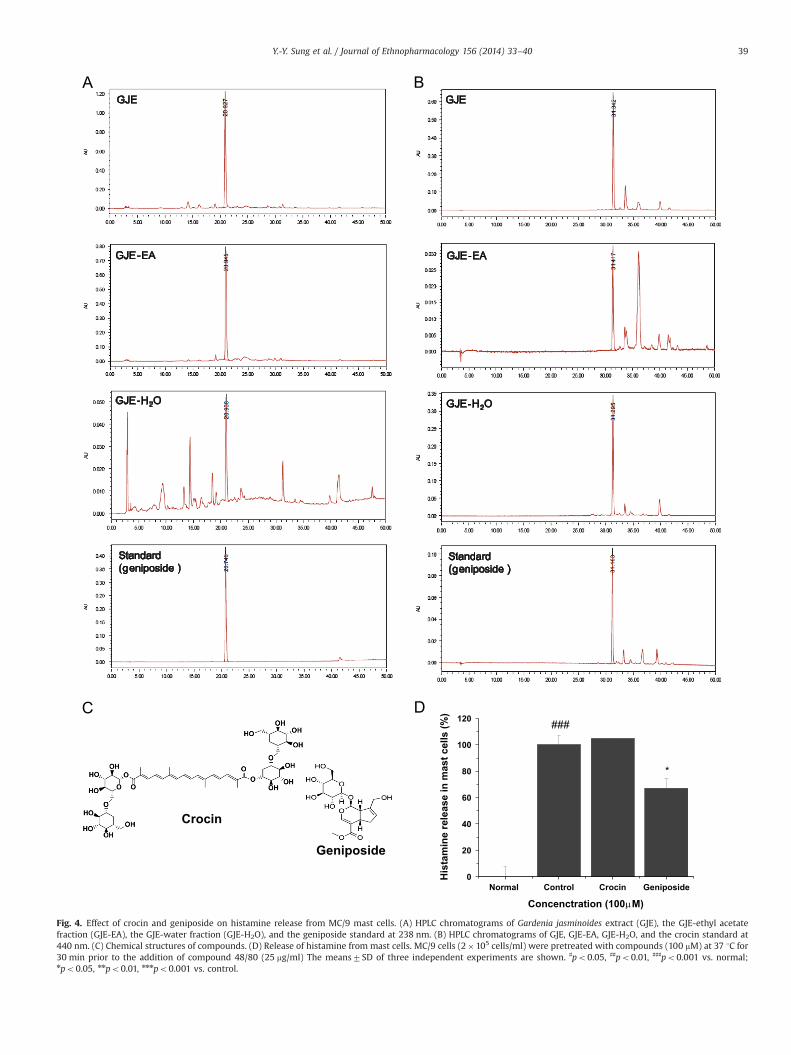
Fig. 4. Effect of crocin and geniposide on histamine release from MC/9 mast cells. (A) HPLC chromatograms of Gardenia jasminoides extract (GJE), the GJE-ethyl acetatefraction (GJE-EA), the GJE-water fraction (GJE-H2O), and the geniposide standard at 238 nm. (B) HPLC chromatograms of GJE, GJE-EA, GJE-H2O, and the crocin standard at440 nm. (C) Chemical structures of compounds. (D) Release of histamine from mast cells. MC/9 cells (2�105 cells/ml) were pretreated with compounds (100 mM) at 37 1C for30 min prior to the addition of compound 48/80 (25 mg/ml) The means7SD of three independent experiments are shown. ♯po0.05, ♯♯po0.01, ♯♯♯po0.001 vs. normal;npo0.05, nnpo0.01, nnnpo0.001 vs. control.
Y.-Y. Sung et al. / Journal of Ethnopharmacology 156 (2014) 33–40 39
studies using in vivo allergic AD models are necessary to verify thatgeniposide exerts its anti-allergic effect by inhibiting inflammatorymediators, such as histamine and cytokines.
In allergic inflammatory disease, Th2 cells secrete cytokines tomediate IgE-dependent mast cell degranulation and activation. IL-4 is a Th2 cell- and mast cell-secreted cytokine that is responsiblefor isotype switching in B cells for IgE synthesis and Th2 celldifferentiation (Bergstedt-Lindqvist et al., 1988). The Th1 cell-secreted cytokine, IFN-γ, inhibits IgE synthesis and Th2 cellproliferation (Leung and Bieber, 2003). Dysregulation of IL-6signaling contributes to the onset and maintenance of inflamma-tory diseases (Heinrich et al., 2003). IL-6, which is produced bymacrophages, T cells, B cells, and keratinocytes in response toexternal immune stimuli (IL-1 and TNF-α), promotes Th2 differ-entiation by upregulating IL-4 expression and inhibits Th1 differ-entiation by upregulating the suppressor of cytokine signaling-1expression (Diehl and Rincon, 2002). In our study, GJE-EAdecreased the serum levels of total IgE, IL-4, and histamine, andit downregulated the mRNA expression of IL-4, IL-6, and TNF-α,but not IFN-γ, in ear lesions. This suggests that GJE-EA attenuatesthe development of AD by inhibiting Th2 cell responses.
ICAM-1 and VCAM-1 are cell-surface glycoproteins that areexpressed on endothelial cells, and they promote leukocyteinfiltration in immune responses by mediating the adhesion ofleukocytes/endothelial cells (Roebuck and Finnegan, 1999). Theyalso contribute to leukocyte accumulation in mice that areexposed to dust mite allergens (Kang et al., 2008), and theexpression of these molecules in the keratinocytes of the skinlesions of AD is induced via mast cell degranulation-secreted TNF-α and histamine (Ackermann and Harvima, 1998). Importantly,GJE-EA decreased the DfE-induced upregulation of these mole-cules in our AD mouse model, thus suggesting that it inhibits themigration of leukocytes to the sites of inflammation in AD bydownregulating the expression of adhesion molecules.
5. Conclusion
In this study, the topical application of GJE or GJE-EA inhibitedthe development of AD-like skin lesions and decreased serum IgEand histamine levels. The inhibition of histamine, cytokines, andadhesion molecules blocked inflammatory cell infiltration, whichcontributed to the anti-allergic effects of GJE and GJE-EA. We alsoreveal for the first time that the active compound of GJE wasgeniposide. These results indicate that Gardenia jasminoides maybe useful in preventing or treating allergic disorders, such as AD.
Acknowledgments
This work was supported by the Construction of the Basis forPractical Application of Herbal Resources (Project K13020), theKorea Institute of Oriental Medicine (KIOM) to the Ministry ofScience, ICT and Future Planning (MSIP), Korea. In addition, thiswork was also partially supported by Efficacy Study of Alter-nativeHerbal Medicine Resources (K14416), the Korea Institute of Orien-tal Medicine (KIOM) to the Ministry of Science, ICT and FuturePlanning (MSIP), Korea.
References
Ackermann, L., Harvima, I.T., 1998. Mast cells of psoriatic and atopic dermatitis skinare positive for TNF-alpha and their degranulation is associated with expressionof ICAM-1 in the epidermis. Archives of Dermatological Research 290, 353–359.
Bergstedt-Lindqvist, S., Moon, H.B., Persson, U., Möller, G., Heusser, C., Severinson, E.,1988. Interleukin 4 instructs uncommitted B lymphocytes to switch to IgG1 andIgE. European Journal of Immunology 18, 1073–1077.
Choi, J.H., Kim, H.G., Jin, S.W., Han, E.H., Khanal, T., Do, M.T., Hwang, Y.P., Choi, J.M.,Chun, S.S., Chung, Y.C., Jeong, T.C., Jeong, H.G., 2013. Topical application ofPleurotus eryngii extracts inhibits 2,4-dinitrochlorobenzene-induced atopicdermatitis in NC/Nga mice by the regulation of Th1/Th2 balance. Food andChemical Toxicology 53, 38–45.
Deng, Y., Guan, M., Xie, X., Yang, X., Xiang, H., Li, H., Zou, L., Wei, J., Wang, D., Deng, X.,2013. Geniposide inhibits airway inflammation and hyperresponsiveness in amouse model of asthma. International Immunopharmacology 17, 561–567.
Diehl, S., Rincon, M., 2002. The two faces of IL-6 on Th1/Th2 differentiation.Molecular Immunology 39, 531–536.
Galli, S.J., Gordon, J.R., Wershil, B.K., 1991. Cytokine production by mast cells andbasophils. Current Opinion in Immunology 3, 865–872.
Guo, Y., Mochizuki, T., Morii, E., Kitamura, Y., Maeyama, K., 1997. Role of mast cellhistamine in the formation of rat paw edema: a microdialysis study. EuropeanJournal of Pharmacology 331, 237–243.
Heinrich, P.C., Behrmann, I., Haan, S., Hermanns, H.M., Müller-Newen, G., Schaper, F.,2003. Principles of interleukin (IL)-6-type cytokine signalling and its regulation.Biochemical Journal 374 (Pt 1), 1–20.
Hwang, S.M., Lee, Y.J., Yoon, J.J., Lee, S.M., Kang, D.G., Lee, H.S., 2010. Gardeniajasminoides inhibits tumor necrosis factor-alpha-induced vascular inflamma-tion in endothelial cells. Phytotherapy Research 24 (Suppl 2), S214–S219.
Jung, W.S., Chae, Y.S., Kim, D.Y., Seo, S.W., Park, H.J., Bae, G.S., Kim, T.H., Oh, H.J., Yun,K.J., Park, R.K., Kim, J.S., Kim, E.C., Hwang, S.Y., Park, S.J., Song, H.J., 2008. Gardeniajasminoides protects against cerulein-induced acute pancreatitis. World Journalof Gastroenterology 14, 6188–6194.
Kang, J.S., Yoon, W.K., Youm, J.K., Jeong, S.K., Park, B.D., Han, M.H., Lee, H., Moon, E.Y.,Han, S.B., Lee, C.W., Lee, K., Park, S.K., Yang, K.H., Kim, H.M., 2008. Inhibition ofatopic dermatitis-like skin lesions by topical application of a novel ceramidederivative, K6PC-9p, in NC/Nga mice. Experimental Dermatology 17, 958–964.
Koo, H.J., Lim, K.H., Jung, H.J., Park, E.H., 2006. Anti-inflammatory evaluation of gardeniaextract, geniposide and genipin. Journal of Ethnopharmacology 103, 496–500.
Leung, D.Y., Bieber, T., 2003. Atopic dermatitis. Lancet 361, 151–160.Leung, D.Y., Boguniewicz, M., Howell, M.D., Nomura, I., Hamid, Q.A., 2004. New
insights into atopic dermatitis. The Journal of Clinical Investigation 113, 651–657.Ma, Z., Otsuyama, K., Liu, S., Abroun, S., Ishikawa, H., Tsuyama, N., Obata, M., Li, F.J.,
Zheng, X., Maki, Y., Miyamoto, K., Kawano, M.M., 2005. Baicalein, a componentof Scutellaria radix from Huang-Lian-Jie-Du-Tang (HLJDT), leads to suppressionof proliferation and induction of apoptosis in human myeloma cells. Blood 105,3312–3318.
Matsuda, H., Watanabe, N., Geba, G.P., Sperl, J., Tsudzuki, M., Hiroi, J., Matsumoto, M.,Ushio, H., Saito, S., Askenase, P.W., Ra, C., 1997. Development of atopic dermatitis-like skin lesion with IgE hyperproduction in NC/Nga mice. International Immunol-ogy 9, 461–466.
Metcalfe, D.D., Baram, D., Mekori, Y.A., 1997. Mast cells. Physiological Reviews 77,1033–1079.
Roebuck, K.A., Finnegan, A., 1999. Regulation of intercellular adhesion molecule-1(CD54) gene expression. Journal of Leukocyte Biology 66, 876–888.
Sawada, E., Yoshida, N., Sugiura, A., Imokawa, G., 2012. Th1 cytokines accentuatebut Th2 cytokines attenuate ceramide production in the stratum corneum ofhuman epidermal equivalents: an implication for the disrupted barriermechanism in atopic dermatitis. Journal of Dermatological Science 68, 25–35.
Sheng, L., Qian, Z., Zheng, S., Xi, L., 2006. Mechanism of hypolipidemic effect ofcrocin in rats: crocin inhibits pancreatic lipase. European Journal of Pharma-cology 543, 116–122.
Spergel, J.M., Paller, A.S., 2003. Atopic dermatitis and the atopic march. The Journalof Allergy and Clinical Immunology 112, S118–S127.
Sung, Y.Y., Yoon, T., Jang, J.Y., Park, S.J., Jeong, G.H., Kim, H.K., 2011. Inhibitory effectsof Cinnamomum cassia extract on atopic dermatitis-like skin lesions induced bymite allergen in NC/Nga mice. Journal of Ethnopharmacology 133, 621–628.
Vestergaard, C., Yoneyama, H., Murai, M., Nakamura, K., Tamaki, K., Terashima, Y.,Imai, T., Yoshie, O., Irimura, T., Mizutani, H., Matsushima, K., 1999. Over-production of Th2-specific chemokines in NC/Nga mice exhibiting atopicdermatitis-like lesions. The Journal of Clinical Investigation 104, 1097–1105.
Wang, G.F., Wu, S.Y., Xu, W., Jin, H., Zhu, Z.G., Li, Z.H., Tian, Y.X., Zhang, J.J., Rao, J.J.,Wu, S.G., 2010. Geniposide inhibits high glucose-induced cell adhesion throughthe NF-kappaB signaling pathway in human umbilical vein endothelial cells.Acta Pharmacologica Sinica 31, 953–962.
Yamamoto, M., Haruna, T., Yasui, K., Takahashi, H., Iduhara, M., Takaki, S., Deguchi,M., Arimura, A., 2007. A novel atopic dermatitis model induced by topicalapplication with dermatophagoides farinae extract in NC/Nga mice. AllergologyInternational 56, 139–148.
Zhou, T., Zhao, W., Fan, G., Chai, Y., Wu, Y., 2007. Isolation and purification of iridoidglycosides from Gardenia jasminoides Ellis by isocratic reversed-phase two-dimensional preparative high-performance liquid chromatography with columnswitch technology. Journal of chromatography B Analytical Technologies in theBiomedical and Life Sciences 858, 296–301.
Y.-Y. Sung et al. / Journal of Ethnopharmacology 156 (2014) 33–4040


![P2Y1 ReceptorActivationoftheTRPV4IonChannelEnhances ...misterx95.myds.me/wordpress/wp-content/uploads/2018/01/... · 2018. 1. 26. · changes in [Ca2] i. Data are presented as F/F](https://img.pdfslide.us/doc/110x75/6115a103c811b409c00f326f/p2y1-receptoractivationofthetrpv4ionchannelenhances-2018-1-26-changes-in.jpg)