Embed Size (px)
Citation preview

http://jdr.sagepub.com/
Journal of Dental Research
http://jdr.sagepub.com/content/early/2010/07/14/0022034510373764The online version of this article can be found at:
DOI: 10.1177/0022034510373764
published online 16 July 2010J DENT RESC.C. Teixeira, E. Khoo, J. Tran, I. Chartres, Y. Liu, L.M. Thant, I. Khabensky, L.P. Gart, G. Cisneros and M. Alikhani
Cytokine Expression and Accelerated Tooth Movement
Published by:
http://www.sagepublications.com
On behalf of:
International and American Associations for Dental Research
can be found at:Journal of Dental ResearchAdditional services and information for
http://jdr.sagepub.com/cgi/alertsEmail Alerts:
http://jdr.sagepub.com/subscriptionsSubscriptions:
http://www.sagepub.com/journalsReprints.navReprints:
http://www.sagepub.com/journalsPermissions.navPermissions:
at Bobst Library, New York University on July 20, 2010jdr.sagepub.comDownloaded from

1
RESEARCH REPORTSBiological
DOI: 10.1177/0022034510373764
Received November 12, 2009; Last revision April 4, 2010; Accepted April 9, 2010
A supplemental appendix to this article is published elec-tronically only at http://jdr.sagepub.com/supplemental.
C.C. Teixeira1,2,3, E. Khoo1, J. Tran1,I. Chartres1, Y. Liu1, L.M. Thant2,I. Khabensky2, L.P. Gart2,G. Cisneros1,3, and M. Alikhani1,3*
1Department of Orthodontics, 2Department of Basic Science and Craniofacial Biology, and 3Consortium for Translational Orthodontic Research, New York University College of Dentistry, 345 East 24th Street, New York, NY 10010, USA; *corresponding author, [email protected]
J Dent Res X(X):xx-xx, XXXX
ABSTRACTIt has been shown that inhibiting the expression of certain cytokines decreases the rate of tooth move-ment. Here, we hypothesized that stimulating the expression of inflammatory cytokines, through small perforations of cortical bone, increases the rate of bone remodeling and tooth movement. Forty-eight rats were divided into 4 groups: 50-cN force applied to the maxillary first molar (O), force application plus soft tissue flap (OF), force appli-cation plus flap plus 3 small perforations of the cortical plate (OFP), and a control group (C). From the 92 cytokines studied, the expression of 37 cytokines increased significantly in all experimen-tal groups, with 21 cytokines showing the highest levels in the OFP group. After 28 days, micro-computed tomography, light and fluorescent microscopy, and immunohistochemistry demon-strated higher numbers of osteoclasts and bone remodeling activity in the OFP group, accompa-nied by generalized osteoporosity and increased rate of tooth movement.
KEY WORDS: osteoperforation, cytokines, bone remodeling, tooth movement.
InTRODuCTIOn
Orthodontic forces induce an aseptic inflammatory response. During early stages of tooth movement, there is an increase in vascular permeabil-
ity and cellular infiltration of leukocytes (Krishnan and Davidovitch, 2006; Meikle, 2006). Migrated immune cells along with native cells such as fibro-blasts and osteoblasts produce inflammatory cytokines that include lym-phocyte- and monocyte-derived factors, colony-stimulating factors, growth factors, and chemotactic factors (Ren and Vissink, 2008; Krishnan and Davidovitch, 2009). High concentrations of inflammatory cytokines such as interleukin-1 (IL-1), IL-2, IL-3, IL-6, IL-8, tumor necrosis factor-α (TNFα), interferon-γ (IFNγ), and osteoclast differentiation factor have been found in the gingival crevicular fluid surrounding moving teeth (Alhashimi et al., 2000; Garlet et al., 2007; Ren et al., 2007).
The role of cytokines during tooth movement is not clear. It has been sug-gested that cytokines and other inflammatory markers, such as prostaglandin E2 (Saito et al., 1991), may activate bone remodeling characterized by bone resorption in the compression region and bone deposition in the tension region of the periodontal ligament (PDL) (Davidovitch et al., 1988; Garletet al., 2007). This is in agreement with previous studies that demonstrated that bone injury which causes cytokine release leads to an accelerated bone turn-over and a decrease in regional bone density (Frost, 1983, 1989a,b; Shih and Norrdin, 1985; Yaffe et al., 1994). One possible mechanism through which inflammatory cytokines may affect bone remodeling is through recruitment of osteoclast precursors from the circulation, their maturation and activation. Many cytokines that promote osteoclast formation and activation, such as IL-1, IL-6, and TNFα (Glantschnig et al., 2003; Seidenberg and An, 2004; Yao et al., 2008), have also been found in crevicular fluid during orthodontic tooth movement (Uematsu et al., 1996; Basaran et al., 2006).
The effect of cytokine expression on bone remodeling is important, since the rate of tooth movement correlates with the efficiency of bone remodeling in the alveolar process. Studies of knockout mice deficient for TNFα recep-tors (Yoshimatsu et al., 2006) showed a slower rate of tooth movement in response to orthodontic forces. Previous reports have shown that anti-inflam-matory medication can decrease the rate of tooth movement (Arias and Marquez-Orozco, 2006).
Based on these findings, we hypothesized that limited and shallow perfo-rations of the buccal cortical plate of the maxilla would be sufficient to increase the expression of inflammatory cytokines, accelerating the bone remodeling process and therefore the rate of tooth movement.
Cytokine Expression and Accelerated Tooth Movement
JDR OnlineFirst, published on July 16, 2010 as doi:10.1177/0022034510373764
at Bobst Library, New York University on July 20, 2010jdr.sagepub.comDownloaded from

2 Teixeira et al. J Dent Res X(X) XXXX
MATERIAlS & METHODS
Animal Study
Forty-eight adult male Sprague-Dawley rats (average body weight of 400 g, 120 days of age) were housed and treated according to a protocol approved by the New York University Institutional Animal Care and Use Committee. Animals were divided into 4 groups (12 rats per group): control, which received coil spring without activation (C), orthodontic force applied to the spring (O), orthodontic force and soft tissue flap (OF), and orthodontic force, soft tissue flap, and shallow perfo-rations of the buccal cortical plate (OFP). The health status and body weight of the rats were evaluated daily, and no significant differences were observed among groups. From each group, 4 animals were used for gene expression studies, 4 for microCT and fluorescent studies, and 4 for demineralized histological studies. Procedures were performed on one side of the maxilla, which allowed the contralateral side to be used as an additional control.
Surgical Procedure
On day 0, all groups were anesthetized with an intraperitoneal injection of ketamine-xylazine (0.09 mL/100 g), and anesthe-sia was verified by lack of response to toe-pinch. All groups were fitted with 50 cN Sentalloy closing coils (DENTSPLY GAC International, Bohemia, NY, USA) tied at both ends to holes drilled in the maxillary incisors and left maxillary first molar with 0.008-inch ligature wire; the coil was activated in groups O, OF, and OFP, but not in the C group. In the OF and OFP groups, a soft tissue flap was raised around the left first molar. Flaps were sealed with cyanoacrylate tissue adhesive (Vetbond, 3M, St. Paul, MN, USA). In the OFP group, the animals received 3 shallow perforations, approximately 0.25 mm in diameter (depth of 0.25 mm), 5 mm mesial to the left first molar with the use of a round bur and handpiece. Animals were checked under general anesthesia twice weekly, and any springs requiring retying (mostly due to continuous eruption of the maxillary incisors) were adjusted. Bone labeling by intra-peritoneal injection of calcein (15 mg/kg) was performed on days 0 and 26 and by demeclocycline (25 mg/kg) on day 14. Animals were killed by CO2 narcosis on day 28; hemimaxillae were collected, then fixed in formaldehyde for 48 hrs before storage in 70% ethanol.
Micro-CT Imaging
Hemimaxillae were scanned with a Scanco MicrocCT (µCT40, Scanco Medical, Bassersdorf, Switzerland). Results were ana-lyzed utilizing µCT V6.0 software on the HP open platform (openVMS Alpha Version 1.3-1 session manager). The area extending from the coronal to the apical root third was analyzed for bony changes. Maxillae were analyzed in fixed coronal and sagittal zones. The ratio of bone volume to total volume (BV/TV) was calculated based on a threshold of 275.
Histology and Immunohistochemistry
Hemimaxillae were collected and fixed in 10% phosphate-buffered formalin and demineralized in a sodium formate (6.8%) and formic acid (50%) solution for 6-8 wks. Following demin-eralization, specimens were dehydrated in alcohol series and embedded in paraffin; 5-µm-thick sections were cut and stained with hematoxylin and eosin (H&E). Consecutive specimens were immunostained with antibodies for tartrate-resistant acid phosphatase (TRAP; Zymed antibodies, Invitrogen, Carlsbad, CA, USA), a marker of osteoclasts, and Vectastain ABC kit (Vector Laboratories, Burlingame, CA, USA) according to the manufacturer’s instructions. As negative control, consecutive sections were exposed to pre-immune serum. Stained sections were scanned on a Scan Scope GL series optical microscope (Aperio, Bristol, UK) at 20x magnification. Osteoclasts were defined as TRAP-positive multinuclear cells on the bone sur-face. The area around the mesio-palatal root of the maxillary first molar was divided into mesial and distal halves, and osteo-clasts in the mesial half were counted. Data were expressed as the number of TRAP-positive cells per 1000 µm2 in the area of PDL and adjacent alveolar bone, excluding the marrow cavities and blood vessels.
For fluorescent microscopy, after formalin fixation, speci-mens were washed overnight in running water, dehydrated in alcohol, cleared in xylene, and embedded in methyl methacry-late according to the method of Erben (Erben, 1997). The sam-ples were sectioned at 5- to 7-µm thickness on a Reichert-Jung Ultracut E microtome and viewed by fluorescent microscopy (Nikon Microscopy, NIS-Elements software, Tokyo, Japan).
RT-PCR Analysis
For RNA extraction, 4 animals from each group were killed by CO2 narcosis at 24 hrs, and the hemimaxillae were dissected and frozen in liquid nitrogen. Isolation of total RNA was performed with the TRIZOL reagent (Life Technologies, New York, NY, USA), and RNA clean-up was performed with an RNeasy Mini Kit (Qiagen Sciences, Valencia, CA, USA) as described previ-ously (Oliveira et al., 2009). All equipment and tools were cleaned with RNaseZap (Sigma, St. Louis, MO, USA). Ninety-two inflammatory cytokines and cytokine receptor genes were analyzed with primers specific for rat genes (see online Appendix for list of genes), with a QuantiTect SYBR Green RT-PCR kit (both Qiagen, Valencia, CA, USA) on a DNA Engine Optican 2 System (MJ Research, Waltham, MA, USA). Each mRNA specimen was tested 3 times. Relative levels of mRNA were calculated and normalized to the level of GAPDH and acidic ribosomal protein mRNA.
Statistical Analysis
Significant differences between test groups and controls were assessed by analysis of variance (ANOVA). Pairwise multiple comparison analysis was performed with Tukey’s post hoc test.
at Bobst Library, New York University on July 20, 2010jdr.sagepub.comDownloaded from

J Dent Res X(X) XXXX Osteoperforation-enhanced Tooth Movement 3
Two-tailed p-values were calcu-lated; p < 0.05 was set as the level of statistical significance.
RESulTS
Osteoperforations Increase the Rate of Tooth Movement
Coil springs were used for mesial movement of the first maxillary molar crown (Fig. 1A). Three shallow perforations were made in the cortical bone, 5 mm mesial to the molar (Fig. 1B). At 28 days, the average crown movement (measured in 12 rats per group) was 0.29 mm in the O and OF groups (Fig. 1C), significantly different from con-trols (p < 0.05). The OFP group showed the greatest mean tooth movement, 0.62 mm, which was significantly higher (p < 0.05 ) than that of the C, O, and OF groups (Fig. 1C).
Osteoperforations Increase Expression of Inflammatory Cytokines
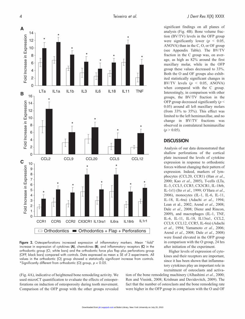
Expression of 92 different cyto-kines/cytokine receptors was studied by RT-PCR, 24 hrs after force application. The expres-sion of 37 cytokines/cytokine receptors increased more than two-fold in the left maxilla of rats in the O, OF, and OFP groups when compared with that in the C group (data not shown). Differences between O and OF groups were not statistically significant. From these 37 cyto-kines, expression of 21 cytokines/cytokine receptors was statis-tically higher in the OFP group than in the O or OF group (p < 0.05) (Fig. 2), with 8 cytokines showing a 1.6- to 2.7-fold increase (Fig. 2A), 5 chemokines showing a 1.6- to 2.8-fold increase (Fig. 2B), and 8 receptors showing a 1.7- to two- fold increase in expression (Fig. 2C). All cytokines/cytokine receptors expressed in the OFP group were also expressed in the O or OF group. Expression of cytokines in the contralateral side of all groups showed no statistically significant differences from group C (data not shown).
Osteoperforations Increase Osteoclast Activity
In both the O and OF groups, application of the orthodontic force stimulated an increase in alveolar bone resorption in the direction of tooth movement and, consequently, an increase in
PDL thickness (Fig. 3A, top row). The OFP group showed increased alveolar bone resorption in the direction of tooth movement (Fig. 3A, top row). Immunohistochemical staining for TRAP-positive osteoclasts (Fig. 3B) revealed an increase in osteoclast number in the OFP group, compared with the OF and O groups (Fig. 3A, bottom row). Quantitative analysis of osteo-clasts in the pressure side (mesial) of alveolar bone adjacent to the mesio-palatal root of the maxillary first molar demonstrated a three-fold increase in numbers of osteoclasts in comparison with the O and OF groups (p < 0.05) (Fig. 3C). The difference between numbers of osteoclasts in the O and OF groups was not statistically significant.
Osteoperforations Increase the Rate of Bone Remodeling and Generalized Osteoporosity
Sagittal sections of specimens viewed under fluorescent micros-copy showed more prominent fluorescence in the OFP group
Figure 1. Osteoperforations increase the rate of tooth movement. (A) Photograph of experimental model. (B) Schematic showing the 3 shallow perforations (0.25 mm diameter and depth) created, 5 mm mesial to the first molar. (C) Representative photographs of rat maxillae showing movement of left first molar at 28 days in the 4 groups. C = control; O = orthodontic force alone; OF = orthodontic force plus flap; OFP = orthodontic force plus flap plus perforations.
at Bobst Library, New York University on July 20, 2010jdr.sagepub.comDownloaded from
4 Teixeira et al. J Dent Res X(X) XXXX
(Fig. 4A), indicative of heightened bone remodeling activity. We used microCT quantification to evaluate the effects of osteoper-forations on induction of osteoporosity during tooth movement. Comparison of the OFP group with the other groups revealed
significant findings on all planes of analysis (Fig. 4B). Bone volume frac-tion (BV/TV) levels in the OFP group were significantly lower (p < 0.05, ANOVA) than in the C, O, or OF group (see Appendix Table). The BV/TV fraction in the C group was, on aver-age, as high as 82% around the first maxillary molar, while in the OFP group these values decreased to 33%. Both the O and OF groups also exhib-ited statistically significant changes in BV/TV levels (p < 0.05, ANOVA) when compared with the C group. Interestingly, in comparison with other groups, the BV/TV fraction in the OFP group decreased significantly (p < 0.05) around all left maxillary molars (from 33% to 35%). This effect was limited to the left hemimaxillae, and no change in BV/TV fractions was observed in contralateral hemimaxillae (p > 0.05).
DISCuSSIOn
Analysis of our data demonstrated that shallow perforations of the cortical plate increased the levels of cytokine expression in response to orthodontic forces without changing their pattern of expression. Indeed, markers of lym-phocytes (CCL20, CCR1) (Han et al., 2000; Kao et al., 2005), T-cells (LTa, IL-3, CCL5, CCR5, CX3CR1, IL-18rb, IL-1r1) (Ito et al., 1999; O’Hara et al., 2006), monocytes (IL-1, IL-6, IL-11, IL-18, IL-6ra) (Adachi et al., 1994; Lean et al., 2002; Arend et al., 2008; Dale et al., 2008; Dienz and Rincon, 2009), and macrophages (IL-1, TNF, IL-6, IL-11, IL-18, IL13ra1, CCL2, CCL9, CCL12, CCR5, IL-6ra) (Adachi et al., 1994; Yamamoto et al., 2006; Arend et al., 2008; Dale et al., 2008) were found elevated in the OFP group in comparison with the O group, 24 hrs after initiation of the experiment.
Higher levels of expression of cyto-kines and their receptors are important, since it has been shown that inflamma-tory cytokines play an important role in recruitment of osteoclasts and activa-
tion of the bone remodeling machinery (Alhashimi et al., 2000; Ren and Vissink, 2008; Krishnan and Davidovitch, 2009). The fact that the number of osteoclasts and the bone remodeling rate were higher in the OFP group in comparison with the O and OF
0
1
2
3
4
5
6
7
8
9
10
CCR1 CCR5 CCR2 CX3CR1 IL13ra1 IL6ra IL18rb IL1r1
* *
*
**
*
*
*
0
2
4
6
8
10
12
14
16
CCL2 CCL9 CCL20 CCL5 CCL12
**
*
**
0
2
4
6
8
10
12
14
LTa IL1a IL1b IL3 IL6 IL18 IL11 TNF
*
*
* *
**
**
Fol
d In
crea
se in
Exp
ress
ion
Fol
d In
crea
se in
Exp
ress
ion
Fol
d In
crea
se in
Exp
ress
ion
Orthodontics Orthodontics + Flap + Perforations
A
B
C
Figure 2. Osteoperforations increased expression of inflammatory markers. Mean “-fold” increase in expression of cytokines (A), chemokines (B), and inflammatory receptors (C) in the orthodontic group (O, white bars) and the orthodontic force plus flap plus perforations group (OFP, black bars) compared with controls. Data expressed as mean ± SE of 3 experiments. All values in the orthodontic (O) group showed a statistically significant increase from controls. *Significantly different from orthodontic (O) group, p < 0.05.
at Bobst Library, New York University on July 20, 2010jdr.sagepub.comDownloaded from

J Dent Res X(X) XXXX Osteoperforation-enhanced Tooth Movement 5
groups supports the possible role of inflammatory cytokines in recruiting osteoclasts into the area.
Similar to previous studies (Verna et al., 1999), we demonstrated that the increase in bone remodeling rate was not limited to the area of the loaded tooth, but extended to the tissues surrounding adjacent teeth. This generalized increase in bone turnover was accompanied by osteoporosity, as reflected by a decrease in bone density around all upper left molars. While a limited number of osteo-perforations had a generalized effect, the effect was not robust enough to cross to the contralateral side.
Since bone remodeling controls the rate of tooth movement, the increase in rate of bone remodeling and osteoporos-ity in response to bone perforations may explain the increase in the rate and mag-nitude of tooth movement observed in this study. One may argue that the effects of the shallow perforations on tooth movement were not a response to an increased cytokine expression, but rather were due to weakening of the bone struc-ture. While we cannot ignore the effects that perforations can have on the physi-cal properties of the bone, this study was designed to minimize this possibility. First, the perforations were small and limited (only 3); therefore, the majority of the cortical bone remained intact. In addition, the perforations were placed far away from the tooth, and could still be observed at the end of the study with remaining bone (about 4 mm) between perforations and the moved tooth. These results further suggest that the perforations do not need to be close to the tooth to be moved to accelerate the rate of move-ment.
It is important to mention that inflammation is “a two-sided sword”, and while it can work to our benefit by accelerating bone remodeling and tooth movement, if uncontrolled, it may also have a destructive effect on the periodontium and tooth structure. We are currently investigating root resorption in response to osteoperfora-tion. While extensive injury to the cortical plate bone, also referred to as corticotomies, is currently being used to accelerate orthodontic tooth movement in private practice, analysis of our data suggests that this approach could be simplified to minimize deleterious side-effects. Therefore, we propose the use of flapless minimal cortical perforations as a means of fine-tuning inflammation levels for enhanced tooth movement, enabling orthodontists to provide more efficient treatment for their patients.
ACKnOWlEDGMEnTS
This investigation was supported by Grants K08DE017426 and R03DE019499 from NIDCR, National Institutes of Health, Bethesda, MD 20892, USA. We would like to acknowledge the Muscular Skeletal Repair and Regeneration Center at the Hospital for Special Surgery, supported by AR046121, for their help with histological preparations.
REFEREnCES
Adachi Y, Okazaki M, Ohno N, Yadomae T (1994). Enhancement of cytokine production by macrophages stimulated with (1-->3)-beta- D-glucan, grifolan (GRN), isolated from Grifola frondosa. Biol Pharm Bull 17:1554-1560.
Alhashimi N, Frithiof L, Brudvik P, Bakhiet M (2000). Orthodontic move-ment induces high numbers of cells expressing IFN-gamma at mRNA and protein levels. J Interferon Cytokine Res 20:7-12.
Figure 3. Osteoperforations increased osteoclast activity. (A) Light microphotographs of H&E-stained section (top row) show differences in PDL thickness (p) and alveolar bone resorption (b) in the area of the mesio-palatal root of the maxillary first molar 28 days post-treatment. TRAP-positive immunohistochemical staining reveals osteoclasts as brown cells (arrowheads) on the mesial alveolar bone surface in the area of the mesio-palatal root of the maxillary first molar (bottom row). (B) High-magnification view of TRAP-positive osteoclast. (C) Changes in number of TRAP-positive cells on the mesial alveolar bone surface of the mesio-palatal root of the maxillary first molar. Each value represents the mean ± SEM of 4 samples. *Significantly different from C group. **Significantly different from C, O, and OF groups; p < 0.05.
at Bobst Library, New York University on July 20, 2010jdr.sagepub.comDownloaded from

6 Teixeira et al. J Dent Res X(X) XXXX
Arend WP, Palmer G, Gabay C (2008). IL-1, IL-18, and IL-33 families of cytokines. Immunol Rev 223:20-38.
Arias OR, Marquez-Orozco MC (2006). Aspirin, acetaminophen, and ibu-profen: their effects on orthodontic tooth movement. Am J Orthod Dentofacial Orthop 130:364-370.
Başaran G, Ozer T, Kaya FA, Hamamci O (2006). Interleukins 2, 6, and 8 levels in human gingival sulcus during orthodontic treatment. Am J Orthod Dentofacial Orthop 130:E1-6.
Dale DC, Boxer L, Liles WC (2008). The phagocytes: neutrophils and monocytes. Blood 112:935-945.
Davidovitch Z, Nicolay OF, Ngan PW, Shanfeld JL (1988). Neurotransmitters, cytokines, and the control of alveolar bone remodeling in orthodontics. Dent Clin North Am 32:411-435.
Dienz O, Rincon M (2009). The effects of IL-6 on CD4 T cell responses. Clin Immunol 130:27-33.
Figure 4. Osteoperforations increased bone remodeling rates and generalized osteoporosity in the entire length of the hemimaxillae. (A) Sagittal sections of maxillae from the 4 groups viewed under fluorescent microscopy showed the rate of bone remodeling in the entire hemimaxillae. The increased intensity of the label in most of the trabecular surface of the OFP group in comparison with other groups indicates that extensive bone remodeling has taken place at 28 days post-treatment. White arrows demonstrate the direction of force application. (B) Schematic indicating axial sections (1, 2, 3) and coronal sections (a, b, c) used in the analysis. (C) Representative coronal sections obtained by microCT analysis showing increased trabecular spacing in the OFP group, indicative of bone remodeling activity. White arrows demonstrate the direction of force application. C = control; O = orthodontic force alone; OF = orthodontic force plus flap; OFP = orthodontic force plus flap plus perforations.
Erben RG (1997). Embedding of bone samples in methylmethacrylate: an improved method suit-able for bone histomorphometry, histochemis-try, and immunohistochemistry. J Histochem Cytochem 45:307-313.
Frost HM (1983). The regional acceleratory phe-nomenon: a review. Henry Ford Hosp Med J 31:3-9.
Frost HM (1989a). The biology of fracture heal-ing. An overview for clinicians. Part I. Clin Orthop Relat Res 248:283-293.
Frost HM (1989b). The biology of fracture heal-ing. An overview for clinicians. Part II. Clin Orthop Relat Res 248:294-309.
Garlet TP, Coelho U, Silva JS, Garlet GP (2007). Cytokine expression pattern in compression and tension sides of the periodontal ligament during orthodontic tooth movement in humans. Eur J Oral Sci 115:355-362.
Glantschnig H, Fisher JE, Wesolowski G, Rodan GA, Reszka AA (2003). M-CSF, TNFalpha and RANK ligand promote osteoclast sur-vival by signaling through mTOR/S6 kinase. Cell Death Differ 10:1165-1177.
Han Y, Wang J, Zhou Z, Ransohoff RM (2000). TGFbeta1 selectively up-regulates CCR1 expression in primary murine astrocytes. Glia 30:1-10.
Ito A, Takii T, Matsumura T, Onozaki K (1999). Augmentation of type I IL-1 receptor expres-sion and IL-1 signaling by IL-6 and gluco-corticoid in murine hepatocytes. J Immunol 162:4260-4265.
Kao CY, Huang F, Chen Y, Thai P, Wachi S, Kim C, et al. (2005). Up-regulation of CC chemo-kine ligand 20 expression in human airway epithelium by IL-17 through a JAK-independent but MEK/NF-kappaB-dependent signaling pathway. J Immunol 175:6676-6685.
Krishnan V, Davidovitch Z (2006). Cellular, molecular and tissue-level reactions to orth-odontic force. Am J Orthod Dentofacial Orthop 129:469.e1-469.e32.
Krishnan V, Davidovitch Z (2009). On a path to unfolding the biological mechanisms of orth-odontic tooth movement. J Dent Res 88:597-608.
Lean JM, Murphy C, Fuller K, Chambers TJ (2002). CCL9/MIP-1gamma and its receptor CCR1 are the major chemokine ligand/recep-tor species expressed by osteoclasts. J Cell Biochem 87:386-393.
Meikle MC (2006). The tissue, cellular, and molecular regulation of orthodontic tooth
movement: 100 years after Carl Sandstedt. Eur J Orthod 28:221-240.O’Hara RM Jr, Benoit SE, Groves CJ, Collins M (2006). Cell-surface and
cytokine biomarkers in autoimmune and inflammatory diseases. Drug Discov Today 11:342-347.
Oliveira SM, Amaral IF, Barbosa MA, Teixeira CC (2009). Engineering endochondral bone: in vitro studies. Tissue Eng Part A 15:625-634.
Ren Y, Vissink A (2008). Cytokines in crevicular fluid and orthodontic tooth movement. Eur J Oral Sci 116:89-97.
Ren Y, Hazemeijer H, de Haan B, Qu N, de Vos P (2007). Cytokine profiles in crevicular fluid during orthodontic tooth movement of short and long durations. J Periodontol 78:453-458.
Saito M, Saito S, Ngan PW, Shanfeld J, Davidovitch Z (1991). Interleukin 1 beta and prostaglandin E are involved in the response of periodontal cells to mechanical stress in vivo and in vitro. Am J Orthod Dentofacial Orthop 99:226-240.
at Bobst Library, New York University on July 20, 2010jdr.sagepub.comDownloaded from

J Dent Res X(X) XXXX Osteoperforation-enhanced Tooth Movement 7
Seidenberg AB, An YH (2004). Is there an inhibitory effect of COX-2 inhibitors on bone healing? Pharmacol Res 50:151-156.
Shih MS, Norrdin RW (1985). Regional acceleration of remodeling during healing of bone defects in beagles of various ages. Bone 6:377-379.
Uematsu S, Mogi M, Deguchi T (1996). Interleukin (IL)-1 beta, IL-6, tumor necrosis factor-alpha, epidermal growth factor, and beta 2-microglobu-lin levels are elevated in gingival crevicular fluid during human orth-odontic tooth movement. J Dent Res 75:562-567.
Verna C, Zaffe D, Siciliani G (1999). Histomorphometric study of bone reactions during orthodontic tooth movement in rats. Bone 24:371-379.
Yaffe A, Fine N, Binderman I (1994). Regional accelerated phenomenon in the mandible following mucoperiosteal flap surgery. J Periodontol 65:79-83.
Yamamoto T, Kita M, Oseko F, Nakamura T, Imanishi J, Kanamura N (2006). Cytokine production in human periodontal ligament cells stimulated with Porphyromonas gingivalis. J Periodontal Res 41:554-559.
Yao Z, Xing L, Qin C, Schwarz EM, Boyce BF (2008). Osteoclast precursor interaction with bone matrix induces osteoclast formation directly by an interleukin-1-mediated autocrine mechanism. J Biol Chem 283:9917-9924.
Yoshimatsu M, Shibata Y, Kitaura H, Chang X, Moriishi T, Hashimoto F,et al. (2006). Experimental model of tooth movement by orthodontic force in mice and its application to tumor necrosis factor receptor-deficient mice. J Bone Miner Metab 24:20-27.
at Bobst Library, New York University on July 20, 2010jdr.sagepub.comDownloaded from