Embed Size (px)
Citation preview
Review Article Open Access
Copetti, J Clinic Experiment Cardiol 2012, S:10 DOI: 10.4172/2155-9880.S10-003
ISSN:2155-9880 JCEC, an open access journalCardiac Arrest and Sudden DeathJ Clinic Experiment Cardiol
*Corresponding author: Roberto Copetti, Emergency Department, Latisana General Hospital, Latisana, Via Libertà 58 -33010 Cassacco (Udine) –Italy, Tel: +393402454399; E-mail: [email protected]
Received April 03, 2013; Accepted April 22, 2013; Published April 25, 2013
Citation: Copetti R (2012) Clinical Integrated Ultrasound in Peri Cardiac Arrest and Cardiac Arrest. J Clinic Experiment Cardiol S10:003. doi:10.4172/2155-9880.S10-003
Copyright: © 2012 Copetti R. This is an open-access article distributed under the terms of the Creative Commons Attribution License, which permits unrestricted use, distribution, and reproduction in any medium, provided the original author and source are credited.
AbstractEarly recognition of the deteriorating patient and prevention of cardiac arrest is the first link in the Chain of
Survival. Once cardiac arrest occurs, fewer than 20% of patients having an in-hospital cardiac arrest will survive to go home. Peri cardiac arrest period is the recognized period, either just before or just after a full cardiac arrest, when the patient’s condition is very unstable and care must be taken to prevent progression or regression into a full cardiac arrest. So all instable patient is at risk of cardiac arrest. A rapid identification of reversible causes of cardiac arrest is crucial for the correct treatment and outcome of these patients. As well to identify precociously the peri cardiac arrest conditions prevents cardiac arrest.
Ultrasound seems to be an ideal tool for a rapid diagnosis. The integration of data that can rapidly be obtain from heart, lung, inferior vena cava, abdomen and leg vein examination are often essential for the diagnosis and treatment.
The role and potentiality of integrated ultrasound in peri and cardiac arrest are considered in this paper.
Clinical Integrated Ultrasound in Peri Cardiac Arrest and Cardiac ArrestRoberto Copetti*
Emergency Department, Latisana General Hospital, Latisana, Italy
Keywords: Emergency ultrasound; Lung ultrasound;Echocardiography; Cardiac arrest; Acute dyspnea; Unexplained shock
IntroductionAccurate assessment and rapid decision-making are essential
in critical care medicine. Often hemodynamic instability identifies a condition of peri cardiac arrest. The causes of hemodynamic instability or cardiac arrest need a rapid identification and treatment. In critical scenarios diagnoses are often difficult and cannot be made with standard physical examination. Portable ultrasound machines are an ideal tool in critical care setting. A multiple-goal problem-based approach represents the main peculiarity of the emergency ultrasound and may be considered an extension of physical examination. The integration of data obtained from the evaluation of the heart, lung, inferior vena cava, leg vein and abdomen often allow a rapidly diagnosis and immediate start of the correct therapy.
In this paper the role of integrated ultrasound will be focused on conditions of peri cardiac arrest and cardiac arrest.
Emergency Echocardiography Echocardiographic evaluation of heart performance is of essential
importance in the management of hemodynamically unstable critically ill patients. In emergency setting the majority of information can be obtained with the subcostal or apical 4-chamber scan.
In emergency situations quantitative evaluations are difficult and often not useful. Qualitative global ventricular function can be assessed by visual inspection alone (“eye balling”). This method has proven very reliable when used by trained clinicians [1]. Real-time visualization of the kinetics and size of the cardiac cavities allows immediate functional diagnosis. In fact, to evaluate whether the cardiac function is normal, moderately or severely impaired is sufficient in most critical cases.
This assessment, moreover, cannot leave out of consideration the valvular function. In the state of severe mitral regurgitation, for example, left ventricular function (LV) may be normal or supernormal.
Significant LV dysfunction is common in critically ill patients and determination of cardiac performance is an integral part of the medical management of the patient.
Transthoracic echocardiography (TTE) provides adequate information on LV function in the majority of intensive care unit patients (> 80%) [2].
Assessment of diastolic function using the evaluation of the ventricular filling pattern is difficult in emergency clinical conditions. The evidence of severe left ventricle hypertrophy or bilateral atrial enlargement in patients with preserved LV function suggests a diastolic dysfunction.
Right ventricle (RV) dysfunction is quite common, although its role is underestimated in critically ill patients [3]. RV dysfunction may be related to intrinsic depression in contractility, acute pulmonary embolism, acute increase in pulmonary vascular resistance (frequent in patients with ARDS), mechanical ventilation, sepsis, and RV infarction [4-7]. RV dysfunction may elucidate the inability of blood volume expansion to improve clinical status in some patients (i.e. septic patients) [7]. Finally, echocardiography may be very useful in monitoring changes of contractility during infusion of vasopressor and inotropic agents.
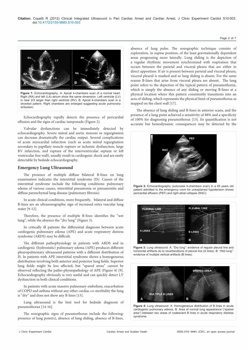
Even rapid qualitative assessment of the dimension of the cardiac chambers is useful in critical ill patients. Normally in apical 4-chambers scan the left ventricle is 2/3 larger than the right while the right and the left atrium have the same dimensions. To observe the right chambers larger than the left in a shocked patient, for example, raises a strong suspicion of massive pulmonary embolism (Figure 1).
LV end systolic cavity obliteration suggests significant hypovolaemia and may precede changes in blood pressure [8].
Journal of Clinical & Experimental CardiologyJo
urna
l of C
linica
l & Experimental Cardiology
ISSN: 2155-9880
Citation: Copetti R (2012) Clinical Integrated Ultrasound in Peri Cardiac Arrest and Cardiac Arrest. J Clinic Experiment Cardiol S10:003. doi:10.4172/2155-9880.S10-003
Page 2 of 7
ISSN:2155-9880 JCEC, an open access journalCardiac Arrest and Sudden DeathJ Clinic Experiment Cardiol
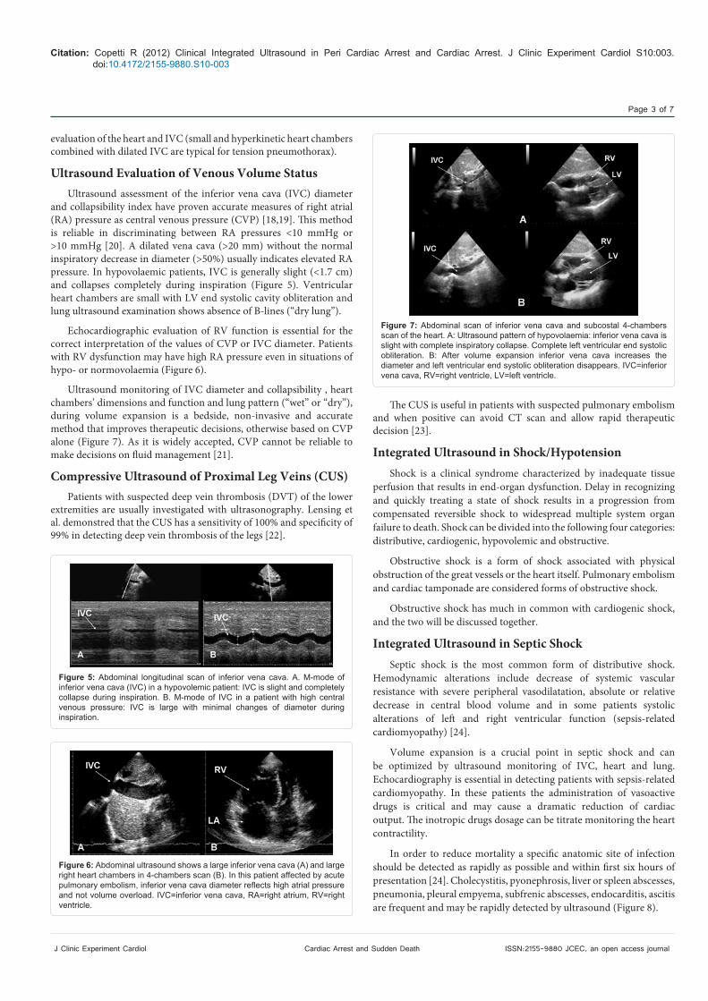
Echocardiography rapidly detects the presence of pericardial effusion and the signs of cardiac tamponade (Figure 2).
Valvular dysfunctions can be immediately detected by echocardiography. Severe mitral and aortic stenosis or regurgitation can decrease dramatically the cardiac output. Several complications of acute myocardial infarction (such as acute mitral regurgitation secondary to papillary muscle rupture or ischemic dysfunction, large RV infarction, and rupture of the interventricular septum or left ventricular free wall), usually result in cardiogenic shock and are easily detectable by bedside echocardiography.
Emergency Lung Ultrasound The presence of multiple diffuse bilateral B-lines on lung
examination indicates the interstitial syndrome (IS). Causes of the interstitial syndrome include the following conditions: pulmonary edema of various causes, interstitial pneumonia or pneumonitis and diffuse parenchymal lung disease (pulmonary fibrosis).
In acute clinical conditions, more frequently, bilateral and diffuse B-lines are an ultrasonographic sign of increased extra vascular lung water [9-12].
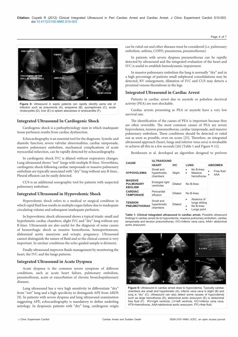
Therefore, the presence of multiple B-lines identifies the “wet lung”, while the absence the “dry lung” (Figure 3).
In critically ill patients the differential diagnosis between acute cardiogenic pulmonary edema (APE) and acute respiratory distress syndrome (ARDS) may be difficult.
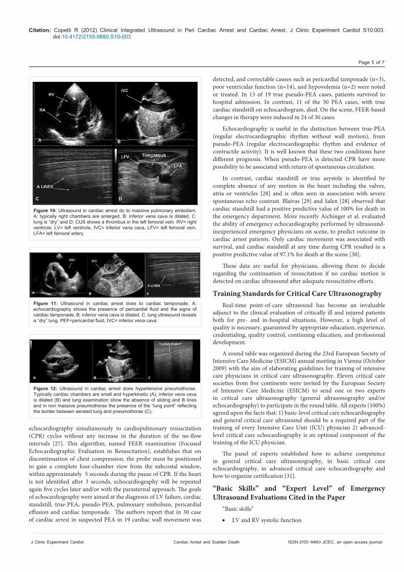
The different pathophysiology in patients with ARDS and in cardiogenic (hydrostatic) pulmonary edema (APE) produces different pleuropulmonary ultrasound patterns with a different distribution of IS. In patients with APE interstitial syndrome shows a homogeneous distribution involving both anterior and posterior lung fields. Superior lung fields might be less affected, but “spared areas” cannot be observed reflecting the patho-physiopathology of APE (Figure 4) [9]. Echocardiography obviously is very useful and can quickly detect LV dysfunction in both clinical conditions.
In patients with acute massive pulmonary embolism, exacerbation of COPD and asthma without any other cardiac co-morbidity the lung is “dry” and does not show any B-lines [13].
Lung ultrasound is the best tool for bedside diagnosis of pneumothorax [14-16].
The sonographic signs of pneumothorax include the following: presence of lung point(s), absence of lung sliding, absence of B-lines,
absence of lung pulse. The sonographic technique consists of exploration, in supine position, of the least gravitationally dependent areas progressing more laterally. Lung sliding is the depiction of a regular rhythmic movement synchronized with respiration that occurs between the parietal and visceral pleura that are either in direct apposition. If air is present between parietal and visceral pleura, visceral pleural is masked and so lung sliding is absent. For the same reason B-lines that arise from visceral pleura are absent. The lung point refers to the depiction of the typical pattern of pneumothorax, which is simply the absence of any sliding or moving B-lines at a physical location where this pattern consistently transitions into an area of sliding, which represents the physical limit of pneumothorax as mapped on the chest wall [17].
The absence of lung sliding and B-lines in anterior scans, and the presence of a lung point achieved a sensitivity of 88% and a specificity of 100% for diagnosing pneumothorax [13]. Its quantification is not accurate but hemodynamic consequences may be detected by the
Figure 1: Echocardiography. A: Apical 4-chambers scan of a normal heart. Right (RA) and left (LA) atrium show the same dimension. Left ventricle (LV) is near 2/3 larger than right ventricle (RV). B: Apical 4-chambers scan in a shocked patient. Right chambers are enlarged suggesting acute pulmonary embolism.
Figure 2: Echocardiography (subcostal 4-chambers scan) in a 65 years old patient admitted to the emergency room for unexplained hypotension shows pericardial effusion (PEF) and right atrial collapse (RAC).
Figure 3: Lung ultrasound. A. “Dry lung”: evidence of regular pleural line and horizontal artifacts do to reverberations of pleural line (A lines). B: “Wet lung”: evidence of multiple vertical artifacts (B lines).
Figure 4: Lung ultrasound. A: Homogeneous distribution of B lines in acute cardiogenic pulmonary edema. B: Area of normal lung appearance (“spared area”) between two areas of coalescent B lines in acute respiratory distress syndrome.
Citation: Copetti R (2012) Clinical Integrated Ultrasound in Peri Cardiac Arrest and Cardiac Arrest. J Clinic Experiment Cardiol S10:003. doi:10.4172/2155-9880.S10-003
Page 3 of 7
ISSN:2155-9880 JCEC, an open access journalCardiac Arrest and Sudden DeathJ Clinic Experiment Cardiol
evaluation of the heart and IVC (small and hyperkinetic heart chambers combined with dilated IVC are typical for tension pneumothorax).
Ultrasound Evaluation of Venous Volume Status Ultrasound assessment of the inferior vena cava (IVC) diameter
and collapsibility index have proven accurate measures of right atrial (RA) pressure as central venous pressure (CVP) [18,19]. This method is reliable in discriminating between RA pressures <10 mmHg or >10 mmHg [20]. A dilated vena cava (>20 mm) without the normal inspiratory decrease in diameter (>50%) usually indicates elevated RA pressure. In hypovolaemic patients, IVC is generally slight (<1.7 cm) and collapses completely during inspiration (Figure 5). Ventricular heart chambers are small with LV end systolic cavity obliteration and lung ultrasound examination shows absence of B-lines (“dry lung”).
Echocardiographic evaluation of RV function is essential for the correct interpretation of the values of CVP or IVC diameter. Patients with RV dysfunction may have high RA pressure even in situations of hypo- or normovolaemia (Figure 6).
Ultrasound monitoring of IVC diameter and collapsibility , heart chambers’ dimensions and function and lung pattern (“wet” or “dry”), during volume expansion is a bedside, non-invasive and accurate method that improves therapeutic decisions, otherwise based on CVP alone (Figure 7). As it is widely accepted, CVP cannot be reliable to make decisions on fluid management [21].
Compressive Ultrasound of Proximal Leg Veins (CUS)Patients with suspected deep vein thrombosis (DVT) of the lower
extremities are usually investigated with ultrasonography. Lensing et al. demonstred that the CUS has a sensitivity of 100% and specificity of 99% in detecting deep vein thrombosis of the legs [22].
The CUS is useful in patients with suspected pulmonary embolism and when positive can avoid CT scan and allow rapid therapeutic decision [23].
Integrated Ultrasound in Shock/HypotensionShock is a clinical syndrome characterized by inadequate tissue
perfusion that results in end-organ dysfunction. Delay in recognizing and quickly treating a state of shock results in a progression from compensated reversible shock to widespread multiple system organ failure to death. Shock can be divided into the following four categories: distributive, cardiogenic, hypovolemic and obstructive.
Obstructive shock is a form of shock associated with physical obstruction of the great vessels or the heart itself. Pulmonary embolism and cardiac tamponade are considered forms of obstructive shock.
Obstructive shock has much in common with cardiogenic shock, and the two will be discussed together.
Integrated Ultrasound in Septic ShockSeptic shock is the most common form of distributive shock.
Hemodynamic alterations include decrease of systemic vascular resistance with severe peripheral vasodilatation, absolute or relative decrease in central blood volume and in some patients systolic alterations of left and right ventricular function (sepsis-related cardiomyopathy) [24].
Volume expansion is a crucial point in septic shock and can be optimized by ultrasound monitoring of IVC, heart and lung. Echocardiography is essential in detecting patients with sepsis-related cardiomyopathy. In these patients the administration of vasoactive drugs is critical and may cause a dramatic reduction of cardiac output. The inotropic drugs dosage can be titrate monitoring the heart contractility.
In order to reduce mortality a specific anatomic site of infection should be detected as rapidly as possible and within first six hours of presentation [24]. Cholecystitis, pyonephrosis, liver or spleen abscesses, pneumonia, pleural empyema, subfrenic abscesses, endocarditis, ascitis are frequent and may be rapidly detected by ultrasound (Figure 8).
Figure 5: Abdominal longitudinal scan of inferior vena cava. A. M-mode of inferior vena cava (IVC) in a hypovolemic patient: IVC is slight and completely collapse during inspiration. B. M-mode of IVC in a patient with high central venous pressure: IVC is large with minimal changes of diameter during inspiration.
Figure 6: Abdominal ultrasound shows a large inferior vena cava (A) and large right heart chambers in 4-chambers scan (B). In this patient affected by acute pulmonary embolism, inferior vena cava diameter reflects high atrial pressure and not volume overload. IVC=inferior vena cava, RA=right atrium, RV=right ventricle.
Figure 7: Abdominal scan of inferior vena cava and subcostal 4-chambers scan of the heart. A: Ultrasound pattern of hypovolaemia: inferior vena cava is slight with complete inspiratory collapse. Complete left ventricular end systolic obliteration. B: After volume expansion inferior vena cava increases the diameter and left ventricular end systolic obliteration disappears. IVC=inferior vena cava, RV=right ventricle, LV=left ventricle.
Citation: Copetti R (2012) Clinical Integrated Ultrasound in Peri Cardiac Arrest and Cardiac Arrest. J Clinic Experiment Cardiol S10:003. doi:10.4172/2155-9880.S10-003
Page 4 of 7
ISSN:2155-9880 JCEC, an open access journalCardiac Arrest and Sudden DeathJ Clinic Experiment Cardiol
Integrated Ultrasound In Cardiogenic ShockCardiogenic shock is a pathophysiology state in which inadequate
tissue perfusion results from cardiac dysfunction.
Echocardiography is an essential tool for the diagnosis. Systolic and diastolic function, severe valvular abnormalities, cardiac tamponade, massive pulmonary embolism, mechanical complications of acute myocardial infarction, can be rapidly detected by echocardiography.
In cardiogenic shock IVC is dilated without respiratory changes. Lung ultrasound shows “wet” lungs with multiple B-lines. Neverthless, cardiogenic shock following cardiac tamponade or massive pulmonary embolism are typically associated with “dry” lung without any B-lines.. Pleural effusion can be easily detected.
CUS is an additional sonographic tool for patients with suspected pulmonary embolism.
Integrated Ultrasound in Hypovolemic ShockHypovolemic shock refers to a medical or surgical condition in
which rapid fluid loss results in multiple organ failure due to inadequate circulating volume and subsequent inadequate perfusion.
In hypovolemic shock ultrasound shows a typical triade: small and hyperkinetic cardiac chambers, slight IVC and “dry” lung without any B-lines. Ultrasounds are also useful for the diagnosis of some causes of hemorrhagic shock as massive hemothorax, hemoperitoneum, abdominal aortic aneurysm and ectopic pregnancy. Ultrasound cannot distinguish the nature of fluid and so the clinical contest is very important. In unclear conditions the echo-guided sample is diriment.
Finally ultrasound improves fluids management by monitoring the heart, the IVC and the lungs patterns.
Integrated Ultrasound in Acute DyspneaAcute dyspnea is the common severe symptom of different
conditions, such as acute heart failure, pulmonary embolism, pneumothorax, acute or exacerbation of chronic bronchopulmonary diseases.
Lung ultrasound has a very high sensitivity in differentiate “dry” from “wet” lung and a high specificity to distinguish APE from ARDS [9]. In patients with severe dyspnea and lung ultrasound examination suggesting APE, echocardiography is mandatory to define underling aetiology. In dyspnoeic patients with “dry” lung, cardiogenic origin
can be ruled out and other diseases must be considered (i.e. pulmonary embolism, asthma, COPD, pneumonia, pneumothorax).
In patients with severe dyspnea pneumothorax can be rapidly detected by ultrasound and the integrated evaluation of the heart and IVC is useful to establish hemodynamic impairment.
In massive pulmonary embolism the lung is normally “dry” and in a high percentage of patients small subpleural consolidations may be detected, RV enlargement, dilatation of IVC and CUS may detects a proximal venous thrombosis in the legs.
Integrated Ultrasound in Cardiac ArrestPatients in cardiac arrest due to asystole or pulseless electrical
activity (PEA) are non shockable.
Cardiac arrests presenting as PEA or asystole have a very low survival rate.
The identification of the causes of PEA is important because they are often reversible. The most common causes of PEA are severe hypovolemia, tension pneumothorax, cardiac tamponade, and massive pulmonary embolism. These conditions should be detected or ruled out as soon as possible, even on scene [25]. Therefore, an integrated ultrasound approach (heart, lungs and inferior vena cava) is invaluable to achieve all this in a few seconds [26] (Table 1 and Figure 9-12).
Breitkreutz et al. developed an algorithm designed to perform
Figure 8: Ultrasound in septic patients can rapidly identify same site of infection such as pneumonia (A), empyema (B), pyonephrosis (C), acute cholecystitis (D), liver (E) or spleen abscesses or endocarditis (F).
Figure 9: Ultrasound in cardiac arrest does to hypovolemia. Typically cardiac chambers are small and hyperkinetic (A), inferior vena cava is slight (B) and lung is “dry” (C). Ultrasound can also detect some causes of hypovolemia such as large hemothorax (D), abdominal aortic aneurysm (E) or abdominal free fluid (F). RV=right ventricle, LV=left ventricle, IVC=inferior vena cava, HTX=hemothorax, AAA=abdominal aortic aneurysm, FFL=free fluid.
CAUSEULTRASOUNDHEART IVC LUNG ABDOMEN
HYPOVOLEMIASmall and hyperkinetic chambers
Slight• No B-lines• Massive
hemothorax
• Free fluid• AAA
MASSIVE PULMONARY EBOLISM
Enlarged right ventricles Dilated No B-lines
CARDIAC TAMPONADE
Pericardial effusion Dilated No B-lines
TENSION PNEUMOTHORAX
Small and hyperkinetic chambers
Dilated
• Absence of lungs sliding
• No B-lines• Lungs point
Table 1: Clinical integrated ultrasound in cardiac arrest. Possible ultrasound findings in cardiac arrest do to hypovolemia, massive pulmonary embolism, cardiac tamponade and tension pneumothorax, IVC=inferior vena cava, AAA= abdominal aortic aneurysm.
Citation: Copetti R (2012) Clinical Integrated Ultrasound in Peri Cardiac Arrest and Cardiac Arrest. J Clinic Experiment Cardiol S10:003. doi:10.4172/2155-9880.S10-003
Page 5 of 7
ISSN:2155-9880 JCEC, an open access journalCardiac Arrest and Sudden DeathJ Clinic Experiment Cardiol
echocardiography simultaneously to cardiopulmonary resuscitation (CPR) cycles without any increase in the duration of the no-flow intervals [27]. This algorithm, named FEER examination (Focused Echocardiographic Evaluation in Resuscitation), establishes that on discontinuation of chest compression, the probe must be positioned to gain a complete four-chamber view from the subcostal window, within approximately 5 seconds during the pause of CPR. If the heart is not identified after 3 seconds, echocardiography will be repeated again five cycles later and/or with the parasternal approach. The goals of echocardiography were aimed at the diagnosis of LV failure, cardiac standstill, true-PEA, pseudo-PEA, pulmonary embolism, pericardial effusion and cardiac tamponade. The authors report that in 30 case of cardiac arrest in suspected PEA in 19 cardiac wall movement was
Figure 10: Ultrasound in cardiac arrest do to massive pulmonary embolism. A: typically right chambers are anlarged, B: inferior vena cava is dilated, C: lung is “dry” and D: CUS shows a thrombus in the left femoral vein. RV= right ventricle, LV= left ventricle, IVC= inferior vena cava, LFV= left femoral vein, LFA= left femoral artery.
Figure 11: Ultrasound in cardiac arrest does to cardiac tamponade. A: echocardiography shows the presence of pericardial fluid and the signs of cardiac tamponade, B: inferior vena cava is dilated. C: lung ultrasound reveals a “dry” lung. PEF=pericardial fluid, IVC= inferior vena cava.
Figure 12: Ultrasound in cardiac arrest does hypertensive pneumothorax. Typically cardiac chambers are small and hyperkinetic (A), inferior vena cava is dilated (B) and lung examination show the absence of sliding and B lines and in non massive pneumothorax the presence of the “lung point” reflecting the border between aerated lung and pneumothorax (C).
detected, and correctable causes such as pericardial tamponade (n=3), poor ventricular function (n=14), and hypovolemia (n=2) were noted or treated. In 13 of 19 true pseudo-PEA cases, patients survived to hospital admission. In contrast, 11 of the 30 PEA cases, with true cardiac standstill on echocardiogram, died. On the scene, FEER-based changes in therapy were induced in 24 of 30 cases.
Echocardiography is useful in the distinction between true-PEA (regular electrocardiographic rhythm without wall motion), from pseudo-PEA (regular electrocardiographic rhythm and evidence of contractile activity). It is well known that these two conditions have different prognosis. When pseudo-PEA is detected CPR have more possibility to be associated with return of spontaneous circulation.
In contrast, cardiac standstill or true asystole is identified by complete absence of any motion in the heart including the valves, atria or ventricles [28] and is often seen in association with severe spontaneous echo contrast. Blaivas [29] and Salen [28] observed that cardiac standstill had a positive predictive value of 100% for death in the emergency department. More recently Aichinger et al. evaluated the ability of emergency echocardiography performed by ultrasound-inexperienced emergency physicians on scene, to predict outcome in cardiac arrest patients. Only cardiac movement was associated with survival, and cardiac standstill at any time during CPR resulted in a positive predictive value of 97.1% for death at the scene [30].
These data are useful for physicians, allowing them to decide regarding the continuation of resuscitation if no cardiac motion is detected on cardiac ultrasound after adequate resuscitative efforts.
Training Standards for Critical Care UltrasonographyReal-time point-of-care ultrasound has become an invaluable
adjunct to the clinical evaluation of critically ill and injured patients both for pre- and in-hospital situations. However, a high level of quality is necessary, guaranteed by appropriate education, experience, credentialing, quality control, continuing education, and professional development.
A round table was organized during the 23rd European Society of Intensive Care Medicine (ESICM) annual meeting in Vienna (October 2009) with the aim of elaborating guidelines for training of intensive care physicians in critical care ultrasonography. Eleven critical care societies from five continents were invited by the European Society of Intensive Care Medicine (ESICM) to send one or two experts in critical care ultrasonography (general ultrasonography and/or echocardiography) to participate in the round table. All experts (100%) agreed upon the facts that: 1) basic-level critical care echocardiography and general critical care ultrasound should be a required part of the training of every Intensive Care Unit (ICU) physician 2) advanced-level critical care echocardiography is an optional component of the training of the ICU physician.
The panel of experts established how to achieve competence in general critical care ultrasonography, in basic critical care echocardiography, in advanced critical care echocardiography and how to organize certification [31].
“Basic Skills” and “Expert Level” of Emergency Ultrasound Evaluations Cited in the Paper
“Basic skills”
• LV and RV systolic function
Citation: Copetti R (2012) Clinical Integrated Ultrasound in Peri Cardiac Arrest and Cardiac Arrest. J Clinic Experiment Cardiol S10:003. doi:10.4172/2155-9880.S10-003
Page 6 of 7
ISSN:2155-9880 JCEC, an open access journalCardiac Arrest and Sudden DeathJ Clinic Experiment Cardiol
• Pericardial effusion and cardiac tamponade
• Qualitative assessment of the dimension of the cardiac chambers
• Volume status evaluation
• “Wet” or “dry” lung
• Pneumothorax
• Pleural effusion
• Abdominal free fluid
• CUS
• Massive pulmonary embolism
• Abdominal aortic aneurysm
• True-PEA, pseudo-PEA
“Expert level”
• Diastolic function
• Valvular disfunction
• Mechanical complications of acute myocardial infarction
• Site of infection in septic patient (cholecystitis, pyonephrosis, liver or spleen abscesses, pneumonia, pleural empyema, subfrenic abscesses, endocarditis)
• Ectopic pregnancy
ConclusionsUltrasound provides a diagnostic modality that allows a rapid
recognition of several critical clinical scenarios. Clinical integrated ultrasound improves the management of critically ill patients and may be considered an extension of physical examination. An integrated ultrasound approach in cardiac arrest and peri cardiac arrest may be rapidly performed at the bedside with relevant clinical consequences in the diagnosis and management of these critical conditions.
References
1. Mueller X, Stauffer JC, Jaussi A, Goy JJ, Kappenberger L (1991) Subjective visual echocardiographic estimate of left ventricular ejection fraction as an alternative to conventional echocardiographic methods: comparison with contrast angiography. Clin Cardiol 14: 898–902.
2. Poelaert J, Schmidt C, Colardyn F (1998) Transoesophageal echocardiography in the critically ill. Anaesthesia 53: 55-68.
3. Enger EL, O’Toole MF (1991) Noncardiogenic mechanisms of right heart dysfunction. J Cardiovasc Nurs 6: 54-69.
4. Vieillard-Baron A, Schmitt JM, Augarde R, Fellahi JL, Prin S, et al. (2001) Acute cor pulmonale in acute respiratory distress syndrome submitted to protective ventilation: incidence, clinical implications, and prognosis. Crit Care Med 29: 1551-1555.
5. Vieillard-Baron A, Page B, Augarde R, Prin S, Qanadli S, et al. (2001) Acute cor pulmonale in massive pulmonary embolism: incidence, echocardiographic pattern, clinical implications and recovery rate. Intensive Care Med 27: 1481-1486.
6. Vieillard-Baron A, Prin S, Chergui K, Dubourg O, Jardin F (2002) Echo-Doppler demonstration of acute cor pulmonale at the bedside in the medical intensive care unit. Am J Respir Crit Care Med 166: 1310-1319.
7. Vieillard-Baron A, Prin S, Chergui K, Dubourg O, Jardin F (2003) Hemodynamic instability in sepsis: bedside assessment by Doppler echocardiography. Am J Respir Crit Care Med 168: 1270-1276.
8. Leung JM, Levine EH (1994) Left ventricular end-systolic cavity obliteration as an estimate of intraoperative hypovolemia. Anesthesiology 81: 1102-1109.
9. Copetti R, Soldati G, Copetti P (2008) Chest sonography: a useful tool to differentiate acute cardiogenic pulmonary edema from acute respiratory distress syndrome. Cardiovasc Ultrasound 6: 16.
10. Lichtenstein D, Mézière G, Biderman P, Gepner A, Barré O (1997) The comet-tail artifact. An ultrasound sign of alveolar-interstitial syndrome. Am J Respir Crit Care Med 156: 1640-1646.
11. Soldati G, Copetti R, Sher S (2009) Sonographic interstitial syndrome: the sound of lung water. J Ultrasound Med 28: 163-174.
12. Reissig A, Copetti R, Kroegel C (2011) Current role of emergency ultrasound of the chest. Crit Care Med 39: 839-845.
13. Lichtenstein DA, Mezière GA (2008) Relevance of lung ultrasound in the diagnosis of acute respiratory failure: the BLUE protocol. Chest 134: 117-125.
14. Lichtenstein DA, Mezière G, Lascols N, Biderman P, Courret JP, et al. (2005) Ultrasound diagnosis of occult pneumothorax. Crit Care Med 33: 1231-1238.
15. Lichtenstein D, Mezière G, Biderman P, Gepner A (2000) The “lung point”: an ultrasound sign specific to pneumothorax. Intensive Care Med 26: 1434-1440.
16. Alsalim W, Lewis D (2009) Towards evidence based emergency medicine: Best BETs from the Manchester Royal Infirmary. BET 1: Is ultrasound or chest x ray best for the diagnosis of pneumothorax in the emergency department? Emerg Med J 26: 434-435.
17. Volpicelli G, Elbarbary M, Blaivas M, Lichtenstein DA, Mathis G (2012) International Liaison Committee on Lung Ultrasound (ILC-LUS) for the International Consensus Conference on Lung Ultrasound (ICC-LUS). International evidence-based recommendations for point-of-care lung ultrasound.Intensive Care Med 38: 577-591.
18. Cheriex EC, Leunissen KM, Janssen JH, Mooy JM, van Hooff JP (1989) Echography of the inferior vena cava is a simple and reliable tool for estimation of ‘dry weight’ in haemodialysis patients. Nephrol Dial Transplant 4: 563-568.
19. Katzarski KS, Nisell J, Randmaa I, Danielsson A, Freyschuss U, et al. (1997) A critical evaluation of ultrasound measurement of inferior vena cava diameter in assessing dry weight in normotensive and hypertensive hemodialysis patients. Am J Kidney Dis 30: 459-465.
20. Kircher BJ, Himelman RB, Schiller NB (1990) Noninvasive estimation of right atrial pressure from the inspiratory collapse of the inferior vena cava. Am J Cardiol 66: 493-496.
21. Marik PE, Baram M, Vahid B (2008) Does central venous pressure predict fluid responsiveness? A systematic review of the literature and the tale of seven mares. Chest 134: 172-178.
22. Lensing AW, Prandoni P, Brandjes D, Huisman PM, Vigo M, et al. (1989) Detection of deep-vein thrombosis by real-time B-mode ultrasonography. N Engl J Med 320: 342-345.
23. Agnelli G, Becattini C (2010) Acute pulmonary embolism. N Engl J Med 363: 266-274.
24. Dellinger RP, Levy MM, Carlet JM, Bion J, Parker MM, et al. (2008) Surviving Sepsis Campaign: international guidelines for management of severe sepsis and septic shock: 2008. Crit Care Med 36: 296-327.
25. Nolan JP, Soar J, Zideman DA, Biarent D, Bossaert LL, et al. (2010) European Resuscitation Council Guidelines for Resuscitation 2010 Section 1. Executive summary. Resuscitation 81: 1219-1276.
26. Hernandez C, Shuler K, Hannan H, Sonyika C, Likourezos A, et al. (2008) C.A.U.S.E.: Cardiac arrest ultra-sound exam--a better approach to managing patients in primary non-arrhythmogenic cardiac arrest. Resuscitation 76: 198-206.
27. Breitkreutz R, Walcher F, Seeger FH (2007) Focused echocardiographic evaluation in resuscitation management: concept of an advanced life support-conformed algorithm. Crit Care Med 35: S150-161.
28. Salen P, Melniker L, Chooljian C, Rose JS, Alteveer J, et al. (2005) Does the presence or absence of sonographically identified cardiac activity predict resuscitation outcomes of cardiac arrest patients? Am J Emerg Med 23: 459-462.
29. Blaivas M, Fox JC (2001) Outcome in cardiac arrest patients found to have

Citation: Copetti R (2012) Clinical Integrated Ultrasound in Peri Cardiac Arrest and Cardiac Arrest. J Clinic Experiment Cardiol S10:003. doi:10.4172/2155-9880.S10-003
Page 7 of 7
ISSN:2155-9880 JCEC, an open access journalCardiac Arrest and Sudden DeathJ Clinic Experiment Cardiol
cardiac standstill on the bedside emergency department echocardiogram. Acad Emerg Med 8: 616-621.
30. Aichinger G, Zechner PM, Prause G, Sacherer F, Wildner G, et al. (2012) Cardiac movement identified on prehospital echocardiography predicts outcome in cardiac arrest patients. Prehosp Emerg Care 16: 251-255.
31. Expert Round Table on Ultrasound in ICU (2011) International expert statement on training standards for critical care ultrasonography. Intensive Care Med 37: 1077-1083.
Thisarticlewasoriginallypublished inaspecial issue,Cardiac Arrest and Sudden Death handledbyEditor(s).Dr.YidongWei,TongjiUniversity,China


![Journal of Clinical & Experimental Copetti, J Clinic Experiment … · 2017-11-17 · make decisions on fluid management [21]. Compressive Ultrasound of Proximal Leg Veins (CUS) Patients](https://img.pdfslide.us/doc/110x75/5e3e31c89f48c22325213d9b/journal-of-clinical-experimental-copetti-j-clinic-experiment-2017-11-17.jpg)