Embed Size (px)
Citation preview
Macrophage Polarisation: A collaboration of Differentiation, Activation and Pre-Programming?Andrew D. Foey*
School of Biomedical & Healthcare Sciences, Peninsula Schools of Medicine & Dentistry, Drake Circus, Plymouth University, Plymouth PL4 8AA, UK*Corresponding author: Andrew D. Foey, School of Biomedical & Healthcare Sciences, Peninsula Schools of Medicine & Dentistry, Drake Circus, Plymouth University,Plymouth PL4 8AA, UK; Tel: +44-1752-584623 ; E-mail: [email protected] date: December 31, 2014; Accepted date: January 27, 2015 ; Published date: February 03, 2015
Copyright: © 2015 Foey AD. This is an open-access article distributed under the terms of the Creative Commons Attribution License, which permits unrestricted use,distribution, and reproduction in any medium, provided the original author and source are credited.
Abstract
Macrophages (Mϕs) exhibit a sliding scale of functional heterogeneity ranging from pro-inflammatory, immuneactivatory and anti-tumoral responses to anti-inflammatory, regulatory and pro-tumoral activity. These effectorresponses are reflected in distinct Mϕ subsets; the M1/classically activated- and M2/alternatively activated subsets.The functional diversity is determined by the combination of Mϕ subset differentiation, activation, signalling and pre-programming in separate monocyte subsets. This diversity in Mϕ subset and functionality is also reflected inmucosal pathologies associated with chronic inflammation (Crohn’s disease, chronic periodontitis) andimmunosuppression observed in solid tumours (oral squamous cell carcinoma). The relative functional plasticitybetween these monocytes and Mϕs represents a realistic therapeutic regimen in the treatment of these Mϕ-drivendiseases. This review will discuss the research evidence that is suggestive of the manipulation of Mϕ polarisationplasticity through pre-programming, differentiation, activation and tolerisation in the therapeutic intervention forchronic inflammation and solid tumours.
Introduction
Macrophages (Mϕs) are phagocytic cells of the innate immunesystem that are present in most tissues of the human body. These cellsexhibit a wide variety of functional characteristics includingphagocytic clearance, microbial killing, antigen processing andpresentation, inflammation, anti-inflammatory processes, tissue repairand immune suppression. This diversity of immune functionality isreflected in macrophage subset heterogeneity. Current understandingcategorises Mϕ subsets according to activation status (classical oralternative) or differentiation (M1 or M2). There are however, subtleand not-so-subtle differences in Mϕ subsets and their phenotypicmarkers when comparing murine and human Mϕ systems, which havebeen described in other seminal reviews [1]. At this stage, cleardelineation of murine Mϕ biology from that of humans would weakenthe overall understanding of Mϕ biology and, as such, this reviewconsiders the combined contribution of murine and human Mϕresearch. This review will focus on the functional role of Mϕ subsetsdriving immune responses with respect to differentiation andactivation stimuli encountered in host tissues as part of homeostaticand pathogenic conditions. Finally, macrophage subset effectorresponses may already be pre-programmed in the monocyte. Currentresearch suggests that differential effector responses are reflected byclassical, intermediate and non-classical monocytes. The overallfunctional impact of tissue macrophages is likely to be reflected by asubtle balance between pre-programmed monocytes, route of Mϕdifferentiation and the activation/suppressive signals encountered inthe local environment; impacting on distinct Mϕ effector subsetpolarisation or switching between functional subsets as a consequenceof plasticity.
Macrophage subsets and effector phenotypesMacrophages exhibit a range of functional characteristics which
include: 1) sampling of the local environment, 2) killing of pathogens,3) inflammation, 4) tissue repair, 5) anti-inflammatory responses orimmune-suppression, 6) instruction and development of specificadaptive immunity via antigen processing and presentation and 7)mobilisation of other innate cells (Nϕs & NKs) and adaptive cellswhich amplify responses at site of infectious/injurious challenge[reviewed in 2]. Mϕs are tissue resident cells, whose behaviour isshaped by the very environment that they inhabit. These tissue Mϕscan be replenished either locally via self-renewal/proliferation or fromthe periphery via bone marrow-derived monocytes [3-6]. It is thislocalised tissue distribution that makes the Mϕ an efficient and centralresponding cell, driving rapid responses to pathogenic infection, tissueinjury and repair [7,8].
The local tissue environment determines Mϕ effector function as aconsequence of a wide variety of activation and differentiation stimuli.This diversity of stimuli results in Mϕ polarisation and the resultingsubsets being described as classically or alternatively activated Mϕs,originally described to be activated by IFNγ/LPS and IL4/IL-13respectively [9,10]. In addition to activation determining Mϕpolarisation and functionality, several groups have described Mϕsubsets to be dependent on differentiation pathways and possibly pre-programmed. Early studies investigating murine immune responses toLeishmania infection demonstrated the C57Bl/6 strain to be resistant(Th1-mediated CMI predominates) whereas Balb/c mice weresusceptible (predominated by a Th2-mediated humoral response). Thisvariation in response to infection was found to be determined by theMϕs rather than the T cell subset [11]. This predominance of Mϕresponse observed in this study lead to the description of M1 and M2subsets where M1 Mϕs activated T cells to secrete IFNγ and theresulting stimulation of Tc and positive feedback to M1. In contrast,
Foey, J Clin Cell Immunol 2015, 6:1http://dx.doi.org/10.4172/2155-9899.1000293
Review Article Open Access
J Clin Cell Immunol Macrophage Polarization ISSN:2155-9899 JCCI, an open access journal
Journal of Clinical & CellularImmunology
M2 Mϕs induced T cells to produce Th2-like cytokines (IL-4 andTGFβ) resulting in humoral responses and amplification of M2activity [12-14]. This latter amplification of M2 activity resulting inwalling off pathogens, as a consequence of matrix deposition andfibrosis. Thus, specific responses to pathogen infection are Mϕ-determined which help tune and are in-turn finely tuned by T cells.Finally, these M1/Th1 and M2/Th2 responses may both occur foroptimally dealing with infection simultaneously or at differentprogression phases of the pathology [15,16]. M1 Mϕs are generallyconsidered to be the predominant subset involved in pathogen killing,hence host defence whereas the M2 subset is associated with repairand maintenance of tissue integrity. Mϕs are pivotal to directing theimmune response where M1s drive T cells towards Th1 cell-mediatedimmunity (CMI) and M2s towards Th2-mediated humoral activity.
The host is under constant challenge by a wide variety of pathogens.The macrophage deals with this ever-changing pathogenic challengeby retaining a heterogeneous functionality through a level of fluidity orplasticity. The degree of plasticity between homeostatic M2 Mϕs andM1 Mϕs is a possible explanation for the ever-increasing number ofMϕ subsets described in the literature. Thus far, Mϕ heterogeneity hasresulted in the description of classically activated, M1s, alternativelyactivated, M2a, M2b, M2c, M2d and regulatory Mϕs [17-19]. Such avariety of subsets exhibiting specific functional heterogeneity has yetto be described in vivo. A likely explanation for this apparent variety ofsubsets/functionality can either is as a consequence of varyingproportions of M1 and M2 Mϕs existing as a heterogeneouspopulation or that these different subsets may be intermediates in asliding scale of plasticity between homeostatic M2 Mϕs and the M1Mϕ. At this stage of our understanding however, these other subsetscannot be ignored (Table 1). M2a (alternative) and M2c (deactivated)are induced by IL4/IL13 and IL10/TGFβ/glucocorticoids respectively,both express arginase activity that is associated with the moreconventional M2 subset. Both of these Mϕ types also express IL-10hi
IL-12lo and the scavenger receptor, MR. The M2b (type II) Mϕ isinduced by immune complex recognition as well as LPS and IL-1β;this Mϕ expresses a similar cytokine profile but differs from M2a andM2c by virtue of expression of iNOS, normally associated with the M1Mϕ subset [reviewed in 8]. This expression of iNOS, yet display of ananti-inflammatory cytokine profile may be suggestive that M2b mayrepresent an intermediate “plastic” state between the canonical M1and M2 subsets. The M2d Mϕ subset was described for an adenosine-mediated switch in phenotype to an M2-like cell. This subsetpolarisation resulting from the synergistic activation by A2R agonistsin combination with agonists of TLR2, TLR4, TLR7 or TLR9; wherethe new M2d effector subset exhibited a phenotype: IL-10hi VEGFhi
iNOShi IL12lo TNFαlo and elevated Arg-1 expression [20,21]. TheseMϕs do not express Ym-1, FIZZ-1 or CD206 but, again, exhibit aphenotype, which falls between the canonical M1 and M2 subsets;whether this Mϕ is proven to exist as a distinct subset or merely anintermediate awaits clarification. In a separate study, ratherconfusingly, Duluc et al. described an ovarian TAM phenotype, whichwas also proposed as M2d [22]. This subset was polarised by LIF, IL-6and OSM and exhibited a regulatory/immunosuppressive phenotype:CD14hi CD163hi CD80lo CD86lo ILT2hi ILT3hi IL10hi TNFαlo IL12lo
CCL18hi PTX3lo CCL1lo CCL17lo CCL22lo. In addition, thissuppressive subset also expressed IDO, VEGF, TGFβ and B7-H4whereas iNOS and Arg-1 were not detected. In contrast, the A2R/TLR-polarised M2d subset was found to express both iNOS and Arg-1,suggestive that these two Mϕ subsets are phenotypically andfunctionally distinct.
Additionally, further Mϕ subsets have been reported, especiallyassociated with investigation of inflammatory pathologies such asatherosclerosis. Whether these further subsets present themselves inthe context of mucosal pathology awaits characterisation. Theseatherosclerotic associated subsets include M4, Mox, HA-mac, M(Hb)and Mhem [23,24]. M4 Mϕs have been shown to be distinct from M1and M2 phenotypes, where polarisation is induced by theatherosclerotic chemokine, Platelet Factor 4 (PF4) or CXCL4, resultingin a phenotype: MRhi CD36lo CCL22hi TNFSF10/TRAILhi TNFαhi/lo
IL10lo CD86hi MMP7hi MMP12hi which is poorly phagocytic [25].Mox subset refers to a phenotype of oxidised Mϕs found inatherosclerotic lesions. This phenotype is polarised by oxidisedphospholipids such as oxLDLs and express the Nrf2-dependent redox-regulated gene product, heme oxygenase-1 (HO-1) and both anti-inflammatory IL-10 and pro-inflammatory IL-1β [26]. The finalputative subsets described in the case of atherosclerosis are HA-mac,M(Hb) and Mhem; all of which are polarised by either haem orhaemoglobin and express CD163. HA-mac were first described byBoyle et al and were found to be located in the hemmorhagic zones ofplaques and defined as CD163hi whereas these Mϕs were lowexpressors of HLA-DR, thus are relatively poor antigen presentingcells [27]. In addition, HA-macs exhibit anti-oxidant and anti-inflammatory behaviour, where HO-1 and IL-10 are expressed andpolarisation is Nrf2-dependent; resulting in tissue repair and a reducedcapacity to form foam cells [28,29]. Thus the Mϕs involved in thisinflammatory disease, Mox, HA-mac, M(Hb) and Mhem, are polarisedby the local environment and express phenotypes that portray bothpro- and anti-atherogenic functionality [23,24]. Whether thisfunctional dichotomy exists in so many distinct subsets in otherpathologies remains to be illucidated. Currently, the plasticityexhibited between M1 and M2 subsets is clear in the context ofmucosal homeostasis and disease states; the existence of M4 and Moxcells may not be involved in mucosal tissues whereas homeostatic anddisease-induced intermediates between these two canonical subsetsmay well parallel these extra subsets described in both tumour- andatherosclerosis-associated Mϕs.
Macrophages can generally be categorised by their ability tometabolise arginine. Mϕs exhibit a specialised biochemical systemutilising L-arginine that allows for functional plasticity between M2homeostatic subset and M1 function, capable of killing of pathogensand pathogen-infected host cells and cancer cells [11,12,30]. Nitricoxide (NO) is produced by Mϕs as part of the innate system’s killingresponse to pathogens. The very nature of innate responses and non-specificity of NO-killing can result in collateral damage to host tissues/cells [31-33]. Thus, Mϕ subsets have been categorised by theexpression and activity of the arginine-metabolising enzymes,inducible nitric oxide synthase (iNOS) and arginase-1 (Arg-1). Arg-1is a signature molecule for the activation state of alternatively activatedor M2 Mϕs [34]. It has been found to be important in immunologicalfunctionality of myeloid cells [4] and that these Arg-1+ expressing cellscontribute to T cell energy, preventing activation of effector Th cells[35,36]. Thus, Arg-1 activity has been associated with previouslydescribed myeloid suppressor cells [37]. The secretion of Arg-1 and itsbreakdown products of arginine are linked with Mϕ functionspreviously described for M2/alternatively activated Mϕs. ExtracellularArg-1 exerts potent anti-inflammatory effects where sustainedexpression of Arg-1 is hypoinflammatory and is limiting to T cellpolarisation via negative regulation of polarising cytokines, IL-6,IL-12p40 subunit (IL-12/IL-23) and IL-10.
Citation: Foey AD (2015) Macrophage Polarisation: A collaboration of Differentiation, Activation and Pre-Programming?. J Clin Cell Immunol 6:293. doi:10.4172/2155-9899.1000293
Page 2 of 15
J Clin Cell Immunol Macrophage Polarization ISSN:2155-9899 JCCI, an open access journal
In addition to being characterised by arginine metabolism, thisdichotomy in Mϕ effector subset functionality can be further definedby a whole plethora of molecules expressed and functional outcomes.In general, M1 Mϕs are iNOS+ hence NO production and anti-microbial functions, they also express high levels of HLA-DR+, co-stimulatory CD86 and IL-12 associated with mediating and polarisingTh1 responses to intracellular-resident pathogens and anti-tumourresponses [38-40]. This high-level expression and protection conferredby IL-12p40 extends to this subunit being shared by IL-23, which isalso produced by M1s, and plays a pivotal role in the differentiationand activation of Th17 cells [41,42]. Additionally, these Mϕs aregenerally STAT1+ TREM-1+ cells expressing IL-8 (CXCL8) andMCP-1 (CCL2), which are responsible for perpetuation ofinflammatory responses through the chemotactic recruitment ofneutrophils and monocytes. M1s also produce a wide array of pro-inflammatory cytokines (TNFα, IL-1β, IL-6, IL-18, IL-23), chemokines(CXCL1,2,3,5,8,10, CCL3,4,5,11,17 and 22), matrix metallo-proteinases (MMP-1,-2,-7,-9 and -12), reactive oxygen species (ROS)and pattern recognition receptors (TLR2, TLR4 and TLR5) [18,43-46].M2 M
ϕ
s, on the other hand, in addition to arginase/ornithine, can becharacterised by their expression of the phagocytic scavenger receptor(Mannose Receptor, CD206), TGFβ (immune regulation/suppression),EGF (tissue repair) and VEGF (angiogenesis). This serves to highlightthe regulatory and reparative nature of these M2 Mϕs. Additionally,M2s can also produce/secrete IL-1β, IL-6, TNFα, IL-10, MMPs andTIMPs; these cytokines/enzymes are less strongly associated with theM2 phenotype and are also expressed by M1 Mϕs [reviewed in 8;47,48], although a predominance of anti-inflammatory and regulatoryfactor production underpins this functionally distinct Mϕ subset. Thesharing of expression of effector molecules to a greater or lesser extentbetween these functionally divergent Mϕ subsets may go some way toexplaining the apparent existence of several subsets/intermediatesbetween M1 and M2s.
Cytokines play a fundamental role in both differentiation andactivation of M1-like and M2-like Mϕs. The growth factors, M-CSFand GM-CSF have been demonstrated to differentially control Mϕlineage populations in homeostatic and inflammatory conditions [49].Indeed, the Th1-derived cytokines, GM-CSF and IFNγ in combinationwith inflammatory stimuli such as LPS or TNFα, polarise Mϕs towardsthe M1 phenotype [50]. Polarisation towards this M1 pro-inflammatory phenotype is also achieved by hypoxic/anoxicenvironments, β-chemokines and the DAG analogue/PKC activator,phorbol myristate acetate (PMA) [51-54]. On the other hand, Th2-derived cytokines, IL-4, IL-13 and IL-21 were described to polariseMϕs to a mannose receptor (CD206)-expressing M2 phenotype[17,55,56]. Extensive research has described many factors, in additionto Th2-derived cytokines, to be M2-polarising; these include IL-10,TGFϕ, M-CSF, Vitamin D3 and immune complexes [reviewed in 57],with one of the first polarising studies describing M-CSF-mediateddifferentiation resulting in the development of Mϕs deficient in IL-12production [58]. Indeed, the immunosuppressive cytokines TGFβ andIL-10 may be responsible for the observed effects ofCD4+CD25+Foxp3+ Tregs in the induction/polarisation of monocytesto alternatively activated M2-like Mϕs [59]. What is relatively unclearis the stage of sensitivity to polarisation and plasticity. It is commonlythought that terminally differentiated cells lose their plasticity, withonly intermediates retaining this ability to polarise according to thetissue environment. Of interest are the early studies of Rees andcolleagues who described first cytokine exposure to irreversiblydetermine previously uncommitted Mϕ responses, where the initialcytokine exposure (IFNγ, TNFα, TGFβ, IL-4, IL-6, IL-10) determinedMϕ response to be pro-inflammatory, anti-inflammatory, phagocyticor anti-microbial (NO production) and failed to be modulated bysubsequent cytokine exposure [60].
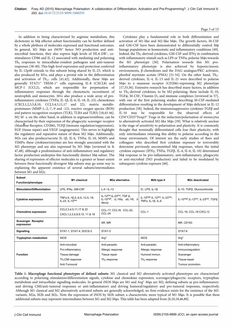
Subset
Function/phenotypeM1 classical M2a alternative M2b type II M2c deactivated
Stimulation/Differentiation LPS, IFNγ, GM-CSF L-4 / IL-13 IC, LPS, IL-1β IL-10, TGFβ, Glucocorticoids
Cytokine expression TNFα,IL-1β,IL-6,IL-12,IL-18,IL-23, IL-10low
IL-12low,IL-23low, TGF β,IL-10high, IL-1Ra, sIL-1R, IIdecoy
IL-10high,IL-12low, IL-23 low,TNFα, IL-1β, IL-6 IL-10high,IL-12low, IL-23low, TGFβ
Chemokine expressionCCL2,3,4,5,11,17 & 22
CXCL1,2,3,5,8,9,10, 11 & 16CCL-17, CCL18, CCL-22,CCL-24 CCL-1 CCL-16, CCL-18 CXCL13
Scavenger Receptorexpression SR, MR MR, CD163
Signalling STAT-1, STAT-4, SOCS-3 STAT-3 STAT-6
Tryptophan metabolism iNOS Arg+ iNOS Arg+
Function
Anti-microbial
Pro-inflammatory
Tissue-damage
Th1CMI response
Anti-Tumoural
Anti-parasitic
Allergic response
Tissue repair
Th2 response
Anti-parasitic
Allergic response
Humoral immun.
Th2 response
Anti-inflammatory
Immunoregulation
Scavenger
Tissue-repair
Tumour promotion
Table 1: Macrophage functional phenotypes of defined subsets: M1 classical and M2 alternatively activated phenotypes are characterisedaccording to polarising stimulation/differentiation signals, cytokine and chemokine expression, scavenger/phagocytic receptors, tryptophanmetabolism and intracellular signalling molecules. In general iNOS Mφs are M1 and Arg+ Mφs are M2; defining subsets as pro-inflammatory
Citation: Foey AD (2015) Macrophage Polarisation: A collaboration of Differentiation, Activation and Pre-Programming?. J Clin Cell Immunol 6:293. doi:10.4172/2155-9899.1000293
Page 3 of 15
J Clin Cell Immunol Macrophage Polarization ISSN:2155-9899 JCCI, an open access journal
and driving CMI/anti-tumoral responses or anti-inflammatory and driving humoral/regulatory and pro-tumoral responses, respectively.Although M1 classical and M2 alternatively activated subsets are generally acknowledged, no firm evidence exists for the existence of the M2-variants, M2a, M2b and M2c. Note the expression of iNOS by M2b subset; a characteristic more typical of M1 Mφs. It is possible that theseadditional subsets may represent intermediates between M1 and M2 Mφs. This table has been adapted from [8,10,18,46,80].
The expression and secretion of effector molecules defines thefunctional responses of M1 and M2 subsets and is integrally-linked tothe manner of cell activation [reviewed in 8]. An efficient Mϕ responseto an infection will thus include both pathogen/tissue destructive andreparative mechanisms mediated by the activity of both M1 and M2Mϕs. Central to this development of appropriate Mϕ immuneresponsiveness is the selective recognition and descrimination ofpathogen associated molecular patterns (PAMPs), danger associatedmolecular patterns (DAMPs) and apoptotic cell associated molecularpatterns (ACAMPs). The recognition of apoptotic cells/ACAMPs byMϕs regulates pro-inflammatory cytokine production and possiblyMϕ polarisation through the induction of TGFβ and PGE2 [61,62].Toll-like receptors (TLRs) mediate responsiveness to PAMPs andDAMPs, hence determining appropriate immune response. TLRsmediate anti-viral, anti-bacterial, anti-fungal or anti-parasiticresponses through involvement of appropriate receptors, adaptorproteins and either MAPK- NFκB- or IRF-dependent signallingpathways [63]. LPS has been shown to be transduced through TLR4which results in the activation of ERK-1,2, JNK, p38 MAPKs as well asNFκB and IRF3 which induce a wide variety of immune geneexpression including TNFα, IL-1β, IL-6, IL-12, IL-10, MHC II andiNOS. Interestingly, these TLR-mediated signals can be negativelyregulated by a wide variety of endogenous inhibitor molecules, whichinclude Myd88s, IRAK-M, IRF4, ST2, TREM2, Tollip, TRIAD3A,p50/p50 NF-κB, suppressor of cytokine signalling 1 (SOCS-1),SOCS-3, SHP1, SHP2 and SIGIRR [64-68]. This range of endogenousinhibitors of TLR signalling becomes more significant whenconsidering the associations of these molecules with regards control ofMϕ polarisation. Alternatively activated, M2-like anti-inflammatoryMϕs have been described to be polarised by IL-4-requiring SHIPdegradation and NFκB inhibition [69,70] whereas IRF5 promotes pro-inflammatory Mϕ polarisation and downstream Th1-Th17 responses[71] and SOCS3 expression is essential for classically activated Mϕs[72].
Distinct signalling components regulate Mϕ polarisation
Mϕ polarisation and effector function is governed by a wealth ofsignal pathways and their component signalling molecules. Suchsignals, which have been previously described to regulate Mϕpolarisation include: NFκB, PI3K/PTEN, STAT3 and SOCS3. There isa reciprocal relationship between the lipid phosphatase, PTEN(phosphatase and tensin homologue deleted on chromosome ten) andPI3K (phosphoinositide 3-kinase) in the polarisation of Mϕ subsets.PTEN has been shown to regulate the expression of Arg-1 inmacrophages, with corresponding downstream modulation of bothinnate and adaptive immune responses [73]. PTEN antagonises theactivity of PI3K where PI3K itself has been demonstrated to functionas a negative regulator of pro-inflammatory cytokine production andiNOS expression, activity and production of nitric oxide (NO) [74,75].PTEN positively regulates TLR-induced IL-6 production; PTENdeletion as well as constitutive activation of PI3K was found to induceArg-1 expression. This is suggestive that PTEN-ve Mϕs expressed afunctional phenotype similar to alternatively activated or M2-like Mϕsin a manner mediated by increased activation of the transcriptionfactors, C/EBPβ and STAT3. IL-10 signalling would appear to be
integrally associated with STAT3 and M2 polarisation, where STAT3activation and IL-10 secretion are linked [76] and the STAT3-inducible cytokines, IL-10 and IL-6, activate Arg-1 expression [77], akey marker of M2/alternatively activated Mϕ polarisation. If STAT3plays a key role in M2 polarisation, it may represent a potentialtherapeutic target for the treatment of inflammatory pathology asevidenced by the conditioned STAT3 KO in mouse Mϕs which wererefractory to IL-10 effects and spontaneously developed chronicenterocolitis [78,79].
The polarisation of M1 Mϕs is transduced by activation of thetranscription factors NFκB and STAT-1 which induce the expressionof M1-associated genes with further control of polarisation throughthe activity of SOCS3 [72]. In addition, the potential for differentiationtowards an M2-like subset is prevented via STAT-1 inhibition ofactivation of the M2-polarising transcription factor, STAT-6 [80],whereas the expressional knock-down (KO) of SOCS3 favours M2polarisation [72]. Indeed Th2 cytokines induce Ym-1 expression (apoorly-defined M2-associated molecule in mice) by a STAT6-dependent mechanism [81]. NFκB has been shown to be integral toMϕ polarisation and effector function; inhibition of which resulted inthe development of an anti-inflammatory M2-like Mϕ phenotype [70].NFκB is also involved in M2 polarisation, where in contrast to p65NFκB subunit involvement with M1 effector function, M2 polarisationprocesses are driven by p50 NFκB subunits [82]. The targeting ofNFκB would appear to be a promising target for manipulation of Mϕpolarisation and has been the subject of intense efforts in the re-education of tumour-associated macrophages (TAMs), originallydescribed as exhibiting a pro-tumoral M2-like phenotype [83].
Activation of the transcription factor, C/EBPβ is associated with thecAMP-dependent activation of CREB; cascades involving thesetranscription factors have been demonstrated to initiate M2 Mϕ-specific gene expression and tissue reparative mechanisms [84]. ThecAMP-activated factor, CREB, is required for full induction of C/EBPβ[84], which transctivates the Arg-1 gene promoter [85]. As was thecase with STAT3, the expression and activity of IL-10 is associatedwith cAMP-mediated responses; whether this signalling pathwaydirectly modulates polarisation or is an indirect consequence of IL-10expression requires further investigation. What is clear is that theprofiles of pro-inflammatory and anti-inflammatory cytokines aredifferentially regulated by cAMP in a manner determined by originalMϕ differentiation signals and activation signals in a PKC/cAMP/CREB axis [86]. In addition to these signalling pathways driving Mϕpolarisation, it is probable that monocytes also display a level ofpolarisation.
Fine control of Mϕ polarisation and functionality is likely to be as aresult of a complex cross-modulation between distinct signallingpathways rather than singular exclusive subset-specific pathwayinvolvement. This subtlety of signal pathway cross-talk driving Mϕpolarisation is potentially demonstrated by a recent study conductedby Arranz et al, who focussed on the involvement of the Akt/PKBfamily of serine/threonine protein kinases. PKB/Akt kinases arepotentially downstream of PI3K, upstream of p70S6K and regulated bycAMP-dependent signals through the activation of PKC isoforms. This
Citation: Foey AD (2015) Macrophage Polarisation: A collaboration of Differentiation, Activation and Pre-Programming?. J Clin Cell Immunol 6:293. doi:10.4172/2155-9899.1000293
Page 4 of 15
J Clin Cell Immunol Macrophage Polarization ISSN:2155-9899 JCCI, an open access journal
breadth of pathway cross-talk is indicative of Akt playing a central rolein Mϕ polarisation. Indeed, in the case of mouse models of LPS-induced endotoxin shock and dextran sodium sulphate (DSS)-inducedcolitis, Akt2 KO resulted in M2 Mϕ polarisation and resistance tothese inflammatory pathologies whereas Akt1 KO polarised Mϕstowards the M1 subset and an increased sensitivity to inducedendotoxin shock and colitis. This polarisation towards M2s as aconsequence of Akt2 KO was found to be due to an increasedexpression of C/EBPβ, a positive regulator of Arg-1 [87]. In addition,tuberous sclerosis complex 1 (TSC1) has been demonstrated tomodulate Mϕ polarisation in a manner that is dependent orindependent of mTOR, the downstream effector of the p70S6Kpathway. TSC1 encourages M2 polarisation in an mTOR-C/EBPβ-dependent manner whereas it suppresses ERK-dependent polarisationtowards the M1 subset in an mTOR-independent manner [88].
Macrophage effector function is pre-programmed inmonocyte subsets
The effector function of macrophages may already be determined inthe monocyte prior to differentiation to the tissue macrophage. Theexistence of pre-programmed monocyte populations has beensuggested in both murine systems and in humans. The followingsection highlights the existence of functionally distinct monocytesubsets, which are linked to homeostatic and inflammatoryenvironments; just how these subsets fit with the established Mϕpolarisation in health and disease is currently no more than hypothesisbut may need to be thoroughly investigated to complete ourunderstanding of Mϕ subsets and functional phenotypes (Figure 1).Two distinct populations of monocytes have been described in mice,on the basis of chemokine receptor expression; a non-inflammatoryCX3CR1hi CCR2- subset and an inflammatory CX3CR1lo CCR2+
subset [89]. With respect to human monocytes, investigationsundertaken by Loems Zeigler-Heitbrock have characterised differentsubsets, which are dependent on the relative expression of CD16, theFcγRIIIa antibody receptor [90,91] ignored (for surface marker,cytokine and effector phenotype analysis of these monocyte subsets,refer to table 2). The monocyte subsets described are the classical(CD14++ CD16- CD163+), intermediate (CD14++ CD16+ CD163+) andnon-classical (CD14+ CD16++ CD163-) monocytes [92; reviewed in93], where the intermediate monocytes are thought to represent anintermediate transitional subset between the classical and non-classicalmonocytes [94]. The classical CD16- monocytes account for 90% ofcirculatory monocytes whereas CD16+ monocytes account for up to10% whilst at rest [91]. The relative numbers of these pro-inflammatory CD16+ monocyte populations have been shown toincrease in malignancy and inflammation, rising up to 50% in sepsisand being significantly raised in RA and representing a major sourceof TNFα [95-98]. These monocytes can also be selectively depletedafter either IgG infusion or glucocorticoid therapy [99,100]. The non-classical CD16+ monocytes exhibit a distinct functional behaviourwhere upon stimulation produce higher amounts of TNFα, IL-12 andlower amounts of IL-10, hence have been referred to as pro-inflammatory monocytes [96,101,102]. In addition, these monocytesubsets display differential migratory responses whereby classicalmonocytes selectively respond to CCL2/MCP-1 and non-classicalmonocytes are refractory to CCL2 and migrate in response toCX3CL1/Fractalkine [103,104]. Finally, CD16+ monocytes also expresshigher amounts of HLA-DR/Class II MHC and a correspondinggreater capacity for antigen presentation, hence T cell activation[91,105]. With respect to these non-classical monocytes, developmentis determined by the activity of and sensitivity to M-CSF whereblockade of the M-CSF-R pathway has been described to selectivelyreduce CD16+ non-classical monocyte numbers [106].
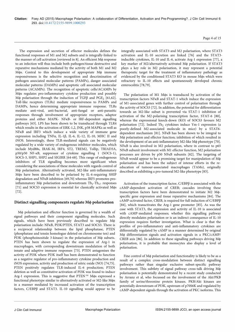
Subset Classical Intermediate Non-Classical
Subset Phenotype CD14hi CD16- CD14hi CD16lo CD14lo CD16hi
Scavenger Receptor expression CD163+ CD163+ CD163-
Cytokine expression TNFα, IL-12, IL-10higher TNFαhigh IL-12high IL-10low
Chemokine recruitment CCL2/MCP-1 CX3CL-1/Fractalkine, CCL2-refractory
Antigen Presentation HLA-DRlow HLA-DRhigh
Tryptophan metabolism iNOS Arg+ iNOS
Function
Anti-microbial
Pro-inflammatory
Tissue-damage
Th1CMI response
Anti-Tumoural
Anti-parasitic
Allergic response
Tissue repair
Th2 response
Anti-parasitic
Allergic response
Humoral immun.
Th2 response
Table 2: Monocyte subset functional phenotypes. Classical CD16-negative and CD16-positive non-classical monocytes can be classifiedaccording to their functional phenotype of scavenger receptor (CD163), cytokine expression, chemokine responsiveness, antigen presentationcapacity (HLA-DR) and arginine metabolism (iNOS or Arg I). The combination of such phenotypes defines monocyte function as pro-inflammatory, CMI-inducing or tissue reparative, induction of humoral immunity. One point to be noted is that the classical and non-classicalsubsets express iNOS whereas the intermediate monocyte subset expresses arginase. Refer to macrophage table 1 earlier. This table has beenadapted from [91,93,94,96,98,101,103,104].
This monocyte system exhibits characteristics, which parallel themacrophage system. Both the monocytes and macrophages exist intwo discrete functional phenotypes and exhibit a level of plasticity
between these subsets, with the monocytes being described to have aclear intermediate subset between the two potential polar subsets.With the realisation that diseases mediated by Mϕ subsets may be
Citation: Foey AD (2015) Macrophage Polarisation: A collaboration of Differentiation, Activation and Pre-Programming?. J Clin Cell Immunol 6:293. doi:10.4172/2155-9899.1000293
Page 5 of 15
J Clin Cell Immunol Macrophage Polarization ISSN:2155-9899 JCCI, an open access journal
controlled by the polarisation/plasticity between M1 and M2, comes afurther complexity that we are likely to have to consider; themanipulation of the subset of the “macrophage progenitor”, themonocyte and how each distinct monocyte subpopulationdifferentiates to distinct Mϕ effector subsets.
Macrophage subsets and pathologyMacrophages play a predominant role in driving many
immunopathological diseases; their pathological function beingdictated by the local tissue environment with respect to the balancebetween polarising activatory, differentiation and suppressive signals.Due to the relative abundance of Mϕ numbers and scientific literature,this section is focussed on the role of Mϕs in the inflammatorypathology of the mouth and intestinal tract and theimmunosuppressive pathology associated with tumour associated Mϕs(TAMs) and solid tumours. Mϕs populate both oral and intestinalmucosae in large numbers [107]. In a homeostatic environment,mucosal Mϕs drive tolerogenic mechanisms whereas, at the same time,maintaining an efficient phagocytic response. This homeostaticmucosal tolerance is associated with Mϕs exhibiting an M2-likephenotype, predominated by the expression of anti-inflammatory,suppressive cytokines and phagocytic scavenger receptors (CD36,CD68 and CD206). These tolerogenic Mϕs maintain a state ofperpetual readiness required for microbial clearance without inducinga localised hyper-inflammatory state [108-110]. In this homeostatictolerogenic state, mucosal Mϕs fail to express the pro-inflammatorycytokines (TNFα, IL-1β, IL-6, IL-8, IL-12, IL-18 and IL-23) whereasTGFβ and IL-10 expression is maintained. This tolerised state isfurther reflected by the lack of expression of CD14/TLRs, FcRs, co-stimulatory molecules (CD40, CD80, CD86) and the pro-inflammatory molecule, TREM-1. Concurrently, there is a markedexpression of the regulatory molecules CD33, CD200R andTGFβRI/RII [reviewed in 57]. This homeostatic /tolerogenic functionof Mϕs is dysregulated in pathology where mucosal tolerance isbroken with respect to inflammatory diseases such as Crohn’s diseaseand Chronic periodontitis and augmented in immune suppression –associated diseases such as colorectal cancer and oral squamous cellcarcinoma. These pathologies exhibit mechanisms aligned to M1- orM2-driven responses. In the context of pro-inflammatory diseases,Mϕs exhibit an inflammatory phenotype that is comparable to the M1subset. These inflammatory Mϕs express a wide variety of effectormolecules, which include: PRRs (CD14, TLR2, TLR4, TLR5), FcRs(CD16, CD32, CD64, CD89), HLA-DR, chemokine receptors (CCR5,CXCR4), CRs and the pro-inflammatory markers/cytokines (TREM-1,TNFα, IL-1β, IL-6, IL-18 and CCL20) [57,111-113].
M1-associated pathology: Crohn’s diseaseCrohn’s disease (CD) is an idiopathic inflammatory bowel disease
(IBD) that is characterised by transmural skip-lesion-associatedinflammatory destruction of the gastro-intestinal tract anywhere fromthe mouth to the anus. CD is characterised by a dysfunctional innateimmune system, which results in inflammatory destruction mediatedby a pathogenic axis of Th1/IL-12 and Th17/IL-23 and the productionof IFNγ, TNFα and IL-17 [114]. This chronic inflammatory disease isassociated with genetic mutations in bacterial-sensing PRRs: NOD2mutations have long-since been described to be a feature of CD whichresulted in dysregulation of and the augmentation of NFκB-mediatedpro-inflammatory cytokine production of TNFα, IL-1γ and IL-12 bymucosal Mϕs [115]. NOD2 has been described to regulate pro-
inflammatory signals transduced through TLR2 [116]. Such abreakdown of regulation observed in CD would result in adysfunctional innate immune response with downstream effects on theadaptive immune system and the commensal microbiota of the gut,which also plays an important role in barrier defences and mucosaltolerance. This total breakdown of barrier integrity and mucosaltolerance, coupled with the bias towards an inflammatory axis of Th1/IL-12 and Th17/IL-23, results in a mucosal environment low inregulatory cytokines IL10 and TGFβ and high in IL-12p40. Thisinflammatory environment is conducive to M1-like Mϕ activation/differentiation with the corresponding up-regulation of pro-inflammatory cytokine and co-stimulatory molecule expression[117,118]. The therapeutic targeting of M1 Mϕs or indeed theaugmentation of M2-mediated responses may represent a realisticregimen in the control of this chronic inflammatory disease.
Chronic periodontitisChronic periodontitis (CP) is a persistent relapsing-remitting
inflammatory disease of the periodontal tissue, which ultimately, ifuntreated, leads to destruction of the periodontium and resultingtooth loss. Like Crohn’s disease, CP is associated with the breakdownof mucosal barrier functionality and tolerance, leading to anuncontrolled inflammatory immune activation response [119]. Theobserved dysbiosis in the oral microbiota results in the perpetualmicrobial challenge; one such prominent microbe driving thisinflammatory pathology is Porphyromonas gingivalis [120,121]. P.gingivalis is an intracellular-resident oral bacteria which infects bothoral epithelial cells and underlying APCs (DCs and Mϕs). Anappropriate host clearing response to such an intracellular pathogenwould be to initiate cell-mediated immunity, mediated by Th1 cells[122,123]. This pathogen however is able to both subvert and suppressappropriate host responses. PG-LPS both exhibits a low endotoxinactivity and can mediate its effects through both TLR2 or TLR4 as wellas changing the appropriate Th1-lead response to that of a non-clearing Th2-mediated humoral response [124-126]. In the case of CP,oral Mϕs exhibit a pro-inflammatory, M1-like cytokine profile: highpro-inflammatory levels (TNFα, IL-1γ, IL-1β, IL-6, IL-8, IL-12, IL-18,IL-32, MCP-1) and low level expression of regulatory cytokines(IL-10) [127]. The Mϕ-driven pathogenic mechanisms that underlieCP is difficult to interpret; Porphyromonas gingivalis, a majorpathogen associated with CP, induces M1 polarisation whereassubverts the adaptive response to be dominated by Th2 cells. At thesame time, Mϕ subsets have been demonstrated to exhibit adifferential sensitivity to endotoxin tolerance (ET); whereby the pro-inflammatory subset, M1 Mϕs, are refractory to ET and thehomeostatic M2-like subset was tolerisable [128]. Such tolerisationmechanisms have already been described for the oral mucosa in CPresulting in down-regulation of TLR2, TLR4, TLR5, MD-2, TNFα,IL-1β, IL-6, IL-8 and IL-10 [129]. This selective Mϕ subset-specificsensitivity to ET, coupled with the relapsing-remitting nature of thischronic inflammatory disease, is normally suggestive thatinflammation/immune activation is tissue-destructive whereasimmune suppression/tolerisation is of benefit to the host via stoppingthese tissue-destructive mechanisms. Future therapeutic interventionwill be reliant on clarification of Mϕ polarisation plasticity, Mϕ subset-specific ET mechanisms and downstream effects on polarisation of Tcell responses (Figure 2).
Citation: Foey AD (2015) Macrophage Polarisation: A collaboration of Differentiation, Activation and Pre-Programming?. J Clin Cell Immunol 6:293. doi:10.4172/2155-9899.1000293
Page 6 of 15
J Clin Cell Immunol Macrophage Polarization ISSN:2155-9899 JCCI, an open access journal
M2-associated pathology: Solid tumoursIn addition to the M1 Mϕ subset being integral to driving
inflammatory pathology, the M2/alternatively activated subset isassociated with suppressive/regulatory mechanisms required fortumorigenesis and progression of solid tumours. High tumourassociated macrophage (TAM) numbers have been indicated as a poorprognostic marker in cancers, in particular in squamous cellcarcinoma [130]. Indeed, Mϕ depletion (M-CSF gene mutation) in amouse model of polyoma virus middle T oncoprotein-inducible breastcancer observed a reduced progression of malignant lesions andmetastases [131]. The Mϕ has thus become a major focus for theunderstanding of cancer; it has been shown to play a central role inneoplastic transformation and tumour progression [132]. Theestablished link between chronic inflammation and cancer, for
example inflammatory bowel disease (IBD) and colorectal cancer(CRC), is suggestive of the Mϕ playing several roles in tumourdevelopment. Which particular Mϕ function is required during eachphase of development is indicative that the range of activities may bereflected by plasticity in subset of TAMs. This inflammation-cancerlink can be exemplified by the malignant transformation of oralepithelial cells resulting in oral cancer such as oral squamous cellcarcinoma (OSCC). The original trigger for cancer or transformationmay have been as a consequence of chronic tissue injury induced by anM1-driven inflammatory disease such as Oral Lichen Planus (OLP),where the pro-inflammatory and anti-microbial (ROS/RNS)environment induces mutagenesis and transformation [reviewed in119].
Citation: Foey AD (2015) Macrophage Polarisation: A collaboration of Differentiation, Activation and Pre-Programming?. J Clin Cell Immunol 6:293. doi:10.4172/2155-9899.1000293
Page 7 of 15
J Clin Cell Immunol Macrophage Polarization ISSN:2155-9899 JCCI, an open access journal
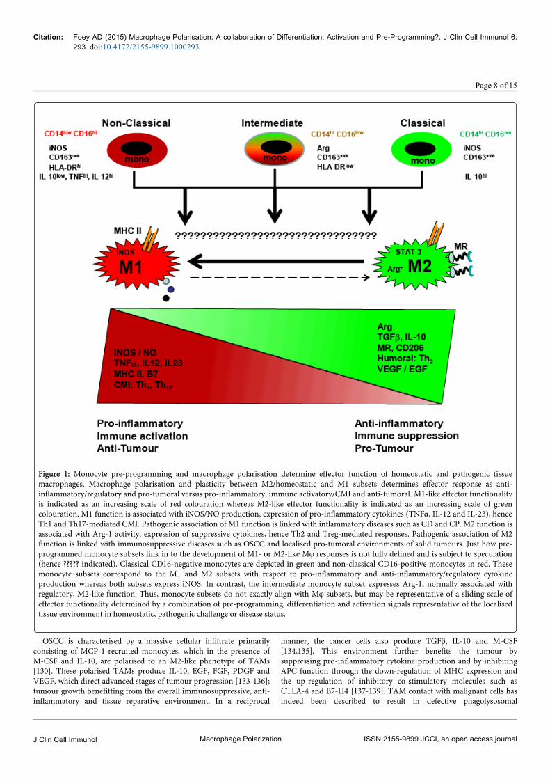
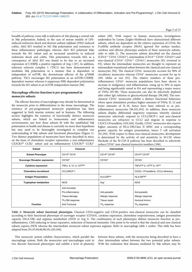
Figure 1: Monocyte pre-programming and macrophage polarisation determine effector function of homeostatic and pathogenic tissuemacrophages. Macrophage polarisation and plasticity between M2/homeostatic and M1 subsets determines effector response as anti-inflammatory/regulatory and pro-tumoral versus pro-inflammatory, immune activatory/CMI and anti-tumoral. M1-like effector functionalityis indicated as an increasing scale of red colouration whereas M2-like effector functionality is indicated as an increasing scale of greencolouration. M1 function is associated with iNOS/NO production, expression of pro-inflammatory cytokines (TNFα, IL-12 and IL-23), henceTh1 and Th17-mediated CMI. Pathogenic association of M1 function is linked with inflammatory diseases such as CD and CP. M2 function isassociated with Arg-1 activity, expression of suppressive cytokines, hence Th2 and Treg-mediated responses. Pathogenic association of M2function is linked with immunosuppressive diseases such as OSCC and localised pro-tumoral environments of solid tumours. Just how pre-programmed monocyte subsets link in to the development of M1- or M2-like Mφ responses is not fully defined and is subject to speculation(hence ????? indicated). Classical CD16-negative monocytes are depicted in green and non-classical CD16-positive monocytes in red. Thesemonocyte subsets correspond to the M1 and M2 subsets with respect to pro-inflammatory and anti-inflammatory/regulatory cytokineproduction whereas both subsets express iNOS. In contrast, the intermediate monocyte subset expresses Arg-1, normally associated withregulatory, M2-like function. Thus, monocyte subsets do not exactly align with Mφ subsets, but may be representative of a sliding scale ofeffector functionality determined by a combination of pre-programming, differentiation and activation signals representative of the localisedtissue environment in homeostatic, pathogenic challenge or disease status.
OSCC is characterised by a massive cellular infiltrate primarilyconsisting of MCP-1-recruited monocytes, which in the presence ofM-CSF and IL-10, are polarised to an M2-like phenotype of TAMs[130]. These polarised TAMs produce IL-10, EGF, FGF, PDGF andVEGF, which direct advanced stages of tumour progression [133-136];tumour growth benefitting from the overall immunosuppressive, anti-inflammatory and tissue reparative environment. In a reciprocal
manner, the cancer cells also produce TGFβ, IL-10 and M-CSF[134,135]. This environment further benefits the tumour bysuppressing pro-inflammatory cytokine production and by inhibitingAPC function through the down-regulation of MHC expression andthe up-regulation of inhibitory co-stimulatory molecules such asCTLA-4 and B7-H4 [137-139]. TAM contact with malignant cells hasindeed been described to result in defective phagolysosomal
Citation: Foey AD (2015) Macrophage Polarisation: A collaboration of Differentiation, Activation and Pre-Programming?. J Clin Cell Immunol 6:293. doi:10.4172/2155-9899.1000293
Page 8 of 15
J Clin Cell Immunol Macrophage Polarization ISSN:2155-9899 JCCI, an open access journal
interactions hence defective tumour antigen processing andpresentation, thus suppressing anti-tumour T cell responses andfacilitating tumour survival [140]. In addition, Treg development isencouraged via the M2-like TAM and OSCC cell expression of IL-10and TGFβ favouring the suppression of host anti-tumour responses[141,142]. Thus, there would appear to be a reciprocal relationshipbetween TAMs and tumour, where the TAMs can modulate tumoursurvival, growth and development and that the tumour cells canmodulate TAM plasticity. Can we limit tumour growth anddevelopment by switching M2-like TAMs to an M1-like subset?Theoretically, at first glance, this might be viewed as an attractiveoption. Practice may be different, given that M1-like TAMs areassociated with malignant transformation through chronicinflammatory injury and that the persistent tumour environment mayjust revert anti-tumoral M1-like Mϕs introduced as a cell-basedtherapy to the pro-tumoral M2-like TAM. Thus treatment of solidtumours by manipulation of polarisation states/plasticity between M1and M2 phenotypes may be an inappropriate regimen for thetreatment of cancer. What may be more realistic is the manipulation ofMϕ subset sensitivity to tolerisation; selectively suppressing polarisedMϕs, which facilitate tumour development in many different tumourenvironments.
Manipulation of Macrophage polarisation: the future?Manipulation of Mϕ polarisation by harnessing differentiation,
activation and suppression signals may offer a potentially realisticregimen for the treatment and management of pro-inflammatory (eg.CD or CP), or immune-suppressive, pro-tumour (eg. OSCC)conditions (refer to figure 1). Effective polarisation and modulation ofpathological mechanisms are likely to result from the delicate balanceof all of these Mϕ-mediating factors, which, if modulated incorrectlymay result in exacerbation of disease processes rather than down-regulation. Indeed, in the case of tumours, TAMs are predominated bythe pro-tumoral M2-like phenotype. Although experimental over-expression of Mϕ IL-12 increased MHC expression, T cell infiltrationand anti-tumour responses [143], attempts to polarise these M2-likeTAMs to a cytotoxic anti-tumour M1 subset have resulted in Mϕpolarisation reverting to the suppressive pro-tumoral M2 subset. Thisis thought to be as a result of the tumour environment expressing awealth of signals which reverse the polarised “therapeutic” M1 subsetto an effector that benefits the tumour. This may be as a consequenceof TAM functional heterogeneity where in invasive areas, TAMsencourage cancer cell motility whereas in stromal and perivascularareas TAMs promote metastasis and in avascular, perinecrotic areashypoxic TAMs stimulate angiogenesis [132,144]. In cancers with apoor prognosis, TAMs accumulate in numbers at sites of hypoxia andnecrosis [145-147]. These TAMs respond to hypoxia by up-regulatingthe expression of HIF-1, HIF-2 and HIF-regulated angiogenic factors[148,149], thus hypoxia may represent a polarising signal whichfavours pro-tumoral function and an M2-like TAM subset [150]. Anadditional confounding factor to the understanding of TAMfunctionality is the characterisation of an additional CD14+ monocytesubset, which expresses Tie-2 (angiopoietin receptor) and is associatedwith tumour angiogenesis [151]. Upon ligation by angiopoeitin-2, thissubset suppresses the release of pro-inflammatory cytokines TNFα andIL-12 via NFκB inhibition by A20-binding inhibitor of NFκBactivation-2 (ABIN-2) [151,152]. This may go some way to highlight
the requirement to manipulate TLR/NFκB signals in the regulation ofTAM plasticity but, in addition, there is a need to fully characterisethis tumour-associated Tie-2 expressing monocyte (TEM) subset andwhere it is placed in the sliding scale of monocyte/macrophagefunctional plasticity. An alternative approach to manipulating M2 toM1, is to encourage M2 polarisation but to manipulate these pro-tumoral Mϕs to act as Trojan horses, acting as delivery systems foranti-tumour cytotoxic drugs. This very approach is currently beinginvestigated where studies have demonstrated Mϕs to be ideal deliverysystems for oncolytic virus, which resulted in the suppression oftumour regrowth and metastasis [153,154].
In addition to the manipulation of polarising activation anddifferentiation signals, Mϕ polarisation to distinct functional subsets islikely to be determined by suppressive signals or tolerisation (Figure2). ET was first described by the observation that LPS pre-treatmentrendered innate immune cells refractory to activation upon LPS re-challenge. ET has since been shown to occur in Mϕs for a range ofcytokine (TNFα, IL-1β) and TLR-mediated (LPS, LTA, PGN,Flagellin) signals [reviewed in 155]. The suppression of Mϕfunctionality could beneficially inhibit harmful inflammatoryresponses whereas at the same time benefit infectious microbes, thusallowing for a favourable environment for the pathogen to recoup itsnumbers through growth. In the case of the oral pathogenPorphyromonas gingivalis, associated with chronic periodontitis, Mϕsubsets were differentially sensitive to PG-LPS-induced ET, where M2swere sensitive to ET and M1s were refractory [128]. As suggestedearlier in the context of TLR-mediated signalling, many endogenoussuppressors exist which can suppress TLR-mediated activatory orpolarising responses. In addition to the endogenous suppressors(MD2, Tollip, IRAK-M, Myd88s, TRIAD3A, SIGIRR), many othersuppressive molecules play a role in regulating Mϕ responses. Theseinclude CD200R, CD47/SIRP1α, Siglecs 3-10, CD32 to name but a few.Ligation of CD200R has been demonstrated to induceimmunosuppressive activity and suppress pro-inflammatory cytokineproduction in models of chronic inflammation such as collagen-induced arthritis [156-158 and reviewed in 159]. CD47-SIRP1αligation also exhibits a suppressive activity by down-regulating IL-12production [160]; this response may be reflective of suppression ofactivity or may alter polarisation status of the Mϕ from M1 to M2. Thetargeting/augmentation of such suppressive molecules may represent arealistic approach in suppressing chronic inflammatory diseases suchas Crohn’s disease and chronic periodontitis but may also facilitatecontrol of Mϕ polarisation in the treatment of solid tumours.
A recurring theme that presents itself in every aspect of the Mϕstory is the ability to recognise immunoglobulin or immune complexes(ICs) through the responsiveness of Mϕ FcRs. FcRs and their ligationwould appear to be involved in monocyte subset responses, Mϕ subsetpolarisation through activation and differentiation and through theinduction of suppressive/regulatory responses. CD16 (FcγRIIIa) isexpressed by both Mϕs and the non-classical subset of monocytes.Activation by immune complexes or immunoglobulin results in analternatively activated M2-like phenotype through the activation ofITAMs present in the intracellular cytoplasmic signalling domain[161], however CD16-ligation has also been shown to induce MϕTNFα production [162].
Citation: Foey AD (2015) Macrophage Polarisation: A collaboration of Differentiation, Activation and Pre-Programming?. J Clin Cell Immunol 6:293. doi:10.4172/2155-9899.1000293
Page 9 of 15
J Clin Cell Immunol Macrophage Polarization ISSN:2155-9899 JCCI, an open access journal
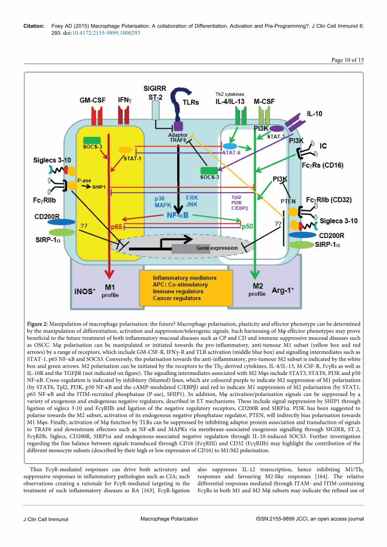
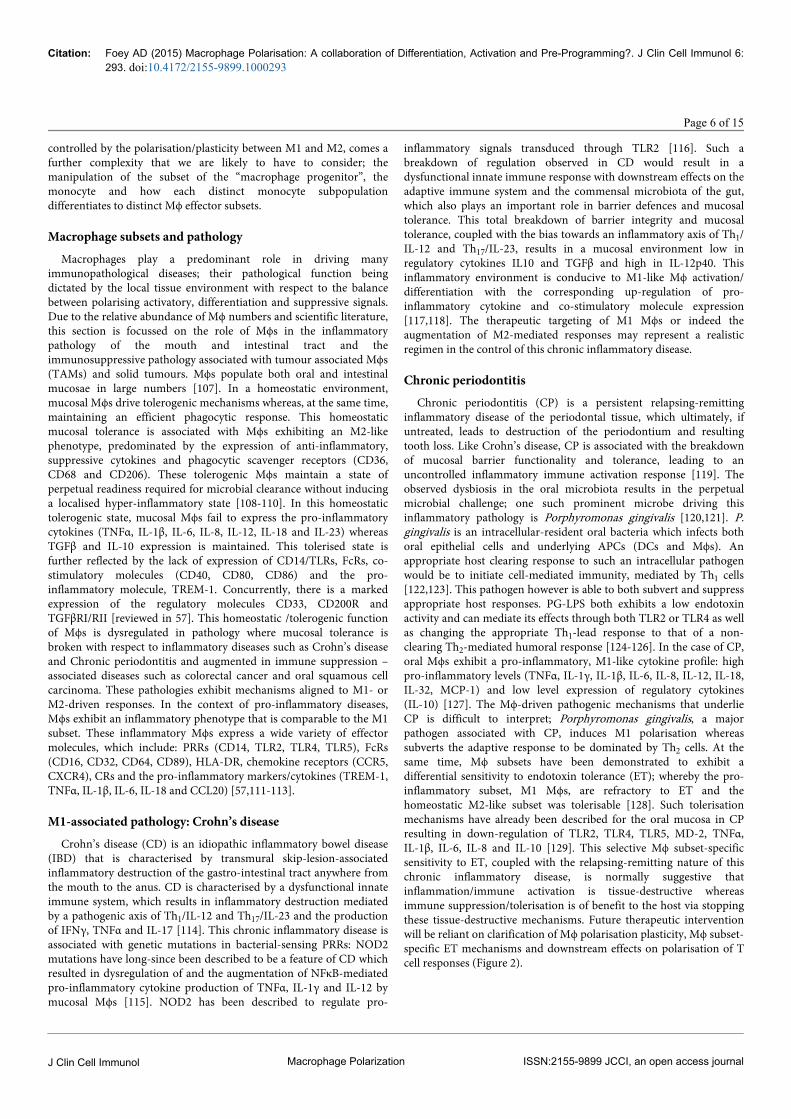
Figure 2: Manipulation of macrophage polarisation: the future? Macrophage polarisation, plasticity and effector phenotype can be determinedby the manipulation of differentiation, activation and suppression/tolerogenic signals. Such harnessing of Mφ effector phenotypes may provebeneficial to the future treatment of both inflammatory mucosal diseases such as CP and CD and immune suppressive mucosal diseases suchas OSCC. Mφ polarisation can be manipulated or initiated towards the pro-inflammatory, anti-tumour M1 subset (yellow box and redarrows) by a range of receptors, which include GM-CSF-R, IFNγ-R and TLR activation (middle blue box) and signalling intermediates such asSTAT-1, p65 NF-κB and SOCS3. Conversely, the polarisation towards the anti-inflammatory, pro-tumour M2 subset is indicated by the whitebox and green arrows. M2 polarisation can be initiated by the receptors to the Th2-derived cytokines, IL-4/IL-13, M-CSF-R, FcγRs as well asIL-10R and the TGFβR (not indicated on figure). The signalling intermediates associated with M2 Mφs include STAT3, STAT6, PI3K and p50NF-κB. Cross-regulation is indicated by inhibitory (blunted) lines, which are coloured purple to indicate M2 suppression of M1 polarisation(by STAT6, Tpl2, PI3K, p50 NF-κB and the cAMP-modulated C/EBPβ) and red to indicate M1 suppression of M2 polarisation (by STAT1,p65 NF-κB and the ITIM-recruited phosphatase (P-ase), SHIP1). In addition, Mφ activation/polarisation signals can be suppressed by avariety of exogenous and endogenous negative regulators, described in ET mechanisms. These include signal suppression by SHIP1 throughligation of siglecs 3-10 and FcγRIIb and ligation of the negative regulatory receptors, CD200R and SIRP1α. PI3K has been suggested topolarise towards the M2 subset, activation of its endogenous negative phosphatase regulator, PTEN, will indirectly bias polarisation towardsM1 Mφs. Finally, activation of Mφ function by TLRs can be suppressed by inhibiting adaptor protein association and transduction of signalsto TRAF6 and downstream effectors such as NF-κB and MAPKs via membrane-associated exogenous signalling through SIGIRR, ST-2,FcγRIIb, Siglecs, CD200R, SIRP1α and endogenous-associated negative regulation through IL-10-induced SOCS3. Further investigationregarding the fine balance between signals transduced through CD16 (FcγRIII) and CD32 (FcγRIIb) may highlight the contribution of thedifferent monocyte subsets (described by their high or low expression of CD16) to M1/M2 polarisation.
Thus FcγR-mediated responses can drive both activatory andsuppressive responses in inflammatory pathologies such as CIA; suchobservations creating a rationale for FcγR-mediated targeting in thetreatment of such inflammatory diseases as RA [163]. FcγR-ligation
also suppresses IL-12 transcription, hence inhibiting M1/Th1responses and favouring M2-like responses [164]. The relativedifferential responses mediated through ITAM- and ITIM-containingFcγRs in both M1 and M2 Mϕ subsets may indicate the refined use of
Citation: Foey AD (2015) Macrophage Polarisation: A collaboration of Differentiation, Activation and Pre-Programming?. J Clin Cell Immunol 6:293. doi:10.4172/2155-9899.1000293
Page 10 of 15
J Clin Cell Immunol Macrophage Polarization ISSN:2155-9899 JCCI, an open access journal
IC-FcγR signalling in the treatment of inflammatory pathologies. Theuse of in vitro immunoglobulin (IVIG) has been adopted for thetreatment of inflammation and autoimmunity [165,166]; whether thisis as a consequence of activation of ITAM-containing FcγRs orregulatory responses through suppression by SHP-1/SHIPphasphatase-recruiting, ITIM-containing FcγRs such as FcγRIIb(CD32) remains to be clarified. Thus, IVIG can potentially be used toeither suppress pathogenic Mϕ-driven responses or can deviate Mϕresponses to a more protective, less pathogenic mechanism. Therelative balance of signals transduced through ITAM- and ITIM-bearing receptors would appear to have a direct effect on Mϕfunctionality; this has been clearly demonstrated whereby SHIPactivity has been shown to repress the generation of alternativelyactivated, M2-like Mϕs thus favouring a pro-inflammatory M1/Th1axis of Mϕ functionality [167]. Another family of receptors which bothpositively and negatively regulate Mϕ responses through ITAM andITIM activity is the sialic acid binding Ig-like lectins, Siglec family[168]. CD33-like ITIM-bearing siglecs are expressed by Mϕs [169].These siglecs exhibit suppressive functionality but, in addition, mayplay a prominent role in Mϕ polarisation. This is supported by siglec 9enhancing Mϕ IL-10 production [170] whereas CD33 responses areblocked by SOCS3 [171], which also targets siglec 7 for proteosomaldegradation [172]. Thus CD33-like siglecs may be involved inpolarisation of M2-driven responses and are blocked in M1-SOCS3expressing Mϕs (see overview diagram of manipulation of Mϕpolarisation, (Figure 2).
In conclusion, Mϕ-driven immune responses would appear to becontrolled by the polarisation of specific effector phenotype beingexpressed and by its level of plasticity and reversibility. The plasticity,hence Mϕ subset, can thus be determined by the tissue environment[173]. Thus plasticity is regulated by a wealth of activation,differentiation and suppression signals to be found in the tissueenvironment. Pro-inflammatory and anti-inflammatory Mϕs areclearly inter-convertible [174] and this plasticity can be controlled byFcγR ligation which can reverse LPS toxicity [175], axis of IKK/NFκBactivation [176], IL-4-induced SHIP degradation [69] and relativecytokine environments. Macrophage polarisation is thus truly acollaboration of differentiation, activation, suppression and pre-programming; further characterisation of which will open up a worldof therapeutic regimens for the treatment of chronic inflammatorydisease and cancer.
References1. Gordon S, Taylor PR (2005) Monocyte and macrophage heterogeneity.
Nat Rev Immunol 5: 953-964.2. Mosser DM, Edwards JP (2008) Exploring the full spectrum of
macrophage activation. Nat Rev Immunol 8: 958-969.3. Varol C, Yona S, Jung S (2009) Origins and tissue-context-dependent
fates of blood monocytes. Immunol Cell Biol 87: 30-38.4. Jenkins SJ, Ruckerl D, Cook PC, Jones LH, Finkelman FD, et al. (2011)
Local macrophage proliferation, rather than recruitment from the blood,is a signature of TH2 inflammation. Science 332: 1284-1288.
5. Epelman S, Lavine KJ2, Randolph GJ3 (2014) Origin and functions oftissue macrophages. Immunity 41: 21-35.
6. Ginhoux F, Jung S (2014) Monocytes and macrophages: developmentalpathways and tissue homeostasis. Nat Rev Immunol 14: 392-404.
7. Adams DO, Hamilton TA (1989) The activated macrophage andgranulomatous inflammation. Curr Top Pathol 79: 151-167.
8. Foey AD (2014) Macrophages – masters of immune activation,suppression and deviation. InTech Publishing, Rijeka, Croatia. ISBN978-953-51-1374-4.
9. Anderson CF, Mosser DM (2002) A novel phenotype for an activatedmacrophage: the type 2 activated macrophage. J Leukoc Biol 72: 101-106.
10. Gordon S (2003) Alternative activation of macrophages. Nat RevImmunol 3: 23-35.
11. Mills CD, Kincaid K, Alt JM, Heilman MJ, Hill AM (2000) M-1/M-2macrophages and the Th1/Th2 paradigm. J Immunol 164: 6166-6173.
12. Nathan CF (1987) Secretory products of macrophages. J Clin Invest 79:319-326.
13. Mills CD (2001) Macrophage arginine metabolism to ornithine/urea ornitric oxide/citrulline: a life or death issue. Crit Rev Immunol 21:399-425.
14. Jenkins SJ, Allen JE (2010) Similarity and diversity in macrophageactivation by nematodes, trematodes, and cestodes. J Biomed Biotechnol2010: 262609.
15. Murray PJ, Wynn TA (2011) Obstacles and opportunities forunderstanding macrophage polarization. J Leukoc Biol 89: 557-563.
16. Murray PJ, Wynn TA (2011) Protective and pathogenic functions ofmacrophage subsets. Nat Rev Immunol 11: 723-737.
17. Stein M, Keshaw S, Harris N, Gordon S (1992) Interleukin 4 potentlyenhances murine macrophage mannose receptor activity: a marker ofalternative immunologic macrophage activation. J Exp Med 176: 287-292.
18. Mantovani A, Sica A, Sozzani S, Allavena P, Vecchi A, et al. (2004) Thechemokine system in diverse forms of macrophage activation andpolarization. Trends Immunol 25: 677-686.
19. Fleming BD, Mosser DM (2011) Regulatory macrophages: setting thethreshold for therapy. Eur J Immunol 41: 2498-2502.
20. Pinhal-Enfield G, Ramanathan M, Hasko G, Vogel SN, Salzman AL, et al.(2003) An angiogenic switch in macrophages involving synergy betweenToll-like receptors 2, 4, 7, and 9 and adenosine A(2A) receptors. Am JPathol 163: 711-721.
21. Ferrante CJ, Pinhal-Enfield G, Elson G, Cronstein BN, Hasko G, et al.(2013) The adenosine-dependent angiogenic switch of macrophages toan M2-like phenotype is independent of interleukin-4 receptor alpha(IL-4Rα) signaling. Inflammation 36: 921-931.
22. Duluc D, Delneste Y, Tan F, Moles MP, Grimaud L, et al. (2007) Tumor-associated leukemia inhibitory factor and IL-6 skew monocytedifferentiation into tumor-associated macrophage-like cells. Blood 110:4319-4330.
23. Colin S, Chinetti-Gbaguidi G, Staels B (2014) Macrophage phenotypes inatherosclerosis. Immunol Rev 262: 153-166.
24. De Paoli F, Staels B, Chinetti-Gbaguidi G (2014) Macrophage phenotypesand their modulation in atherosclerosis. Circ J 78: 1775-1781.
25. Gleissner CA, Shaked I, Little KM, Ley K (2010) CXC chemokine ligand4 induces a unique transcriptome in monocyte-derived macrophages. JImmunol 184: 4810-4818.
26. Kadl A, Meher AK, Sharma PR, Lee MY, Doran AC, et al. (2010)Identification of a novel macrophage phenotype that develops inresponse to atherogenic phospholipids via Nrf2. Circ Res 107: 737-746.
27. Boyle JJ, Harrington HA, Piper E, Elderfield K, Stark J, et al. (2009)Coronary intraplaque hemorrhage evokes a novel atheroprotectivemacrophage phenotype. Am J Pathol 174: 1097-1108.
28. Boyle JJ, Johns M, Lo J, Chiodini A, Ambrose N, et al. (2011) Hemeinduces heme oxygenase 1 via Nrf2: role in the homeostatic macrophageresponse to intraplaque hemorrhage. Arterioscler Thromb Vasc Biol 31:2685-2691.
29. Boyle JJ, Johns M, Kampfer T, Nguyen AT, Game L, et al. (2012)Activating transcription factor 1 directs Mhem atheroprotectivemacrophages through coordinated iron handling and foam cellprotection. Circ Res 110: 20-33.
30. Mills CD, Shearer J, Evans R, Caldwell MD (1992) Macrophage argininemetabolism and the inhibition or stimulation of cancer. J Immunol 149:2709-2714.
Citation: Foey AD (2015) Macrophage Polarisation: A collaboration of Differentiation, Activation and Pre-Programming?. J Clin Cell Immunol 6:293. doi:10.4172/2155-9899.1000293
Page 11 of 15
J Clin Cell Immunol Macrophage Polarization ISSN:2155-9899 JCCI, an open access journal
31. Hibbs JB Jr, Taintor RR, Vavrin Z, Rachlin EM (1988) Nitric oxide: acytotoxic activated macrophage effector molecule. Biochem Biophys ResCommun 157: 87-94.
32. Albina JE, Caldwell MD, Henry WL Jr, Mills CD (1989) Regulation ofmacrophage functions by L-arginine. J Exp Med 169: 1021-1029.
33. Garside P, Hutton AK, Severn A, Liew FY, Mowat AM (1992) Nitricoxide mediates intestinal pathology in graft-vs.-host disease. Eur JImmunol 22: 2141-2145.
34. Gordon S, Martinez FO (2010) Alternative activation of macrophages:mechanism and functions. Immunity 32: 593-604.
35. Makarenkova VP, Bansal V, Matta BM, Perez LA, Ochoa JB (2006)CD11b+/Gr-1+ myeloid suppressor cells cause T cell dysfunction aftertraumatic stress. J Immunol 176: 2085-2094.
36. Munder M, Schneider H, Luckner C, Giese T, Langhans CD, et al. (2006)Suppression of T-cell functions by human granulocyte arginase. Blood108: 1627-1634.
37. Highfill SL, Rodrigues PC, Zhou Q, Goetz CA, Koehn BH, et al. (2010)Bone marrow myeloid-derived suppressor cells (MDSCs) inhibit graft-versus-host disease (GvHD) via an arginase-1-dependent mechanismthat is up-regulated by interleukin-13. Blood 116: 5738-5747.
38. Mosser DM (2003) The many faces of macrophage activation. J LeukocBiol 73: 209-212.
39. Louis J, Himmelrich H, Parra-Lopez C, Tacchini-Cottier F, Launois P(1998) Regulation of protective immunity against Leishmania major inmice. Curr Opin Immunol 10: 459-464.
40. Hölscher C, Atkinson RA, Arendse B, Brown N, Myburgh E, et al. (2001)A protective and agonistic function of IL-12p40 in mycobacterialinfection. J Immunol 167: 6957-6966.
41. Veldhoen M, Hocking RJ, Atkins CJ, Locksley RM, Stockinger B (2006)TGF-β in the context of an inflammatory cytokine milieu supports denovo differentiation of IL-17-producing T cells. Immunity 24: 179-189.
42. Stockinger B, Veldhoen M (2007) Differentiation and function of Th17 Tcells. Curr Opin Immunol 19: 281-286.
43. Gibbs DF, Warner RL, Weiss SJ, Johnson KJ, Varani J (1999)Characterization of matrix metalloproteinases produced by rat alveolarmacrophages. Am J Respir Cell Mol Biol 20: 1136-1144.
44. Gibbs DF, Shanley TP, Warner RL, Murphy HS, Varani J, et al. (1999b)Role of matrix metalloproteinases in models of macrophage-dependentacute lung injury. Evidence for alveolar macrophage as source ofproteinases. Am J Resp Cell Mol Biol 20: 1145-1154.
45. Chizziolini C, Rezzonico R, De Luca C, Burger D, Dayer J-M (2000) Th2cell membrane factors in association with IL-4 enhance matrixmetalloproteinase-1 (MMP-1) while decreasing MMP-9 production bygranulocyte-macrophage colony stimulating factor-differentiated humanmonocytes. J Immunol 164: 5952-5960.
46. Van Ginderachter JA, Movahedi K, Hassanzadeh Ghassabeh G,Meerschaut S, Beschin A, et al. (2006) Classical and alternative activationof mononuclear phagocytes: picking the best of both worlds for tumorpromotion. Immunobiology 211: 487-501.
47. Mills CD, Ley K (2014) M1 and M2 macrophages: the chicken and theegg of immunity. J Innate Immun 6: 716-726.
48. Baay M, Brouwer A, Pauwels P, Peeters M, Lardon F (2011) Tumor cellsand tumor-associated macrophages: secreted proteins as potential targetsfor therapy. Clin Dev Immunol 2011: 565187.
49. Lenzo JC, Turner AL, Cook AD, Vlahos R, Anderson GP, et al. (2012)Control of macrophage lineage populations by CSF-1 receptor and GM-CSF in homeostasis and inflammation. Immunol Cell Biol 90: 429-440.
50. Verreck FA, de Boer T, Langenberg DM, van der Zanden L, OttenhoffTH (2006) Phenotypic and functional profiling of humanproinflammatory type-1 and anti-inflammatory type-2 macrophages inresponse to microbial antigens and IFN-gamma- and CD40L-mediatedcostimulation. J Leukoc Biol 79: 285-293.
51. Albina JE, Henry WL Jr, Mastrofrancesco B, Martin BA, Reichner JS(1995) Macrophage activation by culture in an anoxic environment. JImmunol 155: 4391-4396.
52. Aliberti J, Machado FS, Souto JT, Campanelli AP, Teixeira MM, et al.(1999) chemokines enhance parasite uptake and promote nitric oxide-dependent microbiostatic activity in murine inflammatory macrophagesinfected with Trypanosoma cruzi. Infect Immun 67: 4819-4826.
53. Kielian MC, Cohn ZA (1981) Phorbol myristate acetate stimulatesphagosome-lysosome fusion in mouse macrophages. J Exp Med 154:101-111.
54. Green SP, Phillips WA (1994) Activation of the macrophage respiratoryburst by phorbol myristate acetate: evidence for both tyrosine-kinase-dependent and -independent pathways. Biochim Biophys Acta 1222:241-248.
55. de Waal Malefyt R, Figdor CG, Huijbens R, Mohan-Peterson S, BennettB, et al. (1993) Effects of IL-13 on phenotype, cytokine production, andcytotoxic function of human monocytes. Comparison with IL-4 andmodulation by IFN-gamma or IL-10. J Immunol 151: 6370-6381.
56. Pesce J, Kaviratne M, Ramalingam TR, Thompson RW, Urban JF Jr, et al.(2006) The IL-21 receptor augments Th2 effector function andalternative macrophage activation. J Clin Invest 116: 2044-2055.
57. Foey AD (2012) Mucosal macrophages: phenotype and functionality inhomeostasis and pathology. Nova Science Publishers Inc., New York,USA 4: 121-146.
58. Smith W, Feldmann M, Londei M (1998) Human macrophages inducedin vitro by macrophage colony-stimulating factor are deficient in IL-12production. Eur J Immunol 28: 2498-2507.
59. Tiemessen MM, Jagger AL, Evans HG, van Herwijnen MJ, John S, et al.(2007) CD4+CD25+Foxp3+ regulatory T cells induce alternativeactivation of human monocytes/macrophages. Proc Natl Acad Sci U S A104: 19446-19451.
60. Erwig LP, Kluth DC, Walsh GM, Rees AJ (1998) Initial cytokine exposuredetermines function of macrophages and renders them unresponsive toother cytokines. J Immunol 161: 1983-1988.
61. Fadok VA, Bratton DL, Konowal A, Freed PW, Westcott JY, et al. (1998)Macrophages that have ingested apoptotic cells in vitro inhibitproinflammatory cytokine production through autocrine/paracrinemechanisms involving TGF-?, PGE2 and PAF. J Clin Invest 101: 890-898.
62. Savill J, Dransfield I, Gregory C, Haslett C (2002) A blast from the past:clearance of apoptotic cells regulates immune responses. Nat RevImmunol 2: 965-975.
63. O'Neill LA, Fitzgerald KA, Bowie AG (2003) The Toll-IL-1 receptoradaptor family grows to five members. Trends Immunol 24: 286-290.
64. O'Neill LA (2008) When signaling pathways collide: positive and negativeregulation of toll-like receptor signal transduction. Immunity 29: 12-20.
65. Negishi H, Ohba Y, Yanai H, Takaoka A, Honma K, et al. (2005)Negative regulation of Toll-like-receptor signaling by IRF-4. Proc NatlAcad Sci U S A 102: 15989-15994.
66. Honma K, Udono H, Kohno T, Yamamoto K, Ogawa A, et al. (2005)Interferon regulatory factor 4 negatively regulates the production ofproinflammatory cytokines by macrophages in response to LPS. ProcNatl Acad Sci U S A 102: 16001-16006.
67. Brint EK, Xu D, Liu H, Dunne A, McKenzie AN, et al. (2004) ST2 is aninhibitor of interleukin 1 receptor and Toll-like receptor 4 signaling andmaintains endotoxin tolerance. Nat Immunol 5: 373-379.
68. Wald D, Qin J, Zhao Z, Qian Y, Naramura M, et al. (2003) SIGIRR, anegative regulator of Toll-like receptor-interleukin 1 receptor signaling.Nat Immunol 4: 920-927.
69. Wilson HM, Chettibi S, Jobin C, Walbaum D, Rees AJ, et al. (2005)Inhibition of macrophage nuclear factor-kappaB leads to a dominantanti-inflammatory phenotype that attenuates glomerular inflammationin vivo. Am J Pathol 167: 27-37.
70. Weisser SB, McLarren KW, Voglmaier N, van Netten-Thomas CJ, AntovA, et al. (2011) Alternative activation of macrophages by IL-4 requiresSHIP degradation. Eur J Immunol 41: 1742-1753.
71. Krausgruber T, Blazek K, Smallie T, Alzabin S, Lockstone H, et al. (2011)IRF5 promotes inflammatory macrophage polarization and TH1-TH17responses. Nat Immunol 12: 231-238.
Citation: Foey AD (2015) Macrophage Polarisation: A collaboration of Differentiation, Activation and Pre-Programming?. J Clin Cell Immunol 6:293. doi:10.4172/2155-9899.1000293
Page 12 of 15
J Clin Cell Immunol Macrophage Polarization ISSN:2155-9899 JCCI, an open access journal
72. Liu Y, Stewart KN, Bishop E, Marek CJ, Kluth DC, et al. (2008) Uniqueexpression of suppressor of cytokine signaling 3 is essential for classicalmacrophage activation in rodents in vitro and in vivo. J Immunol 180:6270-6278.
73. Sahin E, Haubenwallner S1, Kuttke M1, Kollmann I1, Halfmann A2, etal. (2014) Macrophage PTEN regulates expression and secretion ofarginase I modulating innate and adaptive immune responses. JImmunol 193: 1717-1727.
74. Díaz-Guerra MJ, Castrillo A, Martín-Sanz P, Boscá L (1999) Negativeregulation by phosphatidylinositol 3-kinase of inducible nitric oxidesynthase expression in macrophages. J Immunol 162: 6184-6190.
75. Fukao T, Koyasu S (2003) PI3K and negative regulation of TLR signaling.Trends Immunol 24: 358-363.
76. Günzl P, Bauer K, Hainzl E, Matt U, Dillinger B, et al. (2010) Anti-inflammatory properties of the PI3K pathway are mediated by IL-10/DUSP regulation. J Leukoc Biol 88: 1259-1269.
77. Haskó G, Kuhel DG, Marton A, Nemeth ZH, Deitch EA, et al. (2000)Spermine differentially regulates the production of interleukin-12 p40and interleukin-10 and suppresses the release of the T helper 1 cytokineinterferon-gamma. Shock 14: 144-149.
78. Riley JK, Takeda K, Akira S, Schreiber RD (1999) Interleukin-10 receptorsignaling through the JAK-STAT pathway. Requirement for two distinctreceptor-derived signals for anti-inflammatory action. J Biol Chem 274:16513-16521.
79. Takeda K, Clausen BE, Kaisho T, Tsujimura T, Terada N, et al. (1999)Enhanced Th1 activity and development of chronic enterocolitis in micedevoid of Stat3 in macrophages and neutrophils. Immunity 10: 39-49.
80. Sica A, Bronte V (2007) Altered macrophage differentiation and immunedysfunction in tumor development. J Clin Invest 117: 1155-1166.
81. Welch JS, Escoubet-Lozach L, Sykes DB, Liddiard K, Greaves DR, et al.(2002) TH2 cytokines and allergic challenge induce Ym1 expression inmacrophages by a STAT6-dependent mechanism. J Biol Chem 277:42821-42829.
82. Porta C, Rimoldi M, Raes G, Brys L, Ghezzi P, et al. (2009) Tolerance andM2 (alternative) macrophage polarization are related processesorchestrated by p50 nuclear factor kappaB. Proc Natl Acad Sci U S A 106:14978-14983.
83. Hagemann T, Lawrence T, McNeish I, Charles KA, Kulbe H, et al. (2008)"Re-educating" tumor-associated macrophages by targeting NF-kappaB. JExp Med 205: 1261-1268.
84. Ruffell D, Mourkioti F, Gambardella A, Kirstetter P, Lopez RG, et al.(2009) A CREB-C/EBPbeta cascade induces M2 macrophage-specificgene expression and promotes muscle injury repair. Proc Natl Acad Sci US A 106: 17475-17480.
85. Pauleau AL, Rutschman R, Lang R, Pernis A, Watowich SS, et al. (2004)Enhancer-mediated control of macrophage-specific arginase Iexpression. J Immunol 172: 7565-7573.
86. Foey AD, Brennan FM (2004) Conventional protein kinase C andatypical protein kinase Czeta differentially regulate macrophageproduction of tumour necrosis factor-alpha and interleukin-10.Immunology 112: 44-53.
87. Arranz A, Doxaki C, Vergadi E, Martinez de la Torre Y, Vaporidi K, et al.(2012) Akt1 and Akt2 protein kinases differentially contribute tomacrophage polarization. Proc Natl Acad Sci U S A 109: 9517-9522.
88. Zhu L, Yang T, Li L, Sun L, Hou Y, et al. (2014) TSC1 controlsmacrophage polarization to prevent inflammatory disease. Nat Commun5: 4696.
89. Geissmann F, Jung S, Littman DR (2003) Blood monocytes consist of twoprincipal subsets with distinct migratory properties. Immunity 19: 71-82.
90. Ziegler-Heitbrock HW, Passlick B, Flieger D (1988) The monoclonalantimonocyte antibody My4 stains B lymphocytes and two distinctmonocyte subsets in human peripheral blood. Hybridoma 7: 521-527.
91. Passlick B, Flieger D, Ziegler-Heitbrock HW (1989) Identification andcharacterization of a novel monocyte subpopulation in human peripheralblood. Blood 74: 2527-2534.
92. Ziegler-Heitbrock L, Ancuta P, Crowe S, Dalod M, Grau V, et al. (2010)Nomenclature of monocytes and dendritic cells in blood. Blood 116:e74-80.
93. Ziegler-Heitbrock L (2014) Monocyte subsets in man and other species.Cell Immunol 289: 135-139.
94. Wong KL, Tai JJ, Wong WC, Han H, Sem X, et al. (2011) Geneexpression profiling reveals the defining features of the classical,intermediate, and nonclassical human monocyte subsets. Blood 118:e16-31.
95. Fingerle G, Pforte A, Passlick B, Blumenstein M, Ströbel M, et al. (1993)The novel subset of CD14+/CD16+ blood monocytes is expanded insepsis patients. Blood 82: 3170-3176.
96. Belge KU, Dayyani F, Horelt A, Siedlar M, Frankenberger M, et al. (2002)The proinflammatory CD14+CD16+DR++ monocytes are a majorsource of TNF. J Immunol 168: 3536-3542.
97. Kawanaka N, Yamamura M, Aita T, Morita Y, Okamoto A, et al. (2002)CD14+,CD16+ blood monocytes and joint inflammation in rheumatoidarthritis. Arthritis Rheum 46: 2578-2586.
98. Ziegler-Heitbrock L (2007) The CD14+ CD16+ blood monocytes: theirrole in infection and inflammation. J Leukoc Biol 81: 584-592.
99. Fingerle-Rowson G, Angstwurm M, Andreesen R, Ziegler-Heitbrock HW(1998) Selective depletion of CD14+ CD16+ monocytes by glucocorticoidtherapy. Clin Exp Immunol 112: 501-506.
100. Siedlar M, Strach M, Bukowska-Strakova K, Lenart M, Szaflarska A, et al.(2011) Preparations of intravenous immunoglobulins diminish thenumber and pro-inflammatory response of CD14+CD16++ monocytesin common variable immunodeficiency (CVID) patients. Clin Immunol139: 122-132.
101. Frankenberger M, Sternsdorf T, Pechumer H, Pforte A, Ziegler-Heitbrock HW (1996) Differential cytokine expression in human bloodmonocyte subpopulations: a polymerase chain reaction analysis. Blood87: 373-377.
102. Szaflarska A, Baj-Krzyworzeka M, Siedlar M, Weglarczyk K, Ruggiero I,et al. (2004) Antitumor response of CD14+/CD16+ monocytesubpopulation. Exp Hematol 32: 748-755.
103. Weber C, Belge KU, von Hundelshausen P, Draude G, Steppich B, et al.(2000) Differential chemokine receptor expression and function inhuman monocyte subpopulations. J Leukoc Biol 67: 699-704.
104. Ancuta P, Rao R, Moses A, Mehle A, Shaw SK, et al. (2003) Fractalkinepreferentially mediates arrest and migration of CD16+ monocytes. J ExpMed 197: 1701-1707.
105. Grage-Griebenow E, Zawatzky R, Kahlert H, Brade L, Flad H, et al.(2001) Identification of a novel dendritic cell-like subset of CD64(+) /CD16(+) blood monocytes. Eur J Immunol 31: 48-56.
106. Korkosz M, Bukowska-Strakova K, Sadis S, Grodzicki T, Siedlar M (2012)Monoclonal antibodies against macrophage colony-stimulating factordiminish the number of circulating intermediate and nonclassical(CD14(++)CD16(+)/CD14(+)CD16(++)) monocytes in rheumatoidarthritis patients. Blood 119: 5329-5330.
107. Hume DA, Allan W, Hogan PG, Doe WF (1987) Immunohistochemicalcharacterisation of macrophages in the liver and gastrointestinal tract:expression of CD4, HLA-DR, OKM1 and the mature macrophagemarker 25F9 in normal and diseased tissue. J Leukocyte Biol 42: 474-484.
108. Smith PD, Ochsenbauer-Jambor C, Smythies LE (2005) Intestinalmacrophages: unique effector cells of the innate immune system.Immunol Rev 206: 149-159.
109. Schenk M, Mueller C (2007) Adaptations of intestinal macrophages to anantigen-rich environment. Semin Immunol 19: 84-93.
110. Platt AM, Mowat AM (2008) Mucosal macrophages and the regulation ofimmune responses in the intestine. Immunol Lett 119: 22-31.
111. Rogler G, Hausmann M, Spöttl T, Vogl D, Aschenbrenner E, et al. (1999)T-cell co-stimulatory molecules are upregulated on intestinalmacrophages from inflammatory bowel disease mucosa. Eur JGastroenterol Hepatol 11: 1105-1111.
Citation: Foey AD (2015) Macrophage Polarisation: A collaboration of Differentiation, Activation and Pre-Programming?. J Clin Cell Immunol 6:293. doi:10.4172/2155-9899.1000293
Page 13 of 15
J Clin Cell Immunol Macrophage Polarization ISSN:2155-9899 JCCI, an open access journal
112. Smith PD, Smythies LE, Mosteller-Barnum M, Sibley DA, Russell MW, etal. (2001) Intestinal macrophages lack CD14 and CD89 and consequentlyare down-regulated for LPS- and IgA-mediated activities. J Immunol 167:2651-2656.
113. Smythies LE, Sellers MT, Clements RH, Mosteller-Barnum M, Meng G,et al. (2005) Human intestinal macrophages display profoundinflammatory anergy despite avid phagocytic and bacteriocidal activity. JClin Invest 115: 66-75.
114. Strober W, Zhang F, Kitani A, Fuss I, Fichtner-Feigl S (2010)Proinflammatory cytokines underlying the inflammation of Crohn'sdisease. Curr Opin Gastroenterol 26: 310-317.
115. Maeda S, Hsu LC, Liu H, Bankston LA, Iimura M, et al. (2005) Nod2mutation in Crohn's disease potentiates NF-kappaB activity and IL-1betaprocessing. Science 307: 734-738.
116. Netea MG, Kullberg BJ, de Jong DJ, Franke B, Sprong T, et al. (2004)NOD2 mediates anti-inflammatory signals induced by TLR2 ligands:implications for Crohn's disease. Eur J Immunol 34: 2052-2059.
117. Bouma G, Strober W (2003) The immunological and genetic basis ofinflammatory bowel disease. Nat Rev Immunol 3: 521-533.
118. Hardy H, Harris J, Lyon E, Beal J, Foey AD (2013) Probiotics, prebioticsand immunomodulation of gut mucosal defences: homeostasis andimmunopathology. Nutrients 5: 1869-1912.
119. Merry R, Belfield L, McArdle P, McLennan A, Crean S, et al. (2012) Oralhealth and pathology: a macrophage account. Br J Oral Maxillofac Surg50: 2-7.
120. Socransky SS, Haffajee AD, Cugini MA, Smith C, Kent RL Jr (1998)Microbial complexes in subgingival plaque. J Clin Periodontol 25:134-144.
121. Ezzo PJ, Cutler CW (2003) Microorganisms as risk indicators forperiodontal disease. Periodontol 2000 32: 24-35.
122. Yilmaz O, Watanabe K, Lamont RJ (2002) Involvement of integrins infimbriae-mediated binding and invasion by Porphyromonas gingivalis.Cell Microbiol 4: 305-314.
123. Jotwani R, Cutler CW (2004) Fimbriated Porphyromonas gingivalis ismore efficient than fimbria-deficient P. gingivalis in entering humandendritic cells in vitro and induces an inflammatory Th1 effectorresponse. Infect Immun 72: 1725-1732.
124. Pulendran B, Kumar P, Cutler CW, Mohamadzadeh M, Van Dyke T, etal. (2001) Lipopolysaccharides from distinct pathogens induce differentclasses of immune responses in vivo. J Immunol 167: 5067-5076.
125. Darveau RP, Pham TT, Lemley K, Reife RA, Bainbridge BW, et al. (2004)Porphyromonas gingivalis lipopolysaccharide contains multiple lipid Aspecies that functionally interact with both toll-like receptors 2 and 4.Infect Immun 72: 5041-5051.
126. Holden JA, Attard TJ, Laughton KM, Mansell A, O’Brien-Simpson NM,Reynolds EC (2014) Porphyromonas gingivalis lipopolysaccharideweakly activates M1 and M2 polarised mouse macrophages but inducesinflammatory cytokines. Infect Immun 82: 4190-4203.
127. Barksby HE, Nile CJ, Jaedicke KM, Taylor JJ, Preshaw PM (2009)Differential expression of immunoregulatory genes in monocytes inresponse to Porphyromonas gingivalis and Escherichia colilipopolysaccharide. Clin Exp Immunol 156: 479-487.
128. Foey AD, Crean S (2013) Macrophage subset sensitivity to endotoxintolerisation by Porphyromonas gingivalis. PLoS One 8: e67955.
129. Muthukuru M, Jotwani R, Cutler CW (2005) Oral mucosal endotoxintolerance induction in chronic periodontitis. Infect Immun 73: 687-694.
130. Koide N, Nishio A, Sato T, Sugiyama A, Miyagawa S (2004) Significanceof macrophage chemoattractant protein-1 expression and macrophageinfiltration in squamous cell carcinoma of the esophagus. Am JGastroenterol 99: 1667-1674.
131. Lin EY, Nguyen AV, Russell RG, Pollard JW (2001) Colony-stimulatingfactor 1 promotes progression of mammary tumors to malignancy. J ExpMed 193: 727-740.
132. Lewis CE, Pollard JW (2006) Distinct role of macrophages in differenttumor microenvironments. Cancer Res 66: 605-612.
133. Lalla RV, Boisoneau DS, Spiro JD, Kreutzer DL (2003) Expression ofvascular endothelial growth factor receptors on tumor cells in head andneck squamous cell carcinoma. Arch Otolaryngol Head Neck Surg 129:882-888.
134. Sica A, Allavena P, Mantovani A (2008) Cancer related inflammation: themacrophage connection. Cancer Lett 267: 204-215.
135. Kuropkat C, Dünne AA, Plehn S, Ossendorf M, Herz U, et al. (2003)Macrophage colony-stimulating factor as a tumor marker for squamouscell carcinoma of the head and neck. Tumour Biol 24: 236-240.
136. Wang F, Arun P, Friedman J, Chen Z, Van Waes C (2009) Current andpotential inflammation targeted therapies in head and neck cancer. CurrOpin Pharmacol 9: 389-395.
137. Kundu N, Fulton AM (1997) Interleukin-10 inhibits tumor metastasis,downregulates MHC class I, and enhances NK lysis. Cell Immunol 180:55-61.
138. de Waal Malefyt R, Haanem J, Spits H, Roncarolo MG, te Velde A, et al.(1991) Interleukin 10 (IL-10) and viral IL-10 strongly reduce antigen-specific human T cell proliferation by diminishing the antigen-presentingcapacity of monocytes via downregulation of class II majorhistocompatibility complex expression. J Exp Med 174: 915-924.
139. Kryczek I, Zou L, Rodriguez P, Zhu G, Wei S, et al. (2006) B7-H4expression identifies a novel suppressive macrophage population inhuman ovarian carcinoma. J Exp Med 203: 871-881.
140. Balm FJ, Drexhage HA, von Blomberg ME, Snow GB (1982)Mononuclear phagocyte function in head and neck cancer: NBT-dyereduction, maturation and migration of peripheral blood monocytes.Laryngoscope 92: 810-814.
141. Young MR, Wright MA, Lozano Y, Matthews JP, Benefield J, et al. (1996)Mechanisms of immune suppression in patients with head and neckcancer: influence on the immune infiltrate of the cancer. Int J Cancer 67:333-338.
142. Levings MK, Bacchetta R, Schulz U, Roncarolo MG (2002) The role ofIL-10 and TGF-beta in the differentiation and effector function of Tregulatory cells. Int Arch Allergy Immunol 129: 263-276.
143. Satoh T, Saika T, Ebara S, Kusaka N, Timme TL, et al. (2003)Macrophages transduced with an adenoviral vector expressinginterleukin 12 suppresses tumour growth and metastasis in a preclinicalmetastatic prostate cancer model. Cancer Res 63: 7853-7860.
144. Biswas SK, Sica A, Lewis CE (2008) Plasticity of macrophage functionduring tumor progression: regulation by distinct molecular mechanisms.J Immunol 180: 2011-2017.
145. Leek RD, Lewis CE, Whitehouse R, Greenall M, Clarke J, et al. (1996)Association of macrophage infiltration with angiogenesis and prognosisin invasive breast carcinoma. Cancer Res 56: 4625-4629.
146. Burton JL, Wells JM, Corke KP (2000) Macrophages accumulate inavascular, hypoxic areas of prostate tumours: implications for thetargeted therapeutic gene delivery to such sites. J Pathol 192: 8A.
147. Negus RP, Stamp GW, Hadley J, Balkwill FR (1997) Quantitativeassessment of the leukocyte infiltrate in ovarian cancer and itsrelationship to the expression of C-C chemokines. Am J Pathol 150:1723-1734.
148. Burke B, Tang N, Corke KP, Tazzyman D, Ameri K, et al. (2002)Expression of HIF-1alpha by human macrophages: implications for theuse of macrophages in hypoxia-regulated cancer gene therapy. J Pathol196: 204-212.
149. White JR, Harris RA, Lee SR, Craigon MH, Binley K, et al. (2004) Geneticamplification of the transcriptional response to hypoxia as a novel meansof identifying regulators of angiogenesis. Genomics 83: 1-8.
150. Murdoch C, Muthana M, Lewis CE (2005) Hypoxia regulatesmacrophage functions in inflammation. J Immunol 175: 6257-6263.
151. De Palma M, Venneri MA, Galli R, Sergi Sergi L, Politi LS, et al. (2005)Tie-2 identifies a hematopoietic lineage of proangiogenic monocytesrequired for tumour vessel formation and a mesenchymal population ofpericyte progenitors. Cancer Cell 8: 211-226.
Citation: Foey AD (2015) Macrophage Polarisation: A collaboration of Differentiation, Activation and Pre-Programming?. J Clin Cell Immunol 6:293. doi:10.4172/2155-9899.1000293
Page 14 of 15
J Clin Cell Immunol Macrophage Polarization ISSN:2155-9899 JCCI, an open access journal
152. Bingle L, Lewis CE, Corke KP, Reed MW, Brown NJ (2006) Macrophagespromote angiogenesis in human breast tumour spheroids in vivo. Br JCancer 94: 101-107.
153. De Palma M1, Lewis CE (2013) Macrophage regulation of tumorresponses to anticancer therapies. Cancer Cell 23: 277-286.
154. Muthana M, Rodrigues S, Chen YY, Welford A, Hughes R, et al. (2013)Macrophage delivery of an oncolytic virus abolishes tumor regrowth andmetastasis after chemotherapy or irradiation. Cancer Res 73: 490-495.
155. Biswas SK, Lopez-Collazo E (2009) Endotoxin tolerance: newmechanisms, molecules and clinical significance. Trends Immunol 30:475-487.
156. Gorczynski RM, Cattral MS, Chen Z, Hu J, Lei J, et al. (1999) Animmunoadhesin incorporating the molecule OX-2 is a potentimmunosuppressant that prolongs allo- and xenograft survival. JImmunol 163: 1654-1660.
157. Gorczynski RM, Chen Z, Yu K, Hu J (2001) CD200 immunoadhesinsuppresses collagen-induced arthritis in mice. Clin Immunol 101:328-334.
158. Simelyte E, Criado G, Essex D, Uger RA, Feldmann M, et al. (2008)CD200-Fc, a novel antiarthritic biologic agent that targetsproinflammatory cytokine expression in the joints of mice with collagen-induced arthritis. Arthritis Rheum 58: 1038-1043.
159. Barclay AN, Wright GJ, Brooke G, Brown MH (2002) CD200 andmembrane protein interactions in the control of myeloid cells. TrendsImmunol 23: 285-290.
160. Armant M, Avice MN, Hermann P, Rubio M, Kiniwa M, et al. (1999)CD47 ligation selectively downregulates human interleukin 12production. J Exp Med 190: 1175-1182.
161. Ravetch JV, Bolland S (2001) IgG Fc receptors. Annu Rev Immunol 19:275-290.
162. Abrahams VM, Cambridge G, Lydyard PM, Edwards JC (2000)Induction of tumor necrosis factor alpha production by adhered humanmonocytes: a key role for Fcgamma receptor type IIIa in rheumatoidarthritis. Arthritis Rheum 43: 608-616.
163. Kleinau S, Martinsson P, Heyman B (2000) Induction and suppression ofcollagen-induced arthritis is dependent on distinct fcgamma receptors. JExp Med 191: 1611-1616.
164. Grazia Cappiello M, Sutterwala FS, Trinchieri G, Mosser DM, Ma X(2001) Suppression of Il-12 transcription in macrophages following Fcgamma receptor ligation. J Immunol 166: 4498-4506.
165. Samuelsson A, Towers TL, Ravetch JV (2001) Anti-inflammatory activityof IVIG mediated through the inhibitory Fc receptor. Science 291:484-486.
166. Bruhns P, Samuelsson A, Pollard JW, Ravetch JV (2003) Colony-stimulating factor-1-dependent macrophages are responsible for IVIGprotection in antibody-induced autoimmune disease. Immunity 18:573-581.
167. Rauh MJ, Ho V, Pereira C, Sham A, Sly LM, et al. (2005) SHIP repressesthe generation of alternatively activated macrophages. Immunity 23:361-374.
168. Crocker PR, Varki A (2001) Siglecs, sialic acids and innate immunity.Trends Immunol 22: 337-342.
169. Lock K, Zhang J, Lu J, Lee SH, Crocker PR (2004) Expression of CD33-related siglecs on human mononuclear phagocytes, monocyte-deriveddendritic cells and plasmacytoid dendritic cells. Immunobiol 209:199-207.
170. Ando M, Tu W, Nishijima K, Iijima S (2008) Siglec-9 enhances IL-10production in macrophages via tyrosine-based motifs. Biochem BiophysRes Commun 369: 878-883.
171. Orr SJ, Morgan NM, Elliott J, Burrows JF, Scott CJ, et al. (2007) CD33responses are blocked by SOCS3 through accelerated proteasomal-mediated turnover. Blood 109: 1061-1068.
172. Orr SJ, Morgan NM, Buick RJ, Boyd CR, Elliott J, et al. (2007) SOCS3targets Siglec 7 for proteasomal degradation and blocks Siglec 7-mediatedresponses. J Biol Chem 282: 3418-3422.
173. Stout RD, Jiang C, Matta B, Tietzel I, Watkins SK, et al. (2005)Macrophages sequentially change their functional phenotype in responseto changes in microenvironmental influences. J Immunol 175: 342-349.
174. Xu W, Zhao X, Daha MR, van Kooten C (2013) Reversible differentiationof pro- and anti-inflammatory macrophages. Mol Immunol 53: 179-186.
175. Gerber JS, Mosser DM (2001) Reversing lipopolysaccharide toxicity byligating the macrophage Fc gamma receptors. J Immunol 166: 6861-6868.
176. Fong CH, Bebien M, Didierlaurent A, Nebauer R, Hussell T, et al. (2008)An antiinflammatory role for IKKbeta through the inhibition of"classical" macrophage activation. J Exp Med 205: 1269-1276.
This article was originally published in a special issue, entitled: "MacrophagePolarization", Edited by David J Vigerust, Vanderbilt University School ofMedicine, USA
Citation: Foey AD (2015) Macrophage Polarisation: A collaboration of Differentiation, Activation and Pre-Programming?. J Clin Cell Immunol 6:293. doi:10.4172/2155-9899.1000293
Page 15 of 15
J Clin Cell Immunol Macrophage Polarization ISSN:2155-9899 JCCI, an open access journal