Embed Size (px)
Citation preview
Open AccessResearch Article
Abdel Aziz et al., J Cell Sci Ther 2011, S2 DOI: 10.4172/2157-7013.S2-004
J Cell Sci Ther Radiotherapy ISSN: 2157-7013 JCEST, an open access journal
Keywords: Breast cancer; Breast reconstruction; Post mastectomyirradiation
IntroductionRadiation therapy is an integral part of the multimodality treatment
of breast cancer and in the recent years, there has been increasing evidence from prospective randomized trials and large meta-analyses supporting greater utilization of radiation therapy for patients at high risk for local-regional relapse after mastectomy [1]. Today, there is broad consensus on indications for postmastectomy radiation that make approximately one-third of patients eligible for treatment after mastectomy [2]. With an increase in indications for postmastectomy radiation, there will be an increased need to consider the special implications of combining radiation with breast reconstruction.
Implants now come in a variety of forms including expander prostheses, with or without detachable valves for one and two stage procedures. The move to less radical mastectomy that spares the pectoralis fascia and acceptance of skin-sparing mastectomy has increased the number of women eligible for implant reconstruction. While there has been considerable progress in the past decade in developing pedicle and free tissue transfer to provide options for women not candidates for or not desiring implants [3].
Immediate reconstruction in general is associated with many advantages to the patient compared with delayed reconstruction. Immediate timing during the mastectomy will provide the patient with an important cosmetic and psychological benefit, not awaking from mastectomy with a complete absence of a breast. Delaying reconstruction until after completion of all adjuvant chemotherapy and radiation may translate into a patient waiting 6-9 months for the procedure [3].
Immediate reconstruction is also associated with avoidance of
a second operation with its associated risks including anesthesia infection and other perioperative complications. The inconvenience and cost of a second hospitalization is also avoided [3]. Although immediate breast reconstruction is ideal for many patients, there are two significant disadvantages with this approach in patients with locally advanced breast cancer. First, radiation can affect the aesthetic outcome of the reconstructed breast [4]. Fat necrosis may occur and may lead to volume loss and hardening of the reconstructed breast and particularly occurs when radiotherapy is given after immediate breast reconstruction using free tissue transfer of skin and fat only (e.g. deep inferior epi-gastric perforator; DIEP flap) [5]. The second major issue with immediate reconstruction concerns the design of radiation fields. The randomized trials showing a survival advantage with postmastectomy radiation included the chest wall, internal mammary lymph nodes, axillary apex and supraclavicular lymph nodes within the radiation fields [4]. To include these targets and minimize dose to the heart and lung, a medial chest wall electron beam field is typically matched to more laterally placed opposed tangent fields. This arrangement is not feasible after reconstruction because the sloping breast contour leads to an imprecise geometric matching of the fields.
*Corresponding author: Hany Mohammed Abdel Aziz, Department of Radiation Oncology & Nuclear Medicine, Faculty of Medicine, Ain Shams University, Egypt, E-mail: [email protected]
Received December 02, 2011; Accepted December 19, 2011; Published December 21, 2011
Citation: Abdel Aziz HM, Reyad AY, Kamel TH, Abdel Aziz AH, Bahl A (2011) The Impact of Immediate Breast Reconstruction on the Technical Delivery of Postmastectomy Radiotherapy. J Cell Sci Ther S2:004. doi:10.4172/2157-7013.S2-004
Copyright: © 2011 Abdel Aziz HM, et al. This is an open-access article distributed under the terms of the Creative Commons Attribution License, which permits unrestricted use, distribution, and reproduction in any medium, provided the original author and source are credited.
AbstractPurpose: To quantify the impact of immediate reconstruction on radiotherapy planning after modified radical
mastectomy & to study radiotherapy complications.
Patients and methods: After surgery, patients submitted to adjuvant radiotherapy with irradiation technique assessment using a semi-quantitative score evaluating the design of radiation fields including five objectives: breadth of chest wall coverage, homogeneity, minimization of lung irradiation, avoidance of heart and Dmax. with assessment of radiation morbidity.
Results: 30 patients were enrolled at Bristol Haematology & Oncology Centre (UK) and Oncology Department, Ain Shams University hospitals (Egypt) between November 2007 and November 2009, with a mean follow up of 14.4 months. 27 patients (90%) had Latissimus Dorsi flaps & 3 (10%) had TRAM flap. The analysis revealed compromise in 24% of the plans; all are moderate compromise. Reconstruction was noticed to compromise chest wall coverage in 27%. Dose homogeneity, Dmax. and minimizing irradiated lung and heart were not affected. Compromises were more common in left side while complications were grades 1 and 2 without major morbidities.
Conclusion: Immediate reconstruction may limit treatment planning of postmastectomy radiotherapy, particularly in providing adequate chest wall coverage; so candidate patients for immediate reconstruction should be aware that the presence of the reconstructed breast could cause technical difficulties.
The Impact of Immediate Breast Reconstruction on the Technical Delivery of Postmastectomy RadiotherapyHany Mohammed Abdel Aziz1*, Atef Youssef Reyad1, Tarek Hussein Kamel1, Ahmed Hassan Abdel Aziz1 and Amit Bahl2
1Department of Radiation Oncology & Nuclear Medicine, Faculty of Medicine, Ain Shams University, Egypt2Bristol Haematology and Oncology Centre, United Kingdom
Jour
nal o
f Cell Science &Therapy
ISSN: 2157-7013 Journal of Cell Science & Therapy
Citation: Abdel Aziz HM, Reyad AY, Kamel TH, Abdel Aziz AH, Bahl A (2011) The Impact of Immediate Breast Reconstruction on the Technical Delivery of Postmastectomy Radiotherapy. J Cell Sci Ther S2:004. doi:10.4172/2157-7013.S2-004
Page 2 of 7
J Cell Sci Ther Radiotherapy ISSN: 2157-7013 JCEST, an open access journal
Alternative field arrangements require either exclusion of the internal mammary lymph nodes as a target volume or acceptance of an increase in the volume of normal tissue irradiated, with a possible increase in the risk of complications [4].
Radiotherapy and breast reconstruction are not incompatible, but careful consideration of their relative timing and technique is important. How to optimally sequence postmastectomy radiation and breast reconstruction is a subject of ongoing research and innovative approaches are still needed to further facilitate patient’s quality of life without compromising their treatments. Plastic surgeons should counsel patient’s before starting their cancer disease treatment and those who choose to have reconstruction need to be informed about risks for specific complications associated with the procedure [6].
Aim of the WorkTo quantify the impact of immediate breast reconstruction
on radiation therapy planning among breast cancer patients who underwent modified radical mastectomy and to study the actuarial incidence of acute and late complications of irradiation.
Patients and MethodsThe present study included a cohort of 30 patients with invasive
breast cancer enrolled at the Bristol Haematology and Oncology centre (UK) as well as Oncology department, Ain Shams University Hospitals during the period from November 2007 till November 2009 (patients accrual 2007- June 2008, follow up till November 2009). All the patients were scheduled to undergo modified radical mastectomy, axillary lymph nodes dissection and immediate breast reconstruction with autologous flap followed by radiation to the chest wall and regional lymphatics if indicated. Systemic neoadjuvant and/or adjuvant therapy if needed was given according to individual patient’s characteristics.
Eligibility criteria
Female with histologically confirmed invasive breast cancer: Patients in whom postmastectomy radiotherapy is indicated: Tumor size > 5cm and/ or pathologically proven axillary lymph node metastasis in more than 3 lymph nodes or positive / close surgical margins.
Age: 18-70 years
ECOG performance status (PS) 0-2
Fit for adjuvant chemotherapy (if indicated), adjuvant endocrine therapy (if indicated) and postoperative irradiation.
A total mastectomy, immediate breast reconstruction with autologous flap and axillary clearance or sentinel lymph node biopsy should be carried out.
All patients must be submitted to full history, complete clinical examination with assessment of ECOG performance status, Laboratory examination including full blood count, liver function tests, renal function tests and radiological work up in the form of Chest X-ray or CT scan, Pelviabdominal ultrasonography or CT (if indicated), Bone scan (when indicated) and Contra lateral mammography & Ultrasonography (if not done in the last 6 months). In addition to the histopathological examination and assessment of estrogen & progesterone receptors status and HER-2 neu overexpression using immunohistochemical assay, then staging was done according to AJCC TNM Staging System (2002).
After undergoing modified radical mastectomy and immediate breast reconstruction patients were submitted to adjuvant chemotherapy (when indicated) and adjuvant radiotherapy including assessment of radiotherapy technique as well as regular assessment of acute and late radiation morbidity. In patients not receiving chemotherapy, radiotherapy was started within 4-8 weeks after the date of mastectomy while those patients receiving chemotherapy, radiotherapy was started within 6 weeks of the end of chemotherapy and 6 months of mastectomy. All chemotherapy was given before radiotherapy.
Simulation and treatment planning
All patients were simulated for the planning of chest wall irradiation in the supine position. Immobilization and fixation were achieved via breast board device (breast wedge); arm pole and orthogonal laser beams to ensure reproducibility of daily setup and to minimize errors. A simulator CT through the centre of the Planning Target Volume (PTV) was used. Treatment was delivered by a pair of tangential fields with wedges as necessary. The entire reconstructed breast and chest wall were included in the irradiated volume which extended medially to the midline or 1cm lateral to the midline if internal mammary inclusion is required, laterally to the mid axillary line and inferiorly to 1-2cm below the level of the inframammary fold and superiorly to the angle of Louis at the level of the second rib when combined with supraclavicular field and at the head of the clavicle if no supraclavicular portal was used. Where a level II axillary clearance has been performed and the axillary nodes are pathologically involved, a single direct anterior field covering the supraclavicular fossa and the apex of the axilla was given. The anterior supraclavicular field was angled 5-10 degrees laterally to avoid the spinal cord. Radiotherapy Dosage and fractionation: 50 Gy in 25 daily fractions over 5 weeks (2 Gy/fraction, 5 fractions/week) using 6 MV megavoltage photons beam.
Assessment of acute and late radiation morbidity
Acute and late morbidity of radiotherapy were assessed using the EORTC/RTOG [7] scale weekly during treatment and at the end of the course of radiotherapy. Subsequent assessments were carried after 3 months then every 6 months for 24 months. Acute side effects which were assessed include:
1) Acute skin and flap morbidity: Erythema, dry/wet desquamation, edema, ulceration, wound infection, dehiscence, flap loss and skin necrosis.
2) Acute lung morbidity: cough, dyspnea, respiratory insufficiency.
3) Acute cardiac morbidity: ECG abnormalities, congestive heart failure, angina pectoris, pericardial disease, arrhythmias.
Whereas late complications which were assessed include:
1) Late skin and flap morbidity: skin atrophy, hyperpigmentation, ulceration, telangiectasia and fat necrosis.
2) Late lung morbidity: fibrosis, pneumonitis, fever, radiographic changes, need for oxygen assistance.
3) Late cardiac morbidity: ECG abnormalities, tachycardia, heart failure, angina pectoris, pericardial disease, arrythmias, cardiac enlargement. Acute and late morbidities were assessed while the patient is on treatment by history and clinical examination
Citation: Abdel Aziz HM, Reyad AY, Kamel TH, Abdel Aziz AH, Bahl A (2011) The Impact of Immediate Breast Reconstruction on the Technical Delivery of Postmastectomy Radiotherapy. J Cell Sci Ther S2:004. doi:10.4172/2157-7013.S2-004
Page 3 of 7
J Cell Sci Ther Radiotherapy ISSN: 2157-7013 JCEST, an open access journal
& ECG if needed while post treatment assessment included history and medical examination every 3-6 months. Chest X-ray and/or chest CT every 6-12 months in addition to ECG / Echocardiography every 12 months.
Assessment of radiotherapy technique
A semi-quantitative scoring tool was used to assess the design of postmastectomy radiation fields. This system assesses five basic objectives of postmastectomy radiotherapy, as follows: (1) breadth of chest wall coverage, (2) dose homogeneity across the planning target volume, (3) minimization of lung irradiation, (4) avoidance of heart structures and (5) Dmax. The scoring system we devised (Table 1) is a modification of the MD Anderson cancer centre scoring system which assessed four objectives: chest wall & internal mammary chain coverage and minimization of lung and heart [8] The primary radiotherapy plans were scored in each category. Each objective was equally weighted. An “optimal” plan is that receiving ≤ 1 point deduction. Plans receiving 1.5 point or more deductions were considered to be “compromised.” Plans with 1.5 to 2 point deductions were considered as “moderately” compromised and plans with 2.5 or more point deductions were considered to have “major” compromises. In any particular patient the choice is based on the technique that provides the maximal therapeutic index for a particular patient’s anatomy and disease. In patients who had right side breast cancer, there is no risk of including part of the heart. The postmastectomy radiotherapy objectives were modified in this subgroup as a result. These plans were scored out of 4 points (breadth of chest wall coverage, lung minimization, dose homogeneity and Dmax). To keep all of the patients on the same scoring system and to treat all patients equally, we took deductions rather than awarding points so that scores could be compared on the basis of their deductions rather than on their total score.
Data management
Patients were identified from outpatient breast clinics, simulators and physics department at Bristol Haematology and Oncology Centre as well as Oncology department, Ain Shams University Hospitals (Egypt). Data collection started 11/2007 using data collection forms were used. Data collected was then analyzed, revised, verified then edited on PC.
ResultsThe present study included a cohort of 30 patients with invasive
breast cancer during the period from November 2007 till November 2009 (patients’ accrual November 2007- June 2008, follow up till November 2009). Follow up duration was 5-24 months with a mean of 14.4 months.
Demographics
Patient and tumour characteristics are summarized in (Table 2).
The results revealed that the age ranged from 30 to 64 years with mean of 52 years (Table 3).
Assessing and scoring postmastectomy radiotherapy plans
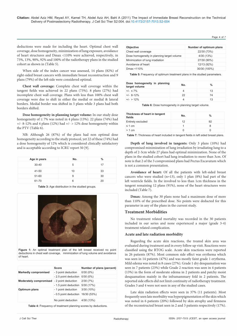
A scoring system was used to assess optimal radiotherapy planning using five parameters (Table 1). Of the 30 radiotherapy plans scored after reconstruction, 76% (23/30) had “optimal scores” (0 - 1 point deduction), example in Figure 1. 24% (7/30) had moderate
compromises (>1- 2 point deductions) and none (0%) had major compromises (more than 2 point deductions) (Table 4).
Point deductions
A full point was deducted for shifting medial and lateral borders. Half point was deducted for irradiating more than 2cm of lung volume and 0.5-point deduction was given for dose homogeneity of 11%. No
Objective No point deduction -0.5 Point deduction -1.0 Point
deduction
Chest wall coverage
Covered from mid sternum medially, to mid axillary line
laterally.
If medial or lateral coverage is not at least to the mid sternum or
mid axillary line(displaced by ≥ 10mm)
If medial and lateral
coverage are deficient
(displaced by ≥ 10mm)
Dose homogeneity in clinical target volume
+/- ≤ 7% +/- 8-12% +/- > 12%
Minimization of lung irradiation
Less than 2-cm of lung thickness involved in chest wall tangential
fields
Maximal lungthickness is 2 cm>2 cm; but ≤3 cm
Maximal lung thicknessis >3 cm
Avoidance of heart
Heart is entirely excluded from tangent fields
If <1 cm of leftventricle is withinthe tangent fields
If >1 cm of leftventricle is treated
intangent fields
D max < 110% 110% - 115% > 115%
Table 1: Radiotherapy plan scoring system guidelines.
Characteristics Number PercentTumour grade123
441214
134047
Tumour Stage123
61113
203743
Lymph nodes Status01-3>3
8139
274330
Surgical Margin involvement-+ or <1mm
1911
6337
Hormone Receptor Statusnegativepositive
921
3070
Her2 neu status_+
255
8317
Lymphovascular invasion-+
237
7723
Menstrual statusPremenopausalPostmenopausal
1416
4753
Type of Flap usedLatissimus DorsiTRAM
273
9010
Treated sideRightLeft
1713
5743
Table 2: Patient and tumour characteristic.
Citation: Abdel Aziz HM, Reyad AY, Kamel TH, Abdel Aziz AH, Bahl A (2011) The Impact of Immediate Breast Reconstruction on the Technical Delivery of Postmastectomy Radiotherapy. J Cell Sci Ther S2:004. doi:10.4172/2157-7013.S2-004
Page 4 of 7
J Cell Sci Ther Radiotherapy ISSN: 2157-7013 JCEST, an open access journal
deductions were made for including the heart. Optimal chest wall coverage, dose homogeneity, minimization of lung exposure, avoidance of heart structures and Dmax <110% were achieved, respectively, in 73%, 13%, 90%, 92% and 100% of the radiotherapy plans in the studied cohort as shown in (Table 5).
When side of the index cancer was assessed, 14 plans (82%) of right-sided breast cancers with immediate breast reconstruction and 9 plans (79%) of the left side were considered optimal.
Chest wall coverage: Complete chest wall coverage within the tangent fields was achieved in 22 plans (73%). 8 plans (27%) had incomplete chest wall coverage. Plans with less than 100% chest wall coverage were due to shift in either the medial or medial & lateral borders. Medial border was shifted in 5 plans while 3 plans had both borders shifted.
Dose homogeneity in planning target volume: In our study dose homogeneity of ≤ 7% was noted in 4 plans (13%). 22 plans (74%) had +/- 8-12% and 4 plans (12%) had +/- > 12% dose homogeneity within the PTV (Table 6).
NB: Although 26 (87%) of the plans had non optimal dose homogeneity according to the study protocol, yet 22 of these (74%) had a dose homogeneity of 12% which is considered clinically satisfactory and is acceptable according to ICRU report 50 [9].
Depth of lung involved in tangents: Only 3 plans (10%) had compromised minimization of lung irradiation by irradiating lung to a depth of 2-3cm while 27 plans had optimal minimization. None of the plans in the studied cohort had lung irradiation to more than 3cm. Of note is that 2 of the 3 compromised plans had Pectus Excavatum which is not a common presentation.
Avoidance of heart: Of all the patients with left-sided breast cancers who were studied (n=13), only 1 plan (8%) had part of the left ventricle fields. In the involved to less than 1cm thickness in the tangent remaining 12 plans (91%), none of the heart structures were included (Table 7).
Dmax: Among the 30 plans none had a maximum dose of more than 110% of the prescribed dose. No points were deducted for this parameter in any of the plans in the current study.
Treatment MorbiditiesNo treatment related mortality was recorded in the 30 patients
included in our series and none experienced a major (grade 3-4) treatment related complication.
Acute and late radiation morbidity
Regarding the acute skin reactions, the treated skin area was evaluated during treatment and in every follow up visit. Reactions were classified using the RTOG scale. Acute skin reactions were reported in 26 patients (87%). Most common side effect was erythema which was seen in 14 patients (47%) and was mostly faint grade 1 erythema. Mild edema was noted in 8 cases (27%). Grade 1 dry desquamation was seen in 7 patients (23%) while Grade 2 reaction was seen in 4 patients (13%) in the form of moderate edema in 2 patients and patchy moist desquamation mainly in the inframammary fold in 2 patients. The reported side effects did not limit continuity of radiotherapy treatment. Grades 3 and 4 were not seen in any of the studied cases.
Late skin radiation effects were seen in 37% (11 patients). Most frequently seen late morbidity was hyperpigmentation of the skin which was noted in 6 patients (20%) followed by skin atrophy and firmness of the reconstructed breast seen in 2 and 3 patients respectively (17%).
Age in years No. %
30-40 5 17
41-50 10 33
51-60 9 30
61-70 6 20
Table 3: Age distribution in the studied groups.
Score Number of plans (percent)Markedly compromised - 3 point deduction 0/30 (0%)
- 2.5 point deduction 0/30 (0%)Moderately compromised - 2 point deduction 2/30 (7%)
- 1.5 point deduction 5/30 (17%)Optimum plans - 1 point deduction 3/30 (10%)
- 0.5 point deduction 16/30 (53%)
No point deduction 4/30 (13%)
Table 4: Frequency of treatment planning scores by deductions.
Figure 1: An optimal treatment plan of the left breast received no point deductions in chest wall coverage, minimization of lung volume and avoidance of heart.
Objective Number of optimum plansChest wall coverage 22/30 (73%)Dose homogeneity in planning target volume 4/30 (13%)Minimization of lung irradiation 27/30 (90%)Avoidance of heart 12/13 (92%)Dmax <110% 30/30 (100%)
Table 5: Frequency of optimum treatment plans in the studied parameters.
Dose homogeneity in planning target volume No. %
+/- ≤ 7% 4 13+/- 8-12% 22 74+/- > 12% 4 13
Table 6: Dose homogeneity in planning target volume.
Thickness of heart in tangent fields No. %
Entirely excluded 12 92≤ 1 cm 1 8> 1 cm 0 0
Table 7: Thickness of heart included in tangent fields in left sided breast plans.
Citation: Abdel Aziz HM, Reyad AY, Kamel TH, Abdel Aziz AH, Bahl A (2011) The Impact of Immediate Breast Reconstruction on the Technical Delivery of Postmastectomy Radiotherapy. J Cell Sci Ther S2:004. doi:10.4172/2157-7013.S2-004
Page 5 of 7
J Cell Sci Ther Radiotherapy ISSN: 2157-7013 JCEST, an open access journal
Telangiectasia (grade 2) was noticed in 1 patient (3%). Grades 3 and 4 were not seen in any patient. Reconstructed flap loss was not reported in the present series.
Lung toxicity: 13 patients (43%) had chest X-ray (10) /CT (3) performed during the follow up period. None of these showed radiological signs of acute pneumonitis. Of the 30 studied cases only 1 (3%) had symptoms of pneumonitis in the form of mild dry cough defined as grade 1 not requiring treatment. When treatment plan was reviewed this patient was found to have been treated with 2 tangents and supraclavicular field. The depth of lung involved in the tangential field was 1.9cm.
Cardiac toxicity: Of the 13 studied left breast cancer cases 4 (30%) had follow up with echo and/or ECG. These are patients who were also receiving adjuvant Trastuzumab. None of the 4 cases had symptoms or signs of acute cardiac morbidity. No ECG changes were noted in the studied cases. 1 patient (8%) had a decline in the left ventricular ejection fraction by 11% (from 68% to 57%) at 18 months post radiotherapy. On reviewing her radiotherapy plan, although she had left side disease yet the heart was entirely excluded from the tangential fields. Trastuzumab could be the likely cause due to its known cardiotoxicity. No symptoms of late cardiac toxicity were noted in the 13 left breast cancer patients.
DiscussionImmediate breast reconstruction offers several advantages as it
is an oncologically safe approach in early stage disease improves the quality of life and can be done contemporaneously as mastectomy. Also it lowers psychosocial morbidity by eliminating the need to experience deformity after mastectomy. Other advantages are superior cosmetic results, decreased surgical morbidity and lower cost [10].
Multiple meta-analyses suggested a significant association between postmastectomy radiation therapy and cardiac mortality [11]. Most radiation oncologists attribute this relationship to techniques that irradiate part of the cardiac silhouette in the tangential fields. The Stockholm group was one of the first to quantify cardiac volume irradiated, dose received and relation to cardiac damage. As the left anterior descending artery is located on the epicardial surface, even minimal cardiac irradiation may subject sensitive targets to high radiation doses [12]. Radiation therapy can also induce pneumonitis and pulmonary fibrosis. The frequency of the effects and their relation to different radiotherapy techniques has been studied more in patients with intact breast than after mastectomy. Nonetheless, the frequency of these effects is related to the volume of lung irradiated, dose per fraction, use of chemotherapy and whether the internal mammary chain was included in the tangential radiation field [13].
When anatomic constraints limit the ability to design a radiation treatment plan that completely covers the regions at risk and simultaneously avoids critical structures, some kind of compromise must be considered. This compromise may be more notable in the presence of a reconstructed breast flap. Although immediate breast reconstruction was shown to be superior compared to other techniques of breast reconstruction by many authors, unfortunately, immediate breast reconstruction and postmastectomy radiation therapy may interact unfavorably with one another. Reconstructions that are subsequently irradiated may have inferior esthetic outcomes and higher complication rates [14]. Also it may pose challenges to optimizing radiotherapy planning and some groups described technical difficulty in delivering PMRT in this subset of patients [15].
The first study to measure the potentially deleterious effect of immediate breast reconstruction and the technical delivery of postmastectomy radiotherapy was carried by Motwani et al. [8] in 2006. In their study 112 treatment plans of patients who had mastectomy with immediate reconstruction with autologous flap followed by radiotherapy were compared with contemporaneous stage-matched patients who had undergone mastectomy without intervening reconstruction. They scored every individual treatment plan to assess whether optimal radiotherapy planning was achievable using four parameters: treatment of the ipsilateral internal mammary chain, breadth of chest wall coverage, minimum involvement of the lung and avoidance of heart. Of the 112 PMRT plans scored after reconstruction, 52% had compromises compared with 7% of matched controls (p < 0.0001). Optimal chest wall coverage, treatment of the ipsilateral internal mammary chain, lung minimization and heart avoidance was achieved in 79%, 45%, 84% and 84% of the plans in the group undergoing immediate reconstruction, compared respectively with 100%, 93%, 97% and 92% of the plans in the control group (p < 0.0001, p < 0.0001, p= 0.0015 and p =0.1435). In patients with reconstructions, 67% of the markedly compromised radiotherapy plans were left-sided (p < 0.16). Patients with right-sided reconstructions had their chest walls and IMC treated with deeper tangential beams at the expense of irradiating more lung. Of the compromised radiation treatment plans with minimization of lung irradiated (17%), 61% of these plans were right sided. Conversely, patients with left-sided reconstructions spared the critical structures (heart and lung), at the expense of suboptimal coverage of chest wall and IMC. Of the compromised radiation treatment plans with poor chest wall coverage (21%), 65% of these plans were left sided. One of the main factors posing difficulty to optimize their plans and contributing to compromises was the intent to treat the internal mammary chain (IMC).
Thus chest wall treatment in their reconstruction cohort was accomplished with tangential fields alone, occasionally using modified blocking when it could provide coverage of the IMC region. Thus, deepening the superior aspect of the photon tangential fields also provided somewhat improved superficial coverage, but frequently at the expense of irradiating more lung and sometimes heart. The impact of this technique limitation resulted in treatment of the IMC that was significantly worse in the reconstruction cohort compared with the matched controls (93% vs. 45%, p =0.0001). To assess the impact of the reconstructed flap on treatment planning if the IMC was not included, they did another analysis of their plans using a scale without IMC involvement, 23% of the treatment plans were still compromised in the reconstruction cohort compared with 2% in the matched controls (p= 0.0001). Of all the patients who underwent reconstruction and whose IMC coverage was compromised, 65% also did not receive a full score in either chest wall breadth, minimization of lung, or avoidance of heart.
They concluded that even if the IMC was not treated, these patients’ plans would still not be optimal and regardless of whether one treats the IMC, immediate breast reconstruction affects the delivery of postmastectomy radiotherapy.
In the present series we used a semi-quantitative scoring tool to assess the design of postmastectomy radiation fields. This system assesses five basic objectives of postmastectomy radiotherapy and the primary radiotherapy plans were scored in each category. Our scoring system that we devised can be viewed as a modification of the MD Anderson cancer centre scoring system implemented by Buchholz
Citation: Abdel Aziz HM, Reyad AY, Kamel TH, Abdel Aziz AH, Bahl A (2011) The Impact of Immediate Breast Reconstruction on the Technical Delivery of Postmastectomy Radiotherapy. J Cell Sci Ther S2:004. doi:10.4172/2157-7013.S2-004
Page 6 of 7
J Cell Sci Ther Radiotherapy ISSN: 2157-7013 JCEST, an open access journal
[4,8,12] which assessed four objectives: chest wall & internal mammary chain coverage and minimum irradiation to lung and heart.
In view of the lack of evidence on the benefit of irradiating the IMC and since the standard practice at Ain Shams University Hospitals and Bristol Oncology hospital generally does not adopt irradiation of the IMC by a separate field, it was not included in the parameters to be assessed in this study. Because dose homogeneity in the planning target volume is a fundamental factor in radiotherapy planning and has been shown to affect the clinical outcome of radiation therapy in terms of both local disease control and radiation toxicity, we viewed it should be taken into account when assessing the planning technique in our study.
According to the ICRU report number 50 issued in 1993, dose across the PTV should be homogeneous as possible and should be kept within +7% and – 5% of prescribed dose for non palliative treatment plans. In the same report Dmax was defined as the maximum dose within the PTV and has to be taken into account for evaluating the homogeneity of the dose distribution and should be considered as part of the optimization criteria [9]. Therefore unlike the MD Anderson group we included Dmax and dose homogeneity in our scoring system and omitted involving the IMC as an objective to be assessed.
In our series of the 30 PMRT plans scored after reconstruction, 76% (23/30) had optimal scores while 24% (7/30) had moderate compromises and none [0%] had major compromises. Bearing in mind that IMC involvement was not included in this study, our outcome is similar to that of the MD Anderson group when they analysed their plans after exclusion of IMC coverage which revealed a compromise in 23% of the plans. Although our cohort was not directly compared to a non reconstructed group yet indirect comparison with the control group from the MD Anderson Cancer centre shows clinically significant difference (24% compromise in our cohort vs. 2% in the MD Anderson control arm).
Complete chest wall coverage which is the most important factor in PMRT was achieved in 22 plans (73%) that is slightly higher than that reported in the MD Anderson’s of 65%. It is worth noting that 7% of the compromised plans in this study were for patients with Pectus excavatum which is an anatomical deformity posing challenge to the radiation oncologist. Although these cases are not typical and relatively uncommon, yet they were eligible as per the study protocol.
Of the 27% of the compromised chest wall coverage, 17% had the left border shifted compared to 10% who had both shifted. This is not a surprising outcome knowing that most of breast cancers occur in the upper outer quadrant, so, if there is a need to minimize lung irradiated it would be more reasonable to shift the medial border rather than the lateral border which is more close to the tumour bed.
In this study we defined an optimal treatment plan as one in which the wedge of lung tissue irradiated was less than 2cm as per the Bristol oncology centre protocol. Because fibrosis, pneumonitis and decreased pulmonary function are all related to the volume of irradiated lung, every centimeter of lung in the tangential field causes a greater volume of lung parenchyma to be irradiated. Cheng et al. [13] reported keeping central lung distance of tangential field to less than 2.8cm in a tangent field technique alone or less than 1.4cm in a tangential field with a separate electron IMC field technique reduces the risk of lung fibrosis. Keeping lung depth to 2cm was achievable in 90% in the studied cohort while 10% had compromised plans, yet none had more than 3cm of lung included. Again, 7% out of the 10% who were compromised where the Pectus excavatum cases.
In terms of dose homogeneity across the PTV, it is universally accepted that it should be within 12% (+7%, -5%) as been recommended by the ICRU, but in this study we defined optimum plans as those having less than 7% dose homogeneity as data from the literature showed that irradiated reconstructed flaps are more prone to radiation side effects and complications as shown earlier. Being tighter in this parameter, only 13% of the plans were considered optimum achieving ≤ 7% homogeneity. Yet, 74% of the assessed plans were contained within 8-12% which is still considered clinically acceptable and none had a maximum dose of more than 110% of the prescribed dose to the PTV. 13% had more than 12% homogeneity, hence had full point deduction.
Of the 13 left sided breast cancer plans, 12 plans (92%) successfully had the heart entirely excluded from the tangential portals and the single plan which included the heart showed less than 1cm involvement. This outcome was higher than that reported by Motwani et al. [8] in their series (85%) and is equal to their control arm which achieved 92%.
When side of the index cancer was assessed, 82% of right-sided breast cancers with immediate breast reconstruction and 69% of the left side were considered optimal. In other terms, 53% of the compromised plans were left sided and 47% were right sided. This higher compromise rate in the left sided group was anticipated since an extra factor (avoidance of heart) was accounted for in this group as the attempt to spare the heart could have led to compromising other parameters. Buchholz et al. [4,8,12] had different outcomes. They had more optimal plans in the right side rather than the left. Optimal plans were noticed in 41% of right-sided breast cancers with immediate breast reconstruction and 51% of the left side. Yet 67% of the major compromised radiotherapy plans were left-sided.
In our study we aimed to evaluate and classify skin reactions and we employed the RTOG scoring criteria to quantify the extent of these reactions. Skin reactions, whether early or late, were the most common and notable side effect in the current study. Acute reaction was reported in 87% of the cases. Results revealed no incidence of degree 3 acute reaction in comparison to 60% and 27% for degrees 1 and 2 respectively. Late reactions were noticed in 36% of the studied population, all had grades 1 and 2. These data are comparable to those found in literature. In a study done in the NCI and NEMROCK to evaluate skin reactions following conservative surgery and radiotherapy, Taher et al. [16] did not report grade 3. Grades 1 and 2 were reported in 60% and 40% respectively while in 2008; Pires et al. [17] underwent a study to Evaluate and classify skin reactions in patients with breast cancer submitted to radiotherapy through the Radiation Therapy Oncology Group [7] criteria. When they looked at the reconstructed subgroup, all of the reported reactions were grades 1 and 2 with incidence rate of 82%. None of their cases developed grade 3.
As regards lung toxicity, literature shows radiation pneumonitis incidence of as many as 5-15% of patients, however most of the studies were evaluating patients who were treated with thoracic irradiation, most often in lung cancer, lymphoma, esophageal cancer or thymoma and data on breast cancer patients has been scarce [18]. In the studied cohort only 3% had symptoms of pneumonitis in the form of mild dry cough defined as grade 1 not requiring treatment. This low incidence rate could be attributed to the minimal lung involvement in the current series. None of the treated cases had more than 3cm of lung wedge included in the tangent portals and 90% had less than 2cm involved. In those who had chest radiographs, signs of interstitial pneumonitis or fibrosis were not observed. Assessing the risk of long-term cardiac toxicity due to PMRT is complex due to the long latency for such side
Citation: Abdel Aziz HM, Reyad AY, Kamel TH, Abdel Aziz AH, Bahl A (2011) The Impact of Immediate Breast Reconstruction on the Technical Delivery of Postmastectomy Radiotherapy. J Cell Sci Ther S2:004. doi:10.4172/2157-7013.S2-004
Page 7 of 7
J Cell Sci Ther Radiotherapy ISSN: 2157-7013 JCEST, an open access journal
effects and the possible contribution of patient-related factors, as well as treatment factors.
Perhaps the most important treatment factor is the volume of heart irradiated. Many techniques used to irradiate the internal mammary nodes (such as the pure photon “hockey-stick” field) included large cardiac volumes, regardless of whether the tumour was left- or right-sided [19]. Registry-based studies also have demonstrated an increase in cardiac mortality for patients treated with left-sided breast cancers [20]. This increase in cardiac deaths has been attributed primarily to ischemic heart disease. This was most clearly demonstrated by a meta-analysis of 10 randomized trials of PMRT initiated before 1975 when older techniques of radiotherapy were used. The standard mortality ratio for heart disease was 1.62 times higher for irradiated patients than for the unirradiated patients [P=0.01]. These findings were confirmed in the larger Oxford overview of radiotherapy trials, which included patients treated with breast-conserving surgery as well [11]. In the present study internal mammary nodal irradiation was not used and among those who had left breast irradiation only 1 patient had part of the heart involved in the tangential field and this was less than 1cm thickness. Thus there was less anticipated risk of cardiac morbidities. None of the studied cases had symptoms or signs of acute cardiac morbidity. 1 patient [8%] had decline of the left ejection fraction yet this patient was on adjuvant therapy with Trastuzumab which is more likely to be the cause of this finding. In fact, it is difficult to assess cardiac morbidity particularly from studies as the present one with relatively short follow up period due to the long latency for such side effects which may extend to 20 years. Finally, the more recent irradiation technique, intensity modulated radiation therapy (IMRT) has been introduced in the adjuvant treatment of breast cancer aiming at reducing acute toxicity and the dose to the critical organs as heart and lung with proved efficacy [21].
ConclusionsImmediate reconstruction may impose limitations on the treatment
planning of postmastectomy radiotherapy, particularly in regard to providing broad adequate coverage of the chest wall and if there can be one conclusion from the published experience, it is that advantages and risks of immediate reconstruction should be fully explored with the patient and a multidisciplinary approach to patient management is required. Multicentre, well powered trials with longer follow up are required to look at the potentially adverse reciprocal effects between radiotherapy and immediate breast reconstruction.
References
1. Ragaz J, Olivotto IA, Spinelli JJ, Phillips N, Jackson SM, et al. (2002) Locoregional radiation therapy in patients with high-risk breast cancer receiving adjuvant chemotherapy: 20-year results of the British Columbia randomized trial. J Natl Cancer Inst 97: 116-126.
2. National Comprehensive Cancer Network. NCCN practice guidelines for breast cancer, 2005.
3. Freedman GM (2005) Implication of planned radiotherapy on breast reconstruction: radiotherapy and plastic surgery with implants. Breast Cancer Online 8: e44.
4. Buchholz TA, Kronowitz SJ, Kuerer HM (2002) Immediate breast reconstruction after skin-sparing mastectomy for the treatment of advanced breast cancer: radiation oncology considerations. Ann Surg Oncol 9: 820-821.
5. Mehta VK, Goffinet D (2004) Postmastectomy radiation therapy after TRAM flap breast reconstruction. Breast J 10: 118-122.
6. Fodor J, Gulyás G, Polgár C, Major T, Kásler M (2003) Radiotherapy and breast reconstruction: the issue of compatibility. Orv Hetil 144: 549-555.
7. Cox JD, Stetz J, Pajak TF (1995) Toxicity criteria of the Radiation Therapy Oncology Group (RTOG) and the European Organization for Research and Treatment of Cancer (EORTC). Int J Radiat Oncol Biol Phys 31: 1341-1346.
8. Motwani SB, Strom EA, Schechter NR, Butler CE, Lee GK, et al. (2006) The impact of immediate breast reconstruction on the Technical delivery of postmastectomy radiotherapy. Int J Radiat Oncol Biol Phys 66: 76-82.
9. ICRU Report 50 (1993) Prescribing, recording and reporting photon beam therapy. Bethesda, MD.
10. Miller MJ (1998) Immediate breast reconstruction. Clin Plast Surg 25: 145-156.
11. Early Breast Cancer Trialists’ Collaborative Group (2002) Radiotherapy for early breast cancer. Cochrane Database Syst Rev CD003647.
12. Storey MR, Munden R, Strom EA, McNeese MD, Buchholz TA (2001) Coronary artery dosimetry in intact left breast irradiation. Cancer J 7: 492-497.
13. Cheng SH, Jian JJ, Chan KY, Tsai SY, Liu MC, et al. (1998) The benefit and risk of postmastectomy radiation therapy in patients with high-risk breast cancer. Am J Clin Oncol 21:12-17.
14. Kronowitz SJ, Robb GL (2004) Breast reconstruction with postmastectomy radiation therapy: current issues. Plast Reconstr Surg 114: 950-960.
15. Schechter NR, Strom EA, Perkins GH, Arzu I, McNeese MD, et al. (2005) Immediate breast reconstruction can impact postmastectomy irradiation. Am J Clin Oncol 28: 485-494.
16. Taher AN, El-Baradie MM, Essa H, Zaki O, Ezzat S (2004) Hypofractionation versus Conventional Fractionation Radiotherapy after Conservative Treatment of Breast Cancer: Early Skin Reactions and Cosmetic Results. J Egypt Natl Canc Inst 16: 178-187.
17. Pires AM, Segreto RA, Segreto HR (2008) RTOG criteria to evaluate acute skin reaction and its risk factors in patients with breast cancer submitted to radiotherapy. Rev Lat Am Enfermagem 16: 844-849.
18. Hurkmans CW, Borger JH, Bos LJ, van der Horst A, Pieters BR, et al. (2000) Cardiac and lung complication probabilities after breast cancer irradiation. Radiother Oncol 55: 145-151.
19. Pierce SM, Recht A, Lingos TI, Abner A, Vicini F, et al. (1992) Long-term radiation complications following conservative surgery (CS) and radiation therapy (RT) in patients with early stage breast cancer. Int J Radiat Oncol Biol Phys 23: 915-923.
20. Paszat LF, Mackillop WJ, Groome PA, Boyd C, Schulze K, et al. (1998) Mortality from myocardial infarction after adjuvant radiotherapy for breast cancer in the surveillance, epidemiology, and end-results cancer registries. J Clin Oncol 16: 2625-2631.
21. McDonald MW, Godette KD, Butker EK, Davis LW, Johnstone PA (2008) Long-term outcomes of IMRT for breast cancer: a single-institution cohort analysis. Int J Radiat Oncol Biol Phys 72: 1031-1040.
Thisarticlewasoriginallypublishedinaspecialissue,RadiotherapyhandledbyEditor(s).Dr.VKrishnanRamanujan,Cedars-SinaiMedicalCenter,USA


![+1cm[width=30mm]logo.pdf +1cm Robust Control Of Under](https://img.pdfslide.us/doc/110x75/628f369cbf717634c42ee7c5/1cmwidth30mmlogopdf-1cm-robust-control-of-under-.jpg)


![+1cm[width=30mm]logo.pdf +1cm Evaluation of Water](https://img.pdfslide.us/doc/110x75/627cce9ad7cf13078b01c655/1cmwidth30mmlogopdf-1cm-evaluation-of-water-.jpg)