Embed Size (px)
Citation preview
Journal of Biomedical Informatics 55 (2015) 55–63
Contents lists available at ScienceDirect
Journal of Biomedical Informatics
journal homepage: www.elsevier .com/locate /y jb in
MorphoCol: An ontology-based knowledgebase for the characterisationof clinically significant bacterial colony morphologies
http://dx.doi.org/10.1016/j.jbi.2015.03.0071532-0464/� 2015 Elsevier Inc. All rights reserved.
⇑ Corresponding author at: ESEI: Escuela Superior de Ingeniería Informática,University of Vigo, Edificio Politécnico, Campus Universitario As Lagoas s/n, 32004Ourense, Spain.
E-mail addresses: [email protected] (A.M. Sousa), [email protected] (M.O. Pereira), [email protected], [email protected] (A. Lourenço).
Ana Margarida Sousa a, Maria Olívia Pereira a, Anália Lourenço a,b,⇑a CEB – Centre of Biological Engineering, LIBRO – Laboratório de Investigação em Biofilmes Rosário Oliveira, University of Minho, Campus de Gualtar, 4710-057 Braga, Portugalb ESEI: Escuela Superior de Ingeniería Informática, University of Vigo, Edificio Politécnico, Campus Universitario As Lagoas s/n, 32004 Ourense, Spain
a r t i c l e i n f o a b s t r a c t
Article history:Received 29 April 201424 January 2015
Accepted 20 March 2015Available online 25 March 2015
Keywords:Colony morphologyAntimicrobial resistanceVirulenceColony morphology ontology
Background: One of the major concerns of the biomedical community is the increasing prevalence ofantimicrobial resistant microorganisms. Recent findings show that the diversification of colonymorphology may be indicative of the expression of virulence factors and increased resistance to antibiotictherapeutics. To transform these findings, and upcoming results, into a valuable clinical decision makingtool, colony morphology characterisation should be standardised. Notably, it is important to establish theminimum experimental information necessary to contextualise the environment that originated thecolony morphology, and describe the main morphological features associated unambiguously.Results: This paper presents MorphoCol, a new ontology-based tool for the standardised, consistent andmachine-interpretable description of the morphology of colonies formed by human pathogenic bacteria.The Colony Morphology Ontology (CMO) is the first controlled vocabulary addressing the specificities ofthe morphology of clinically significant bacteria, whereas the MorphoCol publicly Web-accessibleknowledgebase is an end-user means to search and compare CMO annotated colony morphotypes. Itsultimate aim is to help correlate the morphological alterations manifested by colony-forming bacteriaduring infection with their response to the antimicrobial treatments administered.Conclusions: MorphoCol is the first tool to address bacterial colony morphotyping systematically anddeliver a free of charge resource to the community. Hopefully, it may introduce interesting features ofanalysis on pathogenic behaviour and play a significant role in clinical decision making.Database URL: http://morphocol.org.
� 2015 Elsevier Inc. All rights reserved.
1. Introduction
Human infections involve a complex intertwined interplay ofmicroorganisms. Understanding these interactions as well as thecontinuously emerging mechanisms of antimicrobial resistanceare pressing goals in clinical microbiology.
Recent technologies, such as the enzyme-linked immunosor-bent assay (ELISA), the polymerase chain reaction (PCR) and thematrix-assisted laser desorption/ionisation time of flight massspectrometry (MALDI-TOF MS) have boosted the identificationand characterisation of clinically significant bacteria [1–3].However, the research community is manifesting a renewed inter-est in traditional culture-based strategies like colony morphology
characterisation as more immediate, first-term means of decisionsupport [4,5].
Alterations in the morphology of the microbial colonies,reflected in macroscopically observable features such as form,colour, opacity, size and texture, may support bacteria profilingunder changing and often stressful environments [6–8]. Notably,these morphological features are being increasingly documentedin clinical settings as potential evidences of the expression of viru-lence factors [9–12] and increased resistance to antibiotic thera-peutics [13–15]. For example, mucoid morphotypes [16,17] andsmall colony variants [18–20] are recognised as markedly resistantto a wide range of conventional antibiotics, and are often related tomulti-resistant strains. Therefore, an increasing number of scienti-fic studies are documenting morphotypes of clinically significantbacteria.
In principle, colony morphology characterisation is a simpleprocedure and it is fairly easy to integrate into the analytical pipe-line of any laboratory. Colony morphology is described throughnaked-eye observation and using a magnifying glass, and classified
56 A.M. Sousa et al. / Journal of Biomedical Informatics 55 (2015) 55–63
using several criteria, commonly accepted in clinical microbiology.However, experimental design and morphological annotationshould be consistent in order to allow the systematic comparisonof morphotypes across experiments (and laboratories), species,diseases and clinical samples. Colony morphologies vary widely,depending on the particular behaviour of the microbial speciesunder different test conditions (e.g. different colonisation sites, orantibiotic agents with different modes of action). Also, the mor-phological traits exhibited by the colonies may be significantlyaffected by the procedures taken to isolate and grow the bacteria[21]. Thus, it is very important to establish the minimum set ofinformation to be part of the morphotype description and toemploy harmonised vocabulary in both the biological contextual-isation and the morphological characterisation of the observedcolony.
Scientific literature is the main source of morphotypes, wherethey are often presented as exemplificative figures of what theresearchers observed and are described informally. The fact thatthe description of colony morphologies does not yet follow prede-fined rules of annotation nor makes use of controlled vocabularyhampers the automated classification, integration and inter-pretation of such data.
Researchers are in need of new resources and tools geared tosystematically analyse morphotypes, across infections, body loca-tions, antimicrobial treatments and a number of other conditionsof clinical relevance. Multiple ontologies have been proposed inthe domain of phenotypes. Some are specialised in the character-isation of species (typically, model organisms), such as theMammalian Phenotype ontology (MP) [22], the Worm Phenotypeontology (WPO) [23], the Plant ontology (PO) [24] and theHuman Phenotype ontology (HPO) [25]. Others, like thePhenotype and Trait ontology (PATO) [26], are focused on integrat-ing phenotypes across species, and reuse anatomy and processontologies.
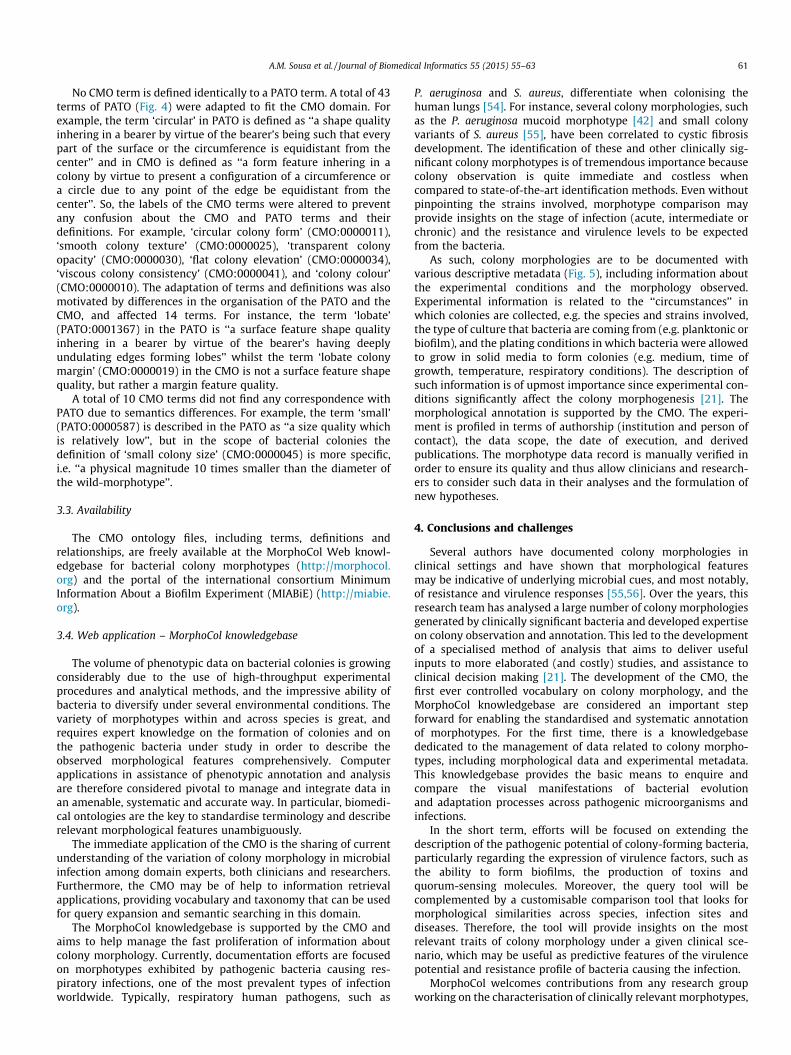
This paper presents the rationale of a novel ontological frame-work in support of the characterisation of the colony morphologyof clinically significant bacteria. The Colony MorphologyOntology (CMO) is introduced as an integrative resource for the
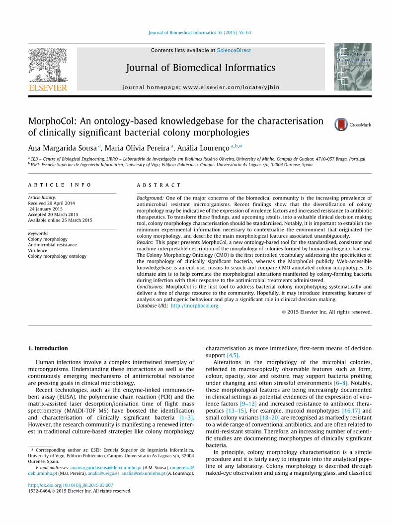
Fig. 1. General workflow of bacterial profiling. Bacteria provided from samples are cultivaincluded. To perform colony morphology characterisation, bacteria from cultures is plated10 main morphological features: form, margin, sheath, type of surface, texture, consissubjected to bacterial profiling using other methods, such as antimicrobial susceptibilit
systematic, transparent and unambiguous characterisation of col-ony morphology traits in support of clinical diagnosis. CMO sup-ports the MorphoCol database, a public Web repository ofpathogenic bacterial morphotypes (http://morphocol.org). Theaim of this repository is to enable the macroscopic observation ofmorphotypes and the comparison of the morphological ‘‘output’’of the species in different scenarios (e.g. antibiotic therapeuticsand body localisation).
The originality of this work lays on addressing colony morpho-typing in a systematic, harmonised and computerised way.Although still in its infancy, MorphoCol aims to pave the way tothe development of advanced clinical decision making applica-tions, which may use morphological features as immediate indica-tors of microbial behaviour (Fig. 1). These indicators can be used toguide more sophisticated (time-consuming and costly) analyses,such as proteome and transcriptome analyses. Also, they may beused to construct decision support models that help clinicians indetermining or anticipating what may be expected in terms of agiven microbial species resistance and resilience in a clinical inci-dent. To the best of our knowledge this is the first public repositorydocumenting bacterial colony morphology systematically.Currently, the system documents respiratory infection traits. Inthe future, it will cover for other major infections regarding the uri-nary tract, bloodstream, chronic wounds, osteomyelitis and bio-material-associated infections. Thus, MorphoCol will be of aid tothe wider community of researchers and clinicians working inclinical microbiology.
2. Design and implementation
2.1. Considerations in ontology design
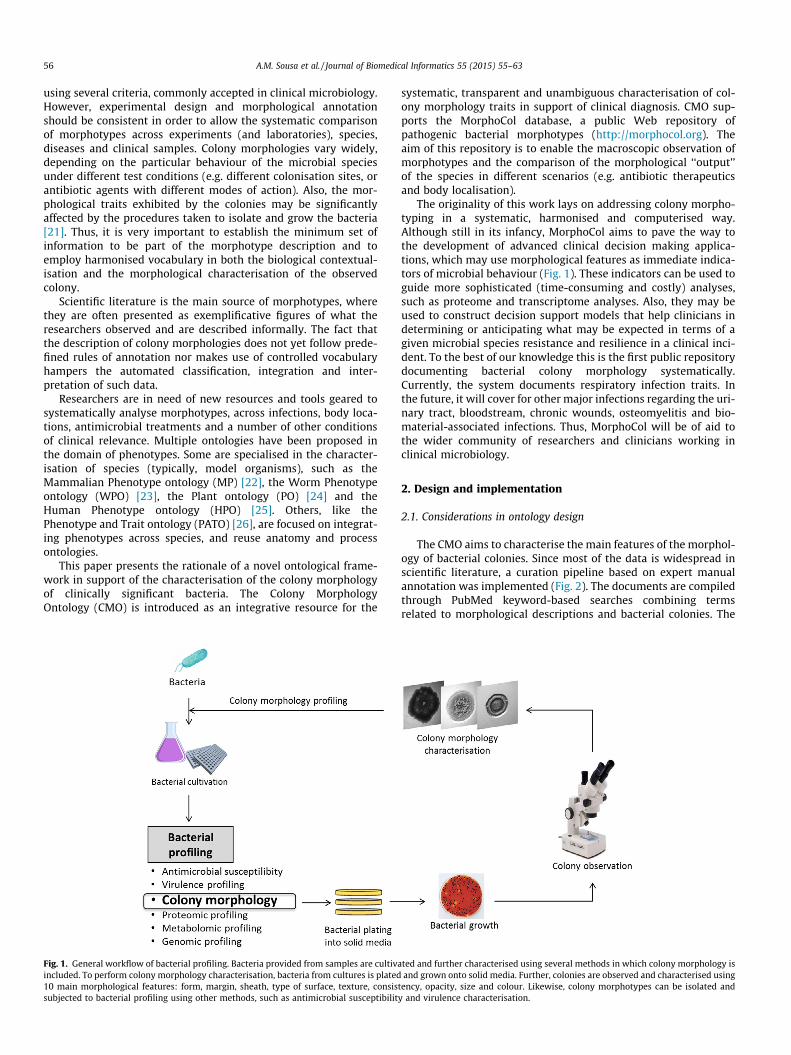
The CMO aims to characterise the main features of the morphol-ogy of bacterial colonies. Since most of the data is widespread inscientific literature, a curation pipeline based on expert manualannotation was implemented (Fig. 2). The documents are compiledthrough PubMed keyword-based searches combining termsrelated to morphological descriptions and bacterial colonies. The
ted and further characterised using several methods in which colony morphology isand grown onto solid media. Further, colonies are observed and characterised using
tency, opacity, size and colour. Likewise, colony morphotypes can be isolated andy and virulence characterisation.
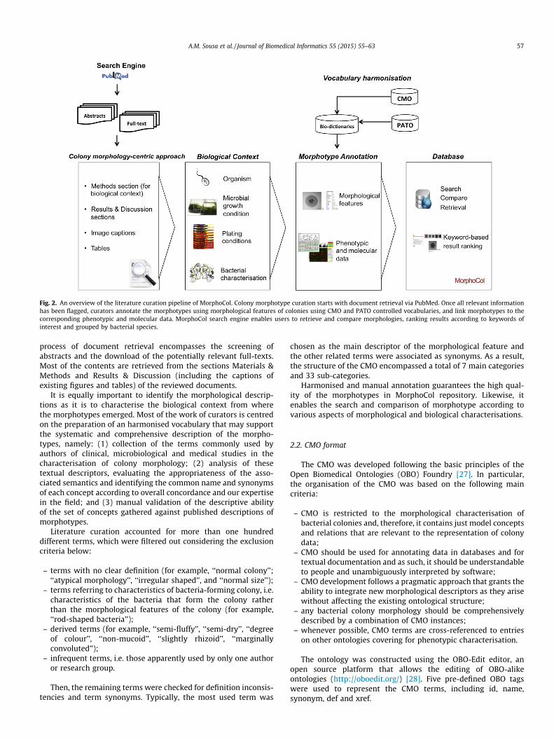
Fig. 2. An overview of the literature curation pipeline of MorphoCol. Colony morphotype curation starts with document retrieval via PubMed. Once all relevant informationhas been flagged, curators annotate the morphotypes using morphological features of colonies using CMO and PATO controlled vocabularies, and link morphotypes to thecorresponding phenotypic and molecular data. MorphoCol search engine enables users to retrieve and compare morphologies, ranking results according to keywords ofinterest and grouped by bacterial species.
A.M. Sousa et al. / Journal of Biomedical Informatics 55 (2015) 55–63 57
process of document retrieval encompasses the screening ofabstracts and the download of the potentially relevant full-texts.Most of the contents are retrieved from the sections Materials &Methods and Results & Discussion (including the captions ofexisting figures and tables) of the reviewed documents.
It is equally important to identify the morphological descrip-tions as it is to characterise the biological context from wherethe morphotypes emerged. Most of the work of curators is centredon the preparation of an harmonised vocabulary that may supportthe systematic and comprehensive description of the morpho-types, namely: (1) collection of the terms commonly used byauthors of clinical, microbiological and medical studies in thecharacterisation of colony morphology; (2) analysis of thesetextual descriptors, evaluating the appropriateness of the asso-ciated semantics and identifying the common name and synonymsof each concept according to overall concordance and our expertisein the field; and (3) manual validation of the descriptive abilityof the set of concepts gathered against published descriptions ofmorphotypes.
Literature curation accounted for more than one hundreddifferent terms, which were filtered out considering the exclusioncriteria below:
– terms with no clear definition (for example, ‘‘normal colony’’;‘‘atypical morphology’’, ‘‘irregular shaped’’, and ‘‘normal size’’);
– terms referring to characteristics of bacteria-forming colony, i.e.characteristics of the bacteria that form the colony ratherthan the morphological features of the colony (for example,‘‘rod-shaped bacteria’’);
– derived terms (for example, ‘‘semi-fluffy’’, ‘‘semi-dry’’, ‘‘degreeof colour’’, ‘‘non-mucoid’’, ‘‘slightly rhizoid’’, ‘‘marginallyconvoluted’’);
– infrequent terms, i.e. those apparently used by only one authoror research group.
Then, the remaining terms were checked for definition inconsis-tencies and term synonyms. Typically, the most used term was
chosen as the main descriptor of the morphological feature andthe other related terms were associated as synonyms. As a result,the structure of the CMO encompassed a total of 7 main categoriesand 33 sub-categories.
Harmonised and manual annotation guarantees the high qual-ity of the morphotypes in MorphoCol repository. Likewise, itenables the search and comparison of morphotype according tovarious aspects of morphological and biological characterisations.
2.2. CMO format
The CMO was developed following the basic principles of theOpen Biomedical Ontologies (OBO) Foundry [27]. In particular,the organisation of the CMO was based on the following maincriteria:
– CMO is restricted to the morphological characterisation ofbacterial colonies and, therefore, it contains just model conceptsand relations that are relevant to the representation of colonydata;
– CMO should be used for annotating data in databases and fortextual documentation and as such, it should be understandableto people and unambiguously interpreted by software;
– CMO development follows a pragmatic approach that grants theability to integrate new morphological descriptors as they arisewithout affecting the existing ontological structure;
– any bacterial colony morphology should be comprehensivelydescribed by a combination of CMO instances;
– whenever possible, CMO terms are cross-referenced to entrieson other ontologies covering for phenotypic characterisation.
The ontology was constructed using the OBO-Edit editor, anopen source platform that allows the editing of OBO-alikeontologies (http://oboedit.org/) [28]. Five pre-defined OBO tagswere used to represent the CMO terms, including id, name,synonym, def and xref.
58 A.M. Sousa et al. / Journal of Biomedical Informatics 55 (2015) 55–63
2.3. MorphoCol knowledgebase
MorphoCol is a publicly Web-accessible knowledgebase thatdocuments bacterial colony morphotypes, as comprehensively aspossible, in order to enable the search and comparison of morpho-types across species (and strains) and conditions (namely, diseasesand colonisation sites). Currently, the main source of information isscientific literature and manual curation grants the high quality ofthe data available. The curation pipeline (Fig. 2) will be graduallyincorporating automatic procedures, namely text mining pro-cesses, now that we have in hand an appropriate terminologicalresource and the minimum set of information necessary to com-prehensively describe a morphotype. Likewise, the knowledgebasewill enable the direct submission of morphotype data by authors,promoting a close interaction with the community.
MorphoCol server runs on a CentOS platform (version 5.6) withApache HTTP server (version 2.2.22), MySQL Community Server(version 5.1.58) and PHP 5.5.3. Apache, MySQL and PHP technologyare open-source and platform-independent software. Moreover,MySQL supports multi-threading and multi-user environments,and thus it is well-suited to support (increasing) real-world data-base usage. Currently, the Web server and all parts of the databaseare hosted at the Centre of Biological Engineering of the Universityof Minho, Portugal.
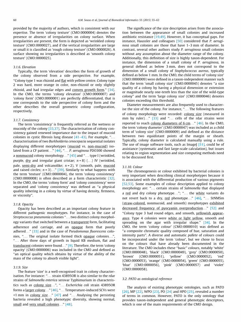
Fig. 3. Examples of colony morphologies exhibiting features that the first version ofthe CMO did not consider: (A) sheath, enveloping part or structure after the marginthat surrounds the colony; dual type of textures, (B) smooth and wrinkled, (C)smooth and rough, and (D) rough and wrinkled, described from the periphery to thecentre. Black bar = 1 mm.
3. Results and discussion
Colony morphotyping is a common technique used in microbio-logical studies of varied purposes. It should be emphasised thatthis study does not propose any colony features. The colonymorphology features mentioned and described are commonlyaccepted by the microbiological community and they can be tracedback to as early as papers published decades ago [29–31].
3.1. CMO structure and contents
The validation of the CMO terms against published morphologi-cal descriptions showed that all terms included in the ontologystructure were valid, but pointed out some additional considera-tions as follows: Pseudomonas aeruginosa colonies exhibit anenveloping part or structure after the margin that surrounds thecolony, which is referred to as ‘sheath’ (Fig. 3A); colonies can exhi-bit more than one type of surface, e.g. P. aeruginosa colonies canexhibit surfaces with smooth and rough zones (Fig. 3B), smoothand wrinkled zones (Fig. 3C), or wrinkled and rough zones(Fig. 3D). To enhance its descriptive abilities, the initial structureof the CMO was extended to a total of 10 main categories and 37sub-categories (Fig. 4).
An important issue whilst defining the organisation of the CMOwas the ability to perform updates without causing major changesin the structure. To this end, the high level nodes of the ontologyrepresent the general concepts behind the morphological featuresmore frequently discussed in the literature, including ‘form’, ‘mar-gin’, ‘type of surface’, ‘texture’, ‘sheath’, ‘opacity’, ‘elevation’, ‘con-sistency’, ‘size’ and ‘colour’. The following subsections introducethese concepts, explaining the semantics adopted by the CMOand discussing the semantic inconsistencies and ambiguities foundin the literature.
3.1.1. FormThe term ‘form’ is commonly used to describe the whole config-
uration of a colony. For example, ‘‘. . . overall surface shape (convex,crater, lobulated, radial, radioumbilicated, radio-umbonated,peaked, rugose, segmented-rugose, segmented-umbilicated,umbilicated, umbilicated with irregular edge, umbilicated with
heaped-up irregular edge, or umbonated).’’ [32], or to differentiatecolonies with certain characteristics, such as ‘‘. . . two distinct
colony morphology variants that switched between a transparent
form, facilitating adherence and carriage, and an opaque form thatpoorly adhered. . .’’ [33]. So, in the CMO, the term ‘colony form’(CMO:0000001) is defined as the geometrical configuration of thecolony – ‘‘a morphological quality inhering in a colony by virtueof having a configuration’’.
3.1.2. MarginThe description of the margin of the colony, also referred to as
edge or border, is typically based on the characteristics of thecolony circumference. For example, ‘‘Colony type 2 was hard, moreorange in colour, non-rhizoid or only slightly rhizoid, and had
irregular edges and convex growth form. Colony type 3 had round
edges, and smooth, yellowish appearance.’’ [34], ‘‘‘fried egg’ SCVs,
with translucent edges surrounding a smaller elevated. . .’’ [19],‘‘other colonies (approximately 60% of the total) were smaller with
somewhat rough edges’’ [35]. In the CMO, the term ‘colony margin’(CMO:0000002) represents the configuration of the limiting borderof the colony and is described as ‘‘a morphological quality inheringin a colony by virtue of having a limit zone’’.
3.1.3. SheathThe description of ‘colony sheath’ is not common in colony
morphology observation. However, this structure is often presentin P. aeruginosa colonies, which are clinically recurrent, and it isquite variable (Fig. 3). Based on our expertise, this may be avaluable element in the description of colony differentiation. So,it was included in the CMO (CMO:0000005) and defined as ‘‘amorphological quality inhering in a colony by virtue of having aclosely enveloping part or structure after the margin and aroundthe colony’’.
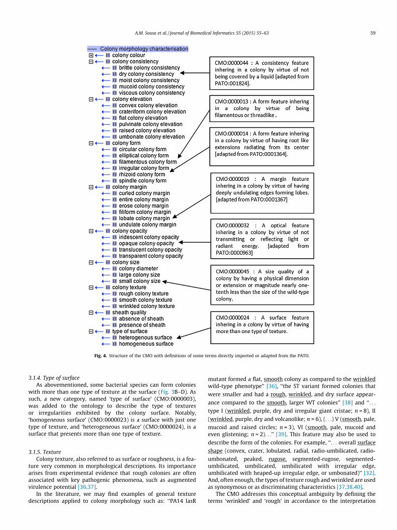
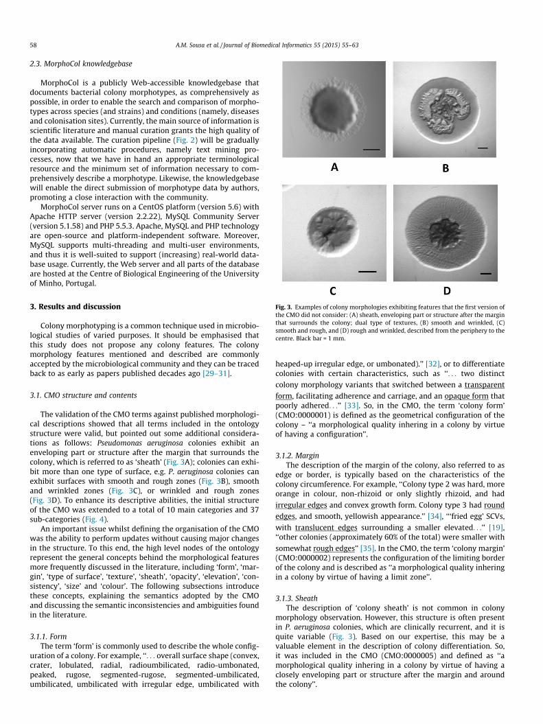
Fig. 4. Structure of the CMO with definitions of some terms directly imported or adapted from the PATO.
A.M. Sousa et al. / Journal of Biomedical Informatics 55 (2015) 55–63 59
3.1.4. Type of surfaceAs abovementioned, some bacterial species can form colonies
with more than one type of texture at the surface (Fig. 3B–D). Assuch, a new category, named ‘type of surface’ (CMO:0000003),was added to the ontology to describe the type of texturesor irregularities exhibited by the colony surface. Notably,‘homogeneous surface’ (CMO:0000023) is a surface with just onetype of texture, and ‘heterogeneous surface’ (CMO:0000024), is asurface that presents more than one type of texture.
3.1.5. TextureColony texture, also referred to as surface or roughness, is a fea-
ture very common in morphological descriptions. Its importancearises from experimental evidence that rough colonies are oftenassociated with key pathogenic phenomena, such as augmentedvirulence potential [36,37].
In the literature, we may find examples of general texturedescriptions applied to colony morphology such as: ‘‘PA14 lasR
mutant formed a flat, smooth colony as compared to the wrinkledwild-type phenotype’’ [36], ‘‘the ST variant formed colonies that
were smaller and had a rough, wrinkled, and dry surface appear-
ance compared to the smooth, larger WT colonies’’ [38] and ‘‘. . .
type I (wrinkled, purple, dry and irregular giant cristae; n = 8), II
(wrinkled, purple, dry and volcanolike; n = 6), (. . .) V (smooth, pale,
mucoid and raised circles; n = 3), VI (smooth, pale, mucoid andeven glistening; n = 2). . .’’ [39]. This feature may also be used to
describe the form of the colonies. For example, ‘‘. . . overall surface
shape (convex, crater, lobulated, radial, radio-umbilicated, radio-
umbonated, peaked, rugose, segmented-rugose, segmented-umbilicated, umbilicated, umbilicated with irregular edge,umbilicated with heaped-up irregular edge, or umbonated)’’ [32].And, often enough, the types of texture rough and wrinkled are usedas synonymous or as discriminating characteristics [37,38,40].
The CMO addresses this conceptual ambiguity by defining theterms ‘wrinkled’ and ‘rough’ in accordance to the interpretation
60 A.M. Sousa et al. / Journal of Biomedical Informatics 55 (2015) 55–63
provided by the majority of authors, which is consistent with ourexpertise. The term ‘colony texture’ (CMO:0000004) denotes thepresence or absence of irregularities on colony surface. Whenirregularities are present, the form is depicted as ‘wrinkled colonytexture’ (CMO:0000027), and if the vertical irregularities are largeor small it is classified as ‘rough colony texture’ (CMO:0000026). Asurface showing no irregularities is described as ‘smooth colonytexture’ (CMO:0000025).
3.1.6. ElevationTypically, the term ‘elevation’ describes the form of growth of
the colony observed from a side perspective. For example,
‘‘Colony type 1 was rhizoid and flat with yellow centre. Colony type2 was hard, more orange in color, non-rhizoid or only slightly
rhizoid, and had irregular edges and convex growth form.’’ [34].In the CMO, the terms ‘colony elevation’ (CMO:0000007) and‘colony form’ (CMO:0000001) are perfectly differentiated, that isone corresponds to the side perspective of colony form and theother describes the overall geometric colony configuration,respectively.
3.1.7. ConsistencyThe term ‘consistency’ is frequently referred as the wetness or
mucoidy of the colony [33,37]. The characterisation of colony con-sistency gained renewed importance due to the impact of mucoidcolonies in cystic fibrosis lung disease [41–43]. For example, ‘‘. . .
characterisation of two Burkholderia cenocepacia sequential isolates
displaying different morphotypes (mucoid vs. non-mucoid) iso-lated from a CF patient. . .’’ [44], ‘‘. . . P. aeruginosa PDO300 showed
a nonmucoid colony morphology. . .’’ [45] and ‘‘. . . type I (wrinkled,
purple, dry and irregular giant cristae; n = 8) (. . .) IV (wrinkled,
pale, semi-dry and volcanolike; n = 2), V (smooth, pale, mucoidand raised circles; n = 3). . .’’ [39]. Similarly to what happens withthe term ‘texture’ (CMO:0000004), the term ‘colony consistency’(CMO:0000008) is often described as a form characteristic [32].In the CMO, the terms ‘colony form’ and ‘colony consistency’ wereseparated and ‘colony consistency’ was defined as ‘‘a physicalquality inhering in a colony by virtue of having density, firmness,or viscosity’’.
3.1.8. OpacityOpacity has been described as an important colony feature in
different pathogenic morphotypes. For instance, in the case ofStreptococcus pneumonia colonies ‘‘. . . two distinct colony morphol-
ogy variants that switched between a transparent form, facilitating
adherence and carriage, and an opaque form that poorlyadhered. . .’’ [33] and in the case of Pseudomonas fluorescens colo-
nies, ‘‘. . . The original isolate formed thick opaque colonies. . .’’,‘‘. . . After three days of growth in liquid KB medium, flat and
translucent colonies were found. . .’’ [9]. Therefore, the term ‘colonyopacity’ (CMO:0000006) was included in the CMO and defined as‘‘an optical quality which obtains by virtue of the ability of themass of the colony to absorb visible light’’.
3.1.9. SizeThe feature ‘size’ is a well-recognised trait in colony character-
isation. For instance: ‘‘. . . strain 43895OR is also similar to the rdarstrains of Salmonella enterica serovar Typhimurium in characteris-
tics such as colony size. . .’’, ‘‘. . . Escherichia coli strain 43895OR
forms a larger colony. . .’’ [46], ‘‘. . . Temperature-induced SCVs were
<1 mm in colony size. . .’’ [47] and ‘‘. . . Analysing the persistingbacteria revealed a high phenotypic diversity, showing normal,
small and very small colonies. . .’’ [48].
The significance of the size description arises from the associa-tion between the appearance of small colonies and increasedantibiotic resistance [19,49]. However, it has conceptual gaps. Forinstance, Haussler and colleagues [50] considered that P. aerugi-nosa small colonies are those that have 1–3 mm of diameter. Incontrast, several other authors study P. aeruginosa small colonieswithout any assumption about the diameter range of the colony.Additionally, this definition of size is highly taxon-dependent. Forinstance, the dimension of a small colony of P. aeruginosa, ingeneral defined as below 3 mm, does not correspond to thedimension of a small colony of Staphylococcus aureus, typicallydefined as below 1 mm. In the CMO, the child terms of ‘colony size’(CMO:0000009) were defined in a taxon-independent manner suchthat the term ‘small colony size’ (CMO:0000045) denotes ‘‘a sizequality of a colony by having a physical dimension or extensionor magnitude nearly one-tenth less than the size of the wild-typecolony’’ and the term ‘large colony size’ (CMO:0000046) denotescolonies exceeding this threshold.
Diameter measurements are also frequently used to character-ise the size of the colony. For instance, ‘‘. . . The following features
of colony morphology were recorded: colony size (measured inmm by ruler). . .’’ [33] and ‘‘. . . cells of the rdar strains were
reported to reach colony diameters of 6 cm. . .’’ [46]. In the CMO,the term ‘colony diameter’ (CMO:0000047) was included as a childterm of ‘colony size’ (CMO:0000009) and defined as the distancebetween two equidistant points of the margin or sheath.Typically, colony diameter is calculated manually using a rule.The use of image software tools, such as ImageJ [51], could be ofassistance (systematic and fast large-scale calculation), but issuesrelating to region segmentation and size computing methods needto be discussed first.
3.1.10. ColourThe chromogenesis or colour exhibited by bacterial colonies is
very important when describing clinical morphotypes because itis associated with the differential production of various pigments[52,53]. Some examples of colour description applied to colonymorphology are: ‘‘. . . certain strains of Salmonella that displayed
a red and dry colony phenotype. . .’’; ‘‘. . . the white variants do
not revert back to a dry, red phenotype. . .’’ [46], ‘‘. . . StNMSm
(straw-colored, nonmucoid, and smooth) morphotypes exhibited
decreased frequency of pyocyanin overproduction. . .’’ [52] and
‘‘Colony type 3 had round edges, and smooth, yellowish appear-
ance. Type 4 colonies were white or light yellow, smooth andspreading on the agar with irregular shape.’’ [34]. In theCMO, the term ‘colony colour’ (CMO:0000010) was defined as‘‘a composite chromatic quality composed of hue, saturation andintensity parts’’. A diverse and automatic pallete of colours couldbe incorporated under the term ‘colour’, but we chose to focuson the colours that have already been documented in theliterature. The CMO includes these ‘‘basic’’ colours, notably ‘white’(CMO:0000048), ‘black’ (CMO:0000049), ‘grey’ (CMO:0000050),‘brown’ (CMO:0000051), ‘yellow’ (CMO:0000052), ‘red’(CMO:0000053), ‘orange’ (CMO:0000054), ‘green’ (CMO:0000055),‘blue’ (CMO:0000056), ‘pink’ (CMO:0000057) and ‘violet’(CMO:0000058).
3.2. PATO as ontological reference
The analysis of existing phenotypic ontologies, such as PATO[26], MP [22], WPO [23], PO [24] and HPO [25], revealed a numberof terms in common. However, PATO is the only ontology thatprovides taxon-independent and general phenotypic descriptors,which is one of the main requirements of the CMO design.
A.M. Sousa et al. / Journal of Biomedical Informatics 55 (2015) 55–63 61
No CMO term is defined identically to a PATO term. A total of 43terms of PATO (Fig. 4) were adapted to fit the CMO domain. Forexample, the term ‘circular’ in PATO is defined as ‘‘a shape qualityinhering in a bearer by virtue of the bearer’s being such that everypart of the surface or the circumference is equidistant from thecenter’’ and in CMO is defined as ‘‘a form feature inhering in acolony by virtue to present a configuration of a circumference ora circle due to any point of the edge be equidistant from thecenter’’. So, the labels of the CMO terms were altered to preventany confusion about the CMO and PATO terms and theirdefinitions. For example, ‘circular colony form’ (CMO:0000011),‘smooth colony texture’ (CMO:0000025), ‘transparent colonyopacity’ (CMO:0000030), ‘flat colony elevation’ (CMO:0000034),‘viscous colony consistency’ (CMO:0000041), and ‘colony colour’(CMO:0000010). The adaptation of terms and definitions was alsomotivated by differences in the organisation of the PATO and theCMO, and affected 14 terms. For instance, the term ‘lobate’(PATO:0001367) in the PATO is ‘‘a surface feature shape qualityinhering in a bearer by virtue of the bearer’s having deeplyundulating edges forming lobes’’ whilst the term ‘lobate colonymargin’ (CMO:0000019) in the CMO is not a surface feature shapequality, but rather a margin feature quality.
A total of 10 CMO terms did not find any correspondence withPATO due to semantics differences. For example, the term ‘small’(PATO:0000587) is described in the PATO as ‘‘a size quality whichis relatively low’’, but in the scope of bacterial colonies thedefinition of ‘small colony size’ (CMO:0000045) is more specific,i.e. ‘‘a physical magnitude 10 times smaller than the diameter ofthe wild-morphotype’’.
3.3. Availability
The CMO ontology files, including terms, definitions andrelationships, are freely available at the MorphoCol Web knowl-edgebase for bacterial colony morphotypes (http://morphocol.org) and the portal of the international consortium MinimumInformation About a Biofilm Experiment (MIABiE) (http://miabie.org).
3.4. Web application – MorphoCol knowledgebase
The volume of phenotypic data on bacterial colonies is growingconsiderably due to the use of high-throughput experimentalprocedures and analytical methods, and the impressive ability ofbacteria to diversify under several environmental conditions. Thevariety of morphotypes within and across species is great, andrequires expert knowledge on the formation of colonies and onthe pathogenic bacteria under study in order to describe theobserved morphological features comprehensively. Computerapplications in assistance of phenotypic annotation and analysisare therefore considered pivotal to manage and integrate data inan amenable, systematic and accurate way. In particular, biomedi-cal ontologies are the key to standardise terminology and describerelevant morphological features unambiguously.
The immediate application of the CMO is the sharing of currentunderstanding of the variation of colony morphology in microbialinfection among domain experts, both clinicians and researchers.Furthermore, the CMO may be of help to information retrievalapplications, providing vocabulary and taxonomy that can be usedfor query expansion and semantic searching in this domain.
The MorphoCol knowledgebase is supported by the CMO andaims to help manage the fast proliferation of information aboutcolony morphology. Currently, documentation efforts are focusedon morphotypes exhibited by pathogenic bacteria causing res-piratory infections, one of the most prevalent types of infectionworldwide. Typically, respiratory human pathogens, such as
P. aeruginosa and S. aureus, differentiate when colonising thehuman lungs [54]. For instance, several colony morphologies, suchas the P. aeruginosa mucoid morphotype [42] and small colonyvariants of S. aureus [55], have been correlated to cystic fibrosisdevelopment. The identification of these and other clinically sig-nificant colony morphotypes is of tremendous importance becausecolony observation is quite immediate and costless whencompared to state-of-the-art identification methods. Even withoutpinpointing the strains involved, morphotype comparison mayprovide insights on the stage of infection (acute, intermediate orchronic) and the resistance and virulence levels to be expectedfrom the bacteria.
As such, colony morphologies are to be documented withvarious descriptive metadata (Fig. 5), including information aboutthe experimental conditions and the morphology observed.Experimental information is related to the ‘‘circumstances’’ inwhich colonies are collected, e.g. the species and strains involved,the type of culture that bacteria are coming from (e.g. planktonic orbiofilm), and the plating conditions in which bacteria were allowedto grow in solid media to form colonies (e.g. medium, time ofgrowth, temperature, respiratory conditions). The description ofsuch information is of upmost importance since experimental con-ditions significantly affect the colony morphogenesis [21]. Themorphological annotation is supported by the CMO. The experi-ment is profiled in terms of authorship (institution and person ofcontact), the data scope, the date of execution, and derivedpublications. The morphotype data record is manually verified inorder to ensure its quality and thus allow clinicians and research-ers to consider such data in their analyses and the formulation ofnew hypotheses.
4. Conclusions and challenges
Several authors have documented colony morphologies inclinical settings and have shown that morphological featuresmay be indicative of underlying microbial cues, and most notably,of resistance and virulence responses [55,56]. Over the years, thisresearch team has analysed a large number of colony morphologiesgenerated by clinically significant bacteria and developed expertiseon colony observation and annotation. This led to the developmentof a specialised method of analysis that aims to deliver usefulinputs to more elaborated (and costly) studies, and assistance toclinical decision making [21]. The development of the CMO, thefirst ever controlled vocabulary on colony morphology, and theMorphoCol knowledgebase are considered an important stepforward for enabling the standardised and systematic annotationof morphotypes. For the first time, there is a knowledgebasededicated to the management of data related to colony morpho-types, including morphological data and experimental metadata.This knowledgebase provides the basic means to enquire andcompare the visual manifestations of bacterial evolutionand adaptation processes across pathogenic microorganisms andinfections.
In the short term, efforts will be focused on extending thedescription of the pathogenic potential of colony-forming bacteria,particularly regarding the expression of virulence factors, such asthe ability to form biofilms, the production of toxins andquorum-sensing molecules. Moreover, the query tool will becomplemented by a customisable comparison tool that looks formorphological similarities across species, infection sites anddiseases. Therefore, the tool will provide insights on the mostrelevant traits of colony morphology under a given clinical sce-nario, which may be useful as predictive features of the virulencepotential and resistance profile of bacteria causing the infection.
MorphoCol welcomes contributions from any research groupworking on the characterisation of clinically relevant morphotypes,
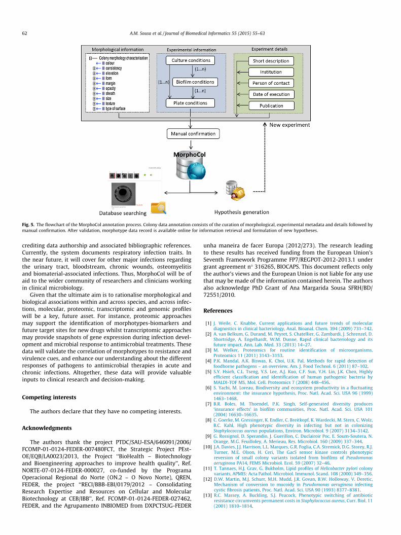
Fig. 5. The flowchart of the MorphoCol annotation process. Colony data annotation consists of the curation of morphological, experimental metadata and details followed bymanual confirmation. After validation, morphotype data record is available online for information retrieval and formulation of new hypotheses.
62 A.M. Sousa et al. / Journal of Biomedical Informatics 55 (2015) 55–63
crediting data authorship and associated bibliographic references.Currently, the system documents respiratory infection traits. Inthe near future, it will cover for other major infections regardingthe urinary tract, bloodstream, chronic wounds, osteomyelitisand biomaterial-associated infections. Thus, MorphoCol will be ofaid to the wider community of researchers and clinicians workingin clinical microbiology.
Given that the ultimate aim is to rationalise morphological andbiological associations within and across species, and across infec-tions, molecular, proteomic, transcriptomic and genomic profileswill be a key, future asset. For instance, proteomic approachesmay support the identification of morphotypes-biomarkers andfuture target sites for new drugs whilst transcriptomic approachesmay provide snapshots of gene expression during infection devel-opment and microbial response to antimicrobial treatments. Thesedata will validate the correlation of morphotypes to resistance andvirulence cues, and enhance our understanding about the differentresponses of pathogens to antimicrobial therapies in acute andchronic infections. Altogether, these data will provide valuableinputs to clinical research and decision-making.
Competing interests
The authors declare that they have no competing interests.
Acknowledgments
The authors thank the project PTDC/SAU-ESA/646091/2006/FCOMP-01-0124-FEDER-007480FCT, the Strategic Project PEst-OE/EQB/LA0023/2013, the Project ‘‘BioHealth – Biotechnologyand Bioengineering approaches to improve health quality’’, Ref.NORTE-07-0124-FEDER-000027, co-funded by the ProgramaOperacional Regional do Norte (ON.2 – O Novo Norte), QREN,FEDER, the project ‘‘RECI/BBB-EBI/0179/2012 – ConsolidatingResearch Expertise and Resources on Cellular and MolecularBiotechnology at CEB/IBB’’, Ref. FCOMP-01-0124-FEDER-027462,FEDER, and the Agrupamento INBIOMED from DXPCTSUG-FEDER
unha maneira de facer Europa (2012/273). The research leadingto these results has received funding from the European Union’sSeventh Framework Programme FP7/REGPOT-2012-2013.1 undergrant agreement n� 316265, BIOCAPS. This document reflects onlythe author’s views and the European Union is not liable for any usethat may be made of the information contained herein. The authorsalso acknowledge PhD Grant of Ana Margarida Sousa SFRH/BD/72551/2010.
References
[1] J. Weile, C. Knabbe, Current applications and future trends of moleculardiagnostics in clinical bacteriology, Anal. Bioanal. Chem. 394 (2009) 731–742.
[2] A. van Belkum, G. Durand, M. Peyret, S. Chatellier, G. Zambardi, J. Schrenzel, D.Shortridge, A. Engelhardt, W.M. Dunne, Rapid clinical bacteriology and itsfuture impact, Ann. Lab. Med. 33 (2013) 14–27.
[3] M. Welker, Proteomics for routine identification of microorganisms,Proteomics 11 (2011) 3143–3153.
[4] P.K. Mandal, A.K. Biswas, K. Choi, U.K. Pal, Methods for rapid detection offoodborne pathogens – an overview, Am. J. Food Technol. 6 (2011) 87–102.
[5] S.Y. Hsieh, C.L. Tseng, Y.S. Lee, A.J. Kuo, C.F. Sun, Y.H. Lin, J.K. Chen, Highlyefficient classification and identification of human pathogenic bacteria byMALDI-TOF MS, Mol. Cell. Proteomics 7 (2008) 448–456.
[6] S. Yachi, M. Loreau, Biodiversity and ecosystem productivity in a fluctuatingenvironment: the insurance hypothesis, Proc. Natl. Acad. Sci. USA 96 (1999)1463–1468.
[7] B.R. Boles, M. Thoendel, P.K. Singh, Self-generated diversity produces‘insurance effects’ in biofilm communities, Proc. Natl. Acad. Sci. USA 101(2004) 16630–16635.
[8] C. Goerke, M. Gressinger, K. Endler, C. Breitkopf, K. Wardecki, M. Stern, C. Wolz,B.C. Kahl, High phenotypic diversity in infecting but not in colonizingStaphylococcus aureus populations, Environ. Microbiol. 9 (2007) 3134–3142.
[9] G. Rossignol, D. Sperandio, J. Guerillon, C. Duclairoir Poc, E. Soum-Soutera, N.Orange, M.G. Feuilloley, A. Merieau, Res. Microbiol. 160 (2009) 337–344.
[10] J.A. Davies, J.J. Harrison, L.L. Marques, G.R. Foglia, C.A. Stremick, D.G. Storey, R.J.Turner, M.E. Olson, H. Ceri, The GacS sensor kinase controls phenotypicreversion of small colony variants isolated from biofilms of Pseudomonasaeruginosa PA14, FEMS Microbiol. Ecol. 59 (2007) 32–46.
[11] T. Tannaes, H.J. Grav, G. Bukholm, Lipid profiles of Helicobacter pylori colonyvariants, APMIS: Acta Pathol. Microbiol. Immunol. Scand. 108 (2000) 349–356.
[12] D.W. Martin, M.J. Schurr, M.H. Mudd, J.R. Govan, B.W. Holloway, V. Deretic,Mechanism of conversion to mucoidy in Pseudomonas aeruginosa infectingcystic fibrosis patients, Proc. Natl. Acad. Sci. USA 90 (1993) 8377–8381.
[13] R.C. Massey, A. Buckling, S.J. Peacock, Phenotypic switching of antibioticresistance circumvents permanent costs in Staphylococcus aureus, Curr. Biol. 11(2001) 1810–1814.

A.M. Sousa et al. / Journal of Biomedical Informatics 55 (2015) 55–63 63
[14] K. Lewis, Persister cells and the riddle of biofilm survival, Biochemistry-Moscow 70 (2005) 267–274.
[15] A.M. Sousa, I. Machado, M.O. Pereira, Phenotypic switching: an opportunity tobacteria thrive, in: A. Mendez-Vilas (Ed.), Science against Microbial Pathogens:Communicating Current Research and Technological Advances, FormatexResearch Center, Spain, 2011.
[16] G. Agarwal, A. Kapil, S.K. Kabra, B.K. Das, S.N. Dwivedi, Characterization ofPseudomonas aeruginosa isolated from chronically infected children with cysticfibrosis in India, BMC Microbiol. 5 (2005) 43.
[17] G. Manno, M. Cruciani, L. Romano, S. Scapolan, M. Mentasti, R. Lorini, L.Minicucci, Antimicrobial use and Pseudomonas aeruginosa susceptibilityprofile in a cystic fibrosis centre, Int. J. Antimicrob. Agents 25 (2005)193–197.
[18] N. Wellinghausen, I. Chatterjee, A. Berger, A. Niederfuehr, R.A. Proctor, B.C.Kahl, Characterization of clinical Enterococcus faecalis small-colony variants, J.Clin. Microbiol. 47 (2009) 2802–2811.
[19] R.A. Proctor, C. von Eiff, B.C. Kahl, K. Becker, P. McNamara, M. Herrmann, G.Peters, Small colony variants: a pathogenic form of bacteria that facilitatespersistent and recurrent infections, Nat. Rev. Microbiol. 4 (2006) 295–305.
[20] Q. Wei, S. Tarighi, A. Dotsch, S. Haussler, M. Musken, V.J. Wright, M. Camara, P.Williams, S. Haenen, B. Boerjan, et al., Phenotypic and genome-wide analysis ofan antibiotic-resistant small colony variant (SCV) of Pseudomonas aeruginosa,PLoS ONE 6 (2011) e29276.
[21] A.M. Sousa, I. Machado, A. Nicolau, M.O. Pereira, Improvements on colonymorphology identification towards bacterial profiling, J. Microbiol. Methods 95(2013) 327–335.
[22] C.L. Smith, C.A. Goldsmith, J.T. Eppig, The mammalian phenotype ontology as atool for annotating, analyzing and comparing phenotypic information,Genome Biol. 6 (2005) R7.
[23] G. Schindelman, J.S. Fernandes, C.A. Bastiani, K. Yook, P.W. Sternberg, Wormphenotype ontology: integrating phenotype data within and beyond the C.elegans community, BMC Bioinformatics 12 (2011) 32.
[24] Y. Yamazaki, P. Jaiswal, Biological ontologies in rice databases. An introductionto the activities in Gramene and Oryzabase, Plant Cell Physiol. 46 (2005) 63–68.
[25] P.N. Robinson, S. Kohler, S. Bauer, D. Seelow, D. Horn, S. Mundlos, The humanphenotype ontology: a tool for annotating and analyzing human hereditarydisease, Am. J. Hum. Genet. 83 (2008) 610–615.
[26] G.V. Gkoutos, E.C. Green, A.M. Mallon, A. Blake, S. Greenaway, J.M. Hancock, D.Davidson, Ontologies for the description of mouse phenotypes, Comp. Funct.Genomics 5 (2004) 545–551.
[27] B. Smith, M. Ashburner, C. Rosse, J. Bard, W. Bug, W. Ceusters, L.J. Goldberg, K.Eilbeck, A. Ireland, C.J. Mungall, et al., The OBO foundry: coordinated evolutionof ontologies to support biomedical data integration, Nat. Biotechnol. 25(2007) 1251–1255.
[28] J. Day-Richter, M.A. Harris, M. Haendel, S. Lewis, OBO-Edit – an ontology editorfor biologists, Bioinformatics 23 (2007) 2198–2200.
[29] E. Ben-Jacob, I. Cohen, D.L. Gutnick, Cooperative organization of bacterialcolonies: from genotype to morphotype, Annu. Rev. Microbiol. 52 (1998) 779–806.
[30] C.H. Zierdt, P.J. Schmidt, Dissociation in Pseudomonas aeruginosa, J. Bacteriol.87 (1964) 1003–1010.
[31] R.C. Clowes, D. Rowley, Genetic studies on small-colony variants of Escherichiacoli K-12, J. Gen. Microbiol. 13 (1955) 461–473.
[32] N. Chantratita, V. Wuthiekanun, K. Boonbumrung, R. Tiyawisutsri, M.Vesaratchavest, D. Limmathurotsakul, W. Chierakul, S. Wongratanacheewin,S. Pukritiyakamee, N.J. White, et al., Biological relevance of colony morphologyand phenotypic switching by Burkholderia pseudomallei, J. Bacteriol. 189 (2007)807–817.
[33] M. Allegrucci, K. Sauer, Characterization of colony morphology variantsisolated from Streptococcus pneumoniae biofilms, J. Bacteriol. 189 (2007)2030–2038.
[34] H.M. Kunttu, L.R. Suomalainen, E.I. Jokinen, E.T. Valtonen, Flavobacteriumcolumnare colony types: connection to adhesion and virulence?, MicrobPathog. 46 (2009) 21–27.
[35] D. Neut, J.G.E. Hendriks, J.R. van Horn, H.C. van der Mei, H.J. Busscher,Pseudomonas aeruginosa biofilm formation and slime excretion on antibiotic-loaded bone cement, Acta Orthop. 76 (2005) 109–114.
[36] R. Gupta, M. Schuster, Quorum sensing modulates colony morphology throughalkyl quinolones in Pseudomonas aeruginosa, BMC Microbiol. 12 (2012) 30.
[37] M. Starkey, J.H. Hickman, L. Ma, N. Zhang, S. De Long, A. Hinz, S. Palacios, C.Manoil, M.J. Kirisits, T.D. Starner, et al., Pseudomonas aeruginosa rugose small-colony variants have adaptations that likely promote persistence in the cysticfibrosis lung, J. Bacteriol. 191 (2009) 3492–3503.
[38] M.J. Kirisits, L. Prost, M. Starkey, M.R. Parsek, Characterization of colonymorphology variants isolated from Pseudomonas aeruginosa biofilms, Appl.Environ. Microbiol. 71 (2005) 4809–4821.
[39] Y.S. Chen, H.H. Lin, C.C. Hung, J.J. Mu, Y.S. Hsiao, Y.L. Chen, Phenotypiccharacteristics and pathogenic ability across distinct morphotypes ofBurkholderia pseudomallei DT, Microbiol. Immunol. 53 (2009) 184–189.
[40] L. Friedman, R. Kolter, Genes involved in matrix formation in Pseudomonasaeruginosa PA14 biofilms, Mol. Microbiol. 51 (2004) 675–690.
[41] R.B. Troxler, W.C. Hoover, L.J. Britton, A.M. Gerwin, S.M. Rowe, Clearance ofinitial mucoid Pseudomonas aeruginosa in patients with cystic fibrosis, Pediatr.Pulmonol. 47 (2012) 1113–1122.
[42] O. Ciofu, L.F. Mandsberg, H. Wang, N. Hoiby, Phenotypes selected duringchronic lung infection in cystic fibrosis patients: implications for thetreatment of Pseudomonas aeruginosa biofilm infections, FEMS Immunol.Med. Microbiol. 65 (2012) 215–225.
[43] P. Greally, P. Whitaker, D. Peckham, Challenges with current inhaledtreatments for chronic Pseudomonas aeruginosa infection in patients withcystic fibrosis, Curr. Med. Res. Opin. 28 (2012) 1059–1067.
[44] I.N. Silva, A.S. Ferreira, J.D. Becker, J.E. Zlosnik, D.P. Speert, J. He, D. Mil-Homens, L.M. Moreira, Mucoid morphotype variation of Burkholderiamultivorans during chronic cystic fibrosis lung infection is correlated withchanges in metabolism, motility, biofilm formation and virulence,Microbiology 157 (2011) 3124–3137.
[45] I.D. Hay, U. Remminghorst, B.H. Rehm, MucR, a novel membrane-associatedregulator of alginate biosynthesis in Pseudomonas aeruginosa, Appl. Environ.Microbiol. 75 (2009) 1110–1120.
[46] G.A. Uhlich, P.H. Cooke, E.B. Solomon, Analyses of the red-dry-roughphenotype of an Escherichia coli O157:H7 strain and its role in biofilmformation and resistance to antibacterial agents, Appl. Environ. Microbiol. 72(2006) 2564–2572.
[47] L.A. Onyango, R.H. Dunstan, J. Gottfries, C. von Eiff, T.K. Roberts, Effect of lowtemperature on growth and ultra-structure of Staphylococcus spp, PLoS ONE 7(2012) e29031.
[48] L. Tuchscherr, E. Medina, M. Hussain, W. Volker, V. Heitmann, S. Niemann, D.Holzinger, J. Roth, R.A. Proctor, K. Becker, et al., Staphylococcus aureusphenotype switching: an effective bacterial strategy to escape host immuneresponse and establish a chronic infection, EMBO Mol. Med. 3 (2011) 129–141.
[49] C. von Eiff, Staphylococcus aureus small colony variants: a challenge tomicrobiologists and clinicians, Int. J. Antimicrob. Agents 31 (2008) 507–510.
[50] S. Haussler, B. Tummler, H. Weissbrodt, M. Rohde, I. Steinmetz, Small-colonyvariants of Pseudomonas aeruginosa in cystic fibrosis, Clin. Infect. Dis.: Off. Publ.Infect. Dis. Soc. Am. 29 (1999) 621–625.
[51] S.M. Hartig, Basic image analysis and manipulation in ImageJ, in: Curr. Protoc.Mol. Biol., John Wiley & Sons, Inc., 2001.
[52] E. Mowat, S. Paterson, J.L. Fothergill, E.A. Wright, M.J. Ledson, M.J. Walshaw,M.A. Brockhurst, C. Winstanley, Pseudomonas aeruginosa population diversityand turnover in cystic fibrosis chronic infections, Am. J. Respir. Crit. Care Med.183 (2011) 1674–1679.
[53] J.L. Fothergill, E. Mowat, M.J. Ledson, M.J. Walshaw, C. Winstanley, Fluctuationsin phenotypes and genotypes within populations of Pseudomonas aeruginosa inthe cystic fibrosis lung during pulmonary exacerbations, J. Med. Microbiol. 59(2010) 472–481.
[54] A.R. Hauser, M. Jain, M. Bar-Meir, S.A. McColley, Clinical significance ofmicrobial infection and adaptation in cystic fibrosis, Clin. Microbiol. Rev. 24(2011) 29–70.
[55] S. Yagci, G. Hascelik, D. Dogru, U. Ozcelik, B. Sener, Prevalence and geneticdiversity of Staphylococcus aureus small-colony variants in cystic fibrosispatients, Clin. Microbiol. Infect.: Off. Publ. Eur. Soc. Clin. Microbiol. Infect. Dis.19 (2013) 77–84.
[56] S. Subramoni, D.T. Nguyen, P.A. Sokol, Burkholderia cenocepacia ShvR-regulatedgenes that influence colony morphology, biofilm formation, and virulence,Infect. Immun. 79 (2011) 2984–2997.