Embed Size (px)
Citation preview

Journal of American Science 2019;15(2) http://www.jofamericanscience.org
31
Measurement of Tracheobronchial Tree Dimensions in Egyptian population and itsCorrelation withSex, Age,
Weight and Height (Computed Tomographic Study)
Amal Mahmoud Elshazly¹, Islam Mahmoud Elshazly²,Asmaaa Y. A. Hussein³ and Ahmed Abdelhamed Hassan⁴
1DepartmentofAnatomy and Embryology, Faculty of Medicine, BenhaUniversity, Egypt;
2Department of DiagnosticRadiology,Faculty of Medicine, Benha University, Egypt;
3Department of Forensic and Toxicology, Faculty of medicine, BenhaUniversity, Egypt;
4Department of anesthesia, Faculty of Medicine, Benha University, Egypt
[email protected]; [email protected];
[email protected]@yahoo.com
Abstract:Measuring measurements of an airway is verysignificant for interventional bronchoscopists, investigators
and clinicians in order to precisely diagnose anatomical anomaliesand detect the pathological alteration along a time
or in reaction to therapies.Treating and exploration of images, thoughtful of topicsconnected to the achievement, and
for any extent liketopics effect on imaging the tracheobronchial tree, is importantforevaluateddimensions precision
and to createactualapplication of advanced techniques. The present workadds to this understandingby providing
accurate measurements of the normative parameters of the tracheobronchial trees in the Egyptianresidentsapplying
multi-planar reconstruction (MPR)andmulti-slice spiral computed tomography (CT).Six hundreds of persons submit
to the Benha University Hospital for performing thoracic CT scans including the dimensions of tracheal and
bronchial tree.Tracheallengths(LT)and its transvers diameter (TrTD) and anteroposterior diameter (APTD), the
lengths of main stem bronchi as right bronchial length (RBL), left bronchial length (LBL), and the sizes of the right
bronchial angle (RBA), left bronchial angle (LBA) were gotten via MPR of CT imaging. Multi-variance analyses
were done to identifypossible associations amongacquired measurements.The anteroposterior tracheal diameter
(APTD) was 20.13 ± 1.61 mm (22.46 ± 1.81mm for male and 17.81 ± 1.12mm for female).The transvers tracheal
diameter(TrTD)was 18.64 ± 1.40 mm (20.92± 1.36 for male and 16.36 ± 1.18 mm for female).The length of the
trachea(TL) was 125.88 ± 2.33mm (130.31 ± 2.22 mm for male and 121.46 ± 2.41mm for female). The mean
lengths of left main stem bronchus (LBL) andthe right main stem bronchus (RBL)were 48.75 ± 1.88 and32.39 ±
1.11 mm respectively. The right bronchus angle and the left bronchus anglewere 35.68 ± 2.11 and 47.77 ± 1.55
degrees, respectively. The differences regarding the gender were significant in all the dimensions measured, there
were significant increasein all parameters with increasing height except for the right and left bronchial angels there
were no significant difference with increasing height,and there were no significant change in alltracheobronchial
parameters with increasing weight and age.
[Amal Mahmoud Elshazly, Islam Mahmoud Elshazly,Asmaaa Y. A. Hussein and Ahmed Abdelhamed Hassan] . Measurement of Tracheobronchial Tree Dimensions in Egyptian population and its Correlation with Sex,
Age, Weight and Height (Computed Tomographic Study).J Am Sci2019;15(2):31-42]. ISSN 1545-1003 (print);
ISSN 2375-7264 (online). http://www.jofamericanscience.org. 5. doi:10.7537/marsjas150219.05.
Keywords: Measurement; Tracheobronchial; Tree; Dimension; Egyptian population; Correlation; Sex; Age; Weight;
Height; Tomographic
1. Introduction
The customary standardvalue and range of
anthropometric differences varied between numerous
races, and also between various ethnic groups inside
the identical race [1], e.g, Caucasian individuals or
Negroid persons have a tendency toshowlarger
physical sizesthan Mongolian persons. For that reason,
the measurements of the tracheobronchial tree may
similarly varyfrom race to race [2].
The facility to determine airway measurements is
significant for interventional bronchoscopists and
investigators, in addition toclinicians, for the purpose
of precisely count structuralanomalies and recording
the alterations which occur along time or occur in as a
result of therapies.Furthermost quantitative airway
dimensions are depended on X-ray computed
tomography and recently, on multidetector computed
tomography [3].
Many studies since the 1950s, have concentrated
on the anatomical structures of the tracheobronchial
tree[4-9]. Anatomically, the trachea is situated at the
height of the 6th
cervical vertebra and bifurcates at the
level of the 4–5th
thoracic vertebra into the rightandleft
main bronchus (RMB&LMB). Advanced investigation
reported that during examining chest computed
tomography (CT) of Asian peoples, the angle of the
the LMB is 43°, where the angle of RMB is 35°. On
the other hand, many factors may be influencing the
variations in the tracheobronchial angles such as race,

Journal of American Science 2019;15(2) http://www.jofamericanscience.org
32
age, and subjects, and the methods used for estimation
of measurement of angle [10].
With regard to the studies which carried out in
the Western countries on the measurements of the
tracheobronchial tree, there are several studies which
dealing with this issue[3-11], and the collected data
from these researches constitute themodernsource of
the tracheobronchial tree measurements in a lot of
textbooks.
Generally, the data concerning the fine
information about the anatomical structure of the
trachea and its branches is essential in the pure
anthropometry area, in addition to in several other
fields. The precise information regarding the
anatomical structure of tracheobronchial tree, in spite
of its importance in the physiology of lung and
chestoperations, it is also essential in the field of
anesthesiology. Determining the tracheobronchial tree
parameters, for instancediameters, angulations, and the
lengths, assist in the improving the surgical
stepslikeinsertion of tube,restoration of the airway
tree, and optimizing the medical instruments, for
instance double-lumen endobronchial tube. A further
clinical suggestion of this investigation is to direct the
airway supervision for bronchus or tracheal
cancerremovalalong the jet ventilation of
interventional fiberoptic operations. It is
commonlyimportant at the moment to confirm there is
sufficient ratio of jet catheter to the diameter of
bronchus or trachea, therefore,sufficientventilation and
oxygenation is sustained, while the hazard of
barotrauma is less [12-13].
In addition, accurate information about a
correlation, or lack thereof, between these dimensions
may decrease the require for expensive, needless, or
invasive diagnostic techniques by supporting the
surgeon with formerdeterminations of
applicabledimensions. For instance,gaining
preciselydimension of the tracheobronchial tree in an
individual can reorganizepossibletroubles in airway
handling and optimize patient care [10].
Because of the mentioned profit, in the present
study, we aimed to do a large scale investigation
demonstrating the anatomical structures of the
tracheobronchial tree in an Egyptian people by
applying multiplaner reconstruction(MPR) and multi-
slice spiral CT. Also, we analyzed
probablerelationships of measurementsusuallyapplied
in illustrating the tracheobronchial tree. The current
worksupplies with a applicableassessmentdimension
of the tracheobronchial tree in an Egyptian peoples
and should offerhelpfuldirection for applicable clinical
practice and medical devices, particularlythe
manufacture of the double-lumen tube.
2. Subject and Methods
The selection of cases was subjected to certain
criteria:
From May 1st, 2013 to January 1st, 2017,
800persons were involved in this study from the
Benha University Hospital, which provides free
medical service to Egyptian population. These persons
underwent CT scans in the hospital, after that their
ages, heights, and weights were recorded under
supervision. 200 patients were excluded from this
study for at least one of the following exclusion
criteria: (1) Non-Egyptian (2) Younger than 16 or
older than 90 (3) Prior diagnosis of compulsive
position, musculoskeletal deformity(4) Presence of
hearing impairment severe enough to preclude
cooperation; and (5) A history of tracheobronchial
surgery. The remaining 600persons continued the
study. Prior to participation, all patients were fully
informed the study and provided their informed,
written consent.
Groups:
For the purpose of studying the changes occur in
tracheobronchial dimensions we further dividing the
whole number of subject (600 subject) into groups
(sexgroups, height groups, weight groups and age
groups)tocompare these dimensionsin thesegroups:
1- Males and females groups (300 subject each)
2- Height groups: we divide the 600 subject into
4 height groups
A- Group1: subject their height varying
from155-165 cm (145 subjects).
B- Group2: subject their height varying from
166-175 cm (165 subjects).
C- Group3: subject their height varying from
176-185 cm (133 subjects).
D- Group4: subject their height varying from
186-195 cm (153 subjects).
3- weightgroups:we divide the 600subject into 6
weightgroups
A- Group1: subject their weight varying from65-
75 kg (95 subjects).
B- Group2: subject their weightvarying from76-
85 kg (106 subjects).
C- Group3: subject their weightvarying from86
– 95 kg (99 subjects).
D- Group4: subject their weight varying from96
– 105 kg (85 subjects).
E- Group5: subject their weight varying from
106 – 115 kg (115 subjects).
F- Group6: subject their weight varying from
116 – 125 kg (100 subjects).
4- Agegroups::we divide the 600subject into 6
weightgroups
A- Group1: subject their age varying from 16-
25 years (65 subjects).

Journal of American Science 2019;15(2) http://www.jofamericanscience.org
33
B- Group2: subject their age varying from26-
35 years (136 subjects).
C- Group3: subject their age varying from 36-
45 years (89 subjects).
D- Group4: subject their age varying from 46-
55 years (105 subjects).
E- Group5: subject their age varying from 56-
65 years (120 subjects).
F- Group6: subject their age varying from 66-
75 years (75 subjects).
Methods:
● The CT apparatus (Scanning unit):
The CT apparatus used in this study was:
Toshiba spiral CT-scan Auklet
System TSX-003A
S#A9582405
Patient couch CBTB-013AA9582410
Console CKCN -007AA9582409
Tube CXB-200BMHU
Gantry slice492074
Tube slice only 10000
Input 24 kilowatt
It was the CT unit of Radiology Department in
Benha University Hospital. The CT scanning unit is
formed of a table, scanning gantry (which includes an
x-ray tube and a detector array), an x-ray generator,
computer, monitor, printer and a viewing consoles.
All the scanned films were viewed and
reexamined carefully by an experienced radiologist to
insure that they are carefully chosen from a healthy
low risk volunteers.
Preparing the Subjects for CT Scanning of the
Chest:
All persons were trained to hold deep breath for
at least 10s before the thorax CT scans during
suspended end inspiration at total lung capacity. The
arms were fully extended above the head. The
acquisition time was selected based on the distance
from the vocal cord to the diaphragm. The scanned CT
images were uploaded to the local area network server
of the Hospital and stored in DICOM format.The
findings were retrospectively reviewed and the
diameters were measured with using ONIS 2.5
software program.
The parameters in use (figure 1):
Figure 1: A CT chest images (axial and multiplanerreconstruction): (A) TrTD (transvers tracheal diameter
sternoclavicular level), (B) APTD (antroposterior tracheal diameter steronoclavicular level), (C) TL (tracheal length)
starting from cricoid cartlage to the carena), (D) RBL (Right main bronchus length), (E) LBL (left main bronchial
length), (F) RBA (right bronchial angle), (G) LBA (left bronchial angle).
Journal of American Science 2019;15(2) http://www.jofamericanscience.org
34
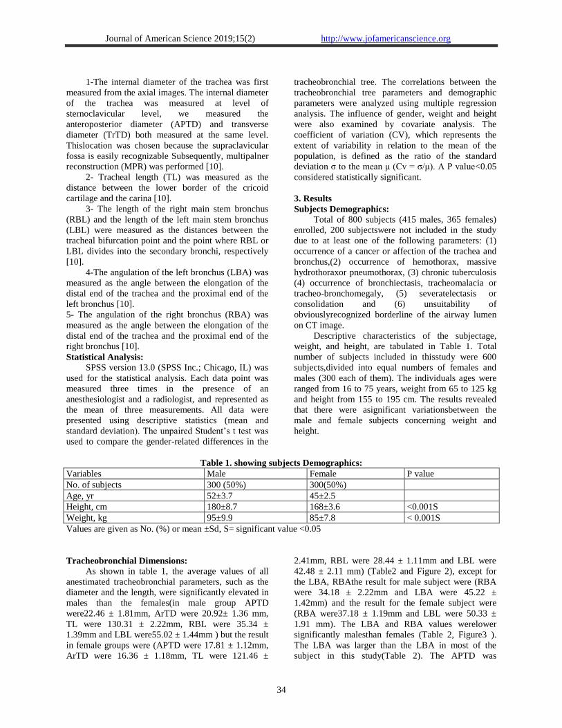
1-The internal diameter of the trachea was first
measured from the axial images. The internal diameter
of the trachea was measured at level of
sternoclavicular level, we measured the
anteroposterior diameter (APTD) and transverse
diameter (TrTD) both measured at the same level.
Thislocation was chosen because the supraclavicular
fossa is easily recognizable Subsequently, multipalner
reconstruction (MPR) was performed [10].
2- Tracheal length (TL) was measured as the
distance between the lower border of the cricoid
cartilage and the carina [10].
3- The length of the right main stem bronchus
(RBL) and the length of the left main stem bronchus
(LBL) were measured as the distances between the
tracheal bifurcation point and the point where RBL or
LBL divides into the secondary bronchi, respectively
[10].
4-The angulation of the left bronchus (LBA) was
measured as the angle between the elongation of the
distal end of the trachea and the proximal end of the
left bronchus [10].
5- The angulation of the right bronchus (RBA) was
measured as the angle between the elongation of the
distal end of the trachea and the proximal end of the
right bronchus [10].
Statistical Analysis:
SPSS version 13.0 (SPSS Inc.; Chicago, IL) was
used for the statistical analysis. Each data point was
measured three times in the presence of an
anesthesiologist and a radiologist, and represented as
the mean of three measurements. All data were
presented using descriptive statistics (mean and
standard deviation). The unpaired Student’s t test was
used to compare the gender-related differences in the
tracheobronchial tree. The correlations between the
tracheobronchial tree parameters and demographic
parameters were analyzed using multiple regression
analysis. The influence of gender, weight and height
were also examined by covariate analysis. The
coefficient of variation (CV), which represents the
extent of variability in relation to the mean of the
population, is defined as the ratio of the standard
deviation σ to the mean μ (Cv = σ/μ). A P value<0.05
considered statistically significant.
3. Results
Subjects Demographics:
Total of 800 subjects (415 males, 365 females)
enrolled, 200 subjectswere not included in the study
due to at least one of the following parameters: (1)
occurrence of a cancer or affection of the trachea and
bronchus,(2) occurrence of hemothorax, massive
hydrothoraxor pneumothorax, (3) chronic tuberculosis
(4) occurrence of bronchiectasis, tracheomalacia or
tracheo-bronchomegaly, (5) severatelectasis or
consolidation and (6) unsuitability of
obviouslyrecognized borderline of the airway lumen
on CT image.
Descriptive characteristics of the subjectage,
weight, and height, are tabulated in Table 1. Total
number of subjects included in thisstudy were 600
subjects,divided into equal numbers of females and
males (300 each of them). The individuals ages were
ranged from 16 to 75 years, weight from 65 to 125 kg
and height from 155 to 195 cm. The results revealed
that there were asignificant variationsbetween the
male and female subjects concerning weight and
height.
Table 1. showing subjects Demographics:
Variables Male Female P value
No. of subjects 300 (50%) 300(50%)
Age, yr 52±3.7 45±2.5
Height, cm 180±8.7 168±3.6 <0.001S
Weight, kg 95±9.9 85±7.8 < 0.001S
Values are given as No. (%) or mean ±Sd, S= significant value <0.05
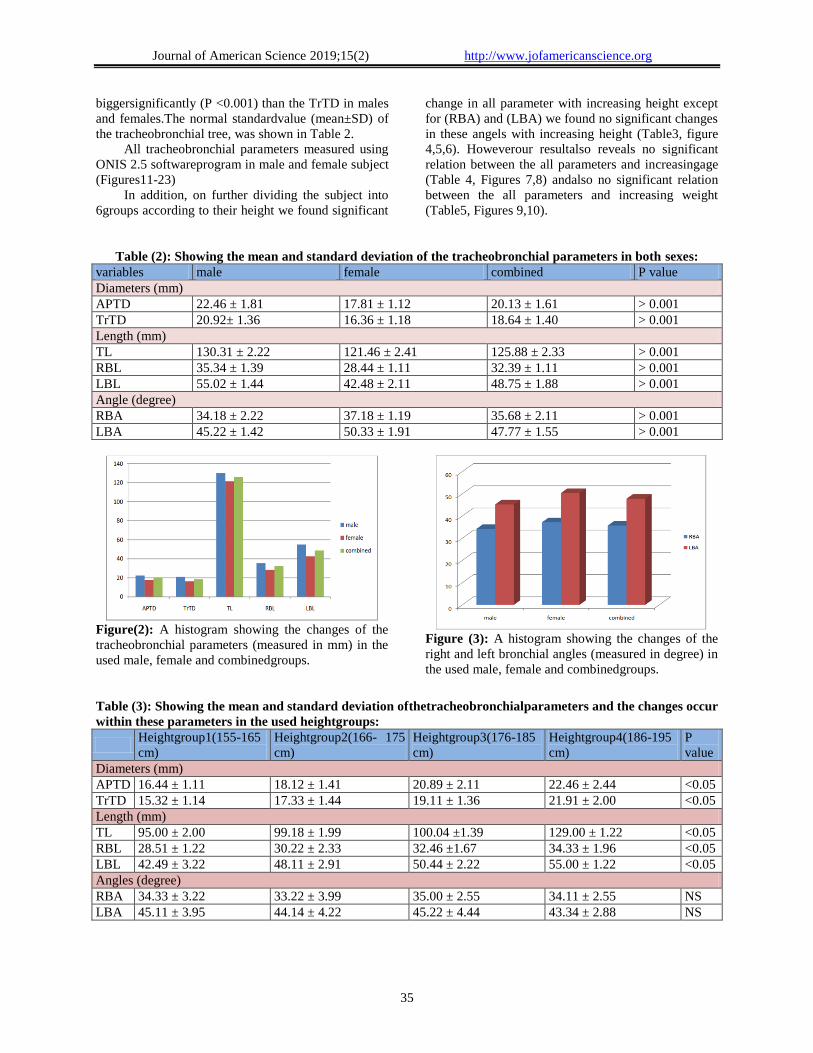
Tracheobronchial Dimensions:
As shown in table 1, the average values of all
anestimated tracheobronchial parameters, such as the
diameter and the length, were significantly elevated in
males than the females(in male group APTD
were22.46 ± 1.81mm, ArTD were 20.92± 1.36 mm,
TL were 130.31 ± 2.22mm, RBL were 35.34 ±
1.39mm and LBL were55.02 ± 1.44mm ) but the result
in female groups were (APTD were 17.81 ± 1.12mm,
ArTD were 16.36 ± 1.18mm, TL were 121.46 ±
2.41mm, RBL were 28.44 ± 1.11mm and LBL were
42.48 ± 2.11 mm) (Table2 and Figure 2), except for
the LBA, RBAthe result for male subject were (RBA
were 34.18 ± 2.22mm and LBA were 45.22 ±
1.42mm) and the result for the female subject were
(RBA were37.18 ± 1.19mm and LBL were 50.33 ±
1.91 mm). The LBA and RBA values werelower
significantly malesthan females (Table 2, Figure3 ).
The LBA was larger than the LBA in most of the
subject in this study(Table 2). The APTD was
Journal of American Science 2019;15(2) http://www.jofamericanscience.org
35
biggersignificantly (P <0.001) than the TrTD in males
and females.The normal standardvalue (mean±SD) of
the tracheobronchial tree, was shown in Table 2.
All tracheobronchial parameters measured using
ONIS 2.5 softwareprogram in male and female subject
(Figures11-23)
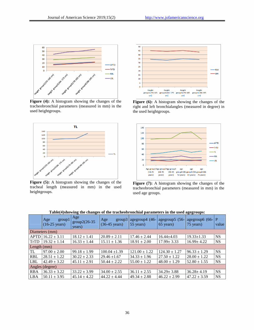
In addition, on further dividing the subject into
6groups according to their height we found significant
change in all parameter with increasing height except
for (RBA) and (LBA) we found no significant changes
in these angels with increasing height (Table3, figure
4,5,6). Howeverour resultalso reveals no significant
relation between the all parameters and increasingage
(Table 4, Figures 7,8) andalso no significant relation
between the all parameters and increasing weight
(Table5, Figures 9,10).
Table (2): Showing the mean and standard deviation of the tracheobronchial parameters in both sexes:
variables male female combined P value
Diameters (mm)
APTD 22.46 ± 1.81 17.81 ± 1.12 20.13 ± 1.61 > 0.001
TrTD 20.92± 1.36 16.36 ± 1.18 18.64 ± 1.40 > 0.001
Length (mm)
TL 130.31 ± 2.22 121.46 ± 2.41 125.88 ± 2.33 > 0.001
RBL 35.34 ± 1.39 28.44 ± 1.11 32.39 ± 1.11 > 0.001
LBL 55.02 ± 1.44 42.48 ± 2.11 48.75 ± 1.88 > 0.001
Angle (degree)
RBA 34.18 ± 2.22 37.18 ± 1.19 35.68 ± 2.11 > 0.001
LBA 45.22 ± 1.42 50.33 ± 1.91 47.77 ± 1.55 > 0.001
Figure(2): A histogram showing the changes of the
tracheobronchial parameters (measured in mm) in the
used male, female and combinedgroups.
Figure (3): A histogram showing the changes of the
right and left bronchial angles (measured in degree) in
the used male, female and combinedgroups.
Table (3): Showing the mean and standard deviation ofthetracheobronchialparameters and the changes occur
within these parameters in the used heightgroups:
Heightgroup1(155-165
cm)
Heightgroup2(166- 175
cm)
Heightgroup3(176-185
cm)
Heightgroup4(186-195
cm)
P
value
Diameters (mm)
APTD 16.44 ± 1.11 18.12 ± 1.41 20.89 ± 2.11 22.46 ± 2.44 <0.05
TrTD 15.32 ± 1.14 17.33 ± 1.44 19.11 ± 1.36 21.91 ± 2.00 <0.05
Length (mm)
TL 95.00 ± 2.00 99.18 ± 1.99 100.04 ±1.39 129.00 ± 1.22 <0.05
RBL 28.51 ± 1.22 30.22 ± 2.33 32.46 ±1.67 34.33 ± 1.96 <0.05
LBL 42.49 ± 3.22 48.11 ± 2.91 50.44 ± 2.22 55.00 ± 1.22 <0.05
Angles (degree)
RBA 34.33 ± 3.22 33.22 ± 3.99 35.00 ± 2.55 34.11 ± 2.55 NS
LBA 45.11 ± 3.95 44.14 ± 4.22 45.22 ± 4.44 43.34 ± 2.88 NS
Journal of American Science 2019;15(2) http://www.jofamericanscience.org
36
Figure (4): A histogram showing the changes of the
tracheobronchial parameters (measured in mm) in the
used heightgroups.
Figure (5): A histogram showing the changes of the
tracheal length (measured in mm) in the used
heightgroups.
Figure (6): A histogram showing the changes of the
right and left bronchialangles (measured in degree) in
the used heightgroups.
Figure (7): A histogram showing the changes of the
tracheobronchial parameters (measured in mm) in the
used age groups.
Table(4)showing the changes of the tracheobronchial parameters in the used agegroups:
Age group1
(16-25 years)
Age
group2(26-35
years)
Age group3
(36-45 years)
agegroup4 (46-
55 years)
agegroup5 (56-
65 years)
agegroup6 (66-
75 years)
P
value
Diameters (mm)
APTD 16.22 ± 3.11 18.12 ± 1.41 20.89 ± 2.11 17.46 ± 2.44 16.44±4.03 19.33±1.33 NS
TrTD 19.32 ± 1.14 16.33 ± 1.44 15.11 ± 1.36 18.91 ± 2.00 17.99± 3.33 16.99± 4.22 NS
Length (mm)
TL 97.00 ± 2.00 99.18 ± 1.99 100.04 ±1.39 121.00 ± 1.22 124.30 ± 1.27 96.33 ± 1.29 NS
RBL 28.51 ± 1.22 30.22 ± 2.33 29.46 ±1.67 34.33 ± 1.96 27.50 ± 1.22 28.00 ± 1.22 NS
LBL 42.49 ± 3.22 45.11 ± 2.91 50.44 ± 2.22 55.00 ± 1.22 48.00 ± 1.29 52.80 ± 1.55 NS
Angles (degree)
RBA 36.33 ± 3.22 33.22 ± 3.99 34.00 ± 2.55 36.11 ± 2.55 34.29± 3.88 36.28± 4.19 NS
LBA 50.11 ± 3.95 45.14 ± 4.22 44.22 ± 4.44 49.34 ± 2.88 46.22 ± 2.99 47.22 ± 3.59 NS

Journal of American Science 2019;15(2) http://www.jofamericanscience.org
37
Figure (8): A histogram showing the changes of the
right and left bronchial angles (measured in degree) in
the used agegroups.
Figure (9): A histogram showing the changes of the
tracheobronchial parameters (measured in mm) in the
used weight groups.
Table (5) showing the changes of the tracheobronchial parameters (measured in mm) in the used
weightgroups.
Weight
group1 (65-
75 kg)
Weight
group2 (76-
85 kg)
Weight
group3 (86-
95 kg)
Weightgroup4
(96-105 kg)
Weightgroup5
(106-115 kg)
Weight group6
(116-125 kg)
P
value
Diameters (mm)
APTD 16.27± 3.61 18.15± 1.21 16.89 ± 2.71 19.66 ± 2.84 17.44±4.83 16.43±1.93 NS
TrTD 15.34± 1.64 19.39± 1.34 16.16± 1.37 17.97± 2.09 18.99± 3.83 15.90± 4.62 NS
Length (mm)
TL 99.08± 2.80 98.18 ± 1.29 106.04 ±1.99 120.00 ± 1.29 123.90 ± 1.29 97.33± 1.49 NS
RBL 28.31 ± 2.22 33.22 ± 1.33 34.46 ±2.67 33.43 ± 1.26 30.70 ± 1.82 32.90 ± 1.42 NS
LBL 48.49 ± 4.22 49.11 ± 391 50.44 ± 322 49.90± 5.22 54.00 ± 1.29 49.80 ± 5.55 NS
Angles (degree)
RBA 36.33 ± 3.22 35.22 ± 3.99 36.02± 2.55 34.11 ± 2.55 36.29± 3.88 33.28 ± 4.19 NS
LBA 45.11 ± 3.55 44.14 ± 4.72 44.22 ± 4.84 49.34 ± 2.48 46.26± 2.79 46.22 ± 3.49 NS
Figure (10): A histogram showing the changes of
theright and left bronchial angles (measured in degree)
in the used weightgroups.
Figure (11): An axial CT chest image at the
sternoclavicular level of a 70 years -old female
showing: anteroposterior tracheal diameter
(APTD)=16.81 mm

Journal of American Science 2019;15(2) http://www.jofamericanscience.org
38
Figure (12): An axial CT chest imageatthe
sternoclavicular level of a 70 years -old female
showing: transverse tracheal diameter (TrTD) =15.36
mm
Figure (13): An axial CT chest image at
thesternoclavicular level of a 49 years -old male
showing:anteroposterior tracheal diameter (APTD) =
21.46 mm
Figure (14): An axial CT chest imageat the
sternoclavicular level of a 49 years -old male showing:
transverse tracheal diameter (TrTD) =19.92 mm
Figure (15): A CT chest imageofmulti-planer
reconstruction (MPR) of a 70 years -old female
showing: left main bronchial length (LBL)= 42.48
mm.
Figure (16): A CT chest imageofmulti-planar
reconstruction (MPR) of a 16 years -old male
showing: left main bronchial length (LBL)= 55.34 mm
Figure (17): A CT chest imageofmulti-planar
reconstruction (MPR) of a 60 years -old female
showing: right main bronchial length (RBL)= 28.51
mm.
Journal of American Science 2019;15(2) http://www.jofamericanscience.org
39
Figure (18): A CT chest imageofmulti-planer
reconstruction (MPR) of a 26 years –old male
showing: right main bronchial length (RBL)= 34.33
mm
Figure (19): A CT chest imageofmulti-planer
reconstruction (MPR) of a 60 years -old female
showing: right main bronchial angle (RBA)= 36.19
degree.
Figure (19): A CT chest imageofmulti-planer
reconstruction (MPR) of a 26 years -old male
showing: right main bronchial angle (RBA)= 34.00
degree.
Figure (20): A CT chest imageofmulti-planer
reconstruction (MPR) of a 60 years -old female
showing: left main bronchial angle (LBA)= 45.00
degree.
Figure (21): A CT chest imageofmulti-planer
reconstruction (MPR) of a 16 years -old male
showing: left main bronchial angle (LBA)= 44.06
degree.
Figure (22): A CT chest imageofmulti-planer
reconstruction (MPR) of a 70 years –old male
showing: tracheal length(TL)= 129.58 mm.

Journal of American Science 2019;15(2) http://www.jofamericanscience.org
40
Figure (23): A CT chest imageofmulti-planer
reconstruction (MPR) of a 25 years –old female
showing: tracheal length(TL)= 121.77 mm.
4. Discussion
The results of current work revealed that the
demotionsof the measured trachea in anumber of
Egyptian population were Theantero-posteriortracheal
diameter (APTD)was 20.13 ± 1.61 mm (22.46 ±
1.81mm for male and 17.81 ± 1.12mm for female ).
The transvers tracheal diameter (TrTD)was 18.64 ±
1.40 mm (20.92± 1.36 for male and 16.36 ± 1.18 mm
for female ). The length of the trachea (TL) was
125.88 ± 2.33mm (130.31 ± 2.22 mm for male and
121.46 ± 2.41 mm for female). The average lengths of
the right main stem bronchus (RBL) and left main
stem bronchus (LBL) were 32.39 ± 1.11 mm and48.75
± 1.88 respectively. The (RBL of male subject were
35.34 ± 1.39 mm and of female subject were 37.18 ±
1.19mm) and (LBL of male subject were 55.02 ± 1.44
mm and of female subject were 42.48 ± 2.11 mm).
The right bronchus angle and the left bronchus angle
were averaged 35.68 ± 2.11 and 47.77 ± 1.55 degrees,
respectively. The (RBA of male subject were 34.18 ±
2.22mm and of female subject were 37.18 ± 1.19mm).
The (LBA of male subject were 45.11 ± 3.95 mm and
of female subject were 50.33 ± 1.91mm).Significant
differences were present among genders in all
estimated parameters,there were significant increasein
all parameters with increasing height exceptfor the
right and left bronchial angels there were no
significant difference with increasing height,and there
were no significant change in alltracheobronchial
parameters with increasing weight and age.
From the results above we found: (1) the male
persons have biggermeasures and length of the
tracheobronchial tree, whereas, the the female persons
have bigger main stem bronchial angles(2) there is a
strong positive relationshipamongthe height of body
and the average tracheal length.(3) TL, RBL, LBL,
APTD,TrTD, RBA and LBA have medical
applications and are more discussed underneath. All
tracheal parameters in the current study were larger
than those measuredfor Chinese populations [10]and
the measurementsreported by other tools as described
on Chinese textbooks [14,15], and also differ
fromother sources [16, 17,18] obtained from other
countries. These results prove the variation of
tracheobronchealdimensions between different ethnic
groups.
To investigate whether gender influences the
tracheal dimensions, we divide the subjects involved
in this study into two large sex group and we analyzed
the tracheobronchial parameters in these groups, a
univariante analysis associatinggender like covariates
was done. We reported that sex had a significant
impact in tracheobronchial dimension. The impact
may be moderatelyreturned to the sexvariation in
height becauseour resultsproposed that height had a
significant influence on these parameters. Some
previous studies [19, 20, 21] demonstrated a
significant relationshipsamong the tracheal length
(TL) and the height merely in youth in growth stage,
there are big individual differences of thelength
(TL)stillbetweenindividuals of the equivalent height.
Additionalresearches are required to clarify other
probable factors dealing with the gender variations of
the (TL).
The current work reported also, a diminish of the
tracheal length (TL) in persons their ages 66 years or
more. This may be attributed to the decrease of fibrous
tissue in elderly peoples. Additionally, the trachea is
much more vertical on lateral projection in youth than
in elderly peoples [22].
Regarding thelength of the Main Bronchi inthe
current study,found that the length of the right main
stem bronchi islargerthan the values recorded on
Chinese peoples in many textbooks [14, 15].Also our
findings on the length of the main stembronchialso
vary from the recorded data from different countries
[19, 20, 21] once morestress the meaning of estimating
anatomical dimensions inside individual ethnic
clusters. The large coefficient of variance
presentwithin the lengths of the right bronchi,in
addition to resultconcerning the frequency of
impression of the right upper lobe from the trachea
wasmore than formerlydocumented[23], they proposed
a high individual differences in the length ofthe right
main stem bronchus. The current work in addition, has
proposition on clinical applications. It has been
observed that Broncho-Cath double lumen tubes
(Mallinckrodt, Athlone, Ireland) create a danger to
personsas soon as the tube length go above the lengths
of the right bronchi by 1cm, where it can induce
trauma to the airway and may lead to rupture of the
cell membranes of the trachea [24]. According to the
safety scoperecorded by Benumof et al. [25], we
showedthat onlythe short persons (their height below
155 cm), who the lengths of their right bronchi are

Journal of American Science 2019;15(2) http://www.jofamericanscience.org
41
1cm shorter than the tubes were in risk.In case of the
patients were subjected for intubation at the right-
sided double-lumen endobronchial tube, the right
upper orifice may simply be blocked by the
endobronchial balloon.
So, the presentresultspowerfullyhold up three
clinical applyproposals: (1) the left sided double-
lumen tube should be appliedat any timeprobable; (2)
anesthesiologists should assessment and examine the
CT image of the thorax prior to intubation with the
purpose ofrecognize the airway tree morphology and
select a best-fit endobronchial tube; (3) Fiberoptic
bronchoscope is suggested to
validateappropriateposition of endobronchial tube.
Regarding the dimension of the trachea and main
stem bronchi the present results revealedthat the
APTD waslarger than (TrTD) is consistent with
previous publications [16,26].
As regarding the angles of the main bronchi with
the trachea our result were in agreement with those
reported previously [10,21,27]. We found that the
averageof right and left bronchial anglescalculated in
the female peoples was higher compared with that in
males, in opposing to a preceding cadaveric
surveillance[20]. This inconsistency may be clarified
by the finding that lungs develop more transversally
than downwards before the chest wall becomes rigid
in females, in addition to the diaphragmatic muscle is
stronger in males than in females. Moreover, a cadaver
differs from a living body, owing to the
comparativelocation of active and inactive diaphragm
asrecorded by Fearon et al. [28].In our study the angle
of the right main stem bronchus wassmaller thanthat
of the left bronchus these data were in agreement
withGrey’s Anatomy [29], who found that the angle of
the right main stem is supposed to be constantlylesser.
We can concluded from our study that we
recorded the normal dimension values of the
tracheobronchial tree for Egyptian citizens from this
work using CT. We supplementary analyzed the
correlations between the basic parameters defining the
tracheobronchial tree. Our data offer a basic
information that can be valuable for improving clinical
practice in the fieldofbronchotrachealintubation. The
current findings discovered also the distinctive aspects
of the anatomical structure of trachea in Egyptian
citizens.The present finding also pointed to a definite
parameters for instance RBL, APTD, LBL,
TL,andTrTDhave a thinallocation, whilefurther
parameters, likeRBA and LBA,display large
individual changeabilitybetween Egyptian citizen. in
spite of thestrong association within the length
oftrachea (TL) and subject height,no precise and
dependable equations were available for predicting the
complete tracheobronchial measurements by usingage,
height, and gender alone or in combination. Computed
tomography and bronchoscopy should still the
mainlyreliable tools for precisely determining the
airway proportions.
Acknowledgments:
The authors express deepest gratitude to Dr. Abd
El-Wanees Al-Awdan, Professor of Anatomy, Benha
Faculty of Medicine, for his constructive advice,
generous help, continuous encouragement and his
sincere caring and kindness which will always be
remembered.
References
1. GilesE,KlepingerLL.:
(Confidenceintervalsforestimatesbasedonlinearre
gressioninforensic,
anthropology).JForensicSci.1988;33(5):1218–
22.PMID:3193077.
2. J. P.Williamson,A. L.James.,M. J.Phillips, D.
D.Sampsone, D. R.Hillman,andP.
R.Eastwood:(Quantifyingtracheobronchialtreedi
mensions:methods,limitationsandemergingtechni
ques)Eur RespirJ2009;34:42–
55DOI:10.1183/09031936.00020408.
3. JessephJE,MerendinoKA.:
(Thedimensionalinterrelationshipsofthemajorcom
ponentsofthehumantracheobronchialtree). Surg
GynecolObstet.1957;105(2):210–214.
4. WailooMP,EmeryJL.:
(Normalgrowthanddevelopmentofthetrachea).
Thorax.1982;37(8):584–587.
5. GriscomNT.:
(Computedtomographicdeterminationoftrachealdi
mensionsinchildrenandadolescents).
Radiology.1982;145(2):361–364.
6. GamsuG,WebbWR.:
(Computedtomographyofthetracheaandmainstem
bronchi). Semin Roentgenol.1983;18(1):51–60.
7. BreatnachE,AbbottGC,FraserRG.:
(Dimensionsofthenormalhumantrachea).AJRAmJ
Roentgenol.1984;142(5):903–906.
8. ThurlbeckA,HorsfieldK.: (
Branchinganglesinthebronchialtreerelatedtoorder
ofbranching).RespirPhysiol.1980;41(2):173–181.
9. HorsfieldK,CummingG.:
(Anglesofbranchinganddiametersofbranchesinthe
humanbronchialtree).BullMathBiophys.1967;29(
2):245–259.
10. MiW,ZhangC,WangH,etal.: (
Measurementandanalysisofthetracheobronchialtr
eeinChinesepopulationusingcomputedtomograph
y).PLoSOne.2015;10(4): e0123177.
11. ZhevnovVN,BodnarchikLV.:
(Dimensionsofthelarynxandtracheainyoungchildr
en). Vestn Khir ImIIGrek.1969;103(9):116–
7.PMID:5354685.

Journal of American Science 2019;15(2) http://www.jofamericanscience.org
42
12. HautmannH,GamarraF,HenkeM,DiehmS,HuberR
M.:
(Highfrequencyjetventilationininterventionalfiber
opticbronchoscopy). Anesth
Analg.2000;90(6):1436–40.PMID:10825336.
13. JetVentilationH.W.In:S-RN,kAJ,editors.The5-
MinuteAnesthesiaConsult.Philadelphia:Lippincot
tWilliams&Wilkins;2013.p.500–1.
14. CaiBQLiLY.:(PUMCRespirology).2ed.Beijing:P
ekingUnionMedicalCollegePress;2011.p.3–5.
15. LiuZY.:(English-
ChineseTextbookofSystematicAnatomy).2ed.Bei
jing:SciencePress;2009.p.143–55.
16. GriscomNT,WohlME.:
(Dimensionsofthegrowingtrachearelatedtoageand
gender).AJRAmJRoentgenol.1986;146(2):233–
7.PMID:3484568.
17. EllisH,LawsonA.:
(Anatomyforanaesthetists).9ed.WestSussex:John
Wiley&Sons;2013.p.42–7.
18. GrilloHC.:
(SurgeryoftheTracheaandBronchi.Diseases,Diagn
osis,ResultsofTreatment).London:BCDeckerInc;
2004.p.43–51.
19. CroteauJR,CookCD.: (Volume-
pressureandlength-
tensionmeasurementsinhumantrachealandbronchi
alsegments). JApplPhysiol.1961;16:170–
2.PMID:13696635.
20. ChunderR,NandiS,GuhaR,SatyanarayanaN.:(Am
orphometricstudyofhumantracheaandprincipalbro
nchiindifferentagegroupsinbothsexesanditsclinica
limplications).NepalMedicalCollegejournal:NM
CJ.2010;12(4):207–14.PMID:21744760.
21. GriscomNT,WohlME.:(Dimensionsofthegrowing
trachearelatedtobodyheight.Length,anteroposteri
orandtransversediameters,cross-
sectionalarea,andvolumeinsubjectsyoungerthan2
0yearsofage).AmRevRespirDis.1985;131(6):840
–4.PMID:4003934.
22. GrilloHC.:
(SurgeryoftheTracheaandBronchi.Diseases,Diagn
osis,ResultsofTreatment).London:BCDeckerInc;
2004.p.43–51.
23. AtwellSW.:
(Majoranomaliesofthetracheobronchialtree:withal
istoftheminoranomalies).
Diseasesofthechest.1967;52(5):611–
5.PMID:6060883.
24. YuceyarL,KaynakK,CanturkE,AykacB.:(Bronchi
alrupturewithaleft-
sidedpolyvinylchloridedoublelumentube).Acta
AnaesthesiolScand.2003;47(5):622–
5.PMID:12699525.
25. BenumofJL,PartridgeBL,SalvatierraC,KeatingJ.:(
Marginofsafetyinpositioningmoderndoublelumen
endotrachealtubes).Anesthesiology.1987;67(5):7
29–38.PMID:3674473.
26. EberleB,WeilerN,VogelN,KauczorHU,Heinrichs
W.: (Computedtomography-
basedtracheobronchialimagereconstructionallows
selectionoftheindividuallyappropriatedouble-
lumentubesize).JCardiothorac Vasc
Anesth.1999;13(5):532–7.PMID:10527220.
27. Munguia-CanalesDA,Ruiz-FloresJ,Vargas-
MendozaGK,Morales-GomezJ,Mendez-
RamirezI,MurataC.:(TrachealdimensionsintheMe
xicanpopulation).CirCir.2011;79(6):505–
10.PMID:22169367.
28. FearonB,WhalenJS.:Trachealdimensionsinthelivi
nginfant(preliminaryreport).AnnOtol Rhinol
Laryngol.1967;76(5):965–74.PMID:6074243.
29. StandringS.Gray'sAnatomy:(TheAnatomicalBasi
sofClinicalPractice),ExpertConsult.40ed:Aubrey
Durkin;2008.p.989–1007.
2/6/2019


![[the American Journal of Cardiology] the American (Bookos.org)](https://img.pdfslide.us/doc/110x75/55cf9c89550346d033aa2ae3/the-american-journal-of-cardiology-the-american-bookosorg.jpg)