Embed Size (px)
Citation preview
PSCMR CET-A.P., India
JOURNAL OF ADVANCED STUDIES IN AGRICULTURAL,
BIOLOGICAL AND ENVIRONMENTAL SCIENCES (JABE)
Editor-in-Chief
Dr. M.KISHORE, Ph.D.,
Professor of Chemistry
Head-Department of Basic Sciences
PSCMRCET, INDIA
http://www.jabe.in
A Peer Reviewed Research Journal
KY PUBLICATIONS
INDIA
www.kypublications.com

JOURNAL OF ADVANCED STUDIES IN AGRICULTURAL, BIOLOGICAL AND ENVIRONMENTAL
SCIENCES (JABE)-EDITORIAL BOARD
Dr Puran Bridgemohan
Associate Professor Department of Crop Sciences
The University of the Trinidad and Tobago, Waterloo Research Centre,
Carapichaima, Trinidad & Tobago
Dr Mulugeta Atnaf Plant breeder
Ethiopian Institute of Agricultural Research Ethiopia
Dr. M.H.Fulekar, Professor & Dean
School of Environment & Sustainable Development Central University of Gujarat
Gandhinagar, Gujarat- 382 030, India
Dr.S.RAMESH KUMAR
Assistant Professor (Horticulture) Department of Horticulture
Vanavarayar Institute of Agriculture, Manakkadavu, Pollachi, Tamil Nadu, India.
Dr. SAINUDEEN PATTAZHY
Associate Professor in Zoology S.N.College
Kollam KERALA, INDIA.
Dr.M.Karthikeyan Dept of Cooperatives
ICDS, Ambo University, Ethiopia
Dr M. S. Joshi
Associate professor of Plant Pathology, Dr. B. S. Konkan Krishi Vidyapeeth,
Dapoli, Ratnagiri (M.S. ) India
Dr. M.S. DHANARAJAN Principal,
Jaya College of Arts and Science, Thirunindravur, Chennai - 602024.India
Dr Gibson Sijumbila
Department of Physiological Sciences, School of Medicine,
University of Zambia, Zambia
Dr. Michael A. Nwachukwu Department of Environmental Technology,
Federal University of Technology Owerri (FUTO) Nigeria.
Dr. Arvind Bijalwan Faculty of Technical Forestry
Indian Institute of Forest Management (IIFM) An Autonomous Institute of Ministry of Environment and Forests
Government of India, Nehru Nagar, Bhopal– 462 003 Madhya Pradesh, India
Dr. AWADHESH KUMAR SHARMA Department of Cardiology
PGIMER & Dr RML Hospital, New Delhi
Dr Puja Rattan Sher-e- Kashmir University of Agricultural Sciences and Technology
of Jammu (SKUAST-J), Main Campus, Chatha, Jammu, J&K 180009, India
Dr D.V. Pathak Scientist
Regional Research Station Bawal
Haryana India
Dr Amjad Ali Channa Faculty of Animal Husbandry and Veterinary Sciences,
Sindh Agriculture University Tandojam. Pakistan.
Dr.Swathi Aluri Department of Biotechnology, College of Science and
Technology, Andhra University, Visakhapatnam - 530003, Andhra Pradesh, India
This work is licensed under a Creative Commons Attribution 4.0 International
License.

Copy Right ©KY Publications Journal of Advanced Studies in Agricultural, Biological and Environmental Sciences (JABE)
A Peer Reviewed & Refereed, International Open Access Journal Vol.1.Issue.1.2014
42
AKPONAH, E
http://www.jabe.in
Research Article
BIOASSESSMENT OF INDOOR AIR IN THREE HOSPITALS (SECONDARY HEALTH CARE
FACILITIES) IN DELTA CENTRAL SENATORIAL DISTRICT, DELTA STATE, NIGERIA
AKPONAH, E1. UBOGU, M2., 1Department of Microbiology, Delta State University, Abraka.
2Department of Biological Sciences, Federal University of Agriculture Makurdi, P.M.B 2373, Makurdi, Benue State, Nigeria
Article History
Received on:11-08-2014
Revised on:23-08-2014
Accepted on:30-08-2014
ABSTRACT
The airborne microflora of indoor air in three hospitals in Delta Central
Senatorial District of Delta State, Nigeria, was assessed by sampling the indoor
air of female, male and children wards as well as theater of each hospital for a
period of sixty days using the exposed plate technique. Thew three hospitals
were designated Hospital A, B and C. Nine hundred and thirteen bacterial
isolates and one hundred and ten fungi isolates were obtained. The bacterial
isolates belonged to seven genera which were Bacillus, Escherichia,
Pseudomonas, Proteus, Staphylococcus, Klebsiella and Streptococcus. The fungi
isolates include Aspergillus spp, Mucor spp and Penicillium spp. Results
obtained revealed that although, the microbial flora (bacteria and fungi) in the
indoor air of various units varied from hospital to hospital, they were highest in
Hospital C and least in Hospital A . However, in all hospitals the microbial load
decreased in the order: female ward ≥ children ward > male ward > theater.
Also, in all the units studied the concentrations of bacteria population were
higher than fungi population. The prevalence of the organisms obtained in the
three hospitals decreased as follows: Staphylococcus aureus > Pseudomonas
spp > Proteus spp > Streptococcus spp > Bacillus spp > Aspergillus spp ≥
Escherichia coli ≥ Klebsiella spp ≥ Mucor spp > Penicillium spp. The result of the
antibiotic susceptibility test demonstrated that isolates belonging to each
bacterial genus possessed varied degree of multiple antibiotics resistance. Also,
survival of both fungi and bacteria decreased with increase in concentration of
dettol and izal.
Key words: Hospital Indoor air microflora bioassessment
Cite this article: AKPONAH, E., UBOGU, M “BIOASSESSMENT OF INDOOR AIR IN THREE HOSPITALS (SECONDARY HEALTH CARE FACILITIES) IN DELTA CENTRAL SENATORIAL DISTRICT, DELTA STATE, NIGERIA”. Journal of Advanced Studies in Agricultural, Biological and Environmental Sciences, 1(1): 2014, 42-51
©KY Publications
INTRODUCTION
Atmospheric pollution is one of the most drastic problems of our age [1]. Air pollution by microbiota entities is
a growing menace to human health throughout the world. Various allergic and infectious ailments of man are
caused by air borne microbes [2]. In recent years, a number of factors have stimulated an increased awareness
of the presence of potentially pathogenic bioaerosols in indoor and outdoor environments and the detrimental
health effects associated with them. Although, indoor environments are considered to be protected, they can
become contaminated with particles that present different and sometimes more serious risks when their
concentrations exceed recommended maximum limits than those related to outdoor exposures [3]. The

Copy Right ©KY Publications Journal of Advanced Studies in Agricultural, Biological and Environmental Sciences (JABE)
A Peer Reviewed & Refereed, International Open Access Journal Vol.1.Issue.1.2014
43
AKPONAH, E
http://www.jabe.in
Research Article
recommended maximum limits are 1000 cfum-3
for the total number of bioaerosol particles as set by the
National Institute of Occupational Safety and Health (NIOSH) while the American conference of governmental
Industrial Hygienists (ACGIH) stipulated that the culturable count for total bacteria should not exceed 500
cfum-3
[4].
The microbiological quality of indoor air in hospitals is an issue with increased emphasis than in other type of
building because of the potential severity of the consequences of nosocomial infections [2]. The problem of
nosocomial infections is generally largest in older hospital which have large wards and poor or no mechanical
ventilation and the situation is even more difficult in developing countries [5,6]. The incidence of hospital
acquired infection is a serious and widespread problem with an estimated 1 in 10 patients acquiring an
infection during a hospital stay.
The level of airborne microbial load of hospital indoor air in Delta Central Senatorial District (DCSD) of Delta
State, Nigeria, is unknown. Microbial monitoring of such environments is important for the quality of life of
humans within the hospital community (such as patients, patient attendant and medical or health workers).
Therefore, this study was aimed at determining the quality and quantity of airborne microflora in three
hospitals in Delta Central Senatorial District and assessing the responses of these organisms to commonly used
antibiotics and disinfectants.
MATERIALS AND METHODS
Study site
Three hospitals (one private and two government owned) in Delta Central Senatorial district were selected for
this study. One each was located in Eku, Abraka and Ughelli and were designated as Hospital A, Hospital B and
Hospital C respectively. The description of the various wards in each of the hospital is as follows:
Hospital A: Although established over 50 years ago, the wards had just been renovated with floors properly
tiled at the time of the experiment. The wards were large, beds were about 2m apart and a window by each
bed. Also the floors of various wards were cleaned routinely (daily) with disinfectant.
Hospital B: Established over 20 years ago, no renovation, floors not tiled but were cleaned with disinfectant on
daily basis. Wards were quite small.
Hospital C: Established over 30 years, no renovation since then and floors not tiled. Athough wards were large,
beds were located less than a meter apart.
Media used: Media used in this study include: nutrient agar (for bacteria isolation), sabauraud dextrose agar
(fungal isolation) and Muller Hinton agar (used to assay the potency of some commonly used antibiotics
against isolates obtained).
Air sampling: The exposed plate technique described by [7] was adopted. Sampling was done once in a day
(usually between 1:00 and 3:00 pm) for 60 days at intervals of Day 1, 2, 3, 7, 14, 21, 28 and 60. This was done
by exposing plate containing the appropriate media in various wards. Exposure was done for 5 minutes after
which, plates were transported in pre-sterilized container to the laboratory for further analytical procedures.
All plates were incubated at room temperature for 24 to 96 hours. At the end of incubation plates were
counted and discrete colonies were sub-cultured for characterization. Fungal colonies were identified based on
the criteria in [8] while [9]as well as [10] were used in bacterial identification.
The total microbial load was estimated thus:
Number of organisms (CFU/ml) = a.1000/ p.t.0.2.
Where a = the number of colonies in the plate
P = the surface area of the petri-dish
t = the duration of exposure of the Petri-dish
Evaluating the response of test isolates to disinfectant
The test tube dilution method was adapted. One milliliter of standardized inocula of test isolate were
respectively introduced into 9ml of various concentration of dettol and izal ( 1%v/v, 5%v/v, 10%v/v, and

Copy Right ©KY Publications Journal of Advanced Studies in Agricultural, Biological and Environmental Sciences (JABE)
A Peer Reviewed & Refereed, International Open Access Journal Vol.1.Issue.1.2014
44
AKPONAH, E
http://www.jabe.in
Research Article
20%v/v). All the test tubes were allowed to stand at room temperature for 24 hours. Survival of bacteria and
fungi was determined by sub-culturing 1ml from various test tube and inoculating into nutrient agar and
Sabauraud dextrose agar plates respectively using the pour plate method. Plates were incubated at room
temperature for 24 to 48 h after which they were observed for growth
Preparation of standard inoculum for the determination of the sensitivity of various test isolate to
disinfectant
A loopful of each bacterial and fungal isolate was introduced into 9ml deionized water and serial dilution was
performed after which 0.1ml from each dilution was introduced into nutrient agar or Sabauraud Dextrose agar
plates (as the case may be) using spread plate technique. Incubation followed immediately at room
temperature for 24 hours and dilution that produced 70-100 colonies were chosen as standard inoculum.
Determination of the sensitivity of bacterial isolates to antibiotics.
Three colonies each of the bacterial test isolate was inoculated into 3ml freshly prepared peptone water. This
was incubated at 37oC until turbidity marched that of 0.1% BaSO4 solution then a swab stick was used to
inoculate onto the surface of freshly prepared nutrient agar plates. A multiple antibiotics disc was placed on
the already seeded agar plate (The disc contained the following antibiotics; ciprofloxacin (10µg), streptomycin
(30 µg) septrin (30 µg), ampliclox(30 µg), zinnacef(20 µg), amoxicillin(30 µg), tarivid(10 µg), gentamycin(10
µg),rocephin(25 µg), augumentin(30 µg), erythromycin(10 µg), perfloxacin(10 µg) and sarfloxacin(10 µg) .
Then, plates were incubated at 37oC for 24h, at the end zones of inhibition were measured. Zones less than
14mm were taken as resistant.

Copy Right ©KY Publications Journal of Advanced Studies in Agricultural, Biological and Environmental Sciences (JABE)
A Peer Reviewed & Refereed, International Open Access Journal Vol.1.Issue.1.2014
45
AKPONAH, E
http://www.jabe.in
Research Article

Copy Right ©KY Publications Journal of Advanced Studies in Agricultural, Biological and Environmental Sciences (JABE)
A Peer Reviewed & Refereed, International Open Access Journal Vol.1.Issue.1.2014
46
AKPONAH, E
http://www.jabe.in
Research Article
Copy Right ©KY Publications Journal of Advanced Studies in Agricultural, Biological and Environmental Sciences (JABE)
A Peer Reviewed & Refereed, International Open Access Journal Vol.1.Issue.1.2014
47
AKPONAH, E
http://www.jabe.in
Research Article
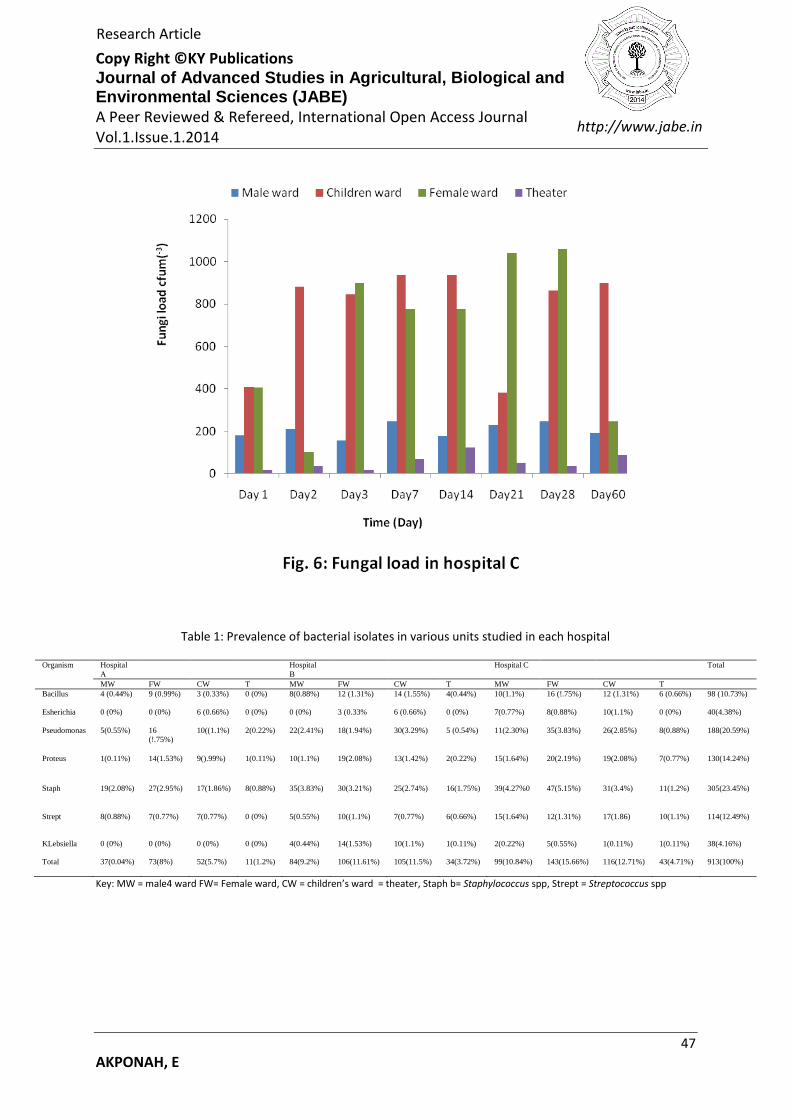
Table 1: Prevalence of bacterial isolates in various units studied in each hospital
Organism Hospital
A
Hospital
B
Hospital C Total
MW FW CW T MW FW CW T MW FW CW T
Bacillus 4 (0.44%) 9 (0.99%) 3 (0.33%) 0 (0%) 8(0.88%) 12 (1.31%) 14 (1.55%) 4(0.44%) 10(1.1%) 16 (!.75%) 12 (1.31%) 6 (0.66%) 98 (10.73%)
Esherichia 0 (0%) 0 (0%) 6 (0.66%) 0 (0%) 0 (0%) 3 (0.33% 6 (0.66%) 0 (0%) 7(0.77%) 8(0.88%)
10(1.1%) 0 (0%) 40(4.38%)
Pseudomonas 5(0.55%) 16
(!.75%)
10((1.1%) 2(0.22%) 22(2.41%) 18(1.94%) 30(3.29%) 5 (0.54%) 11(2.30%) 35(3.83%) 26(2.85%) 8(0.88%)
188(20.59%)
Proteus 1(0.11%) 14(1.53%) 9().99%) 1(0.11%) 10(1.1%) 19(2.08%) 13(1.42%) 2(0.22%) 15(1.64%) 20(2.19%) 19(2.08%) 7(0.77%) 130(14.24%)
Staph 19(2.08%) 27(2.95%) 17(1.86%) 8(0.88%)
35(3.83%) 30(3.21%) 25(2.74%) 16(1.75%) 39(4.27%0 47(5.15%) 31(3.4%) 11(1.2%) 305(23.45%)
Strept 8(0.88%)
7(0.77%) 7(0.77%) 0 (0%) 5(0.55%) 10((1.1%) 7(0.77%) 6(0.66%) 15(1.64%) 12(1.31%) 17(1.86) 10(1.1%) 114(12.49%)
KLebsiella 0 (0%) 0 (0%) 0 (0%) 0 (0%) 4(0.44%) 14(1.53%) 10(1.1%) 1(0.11%) 2(0.22%) 5(0.55%) 1(0.11%) 1(0.11%) 38(4.16%)
Total 37(0.04%) 73(8%) 52(5.7%) 11(1.2%) 84(9.2%) 106(11.61%) 105(11.5%) 34(3.72%) 99(10.84%) 143(15.66%) 116(12.71%) 43(4.71%) 913(100%)
Key: MW = male4 ward FW= Female ward, CW = children’s ward = theater, Staph b= Staphylococcus spp, Strept = Streptococcus spp

Copy Right ©KY Publications Journal of Advanced Studies in Agricultural, Biological and Environmental Sciences (JABE)
A Peer Reviewed & Refereed, International Open Access Journal Vol.1.Issue.1.2014
48
AKPONAH, E
http://www.jabe.in
Research Article
Table 2: Prevalence of fungal isolates in various units studied in each hospital
Organism
Hospital A
Hospital B
Hospital C
Total
MW FW CW T MW FW CW T MW FW CW T
A 3(2.73%) 2(1.82%)
3(2.73%)
0(0%)
4(3.64%)
7(6.36%)
5(4.55%)
0(0%) 5(4.55%)
8(7.27%)
10(9.09%)
1(0.91%)
48(43.64%)
B 0(0%) 4(3.64%)
0(0%) 0(0%)
2(1.82%)
5(4.55%)
3(2.73%)
2(1.82%)
3(2.73%)
5(4.55%)
8(7.27%)
0(0%) 32(29.09%)
C 0(0%) 2(1.82%)
2(1.82%)
0(0%)
3(2.73%)
8(7.27%)
6(5.45%)
2(1.82%)
0(0%) 3(2.73%)
3(2.73%)
1(0.91%)
30(27.27%)
Total 3(2.73%) 8(7.27%)
5(4.55%)
0(0%)
9(8.18%)
20(18.18%)
14(12.73%)
4(3.64%)
8(7.27%)
16(23.64%)
21(19.09%)
2(1.82%)
110(100%)
Key: MW = male ward FW= Female ward, CW = children’s ward, T = theater, a = Aspergillus spp, b =Mucor spp, c =
Penicillium spp
Table 3: Percentage of microbial isolate that showed growth in various concentrations of dettol
Organism Total Number of isolates
Percent that showed growth in various disinfectant concentrations
1 %v/v 5 %v/v 10 %v/v 15 %v/v 20 %v/v
Bacillus spp 98 90(91.84%) 71(72.45%) 31(31.63%) 13(13.27%) 17(17.35%) Escherichia coli 40 30(75%) 27(67.5%) 18(45%) 11(27.5%) 5(12.5%) Pseudomonas spp
188 120(63.83%) 100(53.19%) 92(93.88%) 70(37.23%) 53(28.19%)
Proteus spp 130 100(76.92%) 65(50%) 38(29.23%) 31(23.85%) 19(14.12%) Staphylococcus aureus
305 250(81.97%) 210(68.85%) 109(35.74%) 84(27.54%) 6717.54%)
Streptococcus spp
118 89(78.07%) 80(70.18%) 58(50.88%) 14(15.73%) 20(42.11%)
Klebsiella spp 38 37(97.37%) 30(78.95%) 27(71.05%) 28(75.68%) 16(33.33%) Aspergillus spp 30 30(100%) 21(70%) 26(86.67%) 15(50%) 10(27.08%) Mucor spp 48 40(83.33%) 36(75%) 26(75%) 20(54.17%) 13(27.08%) Penicillium spp 32 20(62.5%) 18(56.25 10(31.25%) 12(37.5%) 10(31.25%)
Table 4: Percentage of microbial isolates that showed growth in various concentrations of izal
Organism Total Number of isolates
Percent that showed growth in various disinfectant concentrations
1 %v/v 5 %v/v 10 %v/v 15 %v/v 20 %v/v
Bacillus spp 98 98(96.94%) 64(65.31%) 10(10.20%) 5(5.10%) 3(3.06%) Escherichia coli 40 32(80%) 15(37.5%) 4(10%) 2(5%) 6(15%) Pseudomonas
spp 188 150(53.19%) 74(39.36%) 50(26.6%) 24(12.76%) 20(10.64%)
Proteus spp 130 102(78.46%) 100(76.92%) 26(20.0%) 10(7.69%) 14(10.77%) Staphylococcus
aureus 305 208(68.2%) 164(53.97%) 61(20.0%) 49(16.07%) 30(9.84%)
Streptococcus spp
118 60(52.63%) 37(32.46%) 41(35.96%) 21(18.42%) 5(4.39%)
Klebsiella spp 38 31(81.58%) 23(60.53%) 15(39.47%)) 16(42.11%) 10(26.32%) Asppergillus spp 30 22(73.33%) 16(53.33%) 10(33.33%) 11(36.67%) 6(20%)
Mucor spp 48 40(83.33%) 29(60.42%) 14(29.11%) 8(16.66%) 7(14.58%) Penicillium spp 32 14(43.75%) 10(56.25 5(15.63%) 2(6.25%) 2(6.25%)

Copy Right ©KY Publications Journal of Advanced Studies in Agricultural, Biological and Environmental Sciences (JABE)
A Peer Reviewed & Refereed, International Open Access Journal Vol.1.Issue.1.2014
49
AKPONAH, E
http://www.jabe.in
Research Article
Table 5: Percentage of Gram positive bacterial isolates that showed resistance to various antibiotics
Organism Total number of isolate
Percent that showed resistance
CIP
STR
SEP
AM
P
ZIN
GEN
RO
C
ERY
PER
Bacillus spp 98 20.41% 10.20% 6.12% 40.82% 4.08% 11.23% 8.16% 13.27% 2.04% Staphylococcus aureus
305 22.95% 16.72% 22.95% 65.57% 15.41% 32.79% 21.97% 7.87% 4.92%
Streptococcus spp
114 14.04% 43.86% 40.35% 32.46% 17.54% 17.54% 16.67% 11.40% 8.77%
Key: CIP = ciprofloxacin, STR = streptomycin, SEP = septrin, AMP = ampliclox, ZIN = zinnacef, GEN = gentamycin,
ROC = rocephin, ERY – erythromycin and PER = perfloxacin.
Table 6: Percentage of Gram negative bacteria isolates that showed resistance to various antibiotics
Organism Total number of isolates
Percent that showed resistance
CIP
STR
SEP
AM
O
TAR
GEN
AU
G
SAR
Escherichia coli
40 72.50% 37.50% 25.00% 75.00% 32.50% 42.50% 63.50% 2.50%
Pseudomonas spp
188 45.74% 31.91% 31.91% 38.83% 26.00% 21.81% 18.62% 15.43%
Proteus spp 130 24.62% 7.69% 3.85% 9.23% 4.62% 6.92% 13.07% 11.54% Klebsiella spp 38 78.95% 73.68% 65.79% 65.79% 71.05% 89.58% 52.63% 47.37%
Key: CIP = ciprofloxacin, STR = streptomycin, SEP = septrin, AMO = amoxicillin, TAR = tarivid, GEN = gentamycin,
AUG = augumentin, perfloxacin and SAR = sarfloxacin.
RESULT
The fungal and bacteria load obtained in the three hospitals studied are presented in Figs. 1 to 6. The total
bacteria load was higher in each ward than the total fungi load. Also the bacterial and fungal load obtained
from the various wards varied from day to day and also from hospital to hospital. In all the hospitals bacterial
and fungal load decreased in the order: Female ward ≥ children ward > male ward > theater. The frequencies
of occurrence (prevalence) of the isolates in each ward in the three hospitals are shown in Tables 1 and 2. A
total of nine hundred and thirteen bacteria (Table 1) and one hundred and ten fungi (Table 2) isolates were
obtained. The bacteria isolates belonged to the following genera: Bacillus, Escherichia, Pseudomonas, Proteus,
Klebsiella, Staphylococcus and Streptococcus while the fungi isolates included Aspergillus, Penicillium and
Mucor. There was no significant difference among the counts of both bacterial and fungi isolates obtained in
the different days of the study in each hospital. However microbial count obtained in Hospital C were higher
than in hospital B and least in hospital A. The prevalence of the organisms obtained in the three hospitals
decreased as follows: Staphylococcus aureus > Pseudomonas spp > Proteus spp > Streptococcus spp > Bacillus
spp > Aspergillus spp ≥ Escherichia coli ≥ Klebsiella spp ≥ Mucor spp > Penicillium spp.

Copy Right ©KY Publications Journal of Advanced Studies in Agricultural, Biological and Environmental Sciences (JABE)
A Peer Reviewed & Refereed, International Open Access Journal Vol.1.Issue.1.2014
50
AKPONAH, E
http://www.jabe.in
Research Article
The result of the effect of disinfectant on the survival of each microbial isolate is shown in Tables 3 and 4. The
percentage of isolates of each organism that showed growth decreased as the concentration of each
disinfectant increased. The results obtained indicated that a high percentage of the isolates of each organism
were susceptible to the disinfectants used. However, the isolates were more susceptible to izal than dettol.
There was a significant difference at p < 0.05 among the percent growth of isolates exposed to various
concentrations of each disinfectant and also between growth of organisms exposed to dettol and izal.
The results of the susceptibility of the gram positive and gram negative bacteria isolates to the tested
antibiotics are presented in Tables 5 and 6 respectively. All the bacteria isolates showed varying degree of
resistance to the antibiotics tested. Percentage of Bacillus, Staphylococcus and Streptococcus isolates that
exhibited resistance to each antibiotics were less than 50% with the exception of ampicillin to which 65.57% of
Staphylococcus aureus isolates showed resistance. Also, of the Gram negative bacteria isolates, the percentage
of Klebsiella isolates that showed resistance to the various antibiotics was highest. This was followed by
Escherichia coli and percentage of Proteus isolates that showed resistance to various antibiotics was least.
DISCUSSION
Although, the microbiological quality of the indoor air of the three hospitals investigated varied, they all
exceeded the set standards as established by NIOSH and ACGIH. The quality of air in these hospitals might
have been influenced by factors such as age of the building, degree of cleanliness and disinfection, type of
disinfectant used, nature of floors (whether tiled or not) and control of influx as well as efflux of people into
the various units. Hence, the airborne bio-contaminants in hospital A that was newly renovated with adequate
ventilation system, was least. The high rates of microbial load observed in hospitals B and C may also be
attributed to the smaller size of the wards (higher number of beds per m3 of ward) [2] and occurrence of
crevices in the floors, ceiling and walls of these hospitals. Also, specific activities like talking, sneezing, walking
and washing can generate airborne biological particulate matter [4].
The high number of bacteria and fungi population observed in female and children wards in all the hospitals
may be attributed to the high number of occupants (patients, patient attendant, personnel and visitors) at all
times while the low microbial population recorded in the theater of the three hospital may be as a result of the
location of theater in these hospitals, controlled entry and the high degree of disinfection that usually occur in
this unit in hospitals. Similarly, [2,11,12] had reported that patient room had the highest microbial count in
hospitals.
The high incidence of Staphylococcus observed in this study could be as a result of the fact that Staphylococcus
is a normal flora of the skin and nasopharynx and thus can easily be shed off by hospital occupants and hence
contribute greatly to the hospital indoor air flora. The prevalence of E coli was quite low but more often was
isolated from the children’s ward. This is likely due to passage of fecal matter into surrounding as well as the
use of focally contaminated water in cleaning floors or other activities in the wards or could even be generated
by toilet flushing. [12]had also reported similar results. The occurrence of pathogens such as Pseudomonas
spp, Proteus spp, Klebsiella spp, Streptococcus spp and Aspergillus spp is suggestive that the patients in the
wards might be responsible for shedding these organisms into the hospital indoor air
In all the units studied, the bacterial load was consistently, higher than the fungi load. This might have been
favored by the high environmental temperature at the period of the study. It could also be attributed to the
larger size of fungi which might have affected their floating capacity.
The result of the antibiotics susceptibility test suggest that many of the bacteria contaminating the indoor air
of the three hospitals are of human origin and that the indiscriminate use of antibiotics may explain the
emergence of antibiotics resistance in these organisms. The resistance to various antibiotics observed in a
great percentage of Klesiella spp may be as a result of the occurrence of capsule in this bacterium.
Additionally, the difference noticed in the susceptibility of organisms to dettol and izal, could possibly be as a
result of the differences in the chemical composition of these disinfectants. However, on a general note, both

Copy Right ©KY Publications Journal of Advanced Studies in Agricultural, Biological and Environmental Sciences (JABE)
A Peer Reviewed & Refereed, International Open Access Journal Vol.1.Issue.1.2014
51
AKPONAH, E
http://www.jabe.in
Research Article
disinfectants reduced the concentration of the population of each organism considerably especially at high
concentrations. The results demonstrated the effectiveness of these disinfectants against fungi and bacteria.
Conclusively, indoor air monitoring that is focused on the presence of bacteria and fungi cannot be over
emphasized. Therefore, efforts should be made to improve the hygiene of hospital environments. It is also
necessary to raise the awareness of medical personnel to reduce the hazard of transmission of potentially
pathogenic airborne microorganisms. Perhaps, this could be by developing a good disinfection strategy which
will be beneficial to reducing the airborne microbial load. Furthermore, it is advisory to obtain an antibiogram
in an event where by any of these organisms are implicated in nosocomial infection outbreak for effective
combat.
REFERENCES
[1]. Bhatin, L. and Vishwakarma, R. (2010). Hospital indoor airborne microflora in private and Government
owned hospitals in Sagar City, India. World J. Med. Sci. 5(3): 65-70.
[2]. Qudiesat, K., Abu-Etten, K., Elkarmi, A., Hamad, M. and Abussaud, M. (2009). Assessment of airborne
pathogens in healthcare settings. Afr. J. Microbiol. Res. 3(2): 066-076.
[3]. Banerjee, D. (2008). Stydy of precipitation chemistry over an industrial city. Int. J. Environ. Sci. Tech.
5(3): 331-338.
[4]. Yassin, M.F. and Alimouqatea, S. (2010). Assessment of airborne bacteria and fungi in an indoor and
outdoor environment. Int. J. Environ.Sci. Tech. 7(3): 535-544.
[5]. Obbard, J.P. and Fang, L.S. (2003). Airborne concentrations of bacteria in a hospital environment in
Singapore. Water, air and Soil Pollut. 144: 333-341.
[6]. Pastuszka, J.S., Marchwinska-Wynwal, E. and Wiazlo, A. (2005). Bacterial aerosol Silesian hoswpital:
Preliminary result. Polish. J. Environ. Studies. 14(6): 883-890.
[7]. Dutkiewiez, J. and Augustowska, M. (2006). Variability of airborne microflora in a hospital ward within
a period of one year. Annals of Agric. Envtal. Med. 13: 99-106.
[8]. [8]Barnet, H.L. and Hunter, B.B. (1972). Illustrated genera of imperfect fungi. 3rd
edn, Burges
Publishing Company, Minneapolis, 208 pp.
[9]. Cruickshank, R.J., P. Dugid, B.P. Marmuon and R.H.A. Swain (1975). Medical Microbiology. 12th
ed
Churchill Livingstone, London. Pp 426-437.
[10]. Cheesebrough, M. (2000). District laboratory practice in tropical countries. Part 2. Cambridge
University Press, UK. 434p
[11]. Ekhaise, F.O., Ighosevwe, O.U. and Ajakpovi, O.D. (2008). Hospital indoor airborne microflora in
private and Government owned hospitals in Benin city, Nigeria. World J. Med. Sci. 3(1): 19-23.
[12]. Jaffal, A.A., Banat, J.M., El Mogheth, A.A., Nsanze, H., benar, A. and Ameen, A.S. (1997). Residential
indoor airborne microbial population in the United Arab Emirates. Environ. Int. 23(4): 529-533.