Embed Size (px)
Citation preview
Jordan Spindle MD
February 23rd, 2012
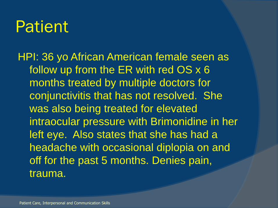
Patient
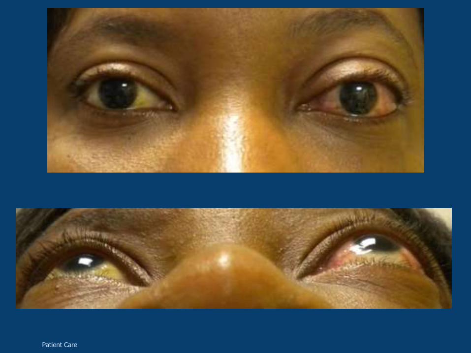
HPI: 36 yo African American female seen as
follow up from the ER with red OS x 6
months treated by multiple doctors for
conjunctivitis that has not resolved. She
was also being treated for elevated
intraocular pressure with Brimonidine in her
left eye. Also states that she has had a
headache with occasional diplopia on and
off for the past 5 months. Denies pain,
trauma.
Patient Care, Interpersonal and Communication Skills

History
POH: none
Gtts: Brimonidine 0/1
PMH: DM (diagnosed 2010), HTN
Meds: Metformin
All: nkda
FH: no glaucoma/blindness
Patient Care, Interpersonal Skills and Communication Skills
EXAM
dVa sc: 20/20, 20/25
Pupils: 32 ou, no apd
EOMs: full ou, no diplopia
CVFs: full ou
Patient Care
Patient Care

Patient Care
Patient Care

SLE
K: clear ou
A/C: d and q ou
P/I: r and r ou
L: clear ou
Patient Care
DFE
V: clear ou
C/D: 0.3/0.3, s and p ou
M: flat ou, +flr ou
V: wnl ou
P: no holes/tears/heme seen ou
Patient Care

Differential Diagnosis
Medical Knowledge
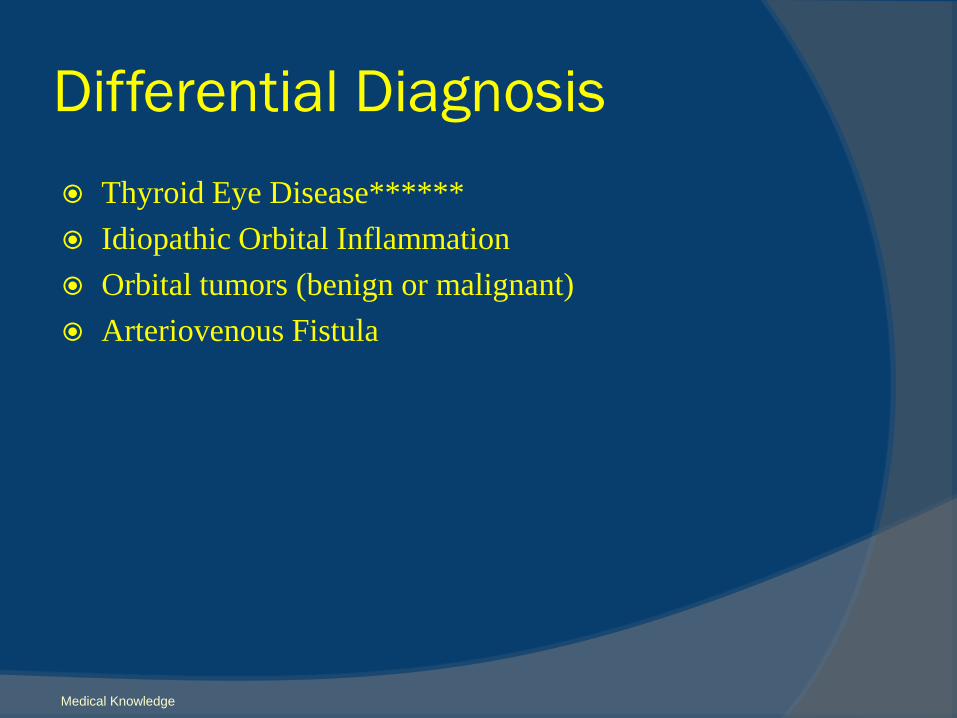
Differential Diagnosis
Thyroid Eye Disease******
Idiopathic Orbital Inflammation
Orbital tumors (benign or malignant)
Arteriovenous Fistula
Medical Knowledge
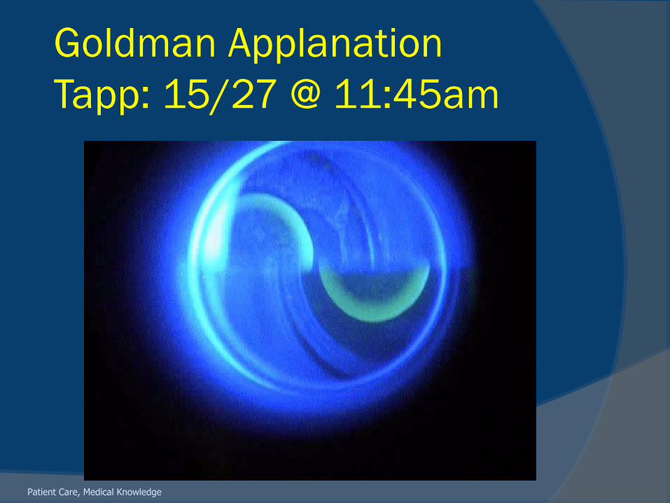
Goldman Applanation
Tapp: 15/27 @ 11:45am
Patient Care, Medical Knowledge
Upon Specific questioning
Patient states that she has been hearing
a wooshing sound for the past month as
well
Ocular auscultation WNL
Patient Care, Interpersonal Skills and Communication Skills

Differential Diagnosis
Thyroid Eye Disease******
Idiopathic Orbital Inflammation
Orbital tumors (benign or malignant)
Arteriovenous Fistula
Medical Knowledge

What would you do next?
Medical Knowledge
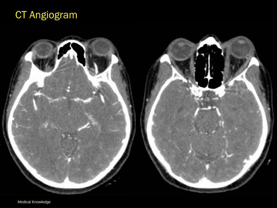
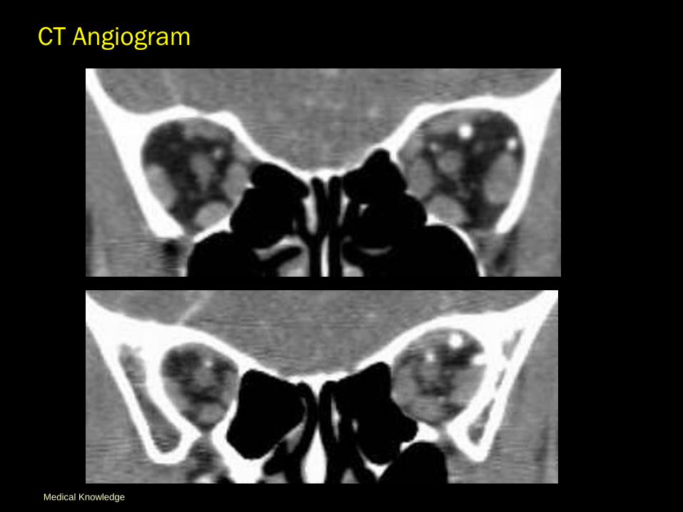
CT Angiogram
Medical Knowledge
CT Angiogram
Medical Knowledge

CT Angiogram
Findings highly suggestive of an
Arteriovenous fistula
Medical Knowledge
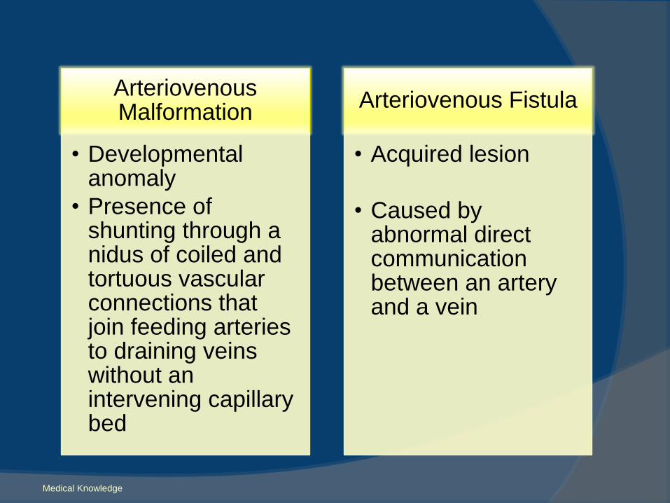
Arteriovenous Malformation
• Developmental anomaly
• Presence of shunting through a nidus of coiled and tortuous vascular connections that join feeding arteries to draining veins without an intervening capillary bed
Arteriovenous Fistula
• Acquired lesion
• Caused by abnormal direct communication between an artery and a vein
Medical Knowledge

Cognard, Djindjian, and BordenMedical Knowledge
Medical Knowledge
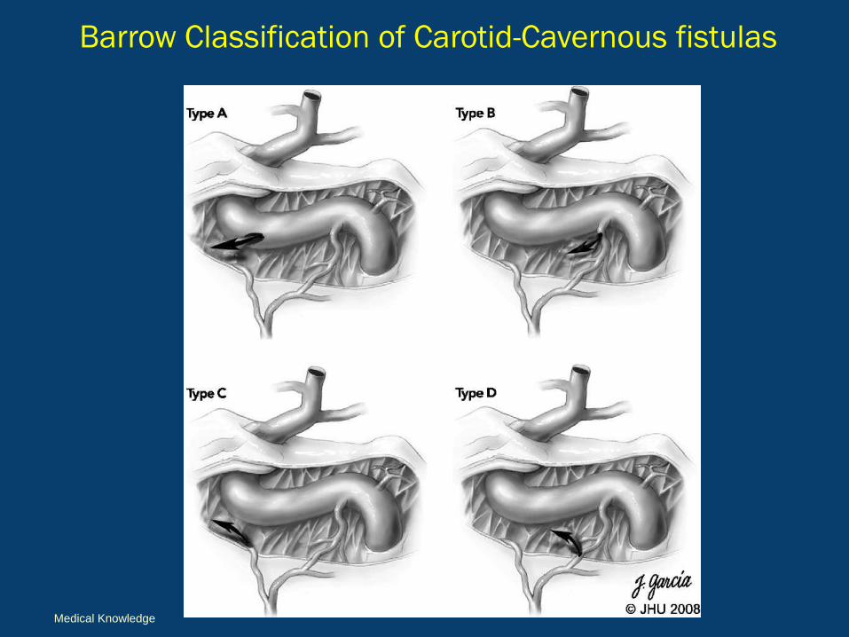
Barrow Classification of Carotid-Cavernous fistulas
Medical Knowledge
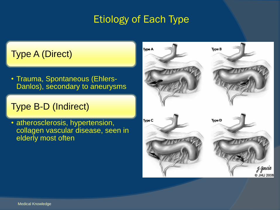
Type A (Direct)
• Trauma, Spontaneous (Ehlers-Danlos), secondary to aneurysms
Type B-D (Indirect)
• atherosclerosis, hypertension, collagen vascular disease, seen in elderly most often
Etiology of Each Type
Medical Knowledge
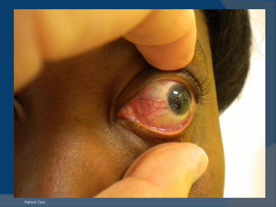
Direct Carotid Cavernous Fistula
High blood-flow rate
Classic triad: pulsatile proptosis, conjunctival
injection/chemosis, and an ocular bruit
Other findings
Tortuous epibulbar vessels
Elevated IOP
Choroidal effusions
Blood in Schlemm’s canal
Cranial nerve dysfunction of III, IV, and most commonly
VI
Enlargement of EOM’s, & dilated SOV
Medical Knowledge
Dural Sinus Fistula
Low blood-flow
Small meningeal arterial branches communicating
with venous drainage
Insidious onset
Findings (same as CC fistula but not as
severe/acute)
Arterialization of episcleral veins
Elevated Intraocular Pressure
Enlargement of EOM’s & dil SOV
Medical Knowledge
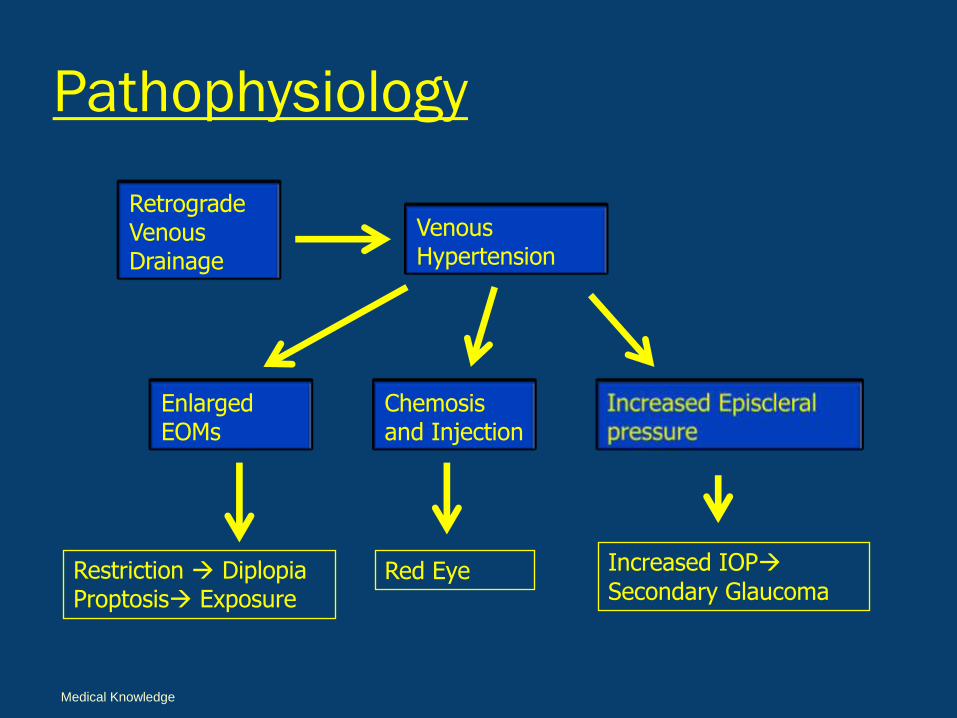
Pathophysiology
Retrograde Venous Drainage
Venous Hypertension
Enlarged EOMs
Chemosisand Injection
Restriction DiplopiaProptosis Exposure
Red Eye Increased IOP
Secondary Glaucoma
Medical Knowledge
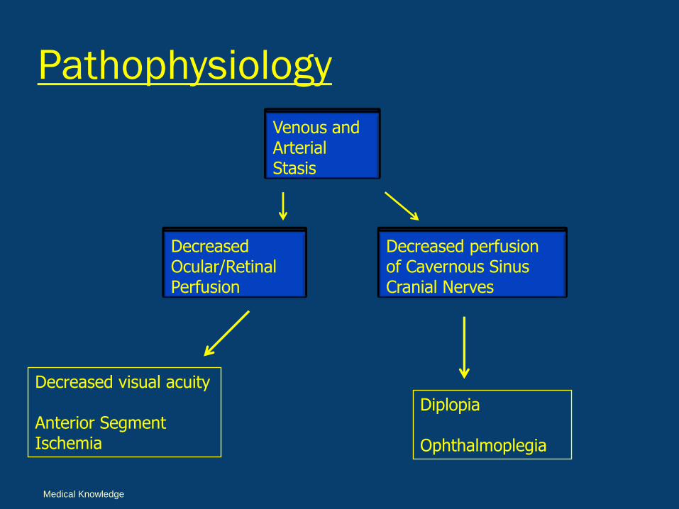
Pathophysiology
Venous and Arterial Stasis
Decreased Ocular/Retinal Perfusion
Decreased perfusion of Cavernous Sinus Cranial Nerves
Diplopia
Ophthalmoplegia
Decreased visual acuity
Anterior Segment Ischemia
Medical Knowledge
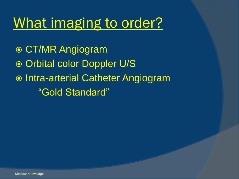
What imaging to order?
CT/MR Angiogram
Orbital color Doppler U/S
Intra-arterial Catheter Angiogram
“Gold Standard”
Medical Knowledge
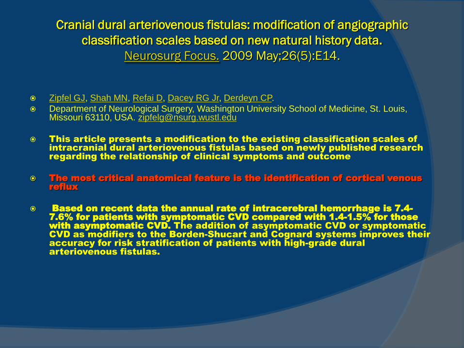
Zipfel GJ, Shah MN, Refai D, Dacey RG Jr, Derdeyn CP.
Department of Neurological Surgery, Washington University School of Medicine, St. Louis, Missouri 63110, USA. [email protected]
This article presents a modification to the existing classification scales of
intracranial dural arteriovenous fistulas based on newly published research
regarding the relationship of clinical symptoms and outcome
The most critical anatomical feature is the identification of cortical venous
reflux
Based on recent data the annual rate of intracerebral hemorrhage is 7.4-
7.6% for patients with symptomatic CVD compared with 1.4-1.5% for those
with asymptomatic CVD. The addition of asymptomatic CVD or symptomatic
CVD as modifiers to the Borden-Shucart and Cognard systems improves their
accuracy for risk stratification of patients with high-grade dural
arteriovenous fistulas.
Cranial dural arteriovenous fistulas: modification of angiographic
classification scales based on new natural history data.
Neurosurg Focus. 2009 May;26(5):E14.

When to Observe
In lesions without CVR (Cortical Venous Reflux)
Asymptomatic or tolerable symptoms
-May close spontaneously (10-60%)
FOLLOW-UP
Serial MRI, MRA and DSA after 3 yr
Any change in symptoms needs evaluation
CONCERNS
2-3% chance of developing CVR
Intra-cerebral hemorrhage
Overall risk of hemorrhage – 1.6% / yr (Brown et al.)
Exacerbation & remission
Medical Knowledge
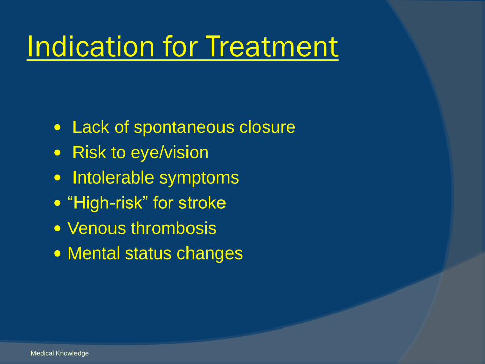
Indication for Treatment
Lack of spontaneous closure
Risk to eye/vision
Intolerable symptoms
“High-risk” for stroke
Venous thrombosis
Mental status changes
Medical Knowledge
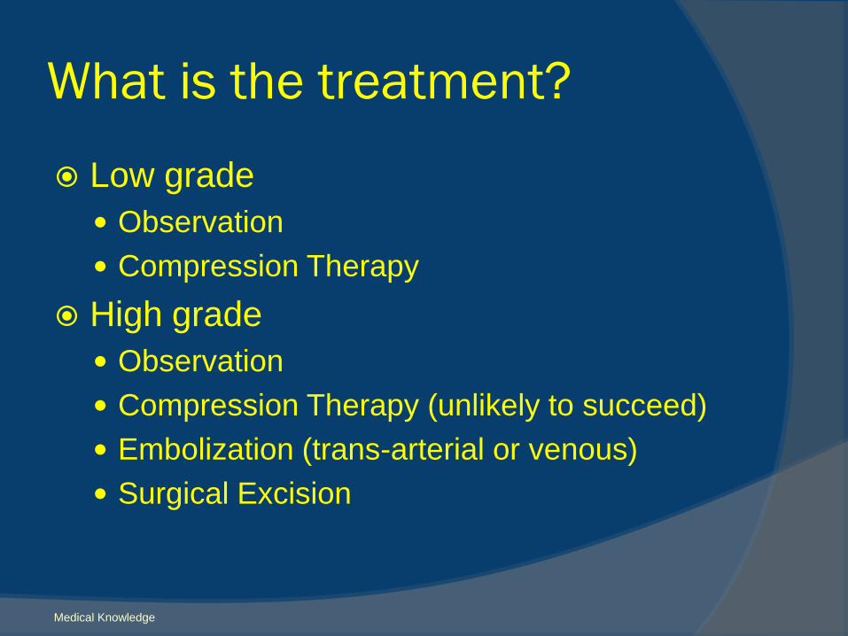
What is the treatment?
Low grade
Observation
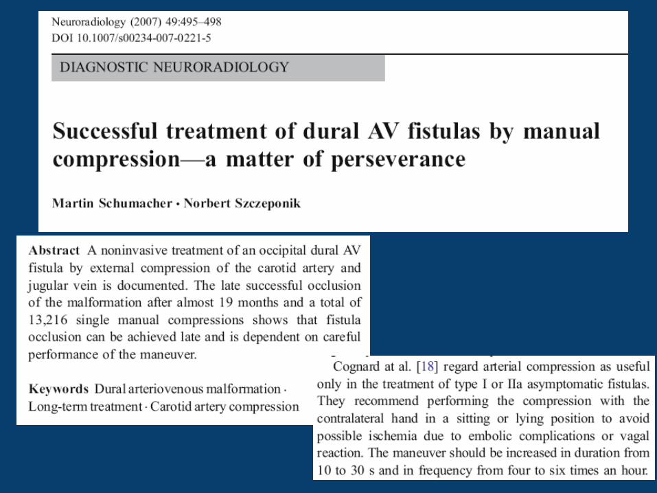
Compression Therapy
High grade
Observation
Compression Therapy (unlikely to succeed)
Embolization (trans-arterial or venous)
Surgical Excision
Medical Knowledge

Intra-arterial Angiogram
Indirect Meningeal feeders
A-V shunting – cavernous pouch and inferior petrosal sinus (IPS)
Patient Care, Medical Knowledge
Courtesy of :Sundeep Mangla, M.D.Director of Interventional NeuroradiologySUNY Downstate

NeuroInterventional Therapy
Early draining vein
Interim coiling Final Post Coiling(AV shunting eliminated)Patient Care, Medical Knowledge
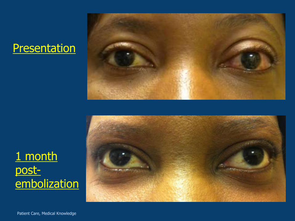
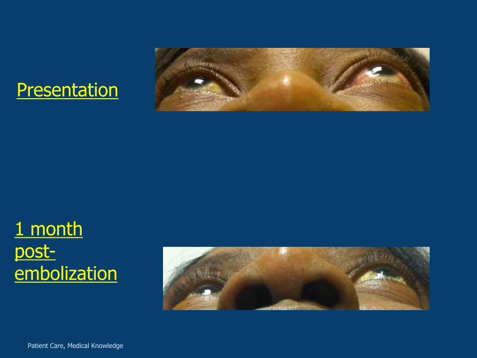
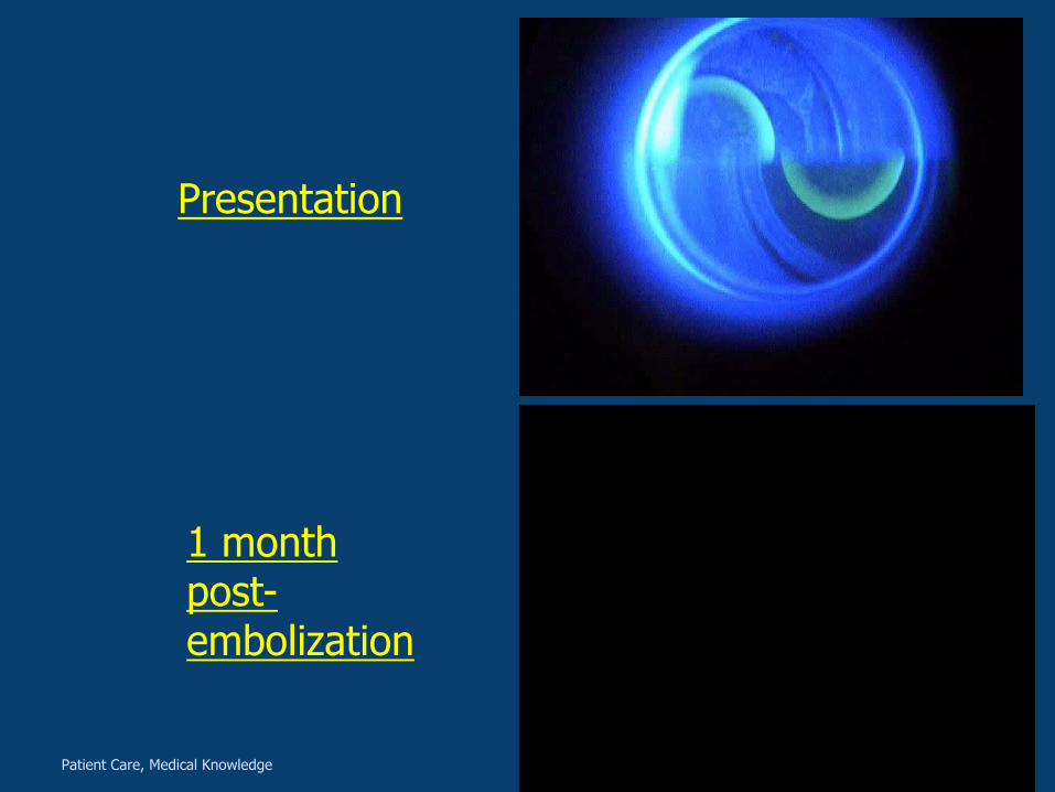
Presentation
1 month post-embolization
Patient Care, Medical Knowledge

Presentation
1 month post-embolization
Presentation
1 month post-embolization
Patient Care, Medical Knowledge
Presentation
1 month post-embolization
Patient Care, Medical Knowledge
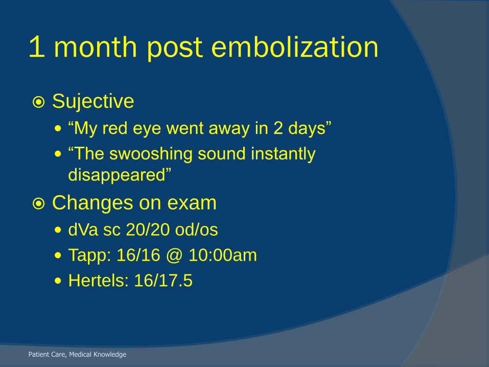
1 month post embolization
Sujective
“My red eye went away in 2 days”
“The swooshing sound instantly
disappeared”
Changes on exam
dVa sc 20/20 od/os
Tapp: 16/16 @ 10:00am
Hertels: 16/17.5
Patient Care, Medical Knowledge
Reflective Practice
This case demonstrated the importance of listening to all of the patient complaints and having the background knowledge to put all the clues together in order to properly diagnose and treat the proper disease etiology.
This patient had an expedited and excellent result and was very happy with the resolution of all her symptoms after treatment.

Patient Care: The case involved thorough patient care and attention to patient's complaints. Once diagnosed, the patient received proper management and care
Medical Knowledge This presentation allowed us to review the presentations, proper evaluation/work up, and different treatments.
Practice-Based Learning and Improvement: This presentation included a current literature search of developing associations and current treatment modalities
Interpersonal and Communication Skills: The patient was treated with respect and every effort was made to communicate with the patient in a timely manner for the proper follow-up
Professionalism: The patient was treated in the proper manner. She was also referred to the proper specialist to treat her condition.
Systems-Based Practice: The patient was discussed in detail with neuroradiologic colleagues in regard to follow up and treatment.
Core Compentencies
Thank you
Dr. Shinder
Dr. Mangla
KCHC staff
References
Sundeep Mangla, M.D.
Director of Interventional Neuroradiology
Dural AVF Classification and Management. Rakesh K. Singh
Arteriovenous Fistulas and Carotid-Cavernous Fistula.
Subramanian, Williams. International Ophtho Clinics. Vol 49. 3 82-
101
Basic Clinical and Sciences Series. Orbit. Pages 64-70
Interventional treatment of carotid cavernous fistula
Richard C. Barry a, Mark Wilkinson b, Rebekah M. Ahmed c,
Charmaine S.M. Lim a, Geoffrey D. Parker b,
a Department of Ophthalmology, Royal Prince Alfred Hospital,
Camperdown, New South Wales, Austra