Embed Size (px)
Citation preview
Joint Examination and Injection Course
Mr Nadim AslamConsultant Orthopaedic Surgeon
Spire Bone and Joint Clinic
Worcestershire Knee and Hip Clinic
KNEE
History & Physical Exam of the Injured Knee
Assessing a knee injury
• Components of the assessment include
– Focused history
– Attentive physical examination
– Thoughtfully ordered tests/studies
Focused History

Differential Diagnosis of Knee Haemarthrosis
ACL tear
Meniscal Tear
Fracture
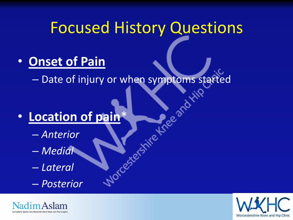
Focused History Questions
• Onset of Pain– Date of injury or when symptoms started
• Location of pain*– Anterior
– Medial
– Lateral
– Posterior

Focused History Questions
• Mechanism of Injury -helps
predict injured structure
– Contact or noncontact injury?*• If contact, what part of the knee was
contacted?
– Anterior blow?
– Valgus force?
– Varus force?
– Was foot of affected knee planted on the ground?**
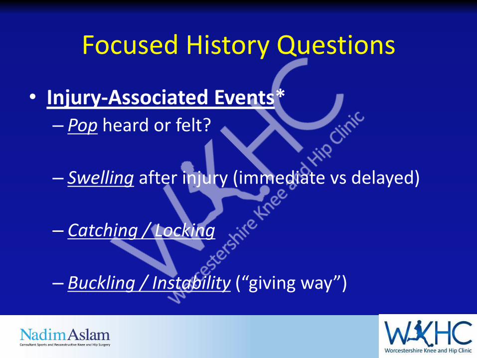
Focused History Questions
• Injury-Associated Events*– Pop heard or felt?
– Swelling after injury (immediate vs delayed)
– Catching / Locking
– Buckling / Instability (“giving way”)

Instability - Example
Patellar dislocation
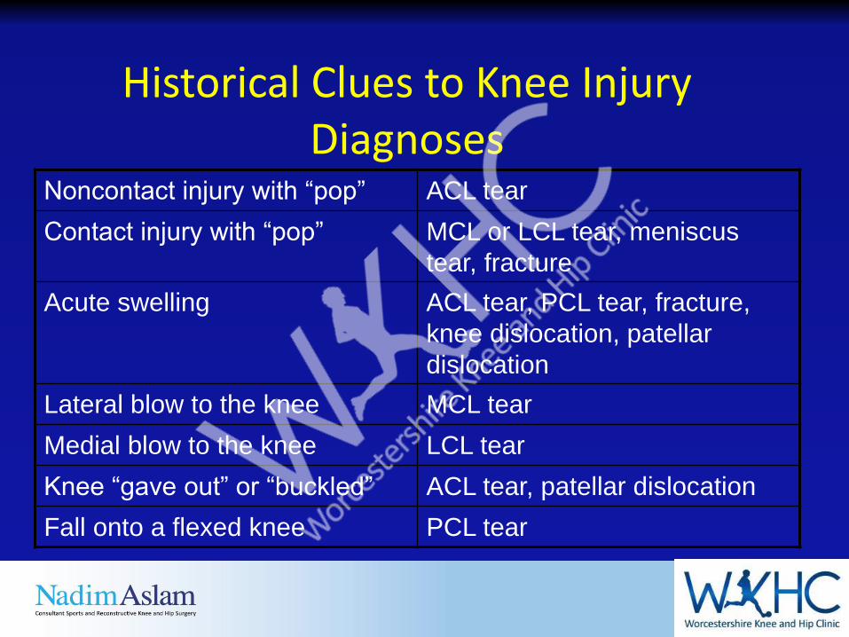
Historical Clues to Knee Injury Diagnoses
Noncontact injury with “pop” ACL tear
Contact injury with “pop” MCL or LCL tear, meniscus
tear, fracture
Acute swelling ACL tear, PCL tear, fracture,
knee dislocation, patellar
dislocation
Lateral blow to the knee MCL tear
Medial blow to the knee LCL tear
Knee “gave out” or “buckled” ACL tear, patellar dislocation
Fall onto a flexed knee PCL tear
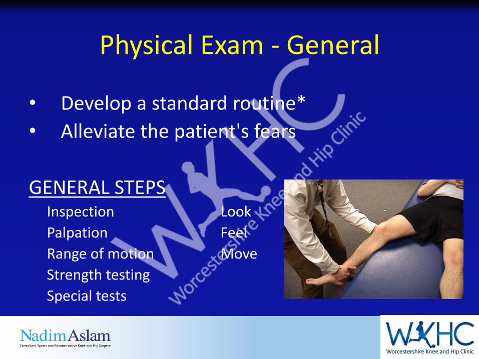
Physical Exam - General
• Develop a standard routine*
• Alleviate the patient's fears
GENERAL STEPSInspection Look
Palpation Feel
Range of motion Move
Strength testing
Special tests
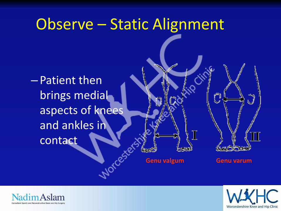
– Patient then brings medial aspects of knees and ankles in contact
Observe – Static Alignment
Genu valgum Genu varum
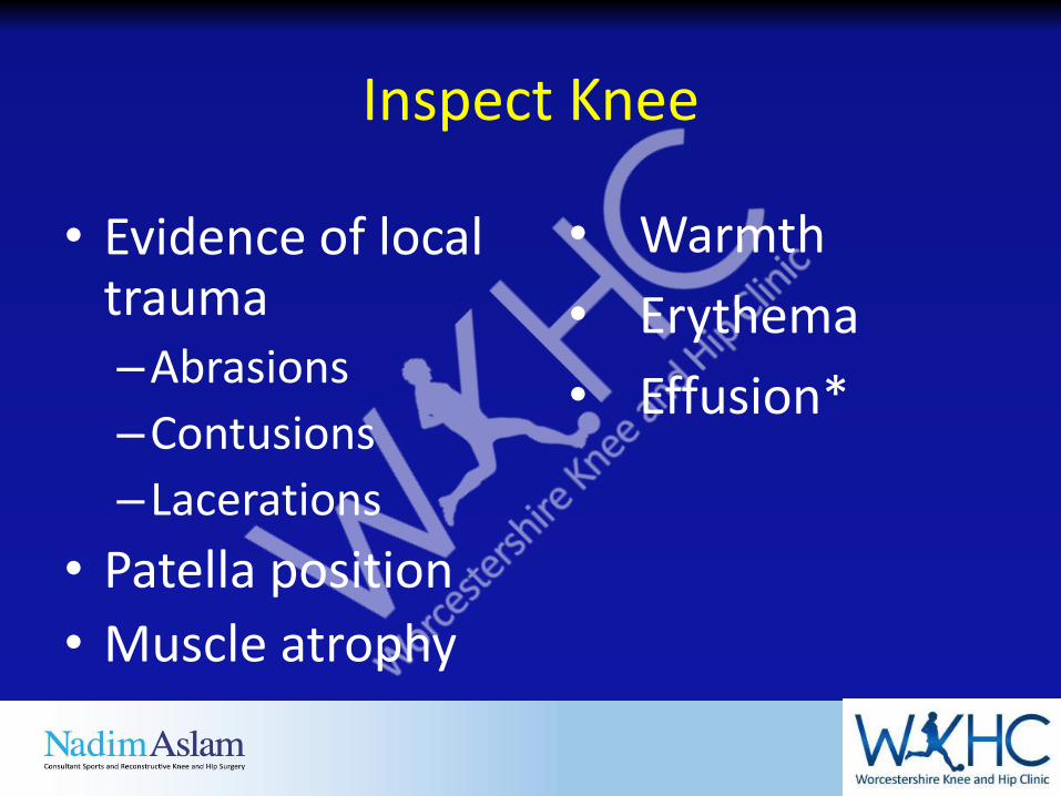
Inspect Knee
• Warmth
• Erythema
• Effusion*
• Evidence of local trauma–Abrasions
–Contusions
–Lacerations
• Patella position
• Muscle atrophy
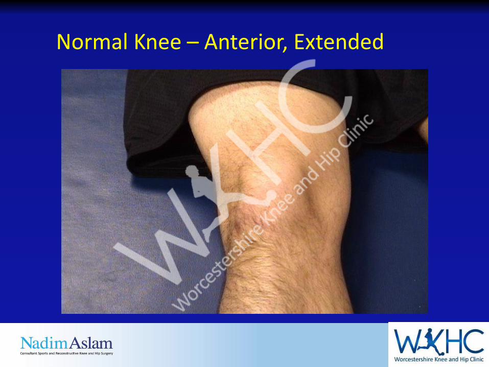
Normal Knee – Anterior, Extended
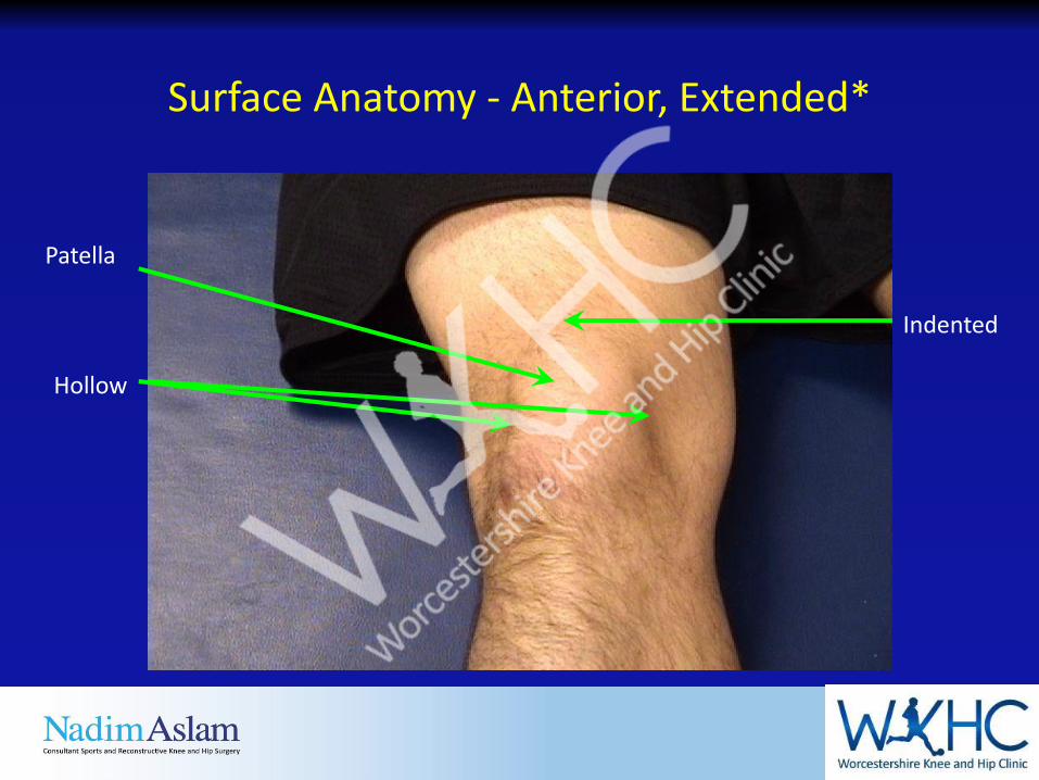
Surface Anatomy - Anterior, Extended*
Patella
Hollow
Indented
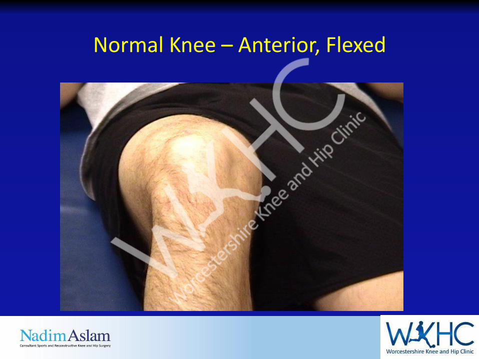
Normal Knee – Anterior, Flexed
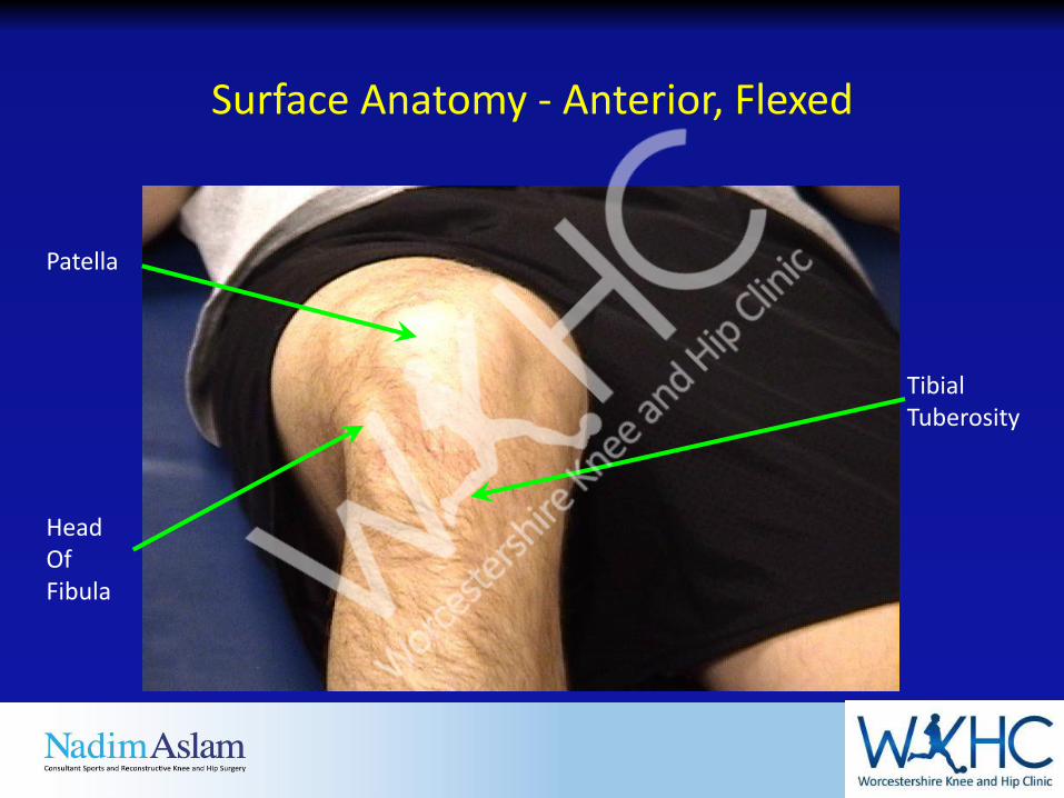
Surface Anatomy - Anterior, Flexed
Head OfFibula
Patella
TibialTuberosity
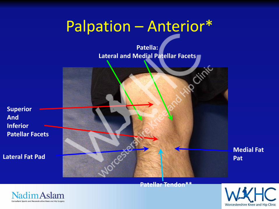
Palpation – Anterior*Patella:
Lateral and Medial Patellar Facets
Superior AndInferior Patellar Facets
Patellar Tendon**
Lateral Fat PadMedial Fat Pat
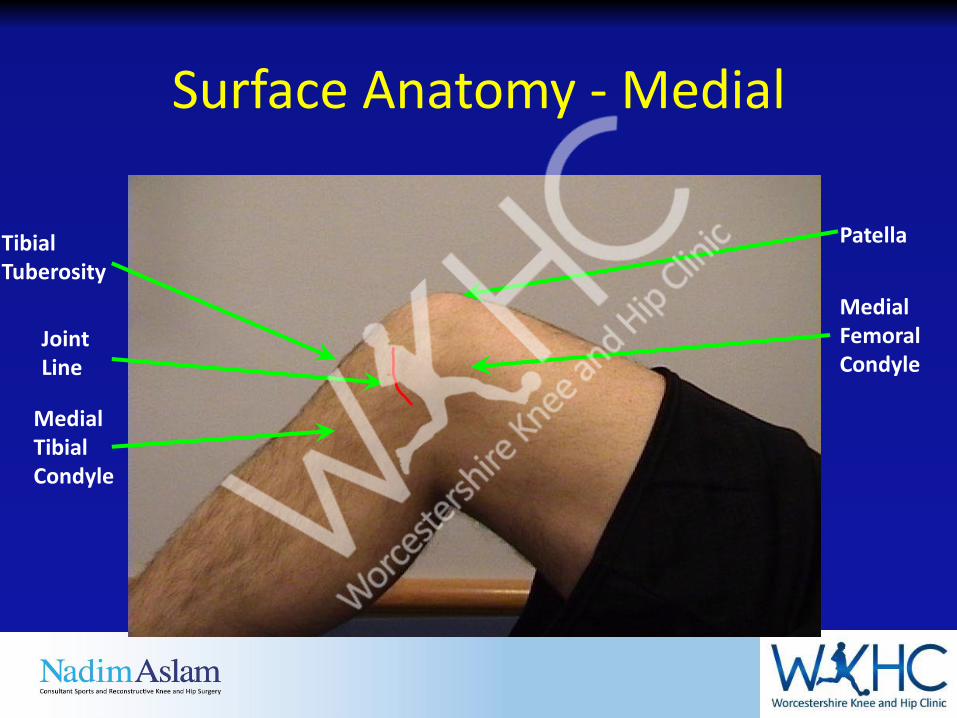
Surface Anatomy - Medial
Medial FemoralCondyle
Patella
JointLine
MedialTibialCondyle
TibialTuberosity
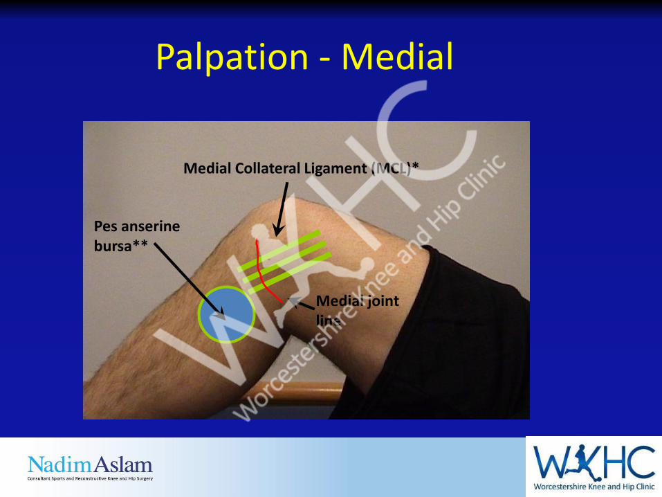
Palpation - Medial
Medial Collateral Ligament (MCL)*
Pes anserine bursa**
Medial joint line
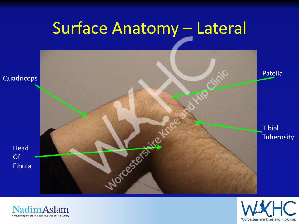
Surface Anatomy – Lateral
Patella
Head OfFibula
TibialTuberosity
Quadriceps

Palpation – Lateral*
Lateral joint line
Lateral Collateral Ligament (LCL)**
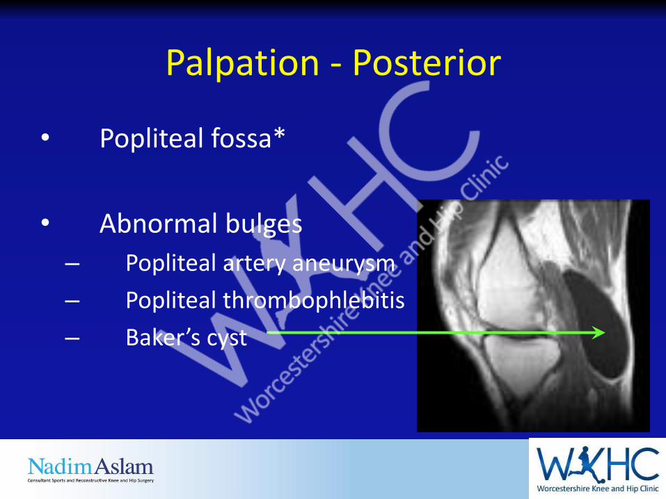
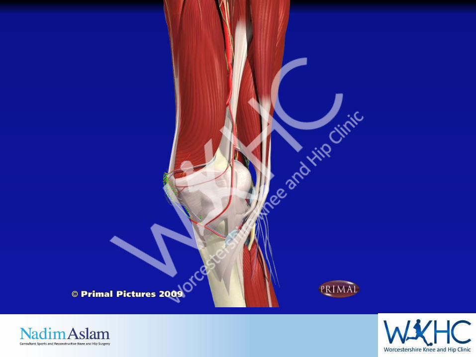
Palpation - Posterior
• Popliteal fossa*
• Abnormal bulges
– Popliteal artery aneurysm
– Popliteal thrombophlebitis
– Baker’s cyst
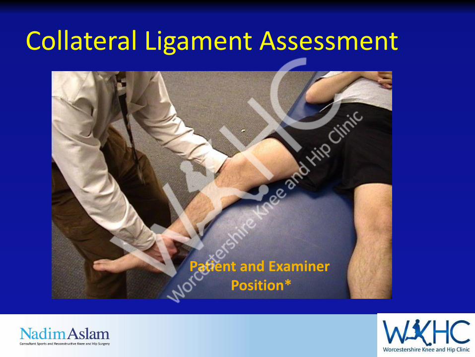
Collateral Ligament Assessment
Patient and ExaminerPosition*
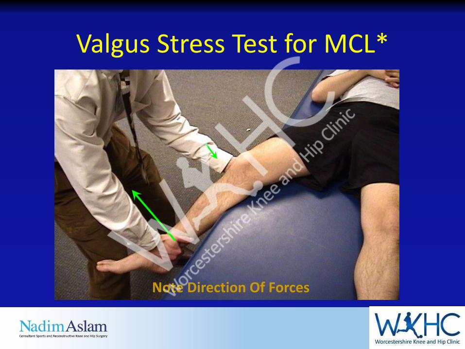
Valgus Stress Test for MCL*
Note Direction Of Forces
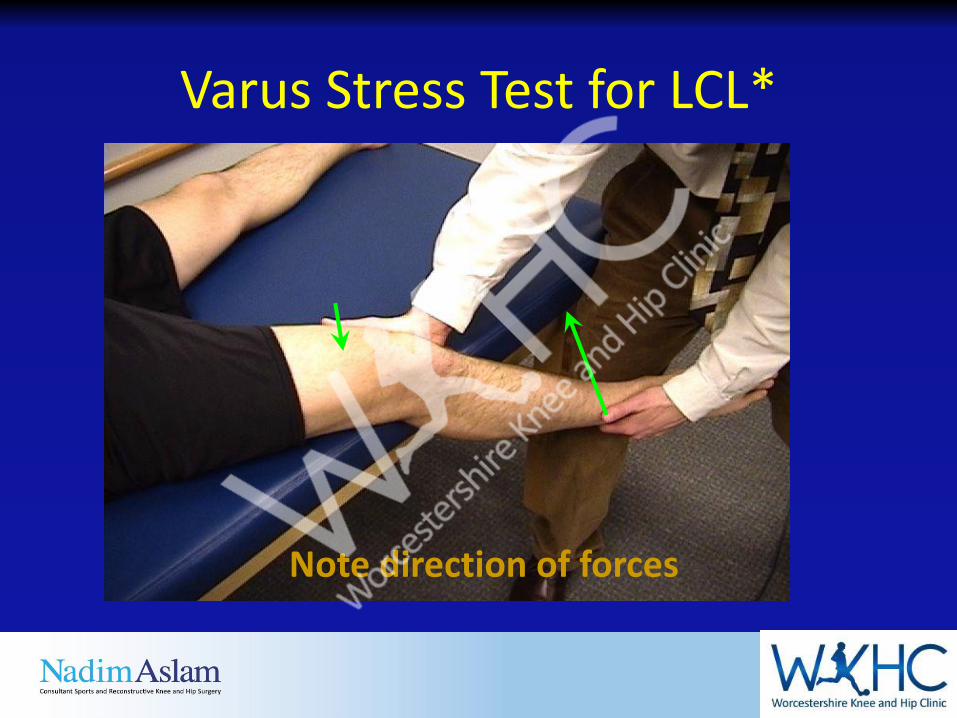
Varus Stress Test for LCL*
Note direction of forces

Lachman Test*
• Patient Position
• Physician hand placement
KNEE PROBLEMS
• OA
• RA
• Gout
• Pyrophosphate disease
• Inflammatory arthropathies
• Pre patella bursitis
• Infra patella bursitis
• Pes Anserinus inflammation
• Popliteal cyst (Bakers)
• Referred from hip
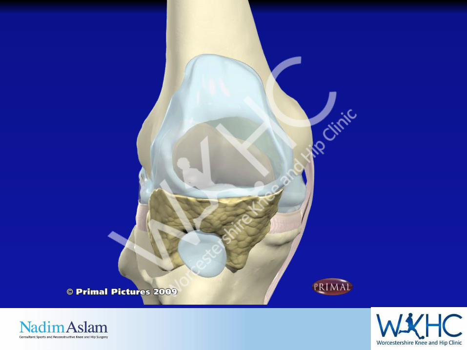
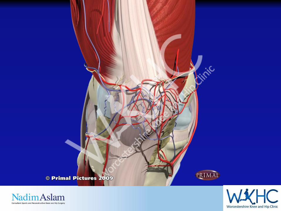
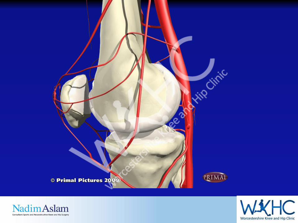
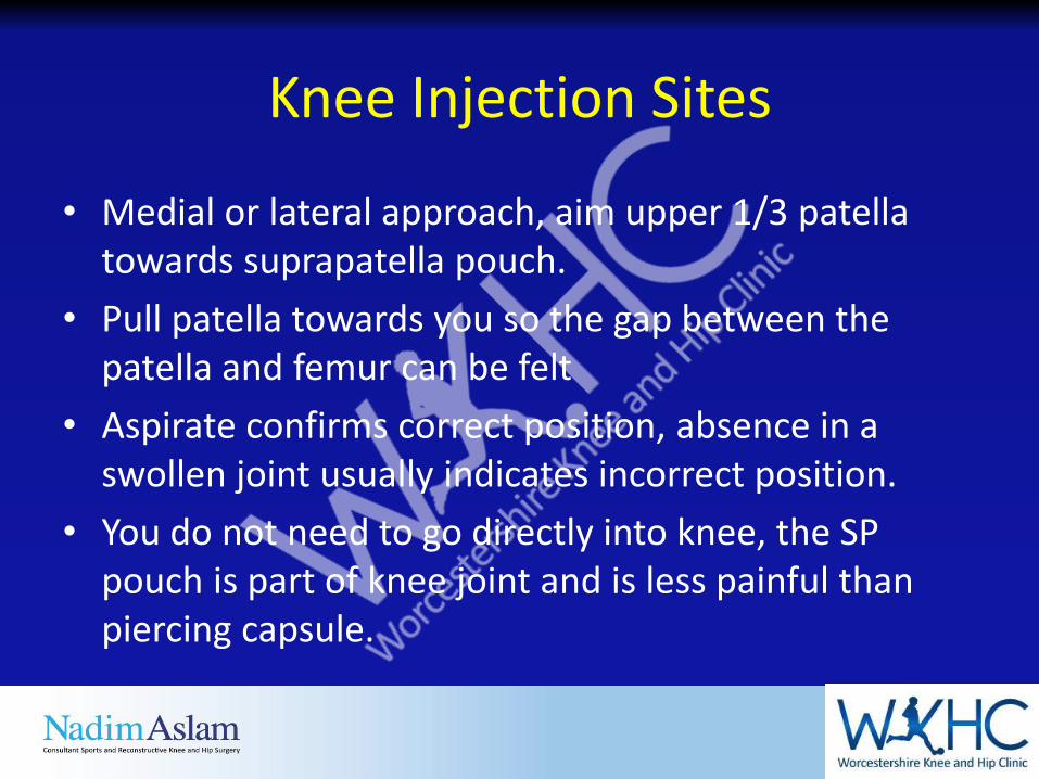
Knee Injection Sites
• Medial or lateral approach, aim upper 1/3 patella towards suprapatella pouch.
• Pull patella towards you so the gap between the patella and femur can be felt
• Aspirate confirms correct position, absence in a swollen joint usually indicates incorrect position.
• You do not need to go directly into knee, the SP pouch is part of knee joint and is less painful than piercing capsule.
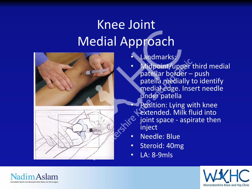
Knee JointMedial Approach
• Landmarks:• Midpoint/upper third medial
patellar border – push patella medially to identify medial edge. Insert needle under patella
• Position: Lying with knee extended. Milk fluid into joint space - aspirate then inject
• Needle: Blue• Steroid: 40mg• LA: 8-9mls
Knee Injection
• If you are in the wrong place DO NOT DIG AROUND LOOKING FOR THE GAP. Main pain caused by needling the periosteum
• Come out re-examine your landmarks and try again after re-cleaning skin and change needle.
Bursae around the knee
• Pre-patella bursa (housemaids knee)
• Infra-patella bursa (preachers knee)
• Popliteal bursa (Bakers ‘cyst’)
• Anserine bursa
Anserine bursa
• Common in OA especially with valgus knee. Also RA.
• Patient localises pain to site and tender
• Inject 40mg Depomedrone and Lidocaine
Politeal cyst/bursa
• Directly connected to knee joint
• Fluid comes from knee
• One way valve, cannot return to knee,
• No need to aspirate bursa will refill.
• After injection, bursa will settle with time (months)
• Rarely requires surgery, only if chronic and obstructing movement significantly.
Referred pain to knee
• If the knee looks normal the pain is persistent remember to check the rotation of the hip (can the patient reach shoe or sock by laterally rotating and flexing hip)
• If reduced, need to xray of hip.
• Knee pain may be the only symptom of significant OA in the hip.