Embed Size (px)
Citation preview
�������� ����� ��
Anthocyanins restore behavioral and biochemical changes caused bystreptozotocin-induced sporadic dementia of Alzheimer’s type
Jessie M. Gutierres, Fabiano B. Carvalho, Maria Rosa C. Schetinger,Patrıcia Marisco, Paula Agostinho, Marılia Rodrigues, Maribel A. Rubin,Roberta Schmatz, Cassia R. da Silva, Giana de P. Cognato, Vera M. Morsch,Cinthia M. Mazzanti, Mauricio Bogo, Carla D. Bonan, Roselia Spanevello
PII: S0024-3205(13)00713-3DOI: doi: 10.1016/j.lfs.2013.11.014Reference: LFS 13803
To appear in: Life Sciences
Received date: 2 August 2013Accepted date: 14 November 2013
Please cite this article as: Gutierres Jessie M., Carvalho Fabiano B., Schetinger MariaRosa C., Marisco Patrıcia, Agostinho Paula, Rodrigues Marılia, Rubin Maribel A.,Schmatz Roberta, da Silva Cassia R., de P. Cognato Giana, Morsch Vera M., MazzantiCinthia M., Bogo Mauricio, Bonan Carla D., Spanevello Roselia, Anthocyanins restorebehavioral and biochemical changes caused by streptozotocin-induced sporadic dementiaof Alzheimer’s type, Life Sciences (2013), doi: 10.1016/j.lfs.2013.11.014
This is a PDF file of an unedited manuscript that has been accepted for publication.As a service to our customers we are providing this early version of the manuscript.The manuscript will undergo copyediting, typesetting, and review of the resulting proofbefore it is published in its final form. Please note that during the production processerrors may be discovered which could affect the content, and all legal disclaimers thatapply to the journal pertain.
ACC
EPTE
D M
ANU
SCR
IPT
ACCEPTED MANUSCRIPT
1
Anthocyanins restore behavioral and biochemical changes caused by
streptozotocin-induced sporadic dementia of Alzheimer’s type
Jessié M. Gutierres1*, Fabiano B. Carvalho1, Maria Rosa C. Schetinger1, Patrícia
Marisco1, Paula Agostinho4, Marília Rodrigues1, Maribel A. Rubin1, Roberta Schmatz1,
Cassia R. da Silva1, Giana de P. Cognato2, Vera M. Morsch1, Cinthia M. Mazzanti¹,
Mauricio Bogo2, Carla D. Bonan2, Roselia Spanevello3*
1 Programa de Pós-Graduação em Bioquímica Toxicológica, Centro de Ciências
Naturais e Exatas, Universidade Federal de Santa Maria, Av. Roraima, 97105-900 -
Santa Maria, RS, Brazil.
2 Laboratório de Neuroquímica e Psicofarmacologia, Faculdade de Biociências,
Pontifícia Universidade Católica do Rio Grande do Sul. Avenida Ipiranga, 6681, 90619-
900 Porto Alegre, RS, Brazil.
3 Centro de Ciências Químicas, Farmacêuticas e de Alimentos, Universidade
Federal de Pelotas, Campus Universitário- Capão do Leão 96010-900 Pelotas, RS,
Brazil.
4 Center for Neuroscience and Cell Biology, Faculty of Medicine, University of Coimbra,
Coimbra, Portugal.
*Corresponding authors:
Gutierres J.M. (Jessié Martins Gutierres) and Spanevello R (Roselia Spanvello)
E-mail address: [email protected]
Phone: +55 55 32209557
ACC
EPTE
D M
ANU
SCR
IPT
ACCEPTED MANUSCRIPT
2
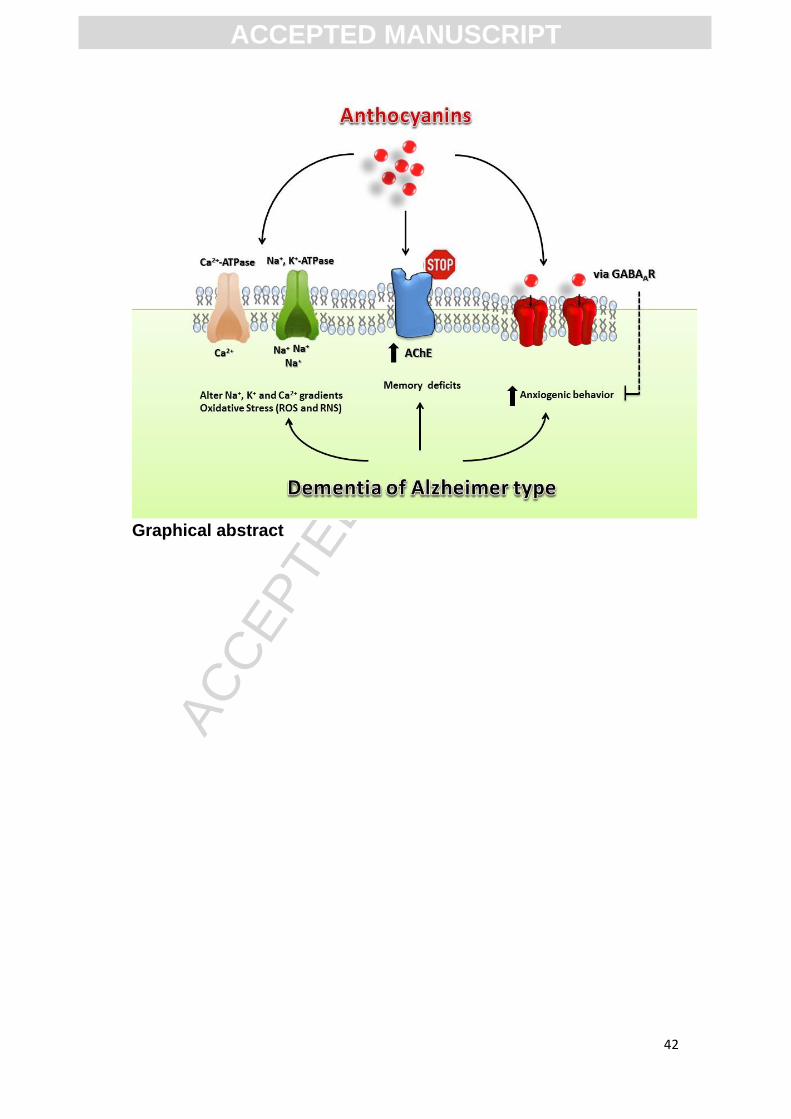
Abstract
Aims: The aim of this study was to analyze if the pre-administration of
anthocyanin on memory and anxiety prevented the effects caused by
intraceberoventicular streptozotocin (icv-STZ) administration-induced sporadic
dementia of Alzheimer’s type (SDAT) in rats. Moreover, we evaluated whether
the levels of nitrite/nitrate (NOx), Na+,K+-ATPase, Ca2+-ATPase and
acethylcholinesterase (AChE) activities in the cerebral cortex (CC) and
hippocampus (HC) are altered in this experimental SDAT.
Main methods: Male Wistar rats were divided in 4 different groups: control
(CTRL), anthocyanin (ANT), streptozotocin (STZ) and streptozotocin+
anthocyanin (STZ+ANT). After seven days of treatment with ANT (200 mg/kg;
oral), the rats were icv-STZ injected (3 mg/kg), and four days later were
performed the behavior parameters and submitted to euthanasia.
Key findings: A memory deficit was found in STZ group, but ANT treatment
showed to prevent this impairment of memory (P<0.05). Our results showed a
higher anxiety in icv-STZ group, but the treatment with ANT showed a per se
effect and prevented the anxiogenic behavior induced by STZ. Our results
reveal that ANT (100µM) tested diplace the specific binding of [H3]
flunitrazepam to benzodiazepinic site of GABAA receptors. AChE, Ca+-ATPase
activities and NOx levels were found to be increased in HC and CC in STZ
group, which was attenuated by ANT (P<0.05). STZ decreased Na+,K+-ATPase
activity and ANT was able to prevent these effects (P<0.05).
Significance: In conclusion, these findings demonstrated that ANT is able to
regulate ions pump activity and cholinergic neurotransmission, as well as able
to enhance memory and act as anxiolytic compound in animals with SDAT.
Keywords: Anxiety-like behavior; Nitric oxide production, Acetylcholinesterase,
Anthocyanin; ICV-streptozotocin; Memory; Rats
ACC
EPTE
D M
ANU
SCR
IPT
ACCEPTED MANUSCRIPT
3
1. Introduction
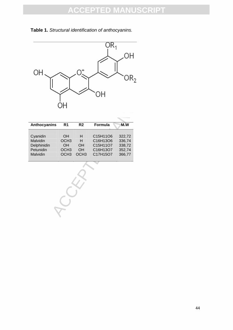
Anthocyanins (ANT) belong to the flavonoid family, which present
phenolic groups in their chemical structure and give colors to a great variety of
flowers and fruits (Table1) (Veitch and Grayer, 2011, Williams and Grayer,
2004, Yoshida et al. , 2009). It has been shown that ANT are potent
antioxidants, and prove to be effective scavengers of reactive oxygen species
(ROS) and reactive nitrogen species (RNS) (Kahkonen and Heinonen, 2003,
Kahkonen et al. , 2001), having a clear neuroprotective role (Min et al. , 2011).
There is evidence that ANT have beneficial effects on memory and cognition
(Shukitt-Hale et al., 2009) improving the memory in old rats and humans
(Andres-Lacueva et al., 2005, Krikorian et al., 2010).
Acetylcholinesterase (AChE) is an important regulatory enzyme that
rapidly hydrolyses the neurotransmitter acetylcholine (ACh) released by the
cholinergic neurons (Paleari et al. , 2008). Several experimental and clinical
studies clearly indicate an undisputed major role of ACh in the regulation of
cognitive functions (Blokland, 1995). Recently, several therapeutic strategies
that enhance AChE activity have been implemented to ameliorate cognitive
disorders. Cognitive disorders also affect the generation of membrane
potentials and the influx of neuronal Ca2+ (Berrocal et al. , 2009, Mata et al. ,
2011).
The Na+,K+-ATPase and the Ca2+-ATPase are key enzymes in the
maintenance of electrolyte gradients in excitable cells and neurons (Jimenez et
al., 2010, Panayiotidis et al., 2010). The former enzyme is responsible for the
active transport of Na+ and K+, and it is necessary to maintain the ionic gradient
across membranes and thus it is essential to regulate neuronal excitability
ACC
EPTE
D M
ANU
SCR
IPT
ACCEPTED MANUSCRIPT
4
(Jimenez, Sanchez, 2010, Jorgensen et al. , 2003, Kaplan, 2002). The Ca2+-
ATPase is one of the most powerful modulators of intracellular Ca2+ levels
(Casteels et al. , 1991, Huang et al. , 2010, Raeymaekers and Wuytack, 1991).
The transient changes in intracellular Ca2+ levels regulate a wide variety of
cellular processes and cells employ both intracellular and extracellular sources
of Ca2+ for the activation of signaling pathways and regulation of many
physiological and pathological processes (Huang, Nagaraja, 2010, Missiaen et
al. , 2000a, Missiaen et al. , 2000b, Ruknudin and Lakatta, 2007).
Alzheimer’s disease (AD) is the most common cause of dementia in the
elderly, and this disease is characterized by abnormalities in glucose
metabolism, reduced glucose utilization and levels of energy rich phosphates
(Hoyer, 2004a, b). The intracerebroventricular (icv) injection of STZ in rats has
been used as a model of sporadic dementia of AD (Sharma and Gupta, 2001)
since it mimics many pathological processes of the disease as impaired brain
glucose and energy and leads to progressive deficits in learning and memory
(Lannert and Hoyer, 1998)
Considering that AD is the most prevalent neurodegenerative disease
worldwide in older adults, we sought to investigate if anthocyanin has the ability
to prevent memory deficits induced by icv administration of STZ. We also
evaluated the levels of nitrite/nitrate and the activities of enzymes AChE,
Na+,K+-ATPase and Ca2+-ATPase, which are known to be altered in AD.
2. Material and Methods
2.1. Chemicals
ACC
EPTE
D M
ANU
SCR
IPT
ACCEPTED MANUSCRIPT
5
Acetylthiocholine, Trizma Base, Acetonitrile, Percoll, Coomassie
Brilliant Blue G and Streptozotocyn (STZ) were purchased from Sigma
Chemical Co (St Luis, MO, USA). Anthocyanins was extracted and purified
from grape skin and are commercially available by Christian Hansen A/S. All
other reagents used in the experiments were of analytical grade and of the
highest purity.
2.2 Animals
Male Wistar rats (3 month year old) weighing 350–400 g were used in
the study. They were kept in the Central Animal House of Federal University of
Santa Maria in colony cages at an ambient temperature of 25±2 °C and relative
humidity 45–55% with 12 h light/dark cycles. They had free access to standard
rodent pelleted diet and water ad libitum. All procedures were carried out ac-
cording to NIH Guide for Care and Use of Laboratory Animals, and Brazilian
Society for Neuroscience and Behavior (SBNeC) recommendations for animal
care. This work was approved by the ethical committee of Federal University of
Santa Maria (23081.003601/2012-63).
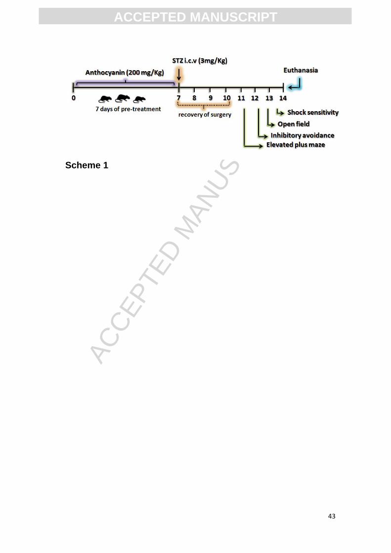
2.3. Administration of drugs to animals
2.3.1. Intracerebroventricular (icv) injection of streptozotocin
Adult male Wistar rats (300–350g) were anaesthetized with thiopental
(180mg/kg). The head was placed in position in the stereotaxic apparatus and a
midline sagittal incision was made in the scalp. The stereotaxic coordinates for
lateral ventricle (Paxinos and Watson, 1986) were measured accurately as
anterio-posterior -0.8mm, lateral 1.5mm and dorso-ventral, -4.0mm relative to
ACC
EPTE
D M
ANU
SCR
IPT
ACCEPTED MANUSCRIPT
6
bregma and ventral from dura with the tooth bar set at 0 mm. Through a skull
hole, a 28-gauge Hamilton® syringe of 10 μl attached to a stereotaxic
apparatus and piston of the syringe was lowered manually into each lateral
ventricle. We used 4 different groups: control (CTRL), anthocyanin (ANT),
streptozotocin (STZ), and streptozotocin plus anthocyanin (STZ+ANT). The STZ
groups received bilateral icv injection of streptozotocin (3 mg/kg, body weight)
which was dissolved in citrate buffer (pH 4.4) (Tiwari et al. , 2009). The
concentration of STZ in citrate buffer was adjusted so as to deliver 5 μl/injection
site of the solution. Rats in the control group received icv injection of same
volume of citrate buffer as in STZ treated (Scheme 1).
2.3.2 Drug administration
Seven to ten animals per group were usually tested in the experiments.
Rats were treated by gavage with anthocyanin (200 mg/kg body weight) daily
per 7 days (around 10 a.m.). The dose of anthocyanin was chosen on the basis
of previous studies indicating neuroprotection (Gutierres et al. , 2012b, Manach
et al. , 2004, Saija et al. , 1990, Varadinova et al. , 2009). The control groups
received only vehicle (2ml/kg gavage of saline, daily per 7 days).
2.4 Behavioral procedure
2.4.1 Elevated plus maze task
At the last day of anthocyanin treatment (7th day), the anxiolytic-like
behavior was evaluated using the task of the elevated plus maze as previously
described (Frussa-Filho et al. , 1999, Rubin, Albach, 2000a). The apparatus
consists of a wooden structure raised to 50 cm from the floor. This apparatus is
ACC
EPTE
D M
ANU
SCR
IPT
ACCEPTED MANUSCRIPT
7
composed of 4 arms of the same size, with two closed-arms (walls 40 cm) and
two open-arms. Initially, the animals were placed on the central platform of the
maze in front an open arm. The animal had 5 minutes to explore the apparatus,
and the time spent and the number of entries in open and closed-arms were
recorded. The apparatus was thoroughly cleaned with 30% ethanol between
each session.
2.4.2 Inhibitory avoidance task
The animals were subjected to training in a step-down inhibitory
avoidance apparatus as previously described (Rubin et al. , 2000b). After that
the animals received icv-STZ (3 mg/kg). Twenty four hours after the training the
animals were subjected to test in a step-down inhibitory avoidance task. Briefly,
the rats were subjected to a single training session in a step-down inhibitory
avoidance apparatus, which consisted of a 25×25×35-cm box with a grid floor
whose left portion was covered by a 7×25-cm platform, 2.5 cm high. The rats
were placed gently on the platform facing the rear left corner, and when the rat
stepped down with all four paws on the grid, a 3-s 0.4-mA shock was applied to
the grid. Retention test took place in the same apparatus 24 h later. Test step-
down latency was taken as a measure of retention, and a cut-off time of 300s
was established.
2.4.3 Open field
Immediately after the inhibitory avoidance test session, the animals
were transferred to an open-field measuring 56×40×30 cm, with the floor
divided into 12 squares measuring 12×12 cm each. The open field session
ACC
EPTE
D M
ANU
SCR
IPT
ACCEPTED MANUSCRIPT
8
lasted for 5 min and during this time, an observer, who was not aware of the
pharmacological treatments, recorded the number of crossing responses and
rearing responses manually. This test was carried out to identify motor
disabilities, which might influence inhibitory avoidance performance at testing
(Gutierres et al. , 2012a).
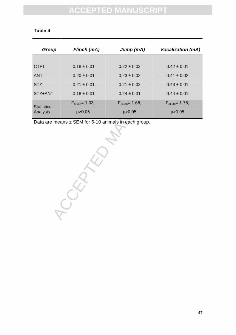
2.4.4 Foot shock sensitivity test
Reactivity to shock was evaluated in the same apparatus used for
inhibitory avoidance, except that the platform was removed and was used to
determine the flinch and jump thresholds in experimentally naïve animals
(Berlese et al. , 2005, Rubin et al. , 2000a). The animals were placed on the
grid and allowed a 3 min habituation period before the start of a series of
shocks (1s) delivered at 10 s intervals. Shock intensities ranged from 0.1 to 0.5
mA in 0.1 mA increments. The adjustments in shock intensity were made in
accordance with each animal's response. The intensity was raised by one unit
when no response occurred and lowered by one unit when a response was
made. A flinch response was defined as withdrawal of one paw from the grid
floor, and a jump response was defined as withdrawal of three or four paws.
Two measurements of each threshold (flinch and jump) were made, and the
mean of each score was calculated for each animal.
2.5 Brain tissue preparation
After behavioral tests, the animals were anesthetized under halothane
atmosphere, euthanized by decapitation and the brain was removed and
separated into cerebral cortex and hippocampus and placed in a solution of
ACC
EPTE
D M
ANU
SCR
IPT
ACCEPTED MANUSCRIPT
9
Tris–HCl 10 mM, pH 7.4, on ice. The brain structures were gently homogenized
in a glass potter in Tris–HCl solution. Aliquots of resulting brain structure
homogenates were stored at −20°C until utilization (Gutierres, Carvalho,
2012a). Protein was determined previously in a strip that varied for each
structure: cerebral cortex (0.7 mg/ml) and hippocampus (0.8 mg/ml), and
determined by Coomassie blue method as previously described (Bradford,
1976), using bovine serum albumin as standard solution.
2.6 Isolation of synaptosomes with a discontinuous Percoll gradient
Synaptosomes were isolated essentially as previously described (Nagy
and Delgado-Escueta, 1984), with a minor modification (Gutierres et al. , 2012c)
using a discontinuous Percoll gradient. The cerebral cortex and hippocampus
were gently homogenized in 10 volumes of an ice-cold medium (medium I)
containing 320 mM sucrose, 0.1 mM EDTA and 5 mM HEPES, pH 7.5, in a
motor driven Teflon-glass homogenizer and then centrifuged at 1,000xg for 10
min. An aliquot of 0.5 mL of the crude mitochondrial pellet was mixed with 4.0
mL of an 8.5% Percoll solution and layered into an isosmotic discontinuous
Percoll/sucrose gradient (10%/16%). The synaptosomes that banded at the
10/16% Percoll interface were collected with a wide-tip disposable plastic
transfer pipette. The synaptosomal fraction was washed twice with an isosmotic
solution consisting of 320 mM sucrose, 5.0 mM HEPES, pH 7.5, and 0.1 mM
EDTA by centrifugation at 15,000 g to remove the contaminating Percoll. The
pellet of the second centrifugation was resuspended in an isosmotic solution to
a final protein concentration of 0.4-0.6 mg/ml. Synaptosomes were prepared
ACC
EPTE
D M
ANU
SCR
IPT
ACCEPTED MANUSCRIPT
10
fresh daily and maintained at 0º-4º throughout the procedure and used to
measure Ca2+-ATPase and AChE activities.
2.7. Assay of Lactate Desydrogenase (LDH)
The integrity of the synaptosomes preparations was confirmed by the
lactate dehydrogenase (LDH) activity, which was obtained after synaptosome
lysis with 0.1 % Triton X-100 and comparing it with an intact preparation, using
the Labtest kit (Labtest, Lagoa Santa, MG, Brasil).
2.8. Determination of AChE activity in brain
The AChE enzymatic assay was determined by a modification of the
spectrophotometric method (Rocha et al., 1993) as previously described
(Ellman et al. , 1961). The reaction mixture contained 100 mM K+-phosphate
buffer, pH 7.5 and 1 mM 5,5′-dithiobisnitrobenzoic acid (DTNB). The method is
based on the formation of the yellow anion, 5,5′-dithio-bis-acid-nitrobenzoic,
measured by absorbance at 412 nm during 2min incubation at 25°C. The
enzyme (40–50 μg of protein) was pre-incubated for 2 min. The reaction was
initiated by adding 0.8 mM acetylthiocholine iodide (AcSCh). All samples were
run in triplicate and the enzyme activity was expressed in μmol AcSCh/h/mg of
protein.
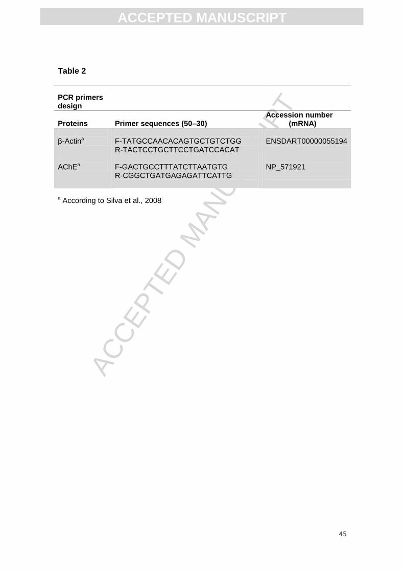
2.9. Analysis of Gene Expression using semiquantitative RT-PCR
The analysis of AChE expression was carried out using
semiquantitative reverse transcriptase polymerase chain reaction (RT-PCR).
The hippocampus and cerebral cortex were dissected under sterile conditions,
and total RNA was extracted using the TRIzol® Reagent (Invitrogen, Carlsbad,
ACC
EPTE
D M
ANU
SCR
IPT
ACCEPTED MANUSCRIPT
11
CA, USA) according to the manufacturer’s instructions. The RNA was quantified
by spectrophotometry, and cDNA was synthesized using the ImProm-II™
Reverse Transcription System (Promega). PCR reactions for the AChE and β-
actin genes were performed using 0.1 μM of the appropriate primers (AChE
forward: 5′- GAC TGC CTT TAT CTT AAT GTG -3′ and reverse: 5′- CGG CTG
ATG AGA GAT TCA TTG -3′; β-actin forward 5′-TAT GCC AAC ACA GTG CTG
TCT GG-3′; and reverse 5′-TAC TCC TGC TTC CTG ATC CAC AT-3′) (see
Table 2), 0.2 μM dNTP, 2 mM MgCl2, and 0.1 U Platinum Taq DNA polymerase
(Invitrogen) in a total volume of 25 μL for AChE and 20 μL for β-actin (Da Silva
et al. , 2008). The following conditions were used for the PCR reactions: 1 min
at 94°C; 1 min at the annealing temperature (54°C for β-actin and 55°C for
AChE) and 1 min at 72°C for 35 cycles. Post-extension at 72°C was performed
for 10 min. For each set of PCR reactions, a negative control was also included.
The PCR products (AChE, 785 bp; β-actin, 210 bp) were analyzed on a 1.5%
agarose gel containing GelRed® (Biotium) and visualized under ultraviolet light.
The Low DNA Mass Ladder (Invitrogen) was used as a molecular marker, and
normalization was performed using β-actin as the constitutive gene. All PCR
analysis was run in triplicates, including negative controls (in which no reverse
transcriptase nor cDNA-containing samples were added in the PCR mix). No
background fluorescence was observed when control samples were analyzed
(data not shown)."
2.10. Na+,K+-ATPase activity measurement
Na+,K+-ATPase activity was measured as previously described (Wyse
et al. , 2000) with minor modifications (Carvalho et al. , 2012). Briefly, the assay
ACC
EPTE
D M
ANU
SCR
IPT
ACCEPTED MANUSCRIPT
12
medium consisted of (in mM) 30 Tris-HCl buffer (pH 7.4), 0.1 EDTA, 50 NaCl, 5
KCl, 6 MgCl2 and 50 μg of protein in the presence or absence of ouabain (1
mM), in a final volume of 350 μL. The reaction was started by the addition of
adenosine triphosphate to a final concentration of 3 mM. After 30 min at 37ºC,
the reaction was stopped by the addition of 70 μL of 50% (w/v) trichloroacetic
acid. Saturating substrate concentrations were used, and reaction was linear
with protein and time. Appropriate controls were included in the assays for non-
enzymatic hydrolysis of ATP. The amount of inorganic phosphate (Pi) released
was quantified colorimetrically, as previously described (Fiske and Subbarow,
1927), using KH2PO4 as reference standard. Specific Na+,K+-ATPase activity
was calculated by subtracting the ouabain-insensitive activity from the overall
activity (in the absence of ouabain) and expressed in nmol of Pi/min/mg of
protein.
2.11. Ca2+-ATPase activity measurement
Ca2+-ATPase activity was measured as previously described (Rohn et
al. , 1993) with minor modifications (Trevisan et al. , 2009). Briefly, the assay
medium consisted of (in mM) 30 Tris-HCl buffer (pH 7.4), 0.1 EGTA, 3 MgCl2
and 100 μg of protein in the presence or absence of 0.4 CaCl2, in a final volume
of 200 μL. The reaction was started by the addition of adenosine triphosphate to
a final concentration of 3 mM. After 60 min at 37ºC, the reaction was stopped by
the addition of 70 μL of 50% (w/v) trichloroacetic acid. Saturating substrate
concentrations were used, and reaction was linear with protein and time.
Appropriate controls were included in the assays for non-enzymatic hydrolysis
of ATP. The amount of inorganic phosphate (Pi) released was quantified
ACC
EPTE
D M
ANU
SCR
IPT
ACCEPTED MANUSCRIPT
13
colorimetrically, as previously described (Fiske and Subbarow, 1927), using
KH2PO4 as reference standard. The Ca2+-ATPase activity was determined by
subtracting the activity measured in the presence of Ca2+ from that determined
in the absence of Ca2+ (no added Ca2+ plus 0.1 mM EGTA) and expressed in
nmol of Pi/min/mg of protein.
2.12 [H3] Flunitrazepam binding assay
To determine if the effect of anthocyanins can be mediated by the
GABAA/BDZ complex, we performed a specific binding assay of the BDZ site of
GABAA receptors using [3H]flunitrazepam according to a previous study (Della-
Pace et al., 2013). Cerebral cortex from each animal was thawed and
homogenized in 10 ml of homogenization buffer A (10 mM Tris-HCl, 300 mM
sucrose, and 2 mM EDTA, pH 7.4) per gram of tissue. This homogenate was
centrifuged at 1,000 X g for 10 min at 4°C. The resulting supernatant was
centrifuged at 16,000 X g for 20 min at 4°C. The resulting pellet was then
resuspended in 1 ml of homogenization buffer and frozen at - 70°C until
analyzed.
2.12.1 Radioligand binding assay
[3H] flunitrazepam binding to benzodiazepinic site of GABAA receptors
was determinedby first washing the cell membrane preparation as follows:
individual aliquots were diluted with five volumes of wash buffer B (50 mM Tris-
HCl and 2 mM EDTA, pH 7.4), mixed, and centrifuged at 16,000 X g for 10 min
at 4°C, and the samples were incubated for 30 min at 37°C. This washing
procedure was repeated twice, and the final pellet was resuspended in binding
ACC
EPTE
D M
ANU
SCR
IPT
ACCEPTED MANUSCRIPT
14
assay buffer C (20 mM HEPES and 1 mM EDTA, pH 7.4). The protein
concentration of each sample was determined by a spectrophotometric protein
dye-binding assay based on the method of Bradford (1976), using bovine serum
albumin as the standard. The incubation was carried out in duplicate in
polycarbonated tubes (total volume 500 µL) containing 50 mM Tris–HCl (pH
7.4), 0.5 mg of protein membrane. Diazepam (0.1 µM) was used as a positive
control. Incubation was started by adding 1 nM [3H] flunitrazepam
(85,8Ci/mmol), and run at ice for 60 min. The reaction was stopped by vacuum
filtration and each filter was washed with 15 mL of cold 10 mM Tris–HCl buffer.
Filters were individually placed in polycarbonated tubes and 1 mL of scintillation
liquid was added. Radioactivity was determined using a Packard Tri-Carb
2100TR liquid scintillation counter. Non-specific binding was determined by
adding 1 µM diazepam to the medium in parallel assays. Specific binding was
considered as the difference between total binding and non-specific binding.
Results were expressed as percentage of specific binding.
2.13. Assay of nitrite plus nitrate (NO2 plus NO3)
For NOx determination, an aliquot (200 μl of samples) was homoge-
nized in 200mM Zn2SO4 and acetonitrile (96%, HPLC grade). After, the homog-
enate was centrifuged at 16,000 xg for 20min at 4°C and supernatant was sepa-
rated for analysis of the NOx content as previously described (Miranda et al. ,
2001). The resulting pellet was suspended in NaOH (6M) for protein determina-
tion.
2.14. Glucose analysis
ACC
EPTE
D M
ANU
SCR
IPT
ACCEPTED MANUSCRIPT
15
The glucose levels were measured using standard enzymatic methods
from Ortho-Clinical Diagnostics® reagents on the fully automated analyzer
(Vitros 950® dry chemistry system; Johnson & Johnson, Rochester, NY, USA).
2.15. Statistical analysis
Statistical analysis of training and test step-down latencies was carried
out by the Scheirer–Ray–Hare extension of the Kruskal–Wallis test
(nonparametric two-way ANOVA). The open field, binding assay and foot shock
sensitivity was analyzed by one-way ANOVA following by student Newman-
Keuls. The other tests were analyzed by two-way ANOVA, followed by Tukey
test, and considered P<0.05 or P<0.001 as significant difference in all
experiments.
3. Results
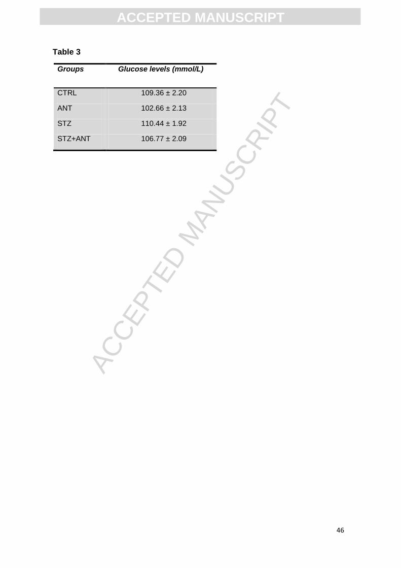
3.1 Glucose Levels
During the complete study there were no differences in body weight and
water consumption in all groups (data not shown). There was no significant
difference between the mean peripheral glucose levels after 3 mg/kg icv-STZ
groups and citrate buffer (pH 4.4) icv injection groups. The mean peripheral
glucose levels were 109.36 ± 2.20 CTRL group, 102.66±2.13 ANT group,
110.44 ± 1.92 STZ group and 106.77±2.09 STZ+ANT group, respectively,
indicating that the dose was sub diabetogenic (Table 3).
3.2 Behavioral tests
3.2.1 Anthocyanin prevents the impairment of memory induced by STZ.
ACC
EPTE
D M
ANU
SCR
IPT
ACCEPTED MANUSCRIPT
16
In this study we used 4 groups of animals: control (CTRL), anthocyanin
(ANT), streptozotocin (STZ), and streptozotocin plus anthocyanin (STZ+ANT).
Figure 1 shows the effect of ANT treatment on the STZ-induced memory
deficits, in the step-down latencies. Statistical analysis of Scheirer–Ray–Hare
test (nonparametric two-way ANOVA) showed a significant difference between
STZ (3 mg/kg) vs ANT (200 mg/kg) or vehicle interaction (CTRL), revealing that
treatment with ANT prevented the impairment of memory induced by STZ [H2 =
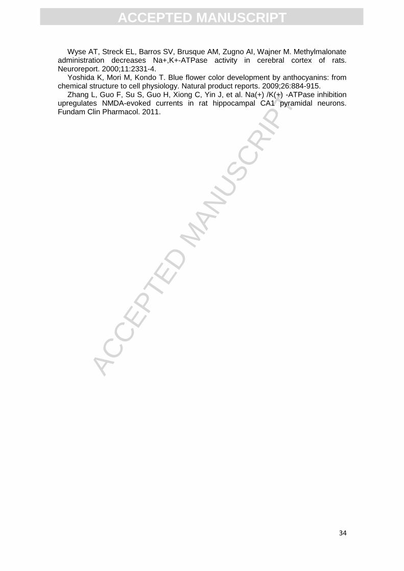
9,75; P< 0.01; Figure 1D]. Statistical analysis of the data obtained during
training showed no difference between the different groups (Figure 1C).
Although, motivational disparities in the training session may account
for differences in inhibitory avoidance testing, experiments were performed to
assess whether STZ or ANT affected shock sensitivity threshold and locomotor
capacity of the animals. Statistical analysis of open-field data (one-way ANOVA)
revealed that STZ did not alter the number of crossing (F (3,42)=0.11, P>0.05;
Figure 1A) or rearing (F (3,42)=1.82, P>0.05; Figure 1B) responses in a
subsequent open-field test session, suggesting that neither STZ nor ANT
caused gross motor disabilities in this task. Moreover, STZ did not alter foot
shock sensitivity, as demonstrated by the similar flinch and jump thresholds
exhibited by the animals. In table 4 it can be seen that neither ANT+STZ
animals nor STZ animals were affected in their motor performances and foot
shock sensitivity: flinch [F (3,30)= 1.33; P>0.05], jump [F (3,30)= 1.66; P>0.05]
and vocalization [F (3,30)= 1.76; P>0.05].
3.2.2 Effect of STZ and anthocyanin treatment on anxiolytic-like behavior
ACC
EPTE
D M
ANU
SCR
IPT
ACCEPTED MANUSCRIPT
17
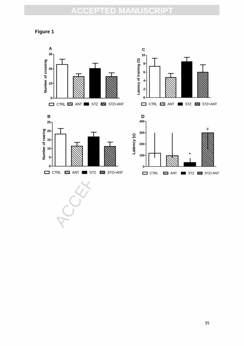
Figure 2 shows the effect of the treatment with anthocyanin and STZ on
anxiolytic-like behavior in the elevated plus maze task. Statistical analysis (two-
way ANOVA) showed a significant CTRL or STZ (3 mg/kg) vs CTRL or ANT
(200 mg/kg) interaction to time spent (s) in open arms [F (1,41)= 6.264; P<0.05;
Figure 2D] and time in closed arms [F (1,41)= 4.925; P<0.05; Figure 2C],
revealing that treatment with ANT prevented the anxiogenic behavior induced
by STZ. However, no significant differences in the number of entries in open
arms [F (1,41)= 0.279; P>0.05; Figure 2B] and in the number of entries in all
arms [F (1,41)= 0.68; P>0.05; Figure 2A] were observed. The number of total
entries in arms suggests that neither icv-STZ nor ANT animals had altered
locomotor activity in the elevated plus maze task.
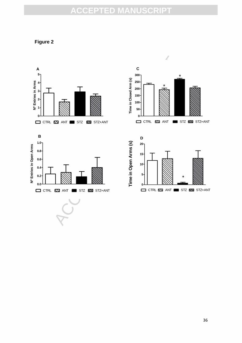
3.3 Binding of [3H] flunitrazepam to benzodiazepinic site assay
Since we observed an anxiolytic effect of ANT in the elevated plus maze
task, we decided to investigate whether the compound can alter the binding of
[3H] flunitrazepam to the benzodiazepinic site of GABAA receptor. The results
presented in Figure 3 reveal that the ANT (100µM) reduced by 43% the [3H]
flunitrazepam binding to benzodiazepinic site of GABAA receptors [F (2,17)=
47.890; P<0.0001] and this result demonstrates that ANT can interact to GABAA
receptors.
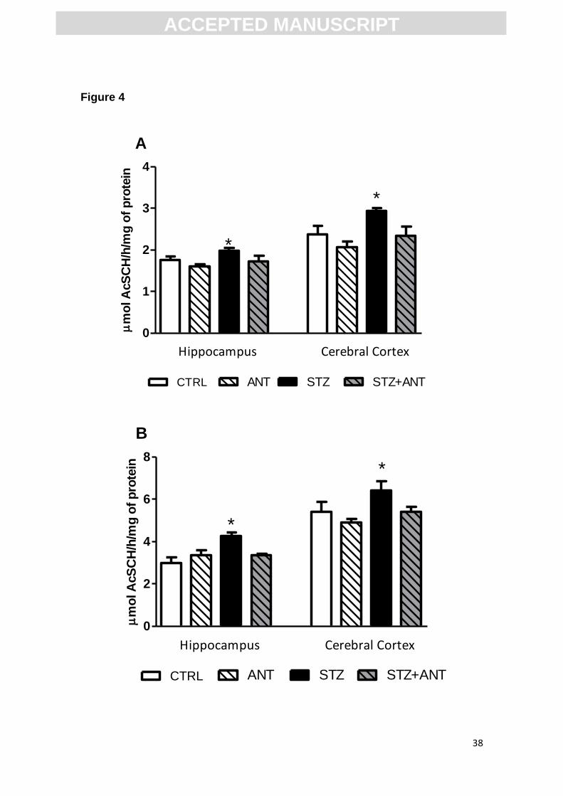
3.4 Activity and expression of acetylcholinesterase
3.4.1 Anthocyanin prevents the increase in AChE activity induced by STZ.
Previous studies report cholinergic impairments in cognitive disorders by
quantification of acetylcholinesterase (AChE) activity. Therefore, we investigat-
ed whether ANT restores AChE activity in the model of SDAT. Figure 4 shows
ACC
EPTE
D M
ANU
SCR
IPT
ACCEPTED MANUSCRIPT
18
the effect of ANT and STZ on the AChE activity in cerebral cortex and hippo-
campus, both in S1 and synaptosomes of rats. We found a significant CTRL or
STZ (3 mg/kg) vs CTRL or ANT (200 mg/kg) interaction, suggesting that the
ANT treatment prevents the increase in AChE activity in S1 fraction of cerebral
cortex [F= (1,28)= 7.973; P<0.05] and hippocampus [F (1,28)= 4.995; P<0.05]
(Figure 4A) induced by icv-STZ.
Importantly, synaptosome fraction analysis showed a significant CTRL or
STZ (3 mg/kg) vs CTRL or ANT (200 mg/kg) interaction, suggesting that the
ANT treatment prevents the increase in AChE activity in the synaptosomes of
cerebral cortex [F (1,28)= 4.760; P<0.05] and hippocampus [F (1,28)= 8. 434;
P<0.01](Figure 4B) induced by icv-STZ.
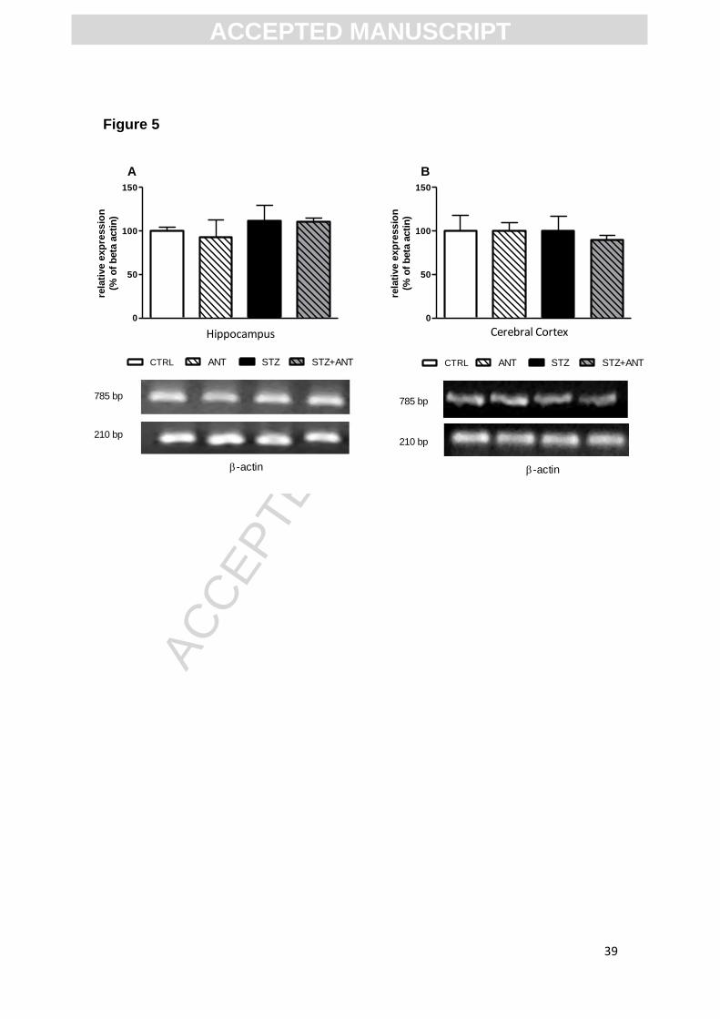
3.4.2 Effect of STZ and anthocyanin treatment on the AChE expression in the
cortex and hippocampus of rats.
Figure 5 shows the effect of ANT and STZ on the AChE expression in
cerebral cortex and hippocampus of rats. No significant differences in AChE
expression between groups were observed in the cerebral cortex [F (1,8)=
0.423; P>0.05] and hippocampus [F (1,8)= 0.140; P>0.05].
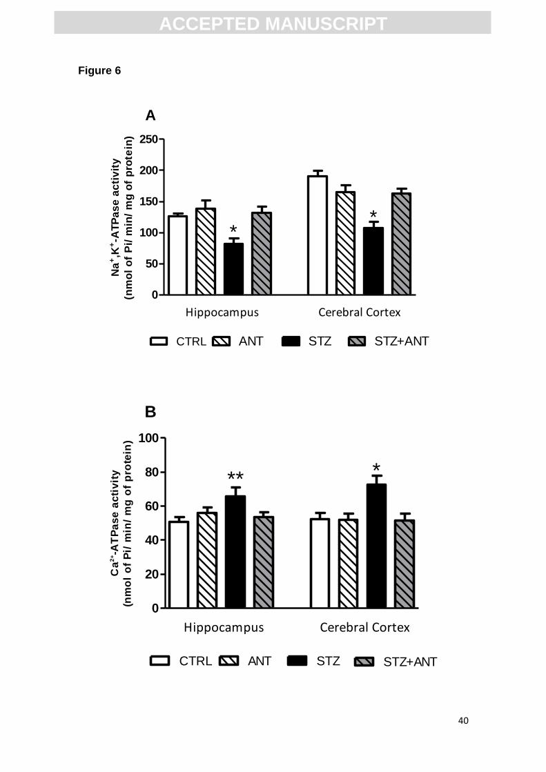
3.5 Anthocyanin prevents the decrease of Na+,K+-ATPase and increase of Ca2+-
ATPase activity induced by STZ.
Na+,K+-ATPase and Ca2+-ATPase are enzymes involved in the control of
neurotransmission, regulating the membrane potential and extracellular calcium
concentrations, respectively. Figure 6 shows the effect of ANT and STZ on the
activity of Na+,K+-ATPase and Ca2+-ATPase in cerebral cortex and
ACC
EPTE
D M
ANU
SCR
IPT
ACCEPTED MANUSCRIPT
19
hippocampus of rats. Statistical analysis (two-way ANOVA) showed a significant
CTRL or STZ (3 mg/kg) vs CTRL or ANT (200 mg/kg) interaction, suggesting
that the ANT treatment prevents the decrease in Na+,K+-ATPase activity in the
cerebral cortex [F (1,28)= 17.760; P<0.001] and hippocampus [F (1,28)= 4.978,
P<0.05] induced by icv-STZ (Figure 6A).
Additionally, two-way ANOVA showed a significant CTRL or STZ (3
mg/kg) vs CTRL or ANT (200 mg/kg) interaction, suggesting that the ANT
treatment prevents the increase of Ca2+-ATPase activity in the cerebral cortex
[F (1,28)= 5.671; P<0.05] and hippocampus [F (1,28)= 5.272; P<0.05] induced
by icv-STZ (Figure 6B).
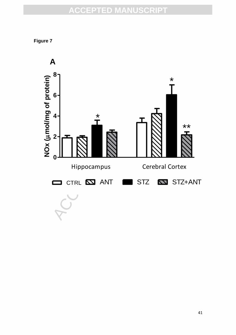
3.6 NOx levels determination
Anthocyanins are known by their antioxidant properties, so in this set of
experiments we investigated if ANT alters nitrite/nitrate content in the brain of
rats. Figure 7 shows the effect of ANT and STZ on the NOx levels production in
cerebral cortex and hippocampus. Statistical analysis (two-way ANOVA)
showed a significant CTRL or STZ (3 mg/kg) vs CRTL or ANT (200 mg/kg)
interaction, suggesting that the ANT treatment prevents the increased NOx
levels both in cerebral cortex [F (1,28)= 8.583; P<0.05] and hippocampus [F
(1,28)= 23.350; P<0.0001] induced by icv-STZ.
4. Discussion
Anthocyanins are flavonoids found in fruits and fruit juices, and have the
capacity to improve memory (Harborne and Williams, 2001, Williams and
ACC
EPTE
D M
ANU
SCR
IPT
ACCEPTED MANUSCRIPT
20
Grayer, 2004, Williams et al. , 2008). Several evidence has demonstrated that
ANT are able to improve memory of old rats in Morris water maze (Andres-
Lacueva, Shukitt-Hale, 2005), and of mice in the inhibitory avoidance task
(Barros et al. , 2006) and eldery humans (Krikorian, Shidler, 2010). There is
evidence that ANT prevents neurotoxicity induced by: i) ethanol in developing
brain mice (Ke et al. , 2011), ii) reperfusion damage model of cerebral ischemia
(Min, Yu, 2011, Shin et al. , 2006), iii) deleterious effects found in models of
Parkinson's (Kim et al. , 2010) and Alzheimer's disease (Shih et al. , 2010).
Additionally, there was a large number of studies’ indicating the
neuroprotective role of ANT, since studies have shown that ANT can be
transported across biological membranes (Passamonti et al., 2005, Talavera et
al.,2005). There are studies announcing gastric absorption and neuroprotective
effects of ANT-rich foods, but there is a gap in the knowledge concerning, for
example, gastric or ANT transport across the blood-brain barrier (BBB) (Kalt et
al.,2008). Another important factor affecting ANT bioavailability and
pharmacokinetic properties are their possible ingestion as pigments
(anthocyanin derivatives), especially when considering wine consumption. A
recent work had already indicated that anthocyanin pyruvic-acid adducts can
rapidly reach rat plasma 15min after oral administration of 400 mg/kg (Faria et
al., 2009a, Faria et al., 2009b). Thus, we investigated whether this natural
compound could prevent some alterations found in a model of SDAT induced by
icv-STZ injection.
Furthermore, a subdiabetogenic dose of STZ (3mg/kg) to rodents
causing a progressive memory impairment, loss and synaptic dysfunction
(Lannert and Hoyer, 1998, Pinton et al. , 2010). Thus, there was no significant
ACC
EPTE
D M
ANU
SCR
IPT
ACCEPTED MANUSCRIPT
21
difference on the mean peripheral glucose levels between CTRL and icv-STZ
groups (Table 3). Our results indicated that icv-STZ impaired the acquisition of
memory in rats trained on the inhibitory avoidance task. Interestingly, we found
out that ANT at 200 mg/kg for 7 days, did not affect the memory of rats and
prevented the memory deficits induced by icv-STZ (Figure. 1D), as assessed by
the inhibitory avoidance task. Furthermore, previous studies from our laboratory
demonstrated that the dose of 200 mg/kg ANT antagonized scopolamine-
induced performance deficits in rats (Gutierres, Carvalho, 2012b) suggesting
that ANT have a close interaction with cholinergic system.
Immediately after inhibitory avoidance test, the animals were subjected
to an open-field test which is widely used for evaluating motor abnormalities
(Belzung and Griebel, 2001). The open field session revealed that the treatment
with icv-STZ or ANT did not alter spontaneous locomotor activity (Figure 1A, B).
Moreover, we observed that the rats of different groups did not show altered
shock sensitivity (Table 4), as verified by their similar flinch, jump and
vocalization thresholds. These data suggest that neither STZ nor ANT
administration caused motor disabilities or altered foot shock sensitivity,
excluding their possibility of interference in step-down latencies of inhibitory
avoidance task.
Besides to assessing the acquisition memory in the inhibitory avoidance
task, we also measured the anxiolytic-like behavior by the elevated plus maze
task, commonly used to study anxiety-related behavior in rodents (Belzung and
Griebel, 2001). Our results showed a higher anxiety in icv-STZ group (3 mg/kg)
(Figure 2C; D) which is in accordance with previous observations that mice
subjected to icv-STZ, in short time (7days) and long time (21 days) treatments,
ACC
EPTE
D M
ANU
SCR
IPT
ACCEPTED MANUSCRIPT
22
have a rise increase in anxious behavior (Pinton et al. , 2011). We found an
anxiolytic effect of treatment of ANT that was observed by time in closed arm.
However, ANT did not change the number of entries and time spent in open
arms. We suggest to investigate anxiolytic effects of anthocyanins per se would
be important to select a range of doses. Additionally, the dose chosen in this
study was able to prevent the anxiogenic behavior caused by icv-STZ
administration (Figure 2C, D). We believe that the mechanism by which ANT
plays anxiolytic effect result in part from an interaction with the GABAergic
system, because the ANT significantly displace the specific binding of [3H]
flunitrazepam to benzodiazepinic site of the GABAA receptor (Figure 3). Several
molecular interactions can be addressed to explain the displacement of specific
binding of [3H] flunitrazepam to benzodiazepinic site of the GABAA including the
indirect effect of ANT on GABA receptor currents and the binding of ANT at
orthosteric or allosteric GABAA binding sites. So, even if we can not assert
where ANT is specifically binding, it is clear that is closely related to GABAergic
pathway. This work is the first to describe a possible location where this
compound may act to promote an anxiolytic effect, suggesting that ANT may be
considered an important pharmacological agent in situations of anxiety.
The pivotal role of cholinergic system in memory is further underlined by
use of AChE inhibitors in AD to prevent memory decline. In this study, we found
that icv-STZ group showed an increase in AChE activity in supernatant and
synaptosomes in relation to all tested groups (Figure 4). This finding is in
conformity with the previous studies showing increase in AChE activity upon
icv-STZ administration (Awasthi et al. , 2010, Tota et al. , 2010, Tota et al. ,
2009), but not AChE expression (Figure 5). However, we cannot exclude that
ACC
EPTE
D M
ANU
SCR
IPT
ACCEPTED MANUSCRIPT
23
the icv-STZ injection can cause changes in the AChE mRNA expression, since
studies with administration of icv-STZ for 21 days was able to alter AChE
expression in the cerebral cortex and hippocampus of rats (Tota et al. , 2012)
and the exposure to STZ in this study was 7 days. In addition, the impairment in
insulin signaling, reduced cholineacetyltransferase (ChAT) activity and
increased oxidative stress induced by icv-STZ injection were associated with
the upregulation of AChE in the brain of rats (de la Monte et al. , 2006, Lester-
Coll et al. , 2006). In the present study, we observed that ANT was able to
prevent the AChE upregulation in hippocampus and cerebral cortex of icv-STZ
animals, without affect per se the AChE activity. This effect of ANT can be
attributed, at least in part, to its potent antioxidant effect.
While it is not evaluated whether glial or neuronal Na+,K+-ATPase and
Ca2+-ATPAse are preferentially affected by STZ, it is conceivable that STZ may
alter both cell types. If this was the case, icv-STZ could alter Na+, K+ and Ca2+
intracellular gradients, facilitating neuronal depolarization and impairing sodium
and potassium gradient-dependent transport processes, such as
neurotransmitter uptake (Benarroch, 2011, Gether et al. , 2006, Gouaux, 2009).
In this view, it is known that a decreased activity and expression of Na+, K+-
ATPase, directly affects the signaling of neurotransmitters, impairing learning
and memory, as well as locomotor activity and anxiety behavior of rats (dos
Reis et al. , 2002, Lingrel et al. , 2007, Moseley et al. , 2007). In vitro studies
showed that the inhibitor of Na+,K+-ATPase, ouabain, increases the Ca2+ influx
into slices of rats brain (Fujisawa et al. , 1965), induces the release of glutamate
by reverse transport of Na+ (Li and Stys, 2001) and cause excitotoxicity in
hippocampal neurons (Lees et al. , 1990). Corroborating these findings, our
ACC
EPTE
D M
ANU
SCR
IPT
ACCEPTED MANUSCRIPT
24
study showed that icv-STZ administration decreased Na+, K+-ATPase activity
and increased Ca2+-ATPase activity (Figure 6), suggesting that a disturbance in
the electrolytic concentrations of Na+ and Ca2+ could lead to excitotoxicity and
neuronal death in animals icv-STZ injected.
Furthermore, it was also found that the inhibition of Na+,K+-ATPase
increase NMDA-mediated currents in the hippocampus (Zhang et al. , 2011). It
is known that NMDA receptor activation increases the nitric oxide (NO)
synthesis by increasing nitric oxide synthase activity (NOS) (Prast and Philippu,
2001, Sattler et al. , 1999). NO is a retrograde messenger which diffuses
through the cellular membranes and activation of guanylate cyclase and PKG
(East and Garthwaite, 1991). Previous studies have demonstrated that
activation of NOS and synthesis of NO are related with the reduction of Na+, K+-
ATPase activity (Boldyrev et al. , 2003, Boldyrev et al. , 2004, Carvalho, Mello,
2012). Our results, show that icv-STZ administration increases the nitrate/nitrite
levels (Figure 7), so these findings may be related to the reduction of Na+, K+-
ATPase in two ways: 1- NO can inhibit the Na+,K+-ATPase activity through its
binding to thiol groups, generating S-nitrosothiol and consequently leading to
the formation of nitrous compounds (Boldyrev and Bulygina, 1997, Boldyrev et
al. , 1997, Lipton et al. , 1993, Lipton et al. , 1994, Takeguchi et al. , 1976); 2-
activation of signaling pathway related with NOS/cGMP/PKG (Carvalho, Mello,
2012).
These studies have stated that in addition to ANT antioxidant effects,
these compounds decrease the levels of NO (Blokland and Jolles, 1993,
Juranic and Zizak, 2005). The data presented in this paper demonstrates that
ANT prevented the augment of NOx levels induced by icv-STZ. Previous
ACC
EPTE
D M
ANU
SCR
IPT
ACCEPTED MANUSCRIPT
25
studies have shown that ANT is able to decrease the iNOS expression as well
as NO production in macrophages and JC77 cells exposed to
lipopolysaccharide induced inflammation (Pergola et al. , 2006, Wang et al. ,
2008). This leads us to believe that the ANT might prevent excitotoxic
mechanisms related with NO synthesis, since the overproduction of reactive
nitrogen species (RNS) results in “nitrosative” stress that contributes to several
pathological processes that underlie neurodegenerative and inflammatory
diseases (Rutkowski et al. , 2007, Valko et al. , 2007).
Besides ANT antioxidant properties we can not discard other ANT
neuroprotective mechanisms in the prevention of the increase in NOx induced
by STZ such as for the affinity of ANT to GABAA receptors. Studies have shown
that compounds which potentiate GABAA receptors (benzodiazepines) prevent
the increase of NO induced by NMDA administration in the cerebellum of rats
(Fedele et al. , 2000). Furthermore, the activation of GABAA receptors protects
neurons against Aβ toxicity in AD-affected regions in mammalian brain (Paula-
Lima et al. , 2005). Recent studies have found a significant reduction of GABA
currents in AD brains, associated with reductions of mRNA and protein of the
principal GABA receptor subunits normally present in the temporal cortex, and
these findings can support a functional remodeling of GABAergic
neurotransmission in the human AD brain (Limon et al., 2012).
Thus, our results suggest that the ANT could exert beneficial actions,
preventing the increase in AChE activity and memory loss induced by icv-STZ.
Interestingly, our results showed, for the first time that ANT has affinity for
GABAA receptors, which may explain the anxiolytic effect per se and counteract
the increased anxiety of icv-STZ animals. Moreover, additional therapeutic
ACC
EPTE
D M
ANU
SCR
IPT
ACCEPTED MANUSCRIPT
26
implications can be attributed to ANT through its capacity to modulate NO
production and regulate Na+, K+-ATPase and Ca+-ATPase activities in
pathological situations. More experiments are already being conducted to
investigate possible biochemical targets of flavonoids, as ANT, in the SDAT.
Acknowledgments
This study was supported by the Christian Hansen LTDA, Conselho Nacional
de Desenvolvimento Científico e Tecnológico (CNPq), Fundação de Amparo à
Pesquisa do Rio Grande do Sul (FAPERGS). We thank Dr. Juliano Ferreira for
the assistance and preparation of radioligant binding assay.
Legends
Table 1. Structural identification of anthocyanins.
Table 2. The primers used for the gene amplification
Table 3. Effects of anthocyanin (200mg/kg) treatment and icv-STZ (3mg/kg)
injection on glucose (mg/dL) levels. Data are reported as means ± S.E.M. with
8-10 rats for group. ANOVA (one-way) followed by Tukey test.
ACC
EPTE
D M
ANU
SCR
IPT
ACCEPTED MANUSCRIPT
27
Table 4. Effect of anthocyanin (200 mg/kg) and icv-STZ (3 mg/kg) on foot shock
sensitivity (flinch, jump and vocalization). Data are reported as means ± S.E.M.
with 8-10 rats for group. ANOVA (two-way) followed by Tukey test.
Scheme 1. Exposure design
Figure 1. Oral administration of anthocyanin (200 mg/kg) once a day during 7
days prevents the impairment of memory induced by icv-STZ (3 mg/kg) in adult
rats. (A) Number of crossing, (B) number of rearing and (C) latency of training
(s) were was reported as means ± S.E.M and analyzed by one or two-way
ANOVA, followed by Tukey test. (D) Latency of test (s) was reported as median
± interquartile range with 8-10 rats for group. *Denotes P<0.05 as compared to
the others groups, # Denotes P<0.05 as compared with icv-STZ group by
Scheirer-Ray-Hare test (nonparametric two-way ANOVA); H2 = 9,75; P< 0,01.
Figure 2. Effects of anthocyanin (200mg/kg) treatment and icv-STZ (3mg/kg)
injection on anxiety-like behavior in the elevated plus maze task: (A) number of
entries in arms; (B) number of entries in open arm; (C) time in closed arms (s)
and (D) percentage of time in open arm. Data are reported as means ± S.E.M.
with 8-10 rats for group. *Denotes P<0.05 as compared to the control (CTRL)
group, ANOVA (two-way) followed by Tukey test.
Figure 3. Anthocyanin (100µM) reduced the specific [3H] flunitrazepam binding
to benzodiazepinic site of GABAA receptors. Data are reported as means ±
S.E.M. *P<0.05 compared with the Diazepan (0.1 µM) and control groups;
***P<0.01 compared with control and ANT groups. ANOVA (one-way) followed
by Tukey test.
Figure 4. AChE activity (A) in supernatant fraction and (B) synaptosomes
fraction of hippocampus and cerebral cortex in CTRL, ANT, STZ and STZ+ANT

ACC
EPTE
D M
ANU
SCR
IPT
ACCEPTED MANUSCRIPT
28
groups. Data are reported as means ± S.E.M. with 8-10 rats for group. *P<0.05
compared with the others groups; ANOVA (two-way) followed by Tukey test.
Figure 5. Relative gene expression pattern of AChE (A) in hippocampus and
(B) cerebral cortex in CTRL, ANT, STZ and STZ+ANT groups. Data are
reported as means ± S.E.M. with 4 rats for group. *P<0.05 compared with the
others groups; ANOVA (two-way) followed by Tukey test. No significative
changes were observed between groups.
Figure 6. Na+,K+-ATPase (A) and Ca+-ATPase (B) activity in hippocampus and
cerebral cortex in CTRL, ANT, STZ and STZ+ANT groups. Data are reported as
means ± S.E.M. with 8-10 rats for group. *P<0.05 compared with the others
groups; ANOVA (two-way) followed by Tukey test.
Figure 7. Effects of anthocyanin and icv-STZ administration on NOx levels in
hippocampus and cerebral cortex of rats. Data are reported as means ± S.E.M.
with 8-10 rats for group. *P<0.05 compared with the others groups; ANOVA
(two-way) followed by Tukey test.
ACC
EPTE
D M
ANU
SCR
IPT
ACCEPTED MANUSCRIPT
29
References
Andres-Lacueva C, Shukitt-Hale B, Galli RL, Jauregui O, Lamuela-Raventos RM, Joseph JA. Anthocyanins in aged blueberry-fed rats are found centrally and may enhance memory. Nutritional neuroscience. 2005;8:111-20.
Awasthi H, Tota S, Hanif K, Nath C, Shukla R. Protective effect of curcumin against intracerebral streptozotocin induced impairment in memory and cerebral blood flow. Life Sci. 2010;86:87-94.
Barros D, Amaral OB, Izquierdo I, Geracitano L, do Carmo Bassols Raseira M, Henriques AT, et al. Behavioral and genoprotective effects of Vaccinium berries intake in mice. Pharmacology, biochemistry, and behavior. 2006;84:229-34.
Belzung C, Griebel G. Measuring normal and pathological anxiety-like behaviour in mice: a review. Behav Brain Res. 2001;125:141-9.
Benarroch EE. Na+, K+-ATPase: functions in the nervous system and involvement in neurologic disease. Neurology. 2011;76:287-93.
Berlese DB, Sauzem PD, Carati MC, Guerra GP, Stiegemeier JA, Mello CF, et al. Time-dependent modulation of inhibitory avoidance memory by spermidine in rats. Neurobiol Learn Mem. 2005;83:48-53.
Berrocal M, Marcos D, Sepulveda MR, Perez M, Avila J, Mata AM. Altered Ca2+ dependence of synaptosomal plasma membrane Ca2+-ATPase in human brain affected by Alzheimer's disease. The FASEB journal : official publication of the Federation of American Societies for Experimental Biology. 2009;23:1826-34.
Blokland A. Acetylcholine: a neurotransmitter for learning and memory? Brain Res Brain Res Rev. 1995;21:285-300.
Blokland A, Jolles J. Spatial learning deficit and reduced hippocampal ChAT activity in rats after an ICV injection of streptozotocin. Pharmacology, biochemistry, and behavior. 1993;44:491-4.
Boldyrev A, Bulygina E, Carpenter D, Schoner W. Glutamate receptors communicate with Na+/K+-ATPase in rat cerebellum granule cells: demonstration of differences in the action of several metabotropic and ionotropic glutamate agonists on intracellular reactive oxygen species and the sodium pump. J Mol Neurosci. 2003;21:213-22.
Boldyrev A, Bulygina E, Gerassimova O, Lyapina L, Schoner W. Functional relationship between Na/K-ATPase and NMDA-receptors in rat cerebellum granule cells. Biochemistry (Mosc). 2004;69:429-34.
Boldyrev AA, Bulygina ER. Na/K-ATPase and oxidative stress. Ann N Y Acad Sci. 1997;834:666-8.
Boldyrev AA, Bulygina ER, Kramarenko GG, Vanin AF. Effect of nitroso compounds on Na/K-ATPase. Biochim Biophys Acta. 1997;1321:243-51.
Bradford MM. A rapid and sensitive method for the quantitation of microgram quantities of protein utilizing the principle of protein-dye binding. Anal Biochem. 1976;72:248-54.
Carvalho FB, Mello CF, Marisco PC, Tonello R, Girardi BA, Ferreira J, et al. Spermidine decreases Na(+),K(+)-ATPase activity through NMDA receptor and protein kinase G activation in the hippocampus of rats. Eur J Pharmacol. 2012;684:79-86.
Casteels R, Wuytack F, Raeymaekers L, Himpens B. Ca(2+)-transport ATPases and Ca(2+)-compartments in smooth muscle cells. Zeitschrift fur Kardiologie. 1991;80 Suppl 7:65-8.
da Silva RS, Richetti SK, da Silveira VG, Battastini AM, Bogo MR, Lara DR, et al. Maternal caffeine intake affects acetylcholinesterase in hippocampus of neonate rats. International journal of developmental neuroscience : the official journal of the International Society for Developmental Neuroscience. 2008;26:339-43.
de la Monte SM, Tong M, Lester-Coll N, Plater M, Jr., Wands JR. Therapeutic rescue of neurodegeneration in experimental type 3 diabetes: relevance to Alzheimer's disease. Journal of Alzheimer's disease : JAD. 2006;10:89-109.
ACC
EPTE
D M
ANU
SCR
IPT
ACCEPTED MANUSCRIPT
30
Della-Pace ID, Rambo LM, Ribeiro LR, Saraiva AL, de Oliveira SM, Silva CR, et al. Triterpene 3beta, 6beta, 16beta trihidroxilup-20(29)-ene protects against excitability and oxidative damage induced by pentylenetetrazol: the role of Na(+),K(+)-ATPase activity. Neuropharmacology. 2013;67:455-64.
dos Reis EA, de Oliveira LS, Lamers ML, Netto CA, Wyse AT. Arginine administration inhibits hippocampal Na(+),K(+)-ATPase activity and impairs retention of an inhibitory avoidance task in rats. Brain Res. 2002;951:151-7.
East SJ, Garthwaite J. NMDA receptor activation in rat hippocampus induces cyclic GMP formation through the L-arginine-nitric oxide pathway. Neurosci Lett. 1991;123:17-9.
Ellman GL, Courtney KD, Andres V, Jr., Feather-Stone RM. A new and rapid colorimetric determination of acetylcholinesterase activity. Biochem Pharmacol. 1961;7:88-95.
Faria A, Pestana D, Azevedo J, Martel F, de Freitas V, Azevedo I, et al. Absorption of anthocyanins through intestinal epithelial cells - Putative involvement of GLUT2. Molecular nutrition & food research. 2009a;53:1430-7.
Faria A, Pestana D, Monteiro R, Teixeira D, Azevedo J, Freitas V. Bioavailability of anthocyanin-pyruvic acid adducts in rat. In International conference on polyphenols and health 2009b:170–1.
Fedele E, Ansaldo MA, Varnier G, Raiteri M. Benzodiazepine-sensitive GABA(A) receptors limit the activity of the NMDA/NO/cyclic GMP pathway: a microdialysis study in the cerebellum of freely moving rats. J Neurochem. 2000;75:782-7.
Fiske CH, Subbarow Y. The Nature of the "Inorganic Phosphate" in Voluntary Muscle. Science. 1927;65:401-3.
Frussa-Filho R, Barbosa-Junior H, Silva RH, Da Cunha C, Mello CF. Naltrexone potentiates the anxiolytic effects of chlordiazepoxide in rats exposed to novel environments. Psychopharmacology (Berl). 1999;147:168-73.
Fujisawa H, Kajikawa K, Ohi Y, Hashimoto Y, Yoshida H. Movement of radioactive calcium in brain slices and influences on it of protoveratrine, ouabain, potassium chloride and cocaine. Jpn J Pharmacol. 1965;15:327-34.
Gether U, Andersen PH, Larsson OM, Schousboe A. Neurotransmitter transporters: molecular function of important drug targets. Trends Pharmacol Sci. 2006;27:375-83.
Gouaux E. Review. The molecular logic of sodium-coupled neurotransmitter transporters. Philosophical transactions of the Royal Society of London Series B, Biological sciences. 2009;364:149-54.
Gutierres JM, Carvalho FB, Rosa MM, Schmatz R, Rodrigues M, Vieira JM, et al. Protective effect of α-Tocopherol on memory deficits and Na+,K+-ATPase and acetylcholinesterase activities in rats with diet-induced hypercholesterolemia. Biomedicine & Aging Pathology. 2012a;2:73-80.
Gutierres JM, Carvalho FB, Schetinger MR, Rodrigues MV, Schmatz R, Pimentel VC, et al. Protective effects of anthocyanins on the ectonucleotidase activity in the impairment of memory induced by scopolamine in adult rats. Life Sci. 2012b;91:1221-8.
Gutierres JM, Kaizer RR, Schmatz R, Mazzanti CM, Vieira JM, Rodrigues MV, et al. alpha-Tocopherol regulates ectonucleotidase activities in synaptosomes from rats fed a high-fat diet. Cell Biochem Funct. 2012c.
Harborne JB, Williams CA. Anthocyanins and other flavonoids. Natural product reports. 2001;18:310-33.
Hoyer S. Causes and consequences of disturbances of cerebral glucose metabolism in sporadic Alzheimer disease: therapeutic implications. Adv Exp Med Biol. 2004a;541:135-52.
Hoyer S. Glucose metabolism and insulin receptor signal transduction in Alzheimer disease. Eur J Pharmacol. 2004b;490:115-25.
Huang H, Nagaraja RY, Garside ML, Akemann W, Knopfel T, Empson RM. Contribution of plasma membrane Ca ATPase to cerebellar synapse function. World journal of biological chemistry. 2010;1:95-102.
ACC
EPTE
D M
ANU
SCR
IPT
ACCEPTED MANUSCRIPT
31
Jimenez T, Sanchez G, Wertheimer E, Blanco G. Activity of the Na,K-ATPase alpha4 isoform is important for membrane potential, intracellular Ca2+, and pH to maintain motility in rat spermatozoa. Reproduction. 2010;139:835-45.
Jorgensen PL, Hakansson KO, Karlish SJ. Structure and mechanism of Na,K-ATPase: functional sites and their interactions. Annu Rev Physiol. 2003;65:817-49.
Juranic Z, Zizak Z. Biological activities of berries: from antioxidant capacity to anti-cancer effects. Biofactors. 2005;23:207-11.
Kahkonen MP, Heinonen M. Antioxidant activity of anthocyanins and their aglycons. Journal of agricultural and food chemistry. 2003;51:628-33.
Kahkonen MP, Hopia AI, Heinonen M. Berry phenolics and their antioxidant activity. Journal of agricultural and food chemistry. 2001;49:4076-82.
Kalt W, Blumberg JB, McDonald JE, Vinqvist-Tymchuk MR, Fillmore SA, Graf BA, et al. Identification of anthocyanins in the liver, eye, and brain of blueberry-fed pigs. Journal of agricultural and food chemistry. 2008;56:705-12.
Kaplan JH. Biochemistry of Na,K-ATPase. Annu Rev Biochem. 2002;71:511-35. Ke Z, Liu Y, Wang X, Fan Z, Chen G, Xu M, et al. Cyanidin-3-glucoside ameliorates
ethanol neurotoxicity in the developing brain. Journal of neuroscience research. 2011;89:1676-84.
Kim HG, Ju MS, Shim JS, Kim MC, Lee SH, Huh Y, et al. Mulberry fruit protects dopaminergic neurons in toxin-induced Parkinson's disease models. The British journal of nutrition. 2010;104:8-16.
Krikorian R, Shidler MD, Nash TA, Kalt W, Vinqvist-Tymchuk MR, Shukitt-Hale B, et al. Blueberry supplementation improves memory in older adults. Journal of agricultural and food chemistry. 2010;58:3996-4000.
Lannert H, Hoyer S. Intracerebroventricular administration of streptozotocin causes long-term diminutions in learning and memory abilities and in cerebral energy metabolism in adult rats. Behav Neurosci. 1998;112:1199-208.
Lees GJ, Lehmann A, Sandberg M, Hamberger A. The neurotoxicity of ouabain, a sodium-potassium ATPase inhibitor, in the rat hippocampus. Neurosci Lett. 1990;120:159-62.
Lester-Coll N, Rivera EJ, Soscia SJ, Doiron K, Wands JR, de la Monte SM. Intracerebral streptozotocin model of type 3 diabetes: relevance to sporadic Alzheimer's disease. Journal of Alzheimer's disease : JAD. 2006;9:13-33.
Li S, Stys PK. Na(+)-K(+)-ATPase inhibition and depolarization induce glutamate release via reverse Na(+)-dependent transport in spinal cord white matter. Neuroscience. 2001;107:675-83.
Lingrel JB, Williams MT, Vorhees CV, Moseley AE. Na,K-ATPase and the role of alpha isoforms in behavior. J Bioenerg Biomembr. 2007;39:385-9.
Lipton SA, Choi YB, Pan ZH, Lei SZ, Chen HS, Sucher NJ, et al. A redox-based mechanism for the neuroprotective and neurodestructive effects of nitric oxide and related nitroso-compounds. Nature. 1993;364:626-32.
Lipton SA, Singel DJ, Stamler JS. Nitric oxide in the central nervous system. Progress in brain research. 1994;103:359-64.
Manach C, Scalbert A, Morand C, Remesy C, Jimenez L. Polyphenols: food sources and bioavailability. The American journal of clinical nutrition. 2004;79:727-47.
Mata AM, Berrocal M, Sepulveda MR. Impairment of the activity of the plasma membrane Ca(2)(+)-ATPase in Alzheimer's disease. Biochem Soc Trans. 2011;39:819-22.
Min J, Yu SW, Baek SH, Nair KM, Bae ON, Bhatt A, et al. Neuroprotective effect of cyanidin-3-O-glucoside anthocyanin in mice with focal cerebral ischemia. Neurosci Lett. 2011;500:157-61.
Miranda KM, Espey MG, Wink DA. A rapid, simple spectrophotometric method for simultaneous detection of nitrate and nitrite. Nitric Oxide. 2001;5:62-71.
ACC
EPTE
D M
ANU
SCR
IPT
ACCEPTED MANUSCRIPT
32
Missiaen L, Callewaert G, Parys JB, Wuytack F, Raeymaekers L, Droogmans G, et al. [Intracellular calcium: physiology and physiopathology]. Verhandelingen - Koninklijke Academie voor Geneeskunde van Belgie. 2000a;62:471-99.
Missiaen L, Robberecht W, van den Bosch L, Callewaert G, Parys JB, Wuytack F, et al. Abnormal intracellular ca(2+)homeostasis and disease. Cell calcium. 2000b;28:1-21.
Moseley AE, Williams MT, Schaefer TL, Bohanan CS, Neumann JC, Behbehani MM, et al. Deficiency in Na,K-ATPase alpha isoform genes alters spatial learning, motor activity, and anxiety in mice. J Neurosci. 2007;27:616-26.
Nagy A, Delgado-Escueta AV. Rapid preparation of synaptosomes from mammalian brain using nontoxic isoosmotic gradient material (Percoll). J Neurochem. 1984;43:1114-23.
Paleari L, Grozio A, Cesario A, Russo P. The cholinergic system and cancer. Seminars in cancer biology. 2008;18:211-7.
Panayiotidis MI, Franco R, Bortner CD, Cidlowski JA. Ouabain-induced perturbations in intracellular ionic homeostasis regulate death receptor-mediated apoptosis. Apoptosis : an international journal on programmed cell death. 2010;15:834-49.
Passamonti S, Vrhovsek U, Vanzo A, Mattivi F. Fast access of some grape pigments to the brain. Journal of agricultural and food chemistry. 2005;53:7029-34.
Paula-Lima AC, De Felice FG, Brito-Moreira J, Ferreira ST. Activation of GABA(A) receptors by taurine and muscimol blocks the neurotoxicity of beta-amyloid in rat hippocampal and cortical neurons. Neuropharmacology. 2005;49:1140-8.
Paxinos G, Watson C. The Rat Brain in Stereotaxic Coordinates. Academic Press, San Diego. 1986.
Pergola C, Rossi A, Dugo P, Cuzzocrea S, Sautebin L. Inhibition of nitric oxide biosynthesis by anthocyanin fraction of blackberry extract. Nitric Oxide. 2006;15:30-9.
Pinton S, da Rocha JT, Gai BM, Nogueira CW. Sporadic dementia of Alzheimer's type induced by streptozotocin promotes anxiogenic behavior in mice. Behav Brain Res. 2011;223:1-6.
Pinton S, da Rocha JT, Zeni G, Nogueira CW. Organoselenium improves memory decline in mice: involvement of acetylcholinesterase activity. Neurosci Lett. 2010;472:56-60.
Prast H, Philippu A. Nitric oxide as modulator of neuronal function. Prog Neurobiol. 2001;64:51-68.
Raeymaekers L, Wuytack F. [The Ca(2+)-transport ATPases of smooth muscle]. Verhandelingen - Koninklijke Academie voor Geneeskunde van Belgie. 1991;53:605-28.
Rocha JB, Emanuelli T, Pereira ME. Effects of early undernutrition on kinetic parameters of brain acetylcholinesterase from adult rats. Acta neurobiologiae experimentalis. 1993;53:431-7.
Rohn TT, Hinds TR, Vincenzi FF. Ion transport ATPases as targets for free radical damage. Protection by an aminosteroid of the Ca2+ pump ATPase and Na+/K+ pump ATPase of human red blood cell membranes. Biochem Pharmacol. 1993;46:525-34.
Rubin MA, Albach CA, Berlese DB, Bonacorso HG, Bittencourt SR, Queiroz CM, et al. Anxiolytic-like effects of 4-phenyl-2-trichloromethyl-3H-1, 5-benzodiazepine hydrogen sulfate in mice. Braz J Med Biol Res. 2000a;33:1069-73.
Rubin MA, Boemo RL, Jurach A, Rojas DB, Zanolla GR, Obregon AD, et al. Intrahippocampal spermidine administration improves inhibitory avoidance performance in rats. Behav Pharmacol. 2000b;11:57-61.
Ruknudin AM, Lakatta EG. The regulation of the Na/Ca exchanger and plasmalemmal Ca2+ ATPase by other proteins. Ann N Y Acad Sci. 2007;1099:86-102.
Rutkowski R, Pancewicz SA, Rutkowski K, Rutkowska J. [Reactive oxygen and nitrogen species in inflammatory process]. Polski merkuriusz lekarski : organ Polskiego Towarzystwa Lekarskiego. 2007;23:131-6.
ACC
EPTE
D M
ANU
SCR
IPT
ACCEPTED MANUSCRIPT
33
Saija A, Princi P, D'Amico N, De Pasquale R, Costa G. Effect of Vaccinium myrtillus anthocyanins on triiodothyronine transport into brain in the rat. Pharmacol Res. 1990;22 Suppl 3:59-60.
Sattler R, Xiong Z, Lu WY, Hafner M, MacDonald JF, Tymianski M. Specific coupling of NMDA receptor activation to nitric oxide neurotoxicity by PSD-95 protein. Science. 1999;284:1845-8.
Sharma M, Gupta YK. Intracerebroventricular injection of streptozotocin in rats produces both oxidative stress in the brain and cognitive impairment. Life Sci. 2001;68:1021-9.
Shih PH, Chan YC, Liao JW, Wang MF, Yen GC. Antioxidant and cognitive promotion effects of anthocyanin-rich mulberry (Morus atropurpurea L.) on senescence-accelerated mice and prevention of Alzheimer's disease. The Journal of nutritional biochemistry. 2010;21:598-605.
Shin WH, Park SJ, Kim EJ. Protective effect of anthocyanins in middle cerebral artery occlusion and reperfusion model of cerebral ischemia in rats. Life sciences. 2006;79:130-7.
Shukitt-Hale B, Cheng V, Joseph JA. Effects of blackberries on motor and cognitive function in aged rats. Nutritional neuroscience. 2009;12:135-40.
Takeguchi CA, Honegger UE, Holland WW, Titus EO. Evidence for subclasses of SH groups in (Na++K+)-ATPase. Life Sci. 1976;19:797-805.
Talavera S, Felgines C, Texier O, Besson C, Gil-Izquierdo A, Lamaison JL, et al. Anthocyanin metabolism in rats and their distribution to digestive area, kidney, and brain. Journal of agricultural and food chemistry. 2005;53:3902-8.
Tiwari V, Kuhad A, Bishnoi M, Chopra K. Chronic treatment with tocotrienol, an isoform of vitamin E, prevents intracerebroventricular streptozotocin-induced cognitive impairment and oxidative-nitrosative stress in rats. Pharmacology, biochemistry, and behavior. 2009;93:183-9.
Tota S, Awasthi H, Kamat PK, Nath C, Hanif K. Protective effect of quercetin against intracerebral streptozotocin induced reduction in cerebral blood flow and impairment of memory in mice. Behav Brain Res. 2010;209:73-9.
Tota S, Kamat PK, Awasthi H, Singh N, Raghubir R, Nath C, et al. Candesartan improves memory decline in mice: involvement of AT1 receptors in memory deficit induced by intracerebral streptozotocin. Behav Brain Res. 2009;199:235-40.
Tota S, Kamat PK, Saxena G, Hanif K, Najmi AK, Nath C. Central angiotensin converting enzyme facilitates memory impairment in intracerebroventricular streptozotocin treated rats. Behav Brain Res. 2012;226:317-30.
Trevisan G, Maldaner G, Velloso NA, Sant'Anna Gda S, Ilha V, Velho Gewehr Cde C, et al. Antinociceptive effects of 14-membered cyclopeptide alkaloids. Journal of natural products. 2009;72:608-12.
Valko M, Leibfritz D, Moncol J, Cronin MT, Mazur M, Telser J. Free radicals and antioxidants in normal physiological functions and human disease. The international journal of biochemistry & cell biology. 2007;39:44-84.
Varadinova MG, Docheva-Drenska DI, Boyadjieva NI. Effects of anthocyanins on learning and memory of ovariectomized rats. Menopause. 2009;16:345-9.
Veitch NC, Grayer RJ. Flavonoids and their glycosides, including anthocyanins. Natural product reports. 2011;28:1626-95.
Wang Q, Xia M, Liu C, Guo H, Ye Q, Hu Y, et al. Cyanidin-3-O-beta-glucoside inhibits iNOS and COX-2 expression by inducing liver X receptor alpha activation in THP-1 macrophages. Life sciences. 2008;83:176-84.
Williams CA, Grayer RJ. Anthocyanins and other flavonoids. Natural product reports. 2004;21:539-73.
Williams CM, El Mohsen MA, Vauzour D, Rendeiro C, Butler LT, Ellis JA, et al. Blueberry-induced changes in spatial working memory correlate with changes in hippocampal CREB phosphorylation and brain-derived neurotrophic factor (BDNF) levels. Free radical biology & medicine. 2008;45:295-305.
ACC
EPTE
D M
ANU
SCR
IPT
ACCEPTED MANUSCRIPT
34
Wyse AT, Streck EL, Barros SV, Brusque AM, Zugno AI, Wajner M. Methylmalonate administration decreases Na+,K+-ATPase activity in cerebral cortex of rats. Neuroreport. 2000;11:2331-4.
Yoshida K, Mori M, Kondo T. Blue flower color development by anthocyanins: from chemical structure to cell physiology. Natural product reports. 2009;26:884-915.
Zhang L, Guo F, Su S, Guo H, Xiong C, Yin J, et al. Na(+) /K(+) -ATPase inhibition upregulates NMDA-evoked currents in rat hippocampal CA1 pyramidal neurons. Fundam Clin Pharmacol. 2011.
ACC
EPTE
D M
ANU
SCR
IPT
ACCEPTED MANUSCRIPT
35
Figure 1
0
10
20
30
A
CTRL ANT STZ STZ+ANT
Nu
mb
er
of
cro
ssin
g
0
2
4
6
8
10
C
CTRL ANT STZ STZ+ANT
Late
ncy o
f tr
ain
ing
(S
)
0
5
10
15
20
25
B
CTRL ANT STZ STZ+ANT
Nu
mb
er
of
reari
ng
0
100
200
300
400
*
D
#
CTRL ANT STZ STZ+ANT
La
ten
cy
(s
)
ACC
EPTE
D M
ANU
SCR
IPT
ACCEPTED MANUSCRIPT
36
Figure 2
0
1
2
3
4
5
A
CTRL ANT STZ STZ+ANT
Nº
En
trie
s i
n A
rms
0
50
100
150
200
250
300
*
*
C
CTRL ANT STZ STZ+ANT
Tim
e i
n C
losed
Arm
(s)
0.0
0.2
0.4
0.6
0.8
1.0
B
CTRL ANT STZ STZ+ANT
Nº
En
trie
s i
n O
pen
Arm
s
0
5
10
15
20
*
D
CTRL ANT STZ STZ+ANT
Tim
e in
Op
en
Arm
s (
s)
ACC
EPTE
D M
ANU
SCR
IPT
ACCEPTED MANUSCRIPT
37
Figure 3
CTRL 100 0.10
50
100
150
Diazepam (M)ANT (M)
***
*
% s
pecif
ic b
ind
ing
[3H
]Flu
nit
razep
an
ACC
EPTE
D M
ANU
SCR
IPT
ACCEPTED MANUSCRIPT
38
Figure 4
0
1
2
3
4
A
*
*
Hippocampus Cerebral Cortex
CTRL STZ STZ+ANTANT
m
ol A
cS
CH
/h/m
g o
f p
rote
in
0
2
4
6
8
B
*
*
Hippocampus Cerebral Cortex
CTRL ANT STZ STZ+ANT
m
ol A
cS
CH
/h/m
g o
f p
rote
in
ACC
EPTE
D M
ANU
SCR
IPT
ACCEPTED MANUSCRIPT
39
Figure 5
0
50
100
150
-actin
785 bp
210 bp
A
CTRL ANT STZ STZ+ANT
Hippocampus
rela
tive e
xp
ressio
n
(% o
f b
eta
acti
n)
0
50
100
150
785 bp
210 bp
B
-actin
ANT STZ STZ+ANTCTRL
Cerebral Cortex
rela
tive e
xp
ressio
n
(% o
f b
eta
acti
n)
ACC
EPTE
D M
ANU
SCR
IPT
ACCEPTED MANUSCRIPT
40
Figure 6
0
50
100
150
200
250
A
**
Hippocampus Cerebral Cortex
CTRL ANT STZ STZ+ANT
Na
+,K
+-A
TP
as
e a
cti
vit
y
(nm
ol
of
Pi/
min
/ m
g o
f p
rote
in)
0
20
40
60
80
100
B
** *
Hippocampus Cerebral Cortex
CTRL ANT STZ STZ+ANT
Ca
2+-A
TP
as
e a
cti
vit
y
(nm
ol
of
Pi/
min
/ m
g o
f p
rote
in)
ACC
EPTE
D M
ANU
SCR
IPT
ACCEPTED MANUSCRIPT
41
Figure 7
0
2
4
6
8
A
* *
Hippocampus Cerebral Cortex
CTRL ANT STZ STZ+ANT
*
**
NO
x (
mo
l/m
g o
f p
rote
in)
ACC
EPTE
D M
ANU
SCR
IPT
ACCEPTED MANUSCRIPT
42
Graphical abstract
ACC
EPTE
D M
ANU
SCR
IPT
ACCEPTED MANUSCRIPT
43
Scheme 1
ACC
EPTE
D M
ANU
SCR
IPT
ACCEPTED MANUSCRIPT
44
Table 1. Structural identification of anthocyanins.
Anthocyanins R1 R2 Formula M.W
Cyanidin
OH
H
C15H11O6
322,72
Malvidin OCH3 H C16H13O6 336,74 Delphinidin OH OH C15H11O7 338,72 Petunidin OCH3 OH C16H13O7 352,74 Malvidin OCH3 OCH3 C17H15O7 366,77
ACC
EPTE
D M
ANU
SCR
IPT
ACCEPTED MANUSCRIPT
45
Table 2
PCR primers design
Proteins
Primer sequences (50–30)
Accession number (mRNA)
β-Actina
F-TATGCCAACACAGTGCTGTCTGG R-TACTCCTGCTTCCTGATCCACAT
ENSDART00000055194
AChEa
F-GACTGCCTTTATCTTAATGTG R-CGGCTGATGAGAGATTCATTG
NP_571921
a According to Silva et al., 2008
ACC
EPTE
D M
ANU
SCR
IPT
ACCEPTED MANUSCRIPT
46
Table 3
Groups Glucose levels (mmol/L)
CTRL 109.36 ± 2.20
ANT 102.66 ± 2.13
STZ 110.44 ± 1.92
STZ+ANT 106.77 ± 2.09
ACC
EPTE
D M
ANU
SCR
IPT
ACCEPTED MANUSCRIPT
47
Table 4
Group
Flinch (mA)
Jump (mA)
Vocalization (mA)
CTRL
0.18 ± 0.01
0.22 ± 0.02
0.42 ± 0.01
ANT 0.20 ± 0.01 0.23 ± 0.02 0.41 ± 0.02
STZ 0.21 ± 0.01 0.21 ± 0.02 0.43 ± 0.01
STZ+ANT 0.18 ± 0.01 0.24 ± 0.01 0.44 ± 0.01
Statistical Analysis
F(3.30)= 1.33;
p>0.05
F(3.30)= 1.66;
p>0.05
F(3.30)= 1.76;
p>0.05
Data are means ± SEM for 6-10 animals in each group.