Embed Size (px)
Citation preview

Reference Series in PhytochemistrySeries Editors: J.- M. Mérillon · K. G. Ramawat
GlucosinolatesJean-Michel MérillonKishan Gopal Ramawat Editors

Reference Series in Phytochemistry
Series EditorsJean-Michel MérillonFaculty of Pharmaceutical SciencesUniversity of BordeauxBordeaux, France
Kishan Gopal RamawatBotany DepartmentM.L. Sukhadia UniversityUdaipur, India

This reference works series provides a platform for all information on plant metab-olites and phytochemicals, their chemistry, properties, applications, and methods. Bythe strictest definition, phytochemicals are chemicals derived from plants. However,the term is often used to describe the large number of secondary metabolic com-pounds found in and derived from plants. These metabolites exhibit a number ofnutritional and protective functions for human welfare such as colorants, fragrancesand flavorings, amino acids, pharmaceuticals, hormones, vitamins and agrochemi-cals. Besides food, fibers, fuel, cloth and shelter, a vast number of wild plants canhence provide important sources for medicines, especially in developing countriesfor their traditional health systems. Natural products have inspired and provided thefoundation to the bulk of FDA-approved compounds and there is tremendousincrease in natural products and natural products derived compounds that havebeen registered against many prevailing diseases. Natural product industry hasshown tremendous growth and is expected to continue to do so in the near future.The present series compiles reference information on various topics and aspectsabout phytochemicals, including their potential as natural medicine, their role aschemo-preventers, in plant defense, their ecological role, their role in plants as wellas for pathogen adaptation, and disease resistance. Volumes in the series also containinformation on methods such as metabolomics, genetic engineering of pathways,molecular farming, and obtaining metabolites from lower organisms and marineorganisms besides higher plants. The books in the series are hence of relevance invarious fields, from chemistry, biology, biotechnology, to pharmacognosy, pharma-cology, botany, or medicine. Each volume is edited by leading experts and containsauthoritative contributions by renowned authors.
More information about this series at http://www.springer.com/series/13872

Jean-Michel MérillonKishan Gopal RamawatEditors
Glucosinolates
With 83 Figures and 23 Tables
EditorsJean-Michel MérillonFaculty of Pharmaceutical SciencesUniversity of BordeauxBordeaux, France
Kishan Gopal RamawatBotany DepartmentM.L. Sukhadia UniversityUdaipur, India
ISBN 978-3-319-25461-6 ISBN 978-3-319-25462-3 (eBook)ISBN 978-3-319-25749-5 (print and electronic bundle)DOI 10.1007/978-3-319-25462-3
Library of Congress Control Number: 2017933570
# Springer International Publishing Switzerland 2017This work is subject to copyright. All rights are reserved by the Publisher, whether the whole or part of thematerial is concerned, specifically the rights of translation, reprinting, reuse of illustrations, recitation,broadcasting, reproduction on microfilms or in any other physical way, and transmission or informationstorage and retrieval, electronic adaptation, computer software, or by similar or dissimilar methodologynow known or hereafter developed.The use of general descriptive names, registered names, trademarks, service marks, etc. in this publicationdoes not imply, even in the absence of a specific statement, that such names are exempt from the relevantprotective laws and regulations and therefore free for general use.The publisher, the authors and the editors are safe to assume that the advice and information in this bookare believed to be true and accurate at the date of publication. Neither the publisher nor the authors or theeditors give a warranty, express or implied, with respect to the material contained herein or for any errorsor omissions that may have been made. The publisher remains neutral with regard to jurisdictional claimsin published maps and institutional affiliations.
Printed on acid-free paper
This Springer imprint is published by Springer NatureThe registered company is Springer International Publishing AGThe registered company address is: Gewerbestrasse 11, 6330 Cham, Switzerland

Preface
Glucosinolates, natural S-glycosides, have attained importance in recent years asnew class of secondary metabolites of profound physiological properties.Glucosinolates are present in the 16 families of order Brassicales includingBrassicaceae which contains several of daily vegetables (cabbage, radish, mustard,cauliflower, broccoli, horseradish, turnip, oilseed rape, etc.). Glucosinolates areaccumulated in all plant parts such as root, shoot, stem, and seed and also containan enzyme called myrosinase (b-thioglucosidase). Glucosinolates have becomeimportant parameter to breed and develop new crop varieties for human welfare.They possess wide ranging properties like bacteriocide, antioxidant, bioherbicideand fungicide, and anticarcinogenic; therefore, this book is a timely compilation ofstate of information about this rapidly developing field.
The book aims to present comprehensive and up-to-date information on this newand developing field. The book comprises of 15 chapters and is divided into threesections, viz.: Part I – Biology, Phytochemistry, Genetics, and Defense; Part II –Biological Activity; and Part III – Analytical and Processing Methods. This com-prehensive reference book presents the sources of glucosinolates, genetics andbreeding of Brassica crops, glucosinolates in food, glucosinolates in plant defense,antimicrobial activity, neuroprotective effects, glucosinolates in atherosclerosis,anticancerous effect and as modulator of drugs, methods of glucosinolates extrac-tion, preparation, processing, and identification by mass spectroscopy. The book willbe a valuable source on glucosinolates.
The book is intended to serve the needs of graduate students, scholars, andresearchers in the field of botany, agriculture, pharmacy, biotechnology, and phyto-chemistry; industrial scientists; and those involved in processing and marketing ofvegetable products.
This work could not be completed without active support of Springer team whotook pains in streamlining the production process. We are particularly indebted toDrs. Lydia Mueller, Sylvia Blago, and Sylvia Jakuscheit for their continuous pro-fessional support throughout the project.
January 2017 J.-M. MérillonK.G. Ramawat
Editors
v

Contents
Part I Biology, Phytochemistry, Genetics, and Defense . . . . . . . . . . 1
1 Glucosinolates: Novel Sources and Biological Potential . . . . . . . . . 3Ivica Blažević, Sabine Montaut, Franko Burčul, and Patrick Rollin
2 Genetics and Breeding of Brassica Crops . . . . . . . . . . . . . . . . . . . . 61Pablo Velasco, Víctor Manuel Rodríguez, Marta Francisco,María Elena Cartea, and Pilar Soengas
3 Glucosinolates in Food . . . . . . . . . . . . . . . . . . . . . . . . . . . . . . . . . . 87Marco Possenti, Simona Baima, Antonio Raffo, Alessandra Durazzo,Anna Maria Giusti, and Fausta Natella
4 Accumulation of Glucosinolates in Broccoli . . . . . . . . . . . . . . . . . . 133Huiying Miao, Jiansheng Wang, Congxi Cai, Jiaqi Chang,Yanting Zhao, and Qiaomei Wang
5 Regulation of Glucosinolate Metabolism: From Model PlantArabidopsis thaliana to Brassica Crops . . . . . . . . . . . . . . . . . . . . . . 163Rehna Augustine and Naveen C. Bisht
6 Glucosinolate and Isothiocyanate Production for Weed Controlin Plasticulture Production System . . . . . . . . . . . . . . . . . . . . . . . . . 201Sanjeev K. Bangarwa and Jason K. Norsworthy
7 Glucosinolates and Plant Defense . . . . . . . . . . . . . . . . . . . . . . . . . . 237Astha Singh
Part II Biological Activity . . . . . . . . . . . . . . . . . . . . . . . . . . . . . . . . . 247
8 Antimicrobial Activity of the Glucosinolates . . . . . . . . . . . . . . . . . 249Federica Saladino, Keliani Bordin, Fernando Bittencourt Luciano,Mónica Fernández Franzón, Jordi Mañes, and Giuseppe Meca
vii

9 Neuroprotective Effects of Glucosinolates . . . . . . . . . . . . . . . . . . . 275Cristina Angeloni, Silvana Hrelia, and Marco Malaguti
10 Antileukemic Activity of Sulforaphane . . . . . . . . . . . . . . . . . . . . . . 301Elena Catanzaro and Carmela Fimognari
11 Sulforaphane and Atherosclerosis . . . . . . . . . . . . . . . . . . . . . . . . . . 319Pon Velayutham Anandh Babu, Chrissa Petersen, and Zhenquan Jia
12 Therapeutic Paradigm Underscoring Glucosinolate Sulforaphanein Chemo- and Radiosensitization of Cancer: Preclinical andClinical Perspective . . . . . . . . . . . . . . . . . . . . . . . . . . . . . . . . . . . . . 339Sanjeev Banerjee and Shivani B. Paruthy
Part III Analytical and Processing Methods . . . . . . . . . . . . . . . . . . 381
13 Changing Trends in the Methodologies of Extraction andAnalysis of Hydrolytic Products of Glucosinolates: A Review . . . . 383Rohit Arora, Sakshi Bhushan, and Saroj Arora
14 Processing and Preparation of Brassica Vegetables and theFate of Glucosinolates . . . . . . . . . . . . . . . . . . . . . . . . . . . . . . . . . . . 407Probo Yulianto Nugrahedi, Matthijs Dekker, and Ruud Verkerk
15 Investigation of Glucosinolates by Mass Spectrometry . . . . . . . . . 431Giuliana Bianco, Raffaella Pascale, Filomena Lelario,Sabino A. Bufo, and Tommaso R. I. Cataldi
Index . . . . . . . . . . . . . . . . . . . . . . . . . . . . . . . . . . . . . . . . . . . . . . . . 463
viii Contents

About the Editors
Prof. Dr. Jean-Michel Mérillon received his Pharm.D.(1979), Ph.D. (1984), and HDR (1992) from the Uni-versity of Tours in France. He joined this same univer-sity as assistant professor in 1981 and became associateprofessor in 1987. In 1993, he moved to the faculty ofPharmacy, University of Bordeaux, France, accepting aposition as full professor. He is currently leading the“study group on biologically active plant substances” atthe Institute of Vine and Wine Sciences, which com-prises 25 scientists and research students. The group hasbeen working on phenolic compounds from vine andwine for many years, mainly complex stilbenes and theirinvolvement in health. Prof. Mérillon has supervised the
doctoral theses of 20 students. He is involved in developing teaching onplantbiology, natural bioactive compounds, and biotechnology.
Prof. Mérillon has published more than 150 research papers in internationallyrecognized journals, resulting in an H index of 38 (documents published between1996 and 2016). He has coedited books and reference works on secondary metab-olites and biotechnology.
Throughout his career, Prof. Mérillon has traveled widely as a senior professor.Scientists from several countries have been and are working in his laboratory, and hisresearch is supported by funding from the Aquitaine Regional Government, theMinistry of Higher Education and Research, and various private companies. In 2004,he founded the technology transfer unit “Polyphenols Biotech,” providing supportfor R&D programs for SMEs and major groups from the cosmetic, pharmaceutical,agricultural, and health-nutrition sectors. Faculty of Pharmacy, Institut des Sciencesde la Vigne et du Vin – CS 50008, University of Bordeaux, Villenave d’Ornon,France.
ix
Prof. Dr. Kishan Gopal Ramawat is former professorand head of the Botany Department, M.L. SukhadiaUniversity, Udaipur, India, and can look back onlongstanding research experience. He received hisPh.D. in Plant Biotechnology in 1978 from the Univer-sity of Jodhpur, India, and afterwards joined the univer-sity as a faculty member. In 1991, he moved to theM.L. Sukhadia University in Udaipur as associate pro-fessor and became professor in 2001. He served as thehead of the Department of Botany (2001–2004,2010–2012); was in charge of the Department of Bio-
technology (2003–2004); was a member of the task force on medicinal and aromaticplants of the Department of Biotechnology, Government of India, New Delhi(2002–2005); and coordinated UGC-DRS and DST-FIST programs (2002–2012).
Prof. Ramawat had done his postdoctoral studies at the University of Tours,France, from 1983 to 1985, and later returned to Tours as visiting professor(1991). He also visited the University of Bordeaux 2, France, several times asvisiting professor (1995, 1999, 2003, 2006, 2010) and in 2005 Poland in anacademic exchange program (2005). Through these visits in France, Prof. Ramawatand Prof. Mérillon established a strong connection, which has resulted in productivecollaborations and several book and reference work publications.
Prof. Ramawat has published more than 170 well-cited peer-reviewed papers andarticles and edited several books and reference works on topics such as the biotech-nology of medicinal plants, secondary metabolites, bioactive molecules, herbaldrugs, and many other topics. His research was funded by several funding agencies.
In his research group, Prof. Ramawat has supervised doctoral theses of 25 stu-dents. He is an active member of several academic bodies, associations, and editorialboards of journals. Botany Department, M.L.Sukhadia University, Udaipur, India.
x About the Editors

Contributors
Cristina Angeloni Dipartimento di Scienze per la Qualità della Vita, Alma MaterStudiorum, Università di Bologna, Rimini, Italy
Rohit Arora Department of Botanical and Environmental Sciences, Guru NanakDev University, Amritsar, Punjab, India
Saroj Arora Department of Botanical and Environmental Sciences, Guru NanakDev University, Amritsar, Punjab, India
Rehna Augustine National Institute of Plant Genome Research (NIPGR), NewDelhi, India
Pon Velayutham Anandh Babu Department of Nutrition and Integrative Physiol-ogy, College of Health, University of Utah, Salt Lake City, UT, USA
Simona Baima CREA-NUT, Consiglio per la Ricerca in Agricoltura e l’Analisidell’Economia Agraria, Food and Nutrition Research Centre, Rome, Italy
Sanjeev Banerjee Department of Pathology (Past), Barbara Ann Karmanos CancerInstitute, Wayne State University School of Medicine, Detroit, MI, USA
Sanjeev K. Bangarwa Department of Crop, Soil, and Environmental Sciences,University of Arkansas, Fayetteville, AR, USA
Sakshi Bhushan Department of Botanical and Environmental Sciences, GuruNanak Dev University, Amritsar, Punjab, India
Giuliana Bianco Dipartimento di Scienze, Università degli Studi della Basilicata,Potenza, Italy
Naveen C. Bisht National Institute of Plant Genome Research (NIPGR), NewDelhi, India
Ivica Blažević Department of Organic Chemistry, Faculty of Chemistry and Tech-nology, University of Split, Split, Croatia
Keliani Bordin School of Agricultural Sciences and Veterinary Medicine,Pontifícia Universidade Católica, São José dos Pinhais, Paraná, Brazil
xi

Sabino A. Bufo Dipartimento di Scienze, Università degli Studi della Basilicata,Potenza, Italy
Franko Burčul Department of Biochemistry, Faculty of Chemistry and Technol-ogy, University of Split, Split, Croatia
Congxi Cai Department of Horticulture, Zhejiang University, Hangzhou, Zhejiang,China
María Elena Cartea Group of Genetics, Breeding and Biochemistry of Brassicas,Misión Biológica de Galicia (CSIC), Pontevedra, Spain
Tommaso R. I. Cataldi Dipartimento di Chimica and Centro InterdipartimentaleSMART, Università degli Studi di Bari Aldo Moro, Campus Universitario, Bari,Italy
Elena Catanzaro Department for Life Quality Studies, Alma Mater Studiorum-University of Bologna, Rimini, Italy
Jiaqi Chang Department of Horticulture, Zhejiang University, Hangzhou,Zhejiang, China
Matthijs Dekker Food Quality and Design Group, Department of Agrotechnologyand Food Sciences, Wageningen University, Wageningen, The Netherlands
Alessandra Durazzo CREA-NUT, Consiglio per la Ricerca in Agricoltura el’Analisi dell’Economia Agraria, Food and Nutrition Research Centre, Rome, Italy
Carmela Fimognari Department for Life Quality Studies, Alma Mater Studiorum-University of Bologna, Rimini, Italy
Marta Francisco Group of Genetics, Breeding and Biochemistry of Brassicas,Misión Biológica de Galicia (CSIC), Pontevedra, Spain
Mónica Fernández Franzón Laboratory of Food Chemistry and Toxicology,Faculty of Pharmacy, University of Valencia, Burjassot, Valencia, Spain
Anna Maria Giusti Department of Experimental Medicine, Medical Physiopathol-ogy, Food Science and Endocrinology Section, Food Science and Human NutritionResearch Unit – University “Sapienza” of Rome, Rome, Italy
Silvana Hrelia Dipartimento di Scienze per la Qualità della Vita, Alma MaterStudiorum, Università di Bologna, Rimini, Italy
Zhenquan Jia Department of Biology, University of North Carolina at Greensboro,Greensboro, NC, USA
Filomena Lelario Dipartimento di Scienze, Università degli Studi della Basilicata,Potenza, Italy
Fernando Bittencourt Luciano School of Agricultural Sciences and VeterinaryMedicine, Pontifícia Universidade Católica, São José dos Pinhais, Paraná, Brazil
xii Contributors

Marco Malaguti Dipartimento di Scienze per la Qualità della Vita, Alma MaterStudiorum, Università di Bologna, Rimini, Italy
Jordi Mañes Laboratory of Food Chemistry and Toxicology, Faculty of Pharmacy,University of Valencia, Burjassot, Valencia, Spain
Giuseppe Meca Laboratory of Food Chemistry and Toxicology, Faculty ofPharmacy, University of Valencia, Burjassot, Valencia, Spain
Huiying Miao Department of Horticulture, Zhejiang University, Hangzhou,Zhejiang, China
Sabine Montaut Department of Chemistry and Biochemistry, BiomolecularSciences Programme, Laurentian University, Sudbury, ON, Canada
Fausta Natella CREA-NUT, Consiglio per la Ricerca in Agricoltura e l’Analisidell’Economia Agraria, Food and Nutrition Research Centre, Rome, Italy
Jason K. Norsworthy Department of Crop, Soil, and Environmental Sciences,University of Arkansas, Fayetteville, AR, USA
Probo Yulianto Nugrahedi Department of Food Technology,SOEGIJAPRANATA Catholic University (Unika) of Semarang, Semarang,Indonesia
Shivani B. Paruthy Department of Surgery, Vardhman Mahavir Medical Collegeand Safdarjung Hospital, New Delhi, Delhi, India
Raffaella Pascale Scuola di Ingegneria, Dipartimento di Scienze, Università degliStudi della Basilicata, Potenza, Italy
Chrissa Petersen Department of Nutrition and Integrative Physiology, College ofHealth, University of Utah, Salt Lake City, UT, USA
Marco Possenti CREA-NUT, Consiglio per la Ricerca in Agricoltura e l’Analisidell’Economia Agraria, Food and Nutrition Research Centre, Rome, Italy
Antonio Raffo CREA-NUT, Consiglio per la Ricerca in Agricoltura e l’Analisidell’Economia Agraria, Food and Nutrition Research Centre, Rome, Italy
Víctor Manuel Rodríguez Group of Genetics, Breeding and Biochemistry ofBrassicas, Misión Biológica de Galicia (CSIC), Pontevedra, Spain
Patrick Rollin Université d’Orléans et CNRS, ICOA, Orléans, France
Federica Saladino Laboratory of Food Chemistry and Toxicology, Faculty ofPharmacy, University of Valencia, Burjassot, Valencia, Spain
Astha Singh Faculty of Agriculture and Environment, The University of Sydney,Sydney, Australia
Pilar Soengas Group of Genetics, Breeding and Biochemistry of Brassicas, MisiónBiológica de Galicia (CSIC), Pontevedra, Spain
Contributors xiii

Pablo Velasco Group of Genetics, Breeding and Biochemistry of Brassicas, MisiónBiológica de Galicia (CSIC), Pontevedra, Spain
Ruud Verkerk Food Quality and Design Group, Department of Agrotechnologyand Food Sciences, Wageningen University, Wageningen, The Netherlands
Jiansheng Wang Department of Horticulture, Zhejiang University, Hangzhou,Zhejiang, China
Qiaomei Wang Department of Horticulture, Zhejiang University, Hangzhou,Zhejiang, China
Yanting Zhao Department of Horticulture, Zhejiang University, Hangzhou,Zhejiang, China
xiv Contributors

Part I
Biology, Phytochemistry, Genetics, andDefense

Glucosinolates: Novel Sourcesand Biological Potential 1Ivica Blažević, Sabine Montaut, Franko Burčul, and Patrick Rollin
AbstractIn this chapter, some of the most recent information on glucosinolate-containingplant families is presented. Glucosinolates (GLs) are structurally homogenoussecondary metabolites present in the Brassicaceae, Capparidaceae, Moringaceae,and Resedaceae families, as well as in other less-studied families of the orderBrassicales. Based on the GL contents, new subdivisions of GL-containing plantsare suggested. It was shown that only a limited number of the reported ca 130 GLsare available in fair quantities, acceptable for further investigation of the biologicalpotential. In recent years, degradation products of a limited number of GLs (e.g.,gluconasturtiin, glucoraphanin, glucomoringin), mostly isothiocyanates, havebeen found to possess real pharmacological activity. Some of the biologicalaspects of GLs and isothiocyanates which have been in recent focus are presented.
KeywordsGlucosinolates • Isothiocyanates • Order Brassicales • Biological activity
I. Blažević (*)Department of Organic Chemistry, Faculty of Chemistry and Technology, University of Split, Split,Croatiae-mail: [email protected]
S. MontautDepartment of Chemistry and Biochemistry, Biomolecular Sciences Programme, LaurentianUniversity, Sudbury, ON, Canadae-mail: [email protected]
F. BurčulDepartment of Biochemistry, Faculty of Chemistry and Technology, University of Split, Split,Croatiae-mail: [email protected]
P. RollinUniversité d’Orléans et CNRS, ICOA, Orléans, Francee-mail: [email protected]
# Springer International Publishing Switzerland 2017J.-M. Mérillon, K.G. Ramawat (eds.), Glucosinolates, Reference Series inPhytochemistry, DOI 10.1007/978-3-319-25462-3_1
3

AbbreviationsAD Alzheimer’s diseaseAla AlanineAPG Angiosperm phylogeny group classificationARE Antioxidant response elementBCAA Branched-chain amino acidsDS-GL Desulfo-glucosinolateESI FTICR MS Electrospray ionization and Fourier transform ion cyclotron
resonance mass spectrometryGC-MS Gas chromatography–mass spectrometryGL GlucosinolateGSH GlutathioneHPLC High-performance liquid chromatographyHPLC-ESI-MS High-performance liquid chromatography–electrospray mass
spectrometryIle IsoleucineITC IsothiocyanateLeu LeucineMet MethionineNrf2 Nuclear factor (erythroid-derived 2)-like 2Phe PhenylalanineRha RhamnoseSeCys SelenocysteineSeMet SelenomethionineTrp TryptophanTyr TyrosineVal Valine
Contents1 Introduction . . . . . . . . . . . . . . . . . . . . . . . . . . . . . . . . . . . . . . . . . . . . . . . . . . . . . . . . . . . . . . . . . . . . . . . . . . . . . . . . . . . 42 Chemical Structure and Beyond . . . . . . . . . . . . . . . . . . . . . . . . . . . . . . . . . . . . . . . . . . . . . . . . . . . . . . . . . . . . . . 53 Occurrence and Novel Sources . . . . . . . . . . . . . . . . . . . . . . . . . . . . . . . . . . . . . . . . . . . . . . . . . . . . . . . . . . . . . . . 30
3.1 Glucosinolate Content in Plants . . . . . . . . . . . . . . . . . . . . . . . . . . . . . . . . . . . . . . . . . . . . . . . . . . . . . . . . 314 Biological Potential . . . . . . . . . . . . . . . . . . . . . . . . . . . . . . . . . . . . . . . . . . . . . . . . . . . . . . . . . . . . . . . . . . . . . . . . . . . 485 Conclusions . . . . . . . . . . . . . . . . . . . . . . . . . . . . . . . . . . . . . . . . . . . . . . . . . . . . . . . . . . . . . . . . . . . . . . . . . . . . . . . . . . . 50References . . . . . . . . . . . . . . . . . . . . . . . . . . . . . . . . . . . . . . . . . . . . . . . . . . . . . . . . . . . . . . . . . . . . . . . . . . . . . . . . . . . . . . . . 51
1 Introduction
Glucosinolates (GLs) represent molecular tags of plants from the order Brassicales.The distribution, biogenesis, and biological activity of GLs, and their most knowndegradation products isothiocyanates (ITCs), have been reviewed over the lastdecade by Fahey et al. [1], Bones and Rossiter [2], Clarke et al. [3], and Agerbirkand Olsen [4]. In this chapter, some of the latest developments in the group of natural
4 I. Blažević et al.

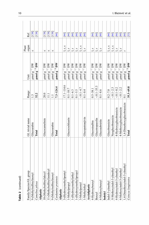
products collectively known as the GLs are presented. Emphasis is placed on theirnatural distribution, abundance level, and biological potential. The lack of commer-cially available authentic standards for most GLs has been pointed out repeatedlyand represents an obvious challenge [3, 5]. In other respects, well-characterizedplants (mostly seeds) are also reliable reference materials [4]. Plant species thatcontain high concentrations of a single or a limited number of GLs represent one ofthe most suitable sources for the extraction and purification of fair amounts of thesecompounds. Tables 1 and 2 list the different types of GLs and their sources, whichare discussed in the text. Dedicated extractive methods allow one to isolate a numberof GLs from adequate plant material, but in many cases, organic synthesis is anecessary alternative to obtain required quantities of GLs. Rollin and Tatibouët’srecent review summarizes the known synthetic approaches developed since the early1960s [6].
In recent years, a number of GL degradation products, mostly ITCs, have beenfound to possess real pharmacological activity, which will be discussed in thefollowing sections. Although GLs offer a structural variety in the aglycone chainR, which may contain alkenyl, indolyl, hydroxyl, carbonyl, or diverse thiofunctions,only few of them have been studied under the diverse angles including their sourceas well as their biological potential. Some of the GL-containing plants are recog-nized for their biological properties, whereas most of them are still not studied. Thisreport includes some of those plants.
2 Chemical Structure and Beyond
Glucosinolates represent thioglucosidic secondary metabolites occurring in theangiosperm plant families, mainly in the order Brassicales. To date, more than ca130 structurally different GLs have been reported [3, 4]. GLs are water-solubleorganic anions that share a common basic structural features (Fig. 1) and bear a sidechain which constitution, depending on plant species, is the sole structural variant:
• β-D-Glucopyrano unit• A O-sulfated anomeric (Z )-thiohydroximate function• A variable aglycone side chain
The aglycone can originate from one of eight natural amino acids according towhich GLs can be classified into: aliphatic (derived from Ala, Leu, Ile, Val, andMet), arylaliphatic (derived from Phe or Tyr), and indolyl GLs (derived from Trp).Many GLs are biosynthesized via extensive changes in the aglycone side chainswhich is due to a wide variety of chemical modifications such as elongation, hydrox-ylation, O-methylation, desaturation, further glycosylation, oxidation, and acylation[1, 7, 8].
Many GLs are derived from chain-elongated derivatives of Met and Phe. Thestructures of the several known aliphatic GLs are derived from the elongated andmodified side chains of Met homologues (Fig. 2).
1 Glucosinolates: Novel Sources and Biological Potential 5

Table 1 Structures of the GLs and plant families which can biosynthesize them in large amount
No. Glucosinolate Aglycone structure (R-)Plantfamily
Aliphatic
1 Glucocapparin CH3- b, d
2 Glucoputranjivin CH3-(CH3)CH- a
3 Glucocochlearin CH3-CH2-(CH3)CH- a
4 Glucocleomin CH3-C(OH)(CH3)-CH2-CH2- d
5 3-(Hydroxymethyl)pentylGL
CH3-CH2-CH(CH2OH)-(CH2)2- a
6 Sinigrin CH2 = CH-CH2- a, b
7 Gluconapin CH2 = CH-(CH2)2- a, e
8 Glucobrassicanapin CH2 = CH-(CH2)3- a
9 Progoitrin (R)-CH2 = CH-CH(OH)-CH2- a
10 Epiprogoitrin (S)-CH2 = CH-CH(OH)-CH2- a
11 Glucoibervirin CH3S-(CH2)3- a
12 Glucoerucin CH3S-(CH2)4- a
13 Glucoberteroin CH3S-(CH2)5- a
14 Glucoiberin CH3SO-(CH2)3- a
15 Glucoraphanin CH3SO-(CH2)4- a
16 Glucoalyssin CH3SO-(CH2)5- a
17 Glucohirsutin CH3SO-(CH2)8- a
18 Glucoarabin CH3SO-(CH2)9- a
19 Glucocamelinin CH3SO-(CH2)10- a
Arylaliphatic
20 Glucotropaeolin C6H5-CH2- a, c, g, l
21 Gluconasturtiin C6H5-CH2-CH2- a,
22 Glucosinalbin pOH-C6H4-CH2- a, l
23 Glucolepigramin mOH-C6H4-CH2- f
24 Glucoaubrietin pCH3O-C6H4-CH2- a, h, l
25 Glucolimnanthin mCH3O-C6H4-CH2- a, f, l
26 Glucobarbarin S-C6H5-CH(OH)-CH2- a
27 Epiglucobarbarin R-C6H5-CH(OH)-CH2- a, i
Indolyl
28 Glucobrassicin
N
CH2
H
a, i
29 Neoglucobrassicin
N
CH2
OCH3
a, j, k
(continued)
6 I. Blažević et al.

Arylaliphatic and indolyl GLs have also been identified together with the O-Glycosylated GLs, e.g., containing L-rhamnose as additional sugar moiety linked tothe aromatic ring.
GC-MS of GL breakdown products (mostly ITCs) and HPLC analysis of thedesulfo-GLs, described in the ISO 9167–1 official method, are extensively used fortheir identification and quantification [9]. However, some GL breakdown productsand desulfo-GLs are unstable in the applied conditions (temperature, pH value, time,and sulfatase enzyme). Some GLs, such as long-chain unsaturated GLs (C8-C10)(Fig. 2), were identified solely by GC-MS of their ITCs [10]. 2-(Methylsulfanyl)ethyl GL was reported without documentation, although it might be expected as abiosynthetic intermediate in plants accumulating 2-(methylsulfinyl)ethyl GL [4].Even in recent literature, there are occasional reports of GL identification solelybased on m/z values from HPLC-MS without the use of authentic references.
Table 1 (continued)
No. Glucosinolate Aglycone structure (R-)Plantfamily
30 4-Hydroxyglucobrassicin
N
H
CH2
OH a
31 4-Methoxyglucobrassicin
N
CH2
OCH3
H
a
O-Glycosylated
32 Glucomoringin(R1 = R2 = R3 = OH) CH2O
OR2
R1
R3H3C
a, g
33 Glucomoringin monoacetyl-isomer I, II, or III (R1, R2, orR3 = OAc)
g
34 2-(α-L-Rhamnopyranosyloxy)benzyl CH2
O Rha
i
a Brassicaceae, b Capparidaceae, c Caricaceae, d Cleomaceae, e Gyrostemonaceae, f Limnanthaceae,gMoringaceae, h Pentadiplandraceae, i Resedaceae, j Salvadoraceae, k Tovariaceae, l Tropaeolaceae
1 Glucosinolates: Novel Sources and Biological Potential 7

Table
2Databaseof
GLsqu
antitypresentin
plantspeciesof
differentfamilies
Fam
ily/Species/GLgrou
pGLtrivialname
Range
Unit
Plant
organ
Ref.
Akaniaceae
Bretschneiderasinensis
Total
0.4–9.2
μmol
g�1DW
[27]
Alip
hatic
2-Hyd
roxy
-2-m
ethy
lpropy
lGlucoconringiin
0.1-0.9
μmol
g�1DW
ba,br,fr,
l[27]
Arylalip
hatic
Benzyl
Glucotrop
aeolin
0.2–7.1
μmol
g�1DW
ba,br,fr,
l[27]
4-Metho
xybenzyl
Glucoaubrietin
0.6
μmol
g�1DW
fr[27]
4-Hyd
roxy
-3-m
etho
xybenzyl
Glucobretschn
eiderin
0.6
μmol
g�1DW
fr[27]
4-Hyd
roxy
benzyl
Glucosinalbin
�0.1
μmol
g�1DW
ba,b
r,l
[27]
Brassicaceae
Arm
oracia
rustican
aTotal
1.6–117.5
μmol
g�1DW
[135
,136]
Alip
hatic
Prop-2-enyl
Sinigrin
0.2–111.9
μmol
g�1DW
i,l,sp,r
[135
–137]
But-3-eny
lGluconapin
<0.1–73
.5μm
olg�
1DW
i,l,sp,r
[135
,137]
Pent-4-enyl
Glucobrassicanapin
<0.1–0.8
μmol
g�1DW
i,l,sp,r
[135
,137]
3-(M
ethy
lsulfiny
l)prop
ylGlucoiberin
0.2–0.6
μmol
g�1DW
i,l,sp,r
[135
,137]
Arylalip
hatic
2-Pheny
lethyl
Gluconasturtiin
0.1–2.6
μmol
g�1DW
l,sp,r
[135
–137]
(R)-2-Hyd
roxy
-2-pheny
lethyl
Glucobarbarin
<0.1
μmol
g�1DW
r[137
]
Indolyl
Indo
l-3-ylmethy
lGlucobrassicin
0.1–2.1
μmol
g�1DW
i,l,sp,r
[135
–137]
4-Metho
xyindo
l-3-ylmethy
l4-Metho
xyglucob
rassicin
<0.1–1.1
μmol
g�1DW
i,l,sp,r
[135
–137]
4-Hyd
roxy
indo
l-3-ylmethy
l4-Hyd
roxy
glucob
rassicin
0.2–0.6
μmol
g�1DW
pl[136
]
8 I. Blažević et al.

Aurinia
leucad
eaTotal
26.0–9
0.1
μmol
g�1DW
[35,
37]
Alip
hatic
But-3-eny
lGluconapin
26.3–4
8.3
μmol
g�1DW
fl,l,s,st
[35,
37]
5-(M
ethy
lsulfiny
l)pentyl
Glucoalyssin
4.8–38
.2μm
olg�
1DW
fl,l,r,s,
st[35,
37]
5-(M
ethy
lsulfany
l)pentyl
Glucoberteroin
0.5–18
.8μm
olg�
1DW
fl,l,r,s,
st[35,
37]
Pent-4-enyl
Glucobrassicanapin
3.6–14
.7μm
olg�
1DW
fl,l,s,st
[35,
37]
4-(M
ethy
lsulfiny
l)bu
tyl
Glucoraph
anin
0.4–3.7
μmol
g�1DW
fl,l,r,s,
st[35,
37]
4-(M
ethy
lsulfany
l)bu
tyl
Glucoerucin
0.4–2.2
μmol
g�1DW
l,r,st
[35,
37]
1-Methy
lpropy
lGlucocochlearin
0.9–1.6
μmol
g�1DW
fl,l,s,st
[35,
37]
Arylalip
hatic
Benzyl
Glucotrop
aeolin
0.3
μmol
g�1DW
l[35,
37]
Aurinia
sinu
ata
Total
21.7–8
6.4
μmol
g�1DW
[35]
Alip
hatic
5-(M
ethy
lsulfiny
l)pentyl
Glucoalyssin
6.5–62
.3μm
olg�
1DW
r,s
[35]
5-(M
ethy
lsulfany
l)pentyl
Glucoberteroin
17.2
μmol
g�1DW
s[35]
Pent-4-enyl
Glucobrassicanapin
6.9–15
.2μm
olg�
1DW
r,s
[35]
Cam
elinaalyssum
Total
28.5
μmol
g�1DW
[138
]
Alip
hatic
10-(Methy
lsulfiny
l)decyl
Glucocamelinin
18.0
μmol
g�1DW
s[138
]
9-(M
ethy
lsulfiny
l)no
nyl
Glucoarabin
8.1
μmol
g�1DW
s[138
]
11-(Methy
lsulfiny
l)un
decyl
2.4
μmol
g�1DW
s[138
]
Cam
elinamicrocarpa
Total
19.8
μmol
g�1DW
[138
]
Alip
hatic
10-(Methy
lsulfiny
l)decyl
Glucocamelinin
14.2
μmol
g�1DW
s[138
]
11-(Methy
lsulfiny
l)un
decyl
4.3
μmol
g�1DW
s[138
]
(con
tinued)
1 Glucosinolates: Novel Sources and Biological Potential 9
Table
2(con
tinue
d)
Fam
ily/Species/GLgrou
pGLtrivialname
Range
Unit
Plant
organ
Ref.
9-(M
ethy
lsulfiny
l)no
nyl
Glucoarabin
1.3
μmol
g�1DW
s[138
]
Cam
elinapilosa
Total
32.2
μmol
g�1DW
[138
]
Alip
hatic
10-(Methy
lsulfiny
l)decyl
Glucocamelinin
20.0
μmol
g�1DW
s[138
]
11-(Methy
lsulfiny
l)un
decyl
11.1
μmol
g�1DW
s[138
]
9-(M
ethy
lsulfiny
l)no
nyl
Glucoarabin
1.1
μmol
g�1DW
s[138
]
Carda
minepratensis
Total
7.3–12
4.4
μmol
g�1DW
[44]
Alip
hatic
3-(H
ydroxy
methy
l)pentyl
17.1–114
.6μm
olg�
1DW
l,r,s
[44]
1-Methy
lpropy
lGlucocochlearin
0.1–10
.7μm
olg�
1DW
l,r
[44]
1-(H
ydroxy
methy
l)ethy
l0.1–6.3
μmol
g�1DW
l,r
[44]
1-(H
ydroxy
methy
l)prop
yl0.1–5.5
μmol
g�1DW
l,r
[44]
3-Methy
lpentyl
<0.1–4.7
μmol
g�1DW
l,r,s
[44]
1-Methy
lethyl
Glucopu
tranjiv
in0.1–0.4
μmol
g�1DW
l,r
[44]
Arylalip
hatic
4-Hyd
roxy
benzyl
Glucosinalbin
0.5–38
.1μm
olg�
1DW
l,r
[44]
Benzyl
Glucotrop
aeolin
<0.1–35
.3μm
olg�
1DW
l,r
[44]
4-Metho
xybenzyl
Glucoaubrietin
0.1–8.6
μmol
g�1DW
l,r
[44]
Indolyl
Indo
l-3-ylmethy
lGlucobrassicin
0.2–7.0
μmol
g�1DW
l,r,s
[44]
N-M
etho
xyindo
l-3-ylmethy
lNeoglucob
rassicin
<0.1–2.3
μmol
g�1DW
l,r,s
[44]
4-Hyd
roxy
indo
l-3-ylmethy
l4-Hyd
roxy
glucob
rassicin
<0.1–0.2
μmol
g�1DW
l,r
[44]
4-Metho
xyindo
l-3-ylmethy
l4-Metho
xyglucob
rassicin
<0.1–2.2
μmol
g�1DW
l,r
[44]
1,4-Dim
etho
xyindo
l-3-ylmethy
l1,4-Dim
etho
xyglucob
rassicin
0.1
μmol
g�1DW
r[44]
Coincya
long
irostra
Total
35.3–6
9.8
μmol
g�1DW
[33]
10 I. Blažević et al.

Alip
hatic
But-3-eny
lGluconapin
2.0–34
.0μm
olg�
1DW
l,r
[33]
(R)-2-Hyd
roxy
but-3-enyl
Progo
itrin
0.1–10
.0μm
olg�
1DW
l,r
[33]
4-(M
ethy
lsulfany
l)bu
tyl
Glucoerucin
8.0
μmol
g�1DW
r[33]
4-(M
ethy
lsulfiny
l)bu
tyl
Glucoraph
anin
0.1–1.6
μmol
g�1DW
l,r
[33]
5-(M
ethy
lsulfiny
l)pentyl
Glucoalyssin
<0.1
μmol
g�1DW
l,r
[33]
Prop-2-enyl
Sinigrin
<0.1
μmol
g�1DW
l[33]
Pent-4-enyl
Glucobrassicanapin
<0.1
μmol
g�1DW
l[33]
Arylalip
hatic
2-Pheny
lethyl
Gluconasturtiin
0.1–28
.0μm
olg�
1DW
l,r
[33]
4-Hyd
roxy
benzyl
Glucosinalbin
0.2
μmol
g�1DW
l,r
[33]
Indolyl
4-Metho
xyindo
l-3-ylmethy
l4-Metho
xyglucob
rassicin
0.2–10
.0μm
olg�
1DW
l,r
[33]
Indo
l-3-ylmethy
lGlucobrassicin
0.2–10
.0μm
olg�
1DW
l,r
[33]
N-M
etho
xyindo
l-3-ylmethy
lNeoglucob
rassicin
0.2
μmol
g�1DW
l[33]
4-Hyd
roxy
indo
l-3-ylmethy
l4-Hyd
roxy
glucob
rassicin
0.2
μmol
g�1DW
l[33]
Coincya
mon
ensis
Total
22.2–4
1.9
μmol
g�1DW
[33]
Alip
hatic
But-3-eny
lGluconapin
0.3–3.0
μmol
g�1DW
l,r
[33]
(R)-2-Hyd
roxy
but-3-enyl
Progo
itrin
1.5
μmol
g�1DW
r[33]
(S)-2-Hyd
roxy
but-3-enyl
Epiprog
oitrin
1.4
μmol
g�1DW
l[33]
4-(M
ethy
lsulfiny
l)bu
tyl
Glucoraph
anin
0.9
μmol
g�1DW
r[33]
4-(M
ethy
lsulfany
l)bu
tyl
Glucoerucin
0.2
μmol
g�1DW
r[33]
5-(M
ethy
lsulfiny
l)pentyl
Glucoalyssin
<0.1
μmol
g�1DW
r[33]
Prop-2-enyl
Sinigrin
<0.1
μmol
g�1DW
l[33]
Arylalip
hatic
2-Pheny
lethyl
Gluconasturtiin
19.0
μmol
g�1DW
r[33]
4-Hyd
roxy
benzyl
Glucosinalbin
10.0–1
7.0
μmol
g�1DW
l,r
[33] (c
ontin
ued)
1 Glucosinolates: Novel Sources and Biological Potential 11
Table
2(con
tinue
d)
Fam
ily/Species/GLgrou
pGLtrivialname
Range
Unit
Plant
organ
Ref.
Indolyl
4-Hyd
roxy
indo
l-3-ylmethy
l4-Hyd
roxy
glucob
rassicin
0.2–5.0
μmol
g�1DW
l,r
[33]
4-Metho
xyindo
l-3-ylmethy
l4-Metho
xyglucob
rassicin
0.2–5.0
μmol
g�1DW
l,r
[33]
N-M
etho
xyindo
l-3-ylmethy
lNeoglucob
rassicin
0.2
μmol
g�1DW
l[33]
Indo
l-3-ylmethy
lGlucobrassicin
0.2
μmol
g�1DW
l[33]
Coincya
rupestris
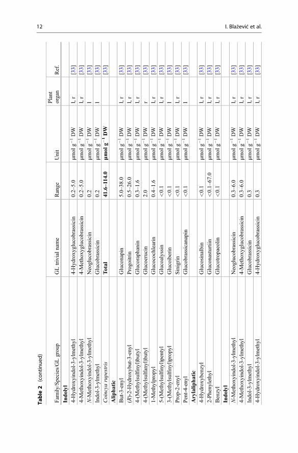
Total
41.6–114
.0μm
olg�
1DW
[33]
Alip
hatic
But-3-eny
lGluconapin
5.0–38
.0μm
olg�
1DW
l,r
[33]
(R)-2-Hyd
roxy
but-3-enyl
Progo
itrin
0.5–26
.0μm
olg�
1DW
l,r
[33]
4-(M
ethy
lsulfiny
l)bu
tyl
Glucoraph
anin
0.3–1.6
μmol
g�1DW
l,r
[33]
4-(M
ethy
lsulfany
l)bu
tyl
Glucoerucin
2.0
μmol
g�1DW
r[33]
1-Methy
lpropy
lGlucocochlearin
0.4–1.6
μmol
g�1DW
l,r
[33]
5-(M
ethy
lsulfiny
l)pentyl
Glucoalyssin
<0.1
μmol
g�1DW
l,r
[33]
3-(M
ethy
lsulfiny
l)prop
ylGlucoiberin
<0.1
μmol
g�1DW
l[33]
Prop-2-enyl
Sinigrin
<0.1
μmol
g�1DW
l,r
[33]
Pent-4-enyl
Glucobrassicanapin
<0.1
μmol
g�1DW
l[33]
Arylalip
hatic
4-Hyd
roxy
benzyl
Glucosinalbin
<0.1
μmol
g�1DW
l,r
[33]
2-Pheny
lethyl
Gluconasturtiin
<0.1–67
.0μm
olg�
1DW
l,r
[33]
Benzyl
Glucotrop
aeolin
<0.1
μmol
g�1DW
l,r
[33]
Indolyl
N-M
etho
xyindo
l-3-ylmethy
lNeoglucob
rassicin
0.3–6.0
μmol
g�1DW
l,r
[33]
4-Metho
xyindo
l-3-ylmethy
l4-Metho
xyglucob
rassicin
0.3–6.0
μmol
g�1DW
l,r
[33]
Indo
l-3-ylmethy
lGlucobrassicin
0.3
μmol
g�1DW
l,r
[33]
4-Hyd
roxy
indo
l-3-ylmethy
l4-Hyd
roxy
glucob
rassicin
0.3
μmol
g�1DW
l,r
[33]
12 I. Blažević et al.

Degenia
velebitica
Total
9.9–99
.1μm
olg�
1DW
[36]
Alip
hatic
5-(M
ethy
lsulfany
l)pentyl
Glucoberteroin
1.3–88
.0μm
olg�
1DW
fl+l,r,
s,st
[36]
5-(M
ethy
lsulfiny
l)pentyl
Glucoalyssin
6.4–8.4
μmol
g�1DW
fl+l,s,
st[36]
4-(M
ethy
lsulfany
l)bu
tyl
Glucoerucin
2.7
μmol
g�1DW
s[36]
Pent-4-enyl
Glucobrassicanapin
2.2
μmol
g�1DW
fl+l
[36]
Arylalip
hatic
4-Metho
xybenzyl
Glucoaubrietin
0.4
μmol
g�1DW
st[36]
Dith
yrea
wislizenii
Total
1.8
μmol
g�1DW
[139
]
Alip
hatic
6-(M
ethy
lsulfany
l)hexy
lGlucolesquerellin
1.5
μmol
g�1DW
fr[139
]
6-(M
ethy
lsulfiny
l)hexy
lGlucohesperin
0.2
μmol
g�1DW
fr[139
]
7-(M
ethy
lsulfany
l)heptyl
<0.1
μmol
g�1DW
fr[139
]
5-(M
ethy
lsulfany
l)pentyl
<0.1
μmol
g�1DW
fr[139
]
Erucastrum
cana
riense
Total
18.7–4
3.3
μmol
g�1DW
[33]
Alip
hatic
Prop-2-enyl
Sinigrin
14.0–3
4.0
μmol
g�1DW
l,r
[33]
3-Methy
lpenthyl
2.1–3.0
μmol
g�1DW
l,r
[33]
But-3-eny
lGluconapin
0.2–0.3
μmol
g�1DW
l,r
[33]
4-(M
ethy
lsulfany
l)bu
tyl
Glucoerucin
<0.1
μmol
g�1DW
l[33]
3-(M
ethy
lsulfiny
l)prop
ylGlucoiberin
<0.1
μmol
g�1DW
l,r
[33]
Indolyl
N-M
etho
xyindo
l-3-ylmethy
lNeoglucob
rassicin
6.9
μmol
g�1DW
r[33]
Indo
l-3-ylmethy
lGlucobrassicin
1.5
μmol
g�1DW
l[33]
Fibigia
triquetra
Total
62.5–1
35.4
μmol
g�1DW
[38] (c
ontin
ued)
1 Glucosinolates: Novel Sources and Biological Potential 13

Table
2(con
tinue
d)
Fam
ily/Species/GLgrou
pGLtrivialname
Range
Unit
Plant
organ
Ref.
Alip
hatic
4-(M
ethy
lsulfany
l)bu
tyl
Glucoerucin
3.5–76
.7μm
olg�
1DW
fl+l,s,
st[38]
But-3-eny
lGluconapin
32.6–6
6.7
μmol
g�1DW
fl+l,s,
st[38]
4-(M
ethy
lsulfiny
l)bu
tyl
Glucoraph
anin
8.0–23
.4μm
olg�
1DW
fl+l,s,
st[38]
1-Methy
lpropy
lGlucocochlearin
1.3–4.3
μmol
g�1DW
fl+l,s,
st[38]
1-Methy
lethyl
Glucopu
tranjiv
in0.7–2.5
μmol
g�1DW
fl+l,s,
st[38]
Pent-4-enyl
Glucobrassicanapin
0.7
μmol
g�1DW
s[38]
Guiraoa
arvensis
Total
61.1–1
60.1
μmol
g�1DW
[33]
Alip
hatic
Prop-2-enyl
Sinigrin
61.0–1
50.0
μmol
g�1DW
l[33]
But-3-eny
lGluconapin
0.1
μmol
g�1DW
l,r
[33]
3-(M
ethy
lsulfiny
l)prop
ylGlucoiberin
<0.1
μmol
g�1DW
l,r
[33]
Indolyl
N-M
etho
xyindo
l-3-ylmethy
lNeoglucob
rassicin
<0.1–5.0
μmol
g�1DW
l,r
[33]
4-Metho
xyindo
l-3-ylmethy
l4-Metho
xyglucob
rassicin
<0.1–5.0
μmol
g�1DW
l,r
[33]
Indo
l-3-ylmethy
lGlucobrassicin
<0.1
μmol
g�1DW
l[33]
4-Hyd
roxy
indo
l-3-ylmethy
l4-Hyd
roxy
glucob
rassicin
<0.1
μmol
g�1DW
l[33]
Hem
icrambe
fruticulosa
Total
46.4–6
4.9
μmol
g�1DW
[33]
Alip
hatic
But-3-eny
lGluconapin
7.0–59
.0μm
olg�
1DW
l,r
[33]
14 I. Blažević et al.

(S)-2-Hyd
roxy
but-3-enyl
Epiprog
oitrin
0.9–8.1
μmol
g�1DW
l,r
[33]
1-Methy
lpropy
lGlucocochlearin
0.7–1.3
μmol
g�1DW
l,r
[33]
4-(M
ethy
lsulfiny
l)bu
tyl
Glucoraph
anin
�0.1
μmol
g�1DW
l,r
[33]
5-(M
ethy
lsulfiny
l)pentyl
Glucoalyssin
<0.1
μmol
g�1DW
l,r
[33]
Prop-2-enyl
Sinigrin
<0.1
μmol
g�1DW
l,r
[33]
3-(M
ethy
lsulfiny
l)prop
ylGlucoiberin
<0.1
μmol
g�1DW
l[33]
Pent-4-enyl
Glucobrassicanapin
<0.1
μmol
g�1DW
l,r
[33]
Arylalip
hatic
2-Pheny
lethyl
Gluconasturtiin
2.0–28
.0μm
olg�
1DW
l,r
[33]
Indolyl
4-Metho
xyindo
l-3-ylmethy
l4-Metho
xyglucob
rassicin
0.4–1.3
μmol
g�1DW
l,r
[33]
4-Hyd
roxy
indo
l-3-ylmethy
l4-Hyd
roxy
glucob
rassicin
0.4–1.3
μmol
g�1DW
l,r
[33]
Indo
l-3-ylmethy
lGlucobrassicin
0.4
μmol
g�1DW
l[33]
N-M
etho
xyindo
l-3-ylmethy
lNeoglucob
rassicin
0.4
μmol
g�1DW
l[33]
Hirschfeldiaincana
Total
2.8–30
.3μm
olg�
1DW
[33]
Alip
hatic
(R)-2-Hyd
roxy
but-3-enyl
Progo
itrin
0.2–8.3
μmol
g�1DW
l,r
[33]
But-3-eny
lGluconapin
2.0–3.0
μmol
g�1DW
l,r
[33]
(S)-2-Hyd
roxy
but-3-enyl
Epiprog
oitrin
0.2
μmol
g�1DW
l[33]
Pent-4-enyl
Glucobrassicanapin
<0.1
μmol
g�1DW
r[33]
5-(M
ethy
lsulfiny
l)pentyl
Glucoalyssin
<0.1
μmol
g�1DW
r[33]
Arylalip
hatic
2-Pheny
lethyl
Gluconasturtiin
0.6–15
.0μm
olg�
1DW
l,r
[33]
Indolyl
N-M
etho
xyindo
l-3-ylmethy
lNeoglucob
rassicin
2.0
μmol
g�1DW
r[33]
4-Metho
xyindo
l-3-ylmethy
l4-Metho
xyglucob
rassicin
2.0
μmol
g�1DW
r[33]
Isatiscanescens
Total
79.5–1
03.3
μmol
g�1DW
[53]
Alip
hatic
(con
tinued)
1 Glucosinolates: Novel Sources and Biological Potential 15

Table
2(con
tinue
d)
Fam
ily/Species/GLgrou
pGLtrivialname
Range
Unit
Plant
organ
Ref.
But-3-eny
lGluconapin
33.5–6
5.1
μmol
g�1DW
fl[53]
Indolyl
Indo
l-3-ylmethy
lGlucobrassicin
33.1–6
8.8
μmol
g�1DW
fl[53]
Isatisindigo
tica
Total
16.0–4
5.0
123.2–
152.0
μmol
g�1DW
μmol
g�1FW
[140
][141
]
Alip
hatic
(R)-2-Hyd
roxy
but-3-enyl
Progo
itrin
2.5–15
.027
.7–6
4.2
μmol
g�1DW
μmol
g�1FW
l,s
s[55,
57,1
40]
[141
]
(S)-2-Hyd
roxy
but-3-enyl
Epiprog
oitrin
0.1–1.3
18.6–115
.4μm
olg�
1DW
μmol
g�1FW
l,s
s[55,
57,1
40]
[141
]
But-3-eny
lGluconapin
<0.1–0.3
0.8–32
.9μm
olg�
1DW
μmol
g�1FW
l s[57,
140]
[141
]
(R)-glucoisatisin/
(S)-epiglucoisatisin
2.1
0.4–1.0
μmol
g�1DW
μmol
g�1FW
s s[57]
[141
]
Indolyl
4-Hyd
roxy
indo
l-3-ylmethy
l4-Hyd
roxy
glucob
rassicin
0.4–40
.02.4–3.4
μmol
g�1DW
μmol
g�1FW
l,s
s[55,
57,1
40]
[141
]
Indo
l-3-ylmethy
lGlucobrassicin
<0.1–4.4
μmol
g�1FW
s[141
]
4-Metho
xyindo
l-3-ylmethy
l4-Metho
xyglucob
rassicin
0.1–0.3
μmol
g�1FW
s[57,
141]
Kremeriella
cordylocarpu
sTotal
13.8–7
7.6
μmol
g�1DW
[33]
Alip
hatic
(S)-2-Hyd
roxy
but-3-enyl
Epiprog
oitrin
<0.1–1.0
μmol
g�1DW
l,s
[33]
But-3-eny
lGluconapin
<0.1–0.6
μmol
g�1DW
l,r
[33]
4-(M
ethy
lsulfiny
l)bu
tyl
Glucoraph
anin
<0.1
μmol
g�1DW
l,r
[33]
5-(M
ethy
lsulfiny
l)pentyl
Glucoalyssin
<0.1
μmol
g�1DW
l,r
[33]
Arylalip
hatic
16 I. Blažević et al.

4-Hyd
roxy
benzyl
Glucosinalbin
11.0–76.0
μmol
g�1DW
l,r
[33]
2-Pheny
lethyl
Gluconasturtiin
0.2
μmol
g�1DW
r[33]
Benzyl
Glucotrop
aeolin
<0.1–0.1
μmol
g�1DW
l[33]
Indolyl
N-M
etho
xyindo
l-3-ylmethy
lNeoglucob
rassicin
<0.1–2.0
μmol
g�1DW
l,r
[33]
4-Metho
xyindo
l-3-ylmethy
l4-Metho
xyglucob
rassicin
<0.1–2.0
μmol
g�1DW
l,r
[33]
Indo
l-3-ylmethy
lGlucobrassicin
<0.1
μmol
g�1DW
l,r
[33]
4-Hyd
roxy
indo
l-3-ylmethy
l4-Hyd
roxy
glucob
rassicin
<0.1
μmol
g�1DW
l,r
[33]
Lepidium
fend
leri
Total
27.5
μmol
g�1DW
[142
]
Alip
hatic
3-(M
ethy
lsulfiny
l)prop
ylGlucoiberin
27.5
μmol
g�1DW
s[142
]
Lepidium
meyenii
Total
3.8–69
.5μm
olg�
1DW
[143
,144]
Alip
hatic
5-(M
ethy
lsulfiny
l)pentyl
Glucoalyssin
0.1–11.2
μmol
g�1DW
h,l,s,sp
[143
,144]
Arylalip
hatic
Benzyl
Glucotrop
aeolin
0.2–40
.9μm
olg�
1DW
h,l,s,sp
[143
–145]
3-Metho
xybenzyl
Glucolim
nanthin
0.2–19
.3μm
olg�
1DW
h,l,s,sp
[143
,144]
4-Hyd
roxy
benzyl
Glucosinalbin
0.2–12
.6μm
olg�
1DW
h,l,s,sp
[143
,144]
4-Metho
xybenzyl
Glucoaubrietin
0.7–6.4
μmol
g�1DW
h,l,sp
[144
]
Indolyl
4-Metho
xyindo
l-3-ylmethy
l4-Metho
xyglucob
rassicin
<0.1–7.6
μmol
g�1DW
h,l,s
[143
,144]
4-Hyd
roxy
indo
l-3-ylmethy
l4-Hyd
roxy
glucob
rassicin
<0.1
μmol
g�1DW
h,l
[143
,144]
Indo
l-3-ylmethy
lGlucobrassicin
<0.1
μmol
g�1DW
h,l
[144
]
Noccaea
caerulescens
Total
0.8–18
6.2
μmol
g�1DW
[146
]
Alip
hatic
Prop-2-enyl
Sinigrin
<0.1–68
.4μm
olg�
1DW
l,s
[46]
Arylalip
hatic
4-Hyd
roxy
benzyl
Glucosinalbin
<0.1–83
.0μm
olg�
1DW
l,s
[46] (c
ontin
ued)
1 Glucosinolates: Novel Sources and Biological Potential 17

Table
2(con
tinue
d)
Fam
ily/Species/GLgrou
pGLtrivialname
Range
Unit
Plant
organ
Ref.
O-G
lycosylated
4-α�
L�
Rhamno
pyrano
syloxy
benzyl*
Glucomoringin*
<0.1–111.0
μmol
g�1DW
l,s
[46]
Cheesem
anii/Pachyclad
onTotal
90.1
μmol
g�1DW
[147
]
Alip
hatic
(S)-2-Hyd
roxy
but-3-enyl
Epiprog
oitrin
47.9
μmol
g�1DW
l[147
]
Prop-2-enyl
Sinigrin
37.1
μmol
g�1DW
l[147
]
6-(M
ethy
lsulfiny
l)hexy
lGlucohesperalin
2.0
μmol
g�1DW
l[147
]
7-(M
ethy
lsulfiny
l)heptyl
Glucoibarin
1.3
μmol
g�1DW
l[147
]
4-(M
ethy
lsulfany
l)bu
tyl
Glucoerucin
0.5
μmol
g�1DW
l[147
]
3-(M
ethy
lsulfany
l)prop
ylGlucoibervirin
0.4
μmol
g�1DW
l[147
]
3-(M
ethy
lsulfiny
l)prop
ylGlucoiberin
0.3
μmol
g�1DW
l[147
]
Indolyl
4-Metho
xyindo
l-3-ylmethy
l4-Metho
xyglucob
rassicin
0.6
μmol
g�1DW
l[147
]
4-Hyd
roxy
indo
l-3-ylmethy
l4-Hyd
roxy
glucob
rassicin
0.3
μmol
g�1DW
l[147
]
Pachyclad
onexile
Total
59.4
μmol
g�1DW
[147
]
Alip
hatic
3-(M
ethy
lsulfiny
l)prop
ylGlucoiberin
46.1
μmol
g�1DW
l[147
]
3-(M
ethy
lsulfany
l)prop
ylGlucoibervirin
9.6
μmol
g�1DW
l[147
]
7-(M
ethy
lsulfiny
l)heptyl
Glucoibarin
1.6
μmol
g�1DW
l[147
]
6-(M
ethy
lsulfiny
l)hexy
lGlucohesperalin
0.9
μmol
g�1DW
l[147
]
4-(M
ethy
lsulfiny
l)bu
tyl
Glucoraph
anin
0.8
μmol
g�1DW
l[147
]
4-(M
ethy
lsulfany
l)bu
tyl
Glucoerucin
0.2
μmol
g�1DW
l[147
]
Indolyl
4-Metho
xyindo
l-3-ylmethy
l4-Metho
xyglucob
rassicin
0.2
μmol
g�1DW
l[147
]
4-Hyd
roxy
indo
l-3-ylmethy
l4-Hyd
roxy
glucob
rassicin
<0.1
μmol
g�1DW
l[147
]
18 I. Blažević et al.

Pachyclad
onno
vae-zeland
iae
Total
102.0
μmol
g�1DW
[147]
Alip
hatic
(S)-2-Hyd
roxy
but-3-enyl
Epiprog
oitrin
42.1
μmol
g�1DW
l[147]
Prop-2-enyl
Sinigrin
31.8
μmol
g�1DW
l[147]
4-(M
ethy
lsulfiny
l)bu
tyl
Glucoraph
anin
10.2
μmol
g�1DW
l[147]
3-(M
ethy
lsulfiny
l)prop
ylGlucoiberin
7.2
μmol
g�1DW
l[147]
But-3-eny
lGluconapin
3.9
μmol
g�1DW
l[147]
4-(M
ethy
lsulfany
l)bu
tyl
Glucoerucin
3.3
μmol
g�1DW
l[147]
8-(M
ethy
lsulfiny
l)octyl
Glucohirsutin
2.5
μmol
g�1DW
l[147]
3-(M
ethy
lsulfany
l)prop
ylGlucoibervirin
1.8
μmol
g�1DW
l[147]
7-(M
ethy
lsulfiny
l)heptyl
Glucoibarin
1.1
μmol
g�1DW
l[147]
7-(M
ethy
lsulfany
l)heptyl
<0.1
μmol
g�1DW
l[147]
Indolyl
4-Metho
xyindo
l-3-ylmethy
l4-Metho
xyglucob
rassicin
0.9
μmol
g�1DW
l[147]
N-M
etho
xyindo
l-3-ylmethy
lNeoglucob
rassicin
0.3
μmol
g�1DW
l[147]
4-Hyd
roxy
indo
l-3-ylmethy
l4-Hyd
roxy
glucob
rassicin
0.1
μmol
g�1DW
l[147]
Pring
leaan
tiscorbutica
Total
52.9–1
48.7
μmol
g�1DW
[34]
Alip
hatic
But-3-eny
lGluconapin
26.1–6
6.2
μmol
g�1DW
l,s
[34]
n-Butyl
5.6–11.8
μmol
g�1DW
l,s
[34]
4-(M
ethy
lsulfany
l)bu
tyl
Glucoerucin
10.1
μmol
g�1DW
s[34]
Prop-2-enyl
Sinigrin
2.9–6.4
μmol
g�1DW
l,s
[34]
4-(M
ethy
lsulfiny
l)bu
tyl
Glucoraph
anin
3.9–5.4
μmol
g�1DW
l,s
[34]
Arylalip
hatic
Benzyl
Glucotrop
aeolin
12.9–5
0.3
μmol
g�1DW
l,s
[34]
Sina
pisbo
ivinii
Total
39.3–4
3.0
μmol
g�1DW
[33]
Alip
hatic
But-3-eny
lGluconapin
27.0–3
5.0
μmol
g�1DW
l,r
[33] (c
ontin
ued)
1 Glucosinolates: Novel Sources and Biological Potential 19

Table
2(con
tinue
d)
Fam
ily/Species/GLgrou
pGLtrivialname
Range
Unit
Plant
organ
Ref.
Prop-2-enyl
Sinigrin
4.0–8.0
μmol
g�1DW
l,r
[33]
4-(M
ethy
lsulfiny
l)bu
tyl
Glucoraph
anin
�0.1
μmol
g�1DW
l,r
[33]
5-(M
ethy
lsulfiny
l)pentyl
Glucoalyssin
<0.1
μmol
g�1DW
l[33]
3-(M
ethy
lsulfiny
l)prop
ylGlucoiberin
<0.1
μmol
g�1DW
l[33]
Pent-4-enyl
Glucobrassicanapin
<0.1
μmol
g�1DW
l[33]
Arylalip
hatic
2-Pheny
lethyl
Gluconasturtiin
6.0
μmol
g�1DW
r[33]
Indolyl
4-Metho
xyindo
l-3-ylmethy
l4-Metho
xyglucob
rassicin
<0.1–1.1
μmol
g�1DW
l,r
[33]
Indo
l-3-ylmethy
lGlucobrassicin
<0.1–1.1
μmol
g�1DW
l,r
[33]
N-M
etho
xyindo
l-3-ylmethy
lNeoglucob
rassicin
<0.1
μmol
g�1DW
l[33]
4-Hyd
roxy
indo
l-3-ylmethy
l4-Hyd
roxy
glucob
rassicin
<0.1
μmol
g�1DW
l[33]
1,4-Dim
etho
xyindo
l-3-ylmethy
l1,4-Dim
etho
xyglucob
rassicin
<0.1
μmol
g�1DW
r[33]
Sina
pispu
bescens
ssp.
indu
rata
andpu
bescens
Total
34.8–8
5.4
μmol
g�1DW
[33]
Alip
hatic
But-3-eny
lGluconapin
27.0–4
1.0
μmol
g�1DW
l,r
[33]
Prop-2-enyl
Sinigrin
<0.1
μmol
g�1DW
l[33]
4-(M
ethy
lsulfiny
l)bu
tyl
Glucoraph
anin
�0.1
μmol
g�1DW
l,r
[33]
(S)-2-Hyd
roxy
but-3-enyl
Epiprog
oitrin
�0.1
μmol
g�1DW
l,r
[33]
5-(M
ethy
lsulfiny
l)pentyl
Glucoalyssin
<0.1
μmol
g�1DW
l,r
[33]
Pent-4-enyl
Glucobrassicanapin
<0.1
μmol
g�1DW
l,r
[33]
Arylalip
hatic
2-Pheny
lethyl
Gluconasturtiin
20.0–5
8.0
μmol
g�1DW
r[33]
Indolyl
20 I. Blažević et al.

4-Hyd
roxy
indo
l-3-ylmethy
l4-Hyd
roxy
glucob
rassicin
0.2–0.8
μmol
g�1DW
l,r
[33]
4-Metho
xyindo
l-3-ylmethy
l4-Metho
xyglucob
rassicin
0.2
μmol
g�1DW
l[33]
Indo
l-3-ylmethy
lGlucobrassicin
0.2
μmol
g�1DW
l[33]
N-M
etho
xyindo
l-3-ylmethy
lNeoglucob
rassicin
0.2
μmol
g�1DW
l[33]
Sina
pisflexuo
saTotal
12.6–3
9.0
μmol
g�1DW
[33]
Arylalip
hatic
4-Hyd
roxy
benzyl
Glucosinalbin
12.0-39.0
μmol
g�1DW
l,r
[33]
Indolyl
4-Metho
xyindo
l-3-ylmethy
l4-Metho
xyglucob
rassicin
<0.1-0.6
μmol
g�1DW
l,r
[33]
Indo
l-3-ylmethy
lGlucobrassicin
<0.1
μmol
g�1DW
l[33]
N-M
etho
xyindo
l-3-ylmethy
lNeoglucob
rassicin
<0.1
μmol
g�1DW
l[33]
4-Hyd
roxy
indo
l-3-ylmethy
l4-Hyd
roxy
glucob
rassicin
<0.1
μmol
g�1DW
l[33]
Trachystom
alaba
sii
Total
65.1–7
0.5
μmol
g�1DW
[33]
Alip
hatic
Prop-2-enyl
Sinigrin
54.0–7
0.0
μmol
g�1DW
l,r
[33]
But-3-eny
lGluconapin
0.1
μmol
g�1DW
l,r
[33]
4-(M
ethy
lsulfiny
l)bu
tyl
Glucoraph
anin
<0.1
μmol
g�1DW
l,r
[33]
3-(M
ethy
lsulfiny
l)prop
ylGlucoiberin
<0.1
μmol
g�1DW
l,r
[33]
Indolyl
N-M
etho
xyindo
l-3-ylmethy
lNeoglucob
rassicin
0.1–8.0
μmol
g�1DW
l,r
[33]
4-Metho
xyindo
l-3-ylmethy
l4-Metho
xyglucob
rassicin
0.1
μmol
g�1DW
l,r
[33]
Indo
l-3-ylmethy
lGlucobrassicin
0.1
μmol
g�1DW
l,r
[33]
4-Hyd
roxy
indo
l-3-ylmethy
l4-Hyd
roxy
glucob
rassicin
0.1
μmol
g�1DW
l,r
[33]
Capparidaceae
Bosciasenega
lensis
Total
2.0–44
0.0
μmol
g�1DW
[68,
148]
Alip
hatic
Methy
lGlucocapp
arin
2.0–44
0.0
μmol
g�1DW
fr,l,s
[68,
148]
Cap
parisovata
Total
11.4–8
4.6
μmol
g�1DW
[65,
67,1
49]
(con
tinued)
1 Glucosinolates: Novel Sources and Biological Potential 21

Table
2(con
tinue
d)
Fam
ily/Species/GLgrou
pGLtrivialname
Range
Unit
Plant
organ
Ref.
Alip
hatic
Methy
lGlucocapp
arin
1.3–80
.4μm
olg�
1DW
bu,fl
,l,
s,ys
[65,
67,1
49]
Prop-2-enyl
Sinigrin
<0.1–29
.8μm
olg�
1DW
bu,fl
,l,
ys[67,
149]
(S)-2-Hyd
roxy
but-3-enyl
Epiprog
oitrin
0.1–8.2
μmol
g�1DW
bu,fl
,l,
ys[67,
149]
But-3-eny
lGluconapin
0.1–2.1
μmol
g�1DW
bu,fl
,l,
ys[67,
149]
3-(M
ethy
lsulfiny
l)prop
ylGlucoiberin
0.1–2.1
μmol
g�1DW
bu,fl
,l,
ys[67,
149]
5-(M
ethy
lsulfiny
l)pentyl
Glucoalyssin
<0.1–1.1
μmol
g�1DW
bu,y
s[149
]
(R)-2-Hyd
roxy
but-3-enyl
Progo
itrin
0.1–1.1
μmol
g�1DW
bu,fl
,l,
ys[67,
149]
(R)-2-Hyd
roxy
pent-4-eny
lGluconapo
leiferin
0.1–0.8
μmol
g�1DW
bu,y
s[149
]
Pent-4-enyl
Glucobrassicanapin
0.1–0.2
μmol
g�1DW
bu,y
s[149
]
Arylalip
hatic
4-Hyd
roxy
benzyl
Glucosinalbin
0.3–0.9
μmol
g�1DW
bu,fl
,l,
ys[67]
2-Pheny
lethyl
Gluconasturtiin
0.1–0.4
μmol
g�1DW
bu,y
s[149
]
Indolyl
4-Hyd
roxy
indo
l-3-ylmethy
l4-Hyd
roxy
glucob
rassicin
<0.1
μmol
g�1DW
bu,y
s[149
]
Indo
l-3-ylmethy
lGlucobrassicin
<0.1–8.2
μmol
g�1DW
bu,fl
,l,
ys[67,
149]
Cap
parisspinosa
subsp.
rupestris
Total
6.8–12
7.1
μmol
g�1DW
[5,1
3,65,
66,1
49]
22 I. Blažević et al.

Alip
hatic
Methy
lGlucocapp
arin
5.4–12
7.1
μmol
g�1DW
bu,s,y
s[5,1
3,65,
66,1
49]
5-(M
ethy
lsulfiny
l)pentyl
Glucoalyssin
0.1–0.5
μmol
g�1DW
bu,y
s[149
]
(R)-2-Hyd
roxy
pent-4-eny
lGluconapo
leiferin
0.1–0.5
μmol
g�1DW
bu,y
s[149
]
Pent-4-enyl
Glucobrassicanapin
0.1–0.4
μmol
g�1DW
bu,y
s[149
]
But-3-eny
lGluconapin
0.1–0.3
μmol
g�1DW
bu,y
s[149
]
(S)-2-Hyd
roxy
but-3-enyl
Epiprog
oitrin
0.1–0.3
μmol
g�1DW
bu,y
s[149
]
Prop-2-enyl
Sinigrin
0.1–0.3
μmol
g�1DW
bu,y
s[149
]
3-(M
ethy
lsulfiny
l)prop
ylGlucoiberin
0.1–0.2
μmol
g�1DW
bu,y
s[149
]
(R)-2-Hyd
roxy
but-3-enyl
Progo
itrin
0.1–0.2
μmol
g�1DW
bu,y
s[149
]
Glyciny
lmethy
lGlyciny
l-glucocapparin
<0.1
μmol
g�1DW
bu[13]
Isop
ropy
l/n-propy
l<0.1
μmol
g�1DW
bu[13]
Mercaptom
ethy
lMercapto-glucocapparin
<0.1
μmol
g�1DW
bu[13]
Disulfany
lmethy
lDisulfany
l-glucocapparin
<0.1
μmol
g�1DW
bu[13]
Trisulfanylmethy
lTrisulfanyl-glucocapp
arin
<0.1
μmol
g�1DW
bu[13]
Arylalip
hatic
2-Pheny
lethyl
Gluconasturtiin
0.1–0.2
μmol
g�1DW
bu,y
s[149
]
Indolyl
4-Hyd
roxy
indo
l-3-ylmethy
l4-Hyd
roxy
glucob
rassicin
<0.1–2.0
μmol
g�1DW
bu,y
s[13,
149]
Indo
l-3-ylmethy
lGlucobrassicin
<0.1–0.5
μmol
g�1DW
bu,y
s[13,
149]
Crataevarelig
iosa
Total
37.3
μmol
g�1DW
[150
]
Alip
hatic
Methy
lGlucocapp
arin
37.3
μmol
g�1DW
l[150
]
Caricaceae
Caricapa
paya
Total
75.0–2
59.2
μmol
g�1DW
[5,1
51]
Arylalip
hatic
Benzyl
Glucotrop
aeolin
75.0–2
59.2
μmol
g�1DW
lx,p
,s[5,1
51]
(con
tinued)
1 Glucosinolates: Novel Sources and Biological Potential 23

Table
2(con
tinue
d)
Fam
ily/Species/GLgrou
pGLtrivialname
Range
Unit
Plant
organ
Ref.
0.3–20
.0μm
olg�
1FW
p,s
[152
–154]
Caricapentag
ona
Total
0.1–10
.0μm
olg�
1DW
[5]
Arylalip
hatic
Benzyl
Glucotrop
aeolin
0.1–10
.0μm
olg�
1DW
s[5]
Cleom
aceae
Cleom
eha
sslerian
aTotal
15.3–2
4.1
μmol
g�1DW
[80]
Alip
hatic
Methy
lGlucocapp
arin
9.9–15
.7μm
olg�
1DW
s[80]
2-Hyd
roxy
-2-m
ethy
lbutyl
Glucocleomin
5.4–8.4
μmol
g�1DW
s[80]
Perito
maarbo
rea
Total
13.8–4
2.9
μmol
g�1FW
[79]
Alip
hatic
Methy
lGlucocapp
arin
13.8–4
2.9
μmol
g�1FW
bu,l,s
[79]
Gyrostemon
aceae
Gyrostemon
ramulosus
Alip
hatic
But-3-eny
lGluconapin
25.0–5
0.0
μmol
g�1DW
s[5]
4-(M
ethy
lsulfiny
l)bu
tyl
Glucoraph
anin
0.1–10
.0μm
olg�
1DW
s[5]
Lim
nanthaceae
Limna
nthesalba
subsp.
alba
andversicolor
Total
29.5–2
04.2
μmol
g�1DW
[81,
82]
Arylalip
hatic
3-Metho
xybenzyl
Glucolim
nanthin
29.5–2
04.2
μmol
g�1DW
s[81,
82]
Limna
nthesdo
uglasii
75.0–1
00.0
μmol
g�1DW
Arylalip
hatic
3-Metho
xybenzyl
Glucolim
nanthin
75.0–1
00.0
μmol
g�1DW
s[5]
24 I. Blažević et al.

Limna
nthesfloccosa
subsp.
belling
eriana
,pum
ila,g
rand
ifolia
,and
californica
Total
86.8–1
68.0
μmol
g�1DW
[81]
Arylalip
hatic
3-Hyd
roxy
benzyl
Glucolepigram
in52
.0–1
34.4
μmol
g�1DW
s[81]
3-Metho
xybenzyl
Glucolim
nanthin
17.4–6
7.2
μmol
g�1DW
s
Limna
nthesgracilis
subsp.
gracilisan
dpa
rishii
Total
75.2–1
86.0
μmol
g�1DW
[81]
Arylalip
hatic
3-Metho
xybenzyl
Glucolim
nanthin
75.2–1
86.0
μmol
g�1DW
s[81]
Limna
nthesmon
tana
Total
74.3–1
75.6
μmol
g�1DW
[81]
Arylalip
hatic
3-Metho
xybenzyl
Glucolim
nanthin
74.3–1
75.6
μmol
g�1DW
s[81]
Moringaceae
Moringa
oleifera
upto
600.0
μmol
g�1DW
[155
]
Alip
hatic
4-(M
ethy
lsulfiny
l)bu
tyl
Glucoraph
anin
<0.1
μmol
g�1DW
l,p,
r[84]
Arylalip
hatic
Benzyl
Glucotrop
aeolin
1.3–50
.7μm
olg�
1DW
l,p,
r[83,
84,1
56]
4-Hyd
roxy
benzyl
Glucosinalbin
<0.1
μmol
g�1DW
l,p,
r,s
[5,8
4,15
6]
O-G
lycosylated
4-(α-L-Rhamno
pyrano
syloxy
)benzyl
Glucomoringin
28.5–4
62.3
μmol
g�1DW
ba,l,r,s,
st[5,8
3,84,
156]
4-(α-L-Rhamno
pyrano
syloxy
)benzylglucosinolate
mon
oacetyl-isom
erIII
2.0–81
.9μm
olg�
1DW
fl,l,st
[83,
84,1
56]
4-(α-L-Rhamno
pyrano
syloxy
)benzylglucosinolate
mon
oacetyl-isom
erI
1.3–8.8
μmol
g�1DW
fl,l,st
[83,
84,1
56]
4-(α-L-Rhamno
pyrano
syloxy
)benzylglucosinolate
mon
oacetyl-isom
erII
1.5–4.6
μmol
g�1DW
fl,l
[83,
84,1
56]
(con
tinued)
1 Glucosinolates: Novel Sources and Biological Potential 25

Table
2(con
tinue
d)
Fam
ily/Species/GLgrou
pGLtrivialname
Range
Unit
Plant
organ
Ref.
3-Hyd
roxy
-4-(α-L-rhamno
pyrano
syloxy
)benzyl
<0.1–1.1
μmol
g�1DW
l,p,
r[84]
Moringa
stenop
etala
Total
19.4–4
48.3
μmol
g�1DW
[83]
Arylalip
hatic
Benzyl
Glucotrop
aeolin
68.1–6
8.8
μmol
g�1DW
r,st
[83]
O-G
lycosylated
4-(α-L-Rhamno
pyrano
syloxy
)benzyl
Glucomoringin
9.3–44
8.3
μmol
g�1DW
ba,l,s,
st[83]
4-(α-L-Rhamno
pyrano
syloxy
)benzylglucosinolate
mon
oacetyl-isom
erIII
8.3–19
.2μm
olg�
1DW
l,st
[83]
4-(α-L-Rhamno
pyrano
syloxy
)benzylglucosinolate
mon
oacetyl-isom
erI
1.0–1.5
μmol
g�1DW
l[83]
4-(α-L-Rhamno
pyrano
syloxy
)benzylglucosinolate
mon
oacetyl-isom
erII
0.8
μmol
g�1DW
l[83]
Pentadiplandraceae
Pentadiplan
drabrazzean
aTotal
2.2–10
9.1
μmol
g�1DW
[87]
Arylalip
hatic
4-Metho
xybenzyl
Glucoaubrietin
38.1–1
09.1
μmol
g�1DW
r,s
[87]
Benzyl
Glucotrop
aeolin
8.1
μmol
g�1DW
r[87]
3,4-Dim
etho
xybenzyl
2.1
μmol
g�1DW
l[87]
3-Metho
xybenzyl
Glucolim
nanthin
1.6
μmol
g�1DW
r[87]
Indolyl
Indo
l-3-ylmethy
lGlucobrassicin
0.1
μmol
g�1DW
l[87]
Resedaceae
Cayluseaab
yssinica
26 I. Blažević et al.
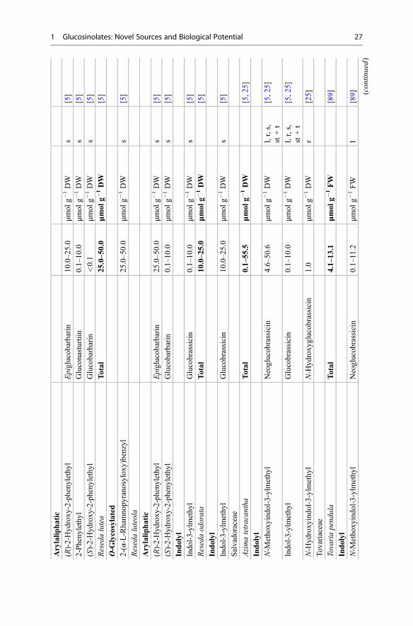
Arylalip
hatic
(R)-2-Hyd
roxy
-2-pheny
lethyl
Epiglucob
arbarin
10.0–2
5.0
μmol
g�1DW
s[5]
2-Pheny
lethyl
Gluconasturtiin
0.1–10
.0μm
olg�
1DW
s[5]
(S)-2-Hyd
roxy
-2-pheny
lethyl
Glucobarbarin
<0.1
μmol
g�1DW
s[5]
Resedalutea
Total
25.0–5
0.0
μmol
g�1DW
[5]
O-G
lycosylated
2-(α-L-Rhamno
pyrano
syloxy
)benzyl
25.0–5
0.0
μmol
g�1DW
s[5]
Resedaluteola
Arylalip
hatic
(R)-2-Hyd
roxy
-2-pheny
lethyl
Epiglucob
arbarin
25.0–5
0.0
μmol
g�1DW
s[5]
(S)-2-Hyd
roxy
-2-pheny
lethyl
Glucobarbarin
0.1–10
.0μm
olg�
1DW
s[5]
Indolyl
Indo
l-3-ylmethy
lGlucobrassicin
0.1–10
.0μm
olg�
1DW
s[5]
Resedaod
orata
Total
10.0–2
5.0
μmol
g�1DW
[5]
Indolyl
Indo
l-3-ylmethy
lGlucobrassicin
10.0–2
5.0
μmol
g�1DW
s[5]
Salvado
raceae
Azimatetracan
tha
Total
0.1–55
.5μm
olg�
1DW
[5,2
5]
Indolyl
N-M
etho
xyindo
l-3-ylmethy
lNeoglucob
rassicin
4.6–50
.6μm
olg�
1DW
l,r,s,
st+t
[5,2
5]
Indo
l-3-ylmethy
lGlucobrassicin
0.1–10
.0μm
olg�
1DW
l,r,s,
st+t
[5,2
5]
N-H
ydroxy
indo
l-3-ylmethy
lN-H
ydroxy
glucob
rassicin
1.0
μmol
g�1DW
r[25]
Tov
ariaceae
Tovariapend
ula
Total
4.1–13
.1μm
olg�
1FW
[89]
Indolyl
N-M
etho
xyindo
l-3-ylmethy
lNeoglucob
rassicin
0.1–11.2
μmol
g�1FW
l[89] (c
ontin
ued)
1 Glucosinolates: Novel Sources and Biological Potential 27

Table
2(con
tinue
d)
Fam
ily/Species/GLgrou
pGLtrivialname
Range
Unit
Plant
organ
Ref.
N-A
cetylin
dol-3-ylmethy
lN-A
cetylglucobrassicin
0.2–3.8
μmol
g�1FW
l[89]
Indo
l-3-ylmethy
lGlucobrassicin
0.2–1.3
μmol
g�1FW
l[89]
4-Metho
xyindo
l-3-ylmethy
l4-Metho
xyglucob
rassicin
<0.1–0.2
μmol
g�1FW
l[89]
4-Hyd
roxy
indo
l-3-ylmethy
l4-Hyd
roxy
glucob
rassicin
<0.1–0.2
μmol
g�1FW
l[89]
Tropaeolaceae
Trop
aeolum
majus
Total
5.9–13
0.0
μmol
g�1DW
[157
,158]
Arylalip
hatic
Benzyl
Glucotrop
aeolin
48.0–1
00.0
μmol
g�1DW
l,r,s,tu
[5,1
57]
3-Metho
xybenzyl
Glucolim
nanthin
0.1–50
.0μm
olg�
1DW
tu[158
]
4-Hyd
roxy
benzyl
Glucosinalbin
<0.1–5.3
μmol
g�1DW
tu[158
]
Indolyl
Indo
l-3-ylmethy
lGlucobrassicin
0.1–10
.0μm
olg�
1DW
s[5]
Trop
aeolum
peregrinum
Alip
hatic
1-Methy
lethyl
Glucopu
tranjiv
in<0.1–2.0
μmol
g�1DW
l,r
[33]
1-Methy
lpropy
lGlucocochlearin
0.6
μmol
g�1DW
l[33]
Arylalip
hatic
4-Hyd
roxy
benzyl
Glucosinalbin
0.5–12
5.0
μmol
g�1DW
l,r,s
[5,3
3]
4-Metho
xybenzyl
Glucoaubrietin
<0.1–50
.0μm
olg�
1DW
l,r,s
[5,3
3]
Benzyl
Glucotrop
aeolin
0.1–10
.0μm
olg�
1DW
r,s
[5,3
3]
Trop
aeolum
tuberosum
Total
36,5–9
0,0
μmol
g�1DW
[159
]
Arylalip
hatic
4-Metho
xybenzyl
Glucoaubrietin
36.5–9
0.0
μmol
g�1DW
tuy
babark,brbranch,bubu
ds,fl
flow
er,frfruit,hhy
pocotyl,iinfl
orescence,lleaf,lxlatex,ppu
lp,plp
lantlet,rroot,s
seed,spsprout,ststem,tthorn,tu
tuber,ys
youn
gshoo
t,μm
olg�
1DW
μmol
g�1driedweigh
t,μm
olg�
1FW
μmol
g�1freshweigh
t,*no
tun
equivo
cally
definedstructure
28 I. Blažević et al.

However, the use of NMR is necessary for the distinction of isomers and it isindispensable for the structure elucidation of previously not isolated anduncharacterized intact GLs [5, 11].
Acylated GLs (either on the side chain or on the glucopyrano unit) by benzoic,cinnamic, p-coumaric, isoferulic, or sinapic acid, as well as some very uncommonand unique structures are also identified [3, 4], but only in very low quantity, sospecific techniques were suggested for their detection [12, 13].
Table 1 includes GLs which structures are undoubtedly known and are found infair amounts in families which include plant species that can be considered as theirnatural sources.
The metabolism of Brassicaceae plants can accept selenium instead of sulfur intoS assimilation pathway. Se assimilation results in the production of the selenoaminoacids: selenomethionine (SeMet) and selenocysteine (SeCys). As a result, the so-called seleno-GLs, defined as GL-like metabolites, are biosynthesized. They werereported from a selenium-tolerant crucifer such as broccoli (Brassica oleracea var.italica) but only when exposed to unusually high levels (100 μmol/L) of sodium
OH
HO
HO
OH
O
S R
N−O3SO
Fig. 1 General structure ofglucosinolates
S CGlc
N
CH2
-O3SO
n S CH3
O
S CGlc
N
CH2
-O3SO
S CH3
O
O
S CGlc
N
CH2
-O3SO
nCH2
n = 2-10 n = 2-12
n = 3-10 n = 1-9
S CGlc
N
CH2
-O3SO
nS CH3
nCH
Fig. 2 General structure of chain-elongated aliphatic GLs [3]
1 Glucosinolates: Novel Sources and Biological Potential 29

selenite and sodium selenate added to nutrient solutions [14, 15]. Recently, severalSe-containing GLs were reported in Se-treated different Brassica spp., i.e., glu-coselenoiberverin, glucoselenoerucin, glucoselenoberteroin, glucoselenoraphanin,and glucoselenonasturtiin [15–18]. However, the detection of these GLs, or theiraglycones, converted into isoselenocyanates was done by using LC-MS, and GC-MS, respectively, and thus not unambiguously identified [16].
3 Occurrence and Novel Sources
Nearly all the GL-producing angiosperm plant families are found in the orderBrassicales (Fig. 3). The Putranjivaceae (Malpighiales) is an enigmatic family,notable for being the only lineage outside of the Brassicales in which plants aresupposed to possess the GL biochemical pathway [19]. Some other families outsideof the order Brassicales, like Phytolaccaceae (Caryophyllales), Pittosporaceae(Apiales), and Rubiaceae (Gentianales) which were reported to contain GLs, andnot characterized by proper methodology, should be reexamined [1, 20, 21].
Fig. 3 Slightly modified phylogenetic tree of the order Brassicales according to the APG III system(by permission from Stevens, Peter F.) [22], with numbers representing the approximate numbers ofgenus and species in each family, * – denotes branches with 50–80% support; all other brancheshave >80% support
30 I. Blažević et al.

3.1 Glucosinolate Content in Plants
Fahey et al. [1] summarized GL data for different species, but without indication ofquantities. Brassica vegetables such as cabbage, broccoli, Brussels sprouts, cauli-flower, and various root vegetables (e.g., radish, turnip) are economically importantmembers of the Brassicaceae family and thus the most investigated GL-containingplants. McNaughton and Marks [23] developed a food database of total GLs in fresh,frozen, boiled, and cooked cruciferous vegetables. Verkerk et al. reviewed crucifer-ous vegetables for the major GLs together with the GL content and the influence onhuman health [24]. A comprehensive analysis of GLs was reported by Bennett et al.[25] which included quantities in seeds. This sound seed-screening has brought tolight clear subdivisions based on the GL content:
(i) Only short- to medium-chain-length aliphatic GLs (C-3, or C-3 and C-4 withtraces of C-5)
(ii) Only long-chain aliphatic GLs(iii) Only simple arylaliphatic GLs [such as benzyl (20), 4-hydroxybenzyl (22),
2-phenylethyl (21) GLs](iv) Highly substituted arylaliphatic GLs (such as 3,4-dihydroxybenzyl, 3,4-
dimethoxybenzyl, and 3,4,5-trimethoxybenzyl GLs)
The GL content in plants varies between cultivars, plant individuals, and part ofthe plants, due to factors such as genetics, environment, and plant nutrients. GLs canbe found in the root, seed, leaf, and stem of the plant, while youngest tissues containthe highest amount. Characterization and quantification of the phytochemical com-position in plant organs, such as leaf and seed, provide a good insight into thepossible functional uses of these materials in agriculture as potential pesticides andin animal and human diets as functional nutraceuticals [26]. Dedicated extractivemethods allow one to isolate a number of GLs from adequate plant material, but inmany cases, organic synthesis is a necessary alternative to obtain fair quantities ofGLs [6].
Although novel sources are revealed continuously by investigating families otherthan Brassicaceae, there exists no database reviewing those studies. Table 2 summarizesthe GL quantities analyzed in all families of the order Brassicales, except for theBrassicaceae plants previously covered by Bennett et al. [25] and Verkerk et al. [24].
3.1.1 Families in the Order Brassicales
AkaniaceaeThe Akaniaceae family comprises two monotypic genera of trees, Bretschneideranative to southwest of China, Vietnam, and Taiwan and Akania native to easternAustralia (Fig. 4) [22].
Montaut et al. recently quantified the GLs in different tissues of Bretschneiderasinensis [27]. This plant is rare and threatened in mainland China. Arylaliphatic GLs,derived from Phe, were mostly found (Table 2), with glucotropaeolin (20) as the
1 Glucosinolates: Novel Sources and Biological Potential 31

major one. The total quantity was very low (highest in the fruit), suggesting that thisspecies is not a good GL source. Spectroscopic data are reported for 3-hydroxy-4-methoxybenzyl GL (glucobretschneiderin), a new GL isolated from the fruit inminor amount [27]. In addition, the aliphatic GL, 2-hydroxy-2-methylpropyl GL,was also quantified in B. sinensis.
Akania bidwillii, endemic to eastern Australia, was analyzed by Mithen et al., butno quantification was reported [28]. Arylaliphatic GLs were also found, withmethoxy-hydroxy-benzyl isomers (major), hydroxybenzyl isomers, methoxybenzyl,and benzyl GLs. However, no aliphatic GLs were detected.
BrassicaceaeThis dominant family encompasses over 330 genera and 3,700 species distributedprimarily in the temperate and alpine areas of all continents except Antarctica(Fig. 5) [29].
The largest genera are Draba (365 species), Cardamine (200 species), Erysimum(225 species), Lepidium (230 species), followed by Alyssum (195 species), Arabis (120),Boechera (110), Physaria (105: inc. Lesquerella), Rorippa (85), Heliophila (80), Isatis(80), Noccaea (80), Thlaspi (55), Biscutella (55), Matthiola (50), Descurainia(50), Hesperis (45), Sisymbrium (45: only Old World), and Brassica (38), etc.The family contains well-known species such as Brassica oleracea (broccoli, cabbage,cauliflower, kale, etc.), Brassica rapa (turnip, Chinese cabbage, etc.), Brassica napus(rapeseed, etc.), Raphanus sativus (common radish), Armoracia rusticana (horseradish),Arabidopsis thaliana (thale cress, a model organism), and many others.
Aliphatic GLsGLs found in these species are derived mostly from the chain-elongated forms ofMet, combined with post-synthesis extensive modifications of the side chain [28].According to Bennett et al.’s report [5], as well as Verkerk et al.’s review of GL
Fig. 4 Akaniaceae world distribution map
32 I. Blažević et al.

concentration ranges [24], C3-C5 aliphatic GLs were a common characteristic ofmost Brassica species. The highest quantities were found in seeds, referring mostlyto sinigrin (6) (>200 μmol g�1 DW), glucoiberin (14) (up to 200 μmol g�1 DW),gluconapin (7) (up to 125 μmol g�1 DW), glucoraphanin (15) (up to 100 μmol g�1
DW), glucoerucin (12) and progoitrin (9) (up to 50 μmol g�1 DW), andGlucoibervirin (11) and glucobrassicanapin (8) (up to 25 μmol g�1 DW). Broccoliseeds are one of the most used sources of 15, a precursor of the biologically relevantR-sulforaphane. In a recent report, De Nicola et al. exploited Tuscan black kaleseeds, rich in 15 (5.1% w/w), as a suitable source for gram-scale production ofthis GL. In a typical experiment, 3.10 g of pure 15, i.e., 1.09 g of enantiopureR-sulforaphane, was obtained from 150 g of defatted Tuscan black kale seed meal[30]. As previously mentioned, selenized Brassica produce seleno-GLs. It is pro-posed that biofortification of Brassica crops with Se can further increase theirchemopreventive activities [31]. Matich et al. investigated the accumulation ofseleno-GLs in different tissues of cauliflower, broccoli, and rape and reported theforage rape roots and the broccoli florets to be the best sources [16]. A high-GLbroccoli floret, containing 29.4 μg g�1 FW of total methylsulfanylalkyl GLs (MeS-GLs), produced after Se-intake produced 32.4 μg g�1 FWof total MeSe-GLs mostlyseleno-glucoerucin (30 μg g�1 FW) [16].
Several recent reports (Table 2) on the sources of aliphatic GLs include Armo-racia rusticana sprouts [32] and Guiraoa arvensis root [33] which are recognized asgood sources of 6 (>100 μmol g�1 DW) (Table 2). These plants can be considered asthe first (i) Bennett et al. subdivision [5], supporting this categorization.
Some of the recent reports also include GLs in endemic species of theBrassicaceae family. Kerguelen cabbage (Pringlea antiscorbutica), a subantarcticendemic perennial plant, from the Kerguelen phytogeographical area in the southernIndian Ocean, is an edible Brassicaceae plant. Having amounts of glucotropaeolin(20) (up to 50.3 μmol g�1 DW) unusually high for Brassicaceae plant and 7 (up to
Fig. 5 Brassicaceae world distribution map
1 Glucosinolates: Novel Sources and Biological Potential 33

66.2 μmol g�1 DW) as the major GLs is considered a good dietary source of GLs(Table 2) [34]. This is an example that combines the first and third categories(i + iii), given by Bennett et al. [5], representing a new subdivision.
The endemic plants included in the Alysseae tribe of the Croatian native flora(Aurinia species, Degenia velebitica, and Fibigia triquetra) are found to have a GLcontent ranging from 9.9 to 135.4 μmol g�1 DW in different plant parts. Havingespecially high content in the seeds (over 4.0% w/w with the highest, 6.1% w/w in F.triquetra), these Alysseae are found to represent a good novel GL source [35–38].Aurinia sinuata and A. leucadia can be found in mountainous areas of Central andSouthern Europe, Russia, and Turkey. The major GLs found in the seeds of theinvestigated plants were glucoalyssin (16) (38.2 μmol g�1 DW) and 7 (39.1 μmol g�1
DW) in A. leucadea, while 16 (62.3 μmol g�1 DW) and 8 (17.2 μmol g�1 DW) in A.sinuata. Degenia velebitica and Fibigia triquetra represent rare Croatian paleoste-noendemic species (Fig. 6). The natural habitat of D. velebitica, the only species ofthe genus Degenia, is restricted to only several localities on the South and MidVelebit massif. D. velebitica seed, having 88.0 μmol g�1 DWof glucoberteroin (13)(ca. 90% of total GLs), represents a good source of this compound [36]. F. triquetra,sometimes confused with D. velebitica, is found wild-growing only on the rockygrounds of Dalmatia. The major GLs found in F. triquetra are 12 (76.7 μmol g�1
DW), 7 (66.7 μmol g�1 DW), and 15 (23.4 μmol g�1 DW) [38]. To a certain extent,these informations bring a different light to the first Bennett et al.’s subdivision,differentiating in C3-C5 GLs possible combination.
Within the Brassicaceae, long-chain aliphatic thiofunctionalized GLs (C7-C10)are generally restricted to a few species such as Arabis, Biscutella, Camelina,Capsella, Nasturtium, as well as certain wild Lepidium and Sinapis species [1, 5].The leaves of three Pachycladon species, endemic to the South Island ofNew Zealand and Tasmania, are found to biosynthesize C7 and/or C8 GLs (Table 2).Biscutella laevigata seed was shown to be a very good source of 8-(methylsulfinyl)octyl GL (17) (75.0–100.0 μmol g�1 DW) [5]. Different Camelina spp. seeds arereported as a source of 10-(methylsulfinyl)decyl GL (up to 25.0 μmol g�1 DW)
Fig. 6 Paleostenoendemic plants growing only in Croatia (Europe): (a) Degenia velebitica(Degen) Hayek (Photo by permission of Berislav Horvatić [39]) and (b) Fibigia triquetra (DC.)Boiss. ex Prantl (Photo by Ivica Blažević)
34 I. Blažević et al.

[5, 33]. In addition, the seeds of Camelina pilosa contain the highest quantity of 11-(methylsulfinyl)undecyl GL (11.1 μmol g�1 DW) (Table 2). These reports are inagreement with the third (iii) Bennett et al.’s subdivision. On the contrary, Agerbirket al. investigated Nasturtium officinale (watercress) of different origin and it wasconcluded to be polymorphic for GL profile and chromosome numbers [40]. Long-chain methylsulfanylalkyl (C5-C7) and methylsulfinylalkyl (C8-C10) GLs werefound (6–28 μmol g�1 DW) the highest being 9-(methylsulfinyl)nonyl GL (18)(21.1 μmol g�1 DW) but in combination with high levels of arylaliphatic GLs –dominantly gluconasturtiin (21) (7.8 – 52.8 μmol g�1 DW) [40].
Some GLs are reported only through their breakdown products, such as thepresence of long-chain C7-C10 unsaturated ITCs in autolyzates of Nasturtiummontanum [10]. Bennett et al. reported the content of C7-C10 thiofunctionalizedGLs in the seed of several Arabis spp. [5]. Blažević et al.’s investigation of Arabisturrita of volatile hydrolysis products isolated from different plant parts (seed, root,leaf) revealed mainly long-chain olefinic (C6-C11) and thiofunctionalized (C8-C10)GLs. The suggested occurrence of alkenyl GLs in a plant, like in the case of N.montanum also accumulating similar Met-derived GLs, seemed likely. However,HPLC-ESI-MS of intact GLs confirmed only thiofunctionalized C8-C10 GLs in A.turrita, and it was suggested that thiofunctionalized ITCs can be converted intoolefinic ITCs under high temperature during GC-MS analyses [41]. Thus, thepresumption of native unsaturated C8-C10 GLs in the case of N. montanum or A.turrita is questionable.
Alkyl GLs are also detected in a few species belonging to the Brassicaceae family[5, 34]. It is suggested that methyl GL (glucocapparin, 1), the simplest GL identifiedin plants, does not occur in this family [28]. However, Griffiths et al. reported, next tothe major glucobrassicin (28) (98%), a small amount of 1 (2%) on the leaf surface ofIsatis tinctoria (containing 0.12 μmol of total GLs per g of leaf material) [42]. Inaddition, by using GC-MS analysis of its degradation product (methyl ITC), 1 wasreported in the seed of Wasabi japonica and Armoracia lapathifolia Gilib [43].
GLs derived from Val, Leu, Ile, and dihomo-Ile are also found. Lunaria spp.,Sisymbrium officinale, and Dentaria pinnata are found to be a source ofglucoputranjivin (2) (up to 50 μmol g�1 DW). A novel GL, 3-(Hydroxymethyl)pentyl GL, was present in high levels in Cardamine pratensis from eastern NorthAmerica and in commercially obtained seeds (114.6 μmol g�1 DW), but was notfound in C. pratensis plants from southern Scandinavia [44].
Arylaliphatic GLsThe arylaliphatic GLs are derived from the aromatic parental amino acids Phe andTyr and are commonly found in plants belonging to the Brassicaceae family. One ofthe highest reported concentrations of glucosinalbin (22) (250 μmol g�1 DW) wasfrom Sinapis alba seeds [45]. It was previously mentioned that, besides the highcontent of C-4 to C-9 aliphatic GLs, P. antiscorbutica and N. officinale also representa good source of 20 and 21, respectively, (up to 50 μmol g�1 DW) [34]. Some of thenovel good sources of arylaliphatic GLs, given in Table 2, are for 22 Noccaeacaerulescens seed and Kremeriella cordylocarpus leaves (83.0 and 76.0 μmol g�1
1 Glucosinolates: Novel Sources and Biological Potential 35

DW, respectively) [33, 46] and for 21 Coincya rupestris root (67.0 μmol g�1 DW)[33]. Barbarea spp. seem to be a good source of 21 (up to 100 μmol g�1 DW) [5, 47,48], glucobarbarin (26) (up to 90 μmol g�1 DW), and epiglucobarbarin (27) (up to55 μmol g�1 DW) [5, 47]. Among these, only K. cordylocarpus and Barbarea spp.are in agreement with the third (iii) Bennett et al.’s subdivision. Other plants, as inthe case of P. antiscorbutica, combine the first and third subdivisions of GL content(i + iii), supporting the new category, combining a short aliphatic and simplearylaliphatic GLs.
Some highly substituted arylaliphatic GLs, such as 3,4-dimethoxybenzyl GL and3,4,5-trimethoxybenzyl GL, are restricted to a few genera, mostly belonging to theBrassicaceae family, such as Coronopus, Lepidium, andMatthiola [1, 49, 50]. 3,4,5-Trimethoxybenzyl GL, identified after its degradation volatile, has been previouslyreported as the sole GL in the seeds of few investigated Lepidium spp. [50–52].Coronopus squamatus seeds of Spanish origin were found to be a good source of3,4,5-trimethoxybenzyl GL (50–75 μmol g�1 DW) [5]. Due to the presence of thisGL, a very close relationship between the Coronopus and Lepidium genera wassuggested [49].
Indolyl GLsIndolyl GLs found in cruciferous plants are derived from Trp and possess a diverselysubstituted heterocyclic moiety. Four main indolyl GLs have been identified in mostcultivated Brassica species: glucobrassicin (28), neoglucobrassicin (29), 4-hydroxyglucobrassicin (30), and 4-methoxyglucobrassicin (31). Isatis canescensflower buds displayed a high 28 content (up to 60 μmol g�1 DW) and represent agood source of this GL. The purification method consisted of two chromatographicsteps, made possible to obtain 28 (purity of 92–95%), with a yield of 21 g kg�1 [53].It seems that Isatis spp. (Table 2) display a high content of short aliphatic and indolylGLs. Some species can produce fair amounts of some additional arylaliphatic orindolyl GLs, next to the short and medium aliphatic GLs, as in the case ofCardamine pratensis, Coincya spp., Hemicrambe fruticulosa, Lepidium meyenii,Isatis spp., Noccaea caerulescens, Pringlea antiscorbutica, and Sinapis pubescens(Table 2). Thus, an additional subdivision can be suggested next to the ones ofBennett et al., combining aliphatic with arylaliphatic or indolyl GLs.
1,4-Dimethoxyglucobrassicin was firstly isolated and confirmed structurally fromthe roots of Barbarea vulgaris R. Br. ssp. arcuata which contained 0.53.1 μmol g�1
DW of this GL [54]. This rare side chain was also recently detected in the root ofCardamine pratensis (0.1 μmol g�1 DW) [44].
Extracts of Isatis tinctoria were found to contain N-sulfoglucobrassicin (11.3 –13.5 μmol g�1 DW) [55]. In addition, Isatis spp. are found to contain the unusualcombination of an aliphatic and an indolyl moiety, known as glucoisatisin/epiglucoisatisin (ca. 1.5 μmol g�1 DW) [56, 57].
O-Glycosylated GLsA O-glycosylated GL, containing L-rhamnose, as additional sugar moiety linked tothe aromatic ring, i.e., 4-α-rhamnosyloxybenzyl GL (glucomoringin; 32, Table 1),
36 I. Blažević et al.

was identified in Noccaea caerulescens. This was the first report of 32 in thisBrassicaceae species where it was the main GL in 10 out of 13 populations collectedfrom different locations in Europe, with the highest concentration in the seeds up to111.0 μmol g�1 DW (Table 2) [46].
O-Acylated GLsUncommon GLs displaying O-acylated sites on the D-glucopyrano unit and/or onthe hydroxyl groups present in the side chain are also detected among Brassicaceaeplants. Those carboxylic esters usually result from acylation by benzoic, cinnamic,p-coumaric, isoferulic, or sinapic acid, mostly conjugated at the primary position ofthe D-gluco unit. However, these GLs, mostly found in the seeds, are detected invery low quantities or their quantity was not reported [3, 4, 12, 40, 47, 57–60].
Recently, Survay et al. reported the 6-p-coumaroyl derivative of 15 isolated frombroccoli florets (0.04 μmol g�1 FW) [59]. However the above structure might beregarded as questionable after retraction of a following paper by the same authors,due to “structure elucidation issues.” In Arabidopsis seeds, esterification on the sidechain of hydroxylated GLs leads to the accumulation of benzoylated and sina-poylated GLs and some of them are identified: 3- and 4-benzoyloxypropyl GLs, 3-and 4-sinapoyloxypropyl GLs, 6'-benzoyl-4-methylsulfanylbutyl GL, 60-benzoyl-4-methylsulfinylbutyl GL, and 60-benzoyl-4-benzoyloxybutyl GL (<6.0 ng mg�1
DW) [61, 62]. From the seeds of Barbarea spp., five 60-isoferuloyl esters of 28,21, (S)-2-hydroxy-2-phenylethyl GL (26), (R)-2-hydroxy-2-phenylethyl GL (27),and (R)-2-hydroxy-2-(4-hydroxyphenyl)-ethyl GL were isolated, and their structureswere elucidated [47]. The acyl group was in all cases found to be a trans isoferuloylgroup at the primary position of the D-glucopyrano unit. The highest level of 60-isoferuloylglucobarbarin was found in B. vulgaris mature seeds (3 μmol g�1 DW)[47]. GLs with diverse acylation patterns are much less abundant and cannot beidentified solely by applying low resolution MS [47]. Thus, Barbarea vulgaris seedextracts were analyzed by reversed-phase liquid chromatography coupled with elec-trospray ionization and Fourier transform ion cyclotron resonance mass spectrome-try (RPLC-ESI FTICR MS), which represents a new procedure for detecting andidentifying intact acylated GLs found in trace amounts in natural plant samples [12].
CapparidaceaeThe Capparidaceae family consists of 16 genera and 480 species, which are mostlydistributed in warm regions all around the world. The largest genera includeCapparis (250), Maerua (100), Boscia (37), and Cadaba (30) (Fig. 7) [22].
Methyl GL (glucocapparin, 1) is the principal GL accumulating in many speciesof the Capparidaceae and Cleomaceae. However, its biosynthesis has not beenexplored. It may derive from Ala, as a novel GL precursor possibly through theaction of CYP79, or it may arise as a result of the single or multiple cleavage of sidechains derived from Met or branched-chain amino acids (BCAA: Leu, Ile, Val) orelongated Phe [28].
The genus Capparis is native to the Mediterranean basin, but is now widelydistributed from Atlantic coasts (Morocco) to Black Sea lands and Caspian Sea.
1 Glucosinolates: Novel Sources and Biological Potential 37

Cultivation of some species of this genus, especially C. spinosa and C. ovata, hasbeen reported from the plain areas and deserts of Afghanistan, India, Indonesia,Nepal, Pakistan, North Africa, Southwest Asia, Australia, and Southern Europe upto an elevation of 1100 m [63]. Generic limits found in Capparidaceae are discussedby Hall et al., suggesting that the New World Capparis will need a new name [64].The sound qualitative GL profiling in Old World and New World Capparidaceaewas reported by Mithen et al. [28]. Old World taxa of the Capparidaceae werereported to have high levels of 1, with three exceptions, namely, Boscia longifoliaand Capparis tomentosa which showed high levels of hydroxymethylbutyl andhydroxyethyl GL, respectively, and Maerua triphylla, where 28 was the mostabundant [28].
In most reports on Capparis spinosa, the highest 1 content was found in the seeds(up to 127 μmol g�1 DW) [5, 65, 66]. ESI FTICRMSwas applied for LC profiling ofintact GLs (specially for the ones with low abundance) in flower buds of C. spinosa[13]. In addition to the GLs already found (1, isopropyl/n-propyl-GLs, mercapto-glucocapparin, 28, and 30) [3, 13], the uncommon glycinyl-glucocapparin and twosulfur-rich GLs (disulfanyl-glucocapparin and trisulfanyl-glucocapparin) were alsoreported (Table 2) [13].
Next to 1, C. ovata can also contain a relatively high amount of 6 (up to29.8 μmol g�1 DW in the seeds, Table 2) [67]. The GL content in B. senegalensis,monitored throughout the whole year in leaves and fruits collected in 4 localities inSenegal, ranged from 2 to 440 μmol g�1. The highest amount was recorded in Janu-ary (dry season) and the lowest between August and November (rainy period) [68].
Whereas all Old World Capparidaceae contain 1, Mithen et al. reported that NewWorld Capparidaceae, including New World Capparis, contain either 1 or severaldistinctive and perhaps unique GLs of complex and unresolved structures, indicatingcontinued innovation in GL biosynthesis [28]. This research has brought to light
Fig. 7 Capparidaceae world distribution map
38 I. Blažević et al.

clear differences between New World and Old World Capparis and can be related tothe subdivisions based on GL content given by Bennett et al. [5]. On the basis of GLqualitative profiles, New World specimens of the Capparidaceae were divided intothree groups:
(a) With high levels of 1 – which can be included as a new subdivision given byBennett et al.
(b) Lack of 1 but presence of GLs with molecular weights consistent with chain-elongated Met or BCAA-derived GLs, including certain GLs, such as oxoheptyland oxooctyl GLs found in C. scabrida and C10-C11 isomers of methylsulfo-nyldecyl and methylsulfonylundecyl GLs found in Steriphoma peruvianum.These GLs do not occur in other families, but can be related to Bennett et al.’ssubdivision (ii), i.e., plants that produce long-chain aliphatic GLs.
(c) Several taxa, such as Morisonia and Atamisquea, have potentially novel GLsthat do not correspond to any known structures, though the structural identitymust remain provisional until further studies [28].
CaricaceaeThe Caricaceae are a family of dicotyledonous angiosperms found in mostlytropical (Andean) America (three genera in Mexico) and Africa (Cylicomorphaonly). It is made up of several genera (4–6) with 34 species or more: Vasconcellea(22), Jacaratia (7), Carica (5), Jarilla (3), and Cylicomorpha (2) (Fig. 8) [22].
Analysis of the rbcL gene sequences by Rodman et al. demonstrated that thefamily Caricaceae belongs to the main GL-containing order Brassicales and thatCaricaceae are apparently a sister family of the Moringaceae [69–72]. DNAsequencing of the nuclear 18S ribosomal RNA gene has yielded the same result[73]. Benzyl GL (20) is the major GL found in the family Caricaceae [1]. Themodification in the biosynthetic route to GLs includes (i) chain elongation of
Fig. 8 Caricaceae world distribution map
1 Glucosinolates: Novel Sources and Biological Potential 39

aliphatic and aromatic amino acids by inserting methylene groups into their sidechains and (ii) metabolic modification of the amino acids (or chain-extended deriv-atives of amino acids) that takes place via an aldoxime intermediate, the same as forcyanogenic glycosides. However, the co-occurrence of GLs and cyanogenic glyco-sides in the same plant is very scarce [74, 75]. Carica papaya L., the most knownplant of this family, includes, next to 20, the cyanogenic glucosides, prunasin,sambunigrin, and tetraphyllin B [76]. Aside from C. papaya, Alliaria petiolata (M.Bieb.) Cavara and Grande (Brassicaceae) is the only other known exception of aplant containing both GLs and cyanogenic glucosides which, next to 6 also containslarge quantities of hydroxynitrile glucoside alliarinoside [75]. In immature papayafruit pulp, the amount of 20 ranged from 163.1 to 259.2 μmol g�1 DWof latex fluid.No myrosinase was found in papaya latex. To explain the variability of benzyl ITCformation in the macerated immature fruit pulp, the author speculated that papain (aproteolytic enzyme also found in the papaya latex) prevents uncontrolled enzymaticproduction of benzyl ITC by inactivating small quantities of myrosinase in the latexthrough proteolysis [77].
CleomaceaeThe Cleomaceae is a small pantropical family. Although historically treated as asubfamily of Capparidaceae sensu lato, Cleomaceae are easily distinguished fromclosely related Capparidaceae sensu stricto [78]. This family comprises about 300species including mostly Cleome (275, including Podandrogyne) and Cleomella(25) genera [22]. These genera were previously included in the familyCapparidaceae, but were raised to a distinct family when DNA evidence suggestedthat the genera included in it are more closely related to the Brassicaceae than theyare to the Capparidaceae. They are found in tropical and warm temperate areas,especially in America (Fig. 9).
Fig. 9 Cleomaceae world distribution map
40 I. Blažević et al.
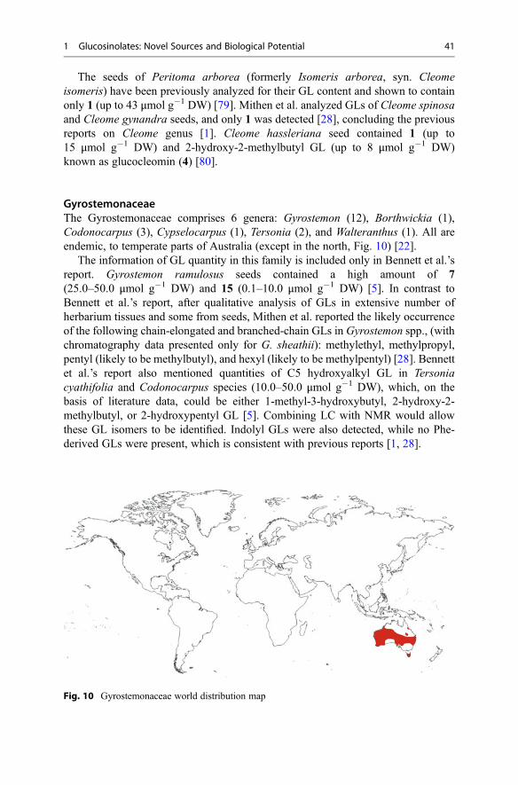
The seeds of Peritoma arborea (formerly Isomeris arborea, syn. Cleomeisomeris) have been previously analyzed for their GL content and shown to containonly 1 (up to 43 μmol g�1 DW) [79]. Mithen et al. analyzed GLs of Cleome spinosaand Cleome gynandra seeds, and only 1 was detected [28], concluding the previousreports on Cleome genus [1]. Cleome hassleriana seed contained 1 (up to15 μmol g�1 DW) and 2-hydroxy-2-methylbutyl GL (up to 8 μmol g�1 DW)known as glucocleomin (4) [80].
GyrostemonaceaeThe Gyrostemonaceae comprises 6 genera: Gyrostemon (12), Borthwickia (1),Codonocarpus (3), Cypselocarpus (1), Tersonia (2), and Walteranthus (1). All areendemic, to temperate parts of Australia (except in the north, Fig. 10) [22].
The information of GL quantity in this family is included only in Bennett et al.’sreport. Gyrostemon ramulosus seeds contained a high amount of 7(25.0–50.0 μmol g�1 DW) and 15 (0.1–10.0 μmol g�1 DW) [5]. In contrast toBennett et al.’s report, after qualitative analysis of GLs in extensive number ofherbarium tissues and some from seeds, Mithen et al. reported the likely occurrenceof the following chain-elongated and branched-chain GLs in Gyrostemon spp., (withchromatography data presented only for G. sheathii): methylethyl, methylpropyl,pentyl (likely to be methylbutyl), and hexyl (likely to be methylpentyl) [28]. Bennettet al.’s report also mentioned quantities of C5 hydroxyalkyl GL in Tersoniacyathifolia and Codonocarpus species (10.0–50.0 μmol g�1 DW), which, on thebasis of literature data, could be either 1-methyl-3-hydroxybutyl, 2-hydroxy-2-methylbutyl, or 2-hydroxypentyl GL [5]. Combining LC with NMR would allowthese GL isomers to be identified. Indolyl GLs were also detected, while no Phe-derived GLs were present, which is consistent with previous reports [1, 28].
Fig. 10 Gyrostemonaceae world distribution map
1 Glucosinolates: Novel Sources and Biological Potential 41
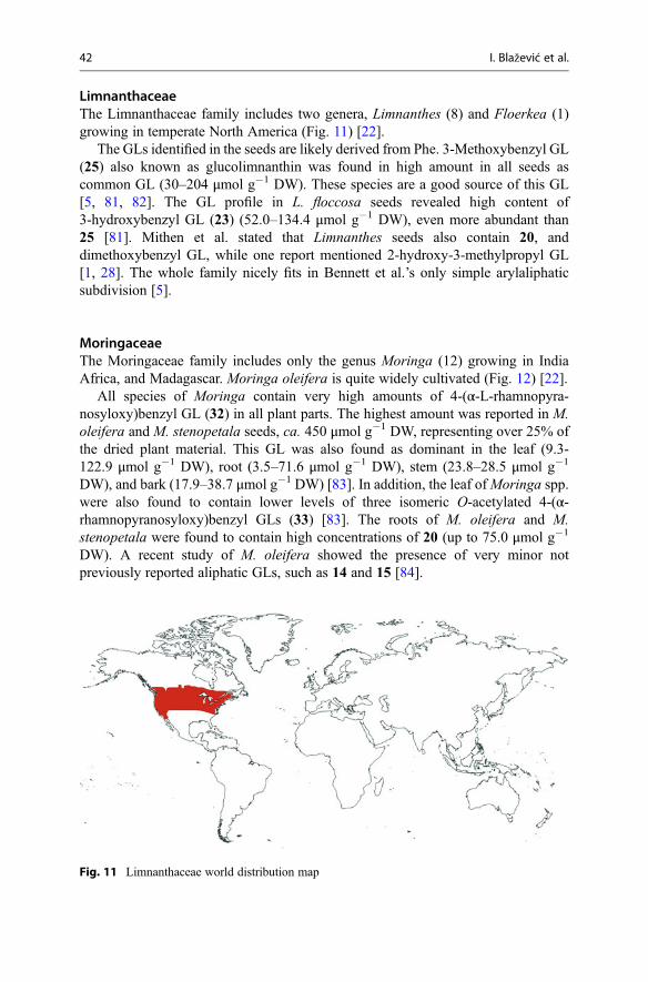
LimnanthaceaeThe Limnanthaceae family includes two genera, Limnanthes (8) and Floerkea (1)growing in temperate North America (Fig. 11) [22].
The GLs identified in the seeds are likely derived from Phe. 3-Methoxybenzyl GL(25) also known as glucolimnanthin was found in high amount in all seeds ascommon GL (30–204 μmol g�1 DW). These species are a good source of this GL[5, 81, 82]. The GL profile in L. floccosa seeds revealed high content of3-hydroxybenzyl GL (23) (52.0–134.4 μmol g�1 DW), even more abundant than25 [81]. Mithen et al. stated that Limnanthes seeds also contain 20, anddimethoxybenzyl GL, while one report mentioned 2-hydroxy-3-methylpropyl GL[1, 28]. The whole family nicely fits in Bennett et al.’s only simple arylaliphaticsubdivision [5].
MoringaceaeThe Moringaceae family includes only the genus Moringa (12) growing in IndiaAfrica, and Madagascar. Moringa oleifera is quite widely cultivated (Fig. 12) [22].
All species of Moringa contain very high amounts of 4-(α-L-rhamnopyra-nosyloxy)benzyl GL (32) in all plant parts. The highest amount was reported in M.oleifera and M. stenopetala seeds, ca. 450 μmol g�1 DW, representing over 25% ofthe dried plant material. This GL was also found as dominant in the leaf (9.3-122.9 μmol g�1 DW), root (3.5–71.6 μmol g�1 DW), stem (23.8–28.5 μmol g�1
DW), and bark (17.9–38.7 μmol g�1 DW) [83]. In addition, the leaf ofMoringa spp.were also found to contain lower levels of three isomeric O-acetylated 4-(α-rhamnopyranosyloxy)benzyl GLs (33) [83]. The roots of M. oleifera and M.stenopetala were found to contain high concentrations of 20 (up to 75.0 μmol g�1
DW). A recent study of M. oleifera showed the presence of very minor notpreviously reported aliphatic GLs, such as 14 and 15 [84].
Fig. 11 Limnanthaceae world distribution map
42 I. Blažević et al.

PentadiplandraceaeThe Pentadiplandraceae family possesses only one genus, Pentadiplandra, whichcontains a single species, Pentadiplandra brazzeana Baillon. P. brazzeana is a plantfound in Western and Central Africa (Fig. 13) [22].
Although some botanical studies of Pentadiplandra genus indicated a strongphylogenetic similarity with the GL-containing Tovaria pendula Ruiz and Pavón(Tovariaceae) [85], the analyses using rbcL, ndhF, and matK sequences showed aweak resolution of the relationship of Pentadiplandraceae and Tovariaceae [86]. DeNicola et al.’s analysis of GLs in different plant parts revealed mostly arylaliphaticGLs having the highest content of glucoaubrietin (24) in the seeds, i.e.,109.1 μmol g�1 DW, representing it as a good source of this GL [87]. The total
Fig. 12 Moringaceae world distribution map
Fig. 13 Pentadiplandraceae world distribution map
1 Glucosinolates: Novel Sources and Biological Potential 43

GLs in the roots were 48.0 μmol g�1 DW which also contained the highest quantityof the same GLs, together with, 20 and 25 (Table 2) [87]. The leaf, with low GLcontent of 2.2 μmol g�1 DW, contained 3,4-dimethoxybenzyl GL, 20, and 28. Thesame qualitative profile of GLs was reported by Mithen et al. [28]. According to theGL content, this family nicely fits in Bennett et al.’s only simple arylaliphaticsubdivision [5].
ResedaceaeThe Resedaceae family is represented by six genera (about 85 species), distributedworldwide [22]. The genus Reseda (68 species) is the largest genera in theResedaceae. Natural areas of occurrence are warm temperate and dry subtropicalparts in the world, especially Mediterranean, Middle East, North African regions,and also Southern Africa, S.W. North America, and S.W. China [22]. With theexception of 4 Reseda species (R. alba, R. lutea, R. luteola, and R. phyteuma),which are worldwide weeds, almost all of the Reseda species are narrowly distrib-uted in the Mediterranean basin (Fig. 14) [88].
Bennett et al. investigated the GLs in the seeds of Reseda species and reportedarylaliphatic and indolyl type GLs in R. luteola and R. odorata, while a O-glycosylated GL was only found in R. lutea [5].
Two arylaliphatic GLs, glucobarbarin (26) and epiglucobarbarin (27), wereidentified in R. luteola ranging 0.1–10.0 and 25.0–50.0 μmol g�1 DW, respectively[5]. The same report included another member of the Resedaceae family, Cayluseaabyssinica, which also contained 21, next to the same arylaliphatic GLs and had thehighest reported 27 content (10.0–25.0 μmol g�1 DW) [5]. Mithen et al. investi-gated the GL profile in fresh leaves and seeds of Reseda species and reportedhydroxyphenylethyl GL (R-/S-isomer not given) and 21 being present in significantamounts such as in R. luteola [28]. Glucobarbarin (26) was identified as the majorGL of the total leaf surface of R. luteola and it accounted for over 90% of the total
Fig. 14 Resedaceae world distribution map
44 I. Blažević et al.
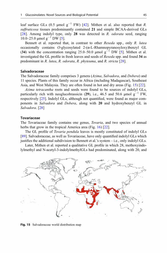
leaf surface GLs (0.5 μmol g�1 FW) [42]. Mithen et al. also reported that R.suffruticosa tissues predominantly contained 21 and simple BCAA-derived GLs[28]. Among indolyl type, only 28 was detected in R. odorata seed, ranging10.0–25.0 μmol g�1 DW [5].
Bennett et al. reported that, in contrast to other Reseda spp., only R. luteaoccasionally contains O-glycosylated 2-(α-L-Rhamnopyranosyloxy)benzyl GL(34) with the concentration ranging 25.0–50.0 μmol g�1 DW [5]. Mithen et al.investigated the GL profile in fresh leaves and seeds of Reseda spp. and found 34 aspredominant in R. lutea, R. odorata, R. phyteuma, and R. stricta [28].
SalvadoraceaeThe Salvadoraceae family comprises 3 genera (Azima, Salvadora, and Dobera) and11 species. Plants of this family occur in Africa (including Madagascar), SoutheastAsia, and West Malaysia. They are often found in hot and dry areas (Fig. 15) [22].
Azima tetracantha roots and seeds were found to be sources of indolyl GLs,particularly rich with neoglucobrassicin (29), i.e., 46.5 and 50.6 μmol g�1 FW,respectively [25]. Indolyl GLs, although not quantified, were found as major com-ponents in Salvadora and Dobera, along with 20 and hydroxybenzyl GL inSalvadora. [28]
TovariaceaeThe Tovariaceae family contains one genus, Tovaria, and two species of annualherbs that grow in the tropical America area (Fig. 16) [22].
The GL profile of Tovaria pendula leaves is mostly constituted of indolyl GLs[89]. Salvadoraceae, as well as Tovariaceae, have only quantified indolyl GLs whichjustifies the additional subdivision to Bennett et al.’s system – i.e., only indolyl GLs.
Later, Mithen et al. reported a qualitative GL profile in which 28, methoxyindo-lylmethyl and N-acetyl-3-indolylmethylGLs had predominated, along with 20, and
Fig. 15 Salvadoraceae world distribution map
1 Glucosinolates: Novel Sources and Biological Potential 45

methylethylGLs [28]. Low levels of pentyl (or methylbutyl) GL, probably derivedfrom chain-elongated BCAA, were also detected [28].
TropaeolaceaeThe Tropaeolaceae family contains one genus, Tropaeolum (105 species), that growsin the New World, especially in Andean parts (Fig. 17) [22].
The GL profile of T. majus, T. peregrinum, and T. tuberosum includedarylaliphatic GLs. Glucotropaeolin (20) and 24 were found as dominant GLs in T.majus seeds, i.e., 100.0, and 50.0 μmol g�1 DW, respectively. On the other hand, T.peregrinum seeds contained high content of 22 and 24, with 125.0 and 50.0 μmol g�1
DW, respectively. Glucoaubrietin (24) was the only arylaliphatic GL found in the
Fig. 16 Tovariaceae world distribution map
Fig. 17 Tropaeolaceae world distribution map
46 I. Blažević et al.

seeds of T. tuberosum, with content up to 90.0 μmol g�1 DW. This family fits into anonly simple arylaliphatic subdivision.
3.1.2 Other Families in the Order BrassicalesReports of other families included in the order Brassicales (Bataceae,Emblingiaceae, Koeberliniaceae, and Setchellanthaceae) are very scarce.
BataceaeThe Bataceae family comprises two species, Batis argillicola and Batis maritima.These plants are distributed in N. Australia and S. New Guinea, tropical America,and the Galapagos Islands [22]. No GL quantification was reported for these plantsto date, as far as the authors know. The most recent report on the analysis of B.maritima revealed small amounts of 28 and 29 [28].
EmblingiaceaeThe Emblingiaceae family includes only Emblingia calceoliflora growing in W.Australia [22]. The only record of GL qualitative profile was given by Mithen et al.,who reported hydroxy and hydroxymethoxybenzyl GLs in the leaf tissue [28]. Thisfinding supports the assignment of this taxon to the Brassicales.
KoeberliniaceaeThe Koeberliniaceae family includes one genus with two species growing in Centraland S.W. North America and Bolivia [22]. Mithen et al.’s study of Koeberliniaspinosa Zucc. (originated from Bolivia and USA) revealed that no GL was present[28], which was consistent with a previous report [73]. This is one family in theBrassicales that lacks GLs, though other evidences firmly place it in that order.
SetchellanthaceaeThe Setchellanthaceae family contains only one species, Setchellanthus caeruleus, ashrub endemic to Mexico, restricted to two small, arid or subarid, and disjunct areasfully 1,000 km apart. The only report by Mithen et al. included qualitative analysesof GLs with the major 2 and hydroxybenzyl GLs and minor hydroxymethylpropyland methylpropyl GL isomers [28].
3.1.3 Families Outside the Order BrassicalesThe nonexistence of GLs in some species of Capparidaceae and species of theKoeberliniaceae family is not the only surprise. The well-known report byDaxenbichler et al. included information of GL-bearing plants outside the orderBrassicales [50]. However, these early reports of GLs included the identification ofunstable p-hydroxybenzyl ITC and ITCs containing an indolyl moiety under thio-cyanate ion release. Such was the case of Bursaria spinosa var. incana (orderApiales, family Pittosporaceae) and Phytolacca americana (order Caryophyllales,family Phytolaccaceae). Phytolacca dioica was reported also to contain 20 from thecorresponding benzyl ITC [50]. Hu et al. reported GLs in the roots of Phytolaccaacinosa [20]. Some reports were discounted after subsequent investigations, such as
1 Glucosinolates: Novel Sources and Biological Potential 47

in the case of edible mushrooms (Agaricus bisporus) [90], Plantago species (orderLamiales, family Plantaginaceae) [91], and Theobroma cacao (order Malvales,family Malvaceae) [92].
On the other hand, the genus Drypetes (order Malpighiales, familyPutranjivaceae) is the only one accepted to possess the GL biochemical pathwayoutside the order Brassicales. The latest reported study of the essential oil from thebark of Drypetes gossweileri (Putranjivaceae) from Gabon found a high concentra-tion of benzyl ITC and benzyl cyanide that can result from 20 degradation [21]. Thesame report included an investigation of the essential oil from the root and bark ofRinorea subintegrifolia (order Malpighiales, family Violaceae), which suggestedthat the plants of the Violaceae family also have the ability to biosynthesize GLs.The most recent report from Hu et al. revealed that Croton tiglium, Phyllanthusemblica, Euphorbia humifusa, E. lathyris, and E. kansui from the Euphorbiaceaefamily and Knoxia valerianoides and Gardenia jasminoides from the Rubiaceaefamily contain GLs [20]. However, the existence of GLs should be reexamined, withmore accurate approaches using standards and more modern techniques (HPLC-MS,NMR) which are indispensable for structure elucidation with regard to GLs outsidethe order Brassicales.
4 Biological Potential
This database which includes all the families in the order Brassicales along withBennett et al.’s. and Verkerk et al.’s works represents the insight into the GL quantityprofile in GL-containing species investigated to date. GLs degrade into variouscompounds, i.e., ITCs and nitriles, but also in other depending on various conditions.This can be induced by the enzyme myrosinase (EC 3.2.1.147), but also thermallyand chemically, which is discussed in the review papers by Bones and Rossiter [2],and Hanschen, et al. [93].
In addition to plant myrosinases, GLs may undergo hydrolysis induced bymyrosinase found in specialist aphids [94, 95], by myrosinase-like enzymes in thebacteria found in the intestinal tract as well as others [96–100]. Although it is foundthat the microbiome can hydrolyze GLs, it seems to be far less efficient than plantmyrosinase and it is possibly limited to few commensal bacteria. Several GLs havebeen found to degrade by using different bacterial strains such as Bacteroides [101],Bacillus [102], Bifidobacterium [103], Clostridium [97], Enterobacter [104, 105],Enterococcus [106], Escherichia [106–108], Lactobacillus [99, 106–108],Lactococcus [108], Staphylococcus [102], and Streptomyces [102]. Those investi-gations included 6 [99, 101–103, 107, 109], 20 [103, 109], and recently 12, 14, and15 [106, 108] which were degraded by different bacterial strains yielding differentamounts of ITCs, nitriles, and/or other metabolites. In some cases interconversioninto another GL occurred [106, 108]. Currently, it is unknown whether the bacteriahave more than one enzyme capable of hydrolyzing GLs [110].
48 I. Blažević et al.
ITC-containing volatile extracts obtained from GL-containing plants [37,111–114] as well as pure ITCs showed wide broad spectra of antimicrobial activity[115]. Sulforaphane and several other ITCs were found to be very active against alarge number of clinical isolates of Helicobacter pylori strains, many of which wereresistant to conventional antibiotics [116–118]. However, the antimicrobial mecha-nisms of ITCs are not well understood. It is known that ITCs are highly reactiveelectrophiles which bind reversibly to thiols (e.g., glutathione or bacillithiol found inbacteria), but when free thiols are not available, they accumulate and cause damage[119, 120]. Questions arise whether some of those bacteria found in human gut getkilled by the ITCs that they produce, as well as if the ones that have no protectivesystem against ITCs are also killed [119].
Earlier studies dealt with the reduction of the GL content from plants due to theirbitterness and some antinutritional properties such as goiter, growth retardation, pooregg production, and liver damage in animals [7]. However, studies showed that, dueto the structural differences of GLs and corresponding ITCs, they can be used in pestcontrol and that they possess a wide array of bioactivities which are the result of hostdefense system triggered by ITCs. Allyl ITC, benzyl ITC, phenylethyl ITC, sulfo-raphane, and moringin are the most widely studied ITCs. Aside previously men-tioned antimicrobial activity [115], they show anticarcinogenic [121, 122],antioxidant and prooxidant activity [123], neuroprotective [124], and other activities.
The information on the GL quantitative profile which also includes plants outsideof the Brassicaceae family enables the creation of a database for estimation of thedietary intake, which in turn would improve epidemiological work on association ofGLs via their breakdown products, mostly ITCs, with various biological activities[125]. Passive diffusion is the most prominent mechanism of ITCs cellular uptakewhich is a major limiting factor for their therapeutic efficacy [122]. At low levels,ITCs trigger a health defense system in mammals through their capacity to modulatethe activities of phase I (e.g., cytochrome P450s) and phase II (e.g., GSH-S-trans-ferase, UDP-glucuronyl transferase) biotransformation enzymes [7, 122, 124]. Thekey trigger is considered to be the activation of Keap1-Nrf2-ARE pathway and Nrf2accumulation and transport to the nucleus. This transcription factor upregulates abroad range of xenobiotic-metabolizing enzymes, antioxidant enzymes, and others,including GSH-S-transferases.
Above all, ITCs are known for their anticancer activity. Showing their strongpotential against various cancers (lung, breast, colon, prostate, ovary), few ITCshave advanced to the clinical trials as potential drug candidates [122]. Studies invitro, as well as preclinical studies, on animal models suggest critical features ofcancer cells such as suppression of cellular proliferation, angiogenesis, metastasis,etc. [122], as approaches in fighting against cancer. By depletion of GSH, ITCs act asprooxidants, i.e., they can indirectly generate reactive oxygen species (ROS), which,as a consequence, induce apoptosis of cancer cells [122, 123].
ITCs are able to pass blood–brain barrier and thus exert neuroprotective effects.They showed activity in both in vitro and in vivo models of neurodegeneration[126–129], but mechanisms involved in the pathogenesis of neurodegenerative
1 Glucosinolates: Novel Sources and Biological Potential 49
diseases remain puzzling [130]. A wide variety of neurodegenerative diseases,including Alzheimer’s (AD), Parkinson’s, and Huntington’s diseases as well asmultiple sclerosis, amyotrophic lateral sclerosis, etc., share common characteristicssuch as oxidative stress, misfolded proteins, excitotoxicity, inflammation, and neu-ronal loss [124, 130]. Among all neurodegenerative disorders, AD accounts for 60%to 70% of cases of dementia [131, 132]. Cholinesterase inhibitors serve as a strategyfor the treatment of AD (as well as some other diseases) and are main pharmacother-apeutics, while very little information on ITCs as cholinesterase inhibitors is avail-able [133, 134] and further studies should be conducted. Strong evidences suggestthat their beneficial effects could be mainly ascribed to the ability to activate Nrf2-ARE pathway. ITCs can also modulate other pathways, such as inflammation andapoptosis, which could be involved in neurodegenerative disease development.
5 Conclusions
After the insight of GL content reports of all GL-producing plants in the orderBrassicales, the Bennett et al.’s subdivisions based on GL content seem to be mostlysupported. However, this can be revised a bit and extended solely by the highest GLcontent into:
(i) Only methyl GL(ii) Only short- to medium-chain-length aliphatic (C3-C5 GLs)(iii) Only long-chain aliphatic GLs(iv) Only simple arylaliphatic (such as benzyl (20), 4-hydroxybenzyl (22), 2-
phenylethyl GL (20))(v) Highly substituted arylaliphatic GLs such as 3,4-dihydroxybenzyl, 3,4-
dimethoxybenzyl, and 3,4,5-trimethoxybenzyl GLs(vi) Only indolyl GLs(vii) Only short- to medium-chain-length aliphatic (C3-C5 GLs) or only long-chain
aliphatic (C8-C10) along with simple arylaliphatic or indolyl GLs
Other GLs present in plants could be considered as specific, or as chemotaxo-nomic markers, as they do not occur as dominant compounds. On the other hand,some plants in the order Brassicales seem to lack the ability to produce GLs(Koeberliniaceae and some Capparidaceae plants). In addition, outside the orderBrassicales, some plants are suggested to produce GLs, but due to the methodologyused, this should still be considered cautiously.
Further elucidation of the protective mechanisms of food and the identification ofactive constituents in GL-containing plants is needed. Additional studies are alsoneeded to determine the amount of ITCs or their metabolites that reach target tissues,and the concentrations needed to exert biological effects.
50 I. Blažević et al.
References
1. Fahey JW, Zalcmann AT, Talalay P (2001) The chemical diversity and distribution ofglucosinolates and isothiocyanates among plants. Phytochemistry 56(1):5–51. doi:10.1016/S0031-9422(00)00316-2
2. Bones AM, Rossiter JT (2006) The enzymic and chemically induced decomposition ofglucosinolates. Phytochemistry 67(11):1053–1067. doi:10.1016/j.phytochem.2006.02.024
3. Clarke DB (2010) Glucosinolates, structures and analysis in food. Anal Methods 2(4):310–325. doi:10.1039/B9AY00280D
4. Agerbirk N, Olsen CE (2012) Glucosinolate structures in evolution. Phytochemistry 77:16–45.doi:10.1016/j.phytochem.2012.02.005
5. Bennett RN, Mellon FA, Kroon PA (2004) Screening crucifer seeds as sources of specificintact glucosinolates using ion-pair high-performance liquid chromatography negative ionelectrospray mass spectrometry. J Agric Food Chem 52(3):428–438. doi:10.1021/jf030530p
6. Rollin P, Tatibouët A (2011) Glucosinolates: the synthetic approach. C R Chim 14(2–3):194–210. doi:10.1016/j.crci.2010.05.002
7. Avato P, Argentieri MP (2015) Brassicaceae: a rich source of health improving phytochemi-cals. Phytochem Rev 2015, 14(6):1019–1033. doi:10.1007/s11101-015-9414-4
8. Sønderby IE, Geu-Flores F, Halkier BA (2010) Biosynthesis of glucosinolates – gene discov-ery and beyond. Trends Plant Sci 15(5):283–290. doi:10.1016/j.tplants.2010.02.005
9. Wathelet J-P, Iori R, Leoni O, Rollin P, Quinsac A, Palmieri S (2004) Guidelines forglucosinolate analysis in green tissues used for biofumigation. Agroindustria 3(3):257–266
10. Songsak T, Lockwood GB (2002) Glucosinolates of seven medicinal plants from Thailand.Fitoterapia 73(3):209–216. doi:10.1016/S0367-326X(02)00061-8
11. Kiddle G, Bennett RN, Botting NP, Davidson NE, Robertson AAB, Wallsgrove RM (2001)High-performance liquid chromatographic separation of natural and synthetic desulphogluco-sinolates and their chemical vadation by UV, NMR and chemical ionisation-MS methods.Phytochem Anal 12(4):226–242. doi:10.1002/pca.589
12. Bianco G, Agerbirk N, Losito I, Cataldi TRI (2014) Acylated glucosinolates with diverse acylgroups investigated by high resolution mass spectrometry and infrared multiphoton dissocia-tion. Phytochemistry 100:92–102. doi:10.1016/j.phytochem.2014.01.010
13. Bianco G, Lelario F, Battista FG, Bufo SA, Cataldi TRI (2012) Identification of glucosinolatesin capers by LC-ESI-hybrid linear ion trap with fourier transform ion cyclotron resonancemass spectrometry (LC-ESI-LTQ-FTICR MS) and infrared multiphoton dissociation. J MassSpectrom 47(9):1160–1169. doi:10.1002/jms.2996
14. Bertelsen F, Gissel-Nielsen G, Kjær A, Skrydstrup T (1988) Selenoglucosinolates in nature:fact or myth? Phytochemistry 27(12):3743–3749. doi:10.1016/0031-9422(88)83010-3
15. Tian M, Xu X, Liu Y, Xie L, Pan S (2016) Effect of Se treatment on glucosinolate metabolismand health-promoting compounds in the broccoli sprouts of three cultivars. Food Chem190:374–380. doi:10.1016/j.foodchem.2015.05.098
16. Matich AJ, McKenzie MJ, Lill RE, McGhie TK, Chen RKY, Rowan DD (2015) Distributionof selenoglucosinolates and their metabolites in Brassica treated with sodium selenate. J AgricFood Chem 63(7):1896–1905. doi:10.1021/jf505963c
17. Avila FW, Yang Y, Faquin V, Ramos SJ, Guilherme LRG, Thannhauser TW, Li L (2014)Impact of selenium supply on Se-methylselenocysteine and glucosinolate accumulation inselenium-biofortified Brassica sprouts. Food Chem 165:578–586. doi:10.1016/j.foodchem.2014.05.134
18. Matich AJ, Matich AJ, McKenzie MJ, Lill RE, Brummell DA, McGhie TK, Chen RKY,Rowan DD (2012) Selenoglucosinolates and their metabolites produced in Brassica spp.fertilised with sodium selenate. Phytochemistry 75:140–152. doi:10.1016/j.phytochem.2011.11.021
1 Glucosinolates: Novel Sources and Biological Potential 51

19. Johnson SD, Griffiths ME, Peter CI, Lawes MJ (2009) Pollinators, “mustard oil” volatiles, andfruit production in flowers of the dioecious tree Drypetes natalensis (Putranjivaceae). Am JBot 96(11):2080–2086. doi:10.3732/ajb.0800362
20. Hu Y, Liang H, Yuan Q, Hong Y (2010) Determination of glucosinolates in 19 Chinesemedicinal plants with spectrophotometry and high-pressure liquid chromatography. Nat ProdRes 24(13):1195–1205. doi:10.1080/14786410902975681
21. Agnaniet H, Mounzeo H, Menut C, Bessiere JM, Criton M (2003) The essential oils of Rinoreasubintegrifolia O. Ktze and Drypetes gossweileri S. Moore occurring in Gabon. FlavourFragance J 18(3):207–210. doi:10.1002/ffj.1185
22. Stevens PF (2001) Angiosperm Phylogeny Website. Version 13, September 2013. http://www.mobot.org/MOBOT/research/APweb/
23. McNaughton SA, Marks GC (2003) Development of a food composition database for theestimation of dietary intakes of glucosinolates, the biologically active constituents of crucif-erous vegetables. Br J Nutr 90(3):687–697. doi:10.1079/BJN2003917
24. Verkerk R, Schreiner M, Krumbein A, Ciska E, Holst B, Rowland I, de Schrijver R, HansenM,Gerhäuser C, Mithen R, Dekker M (2009) Glucosinolates in Brassica vegetables: the influenceof the food supply chain on intake, bioavailability and human health. Mol Nutr Food Res53(Suppl 2):219–265. doi:10.1002/mnfr.200800065
25. Bennett RN, Mellon FA, Rosa EAS, Perkins L, Kroon PA (2004) Profiling glucosinolates,flavonoids, alkaloids, and other secondary metabolites in tissues of Azima tetracantha L.(Salvadoraceae). J Agric Food Chem 52(19):5856–5862. doi:10.1021/jf040091+
26. Berhow MA, Polat U, Glinski JA, Glensk M, Vaughn SF, Isbell T, Ayala-Diaz I, Marek L,Gardner C (2013) Optimized analysis and quantification of glucosinolates from Camelinasativa seeds by reverse-phase liquid chromatography. Ind Crops Prod 43(2013):119–125.doi:10.1016/j.indcrop.2012.07.018
27. Montaut S, ZhangW-D, Nuzillard J-M, De Nicola GR, Rollin P (2015) Glucosinolate diversityin Bretschneidera sinensis of Chinese origin. J Nat Prod. doi:10.1021/acs.jnatprod.5b00338
28. Mithen R, Bennett R, Marquez J (2010) Glucosinolate biochemical diversity and innovation inthe Brassicales. Phytochemistry 71(17–18):2074–2086. doi:10.1016/j.phytochem.2010.09.017
29. Al-Shehbaz Ihsan A (2001) Brassicaceae (Mustard Family). In: eLS. John Wiley & Sons Ltd,Chichester doi:10.1002/9780470015902.a0003690.pub2
30. De Nicola G, Rollin P, Mazzon E, Iori R (2014) Novel gram-scale production of enantiopureR-sulforaphane from Tuscan black kale seeds. Molecules 19(6):6975. doi:10.3390/molecules19066975
31. Abdulah R, Faried A, Kobayashi K, Yamazaki C, Suradji E, Ito K, Suzuki K, Murakami M,Kuwano H, Koyama H (2009) Selenium enrichment of broccoli sprout extract increaseschemosensitivity and apoptosis of LNCaP prostate cancer cells. BMC Cancer 9(1):414.doi:10.1186/1471-2407-9-414
32. Agneta R, Möllers C, De Maria S, Rivelli AR (2014) Evaluation of root yield traits andglucosinolate concentration of different Armoracia rusticana accessions in Basilicata region(southern Italy). Sci Hortic 170:249–255. doi:10.1016/j.scienta.2014.03.025
33. Agerbirk N, Warwick SI, Hansen PR, Hansen PR, Olsen CE (2008) Sinapis phylogeny andevolution of glucosinolates and specific nitrile degrading enzymes. Phytochemistry 69(17):2937–2949. doi:10.1016/j.phytochem.2008.08.014
34. Barillari J, Iori R, Rollin P, Hennion F (2005) Glucosinolates in the subantarctic cruciferKerguelen cabbage (Pringlea antiscorbutica). J Nat Prod 68(2):234–236. doi:10.1021/np049822q
35. Blažević I, De Nicola GR, Montaut S, Rollin P (2013) Glucosinolates in two endemic plantsof the Aurinia genus and their chemotaxonomic significance. Nat Prod Commun 8(10):1463–1466
36. De Nicola GR, Blažević I, Montaut S, Rollin P, Mastelić J, Iori R, Tatibouët A (2011)Glucosinolate distribution in aerial parts of Degenia velebitica. Chem Biodivers 8(11):2090–2096. doi:10.1002/cbdv.201100114
52 I. Blažević et al.

37. Blažević I, Radonić A, Skočibušić M, De Nicola GR, Montaut S, Iori R, Rollin P, Mastelić J,Zekić M, Maravić A (2011) Glucosinolate profiling and antimicrobial screening of Aurinialeucadea (Brassicaceae). Chem Biodivers 8(12):2310–2321. doi:10.1002/cbdv.201100169
38. Blažević I, De Nicola GR, Montaut S, Rollin P, Ruśčić M (2015) Glucosinolate profile ofFibigia triquetra (DC.) Boiss. Ex Prantl, Croatian stenoendemic plant of the brassicaceaefamily. Croat Chem Acta 88(3):307–314. doi:10.5562/cca2687
39. Horvatić B (2015) Degenia velebitica (Degen) Hayek. Flora Croatica baza podataka. On-Line http://hirc.botanic.hr/fcd. Prirodoslovno-matematički fakultet, Sveučilište u Zagrebu,Zagreb
40. Agerbirk N, Olsen CE, Cipollini D, Ørgaard M, Linde-Laursen I, Chew FS (2014) Specificglucosinolate analysis reveals variable levels of epimeric glucobarbarins, dietary precursors of5-phenyloxazolidine-2-thiones, in watercress types with contrasting chromosome numbers.J Agric Food Chem 62(39):9586–9596. doi:10.1021/jf5032795
41. Blazevic I, Montaut S, De Nicola GR, Rollin P (2015) Long-chain glucosinolates from Arabisturrita: enzymatic and non-enzymatic degradations. Nat Prod Commun 10(6):1043–1046
42. Griffiths DW, Deighton N, Birch AE, Patrian B, Baur R, Städler E (2001) Identification ofglucosinolates on the leaf surface of plants from the Cruciferae and other closely relatedspecies. Phytochemistry 57(5):693–700. doi:10.1016/S0031-9422(01)00138-8
43. Radwan HM, El-Missiry MM, Al-SaidWM, Ismael AS, Abdel Shafeek KA, Seif-El-Nasr MM(2007) Investigation of the glucosinolates of Lepidium sativum growing in Egypt and theirbiological activity. Res J Med Med Sci 2(2):127–132
44. Agerbirk N, Olsen CE, Chew FS, Ørgaard M (2010) Variable glucosinolate profiles ofCardamine pratensis (Brassicaceae) with equal chromosome numbers. J Agric Food Chem58(8):4693–4700. doi:10.1021/jf904362m
45. O’Hare TJ, Wong LS, Irving DE (2005) Asian and Western horticultural species of theBrassica family with anti-cancer potential. In: International Society for Horticultural Science(ISHS), Leuven, pp 457–462. doi:10.17660/ActaHortic.2005.694.75
46. de Graaf RM, Krosse S, Swolfs AEM, te Brinke E, Prill N, Leimu R, van Galen PM, Wang Y,Aarts MGM, van Dam NM (2015) Isolation and identification of 4-α-rhamnosyloxy benzylglucosinolate in Noccaea caerulescens showing intraspecific variation. Phytochemistry110:166–171. doi:10.1016/j.phytochem.2014.11.016
47. Agerbirk N, Olsen CE (2011) Isoferuloyl derivatives of five seed glucosinolates in the crucifergenus Barbarea. Phytochemistry 72(7):610–623. doi:10.1016/j.phytochem.2011.01.034
48. Barillari J, Gueyrard D, Rollin P, Iori R (2001) Barbarea verna as a source of 2-phenylethylglucosinolate, precursor of cancer chemopreventive phenylethyl isothiocyanate. Fitoterapia72(7):760–764. doi:10.1016/S0367-326X(01)00320-3
49. Radulović N, Zlatković B, Skropeta D, Palić R (2008) Chemotaxonomy of the peppergrassLepidium coronopus (L.) Al-Shehbaz (syn. Coronopus squamatus) based on its volatileglucosinolate autolysis products. Biochem Syst Ecol 36(10):807–811. doi:10.1016/j.bse.2008.07.006
50. Daxenbichler ME, Spencer GF, Carlson DG, Rose GB, Brinker AM, Powell RG (1991)Glucosinolate composition of seeds from 297 species of wild plants. Phytochemistry 30(8):2623–2638. doi:10.1016/0031-9422(91)85112-D
51. Kjær A, Wagnières M (1971) 3,4,5-Trimethoxybenzylglucosinolate: a constituent of Lepidiumsordidum. Phytochemistry 10(9):2195–2198. doi:10.1016/S0031-9422(00)97218-2
52. Kjær A, Schuster A, Park RJ (1971) Glucosinolates in Lepidium species from Queensland.Phytochemistry 10(2):455–457. doi:10.1016/S0031-9422(00)94076-7
53. Galletti S, Bagatta M, Branca F, Argento S, De Nicola GR, Cianchetta S, Iori R, Ninfali P(2014) Isatis canescens is a rich source of glucobrassicin and other health-promoting com-pounds. J Sci Food Agric 95(1):158–164. doi:10.1002/jsfa.6697
54. Agerbirk N, Petersen BL, Olsen CE, Halkier BA, Nielsen JK (2001) 1,4-Dimethoxygluco-brassicin in Barbarea and 4-hydroxyglucobrassicin in Arabidopsis and Brassica. J Agric FoodChem 49(3):1502–1507. doi:10.1021/jf001256r
1 Glucosinolates: Novel Sources and Biological Potential 53

55. Mohn T, Cutting B, Ernst B, Hamburger M (2007) Extraction and analysis of intactglucosinolates-A validated pressurized liquid extraction/liquid chromatography-mass spec-trometry protocol for Isatis tinctoria, and qualitative analysis of other cruciferous plants.J Chromatogr A 1166(1–2):142–151. doi:10.1016/j.chroma.2007.08.028
56. Fréchard A, Fabre N, Péan C, Montaut S, Fauvel M-T, Rollin P, Fourasté I (2001) Novelindole-type glucosinolates from woad (Isatis tinctoria L.). Tetrahedron Lett 42(51):9015–9017. doi:10.1016/S0040-4039(01)02015-9
57. Mohn T, Hamburger M (2008) Glucosinolate pattern in Isatis tinctoria and I. indigotica seeds.Planta Med 74(8):885–888. doi:10.1055/s-2008-1074554
58. Agerbirk N, Olsen CE, Heimes C, Christensen S, Bak S, Hauser TP (2015) Multiplehydroxyphenethyl glucosinolate isomers and their tandem mass spectrometric distinction ina geographically structured polymorphism in the crucifer Barbarea vulgaris. Phytochemistry115:130–142. doi:10.1016/j.phytochem.2014.09.003
59. Survay NS, Kumar B, Upadhyaya CP, Ko E, Lee C, Choi JN, Yoon D-Y, Jung Y-S, Park SW(2010) Characterization of a cinnamoyl derivative from broccoli (Brassica oleracea L. var.italica) florets. Fitoterapia 81(8):1062–1066. doi:10.1016/j.fitote.2010.06.030
60. Kliebenstein DJ, D’Auria JC, Behere AS, Kim JH, Gunderson KL, Breen JN, Lee G,Gershenzon J, Last RL, Jander G (2007) Characterization of seed-specific benzoyloxygluco-sinolate mutations in Arabidopsis thaliana. Plant J 51(6):1062–1076. doi:10.1111/j.1365-313X.2007.03205.x
61. Lee S, Kaminaga Y, Cooper B, Pichersky E, Dudareva N, Chapple C (2012) Benzoylation andsinapoylation of glucosinolate R-groups in Arabidopsis. Plant J 72(3):411–422. doi:10.1111/j.1365-313X.2012.05096.x
62. Reichelt M, Brown PD, Schneider B, Oldham NJ, Stauber E, Tokuhisa J, Kliebenstein DJ,Mitchell-Olds T, Gershenzon J (2002) Benzoic acid glucosinolate esters and otherglucosinolates from Arabidopsis thaliana. Phytochemistry 59(6):663–671. doi:10.1016/S0031-9422(02)00014-6
63. Gull T, Anwar F, Sultana B, Alcayde MAC, Nouman W (2015) Capparis species: a potentialsource of bioactives and high-value components: a review. Ind Crop Prod 67:81–96.doi:10.1016/j.indcrop.2014.12.059
64. Hall JC (2008) Systematics of Capparaceae and Cleomaceae: an evaluation of the genericdelimitations of Capparis and Cleome using plastid DNA sequence data. Botany 86(7):682–696. doi:10.1139/B08-026
65. Matthäus B, ÖzcanM (2005) Glucosinolates and fatty acid, sterol, and tocopherol compositionof seed oils from Capparis spinosa var. spinosa and Capparis ovata Desf. var. canescens(Coss.) Heywood. J Agric Food Chem 53(18):7136–7141. doi:10.1021/jf051019u
66. Argentieri M, Macchia F, Papadia P, Fanizzi FP, Avato P (2012) Bioactive compounds fromCapparis spinosa subsp. rupestris. Ind Crop Prod 36(1):65–69. doi:10.1016/j.indcrop.2011.08.007
67. Bor M, Ozkur O, Ozdemir F, Turkan I (2009) Identification and characterization of theglucosinolate–myrosinase system in caper (Capparis ovata Desf.). Plant Mol Biol Rep 27(4):518–525. doi:10.1007/s11105-009-0117-0
68. Gueye MT, Seck D, Diallo A, Trisman D, Fischer C, Barthelemy J-P, Wathelet J-P, Lognay G(2013) Development of a performant method for glucocapparin determination in Bosciasenegalensis Lam Ex. Poir.: a study of the variability. Am J Anal Chem 4(2):7. doi:10.4236/ajac.2013.42014
69. Antunes Carvalho F, Renner SS (2012) A dated phylogeny of the papaya family (Caricaceae)reveals the crop’s closest relatives and the family’s biogeographic history. Mol PhylogenetEvol 65(1):46–53. doi:10.1016/j.ympev.2012.05.019
70. Rodman J, Karol K, PRice R, Conti E, Systma K (1994) Nucleotide sequences of rbcL confirmthe capparalean affinity of the Australian endemis Gyrostemonaceae. Aust Syst Bot 7(1):57–69. doi:10.1071/SB9940057
54 I. Blažević et al.

71. Rodman J, Price RA, Karol K, Conti E, Systma KJ, Palmer JD (1993) Nucleotide sequences ofthe rbcL gene indicate Missouri of mustard oil plants. Ann Missouri Bot Gard 80(3):686–699.doi:10.2307/2399854
72. Rodman JE, Karol KG, Price RA, Sytsma KJ (1996) Molecules, morphology, and Dahlgren’sexpanded order capparales. Syst Bot 21(3):289–307
73. Rodman J, Soltis P, Soltis D, Sytsma K, Karol K (1998) Parallel evolution of glucosinolatebiosynthesis inferred from congruent nuclear and plastid gene phylogenies. Am J Bot 85(7):997
74. Verkerk R, Dekker M (2008) Glucosinolates. In: Bioactive compounds in foods, Gilbert J,Senyuva HZ (eds). John Wiley & Sons Ltd, Chichester
75. Frisch T, Motawia MS, Olsen CE, Agerbirk N, Møller BL, Bjarnholt N (2015) Diversifiedglucosinolate metabolism: biosynthesis of hydrogen cyanide and of the hydroxynitrileglucoside alliarinoside in relation to sinigrin metabolism in Alliaria petiolata. Front PlantSci 6:1–16. doi:10.3389/fpls.2015.00926
76. Shahzad A, Shaheen A, Kozgar MI, Sahai A, Sharma S (2013) Phytoactive Compounds fromIn Vitro Derived Tissues. In: Recent trends in biotechnology and therapeutic applications ofmedicinal plants, Shahid M, Shahzad A, Malik A, Sahai A (eds). Springer, New York,doi:10.1007/978-94-007-6603-7
77. Williams DJ, Pun S, Chaliha M, Scheelings P, O’Hare T (2013) An unusual combination inpapaya (Carica papaya): the good (glucosinolates) and the bad (cyanogenic glycosides).J Food Compos Anal 29(1):82–86. doi:10.1016/j.jfca.2012.06.007
78. Patchell MJ, Roalson EH, Hall JC (2014) Resolved phylogeny of Cleomaceae based on allthree genomes. Taxon 63(2):315–328. doi:10.12705/632.17
79. Blua MJ, Hanscom Z 3rd, Collier BD (1988) Glucocapparin variability amongfour populations of Isomeris arborea Nutt. J Chem Ecol 14(2):623–633. doi:10.1007/BF01013911
80. Lazzeri L, Manici LM, Leoni O, Palmieri S (1998) Soil-borne phytopathogenic fungi controlby Cleome hassleriana green manure. In: International Society for Horticultural Science(ISHS), Leuven, pp 53–62. doi:10.17660/ActaHortic.1998.513.5
81. Velasco P, Slabaugh MB, Reed R, Kling J, Kishore VK, Stevens JF, Knapp SJ (2011)Glucosinolates in the new oilseed crop meadowfoam: natural variation in Section Inflexae ofLimnanthes, a new glucosinolate in L. floccosa, and QTL analysis in L. alba. Plant Breed 130(3):352–359. doi:10.1111/j.1439-0523.2010.01830.x
82. Stevens JF, Reed RL, Alber S, Pritchett L, Machado S (2009) Herbicidal activity ofglucosinolate degradation products in fermented meadowfoam (Limnanthes alba) seed meal.J Agric Food Chem 57(5):1821–1826. doi:10.1021/jf8033732
83. Bennett RN, Mellon FA, Foidl N, Pratt JH, Dupont MS, Perkins L, Kroon PA (2003) Profilingglucosinolates and phenolics in vegetative and reproductive tissues of the multi-purpose treesMoringa oleifera L. (Horseradish Tree) andMoringa stenopetala L. J Agric Food Chem 51(12):3546–3553. doi:10.1021/jf0211480
84. Maldini M, Maksoud SA, Natella F, Montoro P, Petretto GL, Foddai M, De Nicola GR, ChessaM, Pintore G (2014) Moringa oleifera: study of phenolics and glucosinolates by massspectrometry. J Mass Spectrom 49(9):900–910. doi:10.1002/jms.3437
85. Ronse De Craene LP (2002) Floral development and anatomy of Pentadiplandra(Pentadiplandraceae): a key genus in the identification of floral morphological trends in thecore Brassicales. Can J Bot 80(5):443–459. doi:10.1139/b02-021
86. Hall JC, Iltis HH, Sytsma KJ (2004) Molecular phylogenetics of core brassicales, placement oforphan genera Emblingia, Forchhammeria, Tirania, and character evolution. Syst Bot 29(3):654–669. doi:10.1600/0363644041744491
87. De Nicola GR, Nyegue M, Montaut S, Iori R, Menut C, Tatibouët A, Rollin P, Ndoyé C, ZolloP-HA (2012) Profile and quantification of glucosinolates in Pentadiplandra brazzeanaBaillon. Phytochemistry 73(1):51–56. doi:10.1016/j.phytochem.2011.09.006
1 Glucosinolates: Novel Sources and Biological Potential 55

88. Martín-Bravo S, Jiménez-Mejías P (2013) Reseda minoica (Resedaceae), a New species fromthe Eastern Mediterranean Region. Ann Bot Fenn 50(1–2):55–60. doi:10.5735/085.050.0108
89. Schraudolf H, Bäuerle R (1986) 1N-acetyl-S-indolylmethylglucosinolate in Seedlings ofTovaria pendula Ruiz et Pay. Z Naturforsch C 41(5–6):526–528. doi:10.1515/znc-1986-5-605
90. MacLeod AJ, Panchasara SD (1983) Volatile aroma components, particularly glucosinolateproducts, of cooked edible mushroom (Agaricus bisporus) and cooked dried mushroom.Phytochemistry 22(3):705–709. doi:10.1016/S0031-9422(00)86966-6
91. Larsen LM, Olsen O, Sørensen H (1983) Failure to detect glucosinolates in Plantago species.Phytochemistry 22(10):2314–2315. doi:10.1016/S0031-9422(00)80170-3
92. Bjerg B, Fenwick GR, Spinks A, Sørensen H (1987) Failure to detect glucosinolates in cocoa.Phytochemistry 26(2):567–568. doi:10.1016/S0031-9422(00)81456-9
93. Hanschen FS, Lamy E, Schreiner M, Rohn S (2014) Reactivity and stability of glucosinolatesand their breakdown products in foods. Angew Chem Int Ed 53(43):11430–11450.doi:10.1002/anie.201402639
94. Kissen R, Rossiter J, Bones A (2009) The “mustard oil bomb”: not so easy to assemble?!localization, expression and distribution of the components of the myrosinase enzyme system.Phytochem Rev 8(1):69–86. doi:10.1007/s11101-008-9109-1
95. Jones AME, Winge P, Bones AM, Cole R, Rossiter JT (2002) Characterization and evolution ofa myrosinase from the cabbage aphid Brevicoryne brassicae. Insect Biochem Mol Biol 32(3):275–284. doi:10.1016/S0965-1748(01)00088-1
96. Shapiro TA, Fahey JW, Wade KL, Stephenson KK, Talalay P (2001) Chemoprotectiveglucosinolates and isothiocyanates of broccoli sprouts: metabolism and excretion in humans.Cancer Epidemiol Biomarkers Prev 10(5):501–508
97. Krul C, Humblot C, Philippe C, Vermeulen M, van Nuenen M, Havenaar R, Rabot S (2002)Metabolism of sinigrin (2-propenyl glucosinolate) by the human colonic microflora in adynamic in vitro large-intestinal model. Carcinogenesis 23(6):1009–1016. doi:10.1093/carcin/23.6.1009
98. Saha S, Hollands W, Teucher B, Needs PW, Narbad A, Ortori CA, Barrett DA, Rossiter JT,Mithen RF, Kroon PA (2012) Isothiocyanate concentrations and interconversion of sulforaph-ane to erucin in human subjects after consumption of commercial frozen broccoli compared tofresh broccoli. Mol Nutr Food Res 56(12):1906–1916. doi:10.1002/mnfr.201200225
99. Palop ML, Smiths JP, ten Brink B (1995) Degradation of sinigrin by Lactobacillus agilis strainR16. Int J Food Microbiol 26(2):219–229. doi:10.1016/0168-1605(95)00123-2
100. Nugon-Baudon L, Rabot S, Wal J-M, Szylit O (1990) Interactions of the intestinal microflorawith glucosinolates in rapeseed meal toxicity: first evidence of an intestinal Lactobacilluspossessing a myrosinase-like activity in vivo. J Sci Food Agric 52(4):547–559. doi:10.1002/jsfa.2740520412
101. Elfoul L, Rabot S, Khelifa N, Quinsac A, Duguay A, Rimbault A (2001) Formation of allylisothiocyanate from sinigrin in the digestive tract of rats monoassociated with a human colonicstrain of Bacteroides thetaiotaomicron. FEMS Microbiol Lett 197(1):99–103. doi:10.1111/j.1574-6968.2001.tb10589.x
102. Brabban AD, Edwards C (1994) Isolation of glucosinolate degrading microorganisms and theirpotential for reducing the glucosinolate content of rapemeal. FEMS Microbiol Lett 119(1–2):83–88. doi:http://dx.doi.org/10.1111/j.1574-6968.1994.tb06871.x
103. Cheng DL, Hashimoto K, Uda Y (2004) In vitro digestion of sinigrin and glucotropaeolin bysingle strains of Bifidobacterium and identification of the digestive products. Food ChemToxicol 42(3):351–357. doi:10.1016/j.fct.2003.09.008
104. Tani N, Ohtsuru M, Hata T (1974) Isolation of myrosinase producing microorganism. AgricBiol Chem 38(9):1617–1622. doi:10.1080/00021369.1974.10861387
105. Tani N, Ohtsuru M, Hata T (1974) Purification and general characteristics of bacterialmyrosinase produced by Enterobacter cloacae. Agric Biol Chem 38(9):1623–1630.doi:10.1080/00021369.1974.10861388
56 I. Blažević et al.

106. Luang-In V, Narbad A, Nueno-Palop C, Mithen R, Bennett M, Rossiter JT (2014) Themetabolism of methylsulfinylalkyl- and methylthioalkyl-glucosinolates by a selection ofhuman gut bacteria. Mol Nutr Food Res 58(4):875–883. doi:10.1002/mnfr.201300377
107. Luang-In V, Narbad A, Cebeci F, Bennett M, Rossiter J (2015) Identification of proteinspossibly involved in glucosinolate metabolism in L. agilis R16 and E. coli VL8. Protein J34(2):135–146. doi:10.1007/s10930-015-9607-0
108. Mullaney JA, Kelly WJ, McGhie TK, Ansell J, Heyes JA (2013) Lactic acid bacteria convertglucosinolates to nitriles efficiently yet differently from enterobacteriaceae. J Agric FoodChem 61(12):3039–3046. doi:10.1021/jf305442j
109. Combourieu B, Elfoul L, Delort AM, Rabot S (2001) Identification of new derivatives ofsinigrin and glucotropaeolin produced by the human digestive microflora using 1H NMRspectroscopy analysis of in vitro incubations. Drug Metab Dispos 29(11):1440–1445
110. Olaimat AN, Sobhi B, Holley RA (2014) Influence of temperature, glucose, and iron onsinigrin degradation by Salmonella and Listeria monocytogenes. J Food Prot 77(12):2133–2138. doi:10.4315/0362-028X.JFP-14-210
111. Blažević I, Radonić A, Mastelić J, ZekićM, SkočibušićM, Maravić A (2010) Glucosinolates,glycosidically bound volatiles and antimicrobial activity of Aurinia sinuata (Brassicaceae).Food Chem 121(4):1020–1028. doi:10.1016/j.foodchem.2010.01.041
112. Blažević I, Radonić A, Mastelić J, Zekić M, Skočibušić M, Maravić A (2010) Hedge mustard(Sisymbrium officinale): chemical diversity of volatiles and their antimicrobial activity. ChemBiodivers 7(8):2023–2034. doi:10.1002/cbdv.200900234
113. Mastelić J, Blažević I, Kosalec I (2010) Chemical composition and antimicrobial activity ofvolatiles from Degenia velebitica, a European stenoendemic plant of the Brassicaceae family.Chem Biodivers 7(11):2755–2765. doi:10.1002/cbdv.201000053
114. Radonić A, Blažević I, Mastelić J, Zekić M, Skočibušić M, Maravić A (2011) Phytochemicalanalysis and antimicrobial activity of Cardaria draba (L.) desv. volatiles. Chem Biodivers8(6):1170–1181. doi:10.1002/cbdv.201000370
115. Dufour V, Stahl M, Baysse C (2015) The antibacterial properties of isothiocyanates. Micro-biology 161(2):229–243. doi:10.1099/mic.0.082362-0
116. Haristoy X, Fahey JW, Scholtus I, Lozniewski A (2005) Evaluation of the antimicrobial effectsof several isothiocyanates on Helicobacter pylori. Planta Med 71(4):326–330. doi:10.1055/s-2005-864098
117. Fahey JW, Stephenson KK, Wade KL, Talalay P (2013) Urease from Helicobacter pylori isinactivated by sulforaphane and other isothiocyanates. Biochem Biophys Res Commun435(1):1–7. doi:10.1016/j.bbrc.2013.03.126
118. Dinkova-Kostova AT, Kostov RV (2012) Glucosinolates and isothiocyanates in health anddisease. Trends Mol Med 18(6):337–347. doi:10.1016/j.molmed.2012.04.003
119. Angelino D, Dosz EB, Sun J, Hoeflinger JL, Van Tassell ML, Chen P, Harnly JM, Miller MJ,Jeffery EH (2015) Myrosinase-dependent and –independent formation and control ofisothiocyanate products of glucosinolate hydrolysis. Front Plant Sci 6:1–6. doi:10.3389/fpls.2015.00831
120. Podhradsky D, Drobnica L, Kristian P (1979) Reactions of cysteine, its derivatives, glutathi-one coenzyme A, and dihydrolipoic acid with isothiocyanates. Experientia 35(2):154–155.doi:10.1007/BF01920581
121. Steinbrecher A, Rohrmann S, Timofeeva M, Risch A, Jansen E, Linseisen J (2010) Dietaryglucosinolate intake, polymorphisms in selected biotransformation enzymes, and risk ofprostate cancer. Cancer Epidemiol Biomarkers Prev 19(1):135–143. doi:10.1158/1055-9965.epi-09-0660
122. Fofaria NM, Ranjan A, Kim S-H, Srivastava SK (2015) Chapter five – Mechanisms of theanticancer effects of isothiocyanates. In: The enzymes, Mechanism of the anticancer effect ofphytochemicals, Bathaie SZ, Fuyuhiko T (eds) Elsevier Inc., Academic press, London,37:111–137. doi:10.1016/bs.enz.2015.06.001
1 Glucosinolates: Novel Sources and Biological Potential 57

123. Valgimigli L, Iori R (2009) Antioxidant and pro-oxidant capacities of ITCs. Environ MolMutagen 50(3):222–237. doi:10.1002/em.20468
124. Giacoppo S, Galuppo M, Montaut S, Iori R, Rollin P, Bramanti P, Mazzon E (2015) Anoverview on neuroprotective effects of isothiocyanates for the treatment of neurodegenerativediseases. Fitoterapia 106:12–21. doi:10.1016/j.fitote.2015.08.001
125. Steinbrecher A, Linseisen J (2009) Dietary intake of individual glucosinolates in participantsof the EPIC-Heidelberg Cohort Study. Ann Nutr Metab 54(2):87–96. doi:10.1159/000209266
126. Clarke JD, Hsu A, Williams DE, Dashwood RH, Stevens JF, Ho E (2011) Metabolism andtissue distribution of sulforaphane in Nrf2 knockout and wild-type mice. Pharm Res28(12):3171–3179. doi:10.1007/s11095-011-0500-z
127. Jazwa A, Rojo AI, Innamorato NG, Hesse M, Fernandez-Ruiz J, Cuadrado A (2011) Pharma-cological targeting of the transcription factor Nrf2 at the basal ganglia provides diseasemodifying therapy for experimental parkinsonism. Antioxid Redox Signal 14(12):2347–2360. doi:10.1089/ars.2010.3731
128. Xiang J, Alesi GN, Zhou N, Keep RF (2012) Protective effects of isothiocyanates on blood-CSF barrier disruption induced by oxidative stress. Am J Physiol Regul Integr Comp Physiol303(1):R1–R7. doi:10.1152/ajpregu.00518.2011
129. Tarozzi A, Morroni F, Bolondi C, Sita G, Hrelia P, Djemil A, Cantelli-Forti G (2012)Neuroprotective effects of erucin against 6-hydroxydopamine-induced oxidative damage ina dopaminergic-like neuroblastoma cell line. Int J Mol Sci 13(9):10899–10910. doi:10.3390/ijms130910899
130. Tarozzi A, Angeloni C, Malaguti M, Morroni F, Hrelia S, Hrelia P (2013) Sulforaphane as apotential protective phytochemical against neurodegenerative diseases. Oxid Med CellLongev, pp 1–10. doi:10.1155/2013/415078
131. Burns A, Iliffe S (2009) Alzheimer’s disease. BMJ 338:467–471. doi:10.1136/bmj.b158132. World Health Organization (2016) Fact sheet N�362 - Dementia. Retrieved on May 25th 2016
from http://www.who.int/mediacentre/factsheets/fs362/en/133. Blazevic I, Burcul F, Ruscic M, Mastelic J (2013) Glucosinolates, volatile constituents, and
acetylcholinesterase inhibitory activity of Alyssoides utriculata. Chem Nat Comd 49(2):374–378. doi:10.1007/s10600-013-0613-1
134. Burčul F, Mekinić IG, Đulović A, Kardum I, Brekalo J, Stojanov D, Ruščić M, Nicola GRD,Montaut S, Rollin P, Blažević I (2015) Isothiocyanates as acetylcholinesterase inhibitors andtheir sources from Croatian wild-growing plants. In: Ukić Š, Bolanča T (eds) 24. Croatianmeeting of chemical engineers. Croatian Socieiety of Chemical Engineers and TechnologistsCroatian Chemical Society, Zagreb, pp 142–143
135. Agneta R, Lelario F, De Maria S, Möllers C, Bufo SA, Rivelli AR (2014) Glucosinolate profileand distribution among plant tissues and phenological stages of field-grown horseradish.Phytochemistry 106:178–187. doi:10.1016/j.phytochem.2014.06.019
136. Radojčić Redovniković I, Peharec P, Krsnik-Rasol M, Delonga K, Brkić K, Vorkapić-Furač J(2008) Glucosinolate profiles, myrosinase and peroxidase activity in horseradish (Armoracialapathifolia Gilib.) plantlets, tumour and teratoma tissues. Food Technol Biotechnol 46(3):317–321.
137. Ahmed ZF, Rizk AM, Hammouda FM, Seif El-Nasr MM (1972) Glucosinolates ofegyptian Capparis species. Phytochemistry 11(1):251–256. doi:10.1016/S0031-9422(00)89999-9
138. Schuster A, Friedt W (1998) Glucosinolate content and composition as parameters of qualityof Camelina seed. Ind Crop Prod 7(2–3):297–302. doi:10.1016/S0926-6690(97)00061-7
139. Montaut S, Grandbois J, Righetti L, Barillari J, Iori R, Rollin P (2009) Updated glucosinolateprofile of Dithyrea wislizenii. J Nat Prod 72(5):889–893. doi:10.1021/np800738w
140. Mohn T, Suter K, Hamburger M (2008) Seasonal changes and effect of harvest onglucosinolates in Isatis leaves. Planta Med 74(5):582–587. doi:10.1055/s-2008-1074504
58 I. Blažević et al.

141. Angelini LG, Tavarini S, Antichi D, Bagatta M, Matteo R, Lazzeri L (2015) Fatty acid andglucosinolate patterns of seed from Isatis indigotica fortune as bioproducts for green chemis-try. Ind Crop Prod 75(part B):51–58. doi:10.1016/j.indcrop.2015.04.010
142. Matthäus B, Angelini LG (2005) Anti-nutritive constituents in oilseed crops from Italy. IndCrop Prod 21(1):89–99. doi:10.1016/j.indcrop.2003.12.021
143. Yábar E, Pedreschi R, Chirinos R, Campos D (2011) Glucosinolate content and myrosinaseactivity evolution in three maca (Lepidium meyenii Walp.) ecotypes during preharvest,harvest and postharvest drying. Food Chem 127(4):1576–1583. doi:10.1016/j.foodchem.2011.02.021
144. Li G, Ammermann U, Quirós C (2001) Glucosinolate contents in maca (Lepidium peruvianumChacón) seeds, sprouts, mature plants and several derived commercial products. Econ Bot55(2):255–262. doi:10.1007/BF02864563
145. Esparza E, Hadzich A, Kofer W, Mithöfer A, Cosio EG (2015) Bioactive maca (Lepidiummeyenii) alkamides are a result of traditional andean postharvest drying practices. Phytochem-istry 116:138–148. doi:10.1016/j.phytochem.2015.02.030
146. Asad SA, Young S, West H (2013) Effect of nickel and cadmium on glucosinolate productionin Thlaspi caerulescens. Pak J Bot 45(S1):495–500
147. Voelckel C, Mirzaei M, Reichelt M, Luo Z, Pascovici D, Heenan PB, Schmidt S, Janssen B,Haynes PA, Lockhart PJ (2010) Transcript and protein profiling identify candidate gene sets ofpotential adaptive significance in New Zealand Pachycladon. BMC Evol Biol 10 (1):1–15.doi:10.1186/1471-2148-10-151
148. Rivera-Vega LJ, Krosse S, de Graaf RM, Garvi J, Garvi-Bode RD, van Dam NM (2015)Allelopathic effects of glucosinolate breakdown products in Hanza [Boscia senegalensis(Pers.) Lam.] processing waste water. Front Plant Sci 6 (July). doi:10.3389/fpls.2015.00532
149. Matthäus B, Özcan M (2002) Glucosinolate composition of young shoots and flower buds ofcapers (Capparis species) growing wild in Turkey. J Agric Food Chem 50(25):7323–7325.doi:10.1021/jf020530+
150. Ikeura H, Kobayashi ZF, Hayata Y (2010) Attractant and oviposition stimulant of Crataevareligiosa Forst. to Pierisrapae. Asian J Plant Sci 9(8):492–497. doi:10.3923/ajps.2010.492.497
151. Tang CS (1973) Localization of benzyl glucosinolate and thioglucosidase in Carica papayafruit. Phytochemistry 12(4):769–773. doi:10.1016/0031-9422(73)80676-4
152. Nakamura Y, Yoshimoto M, Murata Y, Shimoishi Y, Asai Y, Park EY, Sato K, Nakamura Y(2007) Papaya seed represents a rich source of biologically active isothiocyanate. J Agric FoodChem 55(11):4407–4413. doi:10.1021/jf070159w
153. Li Z-Y, Wang Y, Shen W-T, Zhou P (2012) Content determination of benzyl glucosinolate andanti–cancer activity of its hydrolysis product in Carica papaya L. Asian Pac J Trop Med5(3):231–233. doi:10.1016/S1995-7645(12)60030-3
154. Rossetto MRM, Oliveira do Nascimento JR, Purgatto E, Fabi JP, Lajolo FM, Cordenunsi BR(2008) Benzylglucosinolate, benzylisothiocyanate, and myrosinase activity in papaya fruitduring development and ripening. J Agric Food Chem 56(20):9592–9599. doi:10.1021/jf801934x
155. Förster N, Ulrichs C, Schreiner M, M€uller CT, Mewis I (2015) Development of a reliableextraction and quantification method for glucosinolates in Moringa oleifera. Food Chem166(1):456–464. doi:10.1016/j.foodchem.2014.06.043
156. Amaglo NK, Bennett RN, Lo Curto RB, Rosa EAS, Lo Turco V, Giuffrida A, Curto AL, CreaF, Timpo GM (2010) Profiling selected phytochemicals and nutrients in different tissues of themultipurpose tree Moringa oleifera L., grown in Ghana. Food Chem 122(4):1047–1054.doi:10.1016/j.foodchem.2010.03.073
157. Kleinwächter M, Schnug E, Selmar D (2008) The glucosinolate-myrosinase system in nastur-tium (Tropaeolum majus L.): variability of biochemical parameters and screening for clones
1 Glucosinolates: Novel Sources and Biological Potential 59

feasible for pharmaceutical utilization. J Agric Food Chem 56(23):11165–11170. doi:10.1021/jf802053n
158. Ortega OR, Kliebenstein DJ, Arbizu C, Ortega R, Quiros CF (2006) Glucosinolate survey ofcultivated and feral mashua (Tropaeolum tuberosum Ruíz & Pavón) in the cuzco region ofPeru. Econ Bot 60(3):254–264. doi:10.1663/0013-0001(2006)60[254:GSOCAF]2.0.CO;2
159. Ramallo R, Wathelet JP, Le Boulengé E, Torres E, Marlier M, Ledent JF, Guidi A, LarondelleY (2004) Glucosinolates in isaño (Tropaeolum tuberosum) tubers: qualitative and quantitativecontent and changes after maturity. J Sci Food Agric 84(7):701–706. doi:10.1002/jsfa.1691
60 I. Blažević et al.

Genetics and Breeding of Brassica Crops 2Pablo Velasco, Víctor Manuel Rodríguez, Marta Francisco,María Elena Cartea, and Pilar Soengas
AbstractBrassica genus includes very common crops including oilseeds (oilseed rape,mustard) and vegetables (broccoli, cauliflower, Brussels sprouts, cabbage, turnip,Chinese cabbage, pak choi, etc.). Glucosinolates are the major class of secondarymetabolites in the family Brassicaceae, and their hydrolytic products have ben-eficial effects in plant protection and human health. Knowledge on the geneticsand inheritance of these compounds may be used to modify the content and theprofile of glucosinolates. In this review, we summarize the identification of themain genes related to glucosinolate synthesis in crops of the Brassica genus usingdifferent tools, such as syntenic information with the model plant Arabidopsis,whole-genome sequence information, or identification of quantitative trait loci.Breeding programs to decrease total glucosinolate content of seed (oilseeds,mustards) or to increase the content of a specific glucosinolate (glucoraphaninin broccoli) through conventional breeding or genetic engineering are alsoreviewed. Besides, recent studies on genetics of glucosinolates in other cropsthat do not belong to Brassica (Raphanus, Sinapis, etc.) are also presented.
KeywordsBrassica genus • Brassicaceae • Genes • Regulation • Breeding • QTLs
P. Velasco (*) • V.M. Rodríguez • M. Francisco • M.E. Cartea • P. SoengasGroup of Genetics, Breeding and Biochemistry of Brassicas, Misión Biológica de Galicia (CSIC),Pontevedra, Spaine-mail: [email protected]; [email protected]; [email protected];[email protected]; [email protected]
# Springer International Publishing Switzerland 2017J.-M. Mérillon, K.G. Ramawat (eds.), Glucosinolates, Reference Series inPhytochemistry, DOI 10.1007/978-3-319-25462-3_2
61

Contents1 Introduction . . . . . . . . . . . . . . . . . . . . . . . . . . . . . . . . . . . . . . . . . . . . . . . . . . . . . . . . . . . . . . . . . . . . . . . . . . . . . . . . . . . 622 Glucosinolate Synthesis . . . . . . . . . . . . . . . . . . . . . . . . . . . . . . . . . . . . . . . . . . . . . . . . . . . . . . . . . . . . . . . . . . . . . . 65
2.1 Amino Acid Chain Elongation . . . . . . . . . . . . . . . . . . . . . . . . . . . . . . . . . . . . . . . . . . . . . . . . . . . . . . . . . 652.2 Core Structure . . . . . . . . . . . . . . . . . . . . . . . . . . . . . . . . . . . . . . . . . . . . . . . . . . . . . . . . . . . . . . . . . . . . . . . . . . 662.3 Secondary Transformations . . . . . . . . . . . . . . . . . . . . . . . . . . . . . . . . . . . . . . . . . . . . . . . . . . . . . . . . . . . . 66
3 Genetic Regulation of Aliphatic Glucosinolates . . . . . . . . . . . . . . . . . . . . . . . . . . . . . . . . . . . . . . . . . . . . . 664 Genetic Regulation of Indolic Glucosinolates . . . . . . . . . . . . . . . . . . . . . . . . . . . . . . . . . . . . . . . . . . . . . . . 695 Modifying GSL Content in Brassica Crops: Conventional Breeding and Transgenic
Approaches . . . . . . . . . . . . . . . . . . . . . . . . . . . . . . . . . . . . . . . . . . . . . . . . . . . . . . . . . . . . . . . . . . . . . . . . . . . . . . . . . . . 715.1 Conventional Breeding . . . . . . . . . . . . . . . . . . . . . . . . . . . . . . . . . . . . . . . . . . . . . . . . . . . . . . . . . . . . . . . . . 715.2 Transgenic Approaches . . . . . . . . . . . . . . . . . . . . . . . . . . . . . . . . . . . . . . . . . . . . . . . . . . . . . . . . . . . . . . . . . 73
6 Identification of QTLs Related to GSL Biosynthesis . . . . . . . . . . . . . . . . . . . . . . . . . . . . . . . . . . . . . . . . 746.1 Oilseed Crops . . . . . . . . . . . . . . . . . . . . . . . . . . . . . . . . . . . . . . . . . . . . . . . . . . . . . . . . . . . . . . . . . . . . . . . . . . . 756.2 Vegetable Crops . . . . . . . . . . . . . . . . . . . . . . . . . . . . . . . . . . . . . . . . . . . . . . . . . . . . . . . . . . . . . . . . . . . . . . . . 77
7 Genetic Regulation in Other Genus of the Brassicaceae Family . . . . . . . . . . . . . . . . . . . . . . . . . . . . 788 Conclusions . . . . . . . . . . . . . . . . . . . . . . . . . . . . . . . . . . . . . . . . . . . . . . . . . . . . . . . . . . . . . . . . . . . . . . . . . . . . . . . . . . . 80References . . . . . . . . . . . . . . . . . . . . . . . . . . . . . . . . . . . . . . . . . . . . . . . . . . . . . . . . . . . . . . . . . . . . . . . . . . . . . . . . . . . . . . . . 81
1 Introduction
Brassicaceae family represents a monophyletic group including approximately350 genera and 3,700 species, with many economically important plants used asvegetable source (edible and condiment), oilseeds, forage, or ornamental. Thisfamily includes very common known species such as Brassica oleracea (broccoli,cauliflower, Brussels sprouts, cabbages, etc.), Brassica rapa (turnip, Chinese cab-bage, pak choi, etc.), Brassica napus (rapeseed, leaf rape), Sinapis alba (whitemustard), and Raphanus sativus (radishes). Brassica crops were probably domesti-cated in the Neolithic period thousands of years ago. Ancient Greeks, Romans,Indians, and Chinese valued and used them extensively as a source of food andmedicinal compounds. The evolution of Brassica crops was deeply marked by theparticular regional preferences over the centuries. Nowadays, these crops are culti-vated across the world with a production of vegetable and oilseed forms in 2012estimated in 90 and 65 million tons, respectively, in more than 150 countries [1].
Glucosinolates (GSLs) are amino acid-derived natural plant products foundexclusively throughout the Capparales order and the major class of secondarymetabolites found in the family Brassicaceae. The hydrolytic breakdown productsof GSLs, especially isothiocyanates (ITCs), have beneficial effects, such aspreventing cancer in humans, reducing risk of degenerative diseases [2], or enhanc-ing plant protection to abiotic and biotic stresses [3]. GSLs also exhibit certainadverse effects, for example, goiter on animal health [2], which caused the deliberatereduction of GSLs levels in B. napus seeds in the past. However, there is no evidencefor any goitrogenic effect on humans from Brassica consumption [4].
Approximately, 15 GSLs are common in the genus Brassica, while other30 GSLs could be present in the different species of the genus [2] (Table 1). Besides,other Brassicaceae show significant levels of GSLs. More than 150 out of the
62 P. Velasco et al.

Table
1Principalglucosinolates
identifiedin
Brassicavegetablecrop
s
Alip
hatic
GSL
Indo
licGSL
Aromatic
GSL
Crop
GIB
PRO
SIN
GAL
GRA
GNA
GBN
GIV
GER
GNL
GBS
NGBS
4HGBS
4MGBS
GST
Brassicaoleracea
White
cabb
age
++
++
++
++
+�
++
++
+
Savoy
cabb
age
++
+�
++
++
��
++
�+
+
Red
cabb
age
++
+�
++
�+
��
++
��
�Kale
++
+�
++
�+
��
++
++
+
Collard
++
+�
��
�+
+�
+�
��
�Tronchu
dacabb
age
++
++
++
++
��
++
++
+
Broccoli
++
++
++
+�
++
++
++
+
Brussels
sprouts
++
+�
++
�+
��
++
��
�
Cauliflow
er+
++
�+
��
+�
�+
+�
��
Koh
lrabi
++
++
++
�+
��
++
++
�Brassicarapa
Turnip
++
��
�+
+�
++
++
++
+
Turnipgreens
++
�+
++
++
�+
++
+�
+
Turniptops
++
��
�+
++
��
++
+�
+
Chinese
cabb
age
++
��
�+
+�
�+
++
�+
+ (con
tinued)
2 Genetics and Breeding of Brassica Crops 63

Table
1(con
tinue
d) Alip
hatic
GSL
Indo
licGSL
Aromatic
GSL
Brassicana
pus
Swede
�+
��
+�
+�
�+
++
++
+
Leafrape
�+
�+
�+
++
�+
++
�+
+
GIB
glucoiberin(3-m
ethy
lsulfiny
lpropy
l),P
ROprog
oitrin
(2-hyd
roxy
-3-butenyl),SINsinigrin
(2-propeny
l),G
ALglucoalyssin
(5-m
ethy
lsulfiny
lpentyl),G
RA
glucoraphanin(4-m
ethy
lsulfiny
lbutyl),GNAglucon
apin
(3-butenyl),GBN
glucob
rassicanapin
(4-penteny
l),GIV
glucoiberverin
(3-m
ethy
lthioprop
yl),GER
glucoerucin(4-m
ethy
lthiobu
tyl),G
NLglucon
apoleiferin(2-hyd
roxy
-4-penteny
l),G
BSglucob
rassicin(3-ind
olylmethy
l),N
GBSneog
lucobrassicin(1-m
etho
xy-
3-indo
lylm
ethy
l),4H
GBS
4-hy
drox
yglucobrassicin
(4-hyd
roxy
-3-ind
olylmethy
l),4M
GBS
4-metho
xyglucob
rassicin
(4-m
etho
xy-3-ind
olylmethy
l),GST
glucon
asturtiin
(2-pheny
lethyl)
64 P. Velasco et al.

200 GSLs [5] have been identified nowadays in species from Brassicaceae familythat do not belong to the Brassica genus, indicating that different mechanisms ofgenetics regulation and/or biosynthesis must be present in other species and genus.In the last decades the main pathway of GSLs biosynthesis has been well understoodin the model species Arabidopsis thaliana (Arabidopsis), which also belongs toBrassicaceae, and several works investigated this pathway in economic importantcrops belonging to Brassica genus. These studies are mainly based on the discoveryof homologous genes of those of Arabidopsis, since the pathway seems to be ratherwell conserved in the Brassicaceae family [6].
2 Glucosinolate Synthesis
All GSLs share a chemical structure consisting of a β-D-glucopyranose residuelinked via a sulfur atom to a (Z )-N-hydroximinosulfate ester, plus a variable Rgroup derived from one of eight amino acids [7]. GSLs can be classified by theirprecursor amino acids and the types of modification to the R group. Compoundsderived from Ala, Leu, Ile, Met, or Val are called aliphatic GSLs, those derived fromPhe or Tyr are called aromatic GSLs, and those derived from Trp are called indoleGSLs. The R groups of most GSLs are modified from these precursor amino acids,with methionine undergoing an especially wide range of transformations [3]. Mostof the R groups are elongated by one or more methylene moieties. Both elongatedand non-elongated R groups are subject to a wide variety of transformations,including hydroxylation, O-methylation, desaturation, glycosylation, andacylation [7].
The sequence of the biosynthesis of GSLs has been well studied and is divided inthree steps: amino acid chain elongation, core structure, and secondarytransformations.
2.1 Amino Acid Chain Elongation
Initially, the parent amino acid is deaminated to form the corresponding 2-oxo acid.Next is a three-step cycle in which the 2-oxo acid condenses with acetyl-CoA toform a substituted 2-malate derivative, which then isomerizes via a 1,2-hydroxylshift to a 3-malate derivative that undergoes oxidation-decarboxylation to yield a2-oxo acid with one more methylene group than the starting compound. After eachturn of the cycle, the extended 2-oxo acid can be transaminated to form thecorresponding amino acid and enter the second phase of GSL formation. Or it canundergo additional cycles of acetyl-CoA condensation, isomerization, andoxidation-carboxylation, resulting in further elongation. Up to nine cycles areknown to occur in plants. Methylthioalkylmalate synthase (MAM), bile acid: sodiumsymporter family protein 5 (BASS5), and branched-chain aminotransferase (BCAT)are involved in this step [7, 8].
2 Genetics and Breeding of Brassica Crops 65
2.2 Core Structure
The biosynthesis of the core GSL structure involves intermediates common to allGSLs. The intermediates in the pathway from the amino acid to the core structureinclude N-hydroxy amino acids, aldoximes, aci-nitro or nitrile oxide compounds,S-alkyl thiohydroximates, thiohydroximic acids, and desulfoGSLs. The genesresponsible for all these steps, except the S-alkylation, have been identified since2000. Core structure formation of GSLs is accomplished in five steps via oxidationby cytochromes P450 of CYP79 and CYP83, followed by C-S-lyase,S-glucosyltransferase, and sulfotransferase [7, 8].
2.3 Secondary Transformations
The initially formed parent GSL is subject to a wide range of further modifications ofthe R group. The R group of GSLs derived from methionine and its chain-elongatedhomologues is especially subject to further modifications, such as the stepwiseoxidation of the sulfur atom in the methylthioalkyl side chain leading successivelyto methylsulfinylalkyl and methylsulfonylalkyl moieties. Methylsulfinylalkyl sidechains can be further modified by oxidative cleavage to afford alkenyl orhydroxyalkyl chains. The indolic GSL secondary modifications involve a seriesof hydroxylations and methoxylations catalyzed by several CYP family enzymes[9, 10]. Aliphatic and indolic are the major GSLs in the Brassica genus, and studieshave been mainly focused on the elucidation of the genetic regulation of thesecompounds. So far little information is available about the genetic regulation ofaromatic GSL. For this reason, we will focus on the genetics of aliphatic andindolic GSLs.
3 Genetic Regulation of Aliphatic Glucosinolates
Models of the major loci controlling the synthesis of aliphatic GSLs were initiallypostulated in Brassica in the 1990s by four articles labeled genetics of aliphaticGSLs (I, II, III, and IV) [11–14]. In the first one [11], authors evaluated thesegregation of glucosinolate phenotypes using recombinant populations ofB. napus. The results were consistent with a model in which alleles at a singlelocus (GSL-PRO) regulate the presence or absence of propyl GSLs, and those at twoother loci (GSL-ELONG-C and GSL-ELONG-A) regulate side-chain elongation ofthe amino acid derivative which results in the production of butyl and pentyl GSLs.Using the same B. napus population, Parkin et al. [14] investigated the hydroxylationof alkenyl GSLs showing that there are two loci responsible of this step in leaves andseeds. GSL-OH-C at linkage group LG 13 has a major effect, while thehomoeologous locus at LG 3 (GSL-OH-A) has a minor effect. In the third study,Mithen et al. [13] showed in A. thaliana two side-chain modifications produced byalleles in the same locus or in two close loci in the chromosome 4: the conversion of
66 P. Velasco et al.

methylsulfinylalkyl to alkenyl GSLs (GSL-ALK) and the conversion of methylsul-finylpropyl GSLs to hydroxypropyl GSL (GSL-OHP). Cloning and functionalitystudies of the GSL-ALK in B. oleraceawere carried out by Li and Quirós [15] in tworecombinant inbred line populations. In the last paper of this series, Giamoustarisand Mithen [12] used a backcross population from a cross between two B. oleracearelatives to find the model in which 3-methylthiopropyl GSL is sequentiallyconverted to 3-methylsulfinylpropyl, and then to 2-propenyl GSL, by the action ofdominant alleles at two loci (GSL-OXID and GSL-ALK). By RFLP mapping, thesetwo loci were positioned on the same LG homologous to the B. napus LG 19, locifurther confirmed by Hall et al. [16], in Arabidopsis and wild Brassica species.
Based on these studies and others in Arabidopsis, a model where there are fourmajor biosynthetic loci was suggested: GSL-AOP, �ELONG, �OH, and –OX.Contrarily to Arabidopsis, where glucosinolates up to eight carbons occur, inBrassica there are glucosinolates with three, four, and five carbons. For this species,Li et al. [17] suggested a model for GSLs with three and four carbons. In this model,the presence of the dominant allele of the GSL-ELONG gene will result in four-carbon (4C) GSLs, while the presence of the dominant allele for GSL-PRO willresult in three-carbon (3C) GSLs. In 2002, a homologous of the GSL-ELONGmapped in Arabidopsis was found and sequenced in B. oleracea, determining thepresence of 4C GSL [18]. Major GSL genes were definitively found and sequencedin B. oleracea by comparison between a bacterial artificial chromosome (BAC) fromB. oleracea with its homologous region in Arabidopsis [19]. The region inArabidopsis contains a gene family involved in the synthesis of GSL correspondingto three 2-oxoglutarate-dependent dioxygenase (AOP) genes: AOP3 (GS-OHP),AOP (GS-ALK), and AOP1 (null allele). In B. oleracea, two of the genes areduplicated; AOP1.1, AOP1.2, AOP2.1 (BoGSL-ALKa), AOP2.2 (BoGSL-ALKb),and AOP3 are missing.
The GSL-OX locus is responsible for the oxygenation of a methylthioalkyl GSLto its methylsulfinylalkyl structure [13]. In some Arabidopsis populations, there aremultiple QTLs controlling GSL-OX, and a forward genetics screen of Arabidopsismutants also identified multiple modifier loci complicating genetic approaches toidentify the causal basis of this polymorphism [20]. Hansen et al. [21] performed afine-scale mapping of natural variation identifying a subset of candidate genes whichco-expressed with the known aliphatic GSL biosynthetic genes. They identified acrucifer specific family of five flavin monooxygenases that control the conversion ofmethylthioalkyl to methylsulfinylalkyl GSLs. The sequence of two of these geneswas further identified in B. rapa by Wang et al. [8].
In 2009, Zang et al. [22] performed a genome-wide identification of GSLsynthesis and regulation based on sequencing of expressed sequence tags (ESTs)and bacterial artificial chromosomes (BACs) of Chinese cabbage, a B. rapa cultivar,in conjunction with the Arabidopsis sequence. They found 44 genes that contain allthe homologues of Arabidopsis, except for CYP79F2, FMOGS-OX2_4, and AOP3.These genes are present in various copy numbers. Thus, they showed that a highcollinearity in the GSL biosynthesis pathway exists between Arabidopsis andB. rapa despite the difference in gene copy numbers [22]. A more comprehensive
2 Genetics and Breeding of Brassica Crops 67

study was later performed using the assembled genome sequence of B. rapa[8]. Authors found 102 genes related to GSL synthesis in B. rapa, and all genesbut one were mapped to the ten chromosomes. Most of the BrassicaGSL genes existin more than one copy, and 93% of GSL genes exhibit synteny between B. rapa andArabidopsis. In addition, copy number variation of these genes correlates with atriplication event in B. rapa, and the GSL gene content can explain the GSL profilesand accumulation in B. rapa [8].
The sequences of most of the GSL genes were found in Brassica by comparativegenomics with the Arabidopsis genome sequence. In the last years, several studieswere made to prove the function of the main GSL genes in Brassica crops. In thissense, the genes CYP83A1 and CYP83B1, which catalyze the oximes to givealiphatic, aromatic, or indolic GSLs, were cloned from leaves of pak choi(B. rapa), and their expression was studied in different tissues. The expression levelsof these genes were consistent with the GSL accumulation in several cultivars andtissues of this species [23]. In other study, aliphatic GSLs were genetically manip-ulated through homoeologous recombination in backcross lines [24]. Aresynthesized B. napus line, from a cross between B. rapa and B. oleracea, wasbackcrossed with a Chinese cabbage-doubled haploid line, followed by a marker-assisted selection for nonfunctional gene in each backcross generations (BC3F2).Reduction in 5C aliphatic GSLs (gluconapoleiferin, glucoalyssin, and glucobras-sicanapin) was observed in progenies of the recurrent parent that carried theGSL-ELONG� gene. The results suggest that the functional allele had been replacedby the nonfunctional GSL-ELONG� allele from B. oleracea. In B. juncea (AABBgenome), Augustine et al. [25] identified four MYB28 homologues, two fromB. rapa (AA) and two from B. nigra (BB) genomes. Four genes encode functionalMYB28 proteins and resulted in similar aliphatic GSL composition and content.Later, they identified four CYP83A1 homologues and, by expression analysis,confirmed that the four retained ubiquitous, overlapping but distinct expressionprofiles in different tissue and cell types of B. juncea [26]. Another study wasfocused on the expression of the multiple paralogs of aliphatic GSL regulators,such as BrMYB28 and BrMYB29 genes in B. rapa ssp. pekinensis by quantitativereal-time PCR (qRT-PCR) analysis in different tissues and at various developmentalstages. An overlapping gene expression pattern between the BrMYBs as well as theirdownstream genes (DSGs) was found at different developmental stages. Among theBrMYB28 and BrMYB29 paralogous genes, the BrMYB28.3 and BrMYB29.1genes were dominantly expressed in most of the developmental stages, comparedto the other paralogs of the BrMYB genes [27].
The study of the function of GSL genes in B. oleracea has received less attentionuntil recently. The study of two databases, Bolbase and Ensembl Plants, showed 84genes in B. oleracea orthologous to those identified in B. rapa, related to GSLbiosynthesis, transcriptional regulation, and breakdown. Yi et al. [28] evaluated theexpression of these genes in 12 different genotypes of four different groups ofB. oleracea. All of these genes were expressed in different tissues. Most of themwere expressed in leaves of cabbage or kale and florets of cauliflower and only eightexpressed in stems of kohlrabi.
68 P. Velasco et al.

4 Genetic Regulation of Indolic Glucosinolates
Indolic GSLs constitute an important group of tryptophan-derived secondary metab-olites in the Brassicaceae family where they function as defense compounds. Duringthe last years, substantial progress in the biosynthesis, transportation, and functionalproperties of indolic GSLs has been achieved using the model plant Arabidopsis[29–34]. To date, genes from the biosynthetic pathway in Brassica have beenidentified using syntenic information with the model plant Arabidopsis based onsequencing of expressed sequence tags (ESTs), bacterial artificial chromosome(BAC) libraries, whole-genome sequence information, as well as metabolic engi-neering of B. napus [35, 36], B. rapa [8, 10, 22, 23, 37, 38], and B. oleracea [39].
The conversion of amino acids to aldoximes is catalyzed by the cytochrome P450monooxygenase CYP79B2 or CYP79B3 gene products, converting the precursoramino acid tryptophan to indole-3-acetaldoxime [40]. Next, aldoximes are oxidizedto activated compounds (either nitrile oxides or acid nitro compounds) by CYP83A1and CYP83B1 [32]. The function of this CYP family of genes involved on the firststeps of the pathway was addressed in B. rapa performing metabolic engineering.Zang et al. [38] introduced Arabidopsis cDNA coding CYP79B2/CYP79B3 andCYP83B1 in Chinese cabbage plants. They analyzed the GSL content of indepen-dent lines transformed with each double and triple construct, and they found thatoverexpression of CYP79B3 or CYP83B1 did not affect indole GSL accumulationlevels. However, when CYP83B1 was overexpressed together with CYP79B2and/or CYP79B3, the transformed plants accumulated higher levels ofglucobrassicin, hydroxyglucobrassicin, and methoxyglucobrassicin. Thus, coordi-nate expression of the two consecutive enzymes is needed to divert the flux intoindole GSL biosynthesis in B. rapa. Moreover, overexpression of CYP79B2 orCYP79B3 causes indole-3-acetic acid (IAA) overproduction suggesting a principalrole of these genes on both auxin and indole GSL biosynthesis. Further studiesisolated and characterized BrCYP83B1 from leaves of pak choi (B. rapa), and theexpression pattern in different organs and cultivars was studied [23]. Recently, Gaoet al. [39] described, by transcriptome analysis, this CYP family of genes inB. oleracea (BoCYP79B2/BoCYP79B3 and BoCYP83B1). Based on amino acidsequence analysis and sequence alignment, these authors also showed that these keyGSL synthetic genes within Brassicaceae family are highly conserved.
The step between thiohydroximic acids to desulfoGSLs is regulated by glucosyl-transferases of the UGT74 family, UGT74B1, and UGT74C1 [30]. The enzymeUGT74B1 transforms the indolylmethyl-thiohydroximate to the indolylmethyl-desulfoGSL in the indolic GSL pathway. Overexpressing BnUGT74B1 inB. napus lines increased both the aliphatic and indolic GSL levels and showed lesssevere disease symptoms and tissue damage after inoculation with Sclerotiniasclerotiorum and Botrytis cinerea [36]. Homologues of this gene were identifiedfrom cDNA and BAC libraries in B. rapa [22] although they were missing inB. oleracea [39].
The sulfotransferases (SOTs) play a crucial role in the GSL biosynthesis, bycatalyzing the final step of the core GSL formation. Three SOTs were characterized
2 Genetics and Breeding of Brassica Crops 69

(SOT16, SOT17, and SOT18) in Arabidopsis. All three enzymes use desulfoGSLsas substrates, but have different affinities [31]. In this model plant, SOT16 prefersindolic desulfoGSLs as substrate [31, 41]. The recent publication of the completeB. napus genome [42] enabled a detailed investigation of its complete SOT proteinfamily. Based on sequence analysis, Hirschmann and Papenbrock [35] identifiedfour homologues of the Arabidopsis SOT16 in B. napus genome. In vitro, substratespecificity assays of BnSOT16 revealed similar substrate preferences as theirArabidopsis orthologs.
Side-chain modification in indole GSLs occurs through hydroxylations andmethoxylations catalyzed by several enzymes. Four main indolic GSLs have beenidentified in most cultivated Brassica species: the unmodified 3-indolylmethyl GSL(glucobrassicin) and its downstream relatives 4-hydroxy 3-indolylmethyl GSL(hydroxyglucobrassicin), 4-methoxy-3-indolylmethyl GSL (metoxyglucobrassicin),and N-methoxy-3-indolylmethyl GSL (neoglucobrassicin) [3]. The members of theCYP81F family (CYP81F1, CYP81F2, CYP81F3, and CYP81F4) are involved inthe 4-hydroxylation of indolic GSLs. CYP81F2 catalyzes the conversion ofglucobrassicin to hydroxyglucobrassicin, which in turn is converted to metoxyglu-cobrassicin by one or several as yet unknown methyltransferase(s) [9]. Recently,functional characterization of the two identified isoforms coding for CYP81F4 in theB. rapa genome was performed using expression analysis and heterologous com-plementation of the respective Arabidopsis mutant [10]. Moreover, transcriptomeanalysis has led to the identification of BoCYP81F4, BoCYP81F1, and BoCYP81F3in B. oleracea [39].
In Arabidopsis, indolic GSL biosynthesis is regulated by a complex network oftranscription factors (TFs) of MYB family: MYB34, MYB51, and MYB122 [34]. Although all of them upregulate transcription of the core GSL biosyntheticgenes, MYB34 andMYB122 also function as stimulators of auxin biosynthesis [43],whereas MYB51 specifically regulates indolic GSL biosynthesis and plays a role inbiotic stress responses [44]. Wang et al. [8] identified homologous ArabidopsisMYBs related with indolic GSLs using BLASTN and BLASTP, on the basis ofthe draft of B. rapa genome and annotated genes. They reported that each TF ofB. rapa showed more than 70% sequence identity when compared withcorresponding transcription factors in Arabidopsis. Similar studies based on ESTdata set and RNA-seq analysis identified BoMYB34 in kale [45] and BoMYB51 inbroccoli seeds and sprouts [39].
In addition, TFs could respond to specific environmental stimulus altering plantmetabolic activities. Elicitation studies with signaling molecules and mechanicaldamage have shown an enhancement of GSL levels in several Brassica species.Studies in Arabidopsis as well as in B. oleracea (broccoli), B. napus (rutabaga,cabbage, oilseed rape), B. rapa (turnip), and Raphanus sativus (China rose radishand red radish) have suggested that methyl jasmonate and wounding could have adistinct impact on indolic GSL levels [46–50]. In concordance with previousArabidopsis studies [29], it has been reported that also in B. rapa MYBs encodingindolic GSLs, TFs are upregulated upon hormone treatment [51] in B. rapa. Chinesecabbage plants treated with jasmonate, abscisic acid, salicylic acid, and ethanol
70 P. Velasco et al.
increase the expressions of MYB34 and MYB122, subsequently altering the expres-sion levels of CYP79B2/CYP79B3 and the SOT16 finally resulting in enhancedaccumulation of indolic GSLs [51]. Thus, indolic GSL types and contents indifferent organs of the plant are strongly affected by environmental conditions.
5 Modifying GSL Content in Brassica Crops: ConventionalBreeding and Transgenic Approaches
In the last decades, there was a strong interest in developing methods and proceduresto alter levels of specific GSLs in Brassica plants as certain GSLs have desirableproperties in flavor, insect protection, biofumigation, and cancer prevention, whereasothers have undesirable properties such as bitterness and goiter disease in animals[52]. Therefore, the increase of beneficial GSLs and the reduction of detrimentalGSLs are a target in Brassica improvement either to obtain crops with high value andimproved food quality [3, 53, 54] or to obtain useful material to study the biologicaleffects of these compounds. In addition to their structural diversity, there is a high-GSL diversity between families, genera, species, subspecies, and different acces-sions of subspecies [55–57]. This diversity provides the potential to produce newvarieties with optimal GSL composition and content. Different approaches, throughconventional breeding or genetic engineering, have been used to modify the level ofGSLs in cruciferous vegetables.
5.1 Conventional Breeding
5.1.1 Oilseed CropsOilseed rape (Brassica napus L.) is one of the most important crops in the world. It isgrown primarily for its oil which is used both for nutritional and industrial purposes.After oil extraction, the residual seed meal is limited in its usefulness as a proteinsource because of their GSLs and erucic acid content [58]. The presence of GSLs inrapeseed had hindered the use of rapeseed meal in livestock industries due to anti-nutritional effects of its hydrolysis products in animals. GSLs affected the thyroidgland of animals fed on rapeseed meal and reduced its palatability. Therefore,breeding programs for Brassica oil crops have often been related to reduce theseed GSL content. Early forms of domesticated rapeseed contained a high concen-tration of GSL (100 to 180 μmol/g) in their oil-free seed meal. As a result, in the1970s, plant breeders searched germplasm collections for low GSL contents. Thefirst modification of GSL content in the seeds took place by conventional plantbreeding. Low erucic acid and low GSL content varieties of B. napus were obtainedby introgression from other B. napus cultivars [59]. The initial advance came withthe discovery that in the Polish spring rape (B. napus) cultivar “Bronowski,” agenetic block was operated to prevent the GSL accumulation in the seeds. Thisvariety was discovered by the Agriculture Canada Research Station in Saskatoon.This sole genetic source of the low GSL trait has been used to develop all the low
2 Genetics and Breeding of Brassica Crops 71

GSL cultivars in B. napus and B. rapa worldwide by introgression. The world’s firstdouble low (low erucic acid and low GSL content) B. napus and B. rapa cultivars,Tower and Candle, respectively, were developed by pedigree selection in 1970swhich led to a new era for Brassica crop production and its consumption. These newvarieties were designated “canola” (“zero erucic acid, zero GSL”). The canola nameis derived from Canadian oil low acid (Canola Council of Canada, 2010a, http://www.canola-council.org/canola_the_official_definition.aspx). The official defini-tion of canola refers to any rapeseed with less than 2% of erucic acid in the oil andless than 30 μmol/g of air-dried oil-free meal. This definition was changed in 1995 sothe limit was reduced 15 μmol/g of GSL in oil-free seed meal. Consequently, plantbreeders have nearly eliminated erucic acid from the seed oil and have dramaticallyreduced the level of seed GSLs via conventional breeding, allowing the nutritiousseed meal to be used as an animal feed supplement. The development of low erucicacid and low GSL cultivars has also been undertaken for other oilseed Brassicaspecies (e.g., B. juncea) and in other parts of the world for the quality improvementof their oils and seed meals.
Cultivars or lines with low-seed GSLs were selected successfully without perni-cious effects on the GSL content of other tissues (Mithen [4]). The genotypes withlow-seed GSLs do not necessarily have low GSL content in vegetative tissues. Lineswith high-GSL content may have low/high leaf GSL content. There was no corre-lation between the GSL content of leaves, stems, and seeds. Thus, GSL synthesis andaccumulation seem to be under tissue-specific control, and the effect of mutationwhich blocks accumulation of GSLs in seeds is tissue restricted [60]. Depending onthe final use of the crop, this fact gives the possibility to improve cultivars with adouble use: reduced GSL content in seeds for animal feed or oil production andenhanced GSL content in vegetative organs for human consumption.
5.1.2 Vegetable CropsBrassica breeders have been concerned to increase the nutritional profile of thevegetables by increasing GSL content, particularly glucoraphanin, but also otherGSLs related to health properties such as glucoiberin, glucoerucin, and the indolicglucobrassicin. Conventional breeding in vegetable Brassica crops is exemplified bythe production of broccoli with enhanced glucoraphanin content due to the substan-tial body of epidemiological evidence that relate health benefits of broccoli andbiological activity of sulforaphane, the ITC derived from glucoraphanin [61]. Sulfo-raphane has been shown in several in vitro and animal studies to have potentiallyhealth-promoting activities associated with an anticarcinogenic activity [62, 63]. Inthe 1990s, UK groups held a screening of diverse wild B. oleracea species (n = 9)and found that Brassica villosa species contained a high concentration ofglucoraphanin. Faulkner et al. [64] showed that hybrids formed by crossing broccoliinbreds and this wild relative species showed enhanced levels of glucoraphanin andexpressed a higher induction potential of phase II enzymes than the broccoli inbredlines themselves. Later, Mithen et al. [65] reported an enhanced ITC production inbroccoli after the introgression of two genomic segments from B. villosa L., throughseveral breeding cycles. It was shown that a genome segment of B. villosa located on
72 P. Velasco et al.
linkage group 5 enhanced 3C GSLs, whereas segments on linkage groups 2 and9 enhanced all methionine-derived GSLS. In addition to enhanced levels of GSLs,these genotypes have enhanced conversion of GSLs to ITCs through a reduction innitrile production.
The high-glucoraphanin hybrids possessed a high expression of the MYB28allele introgressed from B. villosa segment. These hybrids contained 2.5–3 timesas much glucoraphanin as standard hybrids. The US BroccoSprouts patent (broccolisprouts obtained from GSL-rich genotypes) is an early application of these studies[66]. The high-glucoraphanin hybrids from the USA and the Netherlands programare commercialized as Beneforté® broccoli (a registered trademark of SeminisVegetable Seeds, Inc.). An experimental high-glucoraphanin F1 hybrid from theUK program, identified as HG1, has been used in human intervention studies [67].
Other breeding and selection procedures have been successfully performed forGSL content in different Brassica species. For example, marrow stem kale wassuccessfully improved by low contents of indole GSLs using a full-sib familyselection program [57]. On the other hand, divergent mass selection has been usedas a useful tool in plant breeding to generate groups of individuals that share thesame genetic background but with extreme values for a particular GSL. Stoweet al. (2011) used this type of selection in a rapid cycling variety of B. rapa, andthey found that it is possible to modify the total GSL content of leaves. The researchgroup at Misión Biológica de Galicia (MBG-CSIC) carried out several divergentselection programs in a local population of kale (B. oleracea var. acephala) selectingin parallel for the three major GSLs present in this population, sinigrin, glucoiberin,and glucobrassicin in leaves; in turnip greens (B. rapa) selecting for high and lowgluconapin content; and in leaf rape (B. napus) selecting for high and low GSLcontent with the aim to obtain base germplasm to study the biological effect of theseGSLs. In the B. oleracea selection, authors found a side effect of divergent selectionperformed in leaves in the GSL content of other plant organs as flower buds andseeds and indirect effects of divergent selection performed for the two aliphaticGSLs in the content of other GSLs (unpublished data).
5.2 Transgenic Approaches
The biosynthetic pathway of GSLs has been elucidated in detail, and many of thecorresponding genes have been cloned and characterized in Arabidopsis and Bras-sica. Based on this knowledge, genetic engineering to modify the content of GSLs iscurrently carried on in cruciferous crops acting on different steps of the synthesis anddegradation of GSLs. Transgenic technologies to overexpress or suppress single ormultiple genes allow for rapid and directed engineering of plant metabolism toachieve specific plant traits. Genetic modification by transgene introduction providesan additional route to altering both the levels and types of GSLs.
In this sense, Liu et al. [54] obtained B. napus seeds enriched in glucoraphaninthrough RNAi silencing of the GSL-ALK gene. Introgression of the Arabidopsisgenes related to the core biosynthesis of aliphatic and indolic GSLs into Chinese
2 Genetics and Breeding of Brassica Crops 73
cabbage lines was successfully performed [68]. The overexpression of these genesresulted in the accumulation of some aliphatic (gluconapin and glucobrassicanapin)and some indolyl (glucobrassicin and 4-methoxyglucobrassicin) GSLs in the trans-genic lines.
Metabolic engineering of GSLs in plants can be achieved through variousapproaches targeting either the biosynthetic or the regulatory genes of GSL biosyn-thetic pathway. An attractive approach is the modulation of transcription factors,which appears to be more effective for the control of metabolic pathways than that ofgenes encoding single enzyme in plants. More than 20 genes with potential regula-tory function in GSL metabolism have been identified in the model plantArabidopsis, in the past few years. Further efforts toward this direction will certainlyprovide the required insights to facilitate the modification of the complex GSLbiosynthesis of plants in the near future. Besides, production of GSLs innon-cruciferous plants has been achieved in Nicotiana benthamiana by geneticengineering [69] which opens the field of GSL-enriched foods in different vegetableproducts, focused, for example, in cancer prevention.
The success of plant breeders in reducing levels of GSLs by conventionalbreeding in B. napus and the unpopularity of genetic-modified (GM) crops insome oilseed rape-growing countries has reduced the incentive among the commer-cial sector to target GSL reduction by genetic modification. Likewise, obtaininghigh-GSL broccoli by genetic engineering may not be acceptable to its potentialconsumers. However, conventional breeding is time-consuming, less straightfor-ward, and less predictable than modern and directed genetic engineering. Thedevelopment of molecular markers using sequenced genome information of Bras-sica crops and Arabidopsis will speed marker-assisted selection of target GSLs toincrease beneficial GSLs in Brassica vegetables.
6 Identification of QTLs Related to GSL Biosynthesis
As previously explained, the route of glucosinolate synthesis is well known inArabidopsis and Brassica, especially due to the works made in the last decade.Nevertheless, the importance of glucosinolates in the animal and human consump-tion has been pointed out since the 1970s. Early attempts to localize genomic regionimplied in GSL synthesis were done in the 1990s, and they were partially focused onthe search for QTLs. During these decades, with the development of molecularmarkers, several works have attempted to localize QTLs related to GSL inheritance.The genotype at marker loci linked to QTLs can be used to predict the parentalalleles at the QTLs themselves and thus enable breeders to select for specificdesirable genotypes [70]. Besides, detecting QTLs is a first step in searching andcloning the genes underlying the traits of interest. All the knowledge about genesregulating biosynthesis of GSLs in Arabidopsis, the existing homology amongBrassica species, and the availability of the genome sequences of the main Brassicacrops are tools which have helped on the search for candidate genes for GSLbiosynthesis in Brassica crops. QTL analysis has been applied extensively in
74 P. Velasco et al.

breeding for GSL content in Brassica crops. In this section an overview of this typeof analysis is shown, dissected by species.
6.1 Oilseed Crops
First attempts to elucidate the genetic control of total seed GSL contents in B. napuswere done in the 1990s, in germplasm related to Bronowski. Several authors [58, 71]found between two and four major QTLs controlling the total level of GSLs in seeds.Later, Howell et al. [70] detected four QTLs, GLN1–GLN4, that together explain atleast 76% of the phenotypic variation in the accumulation of GSLs in seeds. Themajor QTLs GSL-1 and GSL-2 and the minor QTL GSL-4 found by Toroseret al. [58] correspond to GLN1, GLN2, and GLN4, respectively. The QTLs gsl-1,gsl-2, and gsl-3 of Uzunova et al. [71] correspond to GLN1, GLN3, and GLN2,respectively. Low GSL spring and winter cultivars related to Bronowski had aspecific RFLP fragment identified by probe wg3f7 which is linked to the GSL-1defined by Toroser et al. [58]. High-GSL cultivars possessed a specific RFLPfragment identified by probe wg7a8, which is linked to the QTL GSL-2 [72]. Sum-marizing, four major QTLs on chromosomes A09, C02, C07, and C09, which weredetected independently by several authors [58, 70, 71, 73], positioned at fourcommon regions at 3.2, 50.0, 39.9, and 2.8 Mb of A09, C02, C07, and C09, formthe basis of the major reduction in seed GSL content which has been achieved inworldwide canola breeding during the past three decades [74]. More recently,105 mQTLs for 16 traits which included total and individual GSLs in leaves andseeds were detected by Feng et al. [75]. Nine mQTLs for total GSLs in seedscorrespond to QTL identified by several authors [58, 70, 71, 76].
Further reductions in seed GSLs require combination of these main effect lociwith additional QTLs that have less prominent effects. Fu et al. [74] identified43 minor QTLs across environments for total seed GSL content in two mappingpopulations involving low GSL parents. Inheritance of QTLs of seed GSL content isalso related to maternal effects. Nine QTLs found by Xu et al. [77] had significantembryo additive main effects, embryo dominant main effects, and/or maternaladditive main effects and could explain 83.8% and 89.7% of their phenotypicvariation, respectively.
Breeding for reducing seed GSL content could have a negative impact in seedyield and resistance to pest and diseases. Six QTLs for seed GSL content weredetected by Quijada et al. [76] in mapping populations derived from crosses betweenspring and winter oilseed rape. The allele coming from the winter oilseed rape in oneof the QTLs that increased seed yield was linked in coupling to a QTL allele forhigh-GSL content, suggesting that the transition of rapeseed into canola could haveresulted in the loss of favorable seed yield alleles. Zhao and Meng [73] studied therelationship of GSL content in seed and Sclerotinia resistance in leaves of a mappingpopulation including Bronowski germplasm. Three QTLs were identified for seedtotal GSL content. Fifteen loci were found to be responsible for different types ofGSLs. No significant correlation was detected between the total GSL content in the
2 Genetics and Breeding of Brassica Crops 75

seeds and the disease resistance in leaves. However, tight linkage exists between aQTL for indolic GSL content and QTLs for the level of disease resistance. However,it did not indicate pleiotropy because the distance of the peaks between the two typesof QTLs was too wide. This linkage might be broken if a recombination happened ata place between the two loci.
Association mapping is a valuable tool for the dissection of QTLs controllingcomplex traits in crop plants. Two different association mapping approaches, GWASand candidate QTL (cQTL), were used to identify SNP markers associated with seedGSL content in a B. napus winter oilseed rape association panel (89 inbred lines)[78]. In the GWAS approach, 17 SNP markers were identified to be significantlyassociated with seed GSL content. As a result of the cQTL analysis, four SNPmarkers were significantly associated with total GSL content. Two of them weremapped to a previously identified QTL on chromosome A9 [70, 73]. Wurschumet al. [79] performed a multiple-line cross QTL mapping and joint linkage associa-tion mapping in a set of 391 doubled haploid (DH) rapeseed progenies, which werederived from nine crosses among ten parental lines, fingerprinted with 253 SNPs.Both approaches detected several additive QTLs for GSL content.
Improvement of quality of the meal through the development of low GSL lineshas been a major objective in the breeding of oilseed rape and mustard (B. juncea).Most of the breeding efforts in this crop have been limited to the exploitation ofvariability available in the adapted pool of elite germplasm. Seeds of rapeseed andmustard are a source of edible oil and have a protein-rich seed meal. High GSLs inthe seed meal when fed to poultry and livestock pose health risks [80]. Hybridiza-tions within a narrow germplasm base have resulted in slackening response toselection and consequently yield stagnation.
Mahmood et al. [81] studied QTLs controlling seed GSL content and found fiveQTLs explaining 29.5–45.1% of the total phenotypic variance. Four QTLs associ-ated with individual GSL content (GSL-A2a, GSL-A2b, GSL-F, GSL-B3) could besuccessfully employed in a MAS breeding program to alter the GSL profile ofB. juncea. Gupta et al. [82] analyzed QTL for total and individual GSLs in leavesand seeds of B. juncea. Three QTLs for total GSLs were found in leaves explaining15–21% phenotypic variance on linkage groups J6 and J14. One QTL in J18explained phenotypic variance of 17% for GSLs in seeds, which is coincident witha QTL reported by Ramchiary et al. [83]. In seeds, the most significant QTL forindividual GSLs was identified in J9 for glucoiberin. In leaves, major QTL explained42% of phenotypic variance for gluconapin.
Future increase in productivity requires introduction of novel alleles from variedecological niches of Eastern Europe and China [82]. Natural variability of B. junceacould be broadly classified into two diverse gene pools––the Indian and the eastEuropean types [80]. Ramchiary et al. [83] introgressed low GSL alleles from an eastEuropean gene pool B. juncea line, Heera, into an Indian gene pool variety, Varuna,and performed a QTL analysis for seed GSLs in early and advanced generationpopulations. Later, Bisht et al. [84] based on the abovementioned work reported finemapping of loci involved with the low GSL trait in B. juncea. Three QTLs, J2Gsl1,J3Gsl2, and J9Gsl3, located in the linkage groups A2, A3, and A9 of B. juncea,
76 P. Velasco et al.

respectively, are the most important loci for breeding low GSL, and all probabilitycould be the “Bronowski gene(s)” which have been introgressed from B. napus intoB. juncea. Candidate genes BjuA.GSL-ELONG.a, BjuA.GSL-ELONG.c, BjuA.GSL-ELONG.d, and BjuA.MYB28.a mapped in the confidence interval of severalQTLs. QTLs for total and individual GSLs were found by Rout et al. [80] in twomapping populations. The first one was derived from a cross between high and lowEuropean GSL lines. The second mapping population was derived from a crossbetween Indian lines with high and low GSL content. QTLs for individual and totalGSLs in seed were mapped in both populations, and a meta-analysis was applied tomerge QTLs from both and to obtain consensus QTLs.
Brassica rapa is another species that can be used as an oilseed crop (oilseed rape).Rahman et al. [85] dissected the genetic control of seed GSL content in oilseed rape,finding three QTLs on linkage groups A2, A7, and A9. The QTL located on A9 iscoincident with other QTLs found in the linkage group 9 of the A genome ofB. juncea [83, 84].
6.2 Vegetable Crops
QTLs for vegetable forms of B. rapa (turnip, turnip tops, cima di rapa, turnip, pakchoi, Chinese cabbage) have been extensively studied. Lou et al. [86] found QTLsfor GSL composition and accumulation in B. rapa leaves in two mappingpopulations involving yellow sarson varieties, pak choi, and Kairiyou Hakata, aJapanese turnip. Sixteen QTLs controlled aliphatic GSLs, three controlled indolicGSL concentration, and three regulated aromatic GSLs.
The integration of metabolomics with transcriptomic and genomic platforms hasfrequently been used as a strategy to identify candidate genes. Del Carpio et al. [87]studied the profile of GSLs and transcript abundance of genes related to GSLsynthesis in the leaves of plants from a mapping population derived from a crossbetween a yellow sarson and a pak choi plant. Significant mQTLs were detected for13 GSLs, showing co-localization mostly in genomic regions on linkage groups A03and A09. Forty-two from 94 probes, representing 25 candidate genes, showed atleast one eQTL. These genes had both cis- and trans-eQTL in most hotspots in thegenomic regions at A03 and A09, with most of the genes showing trans-eQTLeffects at the position of MYB29 in A03 and both UGT74B1 and MYB28 in A09.Bagheri et al. [88] employed nuclear magnetic resonance (NMR) to analyze thegenetic variation for a range of secondary metabolites in B. rapa. In total, six QTLsfor GSLs were mapped to A3, A5, A9, and A10, with two in A3 and two in A5possibly co-locating. The progoitrin QTL presented on A3 at 95 cM co-located withthe map positions of MYB28/MYB29.
Attention has been paid in recent times in breeding for high-glucoraphanincontent in B. oleracea crops. Fourteen QTLs for total and individual GSLs werefound by Brown et al. [89] in a mapping population of broccoli. GSL-ALK wassuggested as the gene underlying variation for GSL12 in C09, while GSL-PROwas associated to a locus in C05 and GSL-ELONG to GSL03 in C02. Eighteen
2 Genetics and Breeding of Brassica Crops 77

meta-QTLs for individual and total GSL content in leaves, flower buds, and seedswere detected by Sotelo et al. [90] in a mapping population derived from a crossbetween a broccoli and Chinese kale inbred lines. GSL-PRO, GSL-ALK, andGSL-OH were proposed as the genes underlying variation for QTL-5.1, QTL-9.2,and QTL-3, respectively. Genes from the indolic GSL pathway (CYP79B2,CYP79B3, ATR1, and CYP81F2) were proposed as candidate genes for QTLs:QTL-1.2, QTL-7.4, QTL-8.1, and QTL-2.1, respectively.
7 Genetic Regulation in Other Genus of the BrassicaceaeFamily
Radish (Raphanus sativus L.) is probably the Brassicaceae species most studiedbesides the Brassica genus. This vegetable is a common crop in Asia where it iscultivated as a root vegetable. Genomic studies have revealed that the genusRaphanus is genetically related to the B. oleracea/rapa linkage; however, synteniesbetween these species are complicated, suggesting extensive genomerearrangements during speciation [91]. Wang et al. [6] performed a de novotranscriptome sequencing experiment in Raphanus using a next-generation sequenc-ing (NGS)-based Illumina paired-end Solexa platform and identified 94 unigeneorthologs to previously identified genes involved in the GSL biosynthetic pathwayand 257 unigenes predicted to encode MYB transcription factors that potentiallycould regulate that pathway. The major biochemical characteristic of this species isthe high content on 4-methylthio-3-butenyl GSL (4MTB-GSL), which accounts formore than 90% of the total GSL content [92, 93]. 4MTB-GSL, with common namesglucoraphasatin, dehydroerucin, or glucodehydroerucin, is a four-carbon aliphaticGSL predominantly contained in roots [94]. Zou et al. [95] used a NGS-basedstrategy to develop a highly saturated genetic map to identify candidate genesinvolved in the biosynthesis of glucoraphasatin. Three QTLs were consistentlyidentified in this experiment. Two of these QTLs were associated to candidategenes (RsMAM3 and RsBCAT4) involved in the amino acid chain elongation,the earliest step of the GSL biosynthetic pathway (Fig. 1). Interestingly, no candidategene was found for the QTL that accounted for the largest contribution of thephenotypic variance. Recently, Ishida et al. [96] identified a spontaneous mutantwith low content of glucoraphasatin from a landrace of white radish that was usedto develop a new cultivar (DPL5) with no detectable glucoraphasatin content.Total GSL content was remarkably reduced in this mutant, and instead ofglucoraphasatin, glucoerucin accounted for more than 90% of the GSL content.Genetic studies reveal that this trait is regulated by a single recessive gene. Authorspostulate that this gene, located at the end of LG 1, encodes for a dehydrogenaseenzyme that produces glucoraphasatin from glucoerucin by a dehydrogenationreaction between C3 and C4.
White or yellow mustard (Sinapis alba L.) is a close relative of Brassica nigraand is largely grown as a condiment crop due to the specific seed’s GSL profile[97, 98]. Similarly to biosynthesis in oilseed rape, the silique cell wall is the major
78 P. Velasco et al.
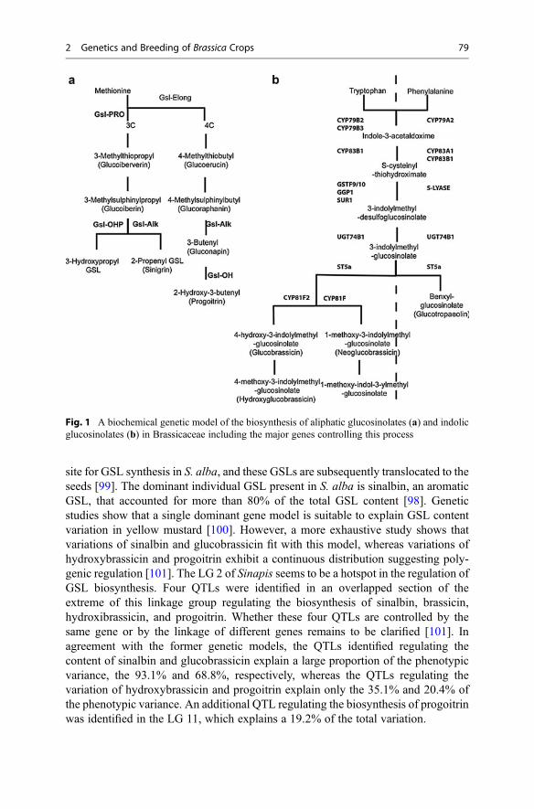
site for GSL synthesis in S. alba, and these GSLs are subsequently translocated to theseeds [99]. The dominant individual GSL present in S. alba is sinalbin, an aromaticGSL, that accounted for more than 80% of the total GSL content [98]. Geneticstudies show that a single dominant gene model is suitable to explain GSL contentvariation in yellow mustard [100]. However, a more exhaustive study shows thatvariations of sinalbin and glucobrassicin fit with this model, whereas variations ofhydroxybrassicin and progoitrin exhibit a continuous distribution suggesting poly-genic regulation [101]. The LG 2 of Sinapis seems to be a hotspot in the regulation ofGSL biosynthesis. Four QTLs were identified in an overlapped section of theextreme of this linkage group regulating the biosynthesis of sinalbin, brassicin,hydroxibrassicin, and progoitrin. Whether these four QTLs are controlled by thesame gene or by the linkage of different genes remains to be clarified [101]. Inagreement with the former genetic models, the QTLs identified regulating thecontent of sinalbin and glucobrassicin explain a large proportion of the phenotypicvariance, the 93.1% and 68.8%, respectively, whereas the QTLs regulating thevariation of hydroxybrassicin and progoitrin explain only the 35.1% and 20.4% ofthe phenotypic variance. An additional QTL regulating the biosynthesis of progoitrinwas identified in the LG 11, which explains a 19.2% of the total variation.
Fig. 1 A biochemical genetic model of the biosynthesis of aliphatic glucosinolates (a) and indolicglucosinolates (b) in Brassicaceae including the major genes controlling this process
2 Genetics and Breeding of Brassica Crops 79

Also belonging to the mustard family, winter cress (Barbarea vulgaris R. Br.)shows a characteristic GSL profile. Three different chemotypes in wild populationshave been described based on the GSL content [102, 103]. These chemotypescontained the same set of GSLs but show differences in the quantitative profile.Van Leur et al. [103] describe two types of wild populations; the so-called BAR typecontains glucobarbarin as the predominant GSL, whereas in the NAS type,gluconasturtiin is the prominent GSL. Genetic analysis indicates that differencesbetween these two chemotypes fit with a simple Mendelian inheritance model of theGSL type, coded by a single gene, with the BAR type dominant over the NAStype. Authors hypothesize that, in the NAS-type plants, an enzyme converting gluco-nasturtiin into glucobarbarin is damaged, has a lower expression level, or is missing. Inwild populations of eastern Denmark, Agerbirk et al. [102] describe a third chemotypeof B. vulgaris, in which glucosibarin is the predominant GSL. Glucosibarin andglucobarbarin are two isomeric forms of 2-hydroxy-2-phenylethyl GSL. These authorsdistinguish two types of wild populations; the G type contains predominantly theS-isomer (glucobarbarin) and therefore could be equivalent to the BAR type previ-ously described, just considering the GSL profile, whereas the P type containspredominantly the R-isomer (glucosibarin). Kuzina et al. [104] crossed twopopulations of each chemotype and constructed a genetic map based on 100 AFLPand 31 microsatellite marker. A QTL associated to glucosibarin level was located onlinkage group 4 which co-localize with a QTL associated to flea beetle resistance and aQTL associated to the high levels of glucobarbarin in the linkage group 5 with asignificant dominant effect.
Several GSLs sparsely present in the Brassica genus have been reported in othergenus of the Brassicaceae family, for instance, glucohesperin, glucoputranjivin, orglucocochlearin, and have been reported in Boechera stricta [105] or4-mercaptobutyl and glucoerucin in the Eruca genus [106, 107]; however, fewgenetic studies have been developed in these genera.
8 Conclusions
In the last decades, knowledge about the importance of glucosinolates in the humanand animal consumption, as well as their role in the plant defense, has beenincreased. This fact led to a high number of works to understand the genetic basesof glucosinolate synthesis. Major genes are well known in Brassica crops, but thereare other genetic factors, showed by QTL analysis, that will need more research.Other aspects, such as the interaction of glucosinolate genes and pathways withenvironmental signals, are a field with an increasing importance nowadays.
Other species from the Brassicaceae family, apart from the genus Brassica, arerecognized as a good source of healthy compounds and may be incorporated into thediet. These species have different glucosinolate patterns. The study of geneticsunderlying these traits is a recent and open field which will render interesting resultsin a close future, for breeding in other brassicaceae. Finally, the extensive knowledgeabout genetics and mode of inheritance of glucosinolates is being incorporated in
80 P. Velasco et al.

breeding programs to modify the content of these healthy metabolites. The sequenc-ing of the main Brassica crops and their existing homologies with Arabidopsis willspeed this process.
References
1. FAOSTAT (2016) Food and agriculture organization of the United Nations. In: FAOSTATdatabase
2. Cartea ME, Velasco P (2007) Glucosinolates in Brassica foods: bioavailability in food andsignificance for human health. Phytochem Rev 7:213–229
3. Fahey JW, Zalcmann AT, Talalay P (2001) The chemical diversity and distribution ofglucosinolates and isothiocyanates among plants. Phytochemistry 56:5–51
4. Mithen RF (2001) Glucosinolates and their degradation products. Adv Bot Res 35:213–2325. Clarke DB (2010) Glucosinolates, structures and analysis in food. Anal Methods 2:310–3256. Wang Y, Pan Y, Liu Z, Zhu X, Zhai L, Xu L, Yu R, Gong Y, Liu L (2013) De novo
transcriptome sequencing of radish (Raphanus sativus L.) and analysis of major genesinvolved in glucosinolate metabolism. BMC Genomics 14:836–848
7. Halkier BA, Gershenzon J (2006) Biology and biochemistry of glucosinolates. Annu RevPlant Biol 57:31
8. Wang H, Wu J, Sun S, Liu B, Cheng F, Sun R, Wang X (2011) Glucosinolate biosyntheticgenes in Brassica rapa. Gene 487:135–142
9. Pfalz M, Vogel H, Kroymann J (2009) The gene controlling the indole glucosinolate modifier1quantitative trait locus alters indole glucosinolate structures and aphid resistance inarabidopsis. Plant Cell 21:985–999
10. Wiesner M, Schreiner M, Zrenner R (2014) Functional identification of genes responsible forthe biosynthesis of 1-methoxy-indol-3-ylmethyl-glucosinolate in Brassica rapa ssp. chinensis.BMC Plant Biol 14:124
11. Magrath R, Bano F, Morgner M, Parkin I, Sharpe A, Lister C, Dean C, Turner J, Lydiate D,Mithen RF (1994) Genetics of aliphatic glucosinolates. I. Side chain elongation in Brassicanapus and Arabidopsis thaliana. Heredity 72:290–299
12. Giamoustaris A, Mithen RF (1996) Genetics of aliphatic glucosinolates. IV. Side-chainmodification in Brassica oleracea. TAG Theor Appl Genet 93:1006–1010
13. Mithen RF, Clarke J, LIister C, Dean C (1995) Genetics of aliphatic glucosinolates. III. Sidechain structure of aliphatic glucosinolates in Arabidopsis thaliana. Heredity 74:210–215
14. Parkin I, Magrath R, Keith D, Sharpe A, Mithen RF, Lydiate D (1994) Genetics of aliphaticglucosinolates. II. Hydroxylation of alkenyl glucosinolates in Brassica napus. Heredity72:594–598
15. Li G, Quiros CF (2003) In planta side-chain glucosinolate modification in Arabidopsis byintroduction of dioxygenase Brassica homolog BoGSL-ALK. TAG Theor Appl Genet106:1116–1121
16. Hall C, McCallum D, Prescott A, Mithen RF (2001) Biochemical genetics of glucosinolatemodification in Arabidopsis and Brassica. TAG Theor Appl Genet 102:369–374
17. Li G, Riaz A, Goyal S, Abel S, Quiros CF (2001) Inheritance of three major genes involved inthe synthesis of aliphatic glucosinolates in Brassica oleracea. J Am Soc Hortic Sci126:427–431
18. Li G, Quiros CF (2002) Genetic analysis, expression and molecular characterization ofBoGSL-ELONG, a major gene involved in the aliphatic glucosinolate pathway of brassicaspecies. Genetics 162:1937–1943
19. Gao M, Li G, Yang B, McCombie WR, Quiros CF (2004) Comparative analysis of a BrassicaBAC clone containing several major aliphatic glucosinolate genes with its correspondingArabidopsis sequence. Genome 47:666–679
2 Genetics and Breeding of Brassica Crops 81

20. Kliebenstein DJ (2009) A quantitative genetics and ecological model system: understandingthe aliphatic glucosinolate biosynthetic network via QTLs. Phytochem Rev 8:243–254
21. Hansen BG, Kliebenstein DJ, Halkier BA (2007) Identification of a flavin-monooxygenase as theS-oxygenating enzyme in aliphatic glucosinolate biosynthesis in Arabidopsis. Plant J 50:902–910
22. Zang YX, Kim HU, Kim JA, Lim MH, Jin M, Lee SC, Kwon SJ, Lee SI, Hong JK, Park THet al (2009) Genome-wide identification of glucosinolate synthesis genes in Brassica rapa.FEBS J 276:3559–3574
23. Zhu B, Wang Z, Yang J, Zhu Z, Wang H (2012) Isolation and expression of glucosinolatesynthesis genes CYP83A1 and CYP83B1 in Pak Choi (Brassica rapa L. ssp. chinensis var.communis (N. Tsen & S.H. Lee) Hanelt). Int J Mol Sci 13:5832–5843
24. Hirani AH, Zelmer CD, McVetty PB, Daayf F, Li G (2013) Homoeologous GSL-ELONG genereplacement for manipulation of aliphatic glucosinolates in Brassica rapa L. by markerassisted selection. Front Plant Sci 4:55
25. Augustine R, Majee M, Gershenzon J, Bisht NC (2013) Four genes encoding MYB28, a majortranscriptional regulator of the aliphatic glucosinolate pathway, are differentially expressed inthe allopolyploid Brassica juncea. J Exp Bot 64:4907–4921
26. Meenu, Augustine R, Majee M, Pradhan AK, Bisht NC (2015) Genomic origin, expressiondifferentiation and regulation of multiple genes encoding CYP83A1, a key enzyme for coreglucosinolate biosynthesis, from the allotetraploid Brassica juncea. Planta 241:651–665
27. Baskar V, Park SW (2015) Molecular characterization of BrMYB28 and BrMYB29paralogous transcription factors involved in the regulation of aliphatic glucosinolate profilesin Brassica rapa ssp. pekinensis. C R Biol 338:434–442
28. Yi GE, Robin AH, Yang K, Park JI, Kang JG, Yang TJ, Nou IS (2015) Identification andexpression analysis of glucosinolate biosynthetic genes and estimation of glucosinolate con-tents in edible organs of Brassica oleracea subspecies. Molecules 20:13089–13111
29. Frerigmann H, Gigolashvili T (2014) MYB34, MYB51, and MYB122 distinctly regulateindolic glucosinolate biosynthesis in Arabidopsis thaliana. Mol Plant 7:814–828
30. Grubb DC, Zipp BJ, Ludwig-M€uller J, Masuno MN, Molinski TF, Abel S (2004) Arabidopsisglucosyltransferase UGT74B1 functions in glucosinolate biosynthesis and auxin homeostasis.Plant J 40:893–908
31. Klein M, Reichelt M, Gershenzon J, Papenbrock J (2006) The three desulfoglucosinolatesulfotransferase proteins in Arabidopsis have different substrate specificities and are differen-tially expressed. FEBS J 273:122–136
32. Naur P, Petersen BL, Mikkelsen MD, Bak S, Rasmussen H, Olsen CE, Halkier BA (2003)CYP83A1 and CYP83B1, two nonredundant cytochrome P450 enzymes metabolizing oximesin the biosynthesis of glucosinolates in Arabidopsis. Plant Physiol 133:63–72
33. Pfalz M, Mikkelsen MD, Bednarek P, Olsen CE, Halkier BA, Kroymann J (2011) Metabolicengineering in nicotiana benthamiana reveals key enzyme functions in arabidopsis indoleglucosinolate modification. Plant Cell 23:716–729
34. Sønderby IE, Geu-Flores F, Halkier BA (2010) Biosynthesis of glucosinolates – gene discov-ery and beyond. Trends Plant Sci 15:283–290
35. Hirschmann F, Papenbrock J (2015) The fusion of genomes leads to more options: a compar-ative investigation on the desulfo-glucosinolate sulfotransferases of Brassica napus andhomologous proteins of Arabidopsis thaliana. Plant Physiol Biochem 91:10–19
36. Zhang Y, Huai D, Yang Q, Cheng Y, Ma M, Kliebenstein DJ, Zhou Y (2015) Overexpressionof three glucosinolate biosynthesis genes in Brassica napus identifies enhanced resistance toSclerotinia sclerotiorum and Botrytis cinerea. PLoS One 10:e0140491
37. Yun-Xiang Z, Myung-Ho L, Beom-Seok P, Seung-Beom H, Doo Hwan K (2008) Metabolicengineering of indole glucosinolates in Chinese cabbage plants by expression of ArabidopsisCYP79B2, CYP79B3, and CYP83B1. Mol Cells 25:231–241
38. Zang Y-X, Lim M-H, Park B-S, Hong S-B, Kim DH (2008) Metabolic engineering of indoleglucosinolates in Chinese cabbage plants by expression of Arabidopsis CYP79B2, CYP79B3,and CYP83B1. Mol Cells 25:231–241
82 P. Velasco et al.

39. Gao J, Yu X, Ma F, Li J (2014) RNA-Seq analysis of transcriptome and glucosinolatemetabolism in seeds and sprouts of broccoli (Brassica oleracea var.). PLoS One 9:e88804
40. Wittstock U, Halkier BA (2000) Cytochrome P450 CYP79A2 from Arabidopsis thalianaL. catalyzes the conversion of L-phenylalanine to phenylacetaldoxime in the biosynthesis ofbenzylglucosinolate. J Biol Chem 275:14659–14666
41. Klein M, Papenbrock J (2009) Kinetics and substrate specificities of desulfo-glucosinolatesulfotransferases in Arabidopsis thaliana. Physiol Plant 135:140–149
42. Chalhoub B, Denoeud F, Liu S, Parkin IAP, Tang H, Wang X, Chiquet J, Belcram H, Tong C,Samans B et al (2014) Early allopolyploid evolution in the post-neolithic Brassica napusoilseed genome. Science 345:950–953
43. Celenza JL, Quiel JA, Smolen GA, Merrikh H, Silvestro AR, Normanly J, Bender J (2005)The arabidopsis ATR1 MYB transcription factor controls indolic glucosinolate homeostasis.Plant Physiol 137:253–262
44. Gigolashvili T, Berger B, Fl€ugge U-I (2009) Specific and coordinated control of indolic andaliphatic glucosinolate biosynthesis by R2R3-MYB transcription factors in Arabidopsisthaliana. Phytochem Rev 8:3–13
45. Araki R, Hasumi A, Nishizawa OI, Sasaki K, Kuwahara A, Sawada Y, Totoki Y, Toyoda A,Sakaki Y, Li Y et al (2013) Novel bioresources for studies of Brassica oleracea: identificationof a kale MYB transcription factor responsible for glucosinolate production. Plant Biotechnol J11:1017–1027
46. Koritsas VM, Lewis JA, Fenwick GR (1991) Glucosinolate responses of oilseed rape, mustardand kale to mechanical wounding and infestation by cabbage stem flea beetle (Psylliodeschrysocephala). Ann Appl Biol 118:209–221
47. Brader G, Tas É, Palva ET (2001) Jasmonate-dependent induction of indole glucosinolates inArabidopsis by culture filtrates of the nonspecific pathogen Erwinia carotovora. Plant Physiol126:849–860
48. Bodnaryk RP (1992) The international journal of plant biochemistry effects of wounding onglucosinolates in the cotyledons of oilseed rape and mustard. Phytochemistry 31:2671–2677
49. Doughty KJ, Kiddle GA, Pye BJ, Wallsgrove RM, Pickett JA (1995) Selective induction ofglucosinolates in oilseed rape leaves by methyl jasmonate. Phytochemistry 38:347–350
50. Baenas N, García-Viguera C, Moreno DA (2014) Biotic elicitors effectively increase theglucosinolates content in Brassicaceae sprouts. J Agric Food Chem 62:1881–1889
51. Thiruvengadam M, Kim S-H, Chung I-M (2015) Exogenous phytohormones increase theaccumulation of health-promoting metabolites, and influence the expression patterns of bio-synthesis related genes and biological activity in Chinese cabbage (Brassica rapa spp.pekinensis). Sci Hortic 193:136–146
52. van Doorn HE, van der Kruk GC, van Holst GJ, Raaijmakers-Ruijs N, Postma E,Groeneweg B, Jongen WHF (1998) The glucosinolates sinigrin and progoitrin are importantdeterminants for taste preference and bitterness of brussels sprouts. J Sci Food Agric 78:30–38
53. Traka M, Mithen RF (2009) Glucosinolates, isothiocyanates and human health. PhytochemRev 8:269–282
54. Liu Z, Hirani A, McVetty PBE, Daayf F, Quiros C, Li G (2012) Reducing progoitrin andenriching glucoraphanin in Brassica napus seeds through silencing of the GSL-ALK genefamily. Plant Mol Biol 79:179–189
55. Cartea ME, Velasco P, Obregon S, Padilla G, de Haro A (2008) Seasonal variation inglucosinolate content in Brassica oleracea crops grown in northwestern Spain. Phytochemis-try 69:403–410
56. Kushad MM, Brown AF, Kurilich AC, Juvik JA, Klein BP, Wallig MA, Jeffery EH (1999)Variation of glucosinolates in vegetable crops of Brassica oleracea. J Agric Food Chem47:1541–1548
57. Bradshaw J, Wilson R (2012) Kale population improvement and cultivar production.Euphytica 184:275–288
2 Genetics and Breeding of Brassica Crops 83

58. Toroser D, Thormann CE, Osborn TC, Mithen R (1995) RFLP mapping of quantitative traitloci controlling seed aliphatic-glucosinolate content in oilseed rape (Brassica napus L). TheorAppl Genet 91:802–808
59. Rucker B, Robbelen G (1994) Inheritance of total and individual glucosinolate contents inseeds of winter oilseed rape (Brassica napus L). Plant Breed 113:206–216
60. Li Y, Kiddle GUY, Bennett R, Doughty K, Wallsgrove R (1999) Variation in the glucosinolatecontent of vegetative tissues of Chinese lines of Brassica napus L. Ann Appl Biol134:131–136
61. Riso P, Del Bo’ C, Vendrame S (2014) Preventive effects of broccoli bioactives: role onoxidative stress and cancer risk. In: Preedy VR (ed) Cancer: oxidative stress and dietaryantioxidants. Elsevier Science Publisher, San Diego, pp 115–126
62. Juge N, Mithen RF, Traka M (2007) Molecular basis for chemoprevention by sulforaphane: acomprehensive review. Cell Mol Life Sci 64:1105–1127
63. Mithen RF, Dekker M, Verkerk R, Rabot S, Johnson IT (2000) The nutritional significance,biosynthesis and bioavailability of glucosinolates in human foods. J Sci Food Agric80:967–984
64. Faulkner K (1998) Selective increase of the potential anticarcinogen 4- methylsulphinylbutylglucosinolate in broccoli. Carcinogenesis 19:605–609
65. Mithen R (2003) Development of isothiocyanate-enriched broccoli, and its enhanced ability toinduce phase 2 detoxification enzymes in mammalian cells. Theor Appl Genet 106:727–734
66. Fahey JW, Zhang Y, Talalay P (1997) Broccoli sprouts: an exceptionally rich source ofinducers of enzymes that protect against chemical carcinogens. Proc Natl Acad Sci U S A94:10367–10372
67. Traka M, Saha S, Huseby S, Kopriva S, Walley P, Barker G, Moore J, Mero G, van denBosch F, Constant H et al (2013) Genetic regulation of glucoraphanin accumulation inBenefortÃ#®broccoli. New Phytol 198:1085–1095
68. Zang Y-X, Kim J-H, Park Y-D, Kim D-H, Hong S-B (2008) Metabolic engineering of aliphaticglucosinolates in Chinese cabbage plants expressing Arabidopsis MAM1, CYP79F1, andCYP83A1. BMB Rep 41:472–478
69. Geu Flores F, Olsen C, Halkier BA (2009) Towards engineering glucosinolates intonon-cruciferous plants. Planta 229:261–270
70. Howell PM, Sharpe AG, Lydiate DJ (2003) Homoeologous loci control the accumulation ofseed glucosinolates in oilseed rape (Brassica napus). Genome 46:454–460
71. Uzunova M, Ecke W, Weissleder K, Robbelen G (1995) Mapping the genome of rapeseed(Brassica napus l).1. Construction of an RFLP linkage map and localization of QTLs for seedglucosinolate content. Theor Appl Genet 90:194–204
72. deQuiroz HC, Mithen R (1996) Molecular markers for low-glucosinolate alleles in oilseedrape (Brassica napus L). Mol Breed 2:277–281
73. Zhao J, Meng J (2003) Detection of loci controlling seed glucosinolate content and theirassociation with Sclerotinia resistance in Brassica napus. Plant Breed 122:19–23
74. Fu Y, Lu K, Qian LW, Mei JQ, Wei DY, Peng XH, Xu XF, Li JN, Frauen M, Dreyer Fet al (2015) Development of genic cleavage markers in association with seed glucosinolatecontent in canola. Theor Appl Genet 128:1029–1037
75. Feng J, Long Y, Shi L, Shi JQ, Barker G, Meng JL (2012) Characterization of metabolitequantitative trait loci and metabolic networks that control glucosinolate concentration in theseeds and leaves of Brassica napus. New Phytol 193:96–108
76. Quijada PA, Udall JA, Lambert B, Osborn TC (2006) Quantitative trait analysis of seed yieldand other complex traits in hybrid spring rapeseed (Brassica napus L.): 1. Identification ofgenomic regions from winter germplasm. Theor Appl Genet 113:549–561
77. Xu JF, Long Y, Wu JG, Xu HM, Zhao ZG, Wen J, Meng JL, Shi CH (2015) QTL identificationon two genetic systems for rapeseed glucosinolate and erucic acid contents over two seasons.Euphytica 205:647–657
84 P. Velasco et al.

78. Gajardo HA, Wittkop B, Soto-Cerda B, Higgins EE, Parkin IAP, Snowdon RJ, Federico ML,Iniguez-Luy FL (2015) Association mapping of seed quality traits in Brassica napus L. usingGWAS and candidate QTL approaches. Mol Breed 35:143
79. Wurschum T, Liu WX, Maurer HP, Abel S, Reif JC (2012) Dissecting the genetic architectureof agronomic traits in multiple segregating populations in rapeseed (Brassica napus L.). TheorAppl Genet 124:153–161
80. Rout K, Sharma M, Gupta V, Mukhopadhyay A, Sodhi YS, Pental D, Pradhan AK (2015)Deciphering allelic variations for seed glucosinolate traits in oilseed mustard (Brassica juncea)using two bi-parental mapping populations. Theor Appl Genet 128:657–666
81. Mahmood T, Ekuere U, Yeh F, Good AG, Stringam GR (2003) Molecular mapping of seedaliphatic glucosinolates in Brassica juncea. Genome 46:753–760
82. Gupta S, Sangha MK, Kaur G, Banga S, Gupta M, Kumar H, Banga SS (2015) QTL analysisfor phytonutrient compounds and the antioxidant molecule in mustard (Brassica juncea L.).Euphytica 201:345–356
83. Ramchiary N, Bisht NC, Gupta V, Mukhopadhyay A, Arumugam N, Sodhi YS, Pental D,Pradhan AK (2007) QTL analysis reveals context-dependent loci for seed glucosinolate trait inthe oilseed Brassica juncea: importance of recurrent selection backcross scheme for theidentification of ‘true’ QTL. Theor Appl Genet 116:77–85
84. Bisht NC, Gupta V, Ramchiary N, Sodhi YS, Mukhopadhyay A, Arumugam N, Pental D,Pradhan AK (2009) Fine mapping of loci involved with glucosinolate biosynthesis in oilseedmustard (Brassica juncea) using genomic information from allied species. Theor Appl Genet118:413–421
85. Rahman H, Kebede B, Zimmerli C, Yang RC (2014) Genetic study and QTL mapping of seedglucosinolate content in Brassica rapa L. Crop Sci 54:537–543
86. Lou P, Zhao J, He H, Hanhart C, Del Carpio DP, Verkerk R, Custers J, Koornneef M, BonnemaG (2008) Quantitative trait loci for glucosinolate accumulation in Brassica rapa leaves. NewPhytol 179:1017–1032
87. Del Carpio DP, Basnet RK, Arends D, Lin K, De Vos RCH, Muth D, Kodde J, Boutilier K,Bucher J, Wang XW et al (2014) Regulatory network of secondary metabolism in Brassicarapa: insight into the glucosinolate pathway. PLoS One 9:e107123
88. Bagheri H, El-Soda M, Kim HK, Fritsche S, Jung C, Aarts MGM (2013) Genetic analysis ofhealth-related secondary metabolites in a Brassica rapa Recombinant inbred line population.Int J Mol Sci 14:15561–15577
89. Brown AF, Yousef GG, Reid RW, Chebrolu KK, Thomas A, Krueger C, Jeffery E, Jackson E,Juvik JA (2015) Genetic analysis of glucosinolate variability in broccoli florets using genome-anchored single nucleotide polymorphisms. Theor Appl Genet 128:1431–1447
90. Sotelo T, Soengas P, Velasco P, Rodriguez VM, Cartea ME (2014) Identification of metabolicQTLs and candidate genes for glucosinolate synthesis in Brassica oleracea leaves, seeds andflower buds. PLoS One 9:e91428
91. Kitashiba H, Li F, Hirakawa H, Kawanabe T, Zou Z, Hasegawa Y, Tonosaki K, Shirasawa S,Fukushima A, Yokoi S et al (2014) Draft sequences of the radish (Raphanus sativus L.)genome. DNA Res 21:481–490
92. Ediage EN, Di Mavungu JD, Scippo ML, Schneider YJ, Larondelle Y, Callebaut A, Robbens J,Van Peteghem C, De Saeger S (2011) Screening, identification and quantification ofglucosinolates in black radish (Raphanus sativus L. niger) based dietary supplements usingliquid chromatography coupled with a photodiode array and liquid chromatography-massspectrometry. J Chromatogr 1218:4395–4405
93. Ishida M, Nagata M, Ohara T, Kakizaki T, Hatakeyama K, Nishio T (2012) Small variation ofglucosinolate composition in Japanese cultivars of radish (Raphanus sativus L.) requiressimple quantitative analysis for breeding of glucosinolate component. Breed Sci 62:63–70
94. Carlson D, Axenbichler M, van Etten C (1985) Glucosinolate in radish cultivars. J Am SocHortic Sci 110:634–638
2 Genetics and Breeding of Brassica Crops 85

95. Zou Z, Ishida M, Li F, Kakizaki T, Suzuki S, Kitashiba H, Nishio T (2013) QTL analysis usingSNP markers developed by next-generation sequencing for identification of candidate genescontrolling 4-methylthio-3-butenyl glucosinolate contents in roots of radish, Raphanus sativusL. PLoS One 8:e53541
96. Ishida M, Kakizaki T, Morimitsu Y, Ohara T, Hatakeyama K, Yoshiaki H, Kohori J, Nishio T(2015) Novel glucosinolate composition lacking 4-methylthio-3-butenyl glucosinolate inJapanese white radish (Raphanus sativus L.). TAG Theor Appl Genet 128:2037–2046
97. Warwick SI, Black LD (1991) Molecular systematics of Brassica and allied genera (SubtribeBrassicinae, Brassiceae) -chloroplast genome and cytodeme congruence. Theor Appl Genet82:81–92
98. Hopkins R, Ekbom B, Henkow L (1998) Glucosinolate content and susceptibility for insectattack of three populations of Sinapis alba. J Chem Ecol 24:1203–1216
99. Liangcheng D, Halkier BA (1998) Biosynthesis of glucosinolates in the developing siliquewalls and seeds of Sinapis alba. Phytochemistry 48:1145–1150
100. Drost W, Rakow G, Raney P (1999) Inheritance of glucosinolate content in yellow mustard(Sinapis alba L.). In: Proceedings of the 10th international rapeseed congress. Canberra
101. Javidfar F, Cheng B (2013) Construction of a genetic linkage map and QTL analysis of erucicacid content and glucosinolate components in yellow mustard (Sinapis alba L.). BMC PlantBiol 13:142
102. Agerbirk N, Ørgaard M, Nielsen JK (2003) Glucosinolates, flea beetle resistance, and leafpubescence as taxonomic characters in the genus Barbarea (Brassicaceae). Phytochemistry63:69–80
103. van Leur H, Raaijmakers CE, van Dam NM (2006) A heritable glucosinolate polymorphismwithin natural populations of Barbarea vulgaris. Phytochemistry 67:1214–1223
104. Kuzina V, Nielsen JK, Augustin JM, Torp AM, Bak S, Andersen SB (2011) Barbarea vulgarislinkage map and quantitative trait loci for saponins, glucosinolates, hairiness and resistance tothe herbivore Phyllotreta nemorum. Phytochemistry 72:188–198
105. Schranz ME, Manzaneda AJ, Windsor AJ, Clauss MJ, Mitchell-Olds T (2009) Ecologicalgenomics of Boechera stricta: identification of a QTL controlling the allocation of methionine-vs branched-chain amino acid-derived glucosinolates and levels of insect herbivory. Heredity102:465–474
106. Bennett RN, Mellon FA, Botting NP, Eagles J, Rosa EA,Williamson G (2002) Identification ofthe major glucosinolate (4-mercaptobutyl glucosinolate) in leaves of Eruca sativa L. (saladrocket). Phytochemistry 61:25–30
107. Iori R, Bernardi R, Gueyrard D, Rollin P, Palmieri S (1999) Formation of glucoraphanin bychemoselective oxidation of natural glucoerucin: a chemoenzymatic route to sulforaphane.Bioorg Med Chem Lett 9:1047–1048
108. Stowe KA, Marquis RJ (2011) Cost of defense: correlated responses to divergent selection forfoliar glucosinolate content in Brassica rapa. Evol Ecol 25:763–775
86 P. Velasco et al.
Glucosinolates in Food 3Marco Possenti, Simona Baima, Antonio Raffo, Alessandra Durazzo,Anna Maria Giusti, and Fausta Natella
AbstractGlucosinolates are secondary plant metabolites that have attracted researcher’sattention due to their potential chemopreventive activity. More than 120 differentglucosinolates have been identified in plants, and several of these compoundshave been studied for the potential anti-cancerogenic effect of their metabolicbreakdown products (mainly ITCs).
Glucosinolates are peculiar of vegetables belonging to Brassicaceae family butare present also in few other species (capers, papaya, and moringa) used forhuman consumption. The type and concentration of glucosinolates in food arehighly variable depending on several factors, such as genetics, cultivation site,cultivar, growth conditions, developmental stage, plant tissue, post-harvest han-dling, and food preparation methods. As types and concentration are also themain determinant of their biological activities, estimates of their content in foodare essential tool to understand if a certain diet is adequate to deliver qualitativelyand quantitatively appropriate glucosinolates and ITCs.
The aim of this chapter is to describe qualitative and quantitative glucosinolatedistribution among commonly eaten food, as well as the effect of the post-harvesthandling on the glucosinolate food content.
M. Possenti (*) • S. Baima • A. Raffo • A. Durazzo • F. NatellaCREA-NUT, Consiglio per la Ricerca in Agricoltura e l’Analisi dell’Economia Agraria, Food andNutrition Research Centre, Rome, Italye-mail: [email protected]; [email protected]; [email protected];[email protected]; [email protected]
A.M. GiustiDepartment of Experimental Medicine, Medical Physiopathology, Food Science andEndocrinology Section, Food Science and Human Nutrition Research Unit – University “Sapienza”of Rome, Rome, Italye-mail: [email protected]
# Springer International Publishing Switzerland 2017J.-M. Mérillon, K.G. Ramawat (eds.), Glucosinolates, Reference Series inPhytochemistry, DOI 10.1007/978-3-319-25462-3_4
87
KeywordsGlucosinolates • Isothiocyanates • Brassicaceae • Food content • Processing
Abbreviations4-GDB 4-[β-D-Glucopyranosyldisulfanyl] butyl glucosinolateDMB Dimeric 4-mercaptobutyl-glucosinolateDW Dry weightFW Fresh weightGLS GlucosinolateITC Isothiocyanate
Contents1 Introduction . . . . . . . . . . . . . . . . . . . . . . . . . . . . . . . . . . . . . . . . . . . . . . . . . . . . . . . . . . . . . . . . . . . . . . . . . . . . . . . . . . 892 Biological Activities of GLS Breakdown Products . . . . . . . . . . . . . . . . . . . . . . . . . . . . . . . . . . . . . . . . 913 Glucosinolates in Food . . . . . . . . . . . . . . . . . . . . . . . . . . . . . . . . . . . . . . . . . . . . . . . . . . . . . . . . . . . . . . . . . . . . . . 954 Vegetables . . . . . . . . . . . . . . . . . . . . . . . . . . . . . . . . . . . . . . . . . . . . . . . . . . . . . . . . . . . . . . . . . . . . . . . . . . . . . . . . . . . 96
4.1 Broccoli (Brassica oleracea convar. botrytis var. cymosa) (commonlyBrassica oleracea ssp. italica) . . . . . . . . . . . . . . . . . . . . . . . . . . . . . . . . . . . . . . . . . . . . . . . . . . . . . . . 96
4.2 Brussels Sprouts(Brassica oleracea var. gemmifera) . . . . . . . . . . . . . . . . . . . . . . . . . . . . . . . . . . . . . . . . . . . . . . . . . 98
4.3 Cabbage (Brassica oleracea convar. capitata) . . . . . . . . . . . . . . . . . . . . . . . . . . . . . . . . . . . . . . 984.4 Cauliflower (Brassica oleracea convar. botrytis var. botrytis) . . . . . . . . . . . . . . . . . . . . . 1064.5 Chinese Cabbage (Brassica rapa ssp. pekinensis,
Also Known as Brassica campestris ssp. pekinensis) . . . . . . . . . . . . . . . . . . . . . . . . . . . . . . 1064.6 Chinese Kale (Brassica oleracea var. alboglabra) . . . . . . . . . . . . . . . . . . . . . . . . . . . . . . . . . 1074.7 Ethiopian Kale (Brassica carinata) . . . . . . . . . . . . . . . . . . . . . . . . . . . . . . . . . . . . . . . . . . . . . . . . . 1074.8 Friarielli or Cime di rapa or Turnip Rape; (B. rapa ssp. campestris Also Known
as B. rapa ssp. sylvestris L. Janch. var. esculenta Hort.) . . . . . . . . . . . . . . . . . . . . . . . . . . . 1074.9 Kale (Brassica oleracea var. acephala) and Rape Kale (Brassica napus) . . . . . . . . . 1084.10 Kohlrabi (Brassica oleracea var. gongylodes) . . . . . . . . . . . . . . . . . . . . . . . . . . . . . . . . . . . . . . 1094.11 Mustard Greens (Brassica juncea) . . . . . . . . . . . . . . . . . . . . . . . . . . . . . . . . . . . . . . . . . . . . . . . . . . 1094.12 Mizuna (Brassica rapa subsp. nipposinica var. chinoleifera Also Known as
Brassica juncea var. japonica) . . . . . . . . . . . . . . . . . . . . . . . . . . . . . . . . . . . . . . . . . . . . . . . . . . . . . . 1104.13 Pak Choi (Brassica rapa ssp. chinensis
Also Known as Brassica campestris L. ssp. Chinensis var. communis) . . . . . . . . . . . 1104.14 Radish (Raphanus sativus) . . . . . . . . . . . . . . . . . . . . . . . . . . . . . . . . . . . . . . . . . . . . . . . . . . . . . . . . . . 1104.15 Rocket (Eruca vesicaria ssp. sativa and Diplotaxis tenuifolia) . . . . . . . . . . . . . . . . . . . . 1114.16 Turnip (Brassica rapa var. rapa) . . . . . . . . . . . . . . . . . . . . . . . . . . . . . . . . . . . . . . . . . . . . . . . . . . . . 1124.17 Sprouts (Various Brassicaceae spp.) . . . . . . . . . . . . . . . . . . . . . . . . . . . . . . . . . . . . . . . . . . . . . . . . . 112
5 Processed Foods . . . . . . . . . . . . . . . . . . . . . . . . . . . . . . . . . . . . . . . . . . . . . . . . . . . . . . . . . . . . . . . . . . . . . . . . . . . . . 1135.1 Colza and Canola oil . . . . . . . . . . . . . . . . . . . . . . . . . . . . . . . . . . . . . . . . . . . . . . . . . . . . . . . . . . . . . . . . . . 1135.2 Mustard . . . . . . . . . . . . . . . . . . . . . . . . . . . . . . . . . . . . . . . . . . . . . . . . . . . . . . . . . . . . . . . . . . . . . . . . . . . . . . . . 1155.3 Sauerkraut . . . . . . . . . . . . . . . . . . . . . . . . . . . . . . . . . . . . . . . . . . . . . . . . . . . . . . . . . . . . . . . . . . . . . . . . . . . . . 115
6 Other Food . . . . . . . . . . . . . . . . . . . . . . . . . . . . . . . . . . . . . . . . . . . . . . . . . . . . . . . . . . . . . . . . . . . . . . . . . . . . . . . . . . . 1156.1 Caper (Capparis spinosa and Capparis ovata) . . . . . . . . . . . . . . . . . . . . . . . . . . . . . . . . . . . . . . . 1156.2 Moringa or Drumstick Tree (Moringa oleifera) . . . . . . . . . . . . . . . . . . . . . . . . . . . . . . . . . . . . . . 1166.3 Papaya (Carica papaya) . . . . . . . . . . . . . . . . . . . . . . . . . . . . . . . . . . . . . . . . . . . . . . . . . . . . . . . . . . . . . . 1176.4 Honey . . . . . . . . . . . . . . . . . . . . . . . . . . . . . . . . . . . . . . . . . . . . . . . . . . . . . . . . . . . . . . . . . . . . . . . . . . . . . . . . . . 1176.5 Impact of Post-Harvest Factors on GLS Content . . . . . . . . . . . . . . . . . . . . . . . . . . . . . . . . . . . . . 118
88 M. Possenti et al.

7 Conclusions . . . . . . . . . . . . . . . . . . . . . . . . . . . . . . . . . . . . . . . . . . . . . . . . . . . . . . . . . . . . . . . . . . . . . . . . . . . . . . . . . . 120References . . . . . . . . . . . . . . . . . . . . . . . . . . . . . . . . . . . . . . . . . . . . . . . . . . . . . . . . . . . . . . . . . . . . . . . . . . . . . . . . . . . . . . . 120
1 Introduction
Glucosinolates (GLSs) are secondary metabolites of vegetables that have attracted agreat interest, due to their possible effects on human health. They are β-thioglucosideN-hydroxysulfates with a side chain (R) and a sulfur-linked β-D-glucopyranose moiety.GLSs have a similar basic structure consisting of a D-thioglucose group linked to asulfonated aldoxime group and a variable side chain derived from amino acids.
GLSs have been divided in three classes based on the structure of different aminoacids precursors:
– Aliphatic GLSs, derived from methionine, isoleucine, leucine, or valine– Aromatic GLSs, derived from phenylalanine or tyrosine– Indole GLSs, derived from tryptophan
The GLSs synthesis is regulated according to the different developmental stagesof the organs and tissues and by the environment. Although more than 120 differentGLSs have been identified, only some of these are present in plants in high quantity.GLS profile varies between species and ecotypes as well as between and withinindividual plants due to the developmental stage, tissue, photoperiod, and environ-mental conditions.
The pungent flavor of the raw edible plants containing GLSs is due to theproducts of GLS enzymatic degradation, which are formed through the activity ofan endogenous enzyme, myrosinase (thioglucohydrolase). Myrosinase is localizedin the cytoplasm instead GLSs are stored in the vacuole. After cell rupture, inducedby chewing during human consumption or by herbivores, insects, or fungalwounding/penetration, GLSs and myrosinase come in contact and hydrolysis occurs.A similar interaction between GLS and myrosinase is obtained by tissue damageduring freezing, thawing, and chopping of edible plants.
The glucosinolate-myrosinase system generates thioglucose, sulfate, and anunstable intermediate which rearranges spontaneously into several degradationproducts. The final composition of the product mix depends on pH, availability of
3 Glucosinolates in Food 89

ferrous ions, and the presence of myrosinase-interacting proteins. In the wide rangeof biological active breakdown products, there are nitriles, isothiocyanates (ITCs),thiocyanates, epithionitriles, and vinyl oxazolidinethiones.
The GLS breakdown products play an important role in the interactionsbetween plants and insects/herbivores; in fact, they can act both as poisons anddeterrents to generalist insects/herbivores or, on the contrary, as signaling mol-ecules to attract specialist insects/herbivores laying their eggs or feeding on thatspecific plant [1, 2]. Several studies have shown that GLSs determine a growthinhibition or represent a feeding deterrent to a wide range of herbivorous animalssuch as birds, slugs, and generalist insects [3, 4]. Moreover, it has been observedthat volatile hydrolysis products of GLSs can attract natural enemies of herbi-vores such as parasitoids and therefore determine an indirect protection of theplant [5, 6].
In the last decades, it was found that the GLS breakdown products might havebeneficial effects on human health, and this is, probably, the reason that hasdetermined a steadily increase in the number of studies on GLS and ITC. Figure 1shows the number of publication per year obtained through a search in Scopus withthe keywords “glucosinolate” or “isothiocyanate” and “health” or “cancer.” Inparticular, the keyword combination “isothiocyanate and cancer” yields 3691 scien-tific articles evidencing the great scientific interest on the anti-cancerogenic activityof these compounds (Scopus, January 2016).
2013
2009
2005
2001
1997
1993
1989
1985
1981
1977
1973
0
50
100
150
200
250
nu
mb
er o
f p
ub
licat
ion
s
ITC and health
ITC and cancer GLS and cancer
GLS and health
300
350
Fig. 1 Chronology of the number of publications appearing in Scopus between 1970 and 2015with the keywords “glucosinolate,” “isothiocyanate,” “health,” and “cancer”
90 M. Possenti et al.
2 Biological Activities of GLS Breakdown Products
In the past years, it has become clear that the consumption of food containing GLSsand/or their degradation products could affect human health. Initially, GLSs havebeen studied for their toxic and goitrogenic activity, and, now, it has become clearthat the metabolic product of progoitrin can inhibit the accumulation of iodide in thethyroid gland predisposing to goiter. A similar goitrogenic activity has been reportedfor thiocyanate ions that are GLS breakdown products common to several GLSs [7];thiocyanate ions are iodine competitors and can inhibit iodine uptake reducingthyroid hormone synthesis [8]. Then a high progoitrin and thiocyanate diet couldbe potentially hazardous for hypothyroid patients, especially in those parts of theworld characterized by a low iodine supply. Due to the toxic and goitrogenic activityof these compounds (especially when used for animal feed), in the 1990s, the plantbreeders started to select plant with low GLS content [9]. However, more recently,this trend has been completely reverted because of the demonstration that several ofthese compounds (especially ITCs) possess interesting health protective capacities,particularly anti-cancerogenic activity (see Table 1). In fact, in vitro and in vivostudies have reported that GLS breakdown products can affect several stages ofcancer development, including the induction of detoxification enzymes (Phase II)and the inhibition of activation enzymes (Phase I) [10]. These enzymes play centralroles in the metabolism, elimination, and detoxification of carcinogen andpro-carcinogen molecules (xenobiotics). In order to minimize the toxic effect ofxenobiotics, Phase I enzymes introduce polar groups into the toxic molecules(sometimes activating them to more hazardous compounds), while Phase II enzymesinactivate these molecules through conjugation reactions that produce more polarand less toxic molecules. Then, the modulation of Phase I and Phase II enzymesoperated by GLS metabolic products can prevent the formation of endogenous andexogenous carcinogens, in turn avoiding carcinogenesis initiation. In addition tomodulation of Phase I and Phase II enzymes, GLS breakdown products can affectcancer initiation, progression, and development through many differentmechanisms:
1. Blocking the cell cycle [11]2. Inducing apoptosis [12, 13]3. Inhibiting angiogenesis [14]4. Inhibiting metastasis and migration of cancer cells [15]5. Decreasing the sensitivity to estrogen (through the downregulation of estrogen
receptor) and then affecting estrogen-dependent cancer (such as breast cancer) [16]6. Inhibiting the NF-kB pathway and then acting as anti-inflammatory agents [17]
Besides anti-cancerogenic activity, the GLS breakdown products have shownprotective effects against cardiovascular disease, neurodegeneration, diabetes, andseveral inflammatory disorders [18]. However, due to their high reactivity, severalITCs can be toxic and act as promoter of carcinogenesis [18, 19], even if the
3 Glucosinolates in Food 91

Table
1Biologicalactiv
ityof
themainGLShy
drolyticprod
ucts
Glucosino
lates
Mainbreakd
ownprod
ucts
Biologicaleffects
Reference
Alip
hatic
Glucobrassicanapin
4-Penteny
l-ITC
Invitroantim
icrobialandantib
acterial
activ
ity[20,
21]
Glucocapp
arin
Methy
l-ITC
Antiproliferativein
cancer
cell
[22]
Progo
itrin/epiprog
oitrin
5-Vinyl-2-oxazol-dinethione
!ox
azolidine-2-thione
Goitrog
enicactiv
ityin
anim
almod
el(inh
ibito
rof
thyrox
inesynthesis)
[23]
Sinigrin
Ally
lITC(A
ITC)
Antiproliferativein
cancer
cell
Indu
ctionof
cellcyclearrestand
apop
tosis
Antim
etastatic
activ
ityin
anim
almod
elAnti-angiog
enicactiv
ityin
anim
almod
elAnti-cancerog
enicactiv
ityin
anim
almod
el
[24]
[25]
[26]
[27]
[26]
Glucoalyssin
Alyssin
(5-(methy
lsulfiny
l)-pentylITC)
Indu
ctionof
apop
tosisin
cancer
cell
[28]
Glucoerucin
Erucin(4-(methy
lthio)-3-bu
tylITC)(M
TBITC)
Indu
ctionof
Phase
IIenzyme
Anti-geno
toxicactiv
ityin
cancer
cell
Antiproliferativein
cancer
cell
Anti-inflam
matoryin
vitroandin
anim
almod
el
[29]
[30]
[31]
[32]
Dehyd
roerucin
(glucoraph
asatin)
4-Methy
lsulfany
l-3-bu
teny
lITC(M
IBITC)
Indu
ctionof
Phase
IIenzyme
Antiproliferativein
cancer
cell
Indu
ctionof
apop
tosisin
cancer
cell
[33]
[34]
[35]
[36]
Glucoerysolin
Erysolin
Indu
ctionof
Phase
IIenzyme
Anti-geno
toxicactiv
ityin
cancer
cell
Indu
ctionof
apop
tosisin
cancer
cell
[29]
[30]
[28]
92 M. Possenti et al.

Glucoiberin
Iberin
Indu
cerof
Phase
IIenzyme
[37]
Glucoiberverin
Iberverin
4-Methy
lthio-3-butenyl-ITC
Anti-mutagen
activ
ityAntiproliferativein
cancer
cell
Anti-cancerog
enicactiv
ityin
anim
almod
el
[38]
[39]
[35]
Glucoraph
anin
Sulforaph
ane
Indu
ctionof
Phase
IIenzyme
Indu
ctionof
cellcyclearrestand
apop
tosis
Inhibitio
nof
histon
edeacetylase
Anti-cancerog
enicactiv
ityin
anim
almod
elAntim
etastatic
activ
ityin
anim
almod
elAnti-inflam
matoryeffectsin
vitroand
invivo
Inhibitio
nof
angiog
enesis
Hyp
ocho
lesterolem
iceffectin
anim
almod
elNeuroprotectiv
eeffectin
vitroand
invivo
Antibacterialactiv
ityagainst
Helicob
acterpylori
[29]
[11]
[40]
[41]
[42]
[43]
[44]
[45]
[46]
[47]
[10]
Indolic
Glucobrassicin
Indo
le-3-carbino
lIndu
ctionof
Phase
IIenzyme
Anti-proliferativein
cancer
cell
Indu
ctionof
apop
tosisin
cancer
cell
Mod
ulationof
estradiolmetabolism
Anti-cancerog
enicactiv
ityin
anim
almod
el
[48]
[49]
[50]
[51]
[52]
Indo
le-3-acetonitrile
Mutagen
precursor
[53]
Thiocyanateions
(SCN� )
Goitrog
enicactiv
ity(iod
inecompetitors)
[23]
(con
tinued)
3 Glucosinolates in Food 93

Table
1(con
tinue
d)
Glucosino
lates
Mainbreakd
ownprod
ucts
Biologicaleffects
Reference
Aromatic
Glucomoringin
Moringin
Anti-cancerog
enicactiv
ityin
anim
almod
elAnti-inflam
matoryactiv
ityin
vitroand
invivo
Neuroprotectiv
eeffectin
vitroand
invivo
[54]
[55]
[56]
Gluconasturtiin
Phenethyl-ITC(PEITC)
Inhibitio
nof
Phase
Ienzymes
Indu
ctionof
Phase
IIenzyme
Anti-proliferativein
cancer
cell
Indu
ctionof
apop
tosisin
cancer
cell
Anti-cancerog
enicactiv
ityin
anim
almod
elAnti-metastatic
activ
ityin
anim
almod
elInhibitio
nof
angiog
enesisin
vitroand
exvivo
Dow
n-regu
latio
nof
estrog
enreceptor
invitro
Anti-inflam
matoryeffectsin
vitro
[57]
[58]
[59]
[60]
[61]
[62]
[63]
[15]
[16]
[64,
65]
Glucosinalbin
4-Hyd
roxy
benzyl-ITC!4-hy
drox
ybenzylalcoho
l+thiocyanate
ions
(SCN�)
Goitrog
enicactiv
ity(iod
inecompetitors)
[23]
Glucotrop
aeolin
Benzyl-ITC(BITC)
Indu
ctionof
Phase
IIenzyme
Anti-proliferativein
cancer
cell
Cancercellapop
tosisindu
cer
Anti-cancerog
enicactiv
ityin
anim
almod
elInhibitio
nof
angiog
enesisin
anim
almod
elAnthelm
intic
activ
ityin
invitromod
el
[66]
[67]
[68]
[69]
[14]
[70]
94 M. Possenti et al.

concentration needed to induce genotoxic and mutagenic activity is generally higherthan those reached in tissue after usual human consumption.
Because the biological activities of GLS breakdown products depend on type (seeTable 1) and concentration, the health effect of the GLS-containing food/dietconsumption is strongly dependent on the types and amount of GLS present inthat particular food/diet. In this respect, the knowledge of GLS dietary intakebecomes crucial to understand if diet is adequate to deliver qualitatively andquantitatively appropriate GLS breakdown products.
An essential tool to achieve this objective is the knowledge of GLS compositionof food and the post-harvest modification of this composition.
3 Glucosinolates in Food
GLSs occur only in dicotyledonous angiosperms and are common in the order ofCapparales, which includes the Brassicaceae family, where they are highly prevalent.Several members of the Brassicaceae family are marketed for animal and humanconsumption, and they are usually eaten by almost all the populations around theworld.
Although GLSs are typical of Brassica vegetables, they are not confined to thisfamily, and at least 500 species of non-cruciferous dicotyledonous angiosperms havebeen reported to contain one or more GLSs, but just few of these species are edibleand used for human consumption (capers, papaya, and moringa). These speciesgenerally possess a distinctive GLS in very high amount (e.g., glucocapparin incappers or glucomoringin in moringa).
The GLS-containing foods greatly differ for the GLS distribution, and generallyeach food contains just few GLS in significant amount; this means that the GLSprofile of broccoli is highly different from those of kale or cauliflower. Furthermore,the GLS content can differ quite significantly even between plants belonging to thesame species, as great qualitative and quantitative difference can be determined bythe cultivar. GLSs are also tissue specific, and great difference in their level can beobserved among different parts of the same plant (generally the highest concentra-tion is found in seeds). Within the same vegetable, different GLS profile and amountcan be determined by the growth stage; in fact, sprouts are richer than adult plants.Finally, within any particular type of vegetable, the GLS content is affected greatlyby growth conditions (from soil to climate), post-harvest treatment, and foodprocessing. While the post-harvest handling may affect indiscriminately all GLSs,environmental clues act prevalently on indolic GLSs, that being induced by stressfulgrowth conditions are also the most variable.
All these factors contribute to the great variability of data found in the severalsurveys carried out for the determination of GLS content in food [71, 72].
The aim of this chapter is to describe this variability and provide information onqualitative and quantitative distribution of GLS among foods, considering bothBrassicaceae and other vegetables and using the most recent literature data. Aparagraph describing the effect of post-harvest treatments will help to understand
3 Glucosinolates in Food 95

the possible changes in GLS composition between the raw vegetable and the food asit is actually consumed.
4 Vegetables
4.1 Broccoli (Brassica oleracea convar. botrytis var. cymosa)(commonly Brassica oleracea ssp. italica)
Broccoli evolved in Europe from a wild cabbage plant. Reports of broccoli growingwild on the shores of the Mediterranean Sea are found in Latin texts since the time ofthe Roman Empire. For this reason, this plant variety was named italica.
The common names broccoli, sprouting broccoli, and calabrese (whose namecomes after the region of Calabria in Italy) are frequently considered equivalent, butactually the terms are not synonymous and are inappropriately used in the markets asthey refer to different types of plants. Calabrese represents only a small part of theitalica group, but it has been the most intensively developed and currently it isrepresented by many cultivars. It produces large (10–20 cm) green heads with thickstalks and is a cool season annual crop harvested from midsummer to mid-autumn.Otherwise, sprouting broccoli has a larger number of heads with many thin stalksand is grown over the winter season and harvested until early spring. Besidesmorphology and seasonality, there is a considerable diversity within this groupalso in the color of florets that can be green, purple, or white.
Broccoli is consumed worldwide by people from both Western and Easterncultures [71] and is one of the most important fresh and processed horticulturalcrops worldwide. A great number of cultivars, adapted to grow at a wide range oflatitudes and seasons (FAOSTAT, http://faostat.fao.org/), are grown in China (themajor producing and exporting country), in USA, and Europe.
The GLS content of broccoli ranges from 47 to 806 mg/100 g FW (Table 1), withan average value of 296 mg/100 g FW. The GLS profile of broccoli is distinct fromthat of other B. oleracea vegetables in that glucoraphanin is the predominant GLSobserved, followed by progoitrin and glucoiberin among aliphatic GLS andglucobrassicin among indolic GLS. The content of indolic GLS is variable amongdifferent studies, probably due to the influence of environmental conditions, but itcontributes to almost half of the total GLS on average (Fig. 2). Remarkably, higherlevels of progoitrin have been reported in the primary commercial Asian cultivarswith respect to the broccoli cultivars widespread in Europe and America [73].
A great variation of glucoraphanin content has been observed among differentbroccoli accessions both within a single study (usually less than 10-fold) andbetween different studies (up to 40-fold).
In an attempt to increase its intake with the diet, great efforts have been made tofind the higher glucoraphanin producer among the best performing cultivars indifferent horticultural conditions or to stimulate its production within a specificcultivar through the application of elicitors [73–75]. A high glucoraphanin
96 M. Possenti et al.

Fig.2
Glucosino
lates’profi
leof
Brassicavegetables
3 Glucosinolates in Food 97

proprietary broccoli, marketed under the brand name Beneforte®, has been devel-oped also through the introgression of chromosomal regions from B. villosa [76].
4.2 Brussels Sprouts (Brassica oleracea var. gemmifera)
The term Brussels sprouts is generally used for the edible, leafy, green buds of theplant belonging to the gemmifera group of Brassica oleracea, which requires a mildcool climate and is widely grown in Europe and North America. Brussels sprouts areusually eaten cooked, mainly boiled, but also steaming, stir-frying, grilling, androasting are common methods for the domestic consumption.
As recent data on GLS content of Brussels sprouts are rather fragmented (gener-ally just one cultivar per paper was analyzed), non-homogenous, and generally lowerthan those measured in earlier studies [71], further studies are needed to betterestablish their mean GLS content.
Based on data collected from recent studies, the mean GLS content of Brusselssprouts ranges from 18 to 390 mg/100 g FW (Table 2), and progoitrin, sinigrin, andglucoiberin represent its main GLSs (Fig. 2), even though different proportions ofthese GLSs have been reported [77].
4.3 Cabbage (Brassica oleracea convar. capitata)
Cabbage is an herbaceous biennial plant with leaves forming a compact head; for thisreason, it is also known as head cabbage or heading cabbage. Cabbage belongs to theBrassica oleracea var. capitata group, in which various types of cabbages arecomprised, such as white head cabbage (Brassica oleracea convar. capitata var.alba), red cabbage (Brassica oleracea convar. capitata var. rubra), and savoycabbage (Brassica oleracea convar. capitata var. sabauda). They differ in size,shape, and color and possess a typical flavor.
Cabbages are cultivated worldwide, being economically and commerciallyimportant for their cheapness, availability in local markets, and consumer prefer-ence. Indeed, they are widely used in human diet, eaten as fresh vegetables in saladsor after boiling and stir-frying. It is also consumed as fermented product, for thepreparation of various recipes such as sauerkraut and kimchi.
The GLS profile of cabbages differs depending on type. The most abundant GLSsfound in red cabbage are sinigrin, glucoiberin, and glucobrassicin; however, the GLSprofile is highly different among the different varieties (Fig. 2).
Among cabbages, the white cabbage appears to contain the highest level of GLSs,with a mean total value of 148 mg/100 g FW. This value is almost double thoseobserved in red cabbage and savoy cabbage (81 mg/100 g FWand 88 mg/100 g FW,respectively) (Table 2).
98 M. Possenti et al.

Table
2Estim
ated
totalglucosinolatecontentof
themostwidelyconsum
edfood
s(m
g/10
0gFW)
Com
mon
name
Botanicalclassificatio
nOrgan
No.
ofaccessions
GLS
References
Weigh
ted
meana
Mean
Min
Max
Brassicavegetables
Broccoli
B.o
leraceaconv
ar.b
otrytis
var.
cimosa
Inflorescences
220
328
(com
mon
lyvar.ita
lica)
110
5[96]
319
814
224
5[78]
1218
157
452
[108]
223
420
525
6[109]
277
4[110
]
118
4[111]
2323
7[74]
580
628
685
8[112
]
359
2[76]
538
621
249
5[73]
143
326
4793
1[73]
170
247
310
00[113
]
656
620
412
66[81]
728
188
551
[114
]
147
[115
]
248
8[116
]
397
3058
[117
]
Brusselssprouts
B.o
leraceavar.gemmifera
Leafbu
ds5
142
119
[77]
139
2[118
]
(con
tinued)
3 Glucosinolates in Food 99
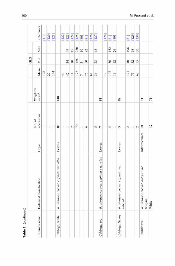
Table
2(con
tinue
d)
Com
mon
name
Botanicalclassificatio
nOrgan
No.
ofaccessions
GLS
References
Weigh
ted
meana
Mean
Min
Max
112
8[119
]
127
[120]
114
4[121]
Cabbage,w
hite
B.o
leraceaconv
ar.cap
itata
var.alba
Leaves
8714
8
144
[122]
142
3449
[123]
214
1017
[124]
7017
212
625
0[125]
17
310
[80]
676
5092
[81]
164
[126]
536
2363
[127]
Cabbage,red
B.oleraceaconv
ar.cap
itata
var.rubra
Leaves
781
117
[128]
510
756
152
[81]
119
1226
[80]
Cabbage,S
avoy
B.o
leraceaconv
ar.cap
itata
var.
saba
uda
Leaves
588
212
140
196
[81]
175
5298
[129]
262
5570
[130]
Cauliflow
erB.o
leraceaconv
ar.b
otrytis
var.
botrytis
Inflorescences
2171
White
1271
100 M. Possenti et al.

210
642
171
[78]
310
383
119
[82,
131]
110
9[79]
191
[120]
115
1[80]
416
1319
[81]
Green
365
285
2314
8[82,
131]
123
[81]
Rom
anesco
448
113
7[82,
131]
319
[81]
Purple
215
4
120
0[82,
131]
110
8[81]
Chinese
cabb
age
B.rap
assp.
pekinensis
Leaves
8819
3
also
know
nas
2317
6[132]
B.cam
pestrisssp.
pekinensis
124
6[84]
159
[133]
6220
3[85]
149
[96]
Chinese
kale
B.o
leraceavar.albo
glab
raFlorets/stems
2838
Leaves
2726
1350
[87]
Stems
2734
1187
[87]
115
0[86]
Ethiopian
kale
B.carinata
Leaves
443
137
[134]
139
[135]
(con
tinued)
3 Glucosinolates in Food 101

Table
2(con
tinue
d)
Com
mon
name
Botanicalclassificatio
nOrgan
No.
ofaccessions
GLS
References
Weigh
ted
meana
Mean
Min
Max
248
[88]
Friarellior
B.rap
assp.
campestris
Florets/sho
ots
896
Cim
edi
rapa
oralso
know
nas
293
[136]
turnip
rape
B.rap
assp.
sylvestrisL.
597
[89]
Janch.
var.esculentaHort.
110
0[137]
Kale
B.o
leraceaconv
ar.a
ceph
ala
Leaves
222
143
153
170
7234
5[138]
4039
[139]
117
7[140]
2415
517
313
[90]
184
[96]
153
[141]
1green
165
[142]
1red
79[142]
Koh
lrabi
B.o
leraceavar.go
ngylod
esStem
tuber
829
6
174
[143]
223
4[91]
210
2[81]
150
[80]
278
4[144]
Mustard
greens
orB.jun
cea
Leaves
812
1
Indian
mustard
253
26113
[94]
1112
6915
5[95]
102 M. Possenti et al.

220
149
354
[145]
162
[146]
120
9[96]
173
6280
[98]
Mizun
aor
B.rap
assp.
nipp
osinicavar.
Leaves
320
Japanese
mustard
chinoleifera
218
532
[94]
125
1932
[98]
Pak
choi
orB.rap
assp.
chinensis
Leaves
3110
1
Bok
choy
also
know
nas
1320
710
731
8[99]
B.cam
pestrisssp.
chinensis
38
613
[94]
215
1218
[147]
157
[148]
374
36112
[149]
79
516
[100]
218
1521
[150]
Radish
Rap
hanu
ssativus
Roo
ts31
505
130
9[143]
8411
[151]
1762
7[152]
344
1[153]
122
[154]
149
[96]
Rapekale
B.n
apus
ssp.
napu
svar.pa
bularia
Leaves
8810
2
36116
629
9[155]
3665
725
8[156]
1615
1[156]
Rocket
Diplotaxistenu
ifolia
Leaves
3031
( con
tinued)
3 Glucosinolates in Food 103
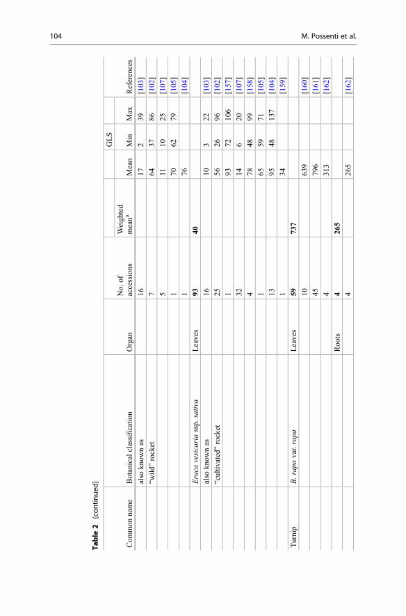
Table
2(con
tinue
d)
Com
mon
name
Botanicalclassificatio
nOrgan
No.
ofaccessions
GLS
References
Weigh
ted
meana
Mean
Min
Max
also
know
nas
1617
239
[103]
“wild
”rocket
764
3786
[102]
511
1025
[107]
170
6279
[105]
176
[104]
Eruca
vesicariassp.
sativa
Leaves
9340
also
know
nas
1610
322
[103]
“cultiv
ated”rocket
2556
2696
[102]
193
7210
6[157]
3214
620
[107]
478
4899
[158]
165
5971
[105]
1395
4813
7[104]
134
[159]
Turnip
B.rap
avar.rapa
Leaves
5973
7
1063
9[160]
4579
6[161]
431
3[162]
Roo
ts4
265
426
5[162]
104 M. Possenti et al.

Other
food
s
Caper
Cap
paris
Flower
buds
435
9
C.o
vata
189
4[163]
112
3[164]
C.spino
sa1
404
[163]
113
[165]
Moringa
orMoringa
oleifera
Leaves
232
5
drum
sticktree
196
[166]
155
5[167]
Seedpu
lp2
4724
186
26[166]
182
2[167]
Papaya
Caricapa
paya
Fruit
337
1⍨1
0[168]
1⍨1
00[169]
10
[170]
Dataof
theoriginalstud
ywereconv
ertedto
mg/10
0gFW
a Meanweigh
tedby
thenu
mberof
accessions
ofeach
stud
y
3 Glucosinolates in Food 105

4.4 Cauliflower (Brassica oleracea convar. botrytis var. botrytis)
Cauliflower is a traditional European crop that is largely cultivated also in NorthAmerica and Asia. The crop is characterized by a hypertrophy of flower branches,that probably has given rise to the name cauliflower (that consists of two Latin words,namely, “caulis” which means stem and “floris” which means flower). It is availableyear-round but is especially abundant in the spring and fall. Cauliflower cultivars showgreat variability: the varieties most known and widespread are white, green,Romanesco, and purple, each characterized by a peculiar leaf shape and color. Mostof the cauliflower cultivars are white and possess a big head, which represents the edibleportion (also known as curd) and is made up of abortive flowers, the stems of which areshort, pulpy, and crowded. Less than 10 years ago though, a green cauliflower wasdeveloped by crossing cauliflower with broccoli; in this new cultivar, the green color isdetermined by the presence of chlorophyll (like in broccoli). Green cauliflower flavor issweet, mild, and nutty and free of the bitterness sometimes found in white cauliflower.
The Italian variety referred to as “broccoli Romanesco” or “Romanesco cabbage”belongs to the group of green cauliflower and is characterized by a unique andunusual growth patterns with multiple compact heads forming a pyramid of spiralingand pointed cones.
The purple cultivar characterized by the presence of the anthocyanin pigments(that confer the characteristic color) is available year-round with a peak seasonduring the winter months. Similar to the green variety, the purple cauliflower ischaracterized by a mild and sweet flavor.
Generally, cauliflower is used as a cooked vegetable, but it can be eaten also in itsraw form.
The predominant GLSs found in cauliflower are sinigrin, glucoiberin, andglucobrassicin (Fig. 2). In white cauliflower, the total GLSs content varied from91 mg/100 g FW to 109 mg/100 g FW (mean value of 78 mg/100 g FW) [78–82](Table 2). Glucoiberin is the predominant GLS ranging from 3 to 43 mg/100 g FW(mean value of 23 mg/100 g FW), followed by sinigrin (mean value of 22 mg/100 gFW) and glucobrassicin (mean value of 14 mg/100 g FW) [80, 81] (Fig. 2).
The mean GLS content in green cauliflower is 65 mg/100 g FW (Table 2), andglucoiberin is the most abundant GLS (mean value of 14.4 mg/100 g FW)[81–83]. Romanesco shows a mean GLS content of 48 mg/100 g FW (Table 2),and glucobrassicin is the predominant GLS (mean value of 14.3 mg/100 g FW) [81,82]. In purple cauliflower, the mean GLS content is 154 mg/100 g FW (Table 2), andglucobrassicin is the most abundant compound (the mean value of 124 mg/100 gFW) [78, 82].
4.5 Chinese Cabbage (Brassica rapa ssp. pekinensis, Also Knownas Brassica campestris ssp. pekinensis)
The pekinensis group is the more common of the two different groups of Brassicarapa used as leafy vegetables in Asia (pekinensis and chinensis), and, within these
106 M. Possenti et al.

two groups, there is a wide range of varieties. The binomial name Brassicacampestris is also used to refer to these cultivars.
Chinese cabbage is one of the most important and popular vegetables in Asia,especially in China, Japan, and Korea. It is the main ingredient of “kimchi,” which isthe traditional fermented food of the Republic of Korea with a consumption procapite of about 120–150 kg of kimchi per year. Analysis performed on fresh leavesof many Korean varieties identified glucobrassicanapin, gluconapin, and4-methoxyglucobrassicin as the main GLS compounds [84, 85] (Fig. 2). The totalGLS content varies largely among varieties of Chinese cabbage, from 49 to 246 mg/100 g FW, with a mean value of about 190 mg/100 g FW (Table 2).
4.6 Chinese Kale (Brassica oleracea var. alboglabra)
Chinese kale, also known as Chinese broccoli or Kai-lan, is a popular Chinese greenvegetable, widely distributed in Southern China and Southeast Asia, even if it is alsopresent in Japan, Europe, and America. It is grown for its bolting thick stems,characterized by a tender and crisp texture and an appreciated flavor, while theconsumption of other edible parts of the plant, such as leaves and sprouts, is becomingincreasingly popular. It is generally eaten after stir-frying, steaming, or boiling.
The mean value of the total GLSs is 38 mg/100 g FW [86, 87] (Table 2), while themost abundant GLS is gluconapin, followed by sinigrin, glucobrassicin, progoitrin,glucoraphanin, and glucoerucin (Fig. 2).
4.7 Ethiopian Kale (Brassica carinata)
Ethiopian kale, also known as Ethiopian mustard, is one of the major traditional leafyvegetables in East Africa (especially in Ethiopia), and its leaves are widely con-sumed in various parts of Africa. The plant is traditionally grown in kitchen gardens,but it is also grown in field and used as an oilseed or fodder crop. Leaves and tenderstems are eaten fresh in salad, boiled in soup, or marinated, while seeds are used asspice or crushed to produce vegetable oil for cooking or for industry.
The mean total GLS content in Ethiopian kale is 43 mg/100 g FW (Table 2) andsinigrin is its predominant and almost sole GLS (Fig. 2); in fact, it accounts for morethan 95% of the total level, while glucobrassicin, gluconapin, glucoalyssin,progoitrin, 4-hydroxyglucobrassicin, 4-methoxyglucobrassicin, and 1-methoxy-glucobrassicin are found in very small amount [88].
4.8 Friarielli or Cime di rapa or Turnip Rape; (B. rapa ssp.campestris Also Known as B. rapa ssp. sylvestris L. Janch. var.esculenta Hort.)
The group Brassica rapa includes also a plant cultivar that is commonly cultivated inSouthern Italy, where it is traditionally known as friarielli or cime di rapa depending
3 Glucosinolates in Food 107

from the region of origin (Campania and Puglia, respectively). Outside of Italy, it isalso known with the names of broccoli rabe, raab, Italian turnip, or turnip rape.
The edible parts of the plant, represented by its turnip tops or shoots (sprouts plusinflorescences), are generally consumed after cooking: boiled, steamed, braised, orserved with pasta or sausages.
The few data reporting the GLS composition of this local Brassica varietyindicate a mean total GLS content of about 95 mg/100 g FW (Table 2). The mostabundant GLSs are the aliphatic gluconapin, glucobrassicanapin, and the indolicglucobrassicin, neoglucobrassicin, and 4-methoxyglucobrassicin (Fig. 2).Progoitrin has been found only in one ecotype, while glucoraphanin is completelyabsent [89].
4.9 Kale (Brassica oleracea var. acephala) and Rape Kale(Brassica napus)
Many different crops are described worldwide as “kale,” some of them being usedfor human consumption while other being grown for fodder. Generally, kale isdefined as a vegetable of the plant species Brassica oleracea var. acephala charac-terized by having leaves (colors ranging from light green to violet brown) along thestem not forming a head. However, also some cultivars of Brassica napus aregenerally known as kale, the Hanover salad, the red Russian, Siberian, and rapekale being good examples. These kinds of kale are generally more tender and have amilder flavor than the Brassica oleracea kales and are well-known varieties in theUSA and in Europe (particularly appreciated in northwestern Spain and in northernPortugal where they are known as “nabicol” and “couve nabica”).
Kale is grown along a wide range of latitudes, as it is robust and can tolerate abroad range of agricultural and climatic conditions (such as cold temperature). It isfrequently consumed after boiling or as an ingredient for stew and soup; however, itmay be eaten also raw for salad use.
Based on recent data, the GLS content of kale (B. oleracea var. acephala) rangesfrom 17 to 345 mg/100 g FW, with an average value of 143 mg/100 g FW (Table 2).Aliphatic GLSs represent the main contributor to the whole GLS profile, whileglucoiberin, sinigrin, and glucobrassicin represent the most abundant GLS (Fig. 2),but different proportions of them have been observed in different studies. Generally,glucoraphanin is present in low amount; however, some kale varieties, such as blackkale, can contain high glucoraphanin level [90].
Rape kale (B. napus) contains similar total GLS level (range, 6–298 mg/100 gFW; average value, 102 mg/100 g FW) (Table 2), but the GLS pattern is quitedifferent; in fact, the principal GLSs are glucobrassicanapin, progoitrin, andgluconapin, while indolic GLSs are present in lower amount (Fig. 2).
108 M. Possenti et al.

4.10 Kohlrabi (Brassica oleracea var. gongylodes)
Kohlrabi is a biennial, herbaceous plant grown as an annual crop that produces anedible turnip-like swollen stem at the base of the plant body. It is closest in form towild cabbage (Brassica oleracea ssp. oleracea), which is the progenitor of all theB. oleracea varieties. Kohlrabi was cultivated since the sixteenth century in thenorthwestern coast of Europe. Today it is produced in Europe, North America,many parts of Asia (India, China, Korea, and Vietnam), and North Africa.Depending on the cultivar, kohlrabi may be purple, red, or white-green in theskin, but the flesh is always white. GLS content varies among the different parts ofthe kohlrabi (skin and flesh), and generally the red varieties have a higher totalcontent of GLSs than the green ones [81, 91, 92]. Beside these differences amongvarieties (ranging from 50 to 784 mg/100 gFW), GLS content data demonstratethat kohlrabi possesses a very high total GLS content, with a mean value of295.5 mg/100 g FW (Table 2). The principal GLSs are raphanin and gluconapin(Fig. 2).
4.11 Mustard Greens (Brassica juncea)
Brassica juncea is an important crop in many parts of the world, being extensivelycultivated in Asia, especially in China and India, primarily for oilseed productionbut also for the production of the leafy vegetable known as mustard greens. Youngleaves of mustard greens are consumed raw in salads or mixed with other saladgreens, while older leaves and stems may be used fresh or processed for potherbsor cooked in a variety of recipes. A remarkable genetic variability has beenreported among strains of Brassica juncea accessions grown in different areas ofthe world, reflecting significant differences in the chemical composition of theirseeds [93].
The total level of GLSs found in mustard green leaves ranges from 26 to 354 mg/100 g FW, with an average level of 121 mg/100 g FW (Table 2). From the fewquantitative data reported in the literature, it seems that the European-type Canadiancultivars of Brassica juncea are characterized by relatively low levels of GLSs (26 to155 mg/100 g FW) [94, 95] when compared to cultivars grown in Asia (208 to354 mg/100 g FW) [96, 97], paralleling well-established differences in the compo-sition of the seeds [93].
The GLS profile is mainly characterized by aliphatic GLSs, such as sinigrin(predominant in European cultivars), gluconapin (predominant in Indian cultivars),and glucobrassicanapin, with minor concentration of indole GLS (glucobrassicin,4-hydroxyglucobrassicin, and 4-methoxyglucobrassicin) and aromatic GLS(gluconasturtiin) (Fig. 2).
3 Glucosinolates in Food 109

4.12 Mizuna (Brassica rapa subsp. nipposinica var. chinoleifera AlsoKnown as Brassica juncea var. japonica)
Mizuna also known as Japanese mustard is traditionally consumed in Japan. Theplant produces many stalks bearing dark green leaves, characterized by a pungent,mild peppery, and spicy flavor. It is consumed raw in salads or cooked in soups, stir-fries, and hot pots and pickled.
Few data reported in the literature show a total GLS content ranging from 5 to32 mg/100 g FW, with an average level of about 20 mg/100 g FW (Table 2). TheGLS profile is dominated by the aliphatic fraction (gluconapin and glucobras-sicanapin), but also indole (glucobrassicin and its derivatives) and aromatic(gluconasturtiin) GLSs have been reported [94] (Fig. 2). Only low levels (less than2 mg/100 g FW) of glucoraphanin have been observed [94, 98].
4.13 Pak Choi (Brassica rapa ssp. chinensis Also Known as Brassicacampestris L. ssp. Chinensis var. communis)
Pak choi is an Asian leafy vegetable, also known as Chinese celery cabbage, bokchoy, and horse’s ear, which has traditionally been highly used in China, but whoseconsumption is also increasing in Europe in salads or Asian-style cooking[99]. Sometimes the species Brassica rapa ssp. chinensis is reported as Brassicacampestris L. ssp. chinensis var. communis, but the two names refer to the samespecies. The fully developed vegetable looks like a short and thick celery, withglossy and crisp green leaves, whose flavor is somewhere between mild cabbage andspinach, and white or pale green chunky stalks. Leaves, preferably if young, can beeaten raw in salads or cooked by brief steaming or stir-frying.
While an average total GLS content of 100 mg/100 g FW results from recent datareported in the literature (Table 1), a remarkable variability can be observed in thetotal GLS content found in the different studies, ranging from 8 [94, 100] to 207 mg/100 g FW [99] (Table 2). This marked variation may be due not only to differentgenetic backgrounds but also to differences in the developmental stages consideredin these studies: prolonging plant growing time, in environmentally controlledconditions, from 10 to 25 days, has been shown to markedly increase the totalGLS content of the leaves at harvest [100].
The aliphatic GLSs are the predominant group in pak choi leaves, withgluconapin, glucobrassicanapin, progoitrin, and glucoalyssin giving the main con-tribution, whereas indolic GLSs (neoglucobrassicin and glucobrassicin) and aro-matic GLSs (gluconasturtiin) are also present at a lower level [99] (Fig. 2).
4.14 Radish (Raphanus sativus)
Radish is a crop of the Brassicaceae family, cultivated in China, Korea, Europe, andAmerica since the fifth century BC. The different varieties of radish diverge in size,
110 M. Possenti et al.

shape, and color. The round radish varieties are more popular in Western countries,and they have many different colors. Instead, the “daikon,” a radish with a longwhite taproot, is more popular in Asia. In the past centuries, radish has been used asmedicinal foods for a lot of diseases, but, nowadays, it is commonly used for dietaryconsumption. The major deterring factor of its use is the bitter and pungent flavorcaused by GLSs and particularly by the high content of dehydroerucin [101]. The totalGLS content varies extensively among varieties of radish ranging from 22 to 627 mg/100 g FW (mean value of 505 mg/100 g FW) (Table 2). Besides dehydroerucin, otherGLSs present in radish are glucoiberin, progoitrin, glucoraphanin, glucobrassicin,4-methoxyglucobrassicin, and 4-hydroxyglucobrassicin (Fig. 2).
4.15 Rocket (Eruca vesicaria ssp. sativa and Diplotaxis tenuifolia)
Rocket (or arugula, rucola, and roquette) is the collective name for a group of leafyvegetable crops, all belonging to the Brassicaceae family and native to the Mediter-ranean geographic area. While traditional consumption of rocket in this area datesback to ancient times, in the past few decades, it has gained increasing popularity inthe salad vegetable market in many countries all over the world [102, 103]. Twomain species are cultivated as salad crops: Eruca vesicaria (more precisely, thesubspecies sativa), belonging to the genus Eruca, and generally referred to as“salad” or “cultivated” rocket, and Diplotaxis tenuifolia, belonging to the genusDiplotaxis, often referred to as “wild” rocket. The two species have similar chemicalcomposition and appearance, and one can be easily mistaken for the other by anonexpert eye and before a certain level of maturity has been achieved. Both speciesshare the distinctive pungent flavor of the leaves that makes them an appreciatedingredient of vegetable salads (simple or in mixtures). Even though rocket is mostcommonly consumed as raw leaves, food use of cooked leaves and also of flowersand sprouted seedlings has also been reported [104, 105].
The total GLS content of rocket leaves ranges from 2 to about 140 mg/100 g offresh weight (Table 2), with an average value of 40 mg/100 g FW and 31 mg/100 gFW for Eruca vesicaria subsp. sativa and Diplotaxis tenuifolia, respectively. Theaverage values reported in Table 2, obtained on a global number of more than120 varieties, are noticeably lower than the range (72–106 mg/100 g FW) previouslyreported by Verkerk and colleagues [72], which was obtained on leaves from a singlecultivar. As for other vegetables from the Brassicaceae family, the total GLS contentis markedly affected by many environmental conditions and physiological factors[102]. In particular, GLS content seems to markedly increase throughout the multipleharvests that are allowed by the ability of this crop to regrow the leaves repeatedlyafter cutting [106].
As regards GLS profile, it is generally dominated by the three aliphatic-derivedcompounds glucosativin, in the monomeric and the two dimeric forms (4-[β-D-glucopyranosyldisulfanyl]-butyl-GLS and dimeric 4-mercaptobutyl-GLS (DMB)),glucoerucin, and glucoraphanin, even though very different proportions of thesethree have been reported [102, 107]. In addition, other aliphatic GLSs (glucoalyssin,
3 Glucosinolates in Food 111

progoitrin/epiprogoitrin, glucoiberverin), aromatic (glucosinalbin), and indole-derived GLS (4-OH-glucobrassicin, glucobrassicin) give a minor contribution tothe global profile (Fig. 2).
4.16 Turnip (Brassica rapa var. rapa)
The turnip is a root vegetable grown in temperate climates worldwide for humanconsumption or to feed livestock. In some countries the turnip leaves are eaten as“turnip greens” (the leaves harvested in vegetative period) or “turnip tops” (thefructiferous stems with the flower buds and the surrounding leaves); they are acommon side dish in the USA (especially in the southeastern of the country), Italy,Spain, and Portugal, but turnip is consumed in Asia too. The edible parts areconsumed boiled and generally served with meat.
The total GLS content varies among the plant organs, being higher in leaves(737 mg/100 g FW on average) than in roots (265 mg/100 g FW on average)(Table 2). Gluconapin is the most abundant GLS compound in all the edible partsof the plant body, but roots contain also progoitrin and gluconapoleiferin in relevantamount (Fig. 2).
4.17 Sprouts (Various Brassicaceae spp.)
Edible sprouts are tasty easy-to-eat food traditionally consumed in Asian countries.In the past years, the consumption of edible sprouts in salads or in Asian-stylecooking has been growing enormously also in Western countries.
Among the large variety of seeds and ready-to-eat sprouted seeds offered by themarket, those of the Brassicaceae family, mainly broccoli (Brassica oleracea ssp.italica) and radish (Raphanus sativus), are among the most popular for the presenceof GLSs. With respect to their conventional mature vegetable counterpart, sproutspresent several advantages. First, sprouts can be grown, simply and inexpensively,all year-round, thus encouraging vegetable consumption. Second, due to theirphysiological state, sprouts are an enriched source of phytochemicals; in fact,GLSs have been found to be higher in Brassicaceae sprouts than in thecorresponding mature vegetables [96, 171]. Third, they are consumed raw, thusavoiding the loss of GLSs usually associated to food processing and cooking. On theother hand, great care must be taken to avoid mold and bacterial contaminations thatcan occur during sprouts’ growth and storage as a great number of serious food-borne illness outbreaks associated to the consumption of raw sprouted seeds havebeen reported in many different countries [172, 173]. During germination, thecombined effect of reactivation of metabolic pathways and dilution due to tissueexpansion dramatically affects the content of bioactive molecules in sprouts[174]. This means that GLSs’ content is higher in seeds and decreases over thegermination period [175–177]. For this reason, sprouts between 5 and 8 days old areconsidered optimum for consumption, balancing biomass yield and size with GLS
112 M. Possenti et al.

content. As for adult plants, the GLS content of sprouts is influenced by both geneticand environmental factors, including production and storage conditions [175]. In anattempt to identify the best performers in terms of GLS content, an increasingnumber of studies have been undertaken aiming at comparing different genotypesunder standardized growth conditions [75, 175, 178, 179]. Significant differences,both qualitative and quantitative, have been reported in the GLS content of sprouts ofdifferent cultivars and species of Brassicaceae. Overall, the GLS pattern of sproutsreflects that of the mature edible part, but almost all the crops exhibited higherconcentrations of GLS in sprouts than in adult parts (the mean total GLS levelranging from 297 to 1297 mg/100 g FW in sprouts and from 193 to 736 mg/100 gFW in adult plants, respectively) (Tables 2 and 3). The total aliphatic GLS concen-tration also was found generally higher in sprouts than in the mature vegetable. Infact, aliphatic GLSs represented more than 90% of the total GLSs in sprouts of mostcrops, whereas they range from 57% to 98% of the total GLSs in adult parts. Therelatively higher concentrations of indolic GLS in mature edible parts are probablydue to the induction of indolic GLS synthesis in the presence of stressful growthconditions.
5 Processed Foods
5.1 Colza and Canola oil
Colza (or rapeseed) oil is a vegetable oil obtained from the seeds of oilseed rape(Brassica napus, Brassica rapa, Brassica juncea). Already in the thirteenth cen-tury, the oil was used for street lighting in the Northern Europe countries, while itsfood use was developed in the mid-nineteenth century, but it had not a lot ofsuccess because of its effects on health. In fact, from 1960 to 1970 years, severalstudies on animals have shown potential health risks (lipidosis, infarction, andcardiac lesions) associated with the use of colza oil [182–187]. The potentiallytoxic element was found to be the erucic acid present in concentrations rangingbetween 30% and 60% depending on the cultivar, the harvesting period, and otherfactors. The GLSs were responsible for the bitter taste of the oil and for thegoitrogenic effect of the meal remaining after oil extraction; due to this effect,the use of meal was drastically reduced in animal feed use. In order to use colza oilin food industry, researchers identified and/or developed different mutants of theoriginal plant. The first and most important varieties of Brassica able to produce anoil with a low content of erucic acid (called “double zero”) have been developed inCanada (University of Manitoba) in 1974. By the use of this new variety, thestandard Canola oil has been developed, the name evoking Canadian oil, which isan oil with less than 2% erucic acid and less than 30 μmol of GLSs per gram ofdefatted and dried meal (the seed must contain <30 μmol/g of any mixture of3-butenyl GLS, 4-pentenyl GLS, 2-hydroxy-3-butenyl GLS, and 2-hydroxy-4-pentenyl GLS per gram DW [188]).
3 Glucosinolates in Food 113

Table 3 Estimated total glucosinolate content of sprouts (7–12 days old, light-grown;mg/100 g FW)
Commonname
Botanicalclassification
No ofaccessions
GSL
ReferencesWeightedmeana Min Max
Broccoli B. oleracea convar.botrytis var. cimosa(commonly var.italica)
9 1297 137 2757 [75, 96,174, 175,177–181]
Brusselssprouts
B. oleracea var.gemmifera
1 1013 [178]
Cabbage,white
B. oleracea convar.capitata var. alba
2 1069 753 1385 [178, 181]
Cabbage,red
B. oleracea convar.capitata var. rubra
3 833 516 1102 [174, 175,178]
Cabbage,savoy
B. oleracea convar.capitata var. sabauda
1 1299 [178]
Cauliflower B. oleracea convar.botrytis var. botrytis
2 1178 886 1470 [96, 178]
Chinesecabbage
B. rapa ssp.pekinensis
1 297 [96]
Gardencress
Lepidium sativum 2 695 174 1216 [175, 178]
Kale B. oleracea convar.acephala
4 1206 [96, 174,178, 179]
Kohlrabi B. oleracea var.gongylodes
2 829 668 990 [175, 178]
Mustardgreens
B. juncea 1 759 [96]
Mustard Sinapis alba 3 834 548 1138 [175, 178,181]
Pak choi B. rapa ssp. chinensis 14 642 353 1156 [96, 99]
Portuguesetronchudacabbage
B. oleracea var.costata
1 598 [174]
Radish Raphanus sativus 16 676 38 2036 [96, 151,175, 176,178, 179]
Rocket Eruca vesicaria ssp.sativa
1 742 [178]
Rutabaga B. napus var.napobrassica
3 458 144 842 [75, 175,178]
Turnip B. rapa var. rapa 4 698 164 1544 [75, 175,178]
Data of the original study were converted to mg/100 g FWaMean weighted by the number of accessions of each study
114 M. Possenti et al.

Canola oil is generally consumed in its liquid form as salad oil, salad dressing,and cooking oil (pure or in blends with other oils), while a minor amount of this oil isused for margarine or shortening production.
5.2 Mustard
Mustard is a popular and widely consumed condiment generally used for sausagesand meats and also as ingredient for sandwiches and salad preparation. It is preparedfrom a mixture of some Brassica seeds that are usually grinded with wine, vinegar,water, salt, and spices. The seeds of Brassica nigra, Brassica juncea, and Sinapisalba are the most frequently used, but also those of Brassica carinata, Brassicacarista, and Brassica eruca can be used in some mustard preparation.
During the preparation process, the GLSs contained in the seeds are degraded bymyrosinase to produce ITCs (responsible for the pungent flavor), so that no GLSscan be found in the end product [189, 190]. The type and level of GLS degradationproducts in mustard and, as consequence, the intensity of the associated pungentflavor greatly depend on the type of GLSs present in the seeds used for itsproduction [189].
5.3 Sauerkraut
Sauerkraut is a very popular fermented product obtained from fermentation ofshredded and salted white cabbage. It is the most well-known German food, but itis largely consumed in all Europe (especially Eastern Europe) and in many parts ofthe USA and Canada. It can be eaten raw or after cooking.
During the fermentation process, cabbage GLSs are degraded, with formation ofITCs and other degradation products (such as indole-3-acetonitrile as well as indole-3-carbinol that reacts with ascorbic acid forming ascorbigen) [123, 191], so that nodetectable amounts of GLSs are present in sauerkraut at the end of the fermentationprocess [126]. The type and the level of GLS degradation products are stronglydependent on the content of native GLSs of the raw materials, as well as on thefermentation conditions.
6 Other Food
6.1 Caper (Capparis spinosa and Capparis ovata)
Capparis is a plant genus of the Capparaceae family that includes several species,among which the most common and studied are Capparis spinosa and Capparisovata. These aromatic plants are largely distributed in the Mediterranean regions butare also present in Asia and Australia.
3 Glucosinolates in Food 115
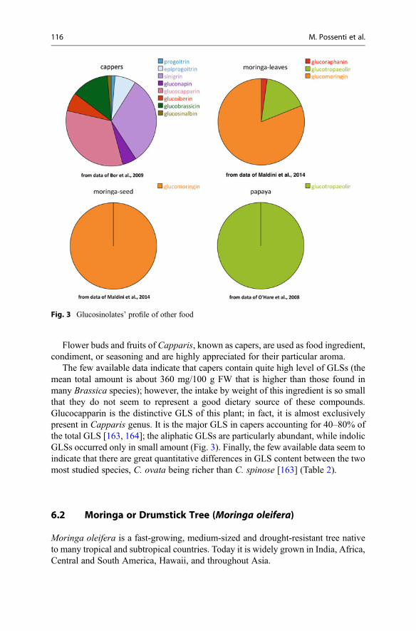
Flower buds and fruits of Capparis, known as capers, are used as food ingredient,condiment, or seasoning and are highly appreciated for their particular aroma.
The few available data indicate that capers contain quite high level of GLSs (themean total amount is about 360 mg/100 g FW that is higher than those found inmany Brassica species); however, the intake by weight of this ingredient is so smallthat they do not seem to represent a good dietary source of these compounds.Glucocapparin is the distinctive GLS of this plant; in fact, it is almost exclusivelypresent in Capparis genus. It is the major GLS in capers accounting for 40–80% ofthe total GLS [163, 164]; the aliphatic GLSs are particularly abundant, while indolicGLSs occurred only in small amount (Fig. 3). Finally, the few available data seem toindicate that there are great quantitative differences in GLS content between the twomost studied species, C. ovata being richer than C. spinose [163] (Table 2).
6.2 Moringa or Drumstick Tree (Moringa oleifera)
Moringa oleifera is a fast-growing, medium-sized and drought-resistant tree nativeto many tropical and subtropical countries. Today it is widely grown in India, Africa,Central and South America, Hawaii, and throughout Asia.
Fig. 3 Glucosinolates’ profile of other food
116 M. Possenti et al.

Seeds, leaves, oil, sap, bark, roots, and flowers are widely used in traditionalIndian medicine since a long time. In addition, many parts of the plant are commonlyused for human consumption (principally fruit and leaves and also flowers, roots,and seed oil) in India, Pakistan, the Philippines, Hawaii, and many parts of Africa.
Very young pods are consumed raw, while mature pods are used in preparation ofsoups and stews. The younger leafy tips and tender leaves, as the young flowers andthe flower buds, are consumed as side dish and in salad. Moringa young leaves are avery rich source of GLSs, with a mean content of 325 mg/100 g FW. Glucomoringinis the main GLS found in Moringa, it is found almost exclusively in this species, andit accounts for about 78% and 99% of the total GLS in leaves and seed pulp,respectively [166, 192] (Fig. 3).
6.3 Papaya (Carica papaya)
Papaya (Carica papaya) belongs to the family of Caricaceae and is one of the mostcultivated plants in tropical and subtropical regions. It is grown in nearly allcountries of the tropical Americas (Central and South America and the state ofHawaii) and in India, Sri Lanka, various Asian countries, as well as the Antilles andtropical Africa. Papaya is available year-round, and the ripe fruit is largely consumedas such or to make fruit salad, drinks, jam, jelly, candies, etc.
While Capparis and Moringa are close relatives of the Brassicaceae family (theyall belong to the order of Capparales), C. papaya is one of the few edible species notrelated to Brassicaceae containing GLSs. Glucotropaeolin seems, at the moment, theunique GLS present in this plant (Fig. 3).
The content of glucotropaeolin is quite different in the papaya tissues; it is veryhigh in seed and leaf, while it is lower in the edible flesh [168, 169]. Theglucotropaeolin content in the pulp gradually decreases during fruit maturation[168], so that its content in fruit is greatly dependent on the maturation stage. Thiscan explain the great data variability observed among studies: from 0 [170] to about100 mg/100 g FW [169]. Because glucotropaeolin imparts an unpalatable flavor tothe fruit (“nasturtium” flavor), the fruits high in glucotropaeolin tend to be dislikedby consumers and then less consumed.
6.4 Honey
Honey represents the unique animal-derived food present in this list ofGLS-containing food. In fact, while GLS degradation products have been reportedoutside the plant kingdom, for example, in cow milk [193], honey is the only animalproduct containing intact GLSs. These GLSs come from the pollen of plantsbelonging to the Brassicaceae family (e.g., Brassica napus or Diplotaxis tenuifolia)that are used for honey production [194]. It has been demonstrated that pollens ofthese plants contain low and variable amount of GLSs (from 0 to about 200 μg/100 gFW) [195], and very low amount of GLSs has been found in some single flower
3 Glucosinolates in Food 117

honey [196]. However, the GLS amount is so low that honey cannot represent adietary source of GLSs.
6.5 Impact of Post-Harvest Factors on GLS Content
In the above sections, GLS content has been mainly discussed as determined in thefreshly harvested vegetables, but it is important to keep in mind that this originalcontent can be, even substantially, affected by the several steps of the food supplychain occurring before actual consumption: transport, storage, industrial processing,and home preparation [72, 197–199]. It is generally agreed that the impact of post-harvest handling and storage is in most cases less pronounced than the effects ofprocessing and preparation, even though the extent of these changes criticallydepends on the type of vegetables, the processing method, and the specificprocessing conditions applied [199]. Transport and storage are key steps in post-harvest life of vegetables before consumer purchase and environmental conditions inthese steps are generally optimized with reference to visual quality. Vegetables ofBrassica species exhibit a broad variety of plant structures, from roots to stems,sprouts, leaves, and inflorescences, with an associated variety in physiology andbiochemistry and, hence, shelf life. In this respect, broccolis are very perishablevegetables, whereas cabbage and Brussels sprouts are less susceptible to senescenceand decay and can sustain a longer shelf life [72]. Time and temperature are the mostimportant factors affecting GLS content during transport and storage: broccoliinflorescences stored for 5 days at 4 �C show a relatively limited decrease in thetotal GLS content (from 4 to 16%), whereas a much marked degradation occurs at20 �C (from 64% to 79%) [200]. A drastic reduction (55%) of glucoraphanin contentmay even occur after 3 days of storage at ambient temperature, while the beneficialeffect of cold storage takes place in broccoli stored both in open boxes and packagedin plastic bags [201]. With regard to broccoli, it has been found that the degree oftotal GLS losses generally corresponds to the degree of visual quality degradation[201]. Interestingly, different classes of GLS are affected in a different way by thesame storage conditions: while glucoraphanin losses are higher (up to 82% after5 days at 20 �C) than average GLS losses, indole GLS tends to be less stronglyaffected by storage [200], and in some studies their content increases during storageat 10 �C [202] or ambient temperature, while all other GLSs decrease [203].
The impact of freezing without previous inactivation of myrosinase may beremarkable: the global freezing-thawing cycle has been reported to reduce bymore than 30% the total GLS content in various Brassica vegetables [204] and tocompletely deplete it in sprouts of sea kale [205]. GLS content of blanched vegeta-bles is not affected by storage in a freezer [197], even though the preliminaryblanching process may have a more or less strong impact (from 2% to 74% losses)depending on blanching time and conditions [72].
In many cases, Brassica vegetables are subjected to industrial or domesticprocessing before consumption. There is a great variety of preparation methodsworldwide, ranging from boiling to steaming, microwave cooking, stir-frying, and
118 M. Possenti et al.

fermentation, the combination of procedures and ingredients changing acrossregions and cultures [199]. In addition, variability in home preparation is furtherwidened by individual preferences about sensory properties of cooked vegetables,which dictate time-temperature settings of the applied domestic cooking processes.Among cooking methods, boiling is probably the most common used for Brassicavegetables, at least in Western countries, and in some instances it may produce asubstantial depletion of the GLS content present in the raw vegetable. Losses of GLSdue to boiling are strongly influenced by the time of cooking, ratio of vegetable towater, and type of vegetable and mainly occur through leaching into cooking water[199]. Regarding the effect of the type of vegetable, it has been observed a progres-sive increase of GLS losses from 58% to 65%, 75%, and 77%, moving from Brusselssprouts to green cabbages, cauliflowers, and broccoli, respectively, after 30 min ofboiling [204]. Much lower, or no, losses have been observed when shorter time isapplied to boiling of broccoli florets (3 to 8 min) and Brussels sprouts (10 min),whereas significant losses (38%) have still been found after a relatively short boilingtime (10 min) on cauliflowers [120, 206]. Steaming, by preventing direct contact toboiling water, results as a much more effective method in preserving the originalGLS content of the freshly harvested vegetable: tests carried out on cabbages,broccoli, cauliflowers, and Brussels sprouts, by varying cooking time up to20 min, have shown either slight decreases or increases in total GLS content[199]. The increase of GLS content may be ascribed to an increased extractabilityof GLS from the food matrix in the analytical process due to the effect of the heattreatment. Microwave cooking has, in general, an intermediate impact when com-pared to boiling and steaming, but, similar to the other methods, the degree of GLSlosses depends on the specific conditions of cooking applied, in terms of time, poweroutput, and addition of water prior to microwave processing. The level of microwavepower is linked to the extent of losses of the total GLS in broccoli florets, whereasslight decreases of total GLS content have been observed on Brussels sprouts,cabbages, and cauliflowers when applying varying power levels [199]. In somecase the application of microwave processing may result in a marked increase(up to 180%) of total GLS content, as observed on red cabbage, probably due toan increased extractability from the plant tissue after heating [207]. Addition ofwater to the vegetable promotes leaching of GLS into the cooking water [208]. Stir-frying is one of the most popular processing methods in several Asian countries;however, relatively few data are available in the literature about the effects of thismethod of cooking. Somewhat inconsistent results have been reported, ranging fromslight losses to substantial reduction of total GLS content in broccoli florets aftervarying times and conditions of stir-frying [199].
Fermentation, as mentioned above, is a quite popular processing method not onlyin some European countries, for the preparation of sauerkraut, but also in Asiancountries, for the preparation of a variety of fermented products [199]. In thepreparation of sauerkraut, fermentation results in complete degradation of GLSjust after 7 days from the start of processing and in the formation of GLS bioactivebreakdown products, such as sulforaphane, ascorbigen, indole-3-carbinol, and manyothers, whose levels undergo further changes along the following storage time
3 Glucosinolates in Food 119

[126]. The content of the GLS breakdown products in the food at the time ofconsumption depends on the level of native GLS in the raw material and also onthe conditions of fermentation (application of starter bacteria cultures, the type ofbacteria strains applied) [72].
7 Conclusions
Among all known phytochemicals, GLSs are of special interest both for their health-related effects and for their relatively high food concentration (ranging from 20 toabout 5000 mg per 100 g FW). However, specific recommendations for increasingGLS-containing food consumption have not been established yet [209]. Among thereasons leading to this lack of specific dietary guidelines, there is the persistence of anumber of uncertainties in the causal relationship between GLS-rich food consump-tion and the health output. First, GLS/ITCs associated to both beneficial anddetrimental health effects coexist in the same food. Second, there are insufficientdata about GLS-containing food consumption. Third, the use of cruciferous con-sumption as a proxy for GLS intake represents an epidemiological study confounder[210]. In fact, cruciferous vegetables contain other bioactive molecules in addition toGLS. Moreover, estimation of GLS daily dietary intake is especially difficult due tothe lack of a comprehensive GLS food composition database, taking into consider-ation also the food processing-induced changes. All these factors contribute to theinsufficient strength of epidemiological evidence (see as examples the relationbetween esophageal and endometrium cancer and cruciferous vegetable consump-tion [211]).
Therefore, a clear qualitative and quantitative description of the presence of GLSsin food represents a prerequisite in order to define GLS dietary guidelines.
Acknowledgments This work was supported by Italian Ministry of Agriculture, Food & Forestry(MiPAAF) grants “NUTRIGEA” (DM 30281 23/12/2009).
References
1. Mewis I, Ulrich C, Schnitzler WH (2002) The role of glucosinolates and their hydrolysisproducts in oviposition and host-plant finding by cabbage webworm, Hellula undalis. EntomolExp Appl 105:129–139. doi:10.1023/A:1022176524227
2. Miles CI, Del Campo ML, Renwick JAA (2005) Behavioral and chemosensory responses to ahost recognition cue by larvae of Pieris rapae. J Comp Physiol A Neuroethol Sensory, Neural,Behav Physiol 191:147–155. doi:10.1007/s00359-004-0580-x
3. Giamoustaris A, Mithen R (1995) The effect of modifying the glucosinolate content of leavesof oilseed rape (Brassica napus ssp. oleifera) on its interaction with specialist and generalistpests. Ann Appl Biol 126:347–363
4. Martin N, M€uller C (2007) Induction of plant responses by a sequestering insect: relationshipof glucosinolate concentration and myrosinase activity. Basic Appl Ecol 8:13–25.doi:10.1016/j.baae.2006.02.001
120 M. Possenti et al.

5. Read DP, Feeny PP, Root RB (1970) Habitat selection by the aphid parasite diaeretiella rapae(hymenoptera: braconidae) and hyperparasite charips brassicae (hymenoptera: cynipidae). CanEntomol 102:1567–1578
6. Mattiacci L, Dicke M, Posthumus MA (1994) Induction of parasitoid attracting synomone inbrussels sprouts plants by feeding of Pieris brassicae larvae: role of mechanical damage andherbivore elicitor. J Chem Ecol 20:2229–2247. doi:10.1007/BF02033199
7. Choi E-J, Zhang P, Kwon H (2014) Determination of goitrogenic metabolites in the serum ofmale wistar rat fed structurally different glucosinolates. Toxicol Res 30:109–116
8. U.S. Department of Health and Human Services PHS (2006) Agency for toxic substances anddisease registry div. of tocicology and environmental medicine 1600 ecifton road ne, MailstopF-32 Atlanta, Georgia 30333, US Dep Heal Hum Serv, p 298
9. Newkirk RW, Classen HL, Tyler RT (1997) Nutritional evaluation of low glucosinolatemustard meals (Brassica juncea) in broiler diets. Poult Sci 76:1272–1277
10. Fahey JW, Haristoy X, Dolan PM et al (2002) Sulforaphane inhibits extracellular, intracellular,and antibiotic-resistant strains of Helicobacter pylori and prevents benzo[a]pyrene-inducedstomach tumors. Proc Natl Acad Sci U S A 99:7610–7615. doi:10.1007/BF02033199
11. Matsui TA, Murata H, Sakabe T et al (2007) Sulforaphane induces cell cycle arrest andapoptosis in murine osteosarcoma cells in vitro and inhibits tumor growth in vivo. OncolRep 18:1263–1268
12. Smith TK, Lund EK, Parker ML et al (2004) Allyl-isothiocyanate causes mitotic block, loss ofcell adhesion and disrupted cytoskeletal structure in HT29 cells. Carcinogenesis25:1409–1415
13. Smith TK (2003) Effects of Brassica vegetable juice on the induction of apoptosis and aberrantcrypt foci in rat colonic mucosal crypts in vivo. Carcinogenesis 24:491–495
14. Boreddy SR, Sahu RP, Srivastava SK (2011) Benzyl isothiocyanate suppresses pancreatictumor angiogenesis and invasion by inhibiting HIF-α/VEGF/Rho-GTPases: pivotal role ofSTAT-3. PLoS One 6, e25799. doi:10.1371/journal.pone.0025799
15. Gupta P, Adkins C, Lockman P, Srivastava SK (2013) Metastasis of breast tumor cells to brainis suppressed by phenethyl isothiocyanate in a novel in vivo metastasis model. PLoS One8:1–9. doi:10.1371/journal.pone.0067278
16. Kang L, Wang Z-Y (2010) Breast cancer cell growth inhibition by phenethyl isothiocyanate isassociated with down-regulation of oestrogen receptor-alpha36. J Cell Mol Med14:1485–1493
17. Xu C, Shen G, Chen C et al (2005) Suppression of NF-kappaB and NF-kappaB-regulated geneexpression by sulforaphane and PEITC through IkappaBalpha, IKK pathway in humanprostate cancer PC-3 cells. Oncogene 24:4486–4495. doi:10.1038/sj.onc.1208656
18. Fimognari C, Turrini E, Ferruzzi L et al (2012) Natural isothiocyanates: genotoxic potentialversus chemoprevention. Mutat Res – Rev Mutat Res 750:107–131. doi:10.1016/j.mrrev.2011.12.001
19. Baasanjav-Gerber C, Monien BH, Mewis I et al (2011) Identification of glucosinolate conge-ners able to form DNA adducts and to induce mutations upon activation by myrosinase. MolNutr Food Res 55:783–792. doi:10.1002/mnfr.201000352
20. JangM, Hong E, Kim G-H (2010) Evaluation of antibacterial activity of 3-butenyl, 4-pentenyl,2-phenylethyl, and benzyl isothiocyanate in Brassica vegetables. J Food Sci 75:M412–M416
21. Blažević I, Radonić A, Mastelić J et al (2010) Glucosinolates, glycosidically bound volatilesand antimicrobial activity of Aurinia sinuata (Brassicaceae). Food Chem 121:1020–1028
22. Kulisic-Bilusic T, Schmöller I, Schn€abele K et al (2012) The anticarcinogenic potential ofessential oil and aqueous infusion from caper (Capparis spinosa L.). Food Chem 132:261–267
23. Vanden Bussche J, Noppe H, Verheyden K et al (2009) Analysis of thyreostats: a history of35 years. Anal Chim Acta 637:2–12
24. Srivastava SK, Xiao D, Lew KL et al (2003) Allyl isothiocyanate, a constituent of cruciferousvegetables, inhibits growth of PC-3 human prostate cancer xenografts in vivo. Carcinogenesis24:1665–1670. doi:10.1093/carcin/bgg123
3 Glucosinolates in Food 121

25. Tsai S-C, Huang WW, Huang CWet al (2012) ERK-modulated intrinsic signaling and G(2)/Mphase arrest contribute to the induction of apoptotic death by allyl isothiocyanate inMDA-MB-468 human breast adenocarcinoma cells. Int J Oncol 41:2065–2072.doi:10.3892/ijo.2012.1640
26. Manesh C, Kuttan G (2003) Effect of naturally occurring allyl and phenyl isothiocyanates inthe inhibition of experimental pulmonary metastasis induced by B16F-10 melanoma cells.Fitoterapia 74:355–363
27. Kumar A, D’Souza SS, Tickoo S et al (2009) Antiangiogenic and proapoptotic activities ofallyl isothiocyanate inhibit ascites tumor growth in vivo. Integr Cancer Ther 8:75–87
28. Kim MJ, Kim SH, Lim SJ (2010) Comparison of the apoptosis-inducing capability ofsulforaphane analogues in human colon cancer cells. Anticancer Res 30:3611–3620. doi:30/9/3611 [pii]
29. Zhang Y, Talalay P, Cho CG, Posner GH (1992) A major inducer of anticarcinogenicprotective enzymes from broccoli: isolation and elucidation of structure. Proc Natl Acad SciU S A 89:2399–2403
30. Lamy E, Schröder J, Paulus S et al (2008) Antigenotoxic properties of Eruca sativa (rocketplant), erucin and erysolin in human hepatoma (HepG2) cells towards benzo(a)pyrene andtheir mode of action. Food Chem Toxicol 46:2415–2421. doi:10.1016/j.fct.2008.03.022
31. Fimognari C, N€usse M, Iori R et al (2004) The new isothiocyanate 4-(methylthio)butylisothiocyanate selectively affects cell-cycle progression and apoptosis induction ofhuman leukemia cells. Invest New Drugs 22:119–129
32. Yehuda H, Soroka Y, Zlotkin-Frušić M et al (2012) Isothiocyanates inhibit psoriasis-relatedproinflammatory factors in human skin. Inflamm Res 61:735–742. doi:10.1007/s00011-012-0465-3
33. Abdull Razis AF, De Nicola GR, Pagnotta E et al (2012) 4-Methylsulfanyl-3-butenylisothiocyanate derived from glucoraphasatin is a potent inducer of rat hepatic phase IIenzymes and a potential chemopreventive agent. Arch Toxicol 86:183–194. doi:10.1007/s00204-011-0750-x
34. Scholl C, Eshelman BD, Barnes DM, Hanlon PR (2011) Raphasatin is a more potent inducerof the detoxification enzymes than its degradation products. J Food Sci 76:504–511.doi:10.1111/j.1750-3841.2011.02078.x
35. Salah-Abbes JB, Abbes S, Abdel-Wahhab MA, Oueslati R (2010) In-vitro free radicalscavenging, antiproliferative and anti-zearalenone cytotoxic effects of 4-(methylthio)-3-butenyl isothiocyanate from Tunisian Raphanus sativus. J Pharm Pharmacol 62:231–239.doi:10.1211/jpp.62.02.0011
36. Wang N, Wang W, Huo P et al (2014) Mitochondria-mediated apoptosis in human lung cancerA549 Cells by 4-Methylsulfinyl-3-butenyl isothiocyanate from radish seeds. Asian Pacific JCancer Prev 15:2133–2139. doi:10.7314/APJCP.2014.15.5.2133
37. Jakubikova J, Bao Y, Bodo J, Sedlak J (2006) Isothiocyanate iberin modulates phase IIenzymes, posttranslational modification of histones and inhibits growth of Caco-2 cells byinducing apoptosis. Neoplasma 53:463–470
38. Wang N, Shen L, Qiu S et al (2010) Analysis of the isothiocyanates present in three ChineseBrassica vegetable seeds and their potential anticancer bioactivities. Eur Food Res Technol231:951–958
39. Ben Salah-Abbes J, Abbes S, Ouanes Z et al (2009) Isothiocyanate from the Tunisian radish(Raphanus sativus) prevents genotoxicity of Zearalenone in vivo and in vitro. Mutat Res –Genet Toxicol Environ Mutagen 677:59–65. doi:10.1016/j.mrgentox.2009.05.017
40. Myzak MC, Karplus PA, Chung F-L, Dashwood RH (2004) A novel mechanism ofchemoprotection by sulforaphane: inhibition of histone deacetylase. Cancer Res64:5767–5774
41. Kanematsu S, Yoshizawa K, Uehara N et al (2011) Sulforaphane inhibits the growth of KPL-1human breast cancer cells in vitro and suppresses the growth and metastasis of orthotopicallytransplanted KPL-1 cells in female athymic mice. Oncol Rep 26:603–608
122 M. Possenti et al.

42. Heiss E, Herhaus C, Klimo K et al (2001) Nuclear factor κB is a molecular target forsulforaphane-mediated anti-inflammatory mechanisms. J Biol Chem 276:32008–32015.doi:10.1074/jbc.M104794200
43. Zhang Y, Kensler TW, Cho CG et al (1994) Anticarcinogenic activities of sulforaphane andstructurally related synthetic norbornyl isothiocyanates. Proc Natl Acad Sci U S A91:3147–3150
44. Sun CC, Li SJ, Yang CL et al (2015) Sulforaphane attenuates muscle inflammation indystrophin-deficient mdx mice via NF-E2-related factor 2 (Nrf2)-mediated inhibition ofNF-κB signaling pathway. J Biol Chem 290:17784–17795
45. Jackson SJT, Singletary KW, Venema RC (2007) Sulforaphane suppresses angiogenesis anddisrupts endothelial mitotic progression and microtubule polymerization. Vascul Pharmacol46:77–84
46. Rodríguez-Cantú LN, Gutiérrez-Uribe JA, Arriola-Vucovich J et al (2011) Broccoli (Brassicaoleracea var. italica) sprouts and extracts rich in glucosinolates and isothiocyanates affectcholesterol metabolism and genes involved in lipid homeostasis in hamsters. J Agric FoodChem 59:1095–1103
47. Morroni F, Tarozzi A, Sita G et al (2013) Neuroprotective effect of sulforaphane in6-hydroxydopamine-lesioned mouse model of Parkinson’s disease. Neurotoxicology36:63–71. doi:10.1016/j.neuro.2013.03.004
48. Adesida A, Edwards LG, Thornalley PJ (1996) Inhibition of human leukaemia 60 cell growthby mercapturic acid metabolites of phenylethyl isothiocyanate. Food Chem Toxicol34:385–392
49. Telang NT, Katdare M, Bradlow HL et al (1997) Inhibition of proliferation and modulation ofestradiol metabolism: novel mechanisms for breast cancer prevention by the phytochemicalindole-3-carbinol. Exp Biol Med 216:246–252
50. Nakamura Y, Yogosawa S, Izutani Y et al (2009) A combination of indol-3-carbinol andgenistein synergistically induces apoptosis in human colon cancer HT-29 cells by inhibitingAkt phosphorylation and progression of autophagy. Mol Cancer 8:100
51. Bjeldanes LF, Kim JY, Grose KR et al (1991) Aromatic hydrocarbon responsiveness-receptoragonists generated from indole-3-carbinol in vitro and in vivo: comparisons with 2,3,7,8-tetrachlorodibenzo-p-dioxin. Proc Natl Acad Sci U S A 88:9543–9547
52. Adwas AA, Elkhoely AA, Kabel AM et al (2016) Anti-cancer and cardioprotective effects ofindol-3-carbinol in doxorubicin-treated mice. J Infect Chemother 22:36–43
53. Wakabayashi K, Nagao M, Ochiai M, et al (1987) Recently identified nitrite-reactive com-pounds in food: occurrence and biological properties of the nitrosated products. IARC Sci Publ84:287–291
54. Brunelli D, Tavecchio M, Falcioni C et al (2010) The isothiocyanate produced fromglucomoringin inhibits NF-kB and reduces myeloma growth in nude mice in vivo. BiochemPharmacol 79:1141–1148
55. Galuppo M, Giacoppo S, De Nicola GR et al (2014) Antiinflammatory activity ofglucomoringin isothiocyanate in a mouse model of experimental autoimmune encephalomy-elitis. Fitoterapia 95:160–174
56. Giacoppo S, Galuppo M, Montaut S et al (2015) An overview on neuroprotective effects ofisothiocyanates for the treatment of neurodegenerative diseases. Fitoterapia 106:12–21.doi:10.1016/j.fitote.2015.08.001
57. Morris CR, Chen SC, Zhou L et al (2004) Inhibition by allyl sulfides and phenethyl isothio-cyanate of methyl-n-pentylnitrosamine depentylation by rat esophageal microsomes, humanand rat CYP2E1, and rat CYP2A3. Nutr Cancer 48:54–63
58. Gupta P, Kim B, Kim SH, Srivastava SK (2014) Molecular targets of isothiocyanates incancer: recent advances. Mol Nutr Food Res 58:1685–1707. doi:10.1002/mnfr.201300684
59. Satyan KS, Swamy N, Dizon DS et al (2006) Phenethyl isothiocyanate (PEITC) inhibitsgrowth of ovarian cancer cells by inducing apoptosis: role of caspase and MAPK activation.Gynecol Oncol 103:261–270
3 Glucosinolates in Food 123

60. Huang C, Ma WY, Li J et al (1998) Essential role of p53 in phenethyl isothiocyanate-inducedapoptosis. Cancer Res 58:4102–4106
61. Stoner G (1998) Inhibition of N’-nitrosonornicotine-induced esophageal tumorigenesis by3-phenylpropyl isothiocyanate. Carcinogenesis 19:2139–2143
62. Solt DB, Chang K, Helenowski I, Rademaker AW (2003) Phenethyl isothiocyanate inhibitsnitrosamine carcinogenesis in a model for study of oral cancer chemoprevention. Cancer Lett202:147–152
63. Xiao D, Singh SV (2007) Phenethyl isothiocyanate inhibits angiogenesis in vitro and ex vivo.Cancer Res 67:2239–2246
64. Rose P, Yen KW, Choon NO, Whiteman M (2005) Beta-phenylethyl and8-methylsulphinyloctyl isothiocyanates, constituents of watercress, suppress LPS inducedproduction of nitric oxide and prostaglandin E2 in RAW 264.7 macrophages. Nitric Oxide –Biol Chem 12:237–243. doi:10.1016/j.niox.2005.03.001
65. Moon PD, Kim HM (2012) Anti-inflammatory effect of phenethyl isothiocyanate, an activeingredient of Raphanus sativus Linne. Food Chem 131:1332–1339. doi:10.1016/j.foodchem.2011.09.127
66. Zhang Y, Talalay P (1998) Mechanism of differential potencies of isothiocyanates as inducersof anticarcinogenic Phase 2 enzymes. Cancer Res 58:4632–4639
67. Srivastava SK, Singh SV (2004) Cell cycle arrest, apoptosis induction and inhibition ofnuclear factor kappa B activation in anti-proliferative activity of benzyl isothiocyanate againsthuman pancreatic cancer cells. Carcinogenesis 25:1701–1709. doi:10.1093/carcin/bgh179
68. Sahu RP, Srivastava SK (2009) The role of STAT-3 in the induction of apoptosis in pancreaticcancer cells by benzyl isothiocyanate. J Natl Cancer Inst 101:176–193
69. Boreddy SR, Pramanik KC, Srivastava SK (2011) Pancreatic tumor suppression by benzylisothiocyanate is associated with inhibition of PI3K/AKT/FOXO pathway. Clin Cancer Res17:1784–1795
70. Kermanshai R, McCarry BE, Rosenfeld J et al (2001) Benzyl isothiocyanate is the chief or soleanthelmintic in papaya seed extracts. Phytochemistry 57:427–435. doi:10.1016/S0031-9422(01)00077-2
71. McNaughton SA, Marks GC (2003) Development of a food composition database for theestimation of dietary intakes of glucosinolates, the biologically active constituents of crucif-erous vegetables. Br J Nutr 90:687–697. doi:10.1079/BJN2003917
72. Verkerk R, Schreiner M, Krumbein A et al (2009) Glucosinolates in Brassica vegetables: theinfluence of the food supply chain on intake, bioavailability and human health. Mol Nutr FoodRes 53(Suppl 2):S219
73. Wang J, Gu H, Yu H et al (2012) Genotypic variation of glucosinolates in broccoli (Brassicaoleracea var. italica) florets from China. Food Chem 133:735–741. doi:10.1016/j.foodchem.2012.01.085
74. Renaud ENC, Lammerts van Bueren ET, Myers JR et al (2014) Variation in Broccoli cultivarphytochemical content under organic and conventional management systems: implications inbreeding for nutrition. PLoS One 9, e95683. doi:10.1371/journal.pone.0095683
75. Baenas N, García-Viguera C, Moreno DA (2014) Biotic elicitors effectively increase theglucosinolates content in Brassicaceae sprouts. J Agric Food Chem 62:1881–1889.doi:10.1021/jf404876z
76. Traka MH, Saha S, Huseby S et al (2013) Genetic regulation of glucoraphanin accumulation inBeneforte broccoli. New Phytol 198:1085–1095. doi:10.1111/nph.12232
77. Zabaras D, Roohani M, Krishnamurthy R et al (2013) Characterisation of taste-active extractsfrom raw Brassica oleracea vegetables. Food Funct 4:592–601. doi:10.1039/c2fo30192j
78. Bhandari S, Kwak J-H (2015) Chemical composition and antioxidant activity in differenttissues of Brassica vegetables. Molecules 20:1228–1243. doi:10.3390/molecules20011228
79. Yi G-E, Robin A, Yang K et al (2015) Identification and expression analysis of glucosinolatebiosynthetic genes and estimation of glucosinolate contents in edible organs of Brassicaoleracea subspecies. Molecules 20:13089–13111. doi:10.3390/molecules200713089
124 M. Possenti et al.

80. Vicas SI, Teusdea AC, Carbunar M et al (2013) Glucosinolates profile and antioxidant capacityof Romanian Brassica vegetables obtained by organic and conventional agricultural practices.Plant Foods Hum Nutr 68:313–321. doi:10.1007/s11130-013-0367-8
81. Verkerk R, Tebbenhoff S, Dekker M (2010) Variation and distribution of glucosinolates in42 cultivars of Brassica oleracea vegetable crops. Acta Hortic 63–70. doi:10.17660/ActaHortic.2010.856.7
82. Volden J, Bengtsson GB, Wicklund T (2009) Glucosinolates, L-ascorbic acid, total phenols,anthocyanins, antioxidant capacities and colour in cauliflower (Brassica oleracea L. ssp.botrytis); effects of long-term freezer storage. Food Chem 112:967–976. doi:10.1016/j.foodchem.2008.07.018
83. Gratacós-Cubarsí M, Ribas-Agustí A, García-Regueiro JA, Castellari M (2010) Simultaneousevaluation of intact glucosinolates and phenolic compounds by UPLC-DAD-MS/MS inBrassica oleracea L. var. botrytis. Food Chem 121:257–263. doi:10.1016/j.foodchem.2009.11.081
84. Hong E, Kim SJ, Kim GH (2011) Identification and quantitative determination ofglucosinolates in seeds and edible parts of Korean Chinese cabbage. Food Chem128:1115–1120. doi:10.1016/j.foodchem.2010.11.102
85. Lee MK, Chun JH, Byeon DH et al (2014) Variation of glucosinolates in 62 varieties ofChinese cabbage (Brassica rapa L. ssp. pekinensis) and their antioxidant activity. LWT – FoodSci Technol 58:93–101. doi:10.1016/j.lwt.2014.03.001
86. Schonhof I, Krumbein A, Br€uckner B (2004) Genotypic effects on glucosinolates and sensoryproperties of broccoli and cauliflower. Nahrung 48:25–33
87. Sun B, Liu N, Zhao Y et al (2011) Variation of glucosinolates in three edible parts of Chinesekale (Brassica alboglabra Bailey) varieties. Food Chem 124:941–947. doi:10.1016/j.foodchem.2010.07.031
88. Schreiner M, Beyene B, Krumbein A, St€utzel H (2009) Ontogenetic changes of 2-propenyland 3-indolylmethyl glucosinolates in Brassica carinata leaves as affected by water supply.J Agric Food Chem 57:7259–7263. doi:10.1021/jf901076h
89. Barbieri G, Pernice R, Maggio A et al (2008) Glucosinolates profile of Brassica rapa L. subsp.Sylvestris L. Janch. var. esculenta Hort. Food Chem 107:1687–1691. doi:10.1016/j.foodchem.2007.09.054
90. Sasaki K, Neyazaki M, Shindo K et al (2012) Quantitative profiling of glucosinolates byLC-MS analysis reveals several cultivars of cabbage and kale as promising sources ofsulforaphane. J Chromatogr B Anal Technol Biomed Life Sci 903:171–176. doi:10.1016/j.jchromb.2012.07.017
91. Park WT, Kim JK, Park S et al (2012) Metabolic profiling of glucosinolates, anthocyanins,carotenoids, and other secondary metabolites in kohlrabi (Brassica oleracea var. gongylodes).J Agric Food Chem 60:8111–8116. doi:10.1021/jf301667j
92. Park M-H, Valan Arasu M, Park N-Y et al (2013) Variation of glucoraphanin andglucobrassicin: anticancer components in Brassica during processing. Food Sci Technol33:624–631. doi:10.1590/S0101-20612013000400005
93. Sodhi YS, Mukhopadhyay A, Arumugam N et al (2002) Genetic analysis of total glucosinolatein crosses involving a high glucosinolate Indian variety and a low glucosinolate line ofBrassica juncea. Plant Breed 121:508–511
94. Krumbein A, Schonhof I, Schreiner M (2005) Composition and contents of phytochemicals(glucosinolates, carotenoids and chlorophylls) and ascorbic acid in selected Brassica species(B. juncea, B. rapa subsp. nipposinica var. chinoleifera, B. rapa subsp. chinensis and B. rapasubsp. rapa). J Appl Bot Food Qual 79:168–174
95. Tong Y, Gabriel-Neumann E, Ngwene B et al (2014) Topsoil drying combined with increasedsulfur supply leads to enhanced aliphatic glucosinolates in Brassica juncea leaves and roots.Food Chem 152:190–196. doi:10.1016/j.foodchem.2013.11.099
96. Bhandari S, Jo J, Lee J (2015) Comparison of glucosinolate profiles in different tissues of nineBrassica crops. Molecules 20:15827–15841. doi:10.3390/molecules200915827
3 Glucosinolates in Food 125

97. Gupta S, Sangha MK, Kaur G, et al (2014) QTL analysis for phytonutrient compounds and theantioxidant molecule in mustard (Brassica juncea L.). Euphytica 345–356. doi:10.1007/s10681-014-1204-3
98. Fallovo C, Schreiner M, Schwarz D et al (2011) Phytochemical changes induced by differentnitrogen supply forms and radiation levels in two leafy Brassica species. J Agric Food Chem59:4198–4207. doi:10.1021/jf1048904
99. Wiesner M (2013) Genotypic variation of the glucosinolate profile in Pak Choi (Brassica rapassp.). J Agric Food Chem 61:1943–1953. doi:10.1021/jf303970k
100. Zhu B, Yang J, Zhu Z (2013) Variation in glucosinolates in pak choi cultivars and variousorgans at different stages of vegetative growth during the harvest period. J Zhejiang Univ Sci B14:309–317. doi:10.1631/jzus.B1200213
101. Visentin M, Tava A, Iori R, Palmieri S (1992) Isolation and identification of trans-4-(Methylthio)-3-butenyl glucosinolate from radish roots (Raphanus sativus L.). J Agri FoodChem 40:1687–1691
102. Bell L, Oruna-Concha MJ, Wagstaff C (2015) Identification and quantification ofglucosinolate and flavonol compounds in rocket salad (Eruca sativa, Eruca vesicaria andDiplotaxis tenuifolia) by LC–MS: highlighting the potential for improving nutritional value ofrocket crops. Food Chem 172:852–861. doi:10.1016/j.foodchem.2014.09.116
103. D’Antuono LF, Elementi S, Neri R (2008) Glucosinolates in diplotaxis and eruca leaves:diversity, taxonomic relations and applied aspects. Phytochemistry 69:187–199. doi:10.1016/j.phytochem.2007.06.019
104. Chun J-H, Arasu MV, Lim Y-P, Kim S-J (2013) Variation of major glucosinolates in differentvarieties and lines of rocket salad. Hortic Environ Biotechnol 54:206–213. doi:10.1007/s13580-013-0122-y
105. Jin J, Koroleva OA, Gibson T et al (2009) Analysis of phytochemical composition andchemoprotective capacity of rocket (Eruca sativa and Diplotaxis tenuifolia) leafy saladfollowing cultivation in different environments. J Agric Food Chem 57:5227–5234.doi:10.1021/jf9002973
106. Bell L, Wagstaff C (2014) Glucosinolates, myrosinase hydrolysis products, and flavonolsfound in rocket (Eruca sativa and Diplotaxis tenuifolia). J Agric Food Chem 62:4481–4492
107. Pasini F, Verardo V, Caboni MF, D’Antuono LF (2012) Determination of glucosinolates andphenolic compounds in rocket salad by HPLC-DAD-MS: evaluation of Eruca sativaMill. andDiplotaxis tenuifolia L. genetic resources. Food Chem 133:1025–1033. doi:10.1016/j.foodchem.2012.01.021
108. Shiva RB, Jung-Ho K (2014) Seasonal variation in phytochemicals and antioxidant activitiesin different tissues of various Broccoli cultivars. Afr J Biotechnol 13:604–615. doi:10.5897/AJB2013.13432
109. Valverde J, Reilly K, Villacreces S et al (2015) Variation in bioactive content in broccoli(Brassica oleracea var. italica) grown under conventional and organic production systems.J Sci Food Agric 95:1163–1171. doi:10.1002/jsfa.6804
110. Brown AF, Yousef GG, Reid RW et al (2015) Genetic analysis of glucosinolate variability inbroccoli florets using genome-anchored single nucleotide polymorphisms. Theor Appl Genet128:1431–1447. doi:10.1007/s00122-015-2517-x
111. Ku KM, Jeffery EH, Juvik J (2014) Optimization of methyl jasmonate application to broccoliflorets to enhance health-promoting phytochemical content. J Sci Food Agric 94:2090–2096.doi:10.1002/jsfa.6529
112. Reilly K, Valverde J, Finn L et al (2014) Potential of cultivar and crop management to affectphytochemical content in winter-grown sprouting broccoli (Brassica oleracea L. var. italica).J Sci Food Agric 94:322–330. doi:10.1002/jsfa.6263
113. Aires A, Fernandes C, Carvalho R et al (2011) Seasonal effects on bioactive compounds andantioxidant capacity of six economically important Brassica vegetables. Molecules16:6816–6832. doi:10.3390/molecules16086816
126 M. Possenti et al.

114. Alanís-Garza PA, Becerra-Moreno A, Mora-Nieves JL et al (2015) Effect of industrial freezingon the stability of chemopreventive compounds in broccoli. Int J Food Sci Nutr 66:282–288.doi:10.3109/09637486.2015.1007451
115. Alarcón-Flores MI, Romero-González R, Martínez Vidal JL et al (2014) Monitoring ofphytochemicals in fresh and fresh-cut vegetables: a comparison. Food Chem 142:392–399.doi:10.1016/j.foodchem.2013.07.065
116. Rodríguez-Hernández MDC, Moreno D, Carvajal M et al (2012) Natural antioxidants inpurple sprouting broccoli under Mediterranean climate. J Food Sci 77:C1058–C1063.doi:10.1111/j.1750-3841.2012.02886.x
117. Dominguez-Perles R, Martinez-Ballesta MC, Riquelme F et al (2011) Novel varieties ofbroccoli for optimal bioactive components under saline stress. J Sci Food Agric91:1638–1647. doi:10.1002/jsfa.4360
118. Tian Q, Rosselot RA, Schwartz SJ (2005) Quantitative determination of intact glucosinolatesin broccoli, broccoli sprouts, Brussels sprouts, and cauliflower by high-performance liquidchromatography-electrospray ionization-tandem mass spectrometry. Anal Biochem343:93–99. doi:10.1016/j.ab.2005.04.045
119. Cieślik E, Leszczyńska T, Filipiak-Florkiewicz A et al (2007) Effects of some technologicalprocesses on glucosinolate contents in cruciferous vegetables. Food Chem 105:976–981.doi:10.1016/j.foodchem.2007.04.047
120. Pellegrini N, Chiavaro E, Gardana C et al (2010) Effect of different cooking methods on color,phytochemical concentration, and antioxidant capacity of raw and frozen Brassica vegetables.J Agric Food Chem 58:4310–4321. doi:10.1021/jf904306r
121. Ciska E, Drabińska N, Honke J, Narwojsz A (2015) Boiled Brussels sprouts: a rich source ofglucosinolates and the corresponding nitriles. J Funct Foods 19:91–99. doi:10.1016/j.jff.2015.09.008
122. Nugrahedi PY, Dekker M, Widianarko B, Verkerk R (2016) Quality of cabbage during longterm steaming; phytochemical, texture and colour evaluation. LWT – Food Sci Technol65:421–427. doi:10.1016/j.lwt.2015.08.034
123. Martinez-Villaluenga C, Peñas E, Frias J et al (2009) Influence of fermentation conditions onglucosinolates, ascorbigen, and ascorbic acid content in white cabbage (Brassica oleracea var.capitata cv. Taler) cultivated in different seasons. J Food Sci. doi:10.1111/j.1750-3841.2008.01017.x
124. Kusznierewicz B, Baczek-Kwinta R, Bartoszek A et al (2012) The dose-dependent influenceof zinc and cadmium contamination of soil on their uptake and glucosinolate content in whitecabbage (Brassica oleracea var. capitata f. alba). Environ Toxicol Chem 31:2482–2489.doi:10.1002/etc.1977
125. Sarikamiş G, Balkaya A, Yanmaz R (2009) Glucosinolates within a collection of white headcabbages (Brassica oleracea var. capitata sub.var. alba) from Turkey. Afr J Biotechnol8:5046–5052
126. Palani K, Harbaum-Piayda B, Meske D et al (2016) Influence of fermentation onglucosinolates and glucobrassicin degradation products in sauerkraut. Food Chem190:755–762. doi:10.1016/j.foodchem.2015.06.012
127. Peñas E, Frias J, Martínez-Villaluenga C, Vidal-Valverde C (2011) Bioactive compounds,myrosinase activity, and antioxidant capacity of white cabbages grown in different locations ofSpain. J Agric Food Chem 59:3772–3779. doi:10.1021/jf200356m
128. Dekker M, Hennig K, Verkerk R (2009) Differences in thermal stability of glucosinolates infive Brassica vegetables. Czech J Food Sci 27:85–88
129. Jakovljević T, Cvjetko M, Sedak M et al (2013) Balance of glucosinolates content under Cdstress in two Brassica species. Plant Physiol Biochem 63:99–106. doi:10.1016/j.plaphy.2012.10.019
130. Fernández-León AM, Lozano M, González D et al (2014) Bioactive compounds content andtotal antioxidant activity of two savoy cabbages. Czech J Food Sci 32:549–554
3 Glucosinolates in Food 127

131. Volden J, Borge GIA, Hansen M et al (2009) Processing (blanching, boiling, steaming) effectson the content of glucosinolates and antioxidant-related parameters in cauliflower (Brassicaoleracea L. ssp. botrytis). LWT – Food Sci Technol 42:63–73. doi:10.1016/j.lwt.2008.05.018
132. Kim JK, Chu SM, Kim SJ et al (2010) Variation of glucosinolates in vegetable crops ofBrassica rapa L. ssp. pekinensis. Food Chem 119:423–428. doi:10.1016/j.foodchem.2009.08.051
133. Hong E, Kim G-H (2014) Variation of glucosinolates composition during seedling and growthstages of Brassica rapa L. ssp pekinensis. Kor J Hort Sci Technol 32(5):730–738
134. Lozano-Baena MD, Tasset I, Obregón-Cano S et al (2015) Antigenotoxicity and tumorgrowing inhibition by leafy Brassica carinata and sinigrin. Molecules 20:15748–15765.doi:10.3390/molecules200915748
135. Bellostas N, Sørensen JC, Sørensen H (2007) Profiling glucosinolates in vegetative andreproductive tissues of four Brassica species of the U-triangle for their biofumigation poten-tial. J Sci Food Agric 87:1586–1594
136. De Pascale S, Maggio A, Pernice R et al (2007) Sulphur fertilization may improve thenutritional value of Brassica rapa L. subsp. sylvestris. Eur J Agron 26:418–424.doi:10.1016/j.eja.2006.12.009
137. Gallo M, Esposito G, Ferracane R et al (2013) Beneficial effects of Trichoderma genusmicrobes on qualitative parameters of Brassica rapa L. subsp. sylvestris L. Janch. var.esculenta Hort. Eur Food Res Technol 236:1063–1071. doi:10.1007/s00217-013-1971-4
138. Cartea ME, Velasco P, Obregón S et al (2008) Seasonal variation in glucosinolate content inBrassica oleracea crops grown in northwestern Spain. Phytochemistry 69:403–410
139. Ferioli F, Giambanelli E, D’Antuono LF et al (2013) Comparison of leafy kale populations fromItaly, Portugal, and Turkey for their bioactive compound content: phenolics, glucosinolates,carotenoids, and chlorophylls. J Sci Food Agric 93:3478–3489. doi:10.1002/jsfa.6253
140. Korus A, Słupski J, Gębczyński P, Banaś A (2014) Effect of preliminary processing andmethod of preservation on the content of glucosinolates in kale (Brassica oleracea L. var.acephala) leaves. LWT – Food Sci Technol 59:1003–1008
141. Hollands WJ, Saha S, Hayran O et al (2013) Lack of effect of bioactive-rich extracts ofpomegranate, persimmon, nettle, dill, kale and Sideritis and isolated bioactives on plateletfunction. J Sci Food Agric 93:3588–3594. doi:10.1002/jsfa.6213
142. Olsen H, Grimmer S, Aaby K et al (2012) Antiproliferative effects of fresh and thermalprocessed green and red cultivars of curly kale (Brassica oleracea L. convar. acephala var.sabellica). J Agric Food Chem 60:7375–7383. doi:10.1021/jf300875f
143. Choi S-H, Ryu D-K, Park S et al (2010) Composition analysis between kohlrabi (Brassicaoleracea var. gongylodes) and radish (Raphanus sativus). Korean J Hortic Sci Technol28:469–475
144. Park WT, Kim YB, Seo JM et al (2013) Accumulation of anthocyanin and associated geneexpression in radish sprouts exposed to light and methyl jasmonate. J Agric Food Chem61:4127–4132. doi:10.1021/jf400164g
145. Gupta S, Sangha MK, Kaur G et al (2014) QTL analysis for phytonutrient compounds and theantioxidant molecule in mustard (Brassica juncea L.). Euphytica 201:345–356. doi:10.1007/s10681-014-1204-3
146. Nugrahedi PY, Verkerk R, Widianarko B, Dekker M (2015) A mechanistic perspective onprocess-induced changes in glucosinolate content in Brassica vegetables: a review. Crit RevFood Sci Nutr 55:823–838. doi:10.1080/10408398.2012.688076
147. Zhu B, Yang J, He Y et al (2015) Glucosinolate accumulation and related gene expression inPak Choi (Brassica rapa L. ssp. chinensis var. communis [N. Tsen & S.H. Lee] Hanelt) inresponse to insecticide application. J Agric Food Chem 63:9683–9689. doi:10.1021/acs.jafc.5b03894
148. Mucha-Pelzer T, Mewis I, Ulrichs C (2010) Response of glucosinolate and flavonoid contentsand composition of Brassica rapa ssp. chinensis (L.) Hanelt to silica formulations used asinsecticides. J Agric Food Chem 58:12473–12480. doi:10.1021/jf102847p
128 M. Possenti et al.

149. He H, Liu L, Song S et al (2003) Evaluation of glucosinolate composition and contents inChinese Brassica vegetables. Acta Hortic 620:85–92
150. Chen X, Zhu J, Gerendás J, Zimmermann N (2008) Glucosinolates in chinese Brassicacampestris vegetables: Chinese cabbage, purple cai-tai, choysum, pakchoi, and turnip.HortScience 43:571–574
151. Hanlon PR, Barnes DM (2011) Phytochemical composition and biological activity of 8 vari-eties of radish (Raphanus sativus L.) sprouts and mature taproots. J Food Sci 76:C185–C192.doi:10.1111/j.1750-3841.2010.01972.x
152. Ishida M, Nagata M, Ohara T et al (2012) Small variation of glucosinolate composition inJapanese cultivars of radish (Raphanus sativus L.) requires simple quantitative analysis forbreeding of glucosinolate component. Breed Sci 62:63–70. doi:10.1270/jsbbs.62.63
153. Kim SJ, Chae SC, Park SU (2013) Glucosinolate accumulation in three important radish(Raphanus sativus) cultivars. Aust J Crop Sci 7:1843–1847
154. Jahangir M, Abdel-Farid IB, de Vos RCH et al (2014) Metabolomic variation of Brassica rapavar. rapa (var. Raapstelen) and Raphanus sativus L. at different developmental stages. Pak JBot 46:1445–1452
155. Font R, del RÌo-Celestino M, Cartea E, de Haro-BailÛn A (2005) Quantification ofglucosinolates in leaves of leaf rape (Brassica napus ssp. pabularia) by near-infrared spectros-copy. Phytochemistry 66:175–185. doi:10.1016/j.phytochem.2004.11.011
156. Cartea ME, Rodríguez VM, De Haro A et al (2008) Variation of glucosinolates and nutritionalvalue in nabicol (Brassica napus pabularia group). Euphytica 159:111–122. doi:10.1007/s10681-007-9463-x
157. Kim S-J, Chiami K, Ishii G (2006) Effect of ammonium: nitrate nutrient ratio on nitrate andglucosinolate contents of hydroponically-grown rocket salad (Eruca sativa Mill.). Soil SciPlant Nutr 52:387–393. doi:10.1111/j.1747-0765.2006.00048.x
158. Villatoro-Pulido M, Priego-Capote F, Álvarez-Sánchez B et al (2013) An approach to thephytochemical profiling of rocket [Eruca sativa (Mill.) Thell]. J Sci Food Agric93:3809–3819. doi:10.1002/jsfa.6286
159. Kim S-J, Ishii G (2007) Effect of storage temperature and duration on glucosinolate, totalvitamin C and nitrate contents in rocket salad (Eruca sativa Mill.). J Sci Food Agric87:966–973
160. Francisco M, Moreno DA, Cartea ME et al (2009) Simultaneous identification ofglucosinolates and phenolic compounds in a representative collection of vegetable Brassicarapa. J Chromatogr A 1216:6611–6619. doi:10.1016/j.chroma.2009.07.055
161. Cartea ME, de Haro A, Obregón S et al (2012) Glucosinolate variation in leaves of Brassicarapa crops. Plant Foods Hum Nutr 67:283–288. doi:10.1007/s11130-012-0300-6
162. Justen VL, Cohen JD, Gardner G, Fritz VA (2011) Seasonal variation in glucosinolateaccumulation in turnip cultivars grown with colored plastic mulches. HortScience46:1608–1614
163. Matth€aus B, Özcan M (2002) Glucosinolate composition of young shoots and flower buds ofcapers (Capparis species) growing wild in Turkey. J Agric Food Chem 50:7323–7325
164. Bor M, Ozkur O, Ozdemir F, Turkan I (2009) Identification and characterization of theglucosinolate–myrosinase system in caper (Capparis ovata desf.). Plant Mol Biol Report27:518–525. doi:10.1007/s11105-009-0117-0
165. Bianco G, Lelario F, Battista FG et al (2012) Identification of glucosinolates in capers by LC-ESI-hybrid linear ion trap with Fourier transform ion cyclotron resonance mass spectrometry(LC-ESI-LTQ-FTICR MS) and infrared multiphoton dissociation. J Mass Spectrom47:1160–1169. doi:10.1002/jms.2996
166. Maldini M, Maksoud S, Natella F et al (2014) Moringa oleifera: study of phenolics andglucosinolates by mass spectrometry. J Mass Spectrom 49:900–910. doi:10.1002/jms.3437
167. Amaglo NK, Bennett RN, Lo Curto RB et al (2010) Profiling selected phytochemicals andnutrients in different tissues of the multipurpose tree Moringa oleifera L., grown in Ghana.Food Chem 122:1047–1054. doi:10.1016/j.foodchem.2010.03.073
3 Glucosinolates in Food 129

168. Rossetto MRM, Do Nascimento JRO, Purgatto E et al (2008) Benzylglucosinolate,benzylisothiocyanate, and myrosinase activity in papaya fruit during development and ripen-ing. J Agric Food Chem 56:9592–9599. doi:10.1021/jf801934x
169. O’Hare TJ, Wong LS, Williams DJ, Pun S (2008) Papaya (Carica papaya) as a source ofglucotropaeolin and its active derivative, benzyl-isothiocyanate. Proc Trop Fruits Hum NutrHeal Conf 2008:197–201
170. Li ZY, Wang Y, Shen WT, Zhou P (2012) Content determination of benzyl glucosinolate andanti-cancer activity of its hydrolysis product in Carica papaya L. Asian Pac J Trop Med5:231–233. doi:10.1016/S1995-7645(12)60030-3
171. Fahey JW, Zhang Y, Talalay P (1997) Broccoli sprouts: an exceptionally rich source ofinducers of enzymes that protect against chemical carcinogens. Proc Natl Acad Sci94:10367–10372. doi:10.1073/pnas.94.19.10367
172. Taormina PJ, Beuchat LR, Slutsker L (1999) Infections associated with eating seed sprouts: aninternational concern. Emerg Infect Dis 5:626–634. doi:10.3201/eid0505.990503
173. Foley C, Harvey E, Bidol SA et al (2013) Outbreak of Escherichia coli O104:H4 infectionsassociated with sprout consumption – Europe and North America, May-July 2011. MorbMortal Wkly Rep 62:1029–1031
174. Vale AP, Santos J, Brito NVet al (2015) Evaluating the impact of sprouting conditions on theglucosinolate content of Brassica oleracea sprouts. Phytochemistry. doi:10.1016/j.phytochem.2015.02.004
175. Baenas N, Moreno DA, García-Viguera C (2012) Selecting sprouts of Brassicaceae foroptimum phytochemical composition. J Agric Food Chem 60:11409–11420. doi:10.1021/jf302863c
176. Yuan G, Wang X, Guo R, Wang Q (2010) Effect of salt stress on phenolic compounds,glucosinolates, myrosinase and antioxidant activity in radish sprouts. Food Chem121:1014–1019. doi:10.1016/j.foodchem.2010.01.040
177. Guo L, Yang R, Zhou Y, Gu Z (2015) Heat and hypoxia stresses enhance the accumulation ofaliphatic glucosinolates and sulforaphane in broccoli sprouts. Eur Food Res Technol.doi:10.1007/s00217-015-2522-y
178. Kusznierewicz B, Iori R, Piekarska A et al (2013) Convenient identification of desulfogluco-sinolates on the basis of mass spectra obtained during liquid chromatography-diode array-electrospray ionisation mass spectrometry analysis: method verification for sprouts of differentBrassicaceae species extract. J Chromatogr A 1278:108–115. doi:10.1016/j.chroma.2012.12.075
179. De Nicola GR, Bagatta M, Pagnotta E et al (2013) Comparison of bioactive phytochemicalcontent and release of isothiocyanates in selected brassica sprouts. Food Chem 141:297–303.doi:10.1016/j.foodchem.2013.02.102
180. Mewis I, Schreiner M, Nguyen CN et al (2012) UV-B irradiation changes specifically thesecondary metabolite profile in broccoli sprouts: induced signaling overlaps with defenseresponse to biotic stressors. Plant Cell Physiol 53:1546–1560. doi:10.1093/pcp/pcs096
181. Piekarska A, Kołodziejski D, Pilipczuk T et al (2014) The influence of selenium additionduring germination of Brassica seeds on health-promoting potential of sprouts. Int J Food SciNutr 65:692–702. doi:10.3109/09637486.2014.917148
182. Roine P, Uksila E, Teir H, Rapola J (1960) Histopathological changes in rats and pigs fedrapeseed oil. Z Ernahrungswiss 1:118–124. doi:10.1007/BF02021352
183. Abdellatif AM (1972) Cardiopathogenic effects of dietary rapeseed oil. Nutr Rev 30:2–6184. Beare-Rogers JL, Nera EA, Heggtveit HA (1971) Cardiac lipid changes in rats fed oils
containing long-chain fatty acids. Can Inst Food Technol J 4:120–124. doi:10.1016/S0008-3860(71)74194-4
185. Kramer JKG, Mahadevan S, Hunt JR et al (1973) Growth rate, lipid composition, metabolismand myocardial lesions of rats fed rapeseed oils (Brassica campestris var. Arlo, Echo andSpan, and B. napus var. Oro). J Nutr 103:1696–1708
130 M. Possenti et al.

186. Aherne FX, Bowland JP, Hardin RT, Christian RG (1976) Performance of myocardial andblood seral changes in pigs fed diets containing high or low erucic acid rapeseed oils. Can JAnim Sci 56:275–284. doi:10.4141/cjas76-032
187. Badawy IH, Atta B, AhmedWM (1994) Biochemical and toxicological studies on the effect ofhigh and low erucic acid rapeseed oil on rats. Nahrung 38:402–411
188. Przybylski R (2011) Vegetable oils in food technology. Wiley-Blackwell, Oxford, UK189. Prchalová J, Kovařík F, Ševčík R et al (2014) Characterization of mustard seeds and paste by
DART ionization with time-of-flight mass spectrometry. J Mass Spectrom 49:811–818190. Paunovic D, Solevic-Knudsen T, Krivokapic M et al (2012) Sinalbin degradation products in
mild yellow mustard paste. Hem Ind 66:29–32191. Ciska E, Pathak DR (2004) Glucosinolate derivatives in stored fermented cabbage. J Agric
Food Chem 52:7938–7943. doi:10.1021/jf048986+192. Bennett RN,Mellon FA, Foidl N et al (2003) Profiling glucosinolates and phenolics in vegetative
and reproductive tissues of the multi-purpose trees Moringa oleifera L. (Horseradish tree) andMoringa stenopetala L. J Agric Food Chem 51:3546–3553. doi:10.1021/jf0211480
193. Papas A, Ingalls JR, Campbell LD (1979) Studies on the effects of rapeseed meal on thyroidstatus of cattle, glucosinolate and iodine content of milk and other parameters. J Nutr109:1129–1139
194. Persano Oddo L, Piana L, Bogdanov S et al (2004) Botanical species giving unifloral honey inEurope. Apidologie 35:S82–S93
195. Ares AM, Nozal MJ, Bernal J (2015) Development and validation of a liquid chromatography-tandem mass spectrometry method to determine intact glucosinolates in bee pollen.J Chromatogr B 1000:49–56. doi:10.1016/j.jchromb.2015.07.017
196. Truchado P, Tourn E, Gallez LM et al (2010) Identification of botanical biomarkers inArgentinean diplotaxis honeys: flavonoids and glucosinolates. J Agric Food Chem58:12678–12685. doi:10.1021/jf103589c
197. Hanschen FS, Lamy E, Schreiner M, Rohn S (2014) Reactivity and stability of glucosinolatesand their breakdown products in foods. Angew Chem Int Ed Engl 53:11430–11450.doi:10.1002/anie.201402639
198. Hennig K, Verkerk R, van Boekel MAJS et al (2014) Food science meets plant science: a casestudy on improved nutritional quality by breeding for glucosinolate retention during foodprocessing. Trends Food Sci Technol 35:61–68. doi:10.1016/j.tifs.2013.10.006
199. Nugrahedi PY, Verkerk R, Widianarko B, Dekker M (2015) A mechanistic perspective onprocess-induced changes in glucosinolate content in Brassica vegetables: a review. Crit RevFood Sci Nutr 55:823–838
200. Rodrigues AS, Rosa EAS (1999) Effect of post-harvest treatments on the level ofglucosinolates in broccoli. J Sci Food Agric 79:1028–1032. doi:10.1002/(SICI)1097-0010(19990515)79:7<1028::AID-JSFA322>3.0.CO;2-I
201. Rangkadilok N, Tomkins B, Nicolas ME et al (2002) The effect of post-harvest and packagingtreatments on glucoraphanin concentration in broccoli (Brassica oleracea var. italica). J AgricFood Chem 50:7386–7391. doi:10.1021/jf0203592
202. Hansen M, Moller P, Sorensen H, Cantwell M (1995) Glucosinolates in broccoli stored undercontrolled atmosphere. J Am Soc Hortic Sci 120:1069–1074
203. Verkerk R, Dekker M, Jongen WM (2001) Post-harvest increase of indolyl glucosinolates inresponse to chopping and storage of Brassica vegetables. J Sci Food Agric 81:953–958
204. Song L, Thornalley PJ (2007) Effect of storage, processing and cooking on glucosinolate contentof Brassica vegetables. Food Chem Toxicol 45:216–224. doi:10.1016/j.fct.2006.07.021
205. Quinsac A, Charrier A, Ribaillier D, Peron JY (1994) Glucosinolates in etiolated sprouts ofsea-kale (Crambe maritima L.). J Sci Food Agric 65:201–207
206. Rungapamestry V, Duncan AJ, Fuller Z, Ratcliffe B (2007) Effect of cooking Brassicavegetables on the subsequent hydrolysis and metabolic fate of glucosinolates. Proc Nutr Soc66:69–81. doi:10.1017/S0029665107005319
3 Glucosinolates in Food 131

207. Verkerk R, Dekker M (2004) Glucosinolates and myrosinase activity in red cabbage (Brassicaoleracea L. var. Capitata f. rubra DC.) after various microwave treatments. J Agric Food Chem52:7318–7323
208. Vallejo F, Tomas-Barberan FA, Garcia-Viguera C (2002) Glucosinolates and vitamin C contentin edible parts of broccoli florets after domestic cooking. Eur Food Res Technol 215:310–316
209. Higdon JV, Delage B, Williams DE, Dashwood RH (2007) Cruciferous vegetables and humancancer risk: epidemiologic evidence and mechanistic basis. Pharmacol Res 55:224–236.doi:10.1016/j.phrs.2007.01.009
210. Agudo A, Ibáñez R, Amiano P et al (2008) Consumption of cruciferous vegetables andglucosinolates in a Spanish adult population. Eur J Clin Nutr 62:324–331
211. AICR (2008) Food, nutrition, physical activity, and the prevention of cancer: a Globalperspective. American institute for cancer research, Washington, Dc.
132 M. Possenti et al.

Accumulation of Glucosinolates in Broccoli 4Huiying Miao, Jiansheng Wang, Congxi Cai, Jiaqi Chang,Yanting Zhao, and Qiaomei Wang
AbstractBroccoli is a rich source of health-promoting antioxidants and anticarcinogenicglucosinolates, which has long been recognized for their outstanding benefits tohuman nutrition and plant defense. The composition and content of glucosinolateare closely associated with the flavor and anticancer activity of broccoli. Up tonow, broccoli is among a few Brassica vegetables, in which the biosyntheticpathway of glucosinolate has been widely studied and has attracted extensiveattention. Recent studies in glucosinolate research have also identified the geneticvariations, as well as the functions of individual glucosinolate profiles and theirdegradation products in broccoli, which provide the basic aims and powerfulstrategies for breeding of broccoli varieties with optimal glucosinolate composi-tion and content. To fully exploit the potentially beneficial effects of broccoli, it isimportant to investigate the glucosinolate variation and metabolism across thewhole food chain, from preharvest production to post-harvest storage, processing,and cooking. This chapter provides a general overview of glucosinolate biosyn-thetic pathway, as well as the genetic variation and function of individualglucosinolate profiles in broccoli, highlights the recent advances in glucosinolateaccumulation of broccoli upon different preharvest and post-harvest handlings,and discusses their potential application in broccoli breeding, production, storage,processing, and consumption.
KeywordsGlucosinolate • Broccoli • Light • Chemical regulation • Post-harvest handling •Processing • Cooking
H. Miao • J. Wang • C. Cai • J. Chang • Y. Zhao • Q. Wang (*)Department of Horticulture, Zhejiang University, Hangzhou, Zhejiang, Chinae-mail: [email protected]; [email protected]; [email protected];[email protected]; [email protected]; [email protected]
# Springer International Publishing Switzerland 2017J.-M. Mérillon, K.G. Ramawat (eds.), Glucosinolates, Reference Series inPhytochemistry, DOI 10.1007/978-3-319-25462-3_16
133
Abbreviations6-BA 6-BenzylaminopurineCA Controlled atmosphereEBR EpibrassinolideESP Epithiospecifier proteinGS GlucosinolateI3C Indole-3-carbinolITC IsothiocyanateJA Jasmonic acidLDPE Low-density polyethyleneM0 No holesM1 Two microholesM2 Four macroholesMAP Modified atmosphere packagingMDA MalondialdehydeMeJA Methyl jasmonateNSP Nitrile-specifier proteinPEF Pulsed electric fieldRH Relative humidity
Contents1 Introduction . . . . . . . . . . . . . . . . . . . . . . . . . . . . . . . . . . . . . . . . . . . . . . . . . . . . . . . . . . . . . . . . . . . . . . . . . . . . . . . . . . 1342 Biosynthetic Pathway of Glucosinolates in Broccoli and Brassica Plants . . . . . . . . . . . . . . . . . 1353 Genetic Variation and Function of Glucosinolate Profiles in Broccoli . . . . . . . . . . . . . . . . . . . . 137
3.1 Genetic Variation of Glucosinolate Composition and Content . . . . . . . . . . . . . . . . . . . . . . . 1373.2 Biological Functions of Individual Glucosinolate Profiles . . . . . . . . . . . . . . . . . . . . . . . . . . . 139
4 Breeding of Broccoli Varieties with Optimal Glucosinolate Composition andContent . . . . . . . . . . . . . . . . . . . . . . . . . . . . . . . . . . . . . . . . . . . . . . . . . . . . . . . . . . . . . . . . . . . . . . . . . . . . . . . . . . . . . . 140
5 Factors Influencing Glucosinolate Accumulation in Broccoli . . . . . . . . . . . . . . . . . . . . . . . . . . . . . 1425.1 Preharvest Factors . . . . . . . . . . . . . . . . . . . . . . . . . . . . . . . . . . . . . . . . . . . . . . . . . . . . . . . . . . . . . . . . . . . . . 1425.2 Post-Harvest Handlings . . . . . . . . . . . . . . . . . . . . . . . . . . . . . . . . . . . . . . . . . . . . . . . . . . . . . . . . . . . . . . . 1445.3 Processing and Cooking . . . . . . . . . . . . . . . . . . . . . . . . . . . . . . . . . . . . . . . . . . . . . . . . . . . . . . . . . . . . . . 148
6 Conclusions . . . . . . . . . . . . . . . . . . . . . . . . . . . . . . . . . . . . . . . . . . . . . . . . . . . . . . . . . . . . . . . . . . . . . . . . . . . . . . . . . . 151References . . . . . . . . . . . . . . . . . . . . . . . . . . . . . . . . . . . . . . . . . . . . . . . . . . . . . . . . . . . . . . . . . . . . . . . . . . . . . . . . . . . . . . . 151
1 Introduction
Broccoli (Brassica oleracea L. var. italica) is an economically important vegetablecrop in many countries and has been highly valued by consumers due to its flavor aswell as its nutritional components like minerals, vitamins, polyphenols, and otherbeneficial phytochemicals, particularly potent anticarcinogenic glucosinolates (GSs)[1–4]. Epidemiological studies have suggested that the consumption of broccolicould lower the risk for the development of certain forms of cancers, which havebeen attributed to glucosinolates and their degradation products [5, 6].
134 H. Miao et al.

Glucosinolates, a group of sulfur- and nitrogen-containing secondary metabolites,mainly belong to the Brassicaceae family. The general structure of glucosinolateconsists of a β-D-thioglucose group, a sulfonated oxime group, and a side chainderived from amino acids [7]. More than 200 glucosinolates have been identified sofar [8] and can be divided into three classes on the basis of their derived aminoacid precursors: aliphatic glucosinolates, indole glucosinolates, and aromaticglucosinolates. Usually, glucosinolates with different structures can be degradedinto various biologically active breakdown products in broken tissues and livingcells of plant, as well as gastrointestinal tract of mammalian [9–14], which contributeto the flavor, anticarcinogenic activity, and resistance of broccoli [15]. As maintainingand enhancing good health through dietary habits have been proposed to deal withincreased lifestyle diseases in current society, optimal glucosinolate profile andcontent in broccoli are expected. The purpose of this chapter is to review the researchfindings related to glucosinolate biosynthetic pathway and variation among differentbroccoli genotypes, as well as the regulation of glucosinolate accumulation along thewhole food chain for better retention of glucosinolates in broccoli.
2 Biosynthetic Pathway of Glucosinolates in Broccoliand Brassica Plants
So far, the biosynthetic pathway of glucosinolates has been successfully elucidatedin Arabidopsis and summarized in several review papers [7, 16]. Generally, aliphaticglucosinolate biosynthesis consists of three separate steps: (i) chain elongation ofprecursor amino acid, (ii) development of the core glucosinolate structure, and (iii)secondary modifications of the amino acid side chains [7, 16, 17]. There are fivereactions required for precursor amino acid elongation: an initial and final transam-ination, acetyl-CoA condensation, isomerization, and oxidative decarboxylation [18,19]. In this process, branched-chain amino acid aminotransferase (BCAT), bile acidtransporter 5 (BAT5), methylthioalkylmalate synthase (MAM), isopropylmalateisomerase (IPMI), and isopropylmalate dehydrogenase (IPMDH) are involved[20–26]. The core glucosinolate structure formation begins with oxidation of pre-cursor amino acids by cytochrome P450 monooxygenases (cytochrome P450s) ofthe CYP79 family and then CYP83 family, followed by further metabolic processcatalyzed by C-S lyase, glucosyltransferase, and sulfotransferase [27–35]. The sec-ondary modifications are attributed to several loci, including GS-OX which cata-lyzes methylthioalkyl to methylsulfinylalkyl glucosinolates, GS-ALK/AOP2 whichcontrols the conversion to alkenyl glucosinolates, GS-OHP/AOP3 which is respon-sible for the production of 3-hydroxypropyl glucosinolates, and GS-OH whichparticipates in the production of 2-hydroxy-3-butenyl glucosinolate in aliphaticglucosinolate biosynthesis [36–38], as well as cytochrome P450 CYP81Fs andindole glucosinolate methyltransferase (IGMT) in indole glucosinolate biosynthesis[39–42]. Furthermore, several regulators of glucosinolate biosynthesis have also
4 Accumulation of Glucosinolates in Broccoli 135

been identified, such as nuclear-localized calmodulin-binding protein IQD1, DOFtranscription factor AtDof1.1, sulfur limitation1 (SLIM1), TU8, and six R2R3-MYBtranscription factors as well as three basic helix-loop-helix (bHLH) transcriptionfactors [43–54].
As the genome sequence of Brassica crops including B. oleracea has beenreported [55], genes related to glucosinolate biosynthesis in Brassica crops wereidentified gradually with the assistance of bioinformatics. The aliphaticglucosinolate biosynthesis is described in Fig. 1 based on previous studies andgenome sequence. BoGSL-ELONG, BoGSL-ELONGL, BoGSL-PRO, and BoGSL-PROL, which control chain elongation of precursor amino acid in B. oleracea, havebeen cloned or inferred [56–59]. Li and Quiros [56] have conducted the genetic
Fig. 1 Aliphatic glucosinolate biosynthesis in Brassica oleracea
136 H. Miao et al.

analysis, expression, and molecular characterization of BoGS-ELONG, which wasresponsible for the synthesis of four-carbon glucosinolates in B. oleracea. However,BoGS-ELONGL, which corresponds to MAM-L in Arabidopsis, was probablynonfunctional, at least in broccoli variety ‘Early Big’ [58]. In addition, geneticanalysis suggested that BoGS-PRO was associated with the biosynthesis of three-carbon side-chain glucosinolates [57], while the function of BoGS-PROL has notbeen assessed yet [59]. BoGS-ALK was also cloned and demonstrated to be respon-sible for alkenylation in side-chain modification in glucosinolate pathway [57]. Fur-thermore, BoCYP79F1, BoCS-lyase, BoS-GT (S-glucosyltransferase), BoGS-OH,and the candidate gene for BoGS-OXID have also been inferred [60, 61].
Although Brassica crops share the similar pathway of glucosinolate biosynthesiswith Arabidopsis, the genes involved are different in sequence identities with theircounterparts in Arabidopsis as well as with other Brassica crops. For instance, MAMfamily contains high copies in B. rapa and B. oleracea, however, with differentexpression patterns, resulting in the fact that the major aliphatic glucosinolates are 3Cand 4C glucosinolates in B. oleracea while 4C and 5C glucosinolates in B. rapa [55].
3 Genetic Variation and Function of Glucosinolate Profilesin Broccoli
3.1 Genetic Variation of Glucosinolate Composition and Content
According to previous reports, there are up to 17 different kinds of glucosinolatesidentified in broccoli (Table 1) with a genetic variation in their composition andcontent [62–65]. Nevertheless, glucoraphanin is the predominant methionine-derived aliphatic glucosinolate in most varieties. In an early report, Carlsonet al. [62] determined the variation of nine glucosinolate profiles in six commercialcultivars and found that glucoraphanin contents were from 29.2 to 88.3 μmol100 g�1 fresh weight (FW). In 1999, Kushad et al. [64] identified 14 glucosinolatesin 50 broccoli accessions, and glucoraphanin levels were in a scope from 0.8 to21.7 μmol g�1 dry weight (DW). Similarly, Farnham et al. [66] demonstrated that theamount of glucoraphanin varied from 0.04 to 2.94 μmol g�1 FW in 71 pure lines and5 hybrid checks. The results from Chinese broccoli germplasms indicated that theglucoraphanin concentrations ranged from 0.06 to 24.17 μmol g�1 DW in 143 purelines and from 1.57 to 5.95 μmol g�1 DW in 5 commercial varieties [65], whileprogoitrin levels in 5 commercial cultivars differed from 1.77 to 6.07 μmol g�1 witha mean of 3.20 μmol g�1 DW. However, reports about high levels of progoitrin orepiprogoitrin were few in primary broccoli cultivars from America [62, 64, 67],England [68], Germany [69], Portugal [70], and Spain [71]. Likewise, relativelyhigher concentration and variation in indole glucosinolate were reported by mostsurveys [65, 68–71] except for Kushad et al. [64]. Generally, the content of aromaticglucosinolates, mainly gluconasturtiin, is much lower than aliphatic or indole group.Moreover, some specific lines with altered glucosinolate profiles were observed inbroccoli germplasms [65]. Former studies have demonstrated that the accumulation
4 Accumulation of Glucosinolates in Broccoli 137

of aliphatic glucosinolates in broccoli head is mainly regulated by genetic factors,while the content of indole glucosinolates is greatly affected by environment andenvironment � genotype interaction [63, 85, 86]. Recently, the identification of twoglucosinolate transporters, GTR1 and GTR2, which are response for the transporta-tion of glucosinolates in Arabidopsis, provided us a useful means to control theallocation of glucosinolates in different tissues [87].
Table 1 Trivial and chemical name of glucosinolates identified in broccoli
Trivial nameChemical name (sidechain)
Corresponding isothiocyanate(ITC)/indole References
Aliphaticglucosinolate
3-Carbon chainlength
Glucoiberverin 3-Methylthiopropyl Iberverin [72]
Glucoiberin 3-Methylsulfinylpropyl
Iberin [73]
Sinigrin 2-Propenyl Allyl isothiocyanate [74]
4-Carbon chainlength
Glucoerucin 4-Methylthiobutyl Erucin [75]
Glucoraphanin 4-Methylsulfinylbutyl
Sulforaphane [76]
Gluconapin 3-Butenyl 3-Butenyl isothiocyanate [77]
Progoitrin (2R)-2-hydroxy-3-butenyl
Oxazolidine-2-thione [78]
Epiprogoitrin (2S)-2-hydroxy-3-butenyl
Oxazolidine-2-thione [79]
5-Carbon chainlength
Glucoalyssin 5-Methylsulfinylpentyl
� �
Glucobrassicanapin 4-Pentenyl 4-Pentenyl isothiocyanate [80]
Gluconapoleiferin 2-Hydroxy-4-pentenyl
� �
Aromaticglucosinolate
Gluconasturtiin 2-Phenylethyl Phenethyl isothiocyanate [81]
Indoleglucosinolate
Glucobrassicin 3-Indolylmethyl Indole-3-carbinol [82]
Neoglucobrassicin N-methoxy-3-indolylmethyl
N-methoxyindole-3-carbinol [83]
4-Hydroxyglucobrassicin
4-Hydroxy-3-indolylmethyl
� �
4-Methoxyglucobrassicin
4-Methoxy-3-indolylmethyl
4-Methoxyindole-3-carbinol [84]
138 H. Miao et al.

The breakdown pathway as well as products of glucosinolates is a leading area inglucosinolate research in recent years. The classical breakdown pathway in plantdepends on classic myrosinase (β-thioglucoside glucohydrolase, TGG) [17], whichis localized in separate plant cells or in separate intracellular compartments withglucosinolates in intact plant tissue while tissue damage brings them together andinitiates TGG-catalyzed glucosinolate hydrolysis [88, 89]. In addition, an atypicalmyrosinase, PENETRATION2 (PEN2), is thought to be involved in a new indoleglucosinolate catabolic pathway in living plant cell [39, 40, 90]. Furthermore, intactglucosinolates could also been degraded by the resident microflora of the gastroin-testinal tract to form ITCs, amines, or nitriles depending on the type of bacterialmyrosinase-like activity involved [10–14].
In Arabidopsis, there are six myrosinase genes with different gene sequences andexpression patterns [9]. Besides, some specific proteins, such as epithiospecifier protein(ESP) and nitrile-specifier protein (NSP), are also involved in the hydrolysis ofglucosinolates, affecting the final breakdown products. Mithen et al. [91] comparedthe ability to induce phase II detoxification enzymes in two broccoli lines at similarglucosinolate level with ‘Marathon’, a standard commercial broccoli cultivar. Theyfound one line showed 80 times the ability to induce quinone reductase compared to‘Marathon’, while the other showed little induction. The detection of breakdownproducts showed that plenty of ITCs (95%) were degraded from glucoiberin andglucoraphanin, the predominant glucosinolates of broccoli, in the former line, whilehigh levels of nitriles, and only 1% ITCs, were formed from glucoiberin andglucoraphanin in the later line. The results of sequences for B. oleracea sp. capitataindicated that the B. oleracea species also had a complex glucosinolate-myrosinasesystem [55]. In vitro, a recombinant broccoli ESP from cv. Packman directedmyrosinase-dependent degradation of epiprogoitrin toward the formation ofepithionitrile and glucoraphanin to sulforaphane nitrile instead of sulforaphane [92]. Inaddition, the variety of breakdown products from aliphatic glucosinolates, mainlyglucoraphanin, in floret and sprout among different broccoli varieties was observed insome studies [91–93]. These researches indicated that the typical glucosinolate-myrosinase was much complex upon tissue damage, and genetic effect played animportant role in hydrolysis of glucosinolates, especially aliphatic glucosinolates.
3.2 Biological Functions of Individual Glucosinolate Profiles
Glucosinolates and their hydrolysis products in broccoli have been considered to bebioactive, while some of them may have adverse effects for human and animal. Thereare in vitro and in vivo evidences that ITCs play multiple roles in cancer prevention.For example, some ITCs from methionine- and aromatic-derived glucosinolates suchas sulforaphane, allyl isothiocyanate, and phenethyl isothiocyanate can stronglyinhibit phase I enzymes and induce phase II enzymes, as well as promote cell cyclearrest and apoptosis in various cancer cell lines [94–100]. Indole-3-carbinol (I3C), abreakdown product from indole glucosinolate, has been found to exertchemoprotective activity through altering estrogen metabolism [101, 102]. Moreover,
4 Accumulation of Glucosinolates in Broccoli 139

ITCs, notably sulforaphane, make contributions to cancer protection via inhibition oftumor invasion and angiogenesis [103], anti-inflammatory [104], as well as immuno-modulatory activities [98]. In addition to these cancer chemopreventive properties,ITCs also play important roles in protection of the central nervous system [105],preventing against cardiovascular disease [106, 107] and bacterial infection [76].
As we have mentioned above, glucosinolates could be degraded to not only ITCsbut also nitriles due to different hydrolytic environments. Previous research verifiedthat quinone reductase and glutathione S-transferase activities in hepatic, colonicmucosal, and pancreatic were induced by high doses of sulforaphane but not bysulforaphane nitrile. Quinone reductase activity of sulforaphane and sulforaphanenitrile treated Hepa 1c1c7 cells also suggested that production of sulforaphane ratherthan sulforaphane nitrile could increase the potential chemoprotective effects ofbroccoli [108].
Glucosinolates and their breakdown products also show some adverse effectsbesides their healthy benefit functions. Indole derivatives (e.g., I3C) have beenclassified as bifunctional inducers [109, 110]. It has been reported that I3C inhibitedthe development of cancer in animals when given before or with a carcinogen,whereas enhanced development of cancer was observed when administered after acarcinogen in some cases [5]. Certain glucosinolates, such as sinigrin, progoitrin,and gluconapoleiferin, are responsible for bitter taste [111], which may decreaseconsumer acceptance. Furthermore, progoitrin has been considered as natural toxi-cants for its derivative has goitrogenic effects on mammals [112]. However, theamount of progoitrin in broccoli is quite low, and the occurrence of these substancesis dependent on many cofactors. In addition, this malnutritional effect can also beavoided by normal iodine intake [113].
Glucosinolates and their hydrolytic products have been demonstrated to becrucial in plant defense response. They can stimulate feeding or oviposition bycrucifer-specialist herbivores [16, 114–116]. Aliphatic glucosinolates were reportedto be important for resistance of plants to pests [117], and sulforaphane functioned innonhost resistance in Arabidopsis-Pseudomonas pathosystem [118]. Moreover,PEN2-dependent potential hydrolysis products of 4-methoxy glucobrassicin werefound to activate innate immunity, leading to protection against fungal penetration[39, 40], while TGG-catalyzed degradation of indole glucosinolates attenuatedmycotoxin fumonisin B1-induced programmed cell death in Arabidopsis [119].
4 Breeding of Broccoli Varieties with Optimal GlucosinolateComposition and Content
A high-glucoraphanin commercial hybrid, Beneforté®, was developed by Mithen’sgroup through genome introgression from the wild species Brassica villosa [120]. Informer surveys of this group, a major quantitative trait locus (QTL) on linkage group2 that determined the concentrations of methionine-derived glucosinolates in thehigh-glucoraphanin hybrids was found, and a microsatellite marker OI12-F02 orig-inated from a wild species Brassica villosa was developed [68, 91]. Recently,
140 H. Miao et al.

researchers of this group cloned the major dominant QTL and identified an MYB28allele resulted in higher expression of MYB28 in the leaves of high-glucoraphaninhybrids compared to the standard broccoli cultivars. There are three SNPs identifiedin theMYB28 allele: two located in the intronic region upstream of exon 3 and one atthe 5’-end of exon 3 [120]. Sotelo et al. [121] used a DH mapping population of F1from a Chinese kale and a DH broccoli line to detect the significant QTLs in leaves,flower buds, and seeds. They identified three loci controlling the content of aliphaticglucosinolates and four loci manipulating the accumulation of indole glucosinolates.Therefore, besides MYB28 allele from Brassica villosa, there might be other gene/locus variation determining the variety of aliphatic glucosinolate level in broccoli. Inaddition, significant general combining ability was observed between glucoraphaninconcentration and total head content in 2-year assessment of 36 combinations bycrossing nine pure parents’ lines [122]. Based on putative high-glucoraphaninmaterials, Gu et al. [86] also obtained sixteen high-glucoraphanin hybrids withacceptable agronomic traits by direct crossbreeding and screened two high-glucoraphanin parental lines.
The basic aims of breeding broccoli varieties with optimal glucosinolate compo-sition and content include the following: (1) to reduce the content of anti-nutritionalcomponent, notably 2-hydroxy-3-butenyl glucosinolate; (2) to improve the contentof beneficial compositions (glucoraphanin, glucoiberin, and glucoerucin) for betternutritional qualtiy, and to regulate the biosynthetic and hydrolytic pathways ofglucosinolate profiles involved in interaction with pathegens and pests for enhancedresistance; (3) to promote the formation of ITCs rather than nitriles; (4) to preserveacceptable agronomic traits for head and considerable seed yield for sprout; and(5) to preserve other beneficial compounds, such as vitamins, minerals, and flavo-noids. The related principles/strategies to reach above aims are as follows:
1. To select the parents without alkenyl glucosinolates through direct detection ormarkers (e.g., BoGS-ALK and BoGS-OH) selection.
2. To detect glucosinolates directly or analyze the critical genes or markers, whichcould regulate the aliphatic glucosinolate accumulation in edible parts. Forexample, the microsatellite marker OI12-F02 or other primers designed for theBoMYB28 allele, which are located in chromosome 2.
3. Due to no available markers for breakdown products variation, direct detection ofbreakdown products is required and enough for determination of the ability todeliver aliphatic glucosinolates to correspondent ITCs.
4. Other nutrients are not affected in the final hybrid. Furthermore, glucosinolatemetabolism is widely involved in various defense reactions, especially againstinsects and pathogens. It is needed to consider the adaptability and resistance ofglucosinolate hybrid in complex environments.
In addition to genotype, glucosinolate contents in broccoli are also influenced bymany factors along the whole food chain from farm to table. Preharvest factors arepivotal to enhancement of glucosinolate accumulation, and suitable post-harvesthandlings are essential for maintaining the glucosinolate content of broccoli products
4 Accumulation of Glucosinolates in Broccoli 141

during the storage after harvest, while processing and cooking are other effectiveways for glucosinolate retention before consumption.
5 Factors Influencing Glucosinolate Accumulationin Broccoli
5.1 Preharvest Factors
Plenty of investigations have been conducted to search useful tools for modulation ofglucosinolate accumulation in broccoli before harvest. Regulation of glucosinolateby light as an environmental factor, as well as by phytohormones, sugars, salinity,and fertilization as chemical regulation, has been widely studied, and many valuableresults were obtained.
5.1.1 LightLight is a crucial environmental factor in plant life, which regulates seed germina-tion, phototropism, and flowering [123, 124]. Recent studies have revealed that lightinfluenced the accumulation of phytochemicals such as vitamin C, phenolic com-pounds and carotenoids, as well as glucosinolates [125–128]. It has been reportedthat UV-B radiation caused an induction of glucosinolate accumulation, especially ofglucoraphanin and 4-methoxy glucobrassicin in broccoli sprouts and florets [129,130]. In contrast, preharvest supplemental far-red light resulted in reduction ofglucosinolate content, whereas supplemental red and blue light did not show asignificant effect in broccoli florets [131].
5.1.2 PhytohormonesAccording to previous studies, glucosinolate accumulation in broccoli can bemanipulated through treatment with several kinds of plant hormones, such asjasmonates, auxin, and brassinosteroids (BRs).
It has been shown that jasmonic acid (JA) and methyl jasmonate (MeJA)enhanced glucosinolate accumulation in broccoli. Liuann et al. [132] reportedthat significantly increased levels of indole glucosinolates including glucobrassicin,neoglucobrassicin, and gluconasturtiin were observed in 250 μM MeJA-treatedbroccoli (B. oleracea var. Green Magic), while the contents of aliphaticglucosinolates remained unchanged. Similar results were also observed in anotherindividual survey, which demonstrated that the content of indole glucosinolateswas increased by 3- to 20-fold upon JA treatment when compared to thecontrol [133].
Since the indole acetaldoxime serves as a common precursor for indoleglucosinolate and indole-3-acid (IAA) biosynthesis, close attention has been paidto the cross talk between these two kinds of indole compounds [134]. In broccoli,exogenous auxin treatment generally resulted in substantially higher levels ofglucosinolates, especially indole glucosinolate. However, the effects of auxin variedwith the types and concentrations used. IAA application at low concentration
142 H. Miao et al.

(0.1 mg L�1) led to the highest content of glucosinolates, followed by 0.1 mg L�1
indole-3-butyric acid (IBA), while 1 mg L�1 naphthalene acetic acid (NAA) treat-ment resulted in the lowest accumulation of glucosinolates [135].
Our previous survey has found that BR treatment downregulates glucosinolateaccumulation in Arabidopsis [136], but a dose effect of BR existed in broccoli. Ourother survey showed that the content of total glucosinolates and glucoraphanin inbroccoli sprouts treated with 2 nM epibrassinolide (EBR) plus 40 mmol/L NaCl wasincreased by 86% and 85%, respectively. However, the reduction in glucosinolatecontent was observed due to the enhanced activity of myrosinase upon 20 nM EBRtreatment alone [137].
5.1.3 SugarsSugar is not only an important source of carbon and energy but also an effectivesignal molecule modulating many developmental and metabolic processes in allphases of plant life cycle [138–143]. Our former studies have shown thatglucosinolate accumulation in broccoli sprouts was enhanced by various kinds ofsugars including sucrose, glucose, fructose, and mannitol [144, 145], with sucrosebeing the most effective one. Though mannitol treatment also led to an increase inglucosinolate content, the effect was not as strong as that resulted from sucrosetreatment, which suggested that sucrose might function as a signal instead of osmoticstress in inducing glucosinolate accumulation [145]. Moreover, different concentra-tions of sugars have been demonstrated to have distinct influences on glucosinolateaccumulation in broccoli sprouts. A relatively higher concentration of 176 mMsucrose and mannitol treatments dramatically increased glucosinolate content,whereas no significant difference was found after 88 mM sucrose or mannitoltreatments [145]. As the glucosinolate level in broccoli is a reflection of twoopposing physiological processes, glucosinolate biosynthesis and hydrolysis bymyrosinase [146], two reasons may account for increased glucosinolates by sugartreatment. For one thing, the biosynthesis of glucosinolate might be induced bysugar treatment. It has been reported that Bo-ELONG, an important biosyntheticgene of aliphatic glucosinolate biosynthesis, was upregulated in broccoli sprouts bysucrose application [144]. For another, the activity of myrosinase was not changedafter sucrose treatment, indicating that the hydrolysis of glucosinolate might not beaffected by sugar treatment [144].
5.1.4 SalinityPlants exhibit morphological or physiological alterations when subjected to saltstress. Besides, several reports indicated that salt stress was an important abioticfactor regulating glucosinolate accumulation in broccoli. The research by Carmenet al. [147] demonstrated that the total glucosinolate content in broccoli leaves wassignificantly increased after salt treatment and application of 80 mM NaCl displayeda more obvious effect when compared with 40 mM NaCl. This result was consistentwith the data obtained from another research, which indicated a tendency of increasefor the total glucosinolates in broccoli leaves upon NaCl treatment at concentrationsof 60 mM and 90 mM [148]. However, studies in broccoli sprouts presented a
4 Accumulation of Glucosinolates in Broccoli 143

contradictory result. We found that application of 40 mM and 80 mM NaCl did notnotably enhance the accumulation of glucoraphanin in 4-day-old broccoli sprouts[149]. Similarly, our another study based on 7-day-old broccoli sprouts showed thattotal glucosinolate level was markedly decreased after treatment with NaCl atconcentrations of 20, 40, and 60 mM [150]. These different responses might bedue to rapid growth of sprouts in which Na+ and C1� ions become utilizable ordifferent broccoli organs used for analysis [150].
5.1.5 FertilizationThe content of glucosinolate, a category of nitrogen- and sulfur- containing second-ary metabolites, could be remarkably affected by nitrogen (N) and sulfur(S) fertilization. It has been demonstrated that levels of glucosinolates in broccoliwere decreased by N fertilization but increased by N deficiency [151–153]. Schonhofet al. [152] reported that the elevation of glucosinolate content by N deficiency wasmainly attributed to the presence of the alkyl glucosinolates, glucoraphanin andglucoiberin. However, in other Brassica crops, such as oilseed rape and Indianmustard, the levels of glucosinolates increased upon N application [154,155]. This distinct response might be due to the difference of species used. Incontrast to N fertilization, S fertilization usually led to an increase of glucosinolatecontent in broccoli in many cases [156]. However, conflicting result has also beendemonstrated by another report which showed that broccoli sprouts did not benefitfrom S fertilization [157].
In addition to N and S fertilization, other fertilizer applications have also beenindicated to influence glucosinolate accumulation. It has been reported that totalglucosinolate contents in two broccoli cultivars (‘Calabrese’ and ‘Southern star’)were notably increased by organic and bioorganic fertilizers supply, respectively[158]. This is probably because organic and bioorganic manure can serve as alter-native mineral fertilizers to improve soil structure [159] and microbialbiomass [160].
Selenium (Se), an essential trace element for humans and mammals, often sub-stitutes for S in physiological and metabolic processes in plants due to their chemicaland physical resemblance [161]. Selenite and selenate salts, which are taken from thesoil by plants through the sulfate absorption pathway, have been shown to improvethe antioxidant status of plants. In addition, selenoglucosinolates displayed a higheranticarcinogenic activity than thioglucosinolates [162]. Se fertilization has beenreported to exert no significant influence on glucosinolate levels in both broccolisprouts [163–165] and florets [166]. Hence, Se fertilization is a good approach toaccumulating selenium while maintaining glucosinolate content.
5.2 Post-Harvest Handlings
Broccoli is a highly perishable product, whose shelf life and visual quality stronglyrely on storage conditions, such as temperature, atmosphere composition, andrelative humidity (RH) [146, 167, 168]. Generally, the declines of their visual quality
144 H. Miao et al.

are accompanied by the loss of nutrients including glucosinolate. Cooling, controlledatmosphere (CA), and modified atmosphere packaging (MAP) are widely usedmethods to extend the shelf life and reduce the nutrient loss. In addition, inhibitingthe action of ethylene by 1-methylcyclopropene (1-MCP) treatment is also aneffective way to improve the shelf life and quality of horticultural crops. The effectsof these post-harvest handlings on glucosinolates of broccoli are discussed here.
5.2.1 TemperatureGlucosinolate contents generally decrease during post-harvest storage, which iscoincided with the loss in visual quality of broccoli, and low temperatures clearlydelay the quality decline [167]. Rangkadilok et al. [169] reported a more than 50%decrease in glucoraphanin concentration in ‘Marathon’ broccoli heads after 7-daystorage at 20 �C. In contrast, no considerable decline was found after 7-day storage at4 �C [169]. These results were consistent with the finding of Rodrigues and Rosa[170] that glucoraphanin levels declined to 82% when principal inflorescence ofbroccoli was left at 20 �C for 5 days but only 31% at 4 �C. The possible explanationis that plant cells are most likely to rapidly become damaged due to the loss ofcellular integrity under high temperature and thus allowed the mixing of myrosinaseand glucosinolates, resulting in the degradation and rapid decrease of glucosinolates.However, contradictory results were found in several other surveys. The study ofHoward et al. [171] showed that sulforaphane decreased by approximately 50% after21-day storage at 4 �C, and the severe decrease occurred within the first 7 days afterharvest. Interestingly, the content of 4-methoxy glucobrassicin in broccoli floretsstored at 10 �C increased from 0.4 μmol � g�1 DW to 1.8 μmol � g�1 DWafter 9-daystorage [172]. Likewise, the contents of 4-hydroxy glucobrassicin and 4-methoxyglucobrassicin in broccoli increased notably after chopping and 48 h storage at roomtemperature [173]. Moreover, the elevated level of 4-methoxy glucobrassicin inbroccoli florets during the first day of storage at 20 �C was also observed by Yuanet al. [174]. It seems that the post-harvest enhancement of some indoleglucosinolates counteracted the myrosinase-mediated degradation. Verkerket al. [173] also proposed a stress-induced increase of glucosinolates by yet unknownmechanism, which plays a vital role in maintaining glucosinolate content of broccoliduring post-harvest storage.
In addition to refrigeration, freezing is widely used in food industry. Usually,products are blanched before freezing, which can inactivate myrosinase as well asother enzymes causing deterioration. Rodrigues and Rosa [170] regarded freezing asthe best method for preserving the glucosinolates in broccoli. However, blanchingbefore freezing led to severe loss of glucosinolates by leaching them into water [175,176]. Considering this, refrigeration might be the better procedure for storage ofbroccoli in comparison with freezing.
Although “cool as soon as possible” is the general recommendation for broccolihandling, delays of several hours before cooling may be encountered in post-harvesthandling, especially when forced air or hydrocooling is employed instead of imme-diate liquid ice cooling in the field. Nevertheless, our former study showed thatkeeping broccoli florets at 20 �C for 6 h before cooling at 5 �C resulted in little
4 Accumulation of Glucosinolates in Broccoli 145

influence on glucoraphanin content as well as shelf life, while keeping them at 20 �Cfor 24 h led to remarkable decline [168].
5.2.2 Controlled Atmosphere StorageCA has been widely used to extend the storage period and maintain the quality ofhorticultural products. Broccoli is one of the commodities that benefit from CAstorage [169, 177]. In regard to the changes of glucosinolate contents in broccoliupon CA treatment, discrepant results were obtained in various broccoli cultivarsunder different storage temperatures combined with diverse O2 and CO2 concentra-tions. Hansen et al. [172] investigated the changes of glucosinolates in ‘Marathon’broccoli florets stored under low O2 and high CO2. Results showed that the totalglucosinolate content increased 42% under air and 21% under 0.5% O2 + 20% CO2
during 7-day storage at 10 °C when compared to freshly harvested broccoli. How-ever, the study of Fernández-León et al. [178] demonstrated that CA storage (10% O2,5% CO2) was effective in lowering the decrease of glucosinolates in broccoli headsduring cooling as well as room temperature (20 C) storage. Similarly, glucoraphaninconcentration in broccoli heads was maintained to a higher level under CA (1.5%O2 + 6% CO2) storage at 4 �C than that under air condition [169]. These wereconsistent with the results of our former survey, which showed that CA conditionswith relative higher CO2 concentrations (10%) and normal O2 concentrations (21%)facilitated the retention of glucoraphanin content at 5 �C [168]. These resultssuggested that the elevated CO2 concentration might favor the induction ofglucoraphanin biosynthesis and/or reduction of its degradation by endogenousmyrosinases. Dunford and Temelli [179] have proposed that elevated CO2 concen-trations caused myrosinase inactivation, which might explain the decreasedglucoraphanin degradation in CA conditions with elevated CO2 concentrations.Contrarily, marked decline in glucoraphanin content was found under 1% O2 or 1%O2 + 10% CO2 treatments compared to air treatment during 20-day storage at 5 �C[168], and 20% CO2 treatment led to a 15% decline of total glucosinolate content in‘Marathon’ florets during 7-day storage at 10 �C [172]. It is known that CYP79F1, acytochrome P450 monooxygenase, is responsible for aldoxime production, a key stepin biosynthesis of glucoraphanin [16, 30, 180]. Therefore, the oxygen dependence ofCYP79F1 action might explain above findings that low concentrations or the absenceof O2 might result in reduced biosynthesis and content of glucoraphanin. Moreover,treatment with 20% CO2 in the absence of O2 brought about severe off-odors, injury,and water soaking of the tissue [172]. Thus, normal level of O2 and enhanced level ofCO2 are better for CA treatment of broccoli.
5.2.3 Modified Atmosphere PackagingCooling and CA are effective in storage of broccoli [181]. However, cooling and CAfacilities are not always available in developing countries, where high temperaturesare often encountered throughout the post-harvest storage, distribution, and market-ing phases of broccoli products [167]. In this case, MAP might be a better choice,which is simple, economical, and also effective in delaying the post-harvest deteri-oration and maintaining the visual quality of broccoli at either low or high
146 H. Miao et al.

temperature [146, 167, 169, 182]. The investigation of Rangkadilok et al. [169]showed that glucoraphanin content in broccoli heads descended significantly in airpackaging at 20 �C with 48% loss at day 3 and 64% loss at day 10, whereas therewere no significant changes in glucoraphanin levels in MAP with no holes at 4 �Cand two microholes at 20 �C for up to 10 days. In this research, products werewrapped using low-density polyethylene (LDPE) bags (40 μm thick), and theatmospheres of approximately 0% O2 and 21% CO2 were reached in the MAPwith microholes after 10 days at 20 �C, while the atmospheres inside the MAP(with no holes) were modified to 0.2% O2 and 15% CO2 after 10 days at 4 �C[169]. However, the glucoraphanin content dropped 48% in ‘Marathon’ heads after7-day storage at 1 �C under MAP using 11 μm LDPE bags, in which the atmospheresreached 17% O2 and 2% CO2 [183].
A survey by Schreiner et al. [184] demonstrated that modified atmosphere, 8% O2
and 14% CO2, could maintain aliphatic and indole glucosinolates in mini broccoliheads for 7 days after an initial decrease in 4 days in mixed packaging of minibroccoli and mini cauliflower in polypropylene food trays. Similarly, the decreasesof aliphatic, indole, and aromatic glucosinolates in ‘Parthenon’ broccoli florets weremore severe when stored in air than that in modified atmospheres undermicroperforated polypropylene plastic after 12 days at 5 �C [182]. We also investi-gated the effects of MAP treatments on glucosinolate contents in ‘Youxiu’ broccoliheads by comparing with non-wrapped florets [146]. The broccoli florets werepackaged by using polyethylene bags (40 μm thick) with no holes (M0), twomicroholes (M1), and four macroholes (M2) and then stored at 4 �C or 20 �C. Theresults showed that all three MAP treatments slowed the decline of glucosinolates inbroccoli florets when compared to those in the control, with M0 being the mostsignificant, followed by M1 and M2 during 23-day storage at 4 �C or 5-day storage at20 �C.
In conclusion, MAP has potential in maintaining glucosinolates in broccoli atboth low and high temperatures, while the differences in types of polyethylene filmused, atmosphere reached in MAP, and genotypes of broccoli had distinct effects inpreserving glucosinolates. In addition, it was worth noting that not only atmospherebut also RH was modulated by CA and MAP. Glucosinolate retention could benefitfrom these two factors as they could prevent cell membrane degradation andsubsequent mixing of glucosinolates with myrosinase [167], though the precisemechanism involved remains to be further investigated.
5.2.4 1-Methylcyclopropene1-MCP has been commercialized and widely utilized in post-harvest handling offruits and vegetables. Ku et al. [185] indicated that application of 1-MCP couldincrease the storage life of broccoli. 1-MCP has also been found to maintainphytochemicals including glucosinolates in broccoli. We found that 1-MCP treat-ment at the concentration of 2.5 μl l�1 reduced the decreasing rate of totalglucosinolates in broccoli florets stored at 20 �C [174]. Similar result was alsoobserved by Fernández-León et al. [178] in broccoli heads under 0.6 μl l�1 1-MCPtreatment. Moreover, the content of total glucosinolate was remarkably enhanced by
4 Accumulation of Glucosinolates in Broccoli 147

application of 1-MCP (25 μl l�1) in broccoli florets stored at 15 �C for 5 days[186]. We found that 1-MCP treatment could inhibit the increase ofmalondialdehyde (MDA) when compared with the control [174]. It is known thatMDA is the product of membrane peroxidation, which could damage the structureand integrity of membrane during the senescence of broccoli florets. Thus, lowerMDA was beneficial for preventing the mixing of glucosinolates with myrosinase,namely, reducing glucosinolates hydrolysis. Moreover, the biosynthesis ofglucosinolate is proved to participate in plant defensive response, and ethylenemight play a role in this process [187]. Finally, it is also possible that the blockageof ethylene action by 1-MCP treatment favored glucosinolate biosynthesis orinhibited some ethylene-related degradative pathways. However, more studies areneeded to elucidate the regulation mechanism of 1-MCP in glucosinolatemetabolism.
In summary, application of 1-MCP is effective in delaying the decrease inglucosinolate contents during storage at low or relatively high temperature. Further-more, 1-MCP has been proved nontoxic for humans and environment [188], and it isactive at very low concentrations with a negligible residue. With no doubt, com-mercialization of 1-MCP will be a new tool to maintain glucosinolates in broccoliduring post-harvest storage.
In addition, light and 6-benzylaminopurine (6-BA) treatments were also used inglucosinolate retention in post-harvest broccoli. Jin et al. [189] reported that thecontent of total glucosinolates in post-harvest broccoli florets was profoundly boostedby light-emitting diode (LED) green light. The levels of glucosinolate and sulforaph-ane in broccoli florets were markedly increased after treatment with 6-BA [190]. It isknown that LED lighting sources are durable with small size and cool emittingsurfaces, which is suitable for plant growth. 6-BA has been considered nontoxic forhuman and environment by the US Environment Protection Agency (EPA). Therefore,their applications in post-harvest handling of broccoli are potential.
5.3 Processing and Cooking
Broccoli is mostly consumed as a processed food, and various processings beforeconsumption might cause the degradation or transformation of the health-promotingcompounds including glucosinolates. Conventional processing and cookingmethods generally affect the content of glucosinolate in broccoli by several aspects:(I) glucosinolate leakage into cooking water, (II) enzymatic hydrolysis bymyrosinase, and (III) glucosinolate breakdown in thermal conditions [191]. Here,we try to summarize the effects of processing and cooking conditions onglucosinolate and its derivative in broccoli.
5.3.1 Blanch-FreezingBroccoli is almost exclusively available to the consumer in two forms, fresh broccoliheads and frozen broccoli florets. Freezing is widely used in broccoli processing as itprovides cheaper product with longer shelf life. In commercial industrial freezing,
148 H. Miao et al.

broccoli usually undergoes blanching prior to freezing, a processing method utilizinghot water or steam to inactivate enzymes that may cause degradative changes andthus limit shelf life severely [192]. Unfortunately, blanching also destroyedmyrosinase, resulting in the disability to form sulforaphane in pre- and post-cookingin frozen broccoli, and substantially reduced sulforaphane bioavailability[193]. Because of the disnatured myrosinase, blanch-frozen broccoli can retain thesame levels of glucosinolates after 90 days of frozen (�20 �C) storage [194]. Fur-thermore, Alanís-Garza et al. [195] reported that glucosinolate content increased inthree tested broccoli cultivars while remained constant in only one cultivar afterblanching and freezing. In order to maximize the production of cancer preventativesulforaphane in broccoli florets, blanching step is optimized to maintain myrosinaseactivity while destroying ESP activity, for a high percentage of glucoraphanin can beconverted to a nitrile with ESP as mentioned above. Perez et al. [196] proposed thatthe optimal blanching conditions to maximize sulforaphane content in broccoliflorets were immersion in water at 57 �C for 13 min, coinciding with the minimumglucosinolates and maximum myrosinase activity.
5.3.2 CookingBroccoli is always cooked before eating. Domestic cooking methods include boiling,steaming, microwaving, stir-frying, and stir-frying followed by boiling (stir-frying/boiling). All these methods could influence the levels of glucosinolates and theirhydrolysis products in broccoli and thus affect its heath protective capacity. Slicingis the common step in food preparation prior to further cooking, which disruptedtissues and facilitated the release of myrosinase, leading to glucosinolate hydrolysisat high degree [146]. General heating methods in cooking such as boiling,microwaving, and steaming would cause decrease of glucosinolate content in broc-coli, and the rate of decrease was higher along with increased cooking time[197]. Sones et al. [198] found that the total glucosinolate content of broccoli boiledfor 10 min was approximately 40% less than that of fresh broccoli. Moreover, thetotal glucosinolate content dropped by 62.0% and 67.7% after 5 and 10 min ofblanching, respectively, with the greatest decline in glucoraphanin (71.58%) beingobserved after 1 min of boiling [197]. Boiling with cold start and hot start also showsdifferent glucosinolate retention. A glucosinolate decrease of 50% was observed inboiling-cold start while 41% loss in boiling-hot start, which provide a suggestion forconsumers in preferring boiling way [199]. The microwave heating process led todistinct results due to the differences in conditions such as cooking time, power, andthe volume of added water [200]. Vallejo et al. [201] reported a reduction of 74% intotal glucosinolate concentration after microwaving broccoli at 1000 W for 5 min,which is the result of glucosinolate leaching into the evaporated water. Likewise, asignificant loss of glucosinolates (62% reduction in glucoraphanin) was alsoobserved in microwaved broccoli in our former study [202]. As for steaming, oursurvey showed that it led to a 36.8% loss of total indole glucosinolates and nosignificant change in the content of total aliphatic glucosinolates [202]. Similarly,steaming caused a decrease of 17% in total glucosinolate content after cooking for22 min [199]. Interestingly, Gliszczynska-Swiglo et al. [203] found that steaming
4 Accumulation of Glucosinolates in Broccoli 149

elevated glucosinolate content in broccoli when compared with the fresh broccoli.Bongoni et al. [199] also reported that steaming increased total glucosinolates’ levelby 17% at the end of cooking, which may be attributed to an increase in theextractability of glucosinolates by processing rather than a real increase in theirtotal content. In addition, the contents of phytochemicals including glucosinolatewere evaluated when fresh broccoli florets were subjected to stir-frying treatments invarious edible oils, and the results indicated that glucosinolate in broccoli stir-friedwith extra-virgin olive, soybean, peanut, or sunflower oil was similar to that in theuncooked sample [204]. We compared the effect of all five domestic cookingmethods on the retention of glucosinolates in broccoli and found that stir-fryingand stir-frying/boiling presented the highest loss of glucosinolates while steamingresulted in the lowest loss [202]. Therefore, steaming method appeared to be the bestway to retain glucosinolates in cooking broccoli.
The loss of glucosinolate in broccoli upon cooking is mainly caused by leaching,for numerous glucosinolates were found in cooking media [205, 206]. Furthermore,heating also leads to thermal degradation of glucosinolate. In general, aliphaticglucosinolates are more stable than indole glucosinolates during cooking [197,201, 202]. The relative thermostability of individual glucosinolates was shown tovary with heating temperature, duo to their respective chemical structures [207,208]. However, thermal degradation might not be the main cause of glucosinolateloss as the content of glucosinolates dropped considerably within a very shortcooking time.
In addition, significant change in sulforaphane content was detected in cookedbroccoli. Matusheski et al. [209] showed that sulforaphane production increasedwhen broccoli was cooked at 60 �C for 5 or 10 min, as temperatures higher than50 �C inactivated ESP. Considering that myrosinase could be inactivated whensubjected to 100 �C for 5–15 min, thus cooking at temperature between 50 �C and90 �C or at 100 �C within 5 min would favor the formation of sulforaphane[209]. This conclusion was also supported by another survey, which found thatsteaming for 1–3 min produced less nitrile and more sulforaphane yield from abroccoli meal [65]. It has also been reported that microwave heating (900 W) for 0.5and 0.75 min increased sulforaphane in production while reduced sulforaphanenitrile content. Furthermore, Ghawi et al. [210] found that adding mustard seedswas proved to intensify sulforaphane formation in cooked broccoli, as these seedscontained a more resilient isoform of myrosinase.
5.3.3 Other Processing MethodsAn early publication by Rosa et al. [211] reported the effect of dehydration processeson glucosinolates in broccoli. They found that 50–65 �C drying of intact broccolimaintained the original glucosinolate content as well as the myrosinase activity.However, the rehydration process caused the hydrolysis of glucosinolates. More-over, as ESP is disnatured during drying (50–65 �C), sulforaphane synthesis shouldbe favored in dehydration processes. A recent study suggested that pulsed electricfield (PEF) treatment could increase glucosinolate content in both floret and stalk ofbroccoli, and the optimal condition was 4 kV cm�1 for 525 and 1000 μs [212]. Other
150 H. Miao et al.

processing conditions, such as pH, also had a significant effect on sulforaphane andsulforaphane nitrile production. A neutral or alkaline pH resulted in predominatesulforaphane production, whereas an acidic pH (3.5, typical of salad dressings) led tomore sulforaphane nitrile [108].
6 Conclusions
In recent years, a great progress has been achieved in elucidating glucosinolatebiosynthetic pathway, as well as its regulation and degradation in broccoli by acombination of molecular, genomic, and bioinformatic approaches, which provides asolid basis for breeding of broccoli for optimal glucosinolate composition andcontent. However, plant secondary metabolites usually have adverse effects besidesbeneficial properties, and several glucosinolate breakdown products have anti-nutritional effects. Therefore, more studies are needed to identify the biologicalactivities of the glucosinolate degradation products in greater detail, so that thebalance of benefit, risk, and consumer preference can be properly defined.
Currently, metabolic engineering of glucosinolate in Brassica crops is underway,providing a feasible method for improving glucosinolate composition and content inthese crops [213–217]. A thorough understanding of glucosinolate biosyntheticpathway and its regulatory network is necessary for regulation of glucosinolate inbroccoli by molecular breeding and metabolic engineering, which is promising forbetter nutrition value and resistance.
In addition, a large number of preharvest factors and post-harvest handlings havebeen clarified to modulate glucosinolate accumulation in broccoli, and attentions arepaid to glucosinolate metabolism in broccoli across the whole food chain, from theproduction to consumption. Suitable environmental conditions such as light, as wellas chemical regulations, help to enhance glucosinolate accumulation during produc-tion before harvest. Post-harvest handlings including cooling, CA, MAP, and 1-MCPtreatments, as well as freezing processing, are effective in attenuating glucosinolateloss during post-harvest package, storage, and processing period, and steaming is therecommended method for best retention of glucosinolate during cooking beforeconsumption in broccoli. However, the underlying mechanism behind regulationof glucosinolate metabolism by different preharvest and post-harvest handlingsremains to be further elucidated.
Acknowledgments This work was supported by grants from National Science Foundation ofChina (NO. 30370974, 31270343, 31470385).
References
1. Kmiecik W, Lisiewska Z, Korus A (2007) Retention of mineral constituents in frozenbrassicas depending on the method of preliminary processing of the raw material andpreparation of frozen products for consumption. Eur Food Res Technol 224:573–579
4 Accumulation of Glucosinolates in Broccoli 151

2. Bhandari S, Kwak J-H (2015) Chemical composition and antioxidant activity in differenttissues of Brassica vegetables. Molecules 20:1228–1243
3. Dominguez-Perles R, Mena P, Garcia-Viguera C, Moreno DA (2014) Brassica foods as adietary source of vitamin C: a review. Crit Rev Food Sci 54:1076–1091
4. Mahn A, Reyes A (2012) An overview of health-promoting compounds of broccoli (Brassicaoleracea var. italica) and the effect of processing. Food Sci Technol Int 18:503–514
5. Higdon J, Delage B, Williams D, Dashwood R (2007) Cruciferous vegetables and humancancer risk: epidemiologic evidence and mechanistic basis. Pharmacol Res 55:224–236
6. Jeffery EH, Araya M (2009) Physiological effects of broccoli consumption. Phytochem Rev8:283–298
7. Sønderby IE, Geu-Flores F, Halkier BA (2010) Biosynthesis of glucosinolates – gene discov-ery and beyond. Trends Plant Sci 15:283–290
8. Clarke DB (2010) Glucosinolates, structures and analysis in food. Anal Methods-UK2:310–325
9. Wittstock U, Burow M (2010) Glucosinolate breakdown in Arabidopsis: mechanism, regula-tion and biological significance. Arabidopsis Book 8, e0134
10. Slominski BA, Campbell LD, Stanger NE (1987) Influence of cecectomy and dietary antibi-otics on the fate of ingested intact glucosinolates in poultry. Can J Anim Sci 67:1117–1124
11. Elfoul L, Rabot S, Khelifa N, Quinsac A, Duguay A, Rimbault A (2001) Formation of allylisothiocyanate from sinigrin in the digestive tract of rats monoassociated with a human colonicstrain of Bacteroides thetaiotaomicron. FEMS Microbiol Lett 197:99–103
12. Maskell I, Smithard R (1994) Degradation of glucosinolates during in vitro incubations ofrapeseed meal with myrosinase (EC 3.2.3.1) and with pepsin (EC 3.4.23.1)-hydrochloric acid,and contents of porcine small intestine and caecum. Brit J Nutr 72:455–466
13. Mullaney JA, Kelly WJ, McGhie TK, Ansell J, Heyes JA (2013) Lactic acid bacteria convertglucosinolates to nitriles efficiently yet differently from enterobacteriaceae. J Agr Food Chem61:3039–3046
14. Rabot S, Nugonbaudon L, Raibaud P, Szylit O (1993) Rapeseed meal toxicity in gnotobioticrats: influence of a whole human faecal flora or single human strains of Escherichia coli andBacteroides vulgatus. Brit J Nutr 70:323–331
15. Abdull Razis AF, Noor NM (2013) Cruciferous vegetables: dietary phytochemicals for cancerprevention. Asian Pac J Cancer P 14:1565–1570
16. Grubb CD, Abel S (2006) Glucosinolate metabolism and its control. Trends Plant Sci11:89–100
17. Halkier BA, Gershenzon J (2006) Biology and biochemistry of glucosinolates. Annu RevPlant Biol 57:303–333
18. Wittstock U, Halkier BA (2002) Glucosinolate research in the Arabidopsis era. Trends PlantSci 7:263–270
19. Mikkelsen MD, Petersen BL, Olsen CE, Halkier BA (2002) Biosynthesis and metabolicengineering of glucosinolates. Amino Acids 22:279–295
20. Schuster J, Knill T, Reichelt M, Gershenzon J, Binder S (2006) Branched-chain aminotrans-ferase4 is part of the chain elongation pathway in the biosynthesis of methionine-derivedglucosinolates in Arabidopsis. Plant Cell 18:2664–2679
21. Gigolashvili T, Yatusevich R, Rollwitz I, Humphry M, Gershenzon J, Fl€ugge U-I (2009) Theplastidic bile acid transporter 5 is required for the biosynthesis of methionine-derivedglucosinolates in Arabidopsis thaliana. Plant Cell 21:1813–1829
22. Kroymann J, Textor S, Tokuhisa JG, Falk KL, Bartram S, Gershenzon J, Mitchell-Olds T(2001) A gene controlling variation in arabidopsis glucosinolate composition is part of themethionine chain elongation pathway. Plant Physiol 127:1077–1088
23. Textor S, de Kraker J-W, Hause B, Gershenzon J, Tokuhisa JG (2007) MAM3 catalyzes theformation of all aliphatic glucosinolate chain lengths in Arabidopsis. Plant Physiol144:60–71
152 H. Miao et al.

24. Knill T, Reichelt M, Paetz C, Gershenzon J, Binder S (2009) Arabidopsis thaliana encodesa bacterial-type heterodimeric isopropylmalate isomerase involved in both Leu biosynthesisand the Met chain elongation pathway of glucosinolate formation. Plant Mol Biol71:227–239
25. Knill T, Schuster J, Reichelt M, Gershenzon J, Binder S (2008) Arabidopsis branched-chainaminotransferase 3 functions in both amino acid and glucosinolate biosynthesis. Plant Physiol146:1028–1039
26. He Y, Mawhinney TP, Preuss ML, Schroeder AC, Chen B, Abraham L, Jez JM, Chen S (2009)A redox‐active isopropylmalate dehydrogenase functions in the biosynthesis of glucosinolatesand leucine in Arabidopsis. Plant J 60:679–690
27. Hull AK, Vij R, Celenza JL (2000) Arabidopsis cytochrome P450s that catalyze the first stepof tryptophan-dependent indole-3-acetic acid biosynthesis. Proc Natl Acad Sci U S A 97:2379–2384
28. Mikkelsen MD, Hansen CH, Wittstock U, Halkier BA (2000) Cytochrome P450 CYP79B2from Arabidopsis catalyzes the conversion of tryptophan to indole-3-acetaldoxime, a precursorof indole glucosinolates and indole-3-acetic acid. J Biol Chem 275:33712–33717
29. Chen S, Glawischnig E, Jørgensen K et al (2003) CYP79F1 and CYP79F2 have distinctfunctions in the biosynthesis of aliphatic glucosinolates in Arabidopsis. Plant J 33:923–937
30. Hansen CH, Wittstock U, Olsen CE, Hick AJ, Pickett JA, Halkier BA (2001) CytochromeP450 CYP79F1 from Arabidopsis catalyzes the conversion of dihomomethionine andtrihomomethionine to the corresponding aldoximes in the biosynthesis of aliphaticglucosinolates. J Biol Chem 276:11078–11085
31. Wittstock U, Halkier BA (2000) Cytochrome P450 CYP79A2 from Arabidopsis thalianaL. catalyzes the conversion of L-phenylalanine to phenylacetaldoxime in the biosynthesis ofbenzylglucosinolate. J Biol Chem 275:14659–14666
32. Naur P, Petersen BL, Mikkelsen MD, Bak S, Rasmussen H, Olsen CE, Halkier BA (2003)CYP83A1 and CYP83B1, two nonredundant cytochrome P450 enzymes metabolizing oximesin the biosynthesis of glucosinolates in Arabidopsis. Plant Physiol 133:63–72
33. Bak S, Tax FE, Feldmann KA, Galbraith DW, Feyereisen R (2001) CYP83B1, a cytochromeP450 at the metabolic branch point in auxin and indole glucosinolate biosynthesis inArabidopsis. Plant Cell 13:101–111
34. Douglas Grubb C, Zipp BJ, Ludwig‐M€uller J, Masuno MN, Molinski TF, Abel S (2004)Arabidopsis glucosyltransferase UGT74B1 functions in glucosinolate biosynthesis and auxinhomeostasis. Plant J 40:893–908
35. Piotrowski M, Schemenewitz A, Lopukhina A, M€uller A, Janowitz T, Weiler EW, Oecking C(2004) Desulfoglucosinolate sulfotransferases from Arabidopsis thaliana catalyze the finalstep in the biosynthesis of the glucosinolate core structure. J Biol Chem 279:50717–50725
36. Hansen BG, Kliebenstein DJ, Halkier BA (2007) Identification of a flavin‐monooxygenase asthe S‐oxygenating enzyme in aliphatic glucosinolate biosynthesis in Arabidopsis. Plant J50:902–910
37. Kliebenstein DJ, Lambrix VM, Reichelt M, Gershenzon J, Mitchell-Olds T (2001) Geneduplication in the diversification of secondary metabolism: tandem 2-oxoglutarate-dependentdioxygenases control glucosinolate biosynthesis in Arabidopsis. Plant Cell 13:681–693
38. Hansen BG, Kerwin RE, Ober JA, Lambrix VM, Mitchell-Olds T, Gershenzon J, Halkier BA,Kliebenstein DJ (2008) A novel 2-oxoacid-dependent dioxygenase involved in the formationof the goiterogenic 2-hydroxybut-3-enyl glucosinolate and generalist insect resistance inArabidopsis. Plant Physiol 148:2096–2108
39. Bednarek P, Piślewska-Bednarek M, Svatoš A et al (2009) A glucosinolate metabolismpathway in living plant cells mediates broad-spectrum antifungal defense. Science323:101–106
40. Clay NK, Adio AM, Denoux C, Jander G, Ausubel FM (2009) Glucosinolate metabolitesrequired for an Arabidopsis innate immune response. Science 323:95–101
4 Accumulation of Glucosinolates in Broccoli 153
41. Pfalz M, Mikkelsen MD, Bednarek P, Olsen CE, Halkier BA, Kroymann J (2011) Metabolicengineering in Nicotiana benthamiana reveals key enzyme functions in Arabidopsis indoleglucosinolate modification. Plant Cell 23:716–729
42. Pfalz M, Vogel H, Kroymann J (2009) The gene controlling the indole glucosinolate modifier1quantitative trait locus alters indole glucosinolate structures and aphid resistance inArabidopsis. Plant Cell 21:985–999
43. Levy M, Wang Q, Kaspi R, Parrella MP, Abel S (2005) Arabidopsis IQD1, a novelcalmodulin-binding nuclear protein, stimulates glucosinolate accumulation and plant defense.Plant J 43:79–96
44. Skirycz A, Reichelt M, Burow M et al (2006) DOF transcription factor AtDof1. 1 (OBP2) ispart of a regulatory network controlling glucosinolate biosynthesis in Arabidopsis. Plant J47:10–24
45. Maruyama-Nakashita A, Nakamura Y, Tohge T, Saito K, Takahashi H (2006) ArabidopsisSLIM1 is a central transcriptional regulator of plant sulfur response and metabolism. Plant Cell18:3235–3251
46. Kim JH, Durrett TP, Last RL, Jander G (2004) Characterization of the Arabidopsis TU8glucosinolate mutation, an allele of TERMINAL FLOWER2. Plant Mol Biol 54:671–682
47. Celenza JL, Quiel JA, Smolen GA, Merrikh H, Silvestro AR, Normanly J, Bender J (2005)The Arabidopsis ATR1 Myb transcription factor controls indolic glucosinolate homeostasis.Plant Physiol 137:253–262
48. Frerigmann H, Gigolashvili T (2014) MYB34, MYB51 and MYB122 Distinctly regulateindolic glucosinolate biosynthesis in Arabidopsis thaliana. Mol Plant 7:814–828.doi:10.1093/mp/ssu004
49. Gigolashvili T, Berger B, Fluegge U-I (2009) Specific and coordinated control of indolic andaliphatic glucosinolate biosynthesis by R2R3-MYB transcription factors in Arabidopsisthaliana. Phytochem Rev 8:3–13
50. Gigolashvili T, Berger B, Mock HP, M€uller C, Weisshaar B, Fl€ugge UI (2007) The transcrip-tion factor HIG1/MYB51 regulates indolic glucosinolate biosynthesis in Arabidopsis thaliana.Plant J 50:886–901
51. Gigolashvili T, Engqvist M, Yatusevich R, M€uller C, Fl€ugge UI (2008) HAG2/MYB76 andHAG3/MYB29 exert a specific and coordinated control on the regulation of aliphaticglucosinolate biosynthesis in Arabidopsis thaliana. New Phytol 177:627–642
52. Gigolashvili T, Yatusevich R, Berger B, M€uller C, Fl€ugge UI (2007) The R2R3‐MYBtranscription factor HAG1/MYB28 is a regulator of methionine‐derived glucosinolate biosyn-thesis in Arabidopsis thaliana. Plant J 51:247–261
53. Schweizer F, Fernández-Calvo P, Zander M et al (2013) Arabidopsis basic helix-loop-helixtranscription factors MYC2, MYC3, and MYC4 regulate glucosinolate biosynthesis, insectperformance, and feeding behavior. Plant Cell 25:3117–3132
54. Frerigmann H, Berger B, Gigolashvili T (2014) bHLH05 is an interaction partner of MYB51and a novel regulator of glucosinolate biosynthesis in Arabidopsis. Plant Physiol 166:349–369
55. Liu SY, Liu YM, Yang XH et al (2014) The Brassica oleracea genome reveals the asymmet-rical evolution of polyploid genomes. Nat Commun 5
56. Li GY, Quiros CF (2002) Genetic analysis, expression and molecular characterization ofBoGSL-ELONG, a major gene involved in the aliphatic glucosinolate pathway of Brassicaspecies. Genetics 162:1937–1943
57. Li G, Quiros CF (2003) In planta side-chain glucosinolate modification in Arabidopsis byintroduction of dioxygenase Brassica homolog BoGSL-ALK. Theor Appl Genet 106:1116–1121
58. Gao MQ, Li GY, McCombie WR, Quiros CF (2005) Comparative analysis of a transposon-rich Brassica oleracea BAC clone with its corresponding sequence in A-thaliana. Theor ApplGenet 111:949–955
59. Gao M, Li G, Potter D, McCombie WR, Quiros CF (2006) Comparative analysis ofmethylthioalkylmalate synthase (MAM) gene family and flanking DNA sequences in Brassicaoleracea and Arabidopsis thaliana. Plant Cell Rep 25:592–598
154 H. Miao et al.
60. Giamoustaris A, Mithen R (1996) Genetics of aliphatic glucosinolates.4. Side-chain modifi-cation in Brassica oleracea. Theor Appl Genet 93:1006–1010
61. Gao M, Li G, Yang B, Qiu D, Farnham M, Quiros C (2007) High-density Brassica oleracealinkage map: identification of useful new linkages. Theor Appl Genet 115:277–287
62. Carlson DG, Daxenbichler ME, Vanetten CH, Kwolek WF, Williams PH (1987)Glucosinolates in crucifer vegetables: broccoli, brussels sprouts, cauliflower, collards, kale,mustard greens, and kohlrabi. J Am Soc Hortic Sci 112:173–178
63. Brown AF, Yousef GG, Jeffrey EH, Klein BP, Wallig MA, Kushad MM, Juvik JA (2002)Glucosinolate profiles in broccoli: variation in levels and implications in breeding for cancerchemoprotection. J Am Soc Hortic Sci 127:807–813
64. Kushad MM, Brown AF, Kurilich AC, Juvik JA, Klein BP, Wallig MA, Jeffery EH (1999)Variation of glucosinolates in vegetable crops of Brassica oleracea. J Agr Food Chem47:1541–1548
65. Wang J, Gu H, Yu H, Zhao Z, Sheng X, Zhang X (2012) Genotypic variation of glucosinolatesin broccoli (Brassica oleracea var. italica) florets from China. Food Chem 133:735–741
66. FarnhamMW, Stephenson KK, Fahey JW (2000) Capacity of broccoli to induce a mammalianchemoprotective enzyme varies among inbred lines. J Am Soc Hortic Sci 125:482–488
67. Baik HY, Juvik J, Jeffery EH, Wallig MA, Kushad M, Klein BP (2003) Relating glucosinolatecontent and flavor of broccoli cultivars. J Food Sci 68:1043–1050
68. Sarikamis G, Marquez J, MacCormack R, Bennett RN, Roberts J, Mithen R (2006) Highglucosinolate broccoli: a delivery system for sulforaphane. Mol Breed 18:219–228
69. Schonhof I, Krumbein A, Bruckner B (2004) Genotypic effects on glucosinolates and sensoryproperties of broccoli and cauliflower. Nahrung 48:25–33
70. Rosa EAS, Rodrigues AS (2001) Total and individual glucosinolate content in 11 broccolicultivars grown in early and late seasons. Hortscience 36:56–59
71. Vallejo F, Tomas-Barberan FA, Benavente-Garcia AG, Garcia-Viguera C (2003) Total andindividual glucosinolate contents in inflorescences of eight broccoli cultivars grown undervarious climatic and fertilisation conditions. J Sci Food Agr 83:307–313
72. Ernst IMA, Palani K, Esatbeyoglu T, Schwarz K, Rimbach G (2013) Synthesis and Nrf2-inducing activity of the isothiocyanates iberverin, iberin and cheirolin. Pharmacol Res70:155–162
73. Jakubikova J, Bao Y, Bodd J, Sedlak J (2006) Isothiocyanate iberin modulates phase IIenzymes, posttranslational modification of histones and inhibits growth of Caco-2 cells byinducing apoptosis. Neoplasma 53:463–470
74. Smith TK, Lund EK, Parker ML, Clarke RG, Johnson IT (2004) Allyl-isothiocyanate causesmitotic block, loss of cell adhesion and disrupted cytoskeletal structure in HT29 cells.Carcinogenesis 25:1409–1415
75. Barillari J, Canistro D, Paolini M, Ferroni F, Pedulli GF, Iori R, Valgimigli L (2005) Directantioxidant activity of purified glucoerucin, the dietary secondary metabolite contained inrocket (Eruca sativa Mill.) seeds and sprouts. J Agr Food Chem 53:2475–2482
76. Fahey JW, Haristoy X, Dolan PM, Kensler TW, Scholtus I, Stephenson KK, Talalay P,Lozniewski A (2002) Sulforaphane inhibits extracellular, intracellular, and antibiotic-resistantstrains ofHelicobacter pylori and prevents benzo a pyrene-induced stomach tumors. Proc NatlAcad Sci U S A 99:7610–7615
77. Jang M, Hong E, Kim G-H (2010) Evaluation of antibacterial activity of 3-Butenyl,4-Pentenyl, 2-Phenylethyl, and benzyl isothiocyanate in Brassica vegetables. J Food Sci 75:M412–M416
78. van Doorn HE, van der Kruk GC, van Holst GJ, Raaijmakers-Ruijs N, Postma E,Groeneweg B, Jongen WHF (1998) The glucosinolates sinigrin and progoitrin are importantdeterminants for taste preference and bitterness of brussels sprouts. J Sci Food Agr 78:30–38
79. ElSayed G, Louveaux A, Mavratzotis M, Rollin P, Quinsac A (1996) Effects of glucobrassicin,epiprogoitrin and related breakdown products on locusts feeding: schouwia purpurea anddesert locust relationships. Entomol Exp Appl 78:231–236
4 Accumulation of Glucosinolates in Broccoli 155

80. Yamada-Kato T, Nagai M, Ohnishi M, Yoshida K (2012) Inhibitory effects of wasabiisothiocyanates on chemical mediator release in RBL-2H3 rat basophilic leukemia cells. J NutrSci Vitaminol 58:303–307
81. Xiao D, Singh SV (2007) Phenethyl isothiocyanate inhibits angiogenesis in vitro and ex vivo.Cancer Res 67:2239–2246
82. Chinni SR, Li YW, Upadhyay S, Koppolu PK, Sarkar FH (2001) Indole3-carbinol (I3C)induced cell growth inhibition, G1 cell cycle arrest and apoptosis in prostate cancer cells.Oncogene 20:2927–2936
83. Stephensen PU, Bonnesen C, Schaldach C, Andersen O, Bjeldanes LF, Vang O (2000) N-methoxyindole-3-carbinol is a more efficient inducer of cytochrome P-450 1A1 in cultured cells than indol-3-carbinol. Nutr Cancer 36:112–121
84. Kronbak R, Duus F, Vang O (2010) Effect of 4-Methoxyindole-3-carbinol on the proliferation ofcolon cancer cells in vitro, when treated alone or in combination with Indole-3-carbinol. J AgricFood Chem 58:8453–8459
85. Farnham MW, Wilson PE, Stephenson KK, Fahey JW (2004) Genetic and environmentaleffects on glucosinolate content and chemoprotective potency of broccoli. Plant Breed123:60–65
86. Gu HH, Wang JS, Yu HF, Zhao ZQ, Sheng XG, Chen JS, Xu YJ (2014) Development andvalidation of high-glucoraphanin broccoli F-1 hybrids and parental lines. J Am Soc Hortic Sci139:460–468
87. Nour-Eldin HH, Andersen TG, Burow M et al (2012) NRT/PTR transporters are essential fortranslocation of glucosinolate defence compounds to seeds. Nature 488:531–534
88. Kelly PJ, Bones A, Rossiter JT (1998) Sub-cellular immunolocalization of the glucosinolatesinigrin in seedlings of Brassica juncea. Planta 206:370–377
89. Andreasson E, Jorgensen LB, Hoglund AS, Rask L, Meijer J (2001) Different myrosinase andidioblast distribution in Arabidopsis and Brassica napus. Plant Physiol 127:1750–1763
90. Lipka V, Dittgen J, Bednarek P et al (2005) Pre-and postinvasion defenses both contribute tononhost resistance in Arabidopsis. Science 310:1180–1183
91. Mithen R, Faulkner K, Magrath R, Rose P, Williamson G, Marquez J (2003) Development ofisothiocyanate-enriched broccoli, and its enhanced ability to induce phase 2 detoxificationenzymes in mammalian cells. Theor Appl Genet 106:727–734
92. Matusheski NV, Swarup R, Juvik JA, Mithen R, Bennett M, Jeffery EH (2006) Epithios-pecifier protein from broccoli (Brassica oleracea L. ssp italica) inhibits formation of theanticancer agent sulforaphane. J Agric Food Chem 54:2069–2076
93. Williams DJ, Critchley C, Pun S, Nottingham S, O'Hare TJ (2008) Epithiospecifier proteinactivity in broccoli: the link between terminal alkenyl glucosinolates and sulphoraphanenitrile. Phytochemistry 69:2765–2773
94. Mahéo K, Morel F, Langouët S, Kramer H, Ferrec EL, Ketterer B, Guillouzo A (1997)Inhibition of cytochromes P-450 and induction of glutathione S-transferases by sulforaphanein primary human and rat hepatocytes. Cancer Res 57:3649–3652
95. Conaway CC, Yang YM, Chung FL (2002) Isothiocyanates as cancer chemopreventiveagents: their biological activities and metabolism in rodents and humans. Curr Drug Metab3:233–255
96. Hecht SS (2000) Inhibition of carcinogenesis by isothiocyanates. Drug Metab Rev32:395–411
97. Nishikawa A, Lee IS, Uneyama C, Furukawa F, Kim HC, Kasahara K, Huh N, Takahashi M(1997) Mechanistic insights into chemopreventive effects of phenethyl isothiocyanate in N-nitrosobis(2-oxopropyl)amine-treated hamsters. Jpn J Cancer Res 88:1137–1142
98. Dinkova-Kostova AT, Kostov RV (2012) Glucosinolates and isothiocyanates in health anddisease. Trends Mol Med 18:337–347
99. Guo ZY, Smith TJ, Wang E, Sadrieh N, Ma Q, Thomas PE, Yang CS (1992) Effects ofphenethyl isothiocyanate, a carcinogenesis inhibitor, on xenobiotic-metabolizing enzymes andnitrosamine metabolism in rats. Carcinogenesis 13:2205–2210
156 H. Miao et al.

100. Riedl MA, Saxon A, Diaz-Sanchez D (2009) Oral sulforaphane increases Phase II antioxidantenzymes in the human upper airway. Clin Immunol 130:244–251
101. Ashok BT, Chen YG, Liu XY, Bradlow HL, Mittelman A, Tiwari RK (2001) Abrogation ofestrogen-mediated cellular and biochemical effects by indole-3-carbinol. Nutr Cancer41:180–187
102. Michnovicz J (1998) Increased estrogen 2-hydroxylation in obese women using oral indole-3-carbinol. Int J Obes 22:227–229
103. Meng QH, Goldberg ID, Rosen EM, Fan SJ (2000) Inhibitory effects of Indole-3-carbinol oninvasion and migration in human breast cancer cells. Breast Cancer Res Treat 63:147–152
104. Heiss E, Herhaus C, Klimo K, Bartsch H, Gerhauser C (2001) Nuclear factor kappa B is amolecular target for sulforaphane-mediated anti-inflammatory mechanisms. J Biol Chem276:32008–32015
105. Tanito M, Masutani H, Kim YC, Nishikawa M, Ohira A, Yodoi J (2005) Sulforaphane inducesthioredoxin through the antioxidant-responsive element and attenuates retinal light damage inmice. Invest Ophthalmol Vis Sci 46:979–987
106. Wu LY, Ashraf MHN, Facci M, Wang R, Paterson PG, Ferrie A, Juurlink BHJ (2004) Dietaryapproach to attenuate oxidative stress, hypertension, and inflammation in the cardiovascularsystem. Proc Natl Acad Sci U S A 101:7094–7099
107. Senanayake GVK, Banigesh A,Wu LY, Lee P, Juurlink BHJ (2012) The dietary phase 2 proteininducer sulforaphane can normalize the kidney epigenome and improve blood pressure inhypertensive rats. Am J Hypertens 25:229–235
108. Matusheski NV, Jeffery EH (2001) Comparison of the bioactivity of two glucoraphaninhydrolysis products found in broccoli, sulforaphane and sulforaphane nitrile. J Agric FoodChem 49:5743–5749
109. Latte KP, Appel KE, Lampen A (2011) Health benefits and possible risks of broccoli – anoverview. Food Chem Toxicol 49:3287–3309
110. Bonnesen C, Eggleston IM, Hayes JD (2001) Dietary indoles and isothiocyanates that aregenerated from cruciferous vegetables can both stimulate apoptosis and confer protectionagainst DNA damage in human colon cell lines. Cancer Res 61:6120–6130
111. Drewnowski A, Gomez-Carneros C (2000) Bitter taste, phytonutrients, and the consumer: areview. Am J Clin Nutr 72:1424–1435
112. Mithen RF, Dekker M, Verkerk R, Rabot S, Johnson IT (2000) The nutritional significance,biosynthesis and bioavailability of glucosinolates in human foods. J Sci Food Agric80:967–984
113. Tripathi MK, Mishra AS (2007) Glucosinolates in animal nutrition: a review. Anim Feed SciTechnol 132:1–27
114. Stadler E, Renwick JAA, Radke CD, Sachdevgupta K (1995) Tarsal contact chemoreceptorresponse to glucosinolates and cardenolides mediating oviposition in Pieris-rapae. PhysiolEntomol 20:175–187
115. Kim JH, Lee BW, Schroeder FC, Jander G (2008) Identification of indole glucosinolatebreakdown products with antifeedant effects on Myzus persicae (green peach aphid). Plant J54:1015–1026
116. Kos M, Houshyani B, Wietsma R, Kabouw P, Vet LEM, van Loon JJA, Dicke M (2012)Effects of glucosinolates on a generalist and specialist leaf-chewing herbivore and an associ-ated parasitoid. Phytochemistry 77:162–170
117. Beekwilder J, van Leeuwen W, van Dam NM et al (2008) The impact of the absence ofaliphatic glucosinolates on insect herbivory in Arabidopsis. Plos One 3, e2068
118. Fan J, Crooks C, Creissen G, Hill L, Fairhurst S, Doerner P, Lamb C (2011) Pseudomonas saxgenes overcome aliphatic isothiocyanate-mediated non-host resistance in Arabidopsis. Science331:1185
119. Zhao Y, Wang J, Liu Y et al (2015) Classic myrosinase-dependent degradation of indoleglucosinolate attenuates fumonisin B1-induced programmed cell death in Arabidopsis. Plant J81:920–933
4 Accumulation of Glucosinolates in Broccoli 157

120. Traka MH, Saha S, Huseby S et al (2013) Genetic regulation of glucoraphanin accumulation inbeneforté® broccoli. New Phytol 198:1085–1095
121. Sotelo T, Soengas P, Velasco P, Rodriguez VM, Cartea ME (2014) Identification of metabolicQTLs and candidate genes for glucosinolate synthesis in Brassica oleracea leaves, seeds andflower buds. Plos One 9, e91428
122. Abercrombie JM, Farnham MW, Rushing JW (2005) Genetic combining ability ofglucoraphanin level and other horticultural traits of broccoli. Euphytica 143:145–151
123. Bae G, Choi G (2008) Decoding of light signals by plant phytochromes and their interactingproteins. Am J Clin Nutr 59:281–311
124. Franklin KA, Quail PH (2010) Phytochrome functions in Arabidopsis development. J Exp Bot61:11–24
125. Steindal ALH, Mølmann J, Bengtsson GB, Johansen TJ (2013) Influence of day length andtemperature on the content of health-related compounds in broccoli (Brassica oleracea L. var.italica). J Agric Food Chem 61:10779–10786
126. Simioni C, Schmidt EC, Felix MR et al (2014) Effects of ultraviolet radiation (UVA + UVB) onyoung gametophytes of Gelidium floridanum: growth rate, photosynthetic pigments, caroten-oids, photosynthetic performance, and ultrastructure. Photochem Photobiol 90:1050–1060
127. Brazaitytė A, Sakalauskienė S, Samuolienė G et al (2015) The effects of LED illuminationspectra and intensity on carotenoid content in Brassicaceae microgreens. Food Chem173:600–606
128. Seo JM, Arasu MV, Kim YB, Sang UP, Kim SJ (2015) Phenylalanine and LED lights enhancephenolic compound production in Tartary buckwheat sprouts. Food Chem 177:204–213
129. Mewis I, Schreiner M, Nguyen CN, Krumbein A, Ulrichs C, Lohse M, Zrenner R (2012)UV-B irradiation changes specifically the secondary metabolite profile in broccoli sprouts:induced signaling overlaps with defense response to biotic stressors. Plant Cell Physiol53:1546–1560
130. Topcu Y, Dogan A, Kasimoglu Z, Sahin-Nadeem H, Polat E, Erkan M (2015) The effects ofUV radiation during the vegetative period on antioxidant compounds and postharvest qualityof broccoli (Brassica oleracea L.). Plant Physiol Biochem 93:56–65
131. Steindal ALH, Johansen TJ, Bengtsson GB, Hagen SF, Mølmann JAB (2015) Impact ofpre-harvest light spectral properties on health- and sensory-related compounds in broccoliflorets. J Sci Food Agric. doi:10.1002/jsfa.7307
132. Liuann G, Juvikjohn A, Jefferyelizabeth H, Bermanbootylisa D, Clintonsteven K, Jr EJW(2014) Enhancement of broccoli indole glucosinolates by methyl jasmonate treatment andeffects on prostate carcinogenesis. J Med Food 17:1177–1182
133. Van DNM, Leontien W, Aleš S (2004) Interactions between aboveground and belowgroundinduction of glucosinolates in two wild Brassica species. New Phytol 161:801–810
134. Yan X, Chen S (2007) Regulation of plant glucosinolate metabolism. Planta 226:1343–1352135. Kim HH, Kwon DY, Uddin MR, Bae H, Kim SJ, Kim YB, Sang UP (2013) Influence of auxins
on glucosinolate biosynthesis in hairy root cultures of broccoli (Brassica oleracea var. italica).Asian J Chem 25:6099–6101
136. Guo R, Qian H, Shen W, Liu L, Zhang M, Cai C, Zhao Y, Qiao J, Wang Q (2013) BZR1 andBES1 participate in regulation of glucosinolate biosynthesis by brassinosteroids inArabidopsis. J Exp Bot 64:2401–2412
137. Guo R, Hou Q, Yuan G, Zhao Y, Wang Q (2014) Effect of 2, 4-epibrassinolide on main health-promoting compounds in broccoli sprouts. LWT Food Sci Technol 58:287–292
138. Dekkers BJW, Schuurmans JAMJ, Smeekens SCM (2004) Glucose delays seed germination inArabidopsis thaliana. Planta 218:579–588
139. Rolland F, Baena-Gonzalez E, Sheen J (2006) Sugar sensing and signaling in plants: conservedand novel mechanisms. Ann Rev Plant Biol Am J Clin Nutr 57:675–709
140. Ramon M, Rolland F, Sheen J (2008) Sugar sensing and signaling. Arabidopsis Book 6, e0117141. Wingler A, Masclauxdaubresse C, Fischer AM (2009) Sugars, senescence, and ageing in
plants and heterotrophic organisms. J Exp Bot 60:1063–1066
158 H. Miao et al.

142. Bolouri-Moghaddam MR, Roy KL, Li X, Rolland F, Ende WVD (2010) Sugar signalling andantioxidant network connections in plant cells. Febs J 277:2022–2037
143. Smeekens S, Ma J, Hanson J, Rolland F (2010) Sugar signals and molecular networkscontrolling plant growth. Curr Opin Plant Biol 13:274–279
144. Guo R, Yuan G, Wang Q (2011) Sucrose enhances the accumulation of anthocyanins andglucosinolates in broccoli sprouts. Food Chem 129:1080–1087
145. Guo R, Yuan G, Wang Q (2011) Effect of sucrose and mannitol on the accumulation of health-promoting compounds and the activity of metabolic enzymes in broccoli sprouts. Sci Hortic-Amsterdam 128:159–165
146. Jia CG, Xu CJ, Wei J, Yuan J, Yuan GF, Wang BL, Wang QM (2009) Effect of modifiedatmosphere packaging on visual quality and glucosinolates of broccoli florets. Food Chem114:28–37
147. Carmen LB, Moreno DA, Micaela C, Cristina GV (2009) Growing hardier crops for betterhealth: salinity tolerance and the nutritional value of broccoli. J Agric Food Chem 57:572–578
148. Zaghdoud C, Alcaraz-López C, Mota-Cadenas C, Martínez-Ballesta MC, Moreno DA,Ferchichi A, Carvajal M (2012) Differential responses of two broccoli (Brassica oleraceaL. var Italica) cultivars to salinity and nutritional quality improvement. Sci World J 2012:291435
149. Guo L, Yang R, Wang Z, Guo Q, Gu Z (2013) Effect of NaCl stress on health-promotingcompounds and antioxidant activity in the sprouts of three broccoli cultivars. Int J Food SciNutr 65:476–481
150. Guo RF, Yuan GF, Wang QM (2013) Effect of NaCl treatments on glucosinolate metabolism inbroccoli sprouts. J Zhejiang Univ Sci B 14:124–131
151. Krumbein A, Schonhof I, R€uhlmann J, Widell S (2001) Influence of sulphur and nitrogensupply on flavour and health-affecting compounds in Brassicaceae. Dev Plant Soil Sci92:294–295
152. Schonhof I, Blankenburg D, M€uller S, Krumbein A (2007) Sulfur and nitrogen supplyinfluence growth, product appearance, and glucosinolate concentration of broccoli. J PlantNutr Soil Sci 170:65–72
153. Xu CJ, Guo RF, Yan HZ, Yuan J, Sun B, Yuan GF, Wang QM (2010) Effect of nitrogenfertilization on ascorbic acid, glucoraphanin content and quinone reductase activity in broccolifloret and stem. J Food Agric Environ 88:179–184
154. Zhao F, Evans EJ, Bilsborrow PE, Syers JK (1993) Influence of sulphur and nitrogen on seedyield and quality of low glucosinolate oilseed rape (Brassica napus L). J Sci Food Agric63:29–37
155. Gerendas J, Podestat J, Stahl T (2009) Interactive effects of sulfur and nitrogen supply on theconcentration of sinigrin and allyl isothiocyanate in Indian mustard (Brassica juncea L.). J AgricFood Chem 57:3837–3844
156. Falk KL, Tokuhisa JG, Gershenzon J (2007) The effect of sulfur nutrition on plantglucosinolate content: physiology and molecular mechanisms. Plant Biol 9:573–581
157. Aires A, Rosa E, Carvalho R (2006) Effect of nitrogen and sulfur fertilization onglucosinolates in the leaves and roots of broccoli sprouts Brassica oleracea var. italica. J SciFood Agr 86:1512–1516
158. Naguib EMM, El-Baz FK, Salama ZA, Hanaa HAEB, Ali HF, Gaafar AA (2012) Enhance-ment of phenolics, flavonoids and glucosinolates of broccoli (Brassica oleracea, var. Italica) asantioxidants in response to organic and bio-organic fertilizers. J Saudi Soc Agric Sci11:135–142
159. Dauda SN, Ajayi FA, Ndor E (2008) Growth and yield of water melon (citrullus lanatus) asaffected by poultry manure application. J Agric Soc Sci 4:121–124
160. Dhull S, Goyal S, Kapoor K, Mundra M (2004) Microbial biomass carbon and microbialactivities of soils receiving chemical fertilizers and organic amendments. Arch Agron Soil Sci50:641–647
161. Sors TG, Ellis DR, Salt DE (2005) Selenium uptake, translocation, assimilation and metabolicfate in plants. Photosynth Res 86:373–389
4 Accumulation of Glucosinolates in Broccoli 159

162. Matich AJ, Mckenzie MJ, Lill RE, Brummell DA, Mcghie TK, Chen RKY, Rowan DD (2012)Selenoglucosinolates and their metabolites produced in Brassica spp. fertilised with sodiumselenate. Phytochemistry 75:140–152
163. Ávila FW, Yong Y, Faquin V, Ramos SJ, Guilherme LRG, Thannhauser TW, Li L (2014)Impact of selenium supply on Se -methylselenocysteine and glucosinolate accumulation inselenium-biofortified Brassica sprouts. Food Chem 165:578–586
164. Tian M, Xu X, Liu Y, Xie L, Pan S (2016) Effect of Se treatment on glucosinolate metabolismand health-promoting compounds in the broccoli sprouts of three cultivars. Food Chem190:374–380
165. Anna P, Dominik K, Tadeusz P et al (2014) The influence of selenium addition duringgermination of Brassica seeds on health-promoting potential of sprouts. Int J Food Sci Nutr65:692–702
166. Sepúlveda I, Barrientos H, Mahn A, Moenne A (2013) Changes in SeMSC, glucosinolates andsulforaphane levels, and in proteome profile in broccoli (Brassica oleracea var. Italica)fertilized with sodium selenate. Molecules 18:5221–5234
167. Jones RB, Faragher JD, Winkler S (2006) A review of the influence of postharvest treatmentson quality and glucosinolate content in broccoli (Brassica oleracea var. italica) heads.Postharvest Biol Technol 41:1–8
168. Xu C-J, Guo D-P, Yuan J, Yuan G-F, Wang Q-M (2006) Changes in glucoraphanin content andquinone reductase activity in broccoli (Brassica oleracea var. italica) florets during coolingand controlled atmosphere storage. Postharvest Biol Technol 42:176–184
169. Rangkadilok N, Tomkins B, Nicolas ME, Premier RR, Bennett RN, Eagling DR, Taylor PWJ(2002) The effect of post-harvest and packaging treatments on glucoraphanin concentration inbroccoli (Brassica oleracea var. italica). J Agric Food Chem 50:7386–7391
170. Rodrigues AS, Rosa EAS (1999) Effect of post-harvest treatments on the level ofglucosinolates in broccoli. J Sci Food Agric 79:1028–1032
171. Howard LA, Jeffery EH, Wallig MA, Klein BP (1997) Retention of phytochemicals in freshand processed broccoli. J Food Sci 62:1098–1104
172. Hansen M, Moller P, Sorensen H, Detrejo MC (1995) Glucosinolates in broccoli stored undercontrolled atmosphere. J Am Soc Hortic Sci 120:1069–1074
173. Verkerk R, Dekker M, Jongen WMF (2001) Post-harvest increase of indolyl glucosinolates inresponse to chopping and storage of Brassica vegetables. J Sci Food Agric 81:953–958
174. Yuan G, Sun B, Yuan J, Wang Q (2010) Effect of 1-methylcyclopropene on shelf life, visualquality, antioxidant enzymes and health-promoting compounds in broccoli florets. Food Chem118:774–781
175. Volden J, Bengtsson GB, Wicklund T (2009) Glucosinolates, l-ascorbic acid, total phenols,anthocyanins, antioxidant capacities and colour in cauliflower (Brassica oleracea L. ssp.botrytis); effects of long-term freezer storage. Food Chem 112:967–976
176. Song LJ, Thornalley PJ (2007) Effect of storage, processing and cooking on glucosinolatecontent of Brassica vegetables. Food Chem Toxicol 45:216–224
177. Guo Y, Gao Z, Li L, Wang Y, Zhao H, Hu M, Li M, Zhang Z (2013) Effect of controlledatmospheres with varying O2/CO2 levels on the postharvest senescence and quality of broccoli(Brassica oleracea L. var. italica) florets. Eur Food Res Technol 237:943–950
178. Fernández-León MF, Fernández-León AM, Lozano M, Ayuso MC, González-Gómez D(2013) Different postharvest strategies to preserve broccoli quality during storage and shelflife: controlled atmosphere and 1-MCP. Food Chem 138:564–573
179. Dunford NT, Temelli F (1996) Effect of supercritical CO2 on myrosinase activity andglucosinolate degradation in canola. J Agric Food Chem 44:2372–2376
180. HansenME, Sorensen H, Cantwell M (2001) Changes in acetaldehyde, ethanol and amino acidconcentrations in broccoli florets during air and controlled atmosphere storage. PostharvestBiol Technol 22:227–237
181. Izumi H, Watada AE, Douglas W (1996) Optimum O2 or CO2 atmosphere for storing broccoliflorets at various temperatures. J Am Soc Hortic Sci 121:127–131
160 H. Miao et al.

182. Fernández-León MF, Fernández-León AM, Lozano M, Ayuso MC, Amodio ML, Colelli G,González-Gómez D (2013) Retention of quality and functional values of broccoli ‘Parthenon’stored in modified atmosphere packaging. Food Control 31:302–313
183. Vallejo F, Tomas-Barberan F, Garcia-Viguera C (2003) Health-promoting compounds inbroccoli as influenced by refrigerated transport and retail sale period. J Agric Food Chem51:3029–3034
184. Schreiner MC, Peters PJ, Krumbein AB (2006) Glucosinolates in mixed-packaged minibroccoli and mini cauliflower under modified atmosphere. J Agric Food Chem 54:2218–2222
185. Ku VVV, Wills RBH (1999) Effect of 1-methylcyclopropene on the storage life of broccoli.Postharvest Biol Technol 17:127–132
186. Xu F, Chen X, Yang Z, Jin P, Wang K, Shang H, Wang X, Zheng Y (2013) Maintaining qualityand bioactive compounds of broccoli by combined treatment with 1-methylcyclopropene and6-benzylaminopurine. J Sci Food Agric 93:1156–1161
187. Mikkelsen MD, Petersen BL, Glawischnig E, Jensen AB, Andreasson E, Halkier BA (2003)Modulation of CYP79 genes and glucosinolate profiles in Arabidopsis by defense signalingpathways. Plant Physiol 131:298–308
188. Luo Z, Xu X, Cai Z, Yan M (2007) Effects of ethylene and 1-methylcyclopropene (1-MCP) onlignification of postharvest bamboo shoot. Food Chem 105:521–527
189. Jin P, Yao D, Xu F, Wang H, Zheng Y (2015) Effect of light on quality and bioactivecompounds in postharvest broccoli florets. Food Chem 172:705–709
190. Xu F, Yang Z, Chen X, Jin P, Wang X, Zheng Y (2012) 6-Benzylaminopurine delayssenescence and enhances health-promoting compounds of harvested broccoli. J Agric FoodChem 60:234–240
191. Rungapamestry V, Duncan AJ, Fuller Z, Ratcliffe B (2007) Effect of cooking brassicavegetables on the subsequent hydrolysis and metabolic fate of glucosinolates. P Nutr Soc66:69–81
192. Andress EL, Harrison JA (2006) So easy to preserve. University of Georgia CooperativeExtension Service, Athens
193. Dosz EB, Jeffery EH (2013) Commercially produced frozen broccoli lacks the ability to formsulforaphane. J Funct Foods 5:987–990
194. Rungapamestry V, Duncan AJ, Fuller Z, Ratcliffe B (2008) Influence of blanching andfreezing broccoli (Brassica oleracea var. italica) prior to storage and cooking on glucosinolateconcentrations and myrosinase activity. Eur Food Res Technol 227:37–44
195. Alanis-Garza PA, Becerra-Moreno A, Mora-Nieves JL, Mora-Mora JP, Jacobo-Velazquez DA(2015) Effect of industrial freezing on the stability of chemopreventive compounds in broccoli.Int J Food Sci Nutr 66:282–288
196. Perez C, Barrientos H, Roman J, Mahn A (2014) Optimization of a blanching step to maximizesulforaphane synthesis in broccoli florets. Food Chem 145:264–271
197. Hwang E-S, Kim G-H (2013) Effects of various heating methods on glucosinolate, carotenoidand tocopherol concentrations in broccoli. Int J Food Sci Nutr 64:103–111
198. Sones K, Heaney RK, Fenwick GR (1984) An estimate of the mean daily intake ofglucosinolates from cruciferous vegetables in the UK. J Sci Food Agric 35:712–720
199. Bongoni R, Verkerk R, Steenbekkers B, Dekker M, Stieger M (2014) Evaluation of differentcooking conditions on broccoli (Brassica oleracea var. italica) to improve the nutritional valueand consumer acceptance. Plant Foods Hum Nutr 69:228–234
200. Lopez-Berenguer C, Carvajal M, Moreno DA, Garcia-Viguera C (2007) Effects of microwavecooking conditions on bioactive compounds present in broccoli inflorescences. J Agric FoodChem 55:10001–10007
201. Vallejo F, Tomas-Barberan FA, Garcia-Viguera C (2002) Glucosinolates and vitamin Ccontent in edible parts of broccoli florets after domestic cooking. Eur Food Res Technol215:310–316
202. Yuan GF, Sun B, Yuan J, Wang QM (2009) Effects of different cooking methods on health-promoting compounds of broccoli. J Zhejiang Univ Sci B 10:580–588
4 Accumulation of Glucosinolates in Broccoli 161
203. Gliszczynska-Swiglo A, Ciska E, Pawlak-Lemanska K, Chmielewski J, Borkowski T,Tyrakowska B (2006) Changes in the content of health-promoting compounds and antioxidantactivity of broccoli after domestic processing. Food Addit Contam 23:1088–1098
204. Moreno DA, Lopez-Berenguer C, Garcia-Viguera C (2007) Effects of stir-fry cooking withdifferent edible oils on the phytochemical composition of broccoli. J Food Sci 72:S64–S68
205. Francisco M, Velasco P, Moreno DA, Garcia-Viguera C, Elena Cartea M (2010) Cookingmethods of Brassica rapa affect the preservation of glucosinolates, phenolics and vitaminC. Food Res Int 43:1455–1463
206. Jones RB, Frisina CL, Winkler S, Imsic M, Tomkins RB (2010) Cooking method significantlyeffects glucosinolate content and sulforaphane production in broccoli florets. Food Chem123:237–242
207. Wathelet JP, Mabon N, Foucart M, Marlier M (1996) Influence of blanching on the quality ofBrussels sprouts (Brassica oleracea L cv gemmifera). Sci Aliment 16:393–402
208. Oerlemans K, Barrett DM, Suades CB, Verkerk R, Dekker M (2006) Thermal degradation ofglucosinolates in red cabbage. Food Chem 95:19–29
209. Matusheski NV, Juvik JA, Jeffery EH (2004) Heating decreases epithiospecifier proteinactivity and increases sulforaphane formation in broccoli. Phytochemistry 65:1273–1281
210. Ghawi SK, Methven L, Niranjan K (2013) The potential to intensify sulforaphane formation incooked broccoli (Brassica oleracea var. italica) using mustard seeds (Sinapis alba). FoodChem 138:1734–1741
211. Rosa EAS, Heaney RK, Fenwick GR, Portas CAM (1997) 7-Methylsulffinylheptyl and8-methylsulfinyloctyl isothiocyanates from watercress are potent inducers of phase IIenzymes. Carcinogenesis 21:1983–1988
212. Aguilo-Aguayo I, Suarez M, Plaza L, Hossain MB, Brunton N, Lyng JG, Rai DK (2015)Optimization of pulsed electric field pre-treatments to enhance health-promotingglucosinolates in broccoli flowers and stalk. J Sci Food Agric 95:1868–1875
213. Zhang Y, Huai D, Yang Q, Cheng Y, Ma M, Kliebenstein DJ, Zhou Y (2015) Overexpressionof three glucosinolate biosynthesis genes in Brassica napus identifies enhanced resistance tosclerotinia sclerotiorum and Botrytis cinerea. Plos One 10, e0140491. doi:10.1371/journal.pone.0140491
214. Augustine R, Mukhopadhyay A, Bisht NC (2013) Targeted silencing of BjMYB28 transcrip-tion factor gene directs development of low glucosinolate lines in oilseed Brassica juncea.Plant Biotechnol J 11:855–866
215. Liu Z, Hirani AH, McVetty PBE, Daayf F, Quiros CF, Li G (2012) Reducing progoitrin andenriching glucoraphanin in Brassica napus seeds through silencing of the GSL-ALK genefamily. Plant Mol Biol 79:179–189
216. Qian H, Sun B, Miao H, Cai C, Xu C, Wang Q (2015) Variation of glucosinolates and quinonereductase activity among different varieties of Chinese kale and improvement ofglucoraphanin by metabolic engineering. Food Chem 168:321–326
217. Liu Z, Hammerlindl J, Keller W, McVetty PBE, Daayf F, Quiros CF, Li G (2011) MAM genesilencing leads to the induction of C3 and reduction of C4 and C5 side-chain aliphaticglucosinolates in Brassica napus. Mol Breed 27:467–478
162 H. Miao et al.

Regulation of Glucosinolate Metabolism:From Model Plant Arabidopsis thalianato Brassica Crops
5
Rehna Augustine and Naveen C. Bisht
AbstractBrassicaceae are blessed with secondary metabolites called glucosinolates whichform the defense arsenal of these plants. Glucosinolates and its degradationproducts are also proved to be beneficial in agriculture and human health eventhough some are known to be detrimental. The type of glucosinolates and itscontent displays huge diversity across different species. The glucosinolate diver-sity is primarily genetically controlled. The profile of glucosinolates also variesdepending on the growth stages and external environment of the plant. Theenvironmental factors include type of pest/pathogen attack, nutrient status ofthe plant, and other abiotic stress factors. The glucosinolate pathway is alsolinked to other major metabolic and signaling pathways resulting in a complexmechanism of regulation. Even though the regulatory mechanism is notcompletely understood, the current chapter integrates the knowledge availablefrom the model plant Arabidopsis and related Brassica crops.
KeywordsBrassica • Glucosinolates • Regulation • Variability • Differential accumulation •Plant defense • Sulfur deficiency • Metabolic cross talk
AbbreviationsABA Abscisic acidBCAT Branched-chain aminotransferasesCYP Cytochrome P450ET EthyleneFMO Flavin monooxygenase
R. Augustine • N.C. Bisht (*)National Institute of Plant Genome Research (NIPGR), New Delhi, Indiae-mail: [email protected]; [email protected]
# Springer International Publishing Switzerland 2017J.-M. Mérillon, K.G. Ramawat (eds.), Glucosinolates, Reference Series inPhytochemistry, DOI 10.1007/978-3-319-25462-3_3
163
Glc GlucoseGSL GlucosinolateGUS β-GlucuronidaseHPLC High-performance liquid chromatographyIPM IsopropylmalateJA Jasmonic acidMAM Methylthioalkyl malate synthaseMeJA Methyl jasmonic acidMet MethionineQTL Quantitative trait lociTrp Tryptophan
Contents1 Introduction . . . . . . . . . . . . . . . . . . . . . . . . . . . . . . . . . . . . . . . . . . . . . . . . . . . . . . . . . . . . . . . . . . . . . . . . . . . . . . . . . . 1642 Biosynthesis of Glucosinolates . . . . . . . . . . . . . . . . . . . . . . . . . . . . . . . . . . . . . . . . . . . . . . . . . . . . . . . . . . . . . 1663 Regulation of Glucosinolate Biosynthesis . . . . . . . . . . . . . . . . . . . . . . . . . . . . . . . . . . . . . . . . . . . . . . . . . . 170
3.1 Direct Transcriptional Regulators of Glucosinolate Biosynthesis . . . . . . . . . . . . . . . . . . . 1713.2 Biosynthetic Genes Controlling Glucosinolate Variability . . . . . . . . . . . . . . . . . . . . . . . . . . . 1753.3 Tissue-Specific Modulation of Glucosinolate Accumulation . . . . . . . . . . . . . . . . . . . . . . . . 1783.4 Environmental Regulation of Glucosinolate Biosynthesis . . . . . . . . . . . . . . . . . . . . . . . . . . . 1793.5 Minerals and Other Abiotic Determinants of Glucosinolate Metabolism . . . . . . . . . . . 1853.6 Cross Talk of Glucosinolate Biosynthesis Pathway with Other
Metabolic Pathways . . . . . . . . . . . . . . . . . . . . . . . . . . . . . . . . . . . . . . . . . . . . . . . . . . . . . . . . . . . . . . . . . . . 1873.7 Regulation of Glucosinolate Transport and Degradation . . . . . . . . . . . . . . . . . . . . . . . . . . . . 188
4 Conclusions and Future Prospects . . . . . . . . . . . . . . . . . . . . . . . . . . . . . . . . . . . . . . . . . . . . . . . . . . . . . . . . . . 189References . . . . . . . . . . . . . . . . . . . . . . . . . . . . . . . . . . . . . . . . . . . . . . . . . . . . . . . . . . . . . . . . . . . . . . . . . . . . . . . . . . . . . . . 189
1 Introduction
Brassicaceae is a large family of plants which consists of >3000 species in 370 gen-era [1]. Brassicaceae, often referred as Cruciferae, includes agriculturally importantmustards, cabbages, broccoli, turnips, cresses, and their many relatives. The genusBrassica which comes under this family is remarkable for containing the largestnumber of agriculturally and horticulturally important crops than any other genus.Crops under the genus Brassica are cultivated since ancient times as vegetables(Brassica oleracea), oilseeds (Brassica napus, Brassica rapa, and Brassica juncea),and condiments (Brassica nigra, Brassica carinata, and B. juncea). The oldestreferences for the cultivation of Brassica crops come from India, China, andJapan. Six economically important Brassica species (B. rapa, B. nigra,B. oleracea, B. juncea, B. napus, and B. carinata) share three major genomes(A, B, and C). B. napus (2n = 38, AACC), B. juncea (2n = 36, AABB), andB. carinata (2n = 34, BBCC) are amphidiploid species resulted from combininggenome sets of the low chromosome number species which are B. rapa (2n = 20,AA), B. nigra (2n = 16, BB), and B. oleracea (2n = 18, CC) [2, 3].
164 R. Augustine and N.C. Bisht

Plants are equipped with a diverse array of natural chemicals to cope up withenvironmental challenges either biotic or abiotic to balance their sessile nature.Among such compounds, glucosinolates (GSL) are one of the widely studied plantsecondary metabolites that contribute toward plant fitness. Glucosinolates (mustardoil glucosides) are a diverse group of nitrogen and sulfur-rich anionic secondarymetabolites derived from amino acids. Chemical structure of glucosinolates consistsof a β-D-glucopyranose residue linked via a sulfur atom to a (Z)-N-hydroximi-nosulfate ester and a variable R group derived from amino acids (Fig. 1).Glucosinolates in intact form are known to be inactive in nature. Hydrolysis ofglucosinolates by β-thioglucoside glucohydrolase (EC 3.2.3.1, myrosinases) yields avariety of products that are responsible for the biological activities of these com-pounds. In plants, myrosinases are located in specialized cells known as myrosincells/idioblast cells of the phloem parenchyma [4], whereas the glucosinolates areheld separately in the vacuoles or S-cells of most plant tissues. The compartmental-ization prevents hydrolysis in natural conditions. This system has been called as“mustard oil bomb” [5] and constitutes a defense system against pests and diseases.
Exist as defense mechanism of plants [6–8], glucosinolates also impart pungencyto Brassica cultivars and hence find its use in culinary purpose from time immemo-rial. However, some glucosinolates and their degradation products have anti-nutritional activities. Hence, development of Brassica varieties low in certainglucosinolates is a key thrust in Brassica breeding program [9, 10]. A majorbreakthrough in glucosinolate research was the discovery that degradation productof glucosinolate, glucoraphanin, i.e., sulforaphane, is a nutritional giant with
Fig. 1 Basic chemical structure of glucosinolate. R variable side chain, S sulfur, N nitrogen andGlcglucose moiety. Right panel shows examples of variable side chain (R)
5 Regulation of Glucosinolate Metabolism: From Model Plant Arabidopsis. . . 165

immense healing properties including anticarcinogenic and antimicrobial activities[11, 12]. Because of its multitude of biological activities, glucosinolate biosynthesisand regulation is an active area of research worldwide. Since the model plantArabidopsis thaliana belongs to Brassicaceae, a great extent of informationfurnished in this chapter is based on Arabidopsis.
2 Biosynthesis of Glucosinolates
Glucosinolate biosynthesis is known to be highly complex, and the use of modelplant Arabidopsis has largely assisted toward our understanding of various stepsinvolved in this biosynthesis [6, 13].
Glucosinolates can be broadly classified based on their biosynthetic precursoramino acid into aliphatic (derived from Ala, Val, Ile, Met, or Val), indolic (derivedfrom Trp), and benzyl (derived from Phe and Tyr) glucosinolates. Till date, morethan 130 glucosinolate structures have been identified across various members ofBrassicaceae family [14], and Arabidopsis itself has about 40 types ofglucosinolates, mainly derived from Met and Trp [15]. Table 1 enlists some of theglucosinolates which are commonly present in model plant Arabidopsis and culti-vated Brassica species. In general, the series of reactions involved in glucosinolatebiosynthesis can be further divided into three distinct phases, viz., chain elongationof the precursor amino acids, core glucosinolate structure formation, and side-chainmodification reactions of the glucosinolate core. However, there is no side-chainelongation that occurs for indolic glucosinolate biosynthesis.
Step I: Side-chain elongation of precursor amino acids: Biosynthesis ofglucosinolates initiates in cytosol with the deamination of the specific amino acidto corresponding α-keto acid/2-keto acid/2-oxo acid by branched-chain aminotrans-ferases (BCATs) [6]. For instance, in the case of methionine-derived glucosinolates,the BCAT4 catalyzes the initial step of chain elongation by converting Met to4-methylthio-2-oxobutanoic acid (MTOB) (Scheme 1a). The initial evidence ofBCATs in deamination of Met was suggested by Diebold et al. [16] based on yeastcomplementation studies. In a more recent study [17], Arabidopsis bcat4 mutantshowed increased accumulation of free Met pool and decreased biosynthesis ofaliphatic glucosinolates, suggesting the role of BCAT4 in the first deaminationstep. After deamination, the resulting α-keto acids are further metabolized in acondensation reaction with acetyl-CoA, catalyzed by methylthioalkyl malatesynthase (MAM1, MAM2, and MAM3 localized in GSL-ELONG locus) presentinside chloroplasts to form substituted 2-malate derivatives [18–20]. MAM1 isinvolved in the production of glucosinolates derived from 1 to 3 chain elongationcycles (i.e., C3–C5 GSL), whereas MAM2 catalyzes the production of glucosinolatesderived from single chain elongation cycle (C3-glucosinolate only). Besides MAM1/2 (At5g23010), the Arabidopsis GS-ELONG locus also harbors a tandemly dupli-cated gene (At5g23020) which encodes another enzyme MAM3 (formerly known asMAM-L), which contributes to the production of all aliphatic glucosinolates,although having higher specificity for longer chain lengths, C5 to C9 [20].
166 R. Augustine and N.C. Bisht

The 2-malate derivative is then isomerized into 3-malate derivative which isfurther converted to homoketo acid by oxidative decarboxylation adding an extracarbon in the side chain than the starting compound (Scheme 1b). Theisopropylmalate isomerases (IPMI1 and IPMI2) and isopropylmalate dehydrogenase(IPMDH1) are known to catalyze these reactions [21, 22]. The resulting homoketoacids are now chain elongated by a single methylene group. This molecule can eitherbe transaminated by BCAT to yield homoMet and enter the core glucosinolatepathway or again can pass through additional elongation cycles resulting in
Table 1 Some of the commonly occurring glucosinolates and their abbreviations
Chemical name Trivial name
Aliphatic GSL
2-Propenyl Sinigrin (SIN)
3-Butenyl Gluconapin (GNA)
4-Pentenyl Glucobrassicanapin (GBN)
Methyl Glucocapparin
3-Hydroxypropyl (3OHP) ———
4-Hydroxybutyl ———
2-Hydroxy-3-butenyl Progoitrin (PRO)
2-Hydroxy-4-pentenyl Gluconapoleiferin (GNL/NAP)
3-Methylsulfinylpropyl (3MSOP) Glucoiberin (IBE)
4-Methylsulfinylbutyl (4MSOB) Glucoraphanin (GRA)
5-Methylsulfinylpentyl (5MSOP) Glucoalyssin (ALY)
6-Methylsulfinylhexyl (6MSOH) Glucohesperin
7-Methylsulfinylheptyl (7MSOH) Glucoibarin
8-Methylsulfinyloctyl (8MSOO) Glucohirsutin
3-Methylsulfonylpropyl Glucocheirolin (CHE)
4-Methylsulfinyl-3-butenyl Glucoraphenin (RAA)
3-Methylthiopropyl (3MTP) Glucoibervirin (IBV)
4-Methylthiobutyl (4MTB) Glucoerucin (ERU)
6-Methylthiohexyl (6MTH) Glucosquerellin
7-Methylthioheptyl (7MTH) ———
8-Methylthiooctyl (8MTO) ———
3-Benzoyloxypropyl (3BzOP) Glucomalcomiin
4-Benzoyloxybutyl (4BzOB) ———
Benzyl GSL
Benzyl Glucotropaeolin (GTL)
2-Phenylethyl (2PE) Gluconasturtiin (NAS)
p-Hydroxybenzyl Glucosinalbin/sinalbin
Indolic GSL
Indol-3-ylmethyl (I3M) Glucobrassicin (GBC)
4-Hydroxyindol-3-ylmethyl (4-OH) 4-Hydroxyglucobrassicin (4-OH)
1-Methoxyindol-3-ylmethyl (1MOI3M) Neoglucobrassicin (NEO)
4-Methoxyindol-3-ylmethyl (4MOI3M) 4-Methoxyglucobrassicin (4ME)
5 Regulation of Glucosinolate Metabolism: From Model Plant Arabidopsis. . . 167

homoketo acids with increased side-chain length up to nine methylene groups [6, 13,23]. Based on knockdown assay in Arabidopsis, BCAT3 was identified as thechloroplast localized enzyme involved in the transamination of 2-oxoacids derivedfrom Met [24]. Thus, BCATs are capable of both deamination and transamination
Scheme 1 The glucosinolate biosynthesis pathway. (a) Chain elongation of precursor amino acid.(b) Formation of glucosinolate core. (c) Side-chain modification reactions. The reactions on the leftpanel show formation of aliphatic glucosinolates and those on right (of b and c) shows formation ofindolic glucosinolates. Enzymes involved in the respective steps are given in red color font. (d)General scheme of chemical reactions involved in each step. 1 precursor amino acid, 2 chain-elongated amino acid, 3 glucosinolate core formation, 4 R group undergoing secondary modifica-tion reactions
168 R. Augustine and N.C. Bisht

reactions. These reactions generate a variety of chain-elongated derivatives ofmethionine.
Step II: Formation of glucosinolate core structure: The first chain elongationstep of glucosinolate biosynthesis starts in cytosol [17], and later steps occur inchloroplast [6]. However, the core structure formation takes place in the cytosol.This implicates the need for a transporter which can shuttle between the cytoplasmand chloroplast membranes. Recently, Gigolashvili et al. [25] have shown that thebile acid transporter, BAT5, is involved in the transport of MTOB and chain-elongated keto acids across the chloroplast membrane before, during, and afterside-chain elongation of keto acids.
Biosynthesis of glucosinolate core structure initiates with the conversion ofprecursor amino acids (chain elongated in the case of aliphatic) to aldoximes bycytochrome P450 monooxygenases of the CYP79 family (Scheme 1c). TheCYP79B2 and CYP79B3 enzymes catalyze the formation of indolic-3-acetaldoxime(IAOx) from Trp [26], whereas CYP79F1 and CYP79F2 are specific to chain-elongated Met substrates [27]. CYP79A2 catalyzes this particular step in the caseof benzyl glucosinolates [28]. The aldoximes are subsequently oxidized into aci-nitro compounds or nitrile oxides by enzymes of the CYP83 family. In Arabidopsis,two nonredundant enzymes, CYP83A1 and CYP83B1, oxidize aliphatic and indolicaldoximes to their corresponding aci-nitro compounds [29]. These compounds arestrong electrophiles and hence can spontaneously react with thiols to formS-alkylthiohydroximate conjugates [23]. Cysteine was suggested to be the thioldonor for the reaction in vivo; however, it is not clear whether this conjugation isenzyme mediated [30]. More recently, Schlaeppi et al. [31] have shown glutathioneas a potential sulfur donor for the reaction. The C-S bond of S-alkylthiohydroximatesis then cleaved by a C-S lyase to yield thiohydroximates, pyruvate, and ammonia.Using bioinformatics approach, Mikkelsen et al. [32] have identified SUR1 as theC-S lyase involved in the glucosinolate biosynthesis. From mutant studies, it is alsosuggested that C-S lyase is a single gene family which lacks side-chain specificity inglucosinolate biosynthesis. Hence, formed thiohydroximates undergo aglucosylation reaction catalyzed by UGT74B1 glucosyltransferase [33] to generatedesulfoglucosinolates. Analysis of ugt74b1 knockout mutant in Arabidopsis showedconsiderably decreased but not completely abolished level of indolic and aliphaticglucosinolates, thus indicating that other UGT activities may also be present inArabidopsis plants. UGT74C1 has been suggested as a candidate gene havingUGT activities involved in glucosinolate biosynthesis and seems to be more specificfor Met-derived thiohydroximates [6, 13]. In the last stage of core structure synthe-sis, desulfoglucosinolates are sulfated by the sulfotransferases (SOT) to form thefinal glucosinolate core structure [13]. The sulfur donor for the reaction is suggestedto be 30-phosphoadenosine-50-phosphosulfate (PAPS). Three Arabidopsissulfotransferases, AtST5a, AtST5c, and AtST5b (SOT16, SOT17 and, SOT18,respectively), have been shown to catalyze this reaction. The AtST5a metabolizestryptophan- and phenylalanine-derived desulfoglucosinolates, whereas AtST5b andAtST5c are specific to aliphatic glucosinolates [34].
5 Regulation of Glucosinolate Metabolism: From Model Plant Arabidopsis. . . 169

Step III: Secondary modifications of glucosinolates: The final stage inglucosinolate biosynthesis is the secondary modifications of the side chain. Theside chain undergoes various modification(s) such as acylation, oxidation, alkyl-ation, and/or esterification reactions. Side-chain modifications of the glucosinolatesoccur through stepwise oxidation of methylthioalkyl moieties to methylsulfinylalkyland then to alkenyl moieties (Scheme 1c). A subclade of flavin monooxygenase(FMOGS-OX1–5) was characterized which catalyze the conversion of methylthioalkylglucosinolates into methylsulfinylalkyl glucosinolates [35, 36]. Twoα-ketoglutarate-dependent dioxygenases (AOP2 and AOP3) are demonstrated tocontrol the conversion of methylsulfinylalkyl to alkenyl- and hydroxyalkylglucosinolates, respectively [37, 38]. A 2-oxo acid-dependent dioxygenase,GS-OH, is demonstrated to convert 3-butenyl to 2-hydroxy-but-3-enyl glucosinolatein Arabidopsis [39]. The side-chain elongation and side-chain modification reactionscontribute to the vast diversity of glucosinolate structures which is described in detailin Sect. 3.2. It has been shown that esterification of Met-derived hydroxylglucosinolates with benzyl CoA give rise to benzyloxy glucosinolates orsinapoylated to corresponding sinapoyloxypropyl or sinapoyloxybutylglucosinolates [40]. Using genetic mapping and T-DNA insertion lines inArabidopsis, benzyl oxidase (BZO1) has been identified to possess the benzoyl-CoA ligase activity [41]. Later, it has been proposed that BZO1 is involved in thesynthesis of benzoate precursor cinnamoyl-CoA, rather than to generate benzoyl-CoA [42].
3 Regulation of Glucosinolate Biosynthesis
Genome sequencing and various genetic and functional screens in Arabidopsis andrelated Brassica crops have led to the identification of almost all the genes involvedin every stage of the glucosinolate biosynthesis [6, 13, 23, 43, 44]. The genetic andmolecular regulation of glucosinolate biosynthesis is highly complex(or quantitative) and not yet completely understood. In recent years, various func-tional studies in model plant Arabidopsis have led to the identification and functionalanalysis of key genes toward regulating the glucosinolate content and profile duringplant growth and developmental stages and in response to various stress and elicitortreatments [45]. Our understanding of molecular regulation of glucosinolate biosyn-thesis in polyploid Brassica crops is still impeding owing to inherent polyploidy andcomplex genetic architecture of these crops. Homologs of some of the glucosinolatecandidate genes are reported from some of the recently sequenced Brassica genomes[43, 44, 46, 47]; however, only a handful of these Brassica homologs has beenfunctionally characterized till date [48–56]. Based on the recent information avail-able from Arabidopsis and other Brassica crops, we have redrawn an updatedscheme for the regulation of glucosinolate metabolism in Brassicaceae species,which is described in detail in the coming sections (Scheme 2).
170 R. Augustine and N.C. Bisht

3.1 Direct Transcriptional Regulators of GlucosinolateBiosynthesis
There are several transcription factors identified which regulate the biosynthesis ofglucosinolates in model plant Arabidopsis and other related Brassica species,including ATR1, IQD1, AtDof1.1, and different members of subgroup-12 ofR2R3-MYB superfamily.
One of the initial studies using molecular-genetic approach, IQD1, a calmodulin-binding nuclear protein, was shown to regulate aliphatic and indolic glucosinolatebiosynthesis in Arabidopsis [57]. Analysis of steady-state messenger RNA levels ofglucosinolate biosynthesis genes in both gain- and loss-of-function mutants inArabidopsis indicated that IQD1 regulates the expression of multiple genes of bothaliphatic and indolic glucosinolate metabolism in a differential manner.
IQD1 was observed to be induced by mechanical stimuli, and hence with the helpof previous literature it may be concluded that IQD1 might integrate early wound
Scheme 2 Regulation of glucosinolate biosynthesis based on current literature. Molecules on
green oval ( ) represent transcriptional regulators. Pathway genes having regulatory role are
given in hexagons. Blue lines indicate direct regulation, whereas red lines with arrows showglucosinolate regulation under prolonged sulfur deficiency. Dotted lines (both blue and black)indicate interaction either positive or negative but not yet fully understood. Other metabolicpathways having cross talk with indolic glucosinolate pathway are also shown. ( ) indicaterepression. ( ) represent protein-protein interaction. Regulation among the major MYB tran-scription factors is also shown
5 Regulation of Glucosinolate Metabolism: From Model Plant Arabidopsis. . . 171

and pathogen/elicitor-induced changes in cytoplasmic Ca2+ concentrations and thusstimulate a wide array of coordinated defense responses, including the upregulationof glucosinolate biosynthesis [58]. Another central transcriptional regulatory com-ponent of glucosinolate biosynthesis proposed is SLIM1 -(Sulfur LIMitation),which represses the biosynthesis of glucosinolates in response to sulfate deficiencythrough the activation of the enzymes catabolizing glucosinolates and hence regu-lating the sulfur (S) assimilatory pathways [59]. It was observed that the slim1mutation altered the expression of metabolic and regulatory genes of glucosinolatebiosynthetic pathways including the initial step Met chain elongation enzymes,CYP79F1/F2, CYP79B2/B3, and CYP83B1, and the indolic transcription factorMYB34 (ATR1). Recently, SLIM1 was shown to be the upstream regulatory com-ponent to all other transcriptional regulators under sulfur deficiency [60, 61]. Skiryczet al. [62] identified transcription factor OBP2, also called as AtDof1.1 (DNA-binding-with-one-finger), as a positive regulator of indolic glucosinolate biosynthe-sis in Arabidopsis. OPB2 was identified initially based on its homology to OPB1 inArabidopsis [63]. It was observed in wild-type Arabidopsis plants that expression ofOBP2 was stimulated by wounding, methyl jasmonic acid (MeJA) treatment, and inresponse to insect feeding which are also known to induce expression ofglucosinolate biosynthetic genes and a subsequent accumulation of glucosinolates.Also, overexpression of OBP2 in transgenic plants resulted in the upregulation ofCYP79B2/B3 and CYP83B1 genes involved in the glucosinolate biosynthetic path-way and an increase in indolic glucosinolates. These findings further confirm thatOBP2 is involved in the regulatory network controlling indolic glucosinolate bio-synthesis in Arabidopsis [62].
In recent years, MYB class of transcription factors have been discovered as one ofthe major regulators of glucosinolate biosynthesis. MYB proteins represent a largefamily of proteins that include the conserved MYBDNA-binding domain. Out of thethree MYB subfamilies, the R2R3-MYB subfamily is largely found in plants whichare involved in a variety of plant functions including developmental processes anddefense responses [64]. The R2R3-MYB family proteins are further classified intosubgroups depending on specific functions they perform. Members of subgroup-12R2R3-MYBs are transcription factors, which include MYB34, MYB51, MYB122,MYB28, MYB29, and MYB76, and are shown to be important regulators ofglucosinolate biosynthesis. MYB34, MYB51, and MYB122 are regulators of indolicglucosinolates, whereas MYB28, MYB29, and MYB76 are involved in the regula-tion of aliphatic glucosinolate biosynthesis in Arabidopsis [65]. Recently two othermembers of R2R3-MYB transcription factors, MYB115 and MYB118 are shown tocontrol benzyloxy glucosinolate pathway in Arabidopsis [66].
Bender and Fink [67] initially identified the R2R3-MYB transcription factor,ALTERED TRYPTOPHAN REGULATION 1 (ATR1/MYB34) as a positive regu-lator of Trp biosynthesis from Arabidopsis. In Brassicaceae, indolic glucosinolatesare derived from the amino acid Trp which is also the precursor for the planthormone IAA. Later, Celenza et al. [68] have shown that ATR1 (MYB34) is a keyregulator of indolic glucosinolate biosynthesis. Even though overexpression ofATR1 caused increase in IAA levels, more prominent elevation was observed for
172 R. Augustine and N.C. Bisht

indolic glucosinolate levels, along with an upregulation of biosynthetic genesCYP79B2/B3 which are also involved in Trp metabolism. Furthermore, the atr1-defective mutation suppressed Trp gene deregulation in a cyp83B1 mutant back-ground confirming the role of ATR1 as a key homeostatic regulator of Trp metab-olism and hence indolic glucosinolates.
Analysis of transcript levels of genes located near the site of insertion of trans-poson in the activation-tagged HIG1-1D (an activation-tagged line high in indol-3-ylmethyl glucosinolate; I3M) mutant showed high level of overexpression ofAt1g18570 compared to the wild type. The product of this gene, a R2R3-MYBtranscription factor, was later coined as HIGH INDOLIC GLUCOSINOLATE1 (HIG1 or MYB51) [69]. To examine the role of HIG1 in glucosinolate biosynthe-sis, the glucosinolate accumulation pattern of HIG1-1D mutant and HIG1/MYB51overexpression lines in Arabidopsis was analyzed. The HIG1-1D mutant accumu-lated up to six-fold higher levels of I3M compared to the wild type. Similarly, theoverexpression lines also showed an enhancement of I3M levels up to eight-foldcompared with the wild type. In addition, there was also an increase in the otherindolic glucosinolates like 4MOI3M and 1MOI3M in these lines compared to thewild type. When transcript profiling of the candidate genes of the indolicglucosinolate and Trp biosynthesis was carried out, it was observed that the expres-sion of the genes DHS1, TSB1, CYP79B2, CYP79B3, CYP83B1, and AtST5a wasactivated by HIG1/MYB51 in trans [69]. The level of major aliphatic glucosinolate4MSOB was found to be lower in HIG1-1D and in HIG1/MYB51 overexpressionlines compared to the wild-type plants which indicate that HIG1/MYB51 could exertopposite effects on the biosynthetic pathways of indolic and aliphatic glucosinolates.Expression of HIG1/MYB51 was shown to respond to mechanical stimuli like touchor wounding, and overexpression of HIG1/MYB51 resulted in increased plantresistance against generalist herbivores. Thus, these findings provide functionalevidence that HIG1/MYB51 is a transcriptional regulator of indolic glucosinolatebiosynthesis genes in A. thaliana.
Another close homolog of MYB51 in Arabidopsis which is involved in theregulation of indolic glucosinolates is MYB122. Ectopic overexpression of HIG2/MYB122 in wild-type Col-0 background resulted in high levels of I3M accumula-tion but not that of other indolic glucosinolates [69]. Attempts to restore the low I3Mlevel of the hig1-1 knockout mutant by overexpression of HIG2/MYB122 showedno restoration of function. However, the expression of some Trp pathway genes likeASA1, TSB1, CYP79B2, CYP79B3, and CYP83B1 was found to be enhanced alongwith elevated auxin levels. These results demonstrate that HIG2/MYB122 has thepotential to upregulate indolic glucosinolate biosynthesis but only in the presence ofa functional HIG1/MYB51 [65].
It was later confirmed that MYB34, MYB51, and MYB122 have distinct butoverlapping functions in the regulation of indolic glucosinolate biosynthesis basedon the expression analysis of these transcription factors in knockout mutants andoverexpression lines. Expression levels of MYB122 was significantly increased inthe myb34 mutant, and expression of both MYB34 and MYB122 was downregulatedin myb51 mutant. However, expression levels of both MYB34 and MYB51 were
5 Regulation of Glucosinolate Metabolism: From Model Plant Arabidopsis. . . 173

unaffected in the myb122 double knockout mutant. In MYB51 overexpression lines,MYB34 and MYB122 transcript levels significantly increased, whereas the transcriptlevel of MYB51 was marginally activated by MYB34 or MYB122 overexpression.The results are conclusive of the fact that the role of MYB34 and MYB122 partiallyoverlap but not of MYB34 and MYB51 [70]. It can be hence inferred that MYB51might be the major regulator of indolic glucosinolate biosynthesis - (Scheme 2).
In Arabidopsis, other members of the subgroup 12 of R2R3-MYB transcriptionfactors, MYB28, MYB29, and MYB76, have been identified as positive regulatorsof aliphatic glucosinolate biosynthesis using an omics-based methodology [71], atrans-activation approach [72, 73], and a quantitative systems biology approach[74]. MYB28 or HIGH ALIPHATIC GLUCOSINOLATE 1 (HAG1) is shown tobe the dominant regulator of aliphatic glucosinolate biosynthesis controlling theexpression of almost all genes of the pathway [13]. The aliphatic glucosinolatecontent as well as transcript levels of aliphatic glucosinolate biosynthetic geneswere elevated in gain-of-function mutants and decreased in RNAi knockdownmutants of HAG1 in Arabidopsis [69, 71, 74]. Strong MYB28/HAG1 promoteractivity was observed in reproductive organs and mature leaves of the plants, themain sites of aliphatic glucosinolate accumulation. Arabidopsis transgenic linesoverexpressing MYB28 showed reduced feeding preference of the generalist lepi-dopteran pest Spodoptera exigua. MYB28 expression was also found to be inducedby glucose both in Arabidopsis and B. juncea [72, 75], suggesting the existence oftranscriptional regulation for integrating carbohydrate availability and defense com-pound allocation upon biotic stress.
It was also demonstrated that other two members of subgroup 12 of R2R3-MYBtranscription factor, namely, HAG2/MYB76 and HAG3/MYB29, were alsoinvolved in the biosynthesis of Met-derived glucosinolates [73]. MYB29 playsonly a minor role in the regulation of aliphatic glucosinolates. There was nosignificant change in the expression of glucosinolate biosynthesis genes orglucosinolate content in myb29 gene knockdown plants suggesting that MYB29may not be essential for basal level glucosinolate biosynthesis. However, the doubleknockout mutant myb28/myb29 abolished the synthesis of aliphatic glucosinolates,indicating the primary importance of both MYB28 and MYB29 in the regulation ofaliphatic glucosinolates [13, 74]. In Arabidopsis, it was observed that on treatmentwith MeJA, the expression of MYB29 was induced but not that of MYB28. Expres-sion of the aliphatic glucosinolate biosynthesis genes CYP79F1, CYP79F2, andCYP83A1 was also upregulated with the induction of MYB29. These results suggestthat MYB28 is essential for basal level synthesis of aliphatic glucosinolates andMYB29 probably has a role in the induction of aliphatic glucosinolate biosynthesisgenes in response to MeJA signaling [71].
Later, Sonderby et al. [13] proposed a new model for the regulation of aliphaticglucosinolates based on genotypic and systems analysis techniques, which suggeststhat MYB28 is the major transcriptional regulator involved in the regulation of bothshort- and long-chain aliphatic glucosinolates, whereas MYB29 and MYB76 mainlyact toward short-chain aliphatic glucosinolates. But under certain conditions,MYB29 and MYB76 can also induce long-chain aliphatic glucosinolates in the
174 R. Augustine and N.C. Bisht

absence of MYB28. The model also propose the presence of additional factors whichcan interact with both MYB29 and MYB76, located probably outside of the tissue,which is needed for the induction of long-chain aliphatic glucosinolates. Thefindings also show that MYB76 is independent of MYB28 and MYB29 in aliphaticglucosinolate biosynthesis regulation and it plays a role in determining the spatialdistribution of aliphatic glucosinolates within the leaf (Scheme 2). Since there is acomplex interplay that exists between these three transcription factors inglucosinolate regulation, understanding their individual role is challenging. There-fore, Li et al. [76] generated transgenic lines expressing each aliphatic MYB genedriven by its own promoter in the myb28myb29 mutant background to get a betterpicture. It was observed that the three MYB genes work differentially in regulatingaliphatic glucosinolate profiles in a tissue-specific manner. Orthologs of MYBtranscription factors have been identified from other Brassica crops, B. rapa ssp.pekinensis [43, 47, 77], B. oleracea var acephala [78], B. oleracea var italica [44,79], and other related B. oleracea varieties including kale and kohlrabi [80] andB. juncea [81].
Recently, Arabidopsis basic helix-loop-helix transcription factors MYC2, MYC3,and MYC4 were identified as direct transcriptional regulators of glucosinolatebiosynthesis [82]. These transcription factors were well documented previously fortheir potential to interact with JAZ repressors to control JA mediated responses,including defense against herbivory [83]. A study by Dombrecht et al. [84] usinggenome-wide transcriptional profiling of wild-type and jasmonate signal transduc-tion mutant jasmonate-insensitive 1 ( jin1/myc2) plants followed by functionalanalysis revealed that MYC2 negatively regulates indolic glucosinolate biosynthesisin response to jasmonic acid (JA) signaling. Later, it was observed that the triplemutant, myc2 myc3 myc4 (myc234), almost completely abolished glucosinolatebiosynthesis and expression of glucosinolate biosynthesis genes was significantlyreduced. However, the expression of known positive regulators MYB28 and MYB29remained unaltered which shows that this regulation is direct and independent ofMYB factors. The pest status of the mutant plants were also altered compared to thewild-type plants. Chromatin immunoprecipitation, yeast two-hybrid, and pulldownexperiments showed that MYC2 binds directly to the promoter of severalglucosinolate biosynthesis genes and MYC2/MYC3/MYC4 can interact withMYBs involved in both aliphatic and indolic glucosinolate biosynthesis (Scheme 2).The study concludes that MYC2, MYC3, and MYC4 are essential for constitutiveand insect-inducible glucosinolate biosynthesis [82].
3.2 Biosynthetic Genes Controlling Glucosinolate Variability
There are over 130 glucosinolate structures documented so far [14], of whichArabidopsis posses about 40 types across different accessions [15]. The hugevariability in the glucosinolate structures follows a complex regulatory mechanism.The existence of the different glucosinolate structures is also controlled by variabil-ity of individual gene functions, especially the genes involved in initial elongation
5 Regulation of Glucosinolate Metabolism: From Model Plant Arabidopsis. . . 175

and side-chain modification reactions (Scheme 2). In addition, the molecular func-tion of genes varies depending on the plant species, accession, allelic condition, andthe polymorphic state of the regulatory network controlling it. The huge variation inglucosinolates is believed to be evolved as a counter mechanism against variety ofpests and diseases and other life-threatening challenges encountered by the plant.
Based on the available literature, there are four major loci that control variation inaliphatic glucosinolate accumulation namely, GS-ELONG, GS-OX, GS-AOP (con-tains QTLs GS-ALK and GS-OHP), and GS-OH [37, 38, 85–87]. GS-ELONG isresponsible for the variation in chain length of glucosinolates and is composed ofthree genes, MAM1, MAM2, and MAM3. Analysis of glucosinolate profiles andknockout studies showed that MAM1 catalyzes the production of aliphaticglucosinolates with two carbon chains and MAM2 plays an important role in theproduction of glucosinolates with a single carbon chain. MAM3 catalyzes theformation of all glucosinolate chain lengths especially those derived from one,five, and six elongation cycles, making this enzyme as the major player ofglucosinolate chain length diversity across Brassicaceae [18, 20]. Moreover,MAM1 and MAM2 never exist together in the same accession except for Sorbo ofArabidopsis [19]. This gene duplication and neo-functionalization in MAM geneshence plays important role toward the diversification of glucosinolate profiles inBrassicaceae.
Through performing analysis on phylogeny and synteny relationships across13 sequenced Brassicaceae species, a recent study has investigated the evolutionof MAM genes, which underwent frequent tandem duplications [88]. The syntenicloci of MAM genes showed two independent lineage-specific evolution routes andwere driven by positive selection after the divergence from Aethionema arabicum,an early branching sister group to the core Brassicaceae group. In the lineage Ispecies (Capsella rubella, Camelina sativa, A. lyrata, and A. thaliana), the MAMloci have evolved three tandem genes encoding enzymes (MAM1, MAM2, andMAM3) responsible for the biosynthesis of aliphatic glucosinolates with differentcarbon chain lengths. In contrast, among the species belonging to lineage II (B. rapa,B. oleracea, Thellungiella salsuginea, T. halophila, Schrenkiella parvula,Sisymbrium irio, and Raphanus sativus), the MAM loci encode enzymes responsiblefor the biosynthesis of short-chain aliphatic glucosinolates only (MAM1/MAM2).Further biochemical studies of MAM enzyme variants from Brassicaceae speciescould be undertaken in the future to understand the biochemical basis ofglucosinolate side-chain variability present in this economically important plantlineage.
Another set of loci that determine glucosinolate variability is involved in side-chain modification reactions. Genetic studies in B. napus and Arabidopsis identifiedimportant loci controlling the conversion of methylthioalkyl glucosinolate tomethylsulfinylalkyl glucosinolate [87]. Later, this locus was designated as GS-OX[38]. Biochemical knowledge combined with the transcriptome co-regulation data-base analysis in Arabidopsis led to the identification of a flavin monooxygenase(FMOGS-OX1), which was mapped to GS-OX locus, and catalyzes the conversion ofmethylthioalkyl glucosinolates to methylsulfinylalkyl glucosinolates [35, 38].
176 R. Augustine and N.C. Bisht

Bacterial overexpression of the recombinant FMOGS-OX protein and analysis of theglucosinolate content in overexpression and knockout mutants of FMOGS-OX1 inArabidopsis further confirmed FMOGS-OX1 as a major enzyme controlling theconversion of methylthioalkyl glucosinolates in the wild-type plant [35]. Phyloge-netic analysis identified a crucifer specific subclade of FMO genes, named FMOGS-
OX1–5, which was involved in the conversion of methylthioalkyl to methylsulfi-nylalkyl glucosinolates [36]. The study also showed that like FMOGS-OX1,FMOGS-OX2 to FMOGS-OX4 are also capable of catalyzing the S-oxygenation inde-pendent of chain length and FMOGS-OX5 is specific for 8-methylthiooctyl (8-MTO)glucosinolates [36].
QTL GS-AOP is yet another important locus modulating glucosinolate variabilityand content [37, 86]. GSL-AOP is the collective name for two tightly linked lociGS-ALK andGS-OHP. Fine scale mapping identified three AOP genes localized withthese loci, namely, AOP1, AOP2, and AOP3. AOP2 and AOP3 are believed to bederived from the ancestral gene AOP1 by gene duplication [37, 89]. Both AOP2 andAOP3 encode a 2-oxoglutarate-dependent dioxygenase which is involved in theconversion of methylsulfinylalkyl glucosinolate to methylalkenyl glucosinolate andhydroxylalkyl glucosinolate, respectively. AOP2 is localized within GS-ALKwhereas AOP3 is localized within GS-OHP locus [37]. Hence the presence, absence,or the allelic variations of these genes determine the type of glucosinolate accumu-lated in a particular plant species. For example, sulforaphane, an isothiocyanatederived by the hydrolysis of glucoraphanin, a methylsulfinylalkyl glucosinolate, hasimmense curing properties in humans including cancer prevention. However, theoccurrence of glucoraphanin is limited to few Brassica species, owing to the non-functional AOP2 gene. In broccoli, a nonfunctional AOP2 homolog has beenidentified which is associated with high glucoraphanin accumulation. In B. rapaand B. juncea, three and four AOP2 gene homologs have been identified, respec-tively, which were found functional and hence explain the reason for the absence ofglucoraphanin and presence of high amounts of alkenyl glucosinolates (sinigrin,gluconapin) in these Brassica crops [44, 46, 48, 50, 55].
It has been shown that, other than its catalysis function, AOP2 genes are alsoinvolved in the regulation of total aliphatic glucosinolate content as well [38, 90,91]. Evidence for this feedback regulation mechanism was initially proposed basedon the analysis of Arabidopsis Ler X Cvi recombinant inbred lines which showed anenhancement of aliphatic glucosinolates up to three-fold in the GS-ALK variantcompared to the GS-OHP or the GS-AOP-null variant which concluded thatGS-AOP has a significant role in controlling the total aliphatic glucosinolate con-centration in the leaf. Similarly, overexpression of AOP2 gene from B. oleracea inArabidopsis ecotype Col-0 which is null for both AOP2 and AOP3 resulted in theenhancement of aliphatic glucosinolates by increasing the expression of almost allbiosynthetic genes and the major transcriptional regulators such as MYB28 andMYB29 [48, 91, 92]. In an effort to enhance glucoraphanin content in oilseedB. juncea through the downregulation of BjuAOP2 gene family, it was observedthat total glucosinolate content was also significantly reduced. This was furthersubstantiated by the downregulation of BjuMYB28 transcription factor gene, the
5 Regulation of Glucosinolate Metabolism: From Model Plant Arabidopsis. . . 177

major positive regulator of aliphatic glucosinolate biosynthesis in B. juncea [55]. Allthese findings corroborate the regulatory role of AOP2 in aliphatic glucosinolateaccumulation. However, the specific role of AOP3 in controlling aliphaticglucosinolate accumulation is less understood. Recently, using QTL mapping andphenotypic analysis of F2 mapping population expressing different AOP3 transgenesconfirmed the regulatory role of AOP3 in aliphatic glucosinolate biosynthesis [15]. Itwas found that AOP3 interact with GSL-ELONG loci to control glucosinolateaccumulation.
The GS-OH locus is involved in the biosynthesis of hydroxylated alkenylglucosinolate (2-hydroxybut-3-enyl glucosinolate) [85, 86, 93]. This glucosinolateproduced by the oxidation of 3-butenyl glucosinolate show wide variation amongdifferent Brassica species indicating a variable control mechanism of biosynthesis[39, 94]. Hansen et al. [39] identified a 2-oxoacid-dependent dioxygenase (2-ODD)required for the formation of 2-hydroxybut-3-enyl glucosinolate from the precursor3-butenyl glucosinolate using the existing natural variation as well as T-DNAmutations available within Arabidopsis populations. It was further suggested that2-ODD could also determine the total content of aliphatic glucosinolates in aparticular plant species.
3.3 Tissue-Specific Modulation of Glucosinolate Accumulation
We have seen that huge diversity of glucosinolates exists among Brassicaceae,primarily controlled by the presence, absence, or allelic variations in regulatoryand biosynthetic genes. Accumulation of glucosinolates is also regulated bothdevelopmentally in various organs and tissues and with the age of the plant[95]. This developmental regulation has important consequences on the plant’sdefense strategy. In most of the Brassicaceae plants, aliphatic glucosinolates arethe predominant glucosinolates found in the aerial tissues, whereas indolicglucosinolates dominates the below ground tissues, i.e., roots. Roots possess higherlevels of benzyl glucosinolate 2-phenylethyl (2-PE), compared to shoots possibly forrendering greater effectiveness and toxicity of its hydrolysis products in soil againstthe microbial invaders. Among the indolic glucosinolates, I3M is predominant inshoots, whereas roots majorly contain methoxy derivatives of indol-3-ylglucosinolate [96].
HPLC analysis of glucosinolate content in various organs of the model plantArabidopsis ecotype Col-0 shows that dormant and germinating seeds contain thehighest concentration of glucosinolates, followed by inflorescences, siliques, leaves,and roots. Glucosinolate content in leaf tissues also decline with age of the tissue[97]. The higher accumulation of glucosinolate content in reproductive tissues waslater supported by the strong activity of the regulatory gene MYB28 in the inflores-cence as evident by promoter GUS analysis. The GUS activity was found decliningwith senescence of leaves [72]. Promoter activity of indolic glucosinolate regulatorsMYB34 andMYB51 showed a different expression profile in different tissue types ofArabidopsis when analyzed histochemically. In general, MYB34 showed highest
178 R. Augustine and N.C. Bisht

expression in meristematic tissues and young flowers, which are the main site ofindolic glucosinolate biosynthesis and accumulation, whereas MYB51 promoteractivity was highest in the vegetative parts of the plants, especially in mature rosetteleaves. MYB51 expression was also observed in roots, but not in mature flowers orsiliques [69].
Tissue-specific expression of transcription factors involved in glucosinolate bio-synthesis has been studied in B. rapa ssp. pekinensis (Chinese cabbage) [77]. Homo-logs of Dof1.1 (Dof1.1-1), MYB34 (MYB34-1, 2, 3), and MYB51 (MYB51-1, 2, 3)showed highest expression in seeds.MYB28-1 showed highest expression in flowerswhereas MYB28-2, MYB28-3, and MYB29 showed peak expression in stem. Theother transcriptional regulators IQD1.1-1 and MYB122 showed maximum expres-sion in the young leaves. Expression of IQD1.1-2 and MYB122-1 was found high inroots. Another study conducted by Baskar and Park [98] in Chinese cabbage showedan overlapping expression pattern between BrMYBs and their downstream genesduring different developmental stages. BrMYB28.3 and BrMYB29.1 gene homologswere found expressed throughout the developmental stages with highest expressionin seedlings and roots, respectively. In allotetraploid B. juncea, four homologs ofMYB28 have been identified as the major transcriptional regulator of aliphaticglucosinolate which exhibited a differential expression pattern in different tissuetypes [81]. All four BjuMYB28 genes showed expression throughout leaf develop-ment but had a lower expression during the younger stages. Among the homologs,two BjuMYB28 genes, namely, BjuBMYB28-1 and �2, had relatively higher tran-script accumulation in the primary and young leaves with the onset of the reproduc-tive phase. A significantly higher expression of all four BjuMYB28 was observed inthe mature and inflorescence leaves. The expression of all the four BjuMYB28 geneswas highest in siliques. In addition sub-functionalization of homologs was alsoobserved within leaves. The expression partitioning of regulators can be correlatedwith differential accumulation of glucosinolates, but not always true.
3.4 Environmental Regulation of Glucosinolate Biosynthesis
As glucosinolates play a vital role in plant fitness, the induction of glucosinolates iscertainly regulated by plant-environment interactions. Both biotic and abiotic factorscan contribute toward the selective activation of glucosinolates leading to its alteredaccumulation, in the plant organs, depending on the external stimuli. Plants are oftenexposed to multiple challenges simultaneously during their lifecycle. The defensemechanism is also regulated differentially when the plant is under a particular stressor a combination of stress factors. These factors result in a complex andinterdependent mechanism of regulation of defense chemicals to cope up withsuch situations (Scheme 2).
It is well established that incidence of pest and diseases affect glucosinolateaccumulation [7, 8, 99–103]. Profiles of glucosinolates also vary greatly with thetype of pests and pathogens. According to literature, indolic glucosinolates areknown to be more important for inducible resistance even though the roles of
5 Regulation of Glucosinolate Metabolism: From Model Plant Arabidopsis. . . 179

aliphatic glucosinolates are also undebatable [100, 103]. Although a large number ofstudies have been carried out in this direction, only a few which describe themolecular basis of glucosinolates-pests-pathogens interaction is focused in thissection. A study correlating the signaling pathways modulating Arabidopsisglucosinolate accumulation and plant responses to insect attack using two phloem-feeding aphids Myzus persicae (generalist) and Brevicoryne brassicae (specialist)and two lepidopteran pests S. exigua Hubner (generalist) and Pieris rapae (special-ist) found that insect feeding causes increase in short-chain aliphatic methylsulfinylglucosinolates in all the first three cases, whereas P. rapae did not alter aliphaticglucosinolate content, but indolic glucosinolates increased slightly. However, geneexpression associated with aliphatic glucosinolate biosynthesis increased after feed-ing by all species, indicating that glucosinolate accumulation is not always regulatedat the level of these gene transcripts even though glucosinolate levels were alteredduring herbivory [104, 105].
It has been demonstrated that infestation of M. persicae (green peach aphid) onArabidopsis causes an overall decrease in the glucosinolate content. However,production of indolic glucosinolate like 4-methoxyindol-3-ylmethyl glucosinolateis detected at the site of infection [100]. Kus´nierczyk et al. [106] studied the changesin gene expression levels of three A. thaliana ecotypes, Landsberg erecta (Ler),Wassilewskija (Ws), and Cape Verde Islands (Cvi) differing in their glucosinolateprofiles, in response to feeding by aphids M. persicae and B. brassicae usingoligonucleotide microarrays and qRT-PCR analysis. In all three ecotypes, indolicglucosinolate genes were upregulated up on both generalist and specialist feeding.The upregulation of the indolic glucosinolate biosynthesis pathway genes washighest in Ler, suggesting a different defense strategy. A concomitant increase inthe indolic glucosinolates was also observed. In another case study in Arabidopsis,glucosinolate mutants with two important pest of Brassicaceae, the diamond backmoth Plutella xylostella (specialist utilizing glucosinolate for host recognition) andHeliothis armigera (generalist pest), glucosinolate induction was observed.P. xylostella feeding induced both aliphatic and indolic glucosinolates, in most ofthe plant lines whereas feeding H. armigera mostly induced indolic glucosinolates[107]. Appel et al. [108] observed that infestation of S. exigua caterpillars onArabidopsis elicited expression of genes involved in aliphatic glucosinolate biosyn-thesis, including MAMs, UGT74B1, UGT74C1, CYP79F1, CYP83A1, and AOP2and indolic glucosinolate genes like CYP79B1, CYP79B2, and CYP79B3. Theexpression of myrosinase-encoding genes, thioglucoside glucohydrolase 1 and2 (TGG1, TGG2), was found downregulated. In a recent study, it was found thatinfestation by cabbage moth Mamestra brassicae increased the levels of indol-3-yl-methyl, 1-methoxy-indol-3-yl-methyl, and total glucosinolates in both wild-typeB. napus and MINELESS (lacking myrosin cells) seedlings. The mRNA transcriptlevels of the B. napus homologs of CYP79B2, CYP79B3, CYP83B1, GSTF9, andSUR1, key genes involved in indolic glucosinolate biosynthesis showed significantupregulation in MINELESS seedlings fed with M. brassicae [109]. These studiessupport the existence of differential regulation of glucosinolate biosynthesis and
180 R. Augustine and N.C. Bisht

accumulation in different Brassica species which is also influenced by the speciesof pest.
Glucosinolate profiles are altered during pathogen attack. Schlaeppi et al. [110]have analyzed the involvement of Trp, indolic glucosinolates, and camalexin in theresistance against Phytophthora brassicae of Arabidopsis. Transcript profilingrevealed that genes involved in the biosynthetic pathway of all the three compoundswere coordinately induced in by P. brassicae infection. In a study conducted byBednarek et al. [101], it was observed that MYB51 upregulates indolic glucosinolatebiosynthesis in response to Flg22, a microbe-associated molecular pattern (MAMP)molecule (these molecules play important role in plant resistance to pathogens), asevident by the significant downregulation of indolic biosynthetic genes likeCYP79B2, CYP79B3, CYP83B1, SUR1, SUR2, UGT74B1, and AtST5a in myb51mutant upon Flg22 induction in contrast with ATR1/MYB34. Two genes, PEN2 andPEN3, were found necessary for glucosinolate induction. Many other studies alsoshowed the upregulation of indolic glucosinolate biosynthetic genes and transcrip-tional factors during pathogen attack [111–113]. Reports on induction of aliphaticglucosinolates by pathogens are limiting. One study suggests that in B. rapa,infection of Xanthomonas campestris pv. campestris (Xcc) induces the productionof aliphatic glucosinolate gluconapin, even though its involvement in resistance isnot clear [102, 114]. On the other hand, overexpression of BnMAM1, BnCYP83A1,and BnUGT74B1 in B. napus conferred greater resistance to fungal pathogensS. sclerotiorum and Botrytis cinerea [115] which shows the involvement of thesegenes in pathogen resistance.
The glucosinolate-mediated defense response is coordinated by various signalingmolecules like jasmonic acid (JA), salicylic acid (SA), or ethylene (ET). These planthormones have a well established role in eliciting plant defense response up on pestand pathogen attack. Apart from these hormones, there are other signaling moleculeslike glucose and mechanical injury/wounding that can also induce glucosinolatebiosynthesis. Kiddle et al. [116] was among the pioneer in demonstrating theinduction of glucosinolates in response to SA application. Soil application of SAinduced glucosinolates, especially the benzyl glucosinolate 2-PE, in the leaves ofoilseed rape (B. napus). Following this, next report came in the subsequent year,showing that spraying of B. napus plants with methyl jasmonate overaccumulatedindolic glucosinolates especially 3-indolyl-methyl and 1-methoxy-3-indolyl-methyl-glucosinolates in a dose-dependent manner [117]. Brader et al. [118] demonstratedthat jasmonate signaling is required for the induction of indolic glucosinolatebiosynthesis in Arabidopsis through the activation of biosynthetic pathway genesin response to attack by the bacterial pathogen Erwinia carotovora. Pathogenicelicitors increased indolic glucosinolate content, particularly 3-indolyl-methyl-glucosinolate in the infected plants. The response is suggested to be mediated byJA as evident by the lack of induction in the jasmonate-insensitive mutant coi1-1.However, in this study, ET and SA could not elicit such a remarkable induction. Aclassic study by Mikkelsen et al. [119] describes the elicitation of glucosinolates inresponse to JA and SA. Wild-type Arabidopsis plants were treated with MeJA and
5 Regulation of Glucosinolate Metabolism: From Model Plant Arabidopsis. . . 181

2,6-dichloro-isonicotinic acid (an SA analog), and the glucosinolate profiles wereanalyzed. The results showed that after MeJA treatment, the content of indolicglucosinolates was increased by three- to four-fold; particularly, the N-methoxy-indol-3-ylmethyl glucosinolate accumulated up to ten-fold in response to MeJAtreatment. Upon SA treatment, 4-methoxy-indol-3-ylmethyl glucosinolate was alsofound to be overaccumulated. In contrast, among aliphatic glucosinolates, only8-methylthiooctyl glucosinolate and 8-methylsulfinyloctyl glucosinolate levelswere elevated. There was a corresponding transcriptional activation of indolicglucosinolate biosynthetic genes like CYP79B2 and CYP79B3 in response tojasmonate and SA. The study draws the conclusion that since different indolicglucosinolate biosynthesis genes are induced by jasmonate and SA, indolicglucosinolates might be responsible for an induced defense response, and thealiphatic glucosinolates might be primarily genetically regulated and not by bioticelicitors.
Cross talk between the signaling molecules and glucosinolate biosynthesis is verycomplex and can depend on the feeding behavior of the pest. It has been previouslyreported that JA and SA possess antagonistic roles [119, 120]. Most of the studiesdescribe the induction using exogenously applied signaling molecules. Mewiset al. [104] evaluated the impact of constitutive and insect-induced changes inglucosinolates following attack by two phloem-feeding aphids (M. persicae andB. brassicae) and a chewing insect S. exigua using mutant and transgenic plantshaving changes in the JA (coi1), SA (npr1, non-expressor of PR genes 1), and ET(etr1, ethylene receptor 1) signaling pathways. Blocking of JA signaling in coi1reduced constitutive glucosinolate concentrations. However, blocking of SA signal-ing at the mediator protein npr1 mutant increased glucosinolate content whileenhanced SA signaling in hrl1 (hypersensitive response-like) mutants reducedconstitutive glucosinolates. There was no significant impact on constitutiveglucosinolate contents on blocking ET signaling (ET Resistant, etr1) and reducingSA concentrations in nahG transgene. It was hence proposed that increasedglucosinolate accumulation in response to insect feeding require functional NPR1and ETR1 but not COI1 or SA. NPR is suggested to be central regulatory protein inthis complex interaction.
Transcriptional regulation of glucosinolates in response to JA, SA, and ACC (theethylene precursor) is reported from Arabidopsis. Promoter GUS lines of the majoraliphatic regulator MYB28 was generated, and the seedlings were treated with theabovementioned signaling molecules [72]. However, none of these molecules couldinduce MYB28 activity. Interestingly, MYB28 was found negatively regulated bySA. In another study, the aliphatic glucosinolate regulator MYB29 was found to beinduced by JA signaling [71]. Recent update on cross talk between indolicglucosinolate regulators and the signaling molecules in Arabidopsis suggests thatMYB51 is the major regulator of indolic glucosinolate biosynthesis upon SA and ETsignaling and MYB34 is the key regulator upon ABA and JA signaling. The role ofMYB122 is rather minor toward JA/ET-induced glucosinolate biosynthesis. How-ever, all the three MYB factors are necessary for the induction of indolicglucosinolates in association with signaling molecules during plant defense [70].
182 R. Augustine and N.C. Bisht

Response of IQD1 was also studied in conjunction with these signaling moleculesJA, SA, and ACC, the results of which showed no significant effect on transcriptlevels of this regulator [57]. However, AtDof1.1 was induced by JA [62].
Research studies have also suggested that glucose is an important signalingmolecule that can induce glucosinolate transcriptional regulatory networks, integrat-ing carbohydrate availability and hormone action during biotic stress [72]. It mightbe due to the fact that biotic stress, wounding, or infection by pathogens can alter thecarbohydrate status of plant tissues. An extracellular invertase is suggested to beinvolved in carbon partitioning and integrating sugar, stress, and hormone signals[121]. Using promoter GUS lines in Arabidopsis, a significant induction of thenative MYB28 promoter activity up on glucose treatment was observed [72]. Micro-array data in Arabidopsis also shows that glucose induction can upregulate MYB28[122]. Miao et al. [123] studied the effect of glucose on aliphatic glucosinolatebiosynthesis in A. thaliana using mutants related to aliphatic glucosinolate biosyn-thesis and regulation, as well as glucose signaling. The study showed a significantenhancement in the individual and total glucosinolate contents up on glucosetreatment, which was further explained by profound upregulation of MYB28 andMYB29, the two key transcriptional regulators of aliphatic glucosinolate biosynthe-sis. There was no enhancement observed for the double mutant myb28myb29.Moreover, the glucose-insensitivemutant (gin2-1) and the ABA-insensitive 5 mutant(abi5-7) showed substantial decrease in the content of total aliphatic glucosinolates.Concomitantly, the expression of MYB28 and MYB29 was also downregulatedcompared to the wild type in these lines. Another sugar-insensitive RGS1 (regulatorof G-protein signaling) mutant (rgs1-2) also showed reduced accumulation of totalaliphatic glucosinolates compared to the wild type. The results are conclusive of thefact that both HXK1- and RGS1-mediated sugar signaling regulate aliphaticglucosinolate biosynthesis through the involvement of transcription factors,MYB28, MYB29, and ABI5.
Interactions between JA, SA, and glucose in the regulation of glucosinolatebiosynthesis genes were also reported. A recent report in Arabidopsis demonstratedthat exogenous application of JA could enhance the glucose-induced glucosinolatebiosynthesis, whereas the synergistic effect of SA and glucose on glucosinolateinduction was comparably less [124]. The enhancement in the glucosinolate accu-mulation was supported by the upregulation of glucosinolate biosynthesis genes andtranscription factors. Among the candidate genes, the expression of CYP79B2,CYP79B3, CYP79F1, CYP79F2, CYP83A1, CYP83B1, UGT74B1, and UGT74C1was upregulated. The expression of transcriptional regulators, MYB28, MYB29,MYB34, and MYB122, also showed enhancement. Induction of indolic and aliphaticglucosinolates after treatment with JA and Glu in JA-insensitive mutants (coi1, jar1,and jin1) and glucose-insensitive mutants (rgs1-2 and abi5-7) was reduced. Theseresults suggest that COI1, JAR1, JIN1, RGS1, and ABI5 are key componentsinvolved in the regulation of glucosinolate accumulation in response to JA andglucose treatments.
Among other phytohormones, brassinosteroids also affect glucosinolate biosyn-thesis [125]. 24-Epibrassinolide (EBR) has been shown to decrease glucosinolate
5 Regulation of Glucosinolate Metabolism: From Model Plant Arabidopsis. . . 183

biosynthesis in Arabidopsis. EBR significantly decreased the contents of bothaliphatic glucosinolates including glucoiberin, glucoraphanin, and glucoerucin,and the indolic glucosinolates glucobrassicin and neoglucobrassicin. Glucosinolateestimation in BR-deficient mutant cpd showed significant enhancement inglucosinolate content, whereas the DWF4-ox transgenic plants overexpressing theBR biosynthetic gene DWF4 showed drastic reduction. It has been also shown thattwo major components in BR signal transduction BZR1 and BES1 are responsiblefor BR-mediated glucosinolate downregulation possibly by downregulating tran-scriptional regulators MYB28, MYB34, and MYB122 [125]. Mechanical damage orwounding is another stimulus which can regulate glucosinolate accumulation.Wounding mimic herbivory, even though few studies defend that, in herbivory,subsequent cascade of reactions may vary as insects probably have evolved strate-gies for counteracting the plants defense mechanism [126]. In Arabidopsis, it hasbeen shown that mechanical stimuli such as touch or wounding can also induce theexpression of MYB28 and MYB51 but not MYB34 or MYB122 [69, 72]. InArabidopsis, expression of IQD1 and AtDof1.1 was also found wound inducible[57, 62].
Other than Arabidopsis, effect of elicitors has been reported from other Brassicaspecies [96], with variable results. It was shown in B. napus that wounding caninduce indolic glucosinolates [127]. Treatment of pak choi plants (B. ssp. chinensis)with different signaling molecules such as MeJA, JA, and methyl salicylate showedinduction of glucosinolates only under JA [128]. Among the glucosinolates, onlyindolic glucosinolates were induced, with a dramatic enhancement of 1-methoxy-indol-3-ylmethyl glucosinolate. The molecular basis of the elevated glucosinolateswere studied by the expression analysis of Arabidopsis orthologs of some of thecandidate genes and major transcriptional regulators of indolic glucosinolates suchas CYP79B2/B3, ST5a, MYB34, MYB51, and MYB122. The high glucosinolatephenotype was well correlated with the enhanced expression of these genes[128]. Similarly, JA and sugars have been found to enhance total glucosinolatelevels in sprouting B. oleracea (broccoli), B. napus (rutabaga cabbage), B. rapa(turnip), and Raphanus sativus (China rose radish) and red radish [129]. In a recentstudy, effect of biotic elicitors on glucosinolate accumulation was studied in poly-ploid B. juncea [75]. Effects of JA, SA, glucose, and wounding were studied in atime course experiment. Total glucosinolates were enhanced under these stressconditions with an optimum accumulation observed 24 h post treatment. In general,all the aliphatic glucosinolates were enhanced mostly by glucose treatment, whereasindolic glucosinolates were highly induced under all the treatments except forglucose. Among the pool of glucosinolates, it was found that the indolicglucosinolates have more profound effect following the elicitor treatments. Indolicglucosinolates like 1-methoxy-indol-3-ylmethyl and its precursor indol-3-methylglucosinolates showed strongest induction. Among the signaling molecules, JAwas found to be the strongest inducer of indolic glucosinolates. Mechanical injuryhad the most pronounced effect on total glucosinolates accumulation. Expressionprofiling of multiple homologs of MYB transcriptional regulators of aliphatic andindolic glucosinolate biosynthesis showed differential expression patterns,
184 R. Augustine and N.C. Bisht

suggesting a complex, coordinated, and overlapping effects toward glucosinolateaccumulation in allotetraploid B. juncea.
3.5 Minerals and Other Abiotic Determinants of GlucosinolateMetabolism
The availability of macro- and micronutrients is vital for crop production. Nutrientstatus of the soil not only affects primary metabolism but also the secondarymetabolism. Nutritional status also affects the feed value of the plants by invaders.Among the major nutrients, NPK (nitrogen, phosphorus, and potassium), effect of Nand P can be both beneficial and detrimental with respect to plant defense [130]. Defi-ciency of potassium (K) makes the plant more susceptible to pest attack, as Kdeficiency increases the concentrations of sugars and amino acid which can attractthe invaders [131]. The effect of other macro- and micronutrients are also depicted.Among all these, the well-studied nutrients with respect to glucosinolate regulationare described in this section.
Since glucosinolates are derived from amino acids, N status can play a major rolein regulation of glucosinolate biosynthesis. MicroRNA profiling under nitrogenstarvation showed that miR826 was induced under N deficiency. Interestingly, thismicroRNA was found to target the AOP2 gene which has a role in aliphaticglucosinolate regulation [132]. Later, the same group [133] identified anothermiRNA (miR5090) from the complementary transcript of theMIR826 gene, evolvedfrom the inverse duplication of their common target gene, AOP2. Transcript levels ofmiR826, miR5090, and AOP2 transcripts in 10-day-old Arabidopsis (accession Cvihaving functional AOP2) seedlings grown in N-deficient medium showed enhancedexpression of both miR826 and miR5090, whereas the expression of AOP2 showeda decline, suggesting that AOP2 transcript level was negatively correlated withmiR826 and miR5090 under N starvation. As expected, glucosinolate content wasalso declined. Most glucosinolate biosynthesis-associated genes were alsodownregulated under N starvation conditions. This nutrient-mediated regulation ofglucosinolate candidate genes might be important for conserving the available N forvital metabolism of plants under stress conditions.
K is an important mineral having effect on glucosinolate accumulation. K defi-ciency induced JA and both indolic and aliphatic glucosinolates in Arabidopsisthrough COI1-dependent and COI1-independent pathways, respectively [134]. Tran-script analysis of K-starved plants showed induction of genes involved in thebiosynthesis of glucosinolates and JA signaling. Transcript and glucosinolate pro-files of K-deficient plants were similar to herbivore attacked plants. The resultssuggest that the under K deficiency, JA pathway is induced as a means of fitnessstrategy of the plant by elevating glucosinolate content, thereby enhancing defensepotential of the plant against insect pests.
Glucosinolates are S-rich secondary metabolites. Due to their high sulfur contentand role in defense, glucosinolates are hypothesized to accumulate less underS-limiting condition, as plants utilize the available S for protein synthesis and
5 Regulation of Glucosinolate Metabolism: From Model Plant Arabidopsis. . . 185
essential functions instead of synthesizing secondary metabolites [135]. Hence,glucosinolates can also be considered as S reserve of Brassicaceae plants [30]. Induc-tion of glucosinolates under S application is well documented in many cruciferousplants [136, 137]. Zhao et al. [138] showed that S deficiency reduced the accumu-lation of the alkenyl glucosinolates compared to that of indolic glucosinolates inB. napus. The condition was reversed up on application of S, in which alkenylglucosinolates were largely induced. The authors suggest that higher induction ofalkenyl glucosinolates in response to S might be due to the requirement of Met fortheir biosynthesis, as compared with the indolic glucosinolates which are derivedfrom Trp. Similar study in turnip also showed that aliphatic glucosinolates, partic-ularly 3-butenyl and 5-pentenyl, were strongly regulated on S application [139]. Inbroccoli (B. oleracea var. italica), differential accumulation pattern of glucosinolatesunder S-rich condition is reported, wherein aliphatic glucosinolates showed higherlevel of induction compared to indolic glucosinolates [140]. Since it is very clear thatS plays an important role in glucosinolate regulation, much research was focused ondiscovering molecular basis of this interaction. Transcriptional regulation ofglucosinolate biosynthesis genes have been reported under S starvation [136, 141].
Combining metabolomics and transcriptomics data of Arabidopsis grown under Sdeficiency, sulfotransferase genes clustering together with known glucosinolatebiosynthesis genes were identified as candidate genes involved in glucosinolatebiosynthesis [142]. Coexpression analysis under S deficiency also identifiedMYB28 as the major transcriptional regulator of aliphatic glucosinolate biosynthesisin Arabidopsis. It has been shown that almost all the genes including the transcrip-tional regulators MYB28, MYB29, and MYB34 were downregulated under Sstarvation [71]. S deficiency reduced the expression of glucosinolate biosynthesisgenes such as BCAT4, MAM1, and MAM3 in SLIM1-dependant manner whichsuggests the possibility of a negative interaction between both pathways. However,the effect on the expression ofMYB28,MYB29, andMYB76 was unclear [59]. Later,it has been shown that all the six MYB transcription factors involved inglucosinolate biosynthesis discovered to date (including MYB28, MYB29,MYB76, MYB34, MYB51, and MYB122) control biosynthesis genes involved inS assimilation such as APK, APK1, and APK [143]. SLIM1 was identified as a majortranscriptional regulator of plant sulfur response, upstream to the MYB factors,affecting glucosinolate metabolism [60]. Another study in Arabidopsis [76] demon-strated that expression of MYB28, MYB29, and MYB76 responded to S stress in adifferential manner in which the expression of MYB29 and MYB76 was repressed,whereas that of MYB28 was induced, which is somewhat in contrast with theprevious observation [71]. A recent study revealed that the decrease in glucosinolatecontent under S deficiency was not coinciding with the transcript levels of theseMYB regulators. Even though the glucosinolate content were declined, the transcriptlevel of MYB34, MYB51, MYB122, and MYB28 almost remained unaltered duringearly time points. Interestingly with time, the transcript levels of these transcriptionfactors increased. However, there was a correlation between the transcript levels ofMYB29 and MYB76 and glucosinolate content. They hypothesized that negativeeffect of SLIM1 on glucosinolate regulatory genes can be overridden by a low
186 R. Augustine and N.C. Bisht
glucosinolate signal inducing the transcription of MYBs in a feedback regulatoryloop, up on continued S starvation (Scheme 2) [61].
In addition, there are other minerals which also affect glucosinolate biosynthesis.This includes calcium [144], molybdenum [145], zinc, boron, selenium, and cad-mium. It has been shown that selenium fortification increases glucosinolate contentin broccoli [146, 147]. Boron is shown to increase content of indolic glucosinolates[148]. Heavy metals like cadmium and zinc are known to enhance biosynthesis ofglucosinolates in a dose-dependent manner in white cabbage plants [149]. Apartfrom minerals, there are other abiotic factors affecting glucosinolate metabolism[150]. These include temperature [151, 152], light conditions [153, 154], CO2
supply [155, 156], drought [157, 158], salinity [159, 160], and soil pH [161].
3.6 Cross Talk of Glucosinolate Biosynthesis Pathway with OtherMetabolic Pathways
Cross talk between glucosinolates with other metabolic pathways has been eluci-dated based on the mutant phenotyping studies. Indolic glucosinolate biosynthesis isestablished to be linked with auxin biosynthesis/phenylpropanoid pathway/shikimicacid pathway (Scheme 2). Since indolic glucosinolate and auxin biosynthesis utilizesthe common amino acid precursor Trp, the link between indolic glucosinolates andauxin has been largely investigated. Loss-of-function mutations in, BUS/SPS/CYP79F1, SUR1/C-S lyase1, SUR2/CYP83B1, and UGT74B1, the genes involvedin glucosinolate core structure synthesis results in auxin-related phenotypes [23]. Inbus, the knockdown mutant of CYP79F1 in Arabidopsis, a decrease in aliphaticglucosinolate was observed, whereas indolic glucosinolate and IAA levels wereelevated [162]. It has been proposed that the level of IAA is regulated by the fluxof indole-3-acetaldoxime through a cytochrome P450, CYP83B1, which serves as ametabolic branch point of auxin and indolic glucosinolate biosynthesis inArabidopsis [163]. Disruption of function of SUR/C-S lyase1 and UGT74B1 alsoresulted in high auxin phenotype possibly due to the diversion of IAOx to IAAsynthesis [164]. IAOx also serves as branch point in the synthesis of phytoalexin,camalexin [165]. It was shown that inhibition of camalexin biosynthesis from IAOxresults in reduced accumulation of indolic glucosinolates and increased levels ofIAA in Arabidopsis [23].
Characterization of ref2 mutant (REF2 encodes CYP83A1 involved in oxidationof aliphatic aldoximes) revealed a metabolic link between the glucosinolate biosyn-thesis and phenylpropanoid pathway [166]. The study suggests that a block atCYP83A1 activity can lead to phenylpropanoid and glucosinolate phenotypes hav-ing decreased COMT and caffeoyl-CoA O-methyltransferase activity as a resultof overaccumulation of Met-derived aldoximes and IAA. A very recent studyusing ref5 (REF5 encoding CYP83B1) mutant showed reduced levels of indolicglucosinolate and enhanced accumulation of indole-3-acetaldoxime (IAOx). Theref5 mutants also showed an altered phenylpropanoid metabolism. Triple mutant,cyp83b1cyp79b2cyp79b3, showed no IAOx accumulation while showing an
5 Regulation of Glucosinolate Metabolism: From Model Plant Arabidopsis. . . 187

enhanced sinapoylmalate, indicating that levels of IAOx or a subsequent metabolitenegatively influence phenylpropanoid accumulation. Analysis of double mutants,ref5ref2, showed a synergistic effect on the phenylpropanoid pathway [167].
Integrating transcriptomics and metabolomics data, it has been demonstrated thatMYB51 and MYB122 have a positive correlation with the proximal network genes(downstream to Trp) that are involved in indolic glucosinolate biosynthesis,camalexin, and IAA biosynthesis. MYB29 and MYB76 showed negative correlationto genes that are involved in IAA and camalexin biosynthesis. Even though veryweak, MYB28 and MYB34 showed positive correlation to the proximal networkgenes that are involved in indolic glucosinolate biosynthesis and negative correlationto genes that are involved in IAA and camalexin biosynthesis [168]. Recently, anovel microRNA, miR10515 has been identified to enhance IAA biosynthesisthrough the IAOx pathway, inhibiting both indolic glucosinolate and camalexinbiosynthetic pathways [169]. However, the cross talk between these molecules ishighly complex and not completely understood and needs further investigation.
3.7 Regulation of Glucosinolate Transport and Degradation
Glucosinolates are synthesized in leaves and are transported through phloem todeveloping seeds [97, 170]. Recently, Nour-Eldin et al. [171] have identified andcharacterized two members of the nitrate/peptide transporter family, GTR1 andGTR2, as high-affinity proton-dependent glucosinolate-specific transporters. Thegtr1 and gtr2 double mutant in A. thaliana abolished glucosinolate accumulationin seeds and showed a concomitant overaccumulation in leaves and silique walls byabout ten-fold, indicating that both transporters are essential for long-distancetransport of glucosinolates. They have also suggested that GTR1 and GTR2 controlthe loading of glucosinolates from the apoplasm into the phloem. The regulatorymechanism of glucosinolate transport is yet unknown.
Glucosinolates are inactive in their native form. During tissue damage,glucosinolates stored in S-cells [172] get mixed with β-glucosidases calledmyrosinases (thioglucoside glucohydrolases, TGGs) which are compartmentalizedin idioblasts or myrosin cells [173]. There are six members of TGGs identified inArabidopsis [174], whereas B. napus have more than 20 myrosinases [175]. Thesehave distinct organ- and tissue-specific activities [176]. Hydrolysis of thioglucosidelinkage of glucosinolates results in the formation of glucose and an unstable agly-cone moiety [6] which can spontaneously rearrange to form isothiocyanates orcatalyzed by an epithiospecifier protein (ESP) to nitriles, epithionitriles [30, 177],depending on the biochemical properties of the specifier protein and the structure ofthe glucosinolate side chain, pH, and cofactors such as ferrous ion [173]. Forexample, if the glucosinolate side chain contains a hydroxyl group at carbon3, oxazoline-2-thione is formed. At low pH, the formation of nitrile is favored,whereas neutral or high pH favors the formation of isothiocyanates. Other proteinslike thiocyanate-forming protein (TFP) and nitrile-specifier proteins (NSPs) [173,178] are also important in determining the type of degradation products. PEN2,
188 R. Augustine and N.C. Bisht

another β-glucosidase, is also demonstrated to take part in indolic glucosinolatehydrolysis [101]. However, regulatory mechanism of glucosinolate degradation atmolecular level is largely unexplored.
4 Conclusions and Future Prospects
We have seen that glucosinolate biosynthesis, diversity, and distribution are con-trolled by multiple factors which primarily include direct transcriptional regulatorsand polymorphism of genetic loci harboring candidate genes for chain elongationand side-chain modifications. Since glucosinolates serve as defense metabolites, theaccumulation is highly affected by biotic challenges. The defense response is largelymediated through signaling molecules such as JA, SA, and ET. Since glucosinolatesare S- and N-rich compounds, status of these nutrients also plays a major role incontrolling glucosinolate metabolism. Availability of metabolites common to bothprimary metabolism and glucosinolate biosynthesis also plays a role in glucosinolateregulation leading to homoeostasis. There are abiotic factors which also influenceglucosinolate metabolism in plants. There might be other possible routes of regula-tion such as post-transcriptional modifications and those involved in transport andregulation. All this interconnected interactions make the clear understanding ofglucosinolate regulation in Brassica crops very complex. Integrating genomics andmetabolomics data, the understanding of this complex regulatory mechanism isunraveled in Arabidopsis to a larger extent. However, the information from otherBrassica crops is highly limiting. More efforts should be made in this direction asBrassicaceae contains large number of economically important crops. Fine tuning ofthe glucosinolate metabolism can lead to the improvement of nutritional quality ofBrassicaceae crops by enhancing the beneficial glucosinolates and reducing thosewith anti-nutritional effects. Customizing the pathway can also improve the defensestrategy of the plant against microbial and insect invaders.
References
1. Beilstein MA, Al-Shehbaz IA, Kellogg EA (2006) Brassicaceae phylogeny and trichomeevolution. Am J Bot 93:607–619
2. Johnston JS, Pepper AE, Hall AE, Hodnett ZJCG, Drabek J, Lopez R, Price HJ (2005)Evolution of genome size in Brassicaceae. Ann Bot 95:229–235
3. Augustine R, Arya GC, Nambiar DM, Kumar R, Bisht NC (2014) Translational genomics inBrassica crops: challenges, progress, and future prospects. Plant Biotechnol Rep 8:65–81
4. Thangstad OP, Gilde B, Chadchawan S, Seem M, Husebye H, Bradley D, Bones AM (2004)Cell specific, cross-species expression of myrosinases in Brassica napus, Arabidopsis thalianaand Nicotiana tabacum. Plant Mol Biol 54:597–611
5. Luthy B, Matile P (1984) The mustard oil bomb – rectified analysis of the subcellularorganization of the myrosinase system. Boichem Physiol Pfl 179:5–12
6. Halkier BA, Gershenzon J (2006) Biology and biochemistry of glucosinolates. Annu RevPlant Biol 57:303–333
5 Regulation of Glucosinolate Metabolism: From Model Plant Arabidopsis. . . 189

7. Clay NK, Adio AM, Carine C, Jander G, Ausubel FM (2009) GSL metabolites required for anArabidopsis innate immune response. Science 323:95–101
8. Hopkins RJ, Van Dam NM, Van Loon JJA (2009) Role of glucosinolates in insect plantrelationships and multitrophic interactions. Annu Rev Entomol 54:57–83
9. Cartea ME, Velasco P (2008) Glucosinolates in Brassica foods: bioavailability in food andsignificance for human health. Phytochem Rev 7:213–229
10. Augustine R, Mukhopadhyay A, Bisht NC (2013) Targeted silencing of BjMYB28 transcrip-tion factor gene directs development of low glucosinolate lines in oilseed Brassica juncea.Plant Biotechnol J 11:855–886
11. Yanaka FJW, Fukumoto A, Nakayama M, Inoue S, Zhang S, Tauchi M, Suzuki H, Hyodo I,Yamamoto M (2009) Dietary sulforaphane-rich broccoli sprouts reduce colonization andattenuate gastritis in Helicobacter pylori infected mice and humans. Cancer Prev Res2:353–360
12. Fahey JW, Wehage SL, Holtzclaw WD, Kensler TW, Egner PA, Shapiro TA, Talalay P (2012)Protection of humans by plant glucosinolates: efficiency of conversion of glucosinolates toisothiocyanates by the gastrointestinal microflora. Cancer Prev Res 5:603–611
13. Sonderby IE, Geu-Flores F, Halkier BA (2010) Biosynthesis of glucosinolates-gene discoveryand beyond. Trends Plant Sci 15:283–290
14. Agerbirk N, Olsen CE (2012) Glucosinolate structures in evolution. Phytochemistry 77:16–4515. Jensen LM, Kliebenstein DJ, BurowM (2015) Investigation of the multifunctional gene AOP3
expands the regulatory network fine-tuning glucosinolate production in Arabidopsis. FrontPlant Sci 6:762
16. Diebold R, Schuster J, Daschner K, Binder S (2002) The branched-chain amino acid trans-aminase gene family in Arabidopsis encodes plastid and mitochondrial proteins. Plant Physiol129:540–550
17. Schuster J, Knill T, Reichelt M, Gershenzon J, Binder S (2006) Branched-chain aminotrans-ferase 4 is part of the chain elongation pathway in the biosynthesis of methionine-derivedglucosinolates in Arabidopsis. Plant Cell 18:2664–2679
18. Kroymann J, Textor S, Tokuhisa JG, Falk KL, Bartram S, Gershenzon J, Mitchell-Olds T(2001) A gene controlling variation in Arabidopsis glucosinolate composition is part of themethionine chain elongation pathway. Plant Physiol 127:1077–1088
19. Benderoth M, Textor S, Windsor AJ, Mitchell-Olds T, Gershenzon J, Kroymann J (2006)Positive selection driving diversification in plant secondary metabolism. Proc Natl Acad SciU S A 103:9118–9123
20. Textor S, de Kraker JW, Hause B, Gershenzon J, Tokuhisa JG (2007) MAM3 catalyzes theformation of all aliphatic glucosinolate chain lengths in Arabidopsis. Plant Physiol 144:60–71
21. He Y, Mawhinney TP, Preuss ML, Schroeder AC, Chen B, Abraham L, Jez JM, Chen S (2009)A redox-active isopropylmalate dehydrogenase functions in the biosynthesis of glucosinolatesand leucine in Arabidopsis. Plant J 60:679–690
22. Sawada Y, Kuwahara A, Nagano M, Narisawa T, Sakata A, Saito K, Hirai MY (2009) Omics-based approaches to methionine side chain elongation in Arabidopsis: characterization of geneencoding methylthioalkylmalate isomerase and methylthioalkylmalate dehydrogenase. PlantCell Physiol 50:1180–1190
23. Grubb CD, Abel S (2006) Glucosinolate metabolism and its control. Trends Plant Sci11:89–100
24. Knill T, Schuster J, Reichelt M, Gershenzon J, Binder S (2008) Arabidopsis branched-chainaminotransferase 3 functions in both amino acid and glucosinolate biosynthesis. Plant Physiol146:1028–1039
25. Gigolashvili T, Yatusevich R, Rollwitz I, Humphry M, Gershenzon J, Flugge UI (2009) Theplastidic bile acid transporter 5 is required for the biosynthesis of methionine-derivedglucosinolates in Arabidopsis thaliana. Plant Cell 21:1813–1829
190 R. Augustine and N.C. Bisht

26. Hull AK, Vij R, Celenza JL (2000) Arabidopsis cytochrome P450s that catalyze the first stepof tryptophan-dependent indole-3-acetic acid biosynthesis. Proc Natl Acad Sci U S A97:2379–2384
27. Chen S, Glawischning E, Jorgensen K, Naur P, Jorgensen B, Olsen CE, Hansen CH,Rasmussen H, Pickett JA, Halkier BA (2003) CYP79F1 and CYP79F2 have distinct functionsin the biosynthesis of aliphatic glucosinolates in Arabidopsis. Plant J 33:923–937
28. Wittstock U, Halkier BA (2000) Cytochrome P450 CYP79A2 from Arabidopsis thalianaL. catalyzes the conversion of Lphenylalanine to phenylacetaldoxime in the biosynthesis ofbenzylglucosinolate. J Biol Chem 275:14659–14666
29. Naur P, Petersen BL, Mikkelsen MD, Bak S, Rasmussen H, Olsen CE, Halkier BA (2003)CYP83A1 and CYP83B1, two nonredundant cytochrome P450 enzymes metabolizing oximesin the biosynthesis of glucosinolates in Arabidopsis. Plant Physiol 133:63–72
30. Wittstock U, Halkier BA (2002) Glucosinolate research in the Arabidopsis era. Trends PlantSci 6:263–270
31. Schlaeppi K, Bodenhausen N, Buchala A, Mauch F, Reymond P (2008) The glutathione-deficient mutant pad2-1 accumulates lower amounts of glucosinolates and is more susceptibleto the insect herbivore Spodoptera littoralis. Plant J 55:774–786
32. Mikkelsen MD, Naur P, Halkier BA (2004) Arabidopsis mutants in the C-S lyase ofglucosinolate biosynthesis establish a critical role for indole-3-acetaldoxime in auxin homeo-stasis. Plant J 37:770–777
33. Grubb CD, Zipp BJ, Ludwig-Muller J, Masuno MN, Molinski TF, Abel S (2004) Arabidopsisglucosyltransferase UGT74B1 functions in glucosinolate biosynthesis and auxin homeostasis.Plant J 40:893–908
34. Klein M, Reichelt M, Gershenzon J, Papenbrock J (2006) The three desulfoglucosinolatesulfotransferase proteins in Arabidopsis have different substrate specificities and are differen-tially expressed. FEBS J 273:122–136
35. Hansen BG, Kliebenstein DJ, Halkier BA (2007) Identification of a flavin-monooxygenase asthe S-oxygenating enzyme in aliphatic glucosinolate biosynthesis in Arabidopsis. Plant J50:902–910
36. Li J, Hansen BG, Ober JA, Kliebenstein DJ, Halkier BA (2008) Subclade of flavin-monooxygenases involved in aliphatic glucosinolate biosynthesis. Plant Physiol148:1721–1733
37. Kliebenstein DJ, Lambrix VM, Reichelt M, Gershenzon J, Mitchell-Olds T (2001) Geneduplication in the diversification of secondary metabolism: tandem 2-oxoglutarate-depen-dent dioxygenases control glucosinolate biosynthesis in Arabidopsis. Plant Cell13:681–693
38. Kliebenstein DJ, Kroymann J, Brown P, Figuth A, Pedersen D, Gershenzon J, Mitchell-Olds T(2001) Genetic control of natural variation in Arabidopsis glucosinolate accumulation. PlantPhysiol 126:811–825
39. Hansen BG, Kerwin RE, Ober JA, Lambrix VM, Mitchell-Olds T, Gershenzon J, Halkier BA,Kliebenstein DJ (2008) A Novel 2-Oxoacid-dependent dioxygenase involved in the formationof the goiterogenic 2-hydroxybut-3-enyl glucosinolate and generalist insect resistance inArabidopsis. Plant Physiol 148:2096–2108
40. Reichelt M, Brown PD, Schneider B, Oldham NJ, Stauber E, Tokuhisa J, Kliebenstein DJ,Mitchell-Olds T, Gershenzon J (2002) Benzoic acid glucosinolate esters and otherglucosinolates from Arabidopsis thaliana. Phytochemistry 9(6):663–671
41. Kliebenstein DJ, D’Auria J, Behere A, Kim J, Gunderson K, Breen J, Lee G, Gershenzon J,Last R, Jander G (2007) Characterization of seed-specific benzoyloxyglucosinolate mutationsin Arabidopsis thaliana. Plant J 51:1062–1076
42. Lee S, Kaminaga Y, Cooper B, Pichersky E, Dudareva N, Chapple C (2012) Benzoylation andsinapoylation of glucosinolate R-groups in Arabidopsis. Plant J 72:411–422
5 Regulation of Glucosinolate Metabolism: From Model Plant Arabidopsis. . . 191

43. Zang YX, Kim HU, Kim JA, Lim MH, Jin M, Lee SC, Kwon SJ, Lee SI, Hong JK, Park TH,Mun JH, Seol YJ, Hong SB, Park BS (2009) Genome-wide identification of glucosinolatesynthesis genes in Brassica rapa. FEBS J 276:3559–3574
44. Liu S, Liu Y, Yang X, Tong C, Edwards D, Parkin I, Zhao M, Yu J, Huang S, Wang X, Yue Zet al (2014) The Brassica oleracea genome reveals the asymmetrical evolution of polyploidgenomes. Nat Commun 5:3930
45. Yan X, Chen S (2007) Regulation of plant glucosinolate metabolism. Planta 226:1343–135246. Bisht NC, Gupta V, Ramchiary N, Sodhi YS, Mukhopadhyay A, Arumugam N, Pental D,
Pradhan AK (2009) Fine mapping of loci involved with glucosinolate biosynthesis in oilseedmustard (Brassica juncea) using genomic information from allied species. Theor Appl Genet118:413–421
47. Wang H, Wu J, Sun S, Liu B, Cheng F, Sun R, Wang X (2011) Glucosinolate biosyntheticgenes in Brassica rapa. Gene 487:135–142
48. Li G, Quiros CF (2003) In planta side-chain glucosinolate modification in Arabidopsis byintroduction of dioxygenase Brassica homolog BoGSLALK. Theor Appl Genet106:1116–1121
49. Liu Z, Hammerlindl J, Keller W, McVetty PBE, Daayf F, Quiros CF, Li G (2011) MAM genesilencing leads to the induction of C3 and reduction of C4 and C5 side-chain aliphaticglucosinolates in Brassica napus. Mol Breed 27:467–478
50. Liu Z, Hirani AH, McVetty PBE, Daayf F, Quiros CF, Li G (2012) Reducing progoitrin andenriching glucoraphanin in Brassica napus seeds through silencing of the GSL-ALK genefamily. Plant Mol Biol 79:179–189
51. Zhu B, Wang Z, Yang J, Zhu Z, Wang H (2012) Isolation and expression of glucosinolatesynthesis genes CYP83A1 and CYP83B1 in Pak Choi (Brassica rapa L. ssp. chinensis var.communis (N. Tsen & S.H. Lee) Hanelt). Int J Mol Sci 13(5):5832–5843
52. Meenu AR, Majee M, Pradhan AK, Bisht NC (2015) Genomic origin, expression differenti-ation and regulation of multiple genes encoding CYP83A1, a key enzyme for coreglucosinolate biosynthesis, from the allotetraploid Brassica juncea. Planta 241:651–665
53. Wiesner M, Schreiner M, Zrenner R (2014) Functional identification of genes responsible forthe biosynthesis of 1-methoxy-indol-3-ylmethyl-glucosinolate in Brassica rapa ssp. chinensis.BMC Plant Biol 8:114–124
54. Qu CM, Li SM, Duan XJ, Fan JH, Jia LD, Zhao HY, Lu K, Li JN, Xu XF, Wang R (2015)Identification of candidate genes for seed glucosinolate content using association mapping inBrassica napus L. Genes (Basel) 6(4):1215–1229
55. Augustine R, Bisht NC (2015) Biofortification of oilseed Brassica juncea with the anti-cancercompound glucoraphanin by suppressing the GSL-ALK gene family. Sci Rep 5:18005
56. Guo L, Yang R, Gu Z (2016) Cloning of genes related to aliphatic glucosinolate metabolismand the mechanism of sulforaphane accumulation in broccoli sprouts under jasmonic acidtreatment. J Sci Food Agric. doi:10.1002/jsfa.7629
57. Levy M, Wang Q, Kaspi R, Parrella MP, Abel S (2005) Arabidopsis IQD1, a novelcalmodulin-binding nuclear protein, stimulates glucosinolate accumulation and plant defense.Plant J 43:79–96
58. Blume B, Nurnberger T, Nass N, Scheel D (2000) Receptor mediated increase in cytoplas-mic free calcium required for activation of pathogen defense in parsley. Plant Cell12:1425–1440
59. Maruyama-Nakashita A, Nakamura Y, Tohge T, Saito K, Takahashi H (2006) ArabidopsisSLIM1 is a central transcriptional regulator of plant sulfur response and metabolism. Plant Cell18:3235–3251
60. Takahashi H, Kopriva S, Giordano M, Saito K, Hell R (2011) Sulfur assimilation in photo-synthetic organisms: molecular functions and regulations of transporters and assimilatoryenzymes. Annu Rev Plant Biol 62:157–184
61. Frerigmann H, Gigolashvili T (2014) Update on the role of R2R3-MYBs in the regulation ofglucosinolates upon sulfur deficiency. Front Plant Sci 5:626
192 R. Augustine and N.C. Bisht
62. Skirycz A, Reichelt M, Burow M, Birkemeyer C, Rolcik J, Kopka J, Zanor MI, Gershenzon J,Strnad M, Szopa J, Mueller-Roeber B, Witt I (2006) DOF transcription factor AtDof1.1(OBP2) is part of a regulatory network controlling glucosinolate biosynthesis in Arabidopsis.Plant J 47:10–24
63. Kang HG, Singh KB (2000) Characterization of salicylic acid-responsive, Arabidopsis DOFdomain proteins: over-expression of OBP3 leads to growth defects. Plant J 21:329–339
64. Dubos C, Stracke R, Grotewold E, Weisshaar B, Martin C, Lepiniec L (2010) MYB transcrip-tion factors in Arabidopsis. Trends Plant Sci 10:573–581
65. Gigolashvili T, Berger B, Flugge UI (2009) Specific and coordinated control of indole andaliphatic glucosinolate biosynthesis by R2R3-MYB transcription factors in Arabidopsisthaliana. Phytochem Rev 8:3–13
66. Zhang Y, Li B, Huai D, Zhou Y, Kliebenstein DJ (2015) The conserved transcription factors,MYB115 and MYB118, control expression of the newly evolved benzoyloxy glucosinolatepathway in Arabidopsis thaliana. Front Plant Sci 6:343
67. Bender J, Fink GR (1998) A Myb homologue, ATR1, activates tryptophan gene expression inArabidopsis. Proc Natl Acad Sci U S A 95:5655–5660
68. Celenza JL, Quiel JA, Smolen GA, Merrikh H, Silvestro AR, Normanly J, Bender J (2005)The Arabidopsis ATR1Myb transcription factor controls indolic glucosinolate homeostasis.Plant Physiol 137:253–262
69. Gigolashvili T, Berger B, Mock HP, Muller C, Weisshaar B, Flugge UI (2007) The transcrip-tion factor HIG1/MYB51 regulates indolic glucosinolate biosynthesis in Arabidopsis thaliana.Plant J 50:886–901
70. Frerigmann H, Gigolashvili T (2014) MYB34, MYB51, and MYB122 distinctly regulateindolic glucosinolate biosynthesis in Arabidopsis thaliana. Mol Plant 7:814–828
71. Hirai MY, Sugiyama K, Sawada Y, Tohge T, Obayashi T, Suzuk A, Araki R, Sakurai N,Suzuki H, Aoki K, Goda H, Nishizawa OI, Shibata D, Saito K (2007) Omics-based identifi-cation of Arabidopsis Myb transcription factors regulating aliphatic GSL biosynthesis. ProcNatl Acad Sci U S A 104:6478–6483
72. Gigolashvili T, Yatusevich R, Berger B, Muller C, Flugge UI (2007) The R2R3-MYBtranscription factor HAG1/MYB28 is a regulator of methionine-derived glucosinolate biosyn-thesis in Arabidopsis thaliana. Plant J 51:247–261
73. Gigolashvili T, Engqvist M, Yatusevich R, Muller C, Flugge UI (2008) HAG2/MYB76 andHAG3/MYB29 exert a specific and coordinated control on the regulation of aliphaticglucosinolate biosynthesis in Arabidopsis thaliana. New Phytol 177:627–642
74. Sønderby IE, Hansen BG, Bjarnholt N, Ticconi C, Halkier BA, Kliebenstein DJ (2007) Asystems biology approach identifies a R2R3MYB gene subfamily with distinct andoverlapping functions in regulation of aliphatic glucosinolate. PLoS One 2, e1322
75. Augustine R, Bisht NC (2015) Biotic elicitors and mechanical damage modulate glucosinolateaccumulation by co-ordinated interplay of glucosinolate biosynthesis regulators in polyploidBrassica juncea. Phytochemistry 117:43–50
76. Li Y, Sawada Y, Hirai A, Sato M, Kuwahara A, Yan X, Hirai MY (2013) Novel insights intothe function of Arabidopsis R2R3-Myb transcription factors regulating aliphatic glucosinolatebiosynthesis. Plant Cell Physiol 54(8):1335–1344
77. Kim B, Li X, Kim SJ, Kim HH, Lee J, Kim HR, Park SU (2013) MYB Transcription factorsregulate glucosinolate biosynthesis in different organs of Chinese Cabbage (Brassica rapa ssp.pekinensis). Molecules 18:8682–8695
78. Araki R, Hasumi A, Nishizawa OI, Sasaki K, Kuwahara A, Sawada Y, Totoki Y, Toyoda A,Sakaki Y, Li Y, Saito K, Ogawa T, Hirai MY (2013) Novel bioresources for studies of Brassicaoleracea: identification of a kale MYB transcription factor responsible for glucosinolateproduction. Plant Biotechnol J 11:1017–1027
79. Hsu FC, Wirtz M, Heppel SC, Bogs J, Krämer U, Khan MS, Bub A, Hell R, Rausch T (2011)Generation of Se-fortified broccoli as functional food: impact of Se fertilization on S metab-olism. Plant Cell Environ 34:192–207
5 Regulation of Glucosinolate Metabolism: From Model Plant Arabidopsis. . . 193

80. Yi GE, Robin AHK, Yang K, Park JI, Kang JG, Yang TJ, Nou IS (2015) Identification andExpression analysis of glucosinolate biosynthetic genes and estimation of glucosinolatecontents in edible organs of Brassica oleracea subspecies. Molecules 20:13089–13111
81. Augustine R, Majee M, Gershenzon J, Bisht NC (2013) Four genes encoding MYB28, a majortranscriptional regulator of aliphatic glucosinolate pathway, are differentially expressed in theallopolyploid Brassica juncea. J Exp Bot 64:4907–4921
82. Schweizer F, Fernández-Calvo P, Zander M, Diez-Diaz M, Fonseca S, Glauser G, Lewsey MG,Ecker JR, Solano R, Reymonda P (2013) Arabidopsis basic Helix-Loop-Helix transcriptionfactors MYC2, MYC3, and MYC4 regulate glucosinolate biosynthesis insect performance,and feeding behavior. Plant Cell 25:3117–3132
83. Fernández-Calvo P, Chini A, Fernández-Barbero G, Chico JM, Gimenez-Ibanez S, Geerinck J,Eeckhout D, Schweizer F, Godoy M, Franco-Zorrilla JM, Pauwels L, Witters E, Puga MI,Paz-Ares J, Goossens A, Reymond P, De Jaeger G, Solano R (2011) The Arabidopsis bHLHtranscription factors MYC3 and MYC4 are targets of JAZ repressors and act additively withMYC2 in the activation of jasmonate responses. Plant Cell 23:701–715
84. Dombrecht B, Xue GP, Sprague SJ, Kirkegaard JA, Ross JJ, Reid JB, Fitt GP, Sewelam N,Schenk PM, Manners JM, Kazan K (2007) MYC2 differentially modulates diverse jasmonatedependent functions in Arabidopsis. Plant Cell 19:2225–2245
85. Parkin I, Magrath R, Keith D, Sharpe A, Mithen R, Lydiate D (1994) Genetics of aliphaticglucosinolates. II. Hydroxylation of alkenyl glucosinolates in Brassica napus. Heredity72:594–598
86. Mithen RF, Clarke J, Lister C, Dean C (1995) Genetics of aliphatic glucosinolates. III. Sidechain structure of aliphatic glucosinolates in Arabidopsis thaliana. Heredity 74:210–215
87. Giamoustaris A, Mithen R (1996) Genetics of aliphatic glucosinolates IV Side-chain modifi-cation in Brassica oleracea. Theor Appl Genet 93:1006–1010
88. Zhang J, Wang X, Cheng F, Wu J, Liang J, YangW,Wang X (2015) Lineage-specific evolutionof Methylthioalkylmalate synthases (MAMs) involved in glucosinolates biosynthesis. FrontPlant Sci 6:18
89. Neal CS, Fredericks DP, Griffiths CA, Neale AD (2010) The characterization of AOP2: a geneassociated with biosynthesis of aliphatic alkenyl glucosinolates in Arabidopsis thaliana. BMCPlant Biol 10:170–186
90. Rohr F, Ulrichs C, Schreiner M, Zrenner R, Mewis I (2012) Responses of Arabidopsis thalianaplant lines differing in hydroxylation of aliphatic glucosinolate side chains to feeding of ageneralist and specialist caterpillar. Plant Physiol Biochem 55:52–59
91. Burow M, Atwell S, Francisco M, Kerwin RE, Halkier BA, Kliebenstein DJ (2015) TheGlucosinolate biosynthetic gene AOP2 mediates feed-back regulation of jasmonic acid sig-naling in Arabidopsis. Mol Plant 8:1201–1212
92. Wentzell AM, Rowe HC, Hansen BG, Ticconi C, Halkier BA, Kliebenstein DJ (2007) Linkingmetabolic QTLs with network and cis-eQTLs controlling biosynthetic pathways. PLoS Genet3, e162
93. Kliebenstein DJ, Gershenzon J, Mitchell-Olds T (2001) Comparative quantitative trait locimapping of aliphatic, indole and benzylic glucosinolate production in Arabidopsis thalianaleaves and seeds. Genetics 159:359–370
94. van Doorn HE, van der Kruk GC, van Holst GJ, Raaijmakers-Ruijs NCME, Postma E,Groeneweg B, Jongen WHF (1998) The glucosinolates sinigrin and progoitrin are importantdeterminants for taste preference and bitterness of Brussels sprouts. J Sci Food Agric 78:30–38
95. Wentzell AM, Kliebenstein DJ (2008) Genotype, age, tissue, and environment regulate thestructural outcome of glucosinolate activation. Plant Physiol 147:415–428
96. van Dam NM, Tytgat TOG, Kirkegaard JA (2009) Root and shoot glucosinolates: a compar-ison of their diversity, function and interactions in natural and managed ecosystems.Phytochem Rev 8:171–186
194 R. Augustine and N.C. Bisht

97. Brown PD, Tokuhisa JG, Reichelt M, Gershenzon J (2003) Variation of glucosinolate accu-mulation among different organs and developmental stages of Arabidopsis thaliana. Phyto-chemistry 62:471–481
98. Baskar V, Park SW (2015) Molecular characterization of BrMYB28 and BrMYB29paralogous transcription factors involved in the regulation of aliphatic glucosinolate profilesin Brassica rapa ssp. pekinensis. C R Bio 338(7):434–442
99. Tierens KF, Thomma BP, Brouwer M, Schmidt J, Kistner K, Porzel A, Mauch-Mani B,Cammue BP, Broekaert WF (2001) Study of the role of antimicrobial glucosinolate-derivedisothiocyanates in resistance of Arabidopsis to microbial pathogens. Plant Physiol125:1688–1699
100. Kim JH, Jander G (2007)Myzus persicae (green peach aphid) feeding on Arabidopsis inducesthe formation of a deterrent indole glucosinolate. Plant J 49:1008–1019
101. Bednarek P, Pislewska-Bednarek M, Svatos A, Schneider B, Doubsky J, Mansurova M,Humphry M, Consonni C, Panstruga R, Sanchez-Vallet A, Molina A, Schulze-Lefert P(2009) A glucosinolate metabolism pathway in living plant cells mediates broad-spectrumantifungal defense. Science 323:101–106
102. Sotelo T, Lema M, Soengas P, Cartea ME, Velasco P (2015) In vitro activity of glucosinolatesand their degradation products against Brassica-pathogenic bacteria and fungi. Appl EnvironMicrobiol 81:432–440
103. Lankau RA, Kliebenstein DJ (2009) Competition, herbivory and genetics interact todetermine the accumulation and fitness consequences of a defense metabolite. J Ecol97:78–88
104. Mewis I, Appel HM, Hom A, Raina R, Schultz JC (2005) Major signalling pathways modulateArabidopsis glucosinolate accumulation and response to both phloem-feeding and chewinginsects. Plant Physiol 138:1149–1162
105. Mewis I, Tokuhisa JG, Schultz JC, Appel HM, Ulrichs C, Gershenzon J (2006) Geneexpression and glucosinolate accumulation in Arabidopsis thaliana in response to generalistand specialist herbivores of different feeding guilds and the role of defense signaling pathways.Phytochemistry 67(22):2450–2462
106. Kusnierczyk A, Winge P, Midelfart H, Armbruster WS, Rossiter JT, Bones AM (2007)Transcriptional responses of Arabidopsis thaliana ecotypes with different glucosinolate pro-files after attack by polyphagous Myzus persicae and oligophagous Brevicoryne brassicae.J Exp Bot 58(10):2537–2552
107. Badenes-Perez FR, Reichelt M, Gershenzon J, Heckel DG (2013) Interaction of glucosinolatecontent of Arabidopsis thaliana mutant lines and feeding and oviposition by generalist andspecialist lepidopterans. Phytochemistry 86:36–43
108. Appel HM, Maqbool SB, Raina S, Jagadeeswaran G, Acharya BR, Hanley JC Jr, Miller KP,Hearnes L, Jones AD, Raina R, Schultz JC (2014) Transcriptional and metabolic signatures ofArabidopsis responses to chewing damage by an insect herbivore and bacterial infection andthe consequences of their interaction. Front Plant Sci 5:441
109. Ahuja I, van Dam NM, Winge P, Traelnes M, Heydarova A, Rohloff J, Langaas M, Bones AM(2015) Plant defence responses in oilseed rape MINELESS plants after attack by the cabbagemoth Mamestra brassicae. J Exp Bot 66:579–592
110. Schlaeppi K, Abou-Mansour E, Buchala A, Mauch F (2010) Disease resistance of Arabidopsisto Phytophthora brassicae is established by the sequential action of indole glucosinolates andcamalexin. Plant J 62(5):840–851
111. Iven T, König S, Singh S, Braus-Stromeyer SA, Bischoff M, Tietze LF, Braus GH,Lipka V, Feussner I, Dröge-Laser W (2012) Transcriptional activation and productionof tryptophan-derived secondary metabolites in Arabidopsis roots contributes to thedefense against the fungal vascular pathogen Verticillium longisporum. Mol Plant5(6):1389–1402
5 Regulation of Glucosinolate Metabolism: From Model Plant Arabidopsis. . . 195

112. van de Mortel JE, de Vos RC, Dekkers E, Pineda A, Guillod L, Bouwmeester K, van Loon JJ,Dicke M, Raaijmakers JM (2012) Metabolic and transcriptomic changes induced inArabidopsis by the rhizobacterium Pseudomonas fluorescens SS101. Plant Physiol 160(4):2173–2188
113. Wei L, Jian H, Lu K, Filardo F, Yin N, Liu L, Qu C, Li W, Du H, Li J (2015) Genome-wideassociation analysis and differential expression analysis of resistance to Sclerotinia stem rot inBrassica napus. Plant Biotechnol J. doi:10.1111/pbi.12501
114. Velasco P, Lema M, Francisco M, Soengas P, Cartea ME (2013) In vivo and in vitro effects ofsecondary metabolites against Xanthomonas campestris pv. campestris. Molecules 18(9):11131–11143
115. Zhang Y, Huai D, Yang Q, Cheng Y, Ma M, Kliebenstein DJ, Zhou Y (2015) Overexpressionof three glucosinolate biosynthesis genes in Brassica napus identifies enhanced resistance toSclerotinia sclerotiorum and Botrytis cinerea. PLoS One 10(10), e0140491
116. Kiddle GA, Doughty KJ, Wallsgrove RM (1994) Salicylic acid induced accumulation ofglucosinolates in oilseed rape (Brassica napus L.) leaves. J Exp Bot 45:1343–1346
117. Doughty KJ, Porter AJR, Morton AM, Kiddle G, Bock CH, Wallsgrove R (1991) Variation inthe glucosinolate content of oilseed rape (Brassica napus L). Ann Appl Biol 118:469–477
118. Brader G, Tas E, Palva ET (2001) Jasmonate-dependent induction of indole glucosinolates inArabidopsis by culture filtrates of the non specific pathogen Erwinia carotovora. Plant Physiol126:849–860
119. Mikkelsen MD, Petersen BL, Glawischnig E, Jensen AB, Andreasson E, Halkier BA (2003)Modulation of CYP79 genes and glucosinolate profiles in Arabidopsis by defense signalingpathways. Plant Physiol 131:298–308
120. Glazebrook J, Chen W, Esters B, Chang HS, Nawrath C, Metraux JP, Zhu T, Katagiri F (2003)Topology of the network integrating salicylate and jasmonate signal transduction derived fromglobal expression phenotyping. Plant J 34:217–228
121. Tauzin AS, Giardina T (2014) Sucrose and invertases, a part of the plant defense response tothe biotic stresses. Front Plant Sci 5:293
122. Li YH, Lee KK, Walsh S, Smith C, Hadingham S, Sorefan K, Cawley G, Bevan MW (2006)Establishing glucose- and ABA-regulated transcription networks in Arabidopsis by microarrayanalysis and promoter classification using a relevance vector machine. Genome Res16:414–427
123. Miao HY, Wei J, Zhao YT, Yan HZ, Sun B, Huang JR, Wang Q (2013) Glucose signallingpositively regulates aliphatic glucosinolate biosynthesis. J Exp Bot 64:1097–1109
124. Guo R, Shen W, Qian H, Zhang M, Liu L, Wang Q (2013) Jasmonic acid and glucosesynergistically modulate the accumulation of glucosinolates in Arabidopsis thaliana. J ExpBot 64:5707–5719
125. Guo R, Qian H, Shen W, Liu L, Zhang M, Cai C, Zhao Y, Qiao J, Wang Q (2013) BZR1 andBES1 participate in regulation of glucosinolate biosynthesis by brassinosteroids inArabidopsis. J Exp Bot 64(8):2401–2412
126. Reymond P, Weber H, Damond M, Farmer EE (2000) Differential gene expression in responseto mechanical wounding and insect feeding in Arabidopsis. Plant Cell 12:707–719
127. Bodnaryk RP (1992) Effects of wounding on glucosinolates in the cotyledons of oilseed rapeand mustard. Phytochemistry 31:2671–2677
128. Wiesner M, Hanschen FS, Schreiner M, Glatt H, Zrenner R (2013) Induced production of1-methoxy-indol-3-ylmethyl glucosinolate by jasmonic acid and methyl jasmonate in sproutsand leaves of pak choi (Brassica rapa ssp. chinensis). Int J Mol Sci 14:14996–15016
129. Baenas N, Garcia-Viguera C, Moreno DA (2014) Biotic elicitors effectively increase theglucosinolates content in Brassicaceae sprouts. J Agric Food Chem 62:1881–1889
130. Fagard M, Launay A, Clément G, Courtial J, Dellagi A, Farjad M, Krapp A, Soulié M-C,Masclaux-Daubresse C (2014) Nitrogen metabolism meets phytopathology. J Exp Bot65:5643–5656
196 R. Augustine and N.C. Bisht

131. Amtmann A, Troufflard S, Armengaud P (2008) The effect of potassium nutrition on pest anddisease resistance in plants. Physiol Plantarum 133:682–691
132. Liang G, He H, Yu D (2012) miR826, a newly identified N-starvation-induced miRNA, wasfound to target the AOP2 gene identification of nitrogen starvation-responsive MicroRNAs inArabidopsis thaliana. PLoS One 7(11):e48951
133. He H, Liang G, Li Y, Wang F, Yu D (2014) Two young MicroRNAs originating from targetduplication mediate nitrogen starvation adaptation via regulation of glucosinolate synthesis inArabidopsis thaliana. Plant Physiol 164(2):853–865
134. Troufflard S, MullenW, Larson TR, Graham IA, Crozier A, Amtmann A, Armengaud P (2010)Potassium deficiency induces the biosynthesis of oxylipins and glucosinolates in Arabidopsisthaliana. BMC Plant Biol 10:172
135. Falk KL, Tokuhisa JG, Gershenzon J (2007) The effect of sulfur nutrition on plant glucosinolatecontent: physiology and molecular mechanisms. Plant Biol 9(5):573–581
136. Nikiforova V, Freitag J, Kempa S, Adamik M, Hesse H, Hoefgen R (2003) Transcriptomeanalysis of sulfur depletion in Arabidopsis thaliana: interlacing of biosynthetic pathwaysprovides response specificity. Plant J 33:633–650
137. Schonhof I, Blankenburg D, Muller S, Krumbein A (2007) Sulfur and nitrogen supplyinfluence growth, product appearance, and glucosinolate concentration of broccoli. J PlantNutr Soil Sci 170:65–72
138. Zhao F, Evans EJ, Bilsborrow PE, Syers JK (1994) Influence of nitrogen and sulphur on theglucosinolate profile of rapeseed (Brassica napus L). J Sci Food Agri 64:295–304
139. Kim SJ, Matsuo T, Watannabe M, Watannabe Y (2002) Effect of nitrogen and sulphurapplication on the glucosinolate concentration in vegetable turnip rape (Brassica rapa L).Soil Sci Plant Nutr 48:43–49
140. Omirou MD, Papadopoulou KK, Papastylianou I, Constantinou M, Karpouzas DG,Asimakopoulos I, Ehaliotis C (2009) Impact of nitrogen and sulfur fertilization on thecomposition of glucosinolates in relation to sulfur assimilation in different plant organs ofbroccoli. J Agric Food Chem 57(20):9408–9417
141. Maruyama-Nakashita A, Inoue E, Watanabe-Takahashi A, Yamaya T, Takahashi H (2003)Transcriptome profiling of sulfur-responsive genes in Arabidopsis reveals global effects ofsulfur nutrition on multiple metabolic pathways. Plant Physiol 132:597–605
142. Hirai MY, Klein M, Fujikawa Y, Yano M, Goodenowe DB, Yamazaki Y, Kanaya S,Nakamura Y, Kitayama M, Suzuki H, Sakurai N, Shibata D, Tokuhisa J, Reichelt M,Gershenzon J, Papenbrock J, Saito K (2005) Elucidation of gene-to-gene and metabolite-to-gene networks in Arabidopsis by integration of metabolomics and transcriptomics. J BiolChem 280:25590–25595
143. Yatusevich R, Mugford SG, Matthewman C, Gigolashvili T, Frerigmann H, Delaney Set al (2009) Genes of primary sulfate assimilation are part of the glucosinolate biosyntheticnetwork in Arabidopsis thaliana. Plant J 62:1–11
144. Vadassery J, Reichelt M, Hause B, Gershenzon J, Boland W, Mithöfer A (2012) CML42-mediated calcium signaling coordinates responses to Spodoptera herbivory and abioticstresses in Arabidopsis. Plant Physiol 159:1159–1175
145. Ide Y, Kusano M, Oikawa A, Fukushima A, Tomatsu H, Saito K, Hirai MY, Fujiwara T (2011)Effects of molybdenum deficiency and defects in molybdate transporter MOT1 on transcriptaccumulation andnitrogen/sulphur metabolism in Arabidopsis thaliana. J Exp Bot 62(4):1483–1497
146. Kim HS, Juvik JA (2011) Effect of selenium fertilization and methyl jasmonate treatment onglucosinolate accumulation in broccoli florets. J Am Soc Hort Sci 136:239–246
147. Avila FW, Yang Y, Faquin V, Ramos SJ, Guilherme LR, Thannhauser TW, Li L (2014) Impactof selenium supply on Se-methylselenocysteine and glucosinolate accumulation in selenium-biofortified Brassica sprouts. Food Chem 165:578–586
5 Regulation of Glucosinolate Metabolism: From Model Plant Arabidopsis. . . 197

148. Shelp BJ, Shattuck VI, McLellan D, Liu L (1992) Boron nutrition and the composition ofglucosinolates and soluble nitrogen compounds in two broccoli (Brassica oleracea var. italica)cultivars. Can J Plant Sci 72:889–899
149. Kusznierewicz B, Bączek-Kwinta R, Bartoszek A, Piekarska A, Huk A, Manikowska A,Antonkiewicz J, Namieśnik J, Konieczka P (2012) The dose-dependent influence of zincand cadmium contamination of soil on their uptake and glucosinolatecontent in white cabbage(Brassica oleracea var. capitata f. alba). Environ Toxicol Chem 31(11):2482–2489
150. Martínez-Ballesta MC, Moreno DA, Carvajal M (2013) The physiological importance ofglucosinolates on plant response to abiotic stress in Brassica. Int J Mol Sci 14:11607–11625
151. Justen VL, Fritz VA (2013) Temperature-induced glucosinolate accumulation is associatedwith expression of BrMYB transcription factors. Hortscience 48:47–52
152. Johansen TJ, Hagen SF, Bengtsson GB, Mølmann JA (2016) Growth temperature affectssensory quality and contents of glucosinolates, vitamin C and sugars in swede roots (Brassicanapus L. ssp. rapifera Metzg.). Food Chem 1(196):228–235
153. Huseby S, Koprivova A, Lee BR, Saha S, Mithen R, Wold AB, Bengtsson GB, Kopriva S(2013) Diurnal and light regulation of sulphur assimilation and glucosinolate biosynthesis inArabidopsis. J Exp Bot 64(4):1039–1048
154. Kim YB, Chun JH, Kim HR, Kim SJ, Lim YP, Park SU (2014) Variation of glucosinolateaccumulation and gene expression of transcription factors at different stages of Chinesecabbage seedlings under light and dark conditions. Nat Prod Commun 9(4):533–537
155. Schonhof I, Klaring HP, Krumbein A, Schreiner M (2007) Interaction between atmosphericCO2 and glucosinolates in broccoli. J Chem Ecol 33:105–114
156. La GX, Fang P, Teng YB, Li YJ, Lin XY (2009) Effect of CO2 enrichment on the glucosinolatecontents under different nitrogen levels in bolting stem of Chinese kale (Brassica alboglabraL.). J Zhejiang Univ Sci B 10(6):454–464
157. Radovich TJK, Kleinhenz MD, Streeter JG (2005) Irrigation timing relative to head develop-ment influences yield components, sugar levels, and glucosinolate concentrations in cabbage.J Am Soc Hortic Sci 130:943–949
158. Schreiner M, Beyene B, Krumbein A, Stutzel H (2009) Ontogenetic changes of 2-propenyland 3-indolylmethyl glucosinolates in Brassica carinata leaves as affected by water supply.J Agric Food Chem 57:7259–7263
159. Steinbrenner AD, Agerbirk N, Orians CM, Chew FS (2012) Transient abiotic stresses lead tolatent defense and reproductive responses over the Brassica rapa life cycle. Chemoecology22:239–250
160. Guo RF, Yuan GF, Wang QM (2013) Effect of NaCl treatments on glucosinolate metabolism inbroccoli sprouts. J Zhejiang Univ Sci B 14(2):124–131
161. Velasco P, Cartea ME, Gonzalez C, Vilar M, Ordas A (2007) Factors affecting theglucosinolate content of kale (Brassica oleracea acephala group). J Agric Food Chem55:955–962
162. Reintanz B, Lehnen M, Reichelt M, Gershenzon J, Kowalczyk M, Sandberg G, Godde M,Uhl R, Pame K (2001) bus, a bushy Arabidopsis CYP79F1 knockout mutant with abolishedsynthesis of short-chain aliphatic glucosinolates. Plant Cell 13:351–367
163. Bak S, Tax FE, Feldmann KA, Galbraith DW, Feyereisen R (2001) CYP83B1, a cytochromeP450 at the metabolic branch point in auxin and indole glucosinolate biosynthesis inArabidopsis. Plant Cell 13:101–111
164. Sugawara S, Hishiyama S, Jikumaru Y, Hanada A, Nishimura T, Koshiba T, Kamiya Y,Kasahara H (2009) Biochemical analyses of indole-3-acetaldoxime-dependent auxin biosyn-thesis in Arabidopsis. Proc Natl Acad Sci U S A 106(13):5430–5435
165. Glawischnig E, Hansen BG, Olsen CE, Halkier BA (2004) Camalexin is synthesized fromindole-3-acetaidoxime, a key branching point between primary and secondary metabolism inArabidopsis. Proc Natl Acad Sci U S A 101:8245–8250
198 R. Augustine and N.C. Bisht

166. Hemm MR, Ruegger MO, Chapple C (2003) The Arabidopsis ref2 mutant is defective in thegene encoding CYP83A1 and shows both phenylpropanoid and glucosinolate phenotypes.Plant Cell 15(1):179–194
167. Kim JI, Dolan WL, Anderson NA, Chapple C (2015) Indole glucosinolate biosynthesis limitsphenylpropanoid accumulation in Arabidopsis thaliana. Plant Cell 27(5):1529–1546
168. Malitsky S, Blum E, Less H, Venger I, Elbaz M, Morin S, Eshed Y, Aharoni A (2008) Thetranscript and metabolite networks affected by the two clades of Arabidopsis glucosinolatebiosynthesis regulators. Plant Physiol 148:2021–2049
169. Kong W, Li Y, Zhang M, Jin F, Li J (2015) A novel Arabidopsis microRNA promotes IAAbiosynthesis via the indole-3-acetaldoxime pathway by suppressing superroot1. Plant CellPhysiol 56(4):715–726
170. Chen S, Petersen BL, Olsen CE, Schulz A, Halkier BA (2001) Long-distance phloem transportof glucosinolates in Arabidopsis. Plant Physiol 127:194–201
171. Nour-Eldin HH, Andersen TG, Burow M, Madsen SR, Jørgensen ME, Olsen CE, Dreyer I,Hedrich R, Geiger D, Halkier BA (2012) NRT/PTR transporters are essential for translocationof glucosinolate defence compounds to seeds. Nature 488(7412):531–534
172. Koroleva OA, Gibson TM, Cramer R, Stain C (2010) Glucosinolate-accumulating S-cells inArabidopsis leaves and flower stalks undergo programmed cell death at early stages ofdifferentiation. Plant J 64(3):456–469
173. Wittstock U, Burow M (2010) Glucosinolate breakdown in Arabidopsis: mechanism, regula-tion and biological significance. Arabidopsis Book 8, e0134
174. Xu Z, Escamilla-Trevino LL, Zeng L, Lalgondar M, Bevan DR, Winkel BSJ, Mohamed A,Cheng C-L, Shih M-C, Poulton JE, Esen A (2004) Functional genomic analysis of Arabidopsisthaliana glycoside hydrolase family 1. Plant Mol Biol 55:343–367
175. Rask L, Andreasson E, Ekbom B, Eriksson S, Pontoppidan B, Meijer J (2000) Myrosinase:gene family evolution and herbivore defense in Brassicaceae. Plant Mol Biol 42:93–113
176. Lenman M, Falk A, Roedin J, Hoeglund A-S, Ek B, Rask L (1993) Differential expression ofmyrosinase gene families. Plant Physiol 103:703–711
177. Kissen R, Hyldbakk E, Wang CWV, Sormo CG, Rossiter JT, Bones AM (2012) Ecotypedependent expression and alternative splicing of epithiospecifier protein (ESP) in Arabidopsisthaliana. Plant Mol Biol 78:361–375
178. Kissen R, Bones AM (2009) Nitrile-specifier proteins involved in glucosinolate hydrolysis inArabidopsis thaliana. J Biol Chem 284:12057–12070
5 Regulation of Glucosinolate Metabolism: From Model Plant Arabidopsis. . . 199

Glucosinolate and IsothiocyanateProduction for Weed Controlin Plasticulture Production System
6
Sanjeev K. Bangarwa and Jason K. Norsworthy
AbstractBrassicaceae cover crops have been studied as biofumigant crops for weed andother soilborne pest control in plasticulture production, especially in the absenceof the commercial fumigant methyl bromide (MeBr). Brassicaceae species syn-thesize a variety of secondary compounds known as glucosinolates (GSLs),which upon hydrolysis are converted into biologically active compoundsknown as isothiocyanates (ITCs). These ITCs are volatile compounds and there-fore can be used for soil fumigation. However, Brassicaceae crops are rarelycomparable to MeBr in terms of weed control, partially because of low andinconsistent ITC production and weed control, especially poor efficacy on nut-sedge species. This chapter compares the biofumigation potential of variousBrassicaceae cover crops and discusses the factors affecting the weed controlfrom biofumigation in plasticulture production. In addition, this chapter describesthe successful integration of synthetic ITCs and plastic mulches for weed controlin commercial plasticulture production.
KeywordsAllelochemicals • Biofumigation • Brassicaceae cover crops • Methyl bromidealternatives • Purple nutsedge (Cyperus rotundus L.) • Yellow nutsedge (Cyperusesculentus L.) • Virtually impermeable film
AbbreviationsGSL GlucosinolateITC IsothiocyanateLDPE Low-density polyethylene mulch
S.K. Bangarwa (*) • J.K. NorsworthyDepartment of Crop, Soil, and Environmental Sciences, University of Arkansas, Fayetteville, AR,USAe-mail: [email protected]; [email protected]
# Springer International Publishing Switzerland 2017J.-M. Mérillon, K.G. Ramawat (eds.), Glucosinolates, Reference Series inPhytochemistry, DOI 10.1007/978-3-319-25462-3_9
201
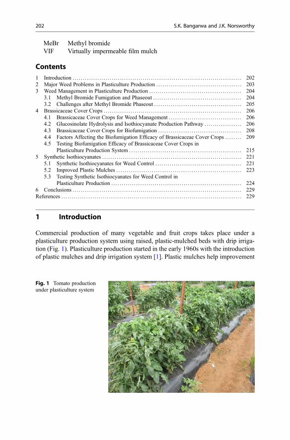
MeBr Methyl bromideVIF Virtually impermeable film mulch
Contents1 Introduction . . . . . . . . . . . . . . . . . . . . . . . . . . . . . . . . . . . . . . . . . . . . . . . . . . . . . . . . . . . . . . . . . . . . . . . . . . . . . . . . . . 2022 Major Weed Problems in Plasticulture Production . . . . . . . . . . . . . . . . . . . . . . . . . . . . . . . . . . . . . . . . . 2033 Weed Management in Plasticulture Production . . . . . . . . . . . . . . . . . . . . . . . . . . . . . . . . . . . . . . . . . . . . 204
3.1 Methyl Bromide Fumigation and Phaseout . . . . . . . . . . . . . . . . . . . . . . . . . . . . . . . . . . . . . . . . . . . 2043.2 Challenges after Methyl Bromide Phaseout . . . . . . . . . . . . . . . . . . . . . . . . . . . . . . . . . . . . . . . . . . 205
4 Brassicaceae Cover Crops . . . . . . . . . . . . . . . . . . . . . . . . . . . . . . . . . . . . . . . . . . . . . . . . . . . . . . . . . . . . . . . . . . . 2064.1 Brassicaceae Cover Crops for Weed Management . . . . . . . . . . . . . . . . . . . . . . . . . . . . . . . . . . . 2064.2 Glucosinolate Hydrolysis and Isothiocyanate Production Pathway . . . . . . . . . . . . . . . . . . 2064.3 Brassicaceae Cover Crops for Biofumigation . . . . . . . . . . . . . . . . . . . . . . . . . . . . . . . . . . . . . . . . 2084.4 Factors Affecting the Biofumigation Efficacy of Brassicaceae Cover Crops . . . . . . . . 2094.5 Testing Biofumigation Efficacy of Brassicaceae Cover Crops in
Plasticulture Production System . . . . . . . . . . . . . . . . . . . . . . . . . . . . . . . . . . . . . . . . . . . . . . . . . . . . . . 2155 Synthetic Isothiocyanates . . . . . . . . . . . . . . . . . . . . . . . . . . . . . . . . . . . . . . . . . . . . . . . . . . . . . . . . . . . . . . . . . . . 221
5.1 Synthetic Isothiocyanates for Weed Control . . . . . . . . . . . . . . . . . . . . . . . . . . . . . . . . . . . . . . . . . . 2215.2 Improved Plastic Mulches . . . . . . . . . . . . . . . . . . . . . . . . . . . . . . . . . . . . . . . . . . . . . . . . . . . . . . . . . . . . 2235.3 Testing Synthetic Isothiocyanates for Weed Control in
Plasticulture Production . . . . . . . . . . . . . . . . . . . . . . . . . . . . . . . . . . . . . . . . . . . . . . . . . . . . . . . . . . . . . . . 2246 Conclusions . . . . . . . . . . . . . . . . . . . . . . . . . . . . . . . . . . . . . . . . . . . . . . . . . . . . . . . . . . . . . . . . . . . . . . . . . . . . . . . . . . 229References . . . . . . . . . . . . . . . . . . . . . . . . . . . . . . . . . . . . . . . . . . . . . . . . . . . . . . . . . . . . . . . . . . . . . . . . . . . . . . . . . . . . . . . 229
1 Introduction
Commercial production of many vegetable and fruit crops takes place under aplasticulture production system using raised, plastic-mulched beds with drip irriga-tion (Fig. 1). Plasticulture production started in the early 1960s with the introductionof plastic mulches and drip irrigation system [1]. Plastic mulches help improvement
Fig. 1 Tomato productionunder plasticulture system
202 S.K. Bangarwa and J.K. Norsworthy
of crop microclimate, crop yield, produce quality, soil and water conservation,efficient nutrient use, pest management, and harvest operation.
Weeds are the major limiting factor in vegetable and fruit production, competingfor resources and harboring harmful crop pests. Besides tolerance issues in high-value vegetable and fruit crops, public concern about herbicidal residues has led tofew herbicide labels in these crops [2]. Further contributing to the limited weedcontrol options in plasticulture production is the phase-out of the highly effective soilfumigant, methyl bromide (MeBr). The loss of MeBr has complicated weed man-agement in plasticulture production, especially difficult-to-control weeds like purple(Cyperus rotundus L.) and yellow nutsedge (Cyperus esculentus L.).
Plants belonging to Brassicaceae family have been tested as an alternative toMeBr because they are known to exhibit allelopathic weed suppression[3–5]. Brassicaceae species synthesize a variety of glucosinolates (GSLs), whichupon tissue decomposition are converted into isothiocyanates (ITCs) [6]. ITCs arebiologically active compounds and have been documented to suppress weeds [7, 8].These ITCs are volatile compounds with biocidal properties and therefore aresuitable for soil biofumigation. However, because weed suppression by Brassicaceaebiofumigant crops is highly variable in field conditions [4, 9, 10], producers tend toprefer chemical alternatives like synthetically produced ITCs.
Synthetically produced ITCs are gaining interest among researchers due to theirlethal activity on several pests. A number of synthetic ITCs are reported to reduce thegermination of several weed species including nutsedge species [11–14]. However,the high volatility of ITCs [7] make them vulnerable to volatilization losses, therebydecreasing weed suppression. One way of reducing the ITC losses and increasingweed efficacy under field conditions is the use of low-permeability plastic mulches.For example, using a virtually impermeable film (VIF) mulch, fumigant rates can bereduced without sacrificing weed control [15]. Most of the previous research workand literature has focused on evaluating synthetic ITCs for weed suppression undergreenhouse or laboratory conditions. However, field research on testing the weedcontrol efficacy of synthetic ITCs is very limited. Therefore, there is interest intesting synthetic ITCs against nutsedge and other problematic weed species, espe-cially in polyethylene-mulched vegetable and fruit crops where MeBr has beenextensively used.
In this chapter, we focus on major weed problems, weed management strategies,and recent advances in our understanding of controlling weeds using Brassicaceaecrops and synthetically produced ITCs in a plasticulture production system based onprevious research findings as well as our research projects conducted in the SouthernUSA.
2 Major Weed Problems in Plasticulture Production
Weeds compete for light, water, nutrients, and space with economically importantcrops. Weeds pose serious threats to vegetable and fruit production by reducing cropyield and quality and interfering with major farm operations, thereby increasing
6 Glucosinolate and Isothiocyanate Production for Weed Control in. . . 203

production costs and resulting in loss of millions of dollars annually [16]. Inplasticulture production, nutsedge species (mainly purple nutsedge and yellownutsedge) are the most problematic weeds because of their perennial life cycle andability to pierce through the plastic mulch due to their sharp pointed tip (Fig. 2)[17–19]. Yield losses of greater than 90% caused by both species in variousvegetable and fruit crops have been well documented [20–24].
The aggressiveness of nutsedge species is mainly associated with their undergroundtubers [25, 26]. These tubers are perennating organs, which store carbohydrates andfacilitate asexual reproduction [27–29]. A single parent tuber of purple nutsedge canproduce 99 tubers in 12 weeks [30]. Similarly, a single tuber of yellow nutsedge canproduce more than 360 tubers within 16 weeks [31]. In addition, nutsedge tubers andfoliage are allelopathic, inhibiting growth of commercial crops [32]. The majority ofpurple nutsedge and yellow nutsedge tubers are found within the top 15 cm of soil [26,33]. The longevity of tubers in the soil is short for both species. Neeser et al. [34]predicated the decay of 99% purple nutsedge tubers in 3.5 years, whereas over 80% ofyellow nutsedge tubers decayed in less than 3 years [35]. Although tuber life is short,even a low density of surviving tubers can rapidly result in a serious weed problem.The above findings indicate that tubers are vital to the success of nutsedge species andthat successful management strategies should concentrate on depleting existing tuberreserves and suppressing new tuber production [36].
3 Weed Management in Plasticulture Production
3.1 Methyl Bromide Fumigation and Phaseout
In a plasticulture production, a preplant fumigant, MeBr, has been widely usedover the last five decades for effective and broad-spectrum pest management,including difficult-to-control weeds like purple and yellow nutsedge [37]. Liquid
Fig. 2 Nutsedge penetrationthrough plastic mulch
204 S.K. Bangarwa and J.K. Norsworthy
MeBr is injected in ground or preformed beds with special knives prior toplanting. It rapidly volatilizes into a gas; therefore, a plastic mulch is placedimmediately over the treated soil to prevent vapor escape and ensure thoroughfumigation of treated soil [38]. However, the standard mulches used are notimpermeable to gases and thereby unable to stop MeBr losses into the environ-ment [39]. Although highly effective, MeBr is listed among the ozone-depletingsubstances in the Montreal Protocol, an international treaty designed to protect theozone layer by phasing out the substances responsible for ozone depletion [40]. Asa result, MeBr is being phased out of USA agriculture since 2005 and is onlyavailable in limited quantity for certain commodities under critical use exemption[41, 42].
3.2 Challenges after Methyl Bromide Phaseout
The phaseout of MeBr has complicated pest problems and negatively affected theUSA vegetable and small fruit production. MeBr is now available in limitedquantity under critical use exemption and therefore USA growers had to signifi-cantly reduce the use of MeBr each year since 2005 [42]. In addition, the restrictedavailability has resulted in increased prices of MeBr, which has increased cost ofcrop production. With the loss of MeBr, vegetable growers are left with few optionsfor controlling many weeds that are capable of reducing yields by more than 90%[20–24]. Purple and yellow nutsedge are among the most troublesome weeds ofplasticulture production in the USA [43] and a major limitation to the adoption ofMeBr alternatives because of the ineffectiveness of current MeBr alternatives onthese weeds.
The currently available fumigants are metam sodium, chloropicrin, and 1,3-dichloropropene. But, these fumigant alternatives may only be short-term replace-ments for MeBr because of narrow spectrum, inconsistent activity especially onnutsedges, uneven distribution in soil profile, and potential issues related to croptolerance and human and environmental safety [37, 44]. Furthermore, chloropic-rin, a leading MeBr alternative, has a stimulatory effect on purple and yellownutsedge emergence [45, 46] that could further exacerbate nutsedge problems andincrease crop yield losses. A number of herbicides have been tested for control ofpurple and yellow nutsedge, but success is limited due to marginal translocationof herbicides to the sites of action and inconsistent control when applied atdifferent stages of growth and under various environmental conditions [47].Most herbicides are registered in agronomic crops, but there are very few optionsin vegetable crops. This is because of high value, small hectarage, and marginalherbicide tolerance of vegetable crops, which in turn increase liability on chem-ical companies [48]. Significant research has been conducted in various locationsthroughout the USA to develop suitable alternatives for MeBr. The researchemphasis has included various nonchemical and chemical strategies and theirsuitable integration.
6 Glucosinolate and Isothiocyanate Production for Weed Control in. . . 205

4 Brassicaceae Cover Crops
4.1 Brassicaceae Cover Crops for Weed Management
Weed management using cover crops has been investigated as a nonchemicalmethod for weed control in vegetable production, especially in the absence ofMeBr. Weed suppression by cover crops is attributed to (1) competition for light,nutrients, and moisture during the cover crop growing cycle and (2) physicalsuppression and allelopathic effect from cover crop residues left on the soil surfaceor incorporated into the soil [49–51].
Brassicaceae cover crops can provide physical weed suppression because of theirfaster canopy development and biomass accumulation early in the season [52,53]. Besides competing for moisture and nutrients, rapid establishment of aBrassicaceae cover crop may result in a high level of nutsedge suppression sinceshoot growth and tuber production in nutsedge species are closely linked to availablephotosynthetically active radiation [54–56]. In plasticulture production,Brassicaceae cover crops can be used to reduce weed infestation before formingbeds and laying plastic mulch. In addition, Brassicaceae cover crop tissues can beincorporated into soil as green manure to provide allelopathic weed suppression afterlaying plastic mulch.
The allelopathic compounds released by Brassicaceae crops has been increas-ingly studied in recent years. These allelopathic compounds, known as ITCs, haveshown allelopathic potential against a number of monocotyledonous and dicotyle-donous plant species [4, 13, 57–59]. These ITCs exhibit non-specific biocidalactivity from interaction with various proteins [60]. As a result, these ITCs arealso effective against other soilborne pests including insects [61–65], nematodes[66–69], and fungi [70–73].
4.2 Glucosinolate Hydrolysis and Isothiocyanate ProductionPathway
Brassicaceae crops synthesize a variety of secondary metabolites known as GSLs intheir cytoplasm and store them in vacuoles. These GSLs are sulfur-rich compoundsand consist of a β-thioglucose moiety, a sulfonated oxime moiety, and a variable sidechain [6]. Brassicaceae plants can synthesize more than 20 different types of GSLs,depending on species and growing conditions [74]. As the structure of side chainvaries, the type of GSL also varies. Depending upon the type of side chain, GSLsproduced by Brassicaceae crops are divided into three major groups: (1) aliphatic,(2) aromatic, and (3) indolyl (Table 1). However, only aliphatic and aromatic GSLsare converted into ITCs upon hydrolysis [75]. GSLs are hydrolyzed by the endog-enous enzyme myrosinase (β-thioglucoside glucohydrolase) to produce ITCs, thio-cyanates, nitriles, epithionitriles, and oxazolidine-2-thiones depending upon side-chain structure and hydrolysis conditions (Scheme 1). GSLs have limited biologicalactivity [76], but ITCs are most phytotoxic among hydrolysis products.
206 S.K. Bangarwa and J.K. Norsworthy

ITCs are a family of compounds composed of nitrogen (N), carbon (C), andsulfur (S), with an alkyl or aryl side chain (R) having the basic structure of R–N =C = S [6]. Plants do not release large amounts of ITCs because GSLs andmyrosinase are physically separated. Therefore, large amounts of ITCs are onlyproduced when plant cells are disrupted and GSLs come in contact with myrosinase
Table 1 Chemical class, common name, chemical name, and variable side-chain structures of mostcommon glucosinolates (GSLs) found in Brassicaceae cropsa
Chemicalclass Common name Chemical name Variable side-chain
Aliphatic GSL
Sinigrin Allyl or 2-Propenyl GSL CH2CHCH2
Gluconapin But-3-enyl GSL CH2CHCH2CH2
Glucobrassicanapin Pent-4-enyl GSL CH2CH(CH2)3Progoitrin 2-Hydroxybut-3-enyl GSL CH2CH(OH)
CHCH2
Gluconapoleiferin 2-Hydroxypent-4-enylGSL
CH2CHCH2(OH)CHCH2
Glucoiberverin 3-Methylthiopropyl GSL CH3S(CH2)3Glucoerucin 4-Methylthiobutyl GSL CH3S(CH2)4Glucoiberin 3-Methylsulfinylpropyl
GSLCH3SO(CH2)3
Glucoraphanin 4-MethylsulfinylbutylGSL
CH3SO(CH2)4
Glucoraphenin 4-MethylsulfinylbutenylGSL
CH3SOCHCH(CH2)2
Glucoalyssin 5-MethylsulfinylpentylGSL
CH3SO(CH2)5
Glucocheirolin 3-MethylsulfonylpropylGSL
CH3SO2(CH2)3
Glucoerysolin 4-MethylsulfonylbutylGSL
CH3SO2(CH2)4
Aromatic GSL
Glucotropaeolin Benzyl GSL C6H5CH2
Gluconasturtiin 2-Phenylethyl GSL C6H5(CH2)2Glucobarbarin 2-Hydroxy-2-phenylethyl
GSLC6H5(OH)CHCH2
Glucosinalbin 4-Hydroxybenzyl GSL 4-OHC6H5CH2
Indole GSL
Glucobrassicin Indol-3-ylmethyl GSL C8H6NCH2
Neoglucobrassicin 1-Methoxyindol-3-ylmethyl GSL
1-(CH3O)C8H5NCH2
4-Hydroxyglucobrassicin 4-Hydroxyindol-3-ylmethyl GSL
4-OHC8H5NCH2
4-Methoxyglucobrassicin 4-Methoxyindol-3-ylmethyl GSL
4-(CH3O)C8H5NCH2
aReferences [38, 74, 82, 86, 87, 92, 95, 105]
6 Glucosinolate and Isothiocyanate Production for Weed Control in. . . 207

[77–79]. Brassicaceae plants utilize these ITCs as a defense mechanism againstattacking herbivores, insects, and pathogens.
4.3 Brassicaceae Cover Crops for Biofumigation
Biofumigation refers to the use of plants containing biologically active volatilecompounds to suppress soilborne pests in agricultural production systems[74]. The biological activity plus volatile nature of ITCs makes them suitable forbiofumigation action [7]. Biofumigation has been successfully implemented in thePacific Northwest of the USA and in Australian production systems and represents apotentially valuable weed management tool for crops [3, 80]. Biofumigation isaccomplished by soil amendment with macerated Brassicaceae tissues or seedmeal. Soil amendment with Brassica species reduced establishment of a widerange of crop and weed species from 23% to 34% and delayed their emergence byapproximately 2 days [81]. Soil amendment with water extracts of Brassica napusshoots inhibited lettuce (Lactuca sativa) seed germination, while root extractsdelayed seed germination [8]. Bell and Muller [78] reported that allelopathic com-pounds released from maceration of black mustard (B. nigra) inhibited the estab-lishment of natural grasses in California. Hemp sesbania (Sesbania herbacea)germination and fresh weight was reduced >95% by chopped leaf tissues offive Brassica species [5]. White mustard (B. hirta)-amended soil reduced weed
Scheme 1 Enzymatic hydrolysis of glucosinolates and production of isothiocyanates and othercompounds
208 S.K. Bangarwa and J.K. Norsworthy

emergence up to 97% in greenhouse trials and provided effective weed suppressionin green pea (Pisum sativum) up to 1 month after incorporation in field trials[57]. Norsworthy et al. [82] reported up to 79% and 48% control of large crabgrass(Digitaria sanguinalis) and Palmer amaranth (Amaranthus palmeri), respectively, at4 weeks after incorporation of Brassicaceae tissues. Yellow nutsedge tuber produc-tion was reduced 71% in wild radish (Raphanus raphanistrum)-amended soil com-pared with no soil amendment [83]. Limited rhizome production was observed inyellow nutsedge tubers when allowed to germinate in wild radish-amended soil[59]. Fall-planted rapeseed, incorporated in the spring, reduced weed density andbiomass by as much as 85% and 96% in potato (Solanum tuberosum),respectively [3].
Green manuring of three Brassicaceae crops (B. juncea, B. napus, and B. hirsuta)reduced total weed biomass by 49% in soybean (Glycine max) at 6 weeks afteremergence [4]. However, some of the weed species were sensitive to a particularBrassica species, while others were not, which indicates the selectivity of these ITCsto certain species [4]. For example, kochia (Kochia scoparia), shepherd’s-purse(Capsella bursa-pastoris), and green foxtail (Setaria viridis) emergences werereduced by all three Brassica species, but redroot pigweed (Amaranthus retroflexus)emergence was reduced by two Brassica species (B. napus and B. hirta), andvelvetleaf (Abutilon theophrasti) emergence was reduced by only one Brassicaspecies (B. hirta). Likewise, the fresh weight of each weed species was affected bythe type of Brassica species [4]. Therefore, testing the biofumigation efficacy ofvarious Brassicaceae cover crops would be helpful to identify the most effectiveBrassicaceae species for biofumigation purposes.
4.4 Factors Affecting the Biofumigation Efficacy of BrassicaceaeCover Crops
Although Brassicaceae crops have potential for weed suppression, the efficacy ofweed suppression via biofumigation is associated with the following factors:(1) biofumigation potential of Brassicaceae crops, (2) GSL to ITC conversionefficiency, (3) intrinsic toxicity of ITCs to target weed species, and (4) concentrationand residence time of ITCs in the amended soil. Let us discuss these factors oneby one.
4.4.1 Biofumigation Potential of Brassicaceae CropsBiofumigation potential of any Brassicaceae crop is linked to the amount of totalGSLs contributed per unit area, which is a function of GSL concentration in plantmaterial and plant biomass per unit area [74, 82]. Glucosinolate type and concen-tration varies in Brassicaceae crops depending upon crop species and variety, plantparts, crop stage, soil and environmental conditions, and management practices.
GSL type and concentration differ among Brassicaceae species and varieties. Forexample, total GSL concentration ranged from 2.4 to 109.9 μmol g�1 dry material inthree Brassica species (B. hirta, B. juncea, and B. oleracea) in a study conducted in
6 Glucosinolate and Isothiocyanate Production for Weed Control in. . . 209

California [84]. B. hirta was high in sinalbin and glucotropaeolin GSLs, whereasB. juncea and B. oleracea were high in sinigrin and glucoraphanin GSL, respec-tively. Likewise, variation in GSL profile was reported in seed meal of three Brassicaspecies (B. napus, B. juncea, and S. alba) and two varieties of B. napus (Athenavs. Sunrise) [85]. However, the GSL variation was relatively less between twovarieties of B. napus.
Field experiments were conducted in Australia to compare the GSL profile of twovarieties (“BQ Mulch,” a commercial biofumigant variety, and “JR049,” a canolaquality variety) of mustard (B. juncea) and two varieties (“Nemflix,” a commercialbiofumigant variety, and “MaximaPlus,” a fodder variety) of winter rape (B. napus)[86]. Results suggested contrasting GSL profiles not only between two Brassicaspecies but also between two varieties within each species. GSL concentration washigher in roots than shoots of winter rape, whereas reverse was true for mustard.Biofumigant variety of winter rape species contributed around 50% ofITC-liberating GSLs in soil, whereas biofumigant variety of mustard contributed100% of ITC-liberating GSLs in soil.
In another study, GSL production was determined in 10 different accessions ofwild radish collected from eight different states of the USA and grown in sameenvironmental conditions [87]. A total of 17 different types of GSLs were identifiedin wild radish accessions, but only four major GSLs (glucoerucin, glucoraphanin,glucobrassicin, and gluconasturtin) contributed more than 90% of total GSLs.Although, total biomass production was not different among wild radish accessions,total GSL concentration ranged from 107.2 μmol plant�1 in the North Carolinaaccession to 618.9 μmol plant�1 in the Mississippi accession. This indicates geneticvariation in GSL production among various wild radish accessions. GSL concentra-tion also varied among various plant parts of wild radish accessions. GSL concen-tration per gram of tissue was highest in primary and secondary branches followedby flowers and then by roots.
A thorough study was conducted in Australia to compare the variation in GSLproduction in roots and shoots of 76 entries from 13 Brassica species/subspeciesgrown in same environment and with same agronomic conditions [74]. GSL con-centration (individual and total) varied four to ten times among and within Brassicaspecies. The type of GSLs present in tissues also varied between species but wasconsistent within species. Roots, shoots, and total biomass varied among Brassicaspecies. Total GSL production per unit area ranged from 0.8 to 45.3 mmol m�2,depending upon total GSL concentration and biomass production of each species.Overall, GSL concentration in roots was either equivalent to or higher than that inshoot tissues. However, roots contributed an average of 23.6% of total GSLs, whichwas directly related to low biomass of root tissues rather than GSL concentration. Ingeneral, shoots produced high biomass than roots in most of Brassica crops;therefore, shoots are considered as major contributor of GSLs per unit area. GSLanalysis also showed that shoot tissues were dominant in aliphatic GSLs, whereasroot tissues were dominant in aromatic GSLs. If we compare the Brassica species, allB. napus, B. campestris, and B. oleracea species contained only 50% of ITCproducing GSLs, whereas B. carinata, B. nigra, B. juncea, B. fruticulosa,
210 S.K. Bangarwa and J.K. Norsworthy

B. tournefortii, S. alba, S. arvensis, Sisymbrium orientale, and Diplotaxis tenuifoliaspecies predominantly contained ITC-liberating GSLs [74]. Therefore, the hugediversity in GSL profiles and GSL contribution per unit area among Brassicaspecies/subspecies/varieties/accessions indicates the importance of careful selectionof a Brassica crop for biofumigation purpose.
GSL concentration in Brassicaceae crops changes with plant growth stage. Forexample, GSL concentration in Brassica species is generally maximum aroundflowering and is declined after flowering and lowest at maturity [75, 87, 88]. Bioas-say study conducted in Australia revealed that biofumigation efficacy of B. rapa/B.napus mixture against soil pathogen increased as plant developmental stageprogressed, with maximum efficacy at flowering stage [89]. This stage is alsoimportant for practical purposes because of high aboveground biomass productionand ease of termination of vegetative tissues and to prevent the Brassica crops fromseeding and becoming weeds in following economically important crop.
The difference in GSL concentrations in Brassicaceae crops is also affected by theenvironmental conditions. For example, spring-planted Brassica species have pro-duced double the amount of GSLs produced from fall-planted Brassica species [90],which could be associated with differences in day length, temperature, rainfall, andlight intensity during growing seasons [91]. Similarly, glucosinolate concentrationvaries between autumn-planted versus spring-planted Brassicaceae crops in theSouthern USA [92]. However, the type of GSLs present did not vary due to plantingdates. In another study conducted in the Southern USA,, total GSL concentrations ina mixture of B. juncea/S. alba varied considerably among three different locations[10]. In addition to environmental conditions, soil conditions such as soil moisture,nutrient status, and biotic factors (insect and diseases) are known to affect the GSLconcentration in Brassicaceae plants [10, 58, 90]. Nutrient deficiency, moisture stressnear flowering stage, and biotic stress like insect/disease attack can increase GSLconcentrations in Brassicaceae crops [91]. However, these stresses can also affect theabove- and belowground biomass production in Brassicaceae crops, which isdirectly linked to biofumigation potential of any Brassicaceae crop.
Crop management practices can also affect the GSL concentration in tissues andGSL contribution per unit area from a Brassicaceae crop. Plant spacing has shownmixed results on GSL concentrations. According to one study, narrow spacing wouldincrease GSL concentration in plant tissues by changing the morphology [93]. Incontrast, an opposite effect of plant spacing was observed on GSL concentration in amixture of B. juncea/S. alba [10]. But, narrow spacing (19-cm wide rows) hasproduced significantly high biomass production than wide spacing (46-cm widerows) [10]. Fertilization is also important for GSL production in Brassicaceae crops.Since GSLs are sulfur-rich compounds, sulfur fertilization would increase the GSLcontent in Brassicaceae tissues. In contrast, nitrogen fertilization at high rates hasdecreased GSL levels in Brassicaceae crops [91]. However, nitrogen fertilization isimportant for high biomass production and in turn high GSL contribution per unitarea. Crop protection practices have not shown any effect on the GSL content ofBrassicaceae crops [91], but they can definitely help in maintaining healthy plantsand maximum biomass production. Therefore, it is important to select management
6 Glucosinolate and Isothiocyanate Production for Weed Control in. . . 211

practices to maintain a balance between GSL concentration in plant tissues andbiomass production to optimize the biofumigation potential of a Brassicaceae crop.
4.4.2 GSL to ITC Conversion EfficiencyUnderstanding the GSL profile and GSL content per unit area of a Brassicaceae cropcan be helpful in predicting the potential ITCs released by Brassicaceae incorpora-tion in the soil [74]. Therefore, high GSL- and biomass-producing Brassicaceaecover crops are expected to produce higher amounts of ITCs in the soil. Since, theactual activity is linked to ITCs [11, 12], it is critical to measure the GSL to ITCconversion efficiency in the Brassicaceae-amended soil. Several of previous studieshave concentrated on measuring GSL profile and content of Brassicaceae crops, butfew studies have attempted assessment of GSL to ITC conversion efficiency. In alaboratory study, the amount and diversity of ITCs released from B. rapa/B. napustissues into soil was less than the potential amount available in biomass [89]. Whenthe same test was conducted in the field, no measurable levels of ITCs were detectedin amended soil [89]. In a field study in Australia, GSL to ITC conversion efficiencyafter incorporation of rape (B. napus) and Indian mustard (B. juncea) ranged from26% to 56% [86]. Low conversion efficiency is believed to be due to incompletehydrolysis of GSLs to ITCs after cover crop incorporation. In same study,un-hydrolyzed GSLs were detected in the soil after Indian mustard incorporation,which constituted up to 13% of total incorporated GSLs. In addition, non-ITCproducing GSLs (indolyl GSLs) were also detected in the soil [86]. It is alsoimportant to remember that not all GSL types present in Brassicaceae crops releaseITCs [75]. For example, some of the Brassicaceae species contained mainly non-ITCproducing GSLs, while other species predominantly contained ITC-liberating GSLs[74]. Also, GSLs can be converted to nitriles instead of ITCs under low pHconditions [94]. In addition, low soil temperatures can slow down myrosinaseenzyme activity and in turn GSL hydrolysis to ITCs [95].
In another field experiment, only 1% or less of the potential GSLs present inIndian mustard (B. juncea) and rapeseed (B. napus) were converted to ITCs[96]. This is because Brassica crops were incorporated into soil without pulveriza-tion. GSLs are physically separated from myrosinase enzyme in a healthy plant cell.Upon cell disruption, GSLs come in contact with myrosinase and release ITCs in thepresence of water. Therefore, complete cell disruption and water are important tomaximize the hydrolysis of GSLs to ITCs. For example, cell disruption by freezingand thawing Indian mustard (B. juncea) leaves increased ITC release efficiency from0.03 to 13.7% [96]. In the same study, ITC production was 1.9-fold higher inamended soil that was waterlogged compared to soil with a water content of�32 kPa [96]. In the field, ITC release can be improved by using a flail mowerwhich can provide thorough maceration of plant tissues before incorporation. Inaddition, the timing of incorporation is also critical to maximize ITC release in soil.ITC concentration in B. napus-amended soil peaked at 30 h after incorporation anddecreased later on, which could be due to losses of released ITCs with time [95]. In abioassay study, the concentration of released ITCs peaked at 4 h after Brassicaincorporation and then declined sharply after 24 h [89]. In a field study in Australia,
212 S.K. Bangarwa and J.K. Norsworthy

GSL to ITC conversion efficiency was maximum immediately after incorporation(56% in B. juncea and 26% in B. napus after 0.5 h) and decreased with time (23% inB. juncea and 10% in B. napus after 6 h) [86]. Similarly, maximum ITC release in thesoil was reported 2 h after incorporation of Brassica tissues in another trial[96]. Hence, the GSL to ITC conversion efficiency in field conditions is affectedby incorporation method and timing, soil conditions, and moisture regime.
4.4.3 Intrinsic Toxicity of ITCs to Target SpeciesThe mode of action of ITCs is not clear to present date, but it is believed that ITCsinteract with glycolysis- and respiration-related enzymes during the germinationprocess and thereby prevent or delay seed germination [79]. In spite of theirbroad-spectrum activity, the degree of toxicity of ITCs varies due to variation inside chain. For example, germination test demonstrated that aromatic ITCs (benzyland 2-phenylethyl ITC) were the most suppressive ITCs [13]. Among alkyl ITCs,herbicidal activity decreased with increased molecular mass [13]. Wolf et al. [97]demonstrated germination inhibition of velvetleaf from benzyl ITC. Another aro-matic ITC, 2-phenylethyl ITCs, inhibited wheat (Triticum aestivum) germination upto 100% [98]. In contrast, other research suggested that aliphatic ITCs are moreeffective than aromatic ITCs [72]. Vaughn and Boydston [5] found aliphatic ITC(methyl, allyl, and 3-butenyl) were effective in suppressing germination of severalspecies, while aromatic ITC (benzyl ITC and 2-phenylethyl) were less inhibitory.Sarwar et al. [72] and Matthiessen and Shackleton [99] concluded that aliphatic ITCsare more volatile and more active in vapor phase while the aromatic ITCs have morecontact toxicity and are more toxic in liquid phase. Petersen et al. [13] reported thatbutyl ITC (aliphatic) was more effective on weeds in vapor phase than liquid phase,while 2-phenylethyl ITC (aromatic) showed opposite effect. Norsworthy andMeehan [12] suggested that aromatic ITCs were more effective than aliphatic ITCson dicot weeds when applied in liquid phase. Regardless of side-chain structure,shorter-chained ITCs are generally more effective than longer-chained ITCs [13, 72,75, 99]. However, the efficacy of ITCs can change dramatically dependent upon soiland environmental conditions as well as agronomic practices. Therefore, it isimportant to select ITC-liberating Brassicaceae crops appropriate to a productionsystem and target species.
In addition, the sensitivity of a target species to a specific ITC varies. Forexample, some of the weed species were sensitive to a particular Brassica species,while others were not, which indicates the selectively of these ITCs to certain species[4]. Norsworthy and Meehan [11, 12] tested eight ITCs on six weed species in agreenhouse and concluded that the effectiveness of ITCs was species specific. Inanother greenhouse study, it was concluded that purple nutsedge is more sensitivethan yellow nutsedge to ITCs [14]. This suggests the importance of considering GSLprofile while selecting a Brassicaceae crop which can deliver ITCs effective on targetspecies. Moreover, seed size of a target species can affect their sensitivity to ITC. Forexample, smaller seeds tended to be more susceptible than larger seeds to methylITC [13]. Yellow nutsedge tubers were more tolerant to ITCs than pittedmorningglory (Ipomoea lacunosa) than Palmar amaranth seeds [12]. Likewise,
6 Glucosinolate and Isothiocyanate Production for Weed Control in. . . 213

sicklepod (Senna obtusifolia) seeds were more tolerant than large crabgrass andTexas panicum (Panicum texanum) [11]. This could be attributed to difference insize of seeds as well as hardness of seed coat among three weed species.
One limitation of Brassicaceae soil amendment could be the phytotoxicity infollowing economically important crops, especially small-seeded crops. For exam-ple, lettuce seed germination was inhibited by 75% when planted within 5 weeks ofsoil amendment with 3% seed meal of S. alba in a growth chamber study [85]. Infield conditions, lettuce emergence was inhibited by planting after 4 weeks of soilamendment with seed meal of B. napus, B. juncea, and S. alba [85]. In contrast,wheat growth was not reduced when planted 4 weeks after Brassicaceae incorpora-tion [100]. In Washington, no injury was observed in potatoes planted around4 weeks after rapeseed (B. napus) green manure [3]. Therefore, there is some speciesselectivity. Transplanted crops have shown better tolerance than seeded crops toITCs. For example, bell pepper (Capsicum annum) injury was less than 5% whenplanted 3 days after soil amended with Brassicaceae crops [82]. In plasticultureproduction, crop injury could be enhanced due to greater retention of toxic ITCs inthe amended soil. Therefore, it is important to establish tolerance level and saferplant-back interval for each production system. For example, no phytotoxicity wasobserved in polyethylene-mulched strawberry when transplanted at least 8 weeksafter incorporation of two Brassicaceae (B. juncea and Eruca sativa) crops [101].
4.4.4 Concentration and Residence Time of ITCs in the Amended SoilLethal concentration of ITC is needed for effective weed control. High ITC concen-trations can completely inhibit seed germination through irreversible reaction withseed enzymes [79]. Large crabgrass seed consistently killed at high concentrations ofmethyl ITC [102]. Norsworthy and Meehan [11, 12] tested eight different ITCs atfive different concentrations (0, 10, 100, 1000, and 10,000 nmole g�1 of soil) on sixweed species in a greenhouse trial. Results indicated that weed emergence wasreduced with increase in ITC concentration and the dose–response relationshipwas depicted by linear, quadratic, and sigmoidal models depending upon ITC typeand weed species [11, 12]. In contrary, low ITC concentrations will not be effectivein killing weed seeds, but low concentration can delay germination by inducingsecondary dormancy in treated seeds. In a greenhouse trial, low ITC concentrationsdelayed germination of smooth pigweed (Amaranthus hybridus), but ungerminatedseeds were viable [13]. In a field trial, low methyl ITC concentrations delayed theemergence of natural weed population by more than 5 weeks [102]. Soil amendmentwith Brassica species delayed emergence of a wide range of crop and weed speciesby approximately 2 days [81]. However, very low concentrations can sometimesstimulate seed germination by breaking dormancy [102]. In a greenhouse trial,emergence of sicklepod, large crabgrass, and Texas panicum was stimulated atsublethal ITC concentrations [11].
In addition to lethal concentration, length of exposure to target species is alsocritical for weed suppression. Longer length of exposure would increase interactiontime between ITC and seed enzymes and thereby would improve the ITC efficacy[12]. One way to increase the length of exposure is to reduce the dissipation of
214 S.K. Bangarwa and J.K. Norsworthy

released ITCs from the treated soil. In general, ITCs have a short life span and ITCsdissipate in environment via volatilization, degradation, and sorption. For example,90% of ITCs dissipated within 24 h of soil treatment [7]. However, the main route ofdissipation is volatilization, especially for short-chained aliphatic ITCs [99]. Forexample, the time for 50% dissipation from soil is 16 h for 2-phenylethyl ITC(aromatic ITC), while it is less than 1 h for n-butyl ITC (aliphatic ITC) [13]. Inlaboratory study, 50% of methyl ITC dissipated in 1–2 days with total loss in 5–7days [102]. Within each ITC group (aliphatic or aromatic), volatility generallydecreases with increase in size of side chain [72, 103]. Hence, low pest efficacyafter cover crop incorporation can been attributed to volatilization losses of ITCsfrom amended soil [104]. Volatilization losses of ITCs can be reduced by watersealing the soil through irrigation or covering the amended soil withlow-permeability plastic mulch.
Besides volatilization losses, soil sorption can reduce active ITC concentrationsin soil possibly due to reaction of ITCs with nucleophilic groups in organic particles[105]. For example, the half-life of allyl ITC ranged from 20 to 60 h in different soiltypes, with enhanced dissipation of allyl ITC from soils with high organic carboncontent [94]. Likewise, the biological activity of various ITCs decreased in thepresence of soil compared to in vitro, with the highest reduction in peat soil followedby loam followed by sandy soil [99]. However, long-chained ITCs are more affectedby soil clay and organic matter compared short-chain ITCs [99]. Microbial degra-dation also plays an important role in ITC dissipation from treated soil. For example,allyl ITC concentration was threefold higher in an autoclaved soil compared tonon-autoclaved soil [106]. Therefore, repeated use of ITCs in same field couldreduce biological activity of ITCs [107, 108]. Hence, several factors can affectbiofumigation efficacy of Brassicaceae crops in amended soil. Although some ofthese factors are in the grower’s control (crop selection, agronomic practices,incorporation method), several other factors related to soil and environmentalconditions are difficult to control. Therefore, it would be a challenging task tounderstand the complex interaction among these factors to achieve optimumbiofumigation efficacy in a field situation.
4.5 Testing Biofumigation Efficacy of Brassicaceae Cover Crops inPlasticulture Production System
Previous studies have tested biofumigation efficacy of Brassicaceae crop either ingreenhouse or field studies in bare ground production system. However, very few ofthe previous research have estimated the biofumigation efficacy of Brassicaceaecrops for weed control in plasticulture production systems. It is expected that thebiofumigation efficacy of Brassicaceae crops can further be improved in aplasticulture system where volatile ITCs are trapped beneath the tarp.
Laboratory and field research were conducted in Arkansas and South Carolinato test the biofumigation efficacy of seven Brassicaceae cover crops in plasticulturetomato (Lycopersicon esculentum) and bell pepper production system [92]. Seven
6 Glucosinolate and Isothiocyanate Production for Weed Control in. . . 215

different Brassicaceae cover crops including Indian mustard (B. juncea) “FumusF-E75” and “Fumus F-L71,” oilseed rape (B. napus) “Humus,” brown mustard(B. juncea) “Southern curled giant,” turnip (B. rapa) “Seventop,” herb cress(Lepidium sativum), and a blend of brown (B. juncea) and white (S. alba) mustard“Caliente” were evaluated for their GSL content and composition, ITC releasedefficiency, and weed efficacy mainly yellow nutsedge. These Brassicaceae species/varieties were selected because of their proven biofumigant action or high GSLcontent or ease of availability. All the cover crops were drill-seeded in fall ofprevious year (2006) or early spring of same year (2007). At mid to late floweringstage in May, all the cover crops were flail mowed (to maximize maceration) andincorporated into the top 7.5 cm of the soil profile using a rototiller (Fig. 3).Immediately after soil incorporation, raised beds were formed and covered withplastic mulch followed by tomato and bell pepper transplanting 1 week later(Fig. 3). GSL analysis demonstrated considerable variation in GSL content andcomposition, depending upon Brassicaceae species/variety, plant parts (shootvs. roots), and production year/location (Table 2). Six different GSLs were detectedin Brassicaceae shoots and roots including three aliphatic GSLs [(2R)-hydroxybut-3-enyl, 2-propenyl, and but-3-enyl] and three aromatic GSLs ( p-hydroxybenzyl,benzyl, and 2-phenylethyl). Total GSLs contributed to the soil by incorporation ofBrassicaceae cover crop tissues were predicted between 47 and 452 nmol g�1 soiland were mainly contributed by the shoots because of a higher proportion of shootbiomass (83 to 93%) than root biomass in all cover crops. From the soil samples,five different ITCs were detected including phenyl, 2-propenyl, propyl, benzyl, and2-phenylethyl ITC. Although the soil samples were taken 3 h to 14 days afterincorporation, the highest ITC concentration (1.6 to 28.4 nmol g�1 soil) wasdetected at 3 h after incorporation, and ITC concentration decreased at latertimings. Similar to previous research, GSL to ITC conversion efficiency was lowand ranged from 1% to 39%, with variation among cover crops and between years(Table 3). Hence, despite of using proper agronomic practices and terminationmethod (flail mower followed by rototiller), ITC release efficiency was low in thistrial. Tomato and bell pepper transplanted 1 week after Brassicaceae incorporationappeared tolerant to ITCs released in beds [92]. However, soil amended with theseBrassicaceae cover crops provided marginal early-season weed (yellow nutsedge)control in plasticulture tomato and bell pepper (Table 4), which could be attributedto the low soil ITC concentration following Brassica incorporation. For example,in the above research, Brassicaceae cover crops can potentially contribute amaximum of 452 nmol g�1 soil total GSLs to the soil. Also, a maximum of 39%conversion of 2-propenyl GSL to respective ITC was achieved in the present study.Assuming a conversion efficiency of 39% for total GSLs, it was estimated that amaximum of 176 nmol g�1 of total ITCs was released in the soil by incorporationof Brassicaceae cover crops. When applied at a label application rate of 358 kg ha�1
with an incorporation depth of 7.5 cm, synthetic fumigant metam sodium canproduce up to 2250 nmol g�1 of methyl ITC (assuming 90% conversion and soilbulk density of 1.48 g cm�3), which is 13 times the molar equivalent of themaximum ITCs produced by Brassicaceae crops in the present study
216 S.K. Bangarwa and J.K. Norsworthy

[92]. Hence, the amount of ITCs produced by Brassicaceae was not sufficient toprovide effective weed control.
In recent years, we have conducted several studies in the Southern USA to test cropsafety and weed efficacy of various biofumigant Brassicaceae crops either alone or incombination with herbicides against nutsedge and other weeds in plasticulture tomatoand bell pepper production [109–112]. Although there was no crop injury, weed
Fig. 3 Brassicaceae cover crop mowing and incorporation followed by plastic laying andtransplanting
6 Glucosinolate and Isothiocyanate Production for Weed Control in. . . 217
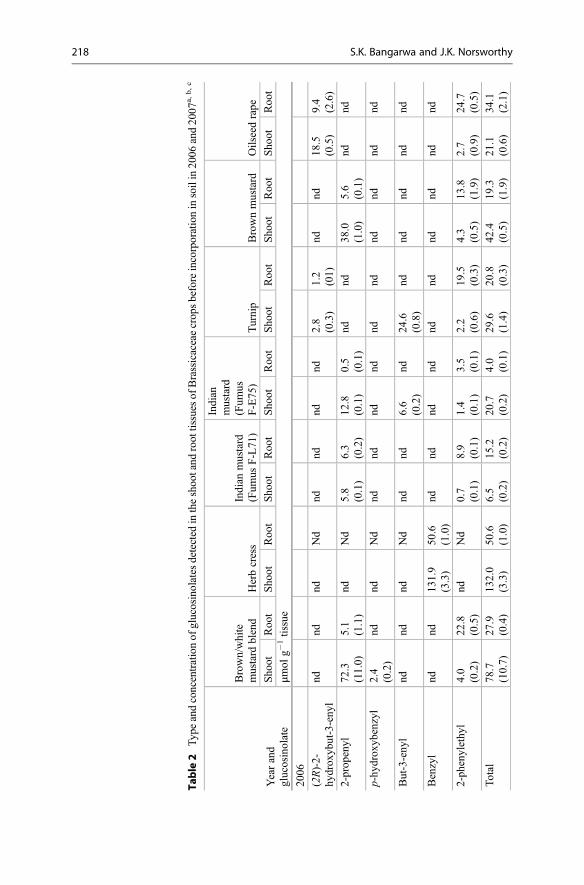
Table
2Typeandconcentrationof
glucosinolates
detected
intheshootand
roottissues
ofBrassicaceaecropsbeforeincorporationinsoilin2006
and2007
a,b,c
Yearand
glucosinolate
Brown/white
mustard
blend
Herbcress
Indian
mustard
(Fum
usF-L71
)
Indian
mustard
(Fum
usF-E75
)Turnip
Brownmustard
Oilseedrape
Sho
otRoo
tSho
otRoo
tSho
otRoo
tSho
otRoo
tSho
otRoo
tSho
otRoo
tSho
otRoo
t
μmol
g�1tissue
2006
(2R)-2-
hydrox
ybut-3-eny
lnd
ndnd
Nd
ndnd
ndnd
2.8
(0.3)
1.2
(01)
ndnd
18.5
(0.5)
9.4
(2.6)
2-prop
enyl
72.3
(11.0)
5.1
(1.1)
ndNd
5.8
(0.1)
6.3
(0.2)
12.8
(0.1)
0.5
(0.1)
ndnd
38.0
(1.0)
5.6
(0.1)
ndnd
p-hy
drox
ybenzyl
2.4
(0.2)
ndnd
Nd
ndnd
ndnd
ndnd
ndnd
ndnd
But-3-eny
lnd
ndnd
Nd
ndnd
6.6
(0.2)
nd24
.6(0.8)
ndnd
ndnd
nd
Benzyl
ndnd
131.9
(3.3)
50.6
(1.0)
ndnd
ndnd
ndnd
ndnd
ndnd
2-ph
enylethy
l4.0
(0.2)
22.8
(0.5)
ndNd
0.7
(0.1)
8.9
(0.1)
1.4
(0.1)
3.5
(0.1)
2.2
(0.6)
19.5
(0.3)
4.3
(0.5)
13.8
(1.9)
2.7
(0.9)
24.7
(0.5)
Total
78.7
(10.7)
27.9
(0.4)
132.0
(3.3)
50.6
(1.0)
6.5
(0.2)
15.2
(0.2)
20.7
(0.2)
4.0
(0.1)
29.6
(1.4)
20.8
(0.3)
42.4
(0.5)
19.3
(1.9)
21.1
(0.6)
34.1
(2.1)
218 S.K. Bangarwa and J.K. Norsworthy

2007
(2R)-2-
hydrox
ybut-3-eny
lnd
ndnd
ndnd
ndnd
nd2.0
(0.1)
2.7
(0.1)
ndnd
11.7
(0.1)
14.4
(0.2)
2-prop
enyl
12.7
(0.1)
ndnd
nd31
.6(1.5)
11.2
(0.1)
13.6
(0.4)
ndnd
nd59
.4(1.1)
ndnd
nd
p-hy
drox
ybenzyl
67.9
(0.3)
ndnd
ndnd
ndnd
ndnd
ndnd
ndnd
nd
But-3-eny
lnd
ndnd
nd13
.2(0.3)
nd23
.0(0.4)
nd21
.4(11.3)
4.7
(0.2)
ndnd
3.6
(0.1)
2.9
(0.1)
Benzyl
4.7
(0.3)
nd13
1.1
(3.0)
12.7
(0.3)
ndnd
ndnd
ndnd
ndnd
ndnd
2-ph
enylethy
l1.9
(0.4)
7.1
(0.5)
ndnd
5.8
(0.2)
60.4
(2.5)
6.1
(0.2)
3.4
(0.5)
2.3
(0.2)
35.3
(1.9)
5.1
(1.1)
0.5
(0.1)
1.1
(0.1)
51.6
(2.6)
Total
87.1
(0.4)
7.1
(0.5)
131.2
(2.9)
12.7
(0.3)
50.7
(1.5)
71.6
(2.5)
42.7
(1.1)
3.5
(0.5)
25.7
(11.3)
42.7
(2.2)
64.5
(2.0)
0.5
(0.1)
16.4
(0.1)
68.9
(2.7)
a Stand
arderrorof
each
meanisinclud
edin
parenthesis
bnd
non-detected
c Reference
[92]
6 Glucosinolate and Isothiocyanate Production for Weed Control in. . . 219
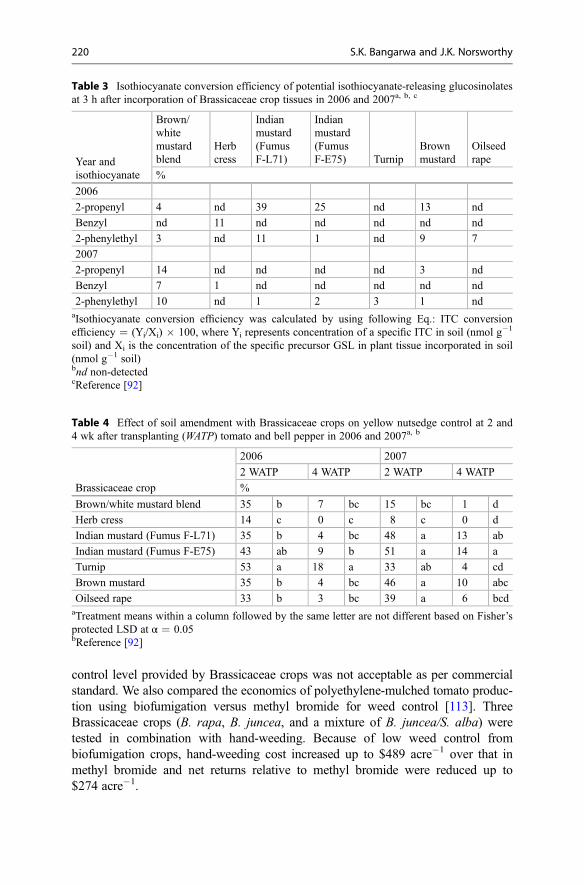
control level provided by Brassicaceae crops was not acceptable as per commercialstandard. We also compared the economics of polyethylene-mulched tomato produc-tion using biofumigation versus methyl bromide for weed control [113]. ThreeBrassicaceae crops (B. rapa, B. juncea, and a mixture of B. juncea/S. alba) weretested in combination with hand-weeding. Because of low weed control frombiofumigation crops, hand-weeding cost increased up to $489 acre�1 over that inmethyl bromide and net returns relative to methyl bromide were reduced up to$274 acre�1.
Table 3 Isothiocyanate conversion efficiency of potential isothiocyanate-releasing glucosinolatesat 3 h after incorporation of Brassicaceae crop tissues in 2006 and 2007a, b, c
Year andisothiocyanate
Brown/whitemustardblend
Herbcress
Indianmustard(FumusF-L71)
Indianmustard(FumusF-E75) Turnip
Brownmustard
Oilseedrape
%
2006
2-propenyl 4 nd 39 25 nd 13 nd
Benzyl nd 11 nd nd nd nd nd
2-phenylethyl 3 nd 11 1 nd 9 7
2007
2-propenyl 14 nd nd nd nd 3 nd
Benzyl 7 1 nd nd nd nd nd
2-phenylethyl 10 nd 1 2 3 1 ndaIsothiocyanate conversion efficiency was calculated by using following Eq.: ITC conversionefficiency = (Yi/Xi) � 100, where Yi represents concentration of a specific ITC in soil (nmol g�1
soil) and Xi is the concentration of the specific precursor GSL in plant tissue incorporated in soil(nmol g�1 soil)bnd non-detectedcReference [92]
Table 4 Effect of soil amendment with Brassicaceae crops on yellow nutsedge control at 2 and4 wk after transplanting (WATP) tomato and bell pepper in 2006 and 2007a, b
Brassicaceae crop
2006 2007
2 WATP 4 WATP 2 WATP 4 WATP
%
Brown/white mustard blend 35 b 7 bc 15 bc 1 d
Herb cress 14 c 0 c 8 c 0 d
Indian mustard (Fumus F-L71) 35 b 4 bc 48 a 13 ab
Indian mustard (Fumus F-E75) 43 ab 9 b 51 a 14 a
Turnip 53 a 18 a 33 ab 4 cd
Brown mustard 35 b 4 bc 46 a 10 abc
Oilseed rape 33 b 3 bc 39 a 6 bcdaTreatment means within a column followed by the same letter are not different based on Fisher’sprotected LSD at α = 0.05bReference [92]
220 S.K. Bangarwa and J.K. Norsworthy

The above field research demonstrates that biofumigation with Brassicaceaecrops could not be used alone for effective weed control in commercial plasticultureproduction system. Even under non-plasticulture production, biofumigation may notbe useful under high weed pressure and in fields with difficult to control weeds likenutsedge species. However, they may still be useful in organic and low-inputproduction systems where weed pressure is low and weed management tools areeither limited or costly. In organic bell pepper production, soil amendment withBrassica species provided early-season control of large crabgrass and Palmer ama-ranth [82]. Farmers can select high GSL-producing varieties of Brassicaceae crops asa component of an integrated weed management program where early-season weedsuppression by Brassicaceae crops can be supplemented by other weed controlstrategies later in season. For example, wild radish cover crop supplemented largecrabgrass control in sweet corn (Zea mays) when used in conjunction with half ratesof herbicides which was equivalent to full herbicide rates in the absence of a covercrop [114]. Likewise, soil amendment with Brassica tissues supplemented theherbicidal control of Johnsongrass (Sorghum halepense) [115]. Other benefits ofBrassicaceae cover crops, such as nutrient recycling, erosion control, moistureconservation, soil health improvement, and insect, nematode, and disease suppres-sion, support a practical application of Brassicaceae cover crops in any productionsystem [56, 116–120]. Australian wheat farmers have observed more yield followedby Brassica crops [121]. In Washington, greater tuber yield was observed in potatoesplanted after rapeseed (B. napus) green manure due to factors other than weedcontrol [3]. Similarly, bell pepper yield was increased after soil amendment withBrassicaceae cover crops in organic bell pepper production in the SouthernUSA [82].
Although Brassicaceae crops have potential for weed suppression, the magnitudeof weed suppression is either low or variable under field conditions [4, 9, 10], whichindicates that biofumigation with Brassicaceae crops alone does not have enoughpotential to replace MeBr. For example, biofumigation with Brassicaceae crops indisease-infected soil led to strawberry (Fragaria � ananassa) yield higher thanuntreated soil, but lower than MeBr fumigation [101]. The low and inconsistentweed and pest control may lead farmers to shift more toward chemical alternativessuch as synthetically produced ITCs.
5 Synthetic Isothiocyanates
5.1 Synthetic Isothiocyanates for Weed Control
Synthetically produced ITCs are gaining interest as a MeBr alternative due to theirbroad-spectrum lethal activity, volatile nature, and shorter persistence in soil com-pared to other alternatives. A number of synthetic ITCs have been reported to reducethe germination of several weed species including nutsedge species (Table 5).Norsworthy and Meehan [11, 12] found effective control of yellow nutsedge andother grass and broadleaf species with eight synthetic ITCs. However, the
6 Glucosinolate and Isothiocyanate Production for Weed Control in. . . 221

Table 5 Herbicidal activity of different isothiocyanates (ITCs) on weeds in greenhouse andlaboratory trialsa
Chemical name of ITC Weeds inhibited
Methyl ITC Dandelion (Taraxacum officinale), large crabgrass (Digitariasanguinalis), smooth pigweed (Amaranthus hybridus), barnyardgrass (Echinochloa crus-galli), spiny sow thistle (Sonchus asper),scentless mayweed (Matricaria inodora), black grass (Alopecurusmyosuroides)
Ethyl ITC Yellow nutsedge (Cyperus esculentus), Palmer amaranth(Amaranthus palmeri), pitted morningglory (Ipomoea lacunosa),sicklepod (Senna obtusifolia), Texas panicum (Panicum texanum),large crabgrass (Digitaria sanguinalis)
Propyl ITC Yellow nutsedge (Cyperus esculentus), Palmer amaranth(Amaranthus palmeri), pitted morningglory (Ipomoea lacunosa),sicklepod (Senna obtusifolia), Texas panicum (Panicum texanum),large crabgrass (Digitaria sanguinalis)
Butyl ITC Dandelion (Taraxacum officinale), yellow nutsedge (Cyperusesculentus), Palmer amaranth (Amaranthus palmeri), pittedmorningglory (Ipomoea lacunosa), sicklepod (Senna obtusifolia),Texas panicum (Panicum texanum), large crabgrass (Digitariasanguinalis))
Allyl or 2-Propenyl ITC Dandelion (Taraxacum officinale), purple nutsedge (Cyperusrotundus), yellow nutsedge (Cyperus esculentus), Palmer amaranth(Amaranthus palmeri), pitted morningglory (Ipomoea lacunosa),sicklepod (Senna obtusifolia), Texas panicum (Panicum texanum),large crabgrass (Digitaria sanguinalis)
3-Methylthiopropyl ITC Yellow nutsedge (Cyperus esculentus), Palmer amaranth(Amaranthus palmeri), pitted morningglory (Ipomoea lacunosa),sicklepod (Senna obtusifolia), Texas panicum (Panicum texanum),large crabgrass (Digitaria sanguinalis)
Phenyl ITC Purple nutsedge (Cyperus rotundus), yellow nutsedge (Cyperusesculentus), Palmer amaranth (Amaranthus palmeri), pittedmorningglory (Ipomoea lacunosa), sicklepod (Senna obtusifolia),Texas panicum (Panicum texanum), large crabgrass (Digitariasanguinalis)
Benzyl ITC Dandelion (Taraxacum officinale), yellow nutsedge (Cyperusesculentus), Palmer amaranth (Amaranthus palmeri), pittedmorningglory (Ipomoea lacunosa), sicklepod (Senna obtusifolia),Texas panicum (Panicum texanum), large crabgrass (Digitariasanguinalis), velvetleaf (Abutilon theophrasti)
2-Phenylethyl ITC Dandelion (Taraxacum officinale), yellow nutsedge (Cyperusesculentus), Palmer amaranth (Amaranthus palmeri), pittedmorningglory (Ipomoea lacunosa), sicklepod (Senna obtusifolia),Texas panicum (Panicum texanum), large crabgrass (Digitariasanguinalis)
Benzoyl ITC Purple nutsedge (Cyperus rotundus), yellow nutsedge (Cyperusesculentus)
o-Tolyl ITC Purple nutsedge (Cyperus rotundus), yellow nutsedge (Cyperusesculentus)
(continued)
222 S.K. Bangarwa and J.K. Norsworthy

effectiveness of ITCs was dependent upon ITC type, concentration, and weedspecies. ITCs reduced yellow nutsedge emergence and shoot density and biomassin purple and yellow nutsedge [12, 14]. Another notable characteristic of ITCs istheir effectiveness in killing dormant weed seeds [102]. This characteristic would beadvantageous under plasticulture production, particularly in depleting dormant nut-sedge tuber reserves in soil. Although the above-mentioned studies were conductedunder greenhouse conditions, they indicate the potential of synthetic ITCs fornutsedge suppression. Limited research has been conducted to evaluate the efficacyof synthetic ITCs, other than methyl, against weeds under field conditions. Thevolatile nature of ITCs makes them vulnerable to volatilization losses in the field, butaids the fumigation effect in plasticulture production. One way of minimizing thelosses of ITC in field condition is by using less permeable plastic mulches.
5.2 Improved Plastic Mulches
The commonly used plastic mulch in vegetable production is low-density polyeth-ylene (LDPE) mulch (Fig. 4a). The low cost and high tensile strength make thismulch popular among growers. However, the LDPE mulches are permeable to gasesand thereby not effective in reducing fumigant losses from treated soil. This problemcan be overcome by using VIF (Fig. 4b), which is 75 times less permeable to MeBrthan LDPE mulch when tested under laboratory conditions [122]. This reducedpermeability is associated with a polymer barrier, ethylene vinyl alcohol, or poly-amide that is incorporated into the center of the VIF mulch. In previous research, VIFmulch reduced methyl bromide emission >40% over an LDPE mulch [123].Concentration of MeBr + chloropicrin under VIF was 6 times higher than underhigh-density polyethylene (HDPE) mulch at 5 days after treatment [124]. Likewise,1,3-dichloropropene and chloropicrin emissions from VIF mulch were reducedby >90% over standard mulch at 24 h after application [125].
Several experiments have been conducted over the past few years to demonstratethat fumigant-use rates can be lowered when using VIF mulch. For example, VIFmulch provided nutsedge control with one-half of the commercially used rate ofMeBr + chloropicrin [124]. In another study, MeBr and 1,3-dichloropropene rateneeded for effective disease suppression in strawberry was reduced by 50% under
Table 5 (continued)
Chemical name of ITC Weeds inhibited
m-Tolyl ITC Purple nutsedge (Cyperus rotundus), yellow nutsedge (Cyperusesculentus)
tert-octyl ITC Purple nutsedge (Cyperus rotundus), yellow nutsedge (Cyperusesculentus)
3-Fluorophenyl ITC Purple nutsedge (Cyperus rotundus), yellow nutsedge (Cyperusesculentus)
aReferences [5, 11–14, 97, 102, 130, 131]
6 Glucosinolate and Isothiocyanate Production for Weed Control in. . . 223
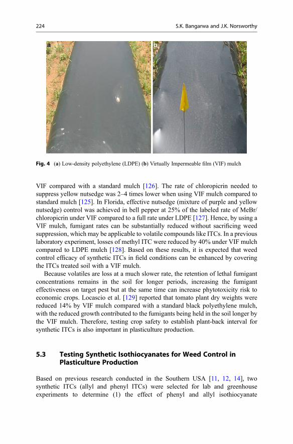
VIF compared with a standard mulch [126]. The rate of chloropicrin needed tosuppress yellow nutsedge was 2–4 times lower when using VIF mulch compared tostandard mulch [125]. In Florida, effective nutsedge (mixture of purple and yellownutsedge) control was achieved in bell pepper at 25% of the labeled rate of MeBr/chloropicrin under VIF compared to a full rate under LDPE [127]. Hence, by using aVIF mulch, fumigant rates can be substantially reduced without sacrificing weedsuppression, which may be applicable to volatile compounds like ITCs. In a previouslaboratory experiment, losses of methyl ITC were reduced by 40% under VIF mulchcompared to LDPE mulch [128]. Based on these results, it is expected that weedcontrol efficacy of synthetic ITCs in field conditions can be enhanced by coveringthe ITCs treated soil with a VIF mulch.
Because volatiles are loss at a much slower rate, the retention of lethal fumigantconcentrations remains in the soil for longer periods, increasing the fumiganteffectiveness on target pest but at the same time can increase phytotoxicity risk toeconomic crops. Locascio et al. [129] reported that tomato plant dry weights werereduced 14% by VIF mulch compared with a standard black polyethylene mulch,with the reduced growth contributed to the fumigants being held in the soil longer bythe VIF mulch. Therefore, testing crop safety to establish plant-back interval forsynthetic ITCs is also important in plasticulture production.
5.3 Testing Synthetic Isothiocyanates for Weed Control inPlasticulture Production
Based on previous research conducted in the Southern USA [11, 12, 14], twosynthetic ITCs (allyl and phenyl ITCs) were selected for lab and greenhouseexperiments to determine (1) the effect of phenyl and allyl isothiocyanate
Fig. 4 (a) Low-density polyethylene (LDPE) (b) Virtually Impermeable film (VIF) mulch
224 S.K. Bangarwa and J.K. Norsworthy

concentration and exposure period on nutsedge tuber viability and (2) to compare theretention of these ITCs in treated soil under LDPE and VIF mulches [130,131]. Greenhouse trial results proved that high rates of phenyl ITCs in sealedenvironment can reduce purple nutsedge tuber viability by 97% [130]. Lab trialsshowed that phenyl isothiocyanate dissipation was lower in soil covered with VIFthan with LDPE mulch and half-life of phenyl ITC under LDPE and VIF mulch was6.09 and 8.88 days, respectively [130]. Similar results were observed for allyl ITC inanother greenhouse study, but mulch effect was not very strong (half-life of0.15 days under LDPE mulch vs. 0.59 days under VIF mulch) [131].
We hypothesized that using higher concentration of synthetic ITCs in combina-tion with VIF plastic mulch can provide weed control equivalent to MeBr in fieldconditions. This hypothesis led to the field testing of allyl and phenyl ITCs underLDPE vs. VIF mulch for weed control (yellow nutsedge, Palmer amaranth, and largecrabgrass) in plasticulture vegetable production in Arkansas and South Carolinafrom 2006–2009 [132–135]. This research provides some of the first information onfield-level efficacy of synthetic ITCs (other than methyl ITC) for weed managementin plasticulture vegetable production. ITCs were applied from 0 to 1500 kg ha�1 raterange and then compared with a standard rate of MeBr [MeBr/chloropicrin (67/33%)at 390 kg ha�1] under LDPE mulch. Liquid ITCs were sprayed on the soil surfaceand immediately incorporated into the soil by using a rototiller before forming bedsand covering them with plastic mulches (LDPE vs. VIF) (Fig. 5). Tomato and bellpeppers were planted 3 weeks after ITCs or MeBr application. Please note that holeswere punched in the plastic 48 h before transplanting to aerate the beds and reducethe crop injury from residual ITCs. Between two synthetic ITCs tested, allyl ITC wasmore effective on weeds and safer on vegetable transplants than phenyl ITC[132–135]. Therefore, we will focus only on allyl ITC results. It is concluded thatcrop injury from allyl ITC under LDPE or VIF mulch can be minimized to accept-able levels by using a plant-back interval of 3 weeks and aerating the beds for 48 hbefore transplanting [132, 133]. Weed control of all species increased with anincrease in allyl ITC rates and this response was described by sigmoidal model(Fig. 6). However, yellow nutsedge was the most tolerant to allyl ITC among thethree weed species tested in the present study (Table 6). Large crabgrass wasintermediate in sensitivity to allyl ITC, whereas Palmer amaranth was least tolerantto allyl ITC among the three species. The less sensitivity of yellow nutsedge to allylITC could be associated with the larger size of tubers compared to the small-seededPalmer amaranth or large crabgrass [12, 13]. Results also proved that allyl ITC cansuccessfully reduce the viable weed propagule density in the bedded soil [132, 133],which will reduce the weed problem in succeeding crop. This will be more valuablefor producers growing more than one crop on the same polyethylene-mulchedbeds [136].
Contrary to our hypothesis, VIF mulch did not improve the weed control fromallyl or phenyl ITC over LDPE mulch, so VIF mulch had no added advantage overLDPE mulch [132–135]. This could be due to little to no differential retention ofthese ITCs under LDPE and VIF mulches. In contrast, we reported higher retentionof phenyl and allyl ITC under VIF than LDPE mulch in previous indoor experiments
6 Glucosinolate and Isothiocyanate Production for Weed Control in. . . 225
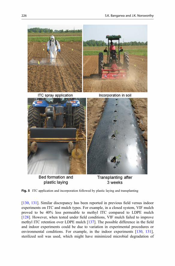
[130, 131]. Similar discrepancy has been reported in previous field versus indoorexperiments on ITC and mulch types. For example, in a closed system, VIF mulchproved to be 40% less permeable to methyl ITC compared to LDPE mulch[128]. However, when tested under field conditions, VIF mulch failed to improvemethyl ITC retention over LDPE mulch [137]. The possible difference in the fieldand indoor experiments could be due to variation in experimental procedures orenvironmental conditions. For example, in the indoor experiments [130, 131],sterilized soil was used, which might have minimized microbial degradation of
Fig. 5 ITC application and incorporation followed by plastic laying and transplanting
226 S.K. Bangarwa and J.K. Norsworthy

ITC compared to field conditions. It is also suspected that the permeability of VIFmulches can change, depending upon changes in the physical and environmentalconditions to which they are exposed [138]. VIF is less flexible than LDPE mulch[139]; therefore, excessive stretching during laying in the field can cause breakdownof the polyamide layer and increase the permeability of VIF mulch to volatile
00
20
40
60
80W
eed
cont
rol (
%)
100
1 2 3
Large crabgrass
Palmer amaranth
Yellow nutsedge
Allyl ITC rate [ln(kg/ha)+1]4 5 6 7 8
Fig. 6 Observed and predicted control of yellow nutsedge, Palmer amaranth, and large crabgrass at6 weeks after transplanting as a function of allyl ITC rate. Symbols and solid line represent meansand predicted response, respectively. The corresponding regression model and individual parame-ters are presented in Table 6 [132]
Table 6 Parameter estimates for regression model of yellow nutsedge, Palmer amaranth, and largecrabgrass control at 6 wk after transplanting, averaged over mulch type (LDPE and VIF) and years(2007 and 2009), and allyl ITC rate needed to obtain weed control equivalent to methyl bromidea, b
Weed species
Parameters estimates (� SE)c
LRmb (� SE)dD C b I
Kg ha�1
Yellow nutsedge 3.97 (1.28) 99.27 (3.26) 2.10 (0.21) 5.77 (0.09) 924 (74)
Palmer amaranth 1.80 (1.69) 99.90 (2.74) 2.44 (0.45) 5.36 (0.09) 888 (225)
Large crabgrass 4.13 (1.59) 99.08 (3.06) 2.65 (0.51) 5.53 (0.12) 651 (118)aAbbreviations: LDPE low-density polyethylene mulch, VIF virtually impermeable filmbReference [132]cRegression model used to predict percent control of each weed species is represented by followingEq.: Y = C + (D – C)/[1 + exp{b(ln x – I)}, where Y represents the percentage control of eachspecies, C is the upper asymptote, D is the lower asymptote, b is the slope of the response, x is the1 + allyl ITC rate (kg ha�1), and I is the natural log transformed allyl ITC rate needed to provide50 % control. SE = approx. standard error of the meandLRmb = allyl ITC rate required to provide percent control of a particular weed species equivalentto methyl bromide. SE = approx. standard error of the mean
6 Glucosinolate and Isothiocyanate Production for Weed Control in. . . 227

substances. Another plausible reason of no effect of mulch type with allyl or phenylITC in our study [132–135] could be related to volatility of ITCs. In contrary to allylor phenyl ITC, VIF mulch has been proved to improve MeBr efficacy over LDPEmulch even under field conditions [124, 127]. This could be attributed to lowervolatility and weaker fumigant activity of allyl (vapor pressure = 0.53 kPa at 20 C)or phenyl ITC (vapor pressure = 0.19 kPa at 20 C) compared to MeBr (vaporpressure = 188 kPa at 20 C), and that is why no clear benefit of low-permeabilityplastic mulch (VIF) was realized in ITC field studies [132–135]. Based on ourresearch, we concluded that regardless of mulch type, allyl ITC at high rates canprovide effective, broad-spectrum weed control (Table 6, Fig. 7) and maintain fruityield equivalent to MeBr [132, 133]. Although, allyl ITC rates are at least 2.4 timesgreater than current fumigants [132, 133] (Table 6), the more consistent season-longweed control provided by allyl ITC could compensate for the higher application rate.However, additional testing is suggested to refine ITC rates and application tech-niques not only to improve the production efficiency but also to minimize input costand human/environmental risk.
Therefore, synthetic ITCs have shown promising results in plasticulture produc-tion system. Numerous ITCs have been identified, but the biological activity of veryfew ITCs has been examined under field conditions. Therefore, field testing ofvarious synthetic ITCs would provide a better understanding for the potential fit ofthese compounds in a plasticulture production system. The knowledge of differentialactivity of ITCs in liquid or vapor phase will be critical for selection of any ITC in aproduction system. For example, short-chain aliphatic ITCs are more active in vaporphase [99] and therefore suitable for fumigation. Conversely, the long-chainedaromatic ITCs have low fumigation potential due to their low volatile nature, butthese ITCs are more toxic in liquid phase [99]. Furthermore, soil, environmental, andmanagement factors should be considered during field testing of ITCs as they candramatically change ITC efficacy. Moreover, to be eligible as fumigant/pesticide,any ITC needs to undergo extensive testing, including toxicology and environmentalfate studies and worker exposure risk assessments.
Fig. 7 Yellow nutsedgecontrol in bell pepper fromallyl ITC at 6 weeks aftertransplanting
228 S.K. Bangarwa and J.K. Norsworthy

6 Conclusions
Weed control is challenging in plasticulture production systems in the absence of thepreplant soil fumigant, MeBr. Plants belonging to Brassicaceae family have theability to release herbicidal compounds (ITCs) in soil after tissue maceration andsoil incorporation and therefore have been suggested as biofumigant crops. How-ever, the amount of ITCs released in soil is affected by several factors like plantgenotype, ontogeny, and morphology, soil and environmental conditions, and agro-nomic and incorporation practices. Under field conditions, the weed control pro-vided by incorporation of Brassicaceae crops in commercial plasticulture productionis not sufficient for growers to rely exclusively on biofumigation. The ineffective-ness of Brassicaceae crops in weed control can be associated with low ITC concen-tration in soil after incorporation. This could be mainly attributed to low productionof GSLs (ITC precursors) by Brassicaceae cover crops, poor GSL to ITC conversionefficiency after tissue incorporation in soil, and loss of released ITCs in treated soil.Therefore, Brassicaceae crops can aid weed management but will not provideeffective, season-long weed control required in a commercial plasticulture produc-tion. Conversely, synthetically produced ITCs at higher rates than produced byplants under natural conditions does have potential for supplying a high level ofweed control. Among synthetic ITCs, allyl ITC proved to be more effective onweeds and comparatively safer than phenyl ITC in plasticulture vegetable produc-tion. However, there does not appear to be any advantage of VIF mulch over LDPEin improving weed control from allyl and phenyl ITCs in the field. Further research isneeded to screen other potential synthetic ITCs and optimize their use rates andapplication techniques to maximize their herbicidal activity in crops produced in aplasticulture system.
References
1. Schrader WL (2000) Plasticulture in California vegetable production. http://anrcatalog.ucanr.edu/pdf/8016.pdf. Accessed 20 Dec 2015
2. Donald WW (2000) Between-row mowing + in-row band-applied herbicides for weed controlin Glycine max. Weed Sci 48:487–500
3. Boydston RA, Hang A (1995) Rapeseed (Brassica napus) green manure crop suppressesweeds in potato (Solanum tuberosum). Weed Technol 9:669–675
4. Krishnan G, Holshouser DL, Nissen SJ (1998) Weed control in soybean (Glycine max) withgreen manure crops. Weed Technol 12:97–112
5. Vaughn SF, Boydston RA (1997) Volatile allelochemicals released by crucifer green manures.J Chem Ecol 23:2107–2116
6. Fenwick GR, Heaney RK, Mullin WJ (1983) Glucosinolates and their breakdown products infood and food plants. Crit Rev Food Sci Nutr 18:123–201
7. Brown PD, Morra MJ (1995) Glucosinolate-containing plant tissues as bioherbicides. J AgriFood Chem 43:3070–3074
8. Brown PD, Morra MJ (1996) Hydrolysis products of glucosinolates in Brassica napus tissuesas inhibitors of seed germination. Plant Soil 181:307–316
6 Glucosinolate and Isothiocyanate Production for Weed Control in. . . 229

9. Haramoto ER, Gallandt ER (2005) Brassica cover cropping: II. Effects on growth andinterference of green bean (Phaseolus vulgaris) and redroot pigweed (Amaranthusretroflexus). Weed Sci 53:702–708
10. Norsworthy JK, Brandenberger L, Burgos NR, Riley MB (2005) Weed suppression inVigna unguiculata with a spring seeded Brassicaceae green manure. Crop Prot24:441–447
11. Norsworthy JK, Meehan JV (2005) Herbicidal activity of eight isothiocyanates on Texaspanicum (Panicum texanum), large crabgrass (Digitaria sanguinalis), and sicklepod (Sennaobtusifolia). Weed Sci 53:515–520
12. Norsworthy JK, Meehan JV (2005) Use of isothiocyanates for suppression of Palmer amaranth(Amaranthus palmeri), pitted morningglory (Ipomoea lacunosa), and yellow nutsedge(Cyperus esculentus). Weed Sci 53:884–890
13. Peterson J, Belz R, Walker F, Hurle K (2001) Weed suppression by release of isothiocyanatesfrom turnip-rape mulch. Agron J 93:37–43
14. Norsworthy JK, Malik MS, Jha P, Oliveira MJ (2006) Effect of isothiocyanates on purple(Cyperus rotundus L.) and yellow nutsedge (Cyperus esculentus L.). Weed Biol Manag6:131–138
15. Santos BM, Gilreath JP, Siham MN (2007) Comparing fumigant retention of polyethylenemulches for nutsedge control in Florida spodosols. HortTechnol 17:308–311
16. Swiader JM, McCollum JP, Ware G (1992) Producing vegetable crops, 4th edn. InterstatePublishers, Danville
17. Chase CA, Sinclair TR, Shilling DG, Gilreath JP, Locascio SJ (1998) Light effects on rhizomemorphogenesis in nutsedges (Cyperus spp.): implications for control by soil solarization. WeedSci 46:575–580
18. Holm LG, Plucknett DL, Pancho JV, Herberger JP (1977) The world’s worst weeds, distribu-tion and biology. University Press of Hawaii, Honolulu
19. Patterson DT (1998) Suppression of purple nutsedge (Cyperus rotundus) with polyethylenefilm mulch. Weed Technol 12:275–280
20. Keeley PE (1987) Interference and interaction of purple and yellow nutsedge (Cyperusrotundus and C. esculentus) with crops. Weed Technol 1:74–81
21. Morales-Payan JP, Santos BM, Stall WM, Bewick TA (1998) Interference of purple nutsedge(Cyperus rotundus) population densities on bell pepper (Capsicum annum) yield as influencedby nitrogen. Weed Technol 12:230–234
22. Morales-Payan JP, Stall WM, Shilling DG, Charuddattan R, Dusky JA, Bewick TA (2003)Above- and belowground interference of purple and yellow nutsedge (Cyperus spp.) withtomato. Weed Sci 51:181–185
23. Motis TN, Locascio SJ, Gilreath JP, Stall WM (2003) Season-long interference of yellownutsedge (Cyperus esculentus) with polyethylene-mulched bell pepper (Capsicum annum).Weed Technol 17:543–549
24. William RD, Warren GF (1975) Competition between purple nutsedge and vegetables. WeedSci 23:317–323
25. Benedixen LE, Nandihalli UB (1987) Worldwide distribution of purple and yellow nutsedge(Cyperus rotundus and C. esculentus). Weed Technol 1:61–65
26. Stoller EW, Sweet RD (1987) Biology and life cycle of purple and yellow nutsedge (Cyperusrotundus and C. esculentus). Weed Technol 1:66–73
27. Horowitz M (1972) Growth, tuber formation, and spread of Cyperus rotundus from singletubers. Weed Res 12:348–363
28. Smith AE (1972) Developmental variation in carbohydrates of purple nutsedge. J RangeManage 24:125–127
29. Stoller EW, Weber EJ (1975) Differential cold tolerance, starch, sugar, protein, and lipid ofyellow and purple nutsedge tubers. Plant Physiol 55:859–863
30. Rao JS (1968) Studies of the development of tubers in nutgrass and their starch content atdifferent depths of soil. Madras Agric J 55:18–23
230 S.K. Bangarwa and J.K. Norsworthy

31. Webster TM (2005) Mulch type affect the growth and tuber production of yellow nutsedge(Cyperus esculentus) and purple nutsedge (Cyperus rotundus). Weed Sci 53:834–838
32. Drost DC, Doll JD (1980) The allelopathic effect of yellow nutsedge (Cyperus esculentus) oncorn (Zea mays) and soybeans (Glycine max). Weed Sci 28:229–233
33. Tumbleson ME, Kommedahl T (1961) Reproductive potential of Cyperus esculentus bytubers. Weeds 9:646–653
34. Neeser C, Aguero R, Swanton CJ (1997) Survival and dormancy of purple nutsedge (Cyperusrotundus) tubers. Weed Sci 45:784–790
35. Anderson WP (1999) Perennial weeds: characteristics and identification of selected herba-ceous species, 1st edn. Iowa State University Press, Ames
36. Stoller EW, Nema DP, Bhan VM (1972) Yellow nutsedge tuber development and seedlingdevelopment. Weed Sci 20:93–97
37. Duniway JM (2002) Status of chemical alternatives of methyl bromide for pre-plant fumiga-tion in soil. Phytopathology 92:1337–1343
38. Ross MA, Lembi CA (1985) Applied weed science, 1st edn. Burgess, Minneapolis,pp 212–213
39. Yates SR, Ernst FF, Gan J, Gao F, Yates MV (1996) Methyl bromide emission from a coveredfield: II. Volatilization. J Environ Qual 25:192–202
40. Anonymous (2015) Montreal protocol. http://en.wikipedia.org/wiki/Montreal_Protocol.Accessed 12 Nov 2015
41. United States Environmental Protection Agency [USEPA] (2016) Ozone layer depletion –regulatory programs: the phaseout of methyl bromide Montreal protocol. http://www3.epa.gov/ozone/mbr/index.html. Accessed 8 Jan 2016
42. United States Environmental Protection Agency [USEPA] (2016) Ozone layer depletion –regulatory programs: critical use exemption information. http://www3.epa.gov/ozone/mbr/cueinfo.html. Accessed 8 Jan 2016
43. Webster TM (2006) Weed survey – southern states: vegetable, fruit and nut crops subsection.Proc South Weed Sci Soc 59:260–277
44. Ajwa HA, Trout T, Mueller J, Wilhelm S, Nelson SD, Soppe R, Shatley D (2002) Applicationof alternative fumigants through drip irrigation systems. Phytopathology 92:1349–1355
45. Gilreath JP, Santos BM, Siham MN, Vaculin P, Herrington M (2006) Effect of VIF on metam,chloropicrin, and 1,3-dichloropicrin alone and in combination on nutsedge population.HortScience 41:506
46. Santos BM, Gilreath JP, Motis TN (2006) Impact of chloropicrin on nutsedge emergencethrough polyethylene mulch. HortTechnol 16:30–32
47. Pereira W, Crabtree G, William RD (1987) Herbicide action on purple and yellow nutsedge(Cyperus rotundus and C. esculentus). Weed Technol 1:92–98
48. Fennimore SA, Doohan DJ (2008) The challenges of specialty crop weed control, futuredirections. Weed Technol 22:364–372
49. Mohler CL, Teasdale JR (1993) Response of weed emergence to rate of Vicia villosa Roth andSecale cereale L. residue. Weed Res 33:487–499
50. Teasdale JR, Mohler CL (1993) Light transmittance, soil temperature, and soil moisture underresidue of hairy vetch and rye. Agron J 85:673–680
51. Weston LA (1996) Utilization of allelopathy for weed management in agroecosystems. AgronJ 88:860–866
52. Beckie HJ, Johnson EN, Blackshaw RE, Gan Y (2008) Weed suppression by canola andmustard cultivars. Weed Technol 22:182–185
53. Brennan EB, Smith RF (2005) Winter cover crop growth and weed suppression on the centralcoast of California. Weed Technol 19:1017–1024
54. Neeser C, Aguero R, Swanton CJ (1997) Incident photosynthetically active radiation as a basisfor integrated management of purple nutsedge (Cyperus rotundus). Weed Sci 45:777–783
55. Santos BM, Morales-Payan JP, Stall WM, Bewick TA, Shilling DG (1997) Effects of shadingon the growth of nutsedges (Cyperus spp.). Weed Sci 45:670–673
6 Glucosinolate and Isothiocyanate Production for Weed Control in. . . 231
56. Stivers-Young L (1998) Growth, nitrogen accumulation, and weed suppression by fall covercrops following early harvest of vegetables. HortScience 33:60–63
57. Al-Khatib K, Libbey C, Boydston R (1998) Weed suppression with Brassica green manurecrops in green pea. Weed Sci 45:439–445
58. Eberlein CV, Morra MJ, Guttieri MJ, Brown PD, Brown J (1998) Glucosinolate production byfive field-grown Brassica napus cultivars used as green manures. Weed Technol 12:712–718
59. Norsworthy JK (2003) Allelopathic potential of wild radish (Raphanus raphanistrum). WeedTechnol 17:307–313
60. Wood JL (1975) Chemistry and biochemistry of thiocyanate acid and its derivatives. In:Newman AA (ed) Biochemistry. Academic, London, pp 156–221
61. Borek V, Elberson LR, McCaffrey JP, Morra MJ (1998) Toxicity of isothiocyanates producedby glucosinolates in Brassicaceae species to black vine weevil eggs. J Agri Food Chem46:5318–5323
62. McCaffrey JP, Williams L, Borek V, Brown PD, Morra MJ (1995) Toxicity of ionicthiocyanate-amended soil to the wireworm, Limonius californicus. J Econ Entomol88:793–797
63. Williams L, Morra M, Brown P, McCaffrey J (1993) Toxicity of allyl isothiocyanate-amendedsoil to Limonius californicus (Mann.) Coleoptera: Elateridae) wireworms. J Chem Ecol19:1033–1046
64. Ahman I (1986) Toxicities of host secondary compounds to eggs of the Brassica specialistDasineura brassicae. J Chem Ecol 12:1481–1488
65. McCloskey C, Isman MB (1993) Influence of foliar glucosinolates in oilseed rape and mustardon feeding and growth of the Bertha armyworm, Mamestra configurata Walker. J Chem Ecol19:249–266
66. Lazzeri L, Tacconi R, Palmieri S (1993) In vitro activity of some glucosinolates and theirreaction products toward a population of the nematode Heterodera schachtii. J Agri FoodChem 41:825–829
67. Lear B (1956) Results of laboratory experiments with Vapam for control of nematodes. PlantDis Rep 40:847–852
68. Mojtahedi H, Santo GS, Hang AN, Wilson JH (1991) Suppression of root-knot nematodepopulations with selected rapeseed cultivars as green manures. J Nematol 23:170–174
69. Potter MJ, Davies K, Rathjen AJ (1998) Suppressive impact of glucosinolates in Brassicavegetative tissues on root lesion nematode Pratylenchus neglectus. J Chem Ecol 24:67–80
70. Harvey SG, Hannahan HN, Sams CE (2002) Indian mustard and allyl isothiocyanate inhibitSclerotium rolfsii. J Am Soc Hortic Sci 127:27–31
71. Muehlchen AM, Rand RE, Parke JL (1990) Evaluation of crucifer green manures for control-ling root rot of peas. Plant Dis 64:651–654
72. Sarwar M, Kirkegaard JA, Wong PTW, Desmarchelier JM (1998) Biofumigation potential ofbrassicas. Part III: in vitro toxicity of isothiocyanates to soil-borne fungal pathogens. Plant Soil201:103–112
73. Smolinska U, Knudsen GR, Morra MJ, Borek V (1997) Inhibition of Aphanomyces euteichesf. sp. pisi by volatile allelochemicals from Brassica napus seed meal. Plant Soil 81:288–292
74. Kirkegaard JA, Sarwar M (1998) Biofumigation potential of Brassicas. I. Variation inglucosinolate profiles of diverse field-grown Brassicas. Plant Soil 201:71–89
75. Matthiessen JN, Kirkegaard JA (2006) Biofumigation and enhanced biodegradation: oppor-tunity and challenge in soilborne pest and disease management. Crit Rev Plant Sci 25:235–265
76. Chew FS (1988) Biological effects of glucosinolates. In: Cutler HG (ed) Biologically activenatural products: potential use in agriculture, vol 380, ACS symposium series. AmericanChemical Society, Washington, DC, pp 155–181
77. Bjorkman R (1976) Properties and function of plant myrosinase. In: Vaughan JG, MacLeodAJ, Jones BMG (eds) The biology and chemistry of the Cruciferae. Academic, London,pp 191–203
232 S.K. Bangarwa and J.K. Norsworthy

78. Bell DT, Muller CH (1973) Dominance of California annual grasslands by Brassica nigra. AmMidl Nat 90:277–299
79. Drobnica L, Kristian P, Augustin J (1977) The chemistry of the NCS group. In: Patai S(ed) The chemistry of cyanates and their derivatives, part 2. Wiley, New York, pp 1003–1197
80. Kirkegaard JA, Sarwar M (1999) Glucosinolate profiles of Australian canola (Brassica napusannua L) and Indian mustard (Brassica juncea L) cultivars: implications for biofumigation.Aust J Agric Res 50:315–324
81. Haramoto ER, Gallandt ER (2005) Brassica cover cropping: I. Effects of weed and cropestablishment. Weed Sci 53:695–701
82. Norsworthy JK, Malik MS, Jha P, Riley MB (2007) Suppression of Digitaria sanguinalis andAmaranthus palmeri using autumn-sown glucosinolate-producing cover crops in organicallygrown bell pepper. Weed Res 47:425–432
83. Norsworthy JK, Meehan JT (2005) Wild radish-amended soil effects on yellow nutsedge(Cyperus esculentus) interference with tomato and bell pepper. Weed Sci 53:77–83
84. Zasada IA, Ferris H (2004) Nematode suppression with brassicaceous amendments: applica-tion based upon glucosinolate profiles. Soil Biol Biochem 36:1017–1024
85. Rice AR, Johnson-Maynard JL, Thill DC, Morra MJ (2007) Vegetable crop emergence andweed control following amendment with different Brassicaceae seed meals. Renewable AgricFood Syst 22:204–212
86. Gimsing AN, Kirkegaard JA (2006) Glucosinolate and isothiocyanate concentration in soilfollowing incorporation of Brassica biofumigants. Soil Biol Biochem 38:2255–2264
87. Malik MS, Riley MB, Norsworthy JK, Bridges W (2010) Variation of glucosinolates in wildradish (Raphanus raphanistrum) accessions. J Agric Food Chem 58:11628–11632
88. Haramoto ER, Gallandt ER (2004) Brassica cover cropping for weed management: a review.Renewable Agric Food Syst 19:187–198
89. Mattner SW, Porter IJ, Gounder RK, Shanks AL, Wren DJ, Allen D (2008) Factors that impacton the ability of biofumigants to suppress fungal pathogens and weeds of strawberry. Crop Prot27:1165–1173
90. Sarwar M, Kirkegaard JA (1998) Biofumigation potential of brassicas. II – Effect of environ-ment and ontogeny on glucosinolate production and implications for screening. Plant Soil201:91–101
91. Rosa EAS, Heaney RK, Fenwick GR, Portas CAM (1997) Glucosinolates in crop plants.Hortic Rev 19:99–215
92. Bangarwa SK, Norsworthy JK, Mattice JD, Gbur EE (2011) Glucosinolate and isothiocyanateproduction from Brassicaceae cover crops in a plasticulture production system. Weed Sci59:247–254
93. MacLeod AJ, Nussbaum ML (1977) The effects of differential horticultural practices on thechemical flavor composition of some cabbage cultivars. Phytochem 16:861–865
94. Borek VM, Morra J, Brown PD, McCaffrey JP (1995) Transformation of the glucosinolate-derived allelochemicals allyl isothiocyanate and allyl nitrile in soil. J Agric Food Chem43:1935–1940
95. Gardiner JB, Morra MJ, Eberlein CV, Brown PD, Borek V (1999) Allelochemicals released insoil following incorporation of rapeseed (Brassica napus) green manures. J Agric Food Chem47:3837–3842
96. Morra M, Kirkegaard JA (2002) Isothiocyanate release from soil-incorporated Brassicatissues. Soil Biol Biochem 34:1683–1690
97. Wolf RB, Spencer GF, Kwolek WF (1984) Inhibition of velvetleaf (Abutilon theophrasti)germination and growth by benzyl isothiocyanate, a natural toxicant. Weed Sci 32:612–615
98. Bialy Z, Oleszek W, Lewis J, Fenwick GR (1990) Allelopathic potential of glucosinolates(mustard oil glycosides) and their degradation products against wheat. Plant Soil 129:277–281
99. Matthiessen JN, Shackleton MA (2005) Biofumigation: environmental impacts on the biolog-ical activity of diverse pure and plant-derived isothiocyanates. Pest Manag Sci 61:1043–1051
6 Glucosinolate and Isothiocyanate Production for Weed Control in. . . 233
100. Kirkegaard JA, Gardner PA, Angus JF, Koetz E (1994) Effect of Brassica break crops on thegrowth and yield of wheat. Aust J Agric Res 45:539–545
101. Lazzeri L, Baruzzi G, Malaguti L, Antoniacci L (2003) Replacing methyl bromide in annualstrawberry production with glucosinolate-containing green manure crops. Pest Manag Sci59:983–990
102. Teasdale JR, Taylorson RB (1986) Weed seed response to methyl isothiocyanate and metham.Weed Sci 34:520–524
103. Boublik T, Fried V, Hala E (1973) The vapor pressure of pure substances. Elsevier,Amesterdam
104. Saeed IAM, Rouse DI, Harkin JM (2000) Methyl isothiocyanate volatilization from fieldstreated with metam-sodium. Pest Manag Sci 56:813–817
105. Brown PD, Morra MJ (1997) Control of soil-borne plant pests using glucosinolate-containingplants. Adv Agron 61:167–231
106. Price AJ, Charron CS, Saxton AM, Sams CE (2005) Allyl isothiocyanate and carbon dioxideproduced during degradation of Brassica juncea tissue in different soil conditions.HortScience 40:1734–1739
107. Primo PD, Gamliel A, Austerweil M, Steiner B, Beniches M, Peretz-Alon I, Katan J (2003)Accelerated degradation of metam sodium and dazomet in soil: characterization and conse-quences for pathogen control. Crop Prot 22:635–646
108. Smelt JH, Crum SJH, Teunissen WH (1989) Accelerated transformation of the fumigantmethyl isothiocyanate in soil after repeated applications of metham-sodium. J Environ SciHealth 24:437–455
109. Bangarwa SK, Norsworthy JK, Gbur EE (2009) Integration of a Brassicaceae cover crop withherbicides in plasticulture tomato. Weed Technol 23:280–286
110. Bangarwa SK, Norsworthy JK, Gbur EE (2009) Cover crop and herbicide combinations forweed control in polyethylene-mulched bell pepper. HortTechnol 19:405–410
111. Bangarwa SK, Norsworthy JK, Mattice JD, Gbur EE (2011) Yellow nutsedge interference inpolyethylene-mulched bell pepper as influenced by turnip soil amendment. Weed Technol25:466–472
112. Bangarwa SK, Norsworthy JK, Gbur EE (2012) Effect of turnip soil amendment on yellownutsedge interference in polyethylene-mulched tomato. Weed Technol 26:364–370
113. Bangarwa SK, Norsworthy JK, Rainey RL, Gbur EE (2010) Economic returns in plasticulturetomato production from Brassicaceae cover crops as a methyl bromide alternative for weedmanagement. Hort Technol 20(20):764–771
114. Malik MS, Norsworthy JK, Culpepper AS, Riley MB, Bridges W (2008) Use of wild radish(Raphanus raphanistrum) and rye cover crops for weed suppression in sweet corn. Weed Sci56:588–595
115. Uremis I, Arslan M, Uludag A, Sangun MKL (2009) Allelopathic potentials of residues of sixBrassica species on johnsongrass (Sorghum halepense). Afr J Biotechnol 8:3497–3501
116. Birch ANE, Griffiths DW, Hopkins RJ, Smith WHM, Mckinlay RG (1992) Glucosinolateresponse of swede, kale, forage and oilseed rape to root damage by turnip root fly (Deliafloralis) larvae. J Sci Food Agric 60:1–9
117. Clark A (2007) Managing cover corps profitably, 3rd edn. Sustainable Agriculture Network,Beltsville, pp 81–84
118. Hartz TK, Johnstone PR, Miyao EM, Davis RM (2005) Mustard cover crops are ineffective insuppressing soilborne disease or improving processing tomato yield. HortScience40:2016–2019
119. Lazzeri L, Manici L (2001) Allelopathic effect of glucosinolate-containing plant green manureon Pythium sp. and total fungal population in soil. HortScience 36:1283–1289
120. Roubtsova T, Lopez-Perez JA, Edwards S, Ploeg A (2007) Effect of broccoli (Brassicaoleracea) tissue, incorporated at different depths in a soil column, on Meloidogyne incognita.J Nematol 39:111–117
234 S.K. Bangarwa and J.K. Norsworthy

121. Angus JF, van Herwaaden AF, Howe GN (1994) Productivity and break-crop effect of wintergrowing oilseeds. Aust J Exp Agric 31:669–677
122. Yates SR, Gan J, Papiernik SK, Dungan R, Wang D (2002) Reducing the fumigant emissionafter soil application. Phytopathology 92:1344–1348
123. Wang D, Yates SR, Ernst FF, Gan J, Jury WA (1997) Reducing methyl bromide emission witha high barrier plastic film and reduced dosages. Environ Sci Technol 31:3686–3691
124. Santos BM, Gilreath JP, Siham MN, Esmel CE (2006) Methyl bromide rate reduction andmulch effect on nutsedge control. HortScience 41:505–506
125. Ajwa HA (2005) Evaluation of fumigant efficacy with VIF plastic. http://www.reeis.usda.gov/web/crisprojectpages/0192490-evaluation-of-fumigant-efficacy-with-vif-plastic.html. Accessed7 Jan 2016
126. Cal D, Martinez-Treceno A, Lopez-Aranda JM, Melgarejo P (2004) Chemical alternatives tomethyl bromide in Spanish strawberry nurseries. Plant Dis 88:210–214
127. Motis TN, Gilreath JP, Noling JW (2003) Nutsedge control and bell pepper production withreduced rates of methyl bromide applied under virtually impermeable film. Proc South WeedSci Soc 56:110
128. Austerweil M, Steiner B, Gamliel A (2006) Permeation of soil fumigants through agriculturalplastic films. Phytoparasitica 34:491–501
129. Locascio SJ, Dickson DW, Rosskopf E (2001) Alternative fumigants applied with PE and VIFmulches for tomato. In: Proceedings 2001 annual international research conference on methylbromide alternatives and emissions reductions. San Diego, California, pp 17.1–17.4
130. Bangarwa SK, Norsworthy JK, Gbur EE, Mattice JD (2010) Phenyl isothiocyanate perfor-mance on purple nutsedge under virtually impermeable mulch. HortTechnol 20:402–408
131. Bangarwa SK, Norsworthy JK (2014) Purple nutsedge control with allyl isothiocyanate undervirtually impermeable film mulch. Weed Technol 28:200–205
132. Bangarwa SK, Norsworthy JK, Gbur EE, Zhang J, Habtom T (2011) Allyl isothiocyanate: amethyl bromide replacement in polyethylene-mulched bell pepper. Weed Technol 25:90–96
133. Bangarwa SK, Norsworthy JK, Gbur EE (2012) Allyl Isothiocyanate as a methyl bromidealternative for weed management in polyethylene-mulched tomato. Weed Technol 26:449–454
134. Bangarwa SK, Norsworthy JK, Gbur EE (2012) Comparison of the herbicidal activity ofphenyl isothiocyanate with methyl bromide in polyethylene-mulched tomato. Weed Technol26:666–672
135. Bangarwa SK, Norsworthy JK, Gbur EE (2012) Herbicidal performance of phenyl isothiocy-anate in polyethylene-mulched bell pepper. Weed Technol 26:763–768
136. Goldy R (2012) Double cropping in a plasticulture system. http://msue.anr.msu.edu/news/double_cropping_in_a_plasticulture_system. Accessed 8 Jan 2016
137. Hadiri NE, Ammati M, Chgoura M, Mounir K (2003) Behavior of methyl isothiocyanate insoils under field conditions in Morocco. Chemosphere 52:927–932
138. Noling JW, Botts DA, McRae AW (2011) Alternatives to methyl bromide soil fumigation forFlorida vegetable production. http://edis.ifas.ufl.edu/pdffiles/CV/CV29000.pdf. Accessed8 Mar 2012
139. Noling JW (2002) The practical realities of alternative of methyl bromide: concluding remarks.Phytopathology 92:1373–1375
6 Glucosinolate and Isothiocyanate Production for Weed Control in. . . 235

Glucosinolates and Plant Defense 7Astha Singh
AbstractGlucosinolates are unique secondary metabolites present in the members offamily Brassicaceae. The major role of glucosinolates in plants is believed tobe responses to external or environmental stimuli. Glucosinolates are alsoinvolved in communicating and triggering a range of information pertaining toplant defense against insects, some food bacteria, and against some fungi.Glucosinolates are hydrolyzed by the enzyme myrosinase on injury to plant toproduce isothiocyanates and subsequently by PAL to toxic compounds injuriousto the pathogen. In this review, the role of glucosinolates in plant defense has beendiscussed with possible involvement of PAL enzyme.
KeywordsGlucosinolates • Plant defense • Brassicas • Bion • SAR • Isothiocyanates •Phenylalanine ammonia lyase • Phosphonate • Broccoli
AbbreviationsHAG High aliphatic glucosinolatePAL Phenylalanine ammonia lyaseSA Salicylic acidSAR Systemic acquired resistance
Contents1 Introduction . . . . . . . . . . . . . . . . . . . . . . . . . . . . . . . . . . . . . . . . . . . . . . . . . . . . . . . . . . . . . . . . . . . . . . . . . . . . . . . . . . 2382 Chemical Structure of Glucosinolates . . . . . . . . . . . . . . . . . . . . . . . . . . . . . . . . . . . . . . . . . . . . . . . . . . . . . . 2383 Interaction Between Plant Defense and Phytochemicals . . . . . . . . . . . . . . . . . . . . . . . . . . . . . . . . . . . 2384 Biosynthetic Pathways . . . . . . . . . . . . . . . . . . . . . . . . . . . . . . . . . . . . . . . . . . . . . . . . . . . . . . . . . . . . . . . . . . . . . . 239
A. Singh (*)Faculty of Agriculture and Environment, The University of Sydney, Sydney, Australiae-mail: [email protected]
# Springer International Publishing Switzerland 2017J.-M. Mérillon, K.G. Ramawat (eds.), Glucosinolates, Reference Series inPhytochemistry, DOI 10.1007/978-3-319-25462-3_8
237

5 Environmental Regulation of Glucosinolate Biosynthesis . . . . . . . . . . . . . . . . . . . . . . . . . . . . . . . . . 2406 Phenylalanine Ammonia Lyase Activity . . . . . . . . . . . . . . . . . . . . . . . . . . . . . . . . . . . . . . . . . . . . . . . . . . . . 2417 Conclusions . . . . . . . . . . . . . . . . . . . . . . . . . . . . . . . . . . . . . . . . . . . . . . . . . . . . . . . . . . . . . . . . . . . . . . . . . . . . . . . . . . 243References . . . . . . . . . . . . . . . . . . . . . . . . . . . . . . . . . . . . . . . . . . . . . . . . . . . . . . . . . . . . . . . . . . . . . . . . . . . . . . . . . . . . . . . 243
1 Introduction
Glucosinolates are immensely unique secondary metabolites found in Brassicaceaefamily plants that are hydrolyzed by the enzyme myrosinase to produce isothiocyanateswhich is an intermediary compound [1]. Once hydrolysis occurs, toxic electrophilichydrolysis products can be conjugated to glutathione and various amino acids [2]. Themajor role of glucosinolates in plants is believed to be responses to external orenvironmental stimuli. It is also involved in communicating and switching a range ofinformation pertaining to plant defense [3–5] and to insect-attractant signals [6]. Signif-icant research has been carried out regarding the relationship between plant resistanceand glucosinolates [7], the role of insect attack [8, 9], water stress [10] on glucosinolatelevels, plant age, and species [11] on glucosinolate content. However, major insights arestill required to unfold the complex systems that these compounds are involved in andtheir fate through the course of the plant’s life.
Glucosinolates are known to act as biopesticides and insect deterrents and have alsobeen found to be beneficial to human health as antimicrobial and anticancerouscompounds. Glucosinolates are considered to play a role in plant defense against insects[5, 12], some food bacteria [13], and some fungi [14, 15]. These compounds have beenreported to have bio-herbicidal potential and are used as biofunigants [16, 17].
2 Chemical Structure of Glucosinolates
In relation to the chemical structure of these compounds, there is a central carbonatom which is bound via a sulfur atom. This is attached to the thioglucose group thatmakes a sulfated ketoxine and via a nitrogen atom to a sulfate group. An additionalside group that is bound to the central atom is also involved, and the difference inglucosinolates occurs due to this side group. This side group is responsible for thevariation in any activity of the glucosinolates [18, 19]. The basic structure ofglucosinolates contains the side chain R, which symbolizes a variable structurethat can be aliphatic or aromatic [20]. In this review, the role of glucosinolates inplant defense has been discussed with possible involvement of PAL enzyme.
3 Interaction Between Plant Defense and Phytochemicals
Plants have an array of defense mechanisms which occur soon after the pathogenattack that lead to formation of a wide range of phytochemicals and by products.These chemicals help the plant to respond to the incompatible interaction[21–23]. One of these defense responses involves phenylpropanoid metabolism in
238 A. Singh

which phenylalanine conversion to cinnamic acid and ammonia is catalyzed by theenzyme phenylalanine ammonia lyase (PAL). The phenylpropanoid metabolismleads to various signaling pathways [24], one of which has the end product salicylicacid. Salicylic acid is a signaling molecule that activates the systemic acquiredresistance (SAR) of the plant [25].
Broccoli and Indian mustard roots have been shown to accumulate high amountsof glucosinolates as a result of club root and white rust infections [26, 27]. Also,Arabidopsis thaliana has a defense mechanism that involves synthesis oftryptophan-derived indole glucosinolates in turn enhancing the nonhost resistanceof the plant against a biotrophic fungus [28]. Research has shown a role oftryptophan-derived secondary metabolites in disease resistance of Arabidopsisthaliana to Phytophthora brassicae that induces genes encoding enzymes involvedin aromatic glucosinolates biosynthesis in response to this pathogen. Therefore,disease resistance of Arabidopsis to P. Brassicae is established by aromaticglucosinolate activity [29].
The above findings suggest a role of phytochemicals in resistance, and there is apotential advantage from manipulation in the levels of the beneficial phytochemicals(in this case glucosinolates). Metabolism of glucosinolates involves precursor aminoacid phenylalanine, and its deamination is catalyzed by PAL to form trans-cinnamicacid [30].
4 Biosynthetic Pathways
Understanding biosynthetic pathways of glucosinolate formation is far from clear.Significant research has been carried out relating to immunohistochemical localiza-tion of glucosinolates. In in situ localization, glucosinolates are lost to the materialthat was used to fix them if we follow conventional fixation and dehydrationprocedures. Therefore, analysis was done by using ELISA and raising antibodiesagainst complexes of glucosinolates [31]. Another piece of work has reported thevacuolar localization of glucobrassicin and sinigrin [32] (types of aliphaticGlucosinolates).
A key regulatory enzyme in the biosynthesis of aromatic glucosinolates is PAL, theenzyme that diverts phenylalanine from primary metabolism to secondary metabolism.PAL converts phenylalanine to cinnamic acid, the basic building block forphenylpropanoids and alkaloids and the side chains of aromatic glucosinolates[33]. PAL activity increases following a range of environmental stresses, includingpathogen attack [34], leading to the formation of phytoalexins, lignin, and salicylicacid, a key regulator of SAR [35]. Thus, changes in the levels of aromatic glucosinolatesmay result from changes in PAL activity or a switch in the balance of pathways usingcinnamic acid (Fig. 1) [36]. Phenylalanine is converted to cinnamic acid via PALenzyme and is followed by various pathways like phenylpropanoid metabolism, ligninformation, SA metabolism, and aromatic glucosinolate biosynthesis.
7 Glucosinolates and Plant Defense 239
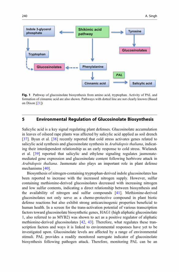
5 Environmental Regulation of Glucosinolate Biosynthesis
Salicylic acid is a key signal regulating plant defenses. Glucosinolate accumulationin leaves of oilseed rape plants was affected by salicylic acid applied as soil drench[37]. Byun et al. [38] recently reported that cold stress activates genes related tosalicylic acid synthesis and glucosinolate synthesis in Arabidopsis thaliana, indicat-ing their interdependent relationship as an early response to cold stress. Wielaneket al. [39] reported that salicylic and ethylene signaling regulates jasmonate-mediated gene expression and glucosinolate content following herbivore attack inArabidopsis thaliana. Jasmonate also plays an important role in plant defensemechanisms [40].
Biosynthesis of nitrogen-containing tryptophan-derived indole glucosinolates hasbeen reported to increase with the increased nitrogen supply. However, sulfurcontaining methionine-derived glucosinolates decreased with increasing nitrogenand low sulfur contents, indicating a direct relationship between biosynthesis andthe availability of nitrogen and sulfur compounds [41]. Methionine-derivedglucosinolates not only serve as a chemo-protective compound in plant bioticdefense reactions but also exhibit strong anticarcinogenic properties beneficial tohuman health. In a screen for the trans-activation potential of various transcriptionfactors toward glucosinolate biosynthetic genes, HAG1 (high aliphatic glucosinolate1, also referred to as MYB2) was shown to act as a positive regulator of aliphaticmethionine-derived glucosinolates [42, 43]. Therefore, what regulates these tran-scription factors and ways it is linked to environmental responses have yet to beinvestigated upon. Glucosinolate levels are affected by a range of environmentalstimuli. PAL provides a readily monitored surrogate indicator of glucosinolatebiosynthesis following pathogen attack. Therefore, monitoring PAL can be an
Shikimic acidpathway
Tryptophan
Indole 3-glycerolphosphate Tyrosine
Glucosinolates
Glucosinolates Phenylalanine
Cinnamic acid Salicylic acid
PAL
Fig. 1 Pathway of glucosinolate biosynthesis from amino acid, tryptophan. Activity of PAL andformation of cinnamic acid are also shown. Pathways with dotted line are not clearly known (Basedon Dixon [21])
240 A. Singh

effective indication of glucosinolate metabolism post-pathogen attack and wasbrought into action by Singh et al. [27].
6 Phenylalanine Ammonia Lyase Activity
Phenylalanine is a precursor of some aromatic glucosinolates during initial biosyn-thesis steps [44], and the enzyme PAL is a key enzyme in phenolic metabolism ofplants. PAL activity can be considered as an indirect indicator of aromaticglucosinolate synthesis; however, there are other pathways where PAL is involved,for example, SA pathway and phenylpropanoid metabolism.
PAL activity is affected by wounding of the plant; PAL accumulation was found24 h after wounding of maize mesocotyls [45]. PAL activity has also been shown tobe affected by chilling injury; the greater the injury, the more is the activity [46]. PALactivity is known to increase soon after the plant is challenged by a pathogen. As inthe case of cassava following elicitation by yeast, PAL was induced after 15 h[24]. Another example of induction of PAL activity after inoculation was wheatseedlings against stripe rust [47].
The defense activator, Bion®, has also been known to affect PAL activity 6, 9, and12 days after spraying in tomato as a defense response against Xanthomonasvesicatoria [48] as well as after treating wheat seedlings with Bion® as a controlagainst stripe rust [47]. Young et al. [49] have shown that phenolic levels inconventionally grown vegetable crops and organically grown vegetable crops,which are environmentally stressed, were high. This was because of increasedactivity of PAL following which more phenolics are synthesized. The PAL catalyzedconversion from phenylalanine to cinnamate, and then cinnamic acid has beenshown.
Phosphonate has been shown to activate defense responses in Arabidopsis as aresult of Phytophthora palmivora challenge [50]. Induction of similar defenseresponses may explain PAL activity increases in phosphonate-treated broccoli leavesin the current research. Also phosphonate treatment has been shown to activatedefense responses in Lambertia spp. against Phytophthora cinnamomi, and follow-ing which PAL activity increased 24 h after inoculation [51] (Fig. 2).
Cinnamic acid is a white crystalline acid obtained naturally from the oil ofcinnamon. Cinnamic acid is also a self-inhibitor produced by fungal spores toprevent germination [52]. In relation to disease and PAL activity, research suggeststhat PAL activity increases significantly in all inoculated genotypes of wheat in thefirst few hours after inoculation with Erysiphe [53]. This implies that infectionstimulates PAL activity that is shown in the current research. Also many studies ofPAL-deficient mutants that are compromised in resistance reveal similar results[54]. These studies indicate that mutants with deficiency in PAL levels affect thephenylpropanoid pathway and the plant’s ability to resist pathogens [55, 56].
Summing up the literature and various pathways, the following figure (Fig. 3)indicates that PAL activity can be monitored to derive more information aboutformation of phenolics (e.g., salicylic acid and some glucosinolates).
7 Glucosinolates and Plant Defense 241

There can be two possible circumstances, one in which there is already accumu-lation of glucosinolate in the crop and the other where it follows stress in the form ofpathogen attack in the plant. In the first case, glucosinolates are synthesized fromamino acid precursors as the normal course. These glucosinolates are in compart-ments within vacuoles; after injury or chewing, these compartments break, andmyrosinase acts to form isothiocyanates which in turn has possible beneficial effectsfor human health. On the other hand, if there is stress in the plant, i.e., pathogen
Fig. 2 Conversion of phenylalanine to cinnamic acid, catalyzed by PAL as measured at absorbance270 nm in a UV-vis spectrophotometer (Nanodrop 2000c, thermo scientific)
Relationship between glucosinolates, PAL and phenolics
recognition
myrosinase
Glucosinolates
Increase inPAL
Phenolicdefences
Isothiocynates
Aminoacids(Phenylalanine)
Fig. 3 Relationship between glucosinolates and PAL activity. PAL and glucosinolates are derivedfrom the same precursor that links the two
242 A. Singh

infection, phenylalanine is accumulated as a result of infection in turn accumulatingmore glucosinolates. Increase in PAL activity was found after inoculation of Bras-sica juncea with Albugo candida which suggested that pathogen infection triggersthe accumulation of PAL in Brassica crops [57]. This derivative pertains to havingthe potential to be beneficial to human health.
Bion® delayed the symptoms of white rust for 2 days, while phosphonate delayedsymptoms for 3 days. This occured possibly due to the phenolics and phytochem-icals formed after infection and defense response induction due the defense activatortreatments. One of these hydrolysis compounds has been studied by measuring thePAL activity. The increase in the PAL activity after inoculation and Bion® andphosphonate treatment supports the role for PAL in plant defense in broccoli and thatincrease in PAL activity is a marker of plant defense [58].
PAL activity in both root and stem tissues were analyzed in chickpea varietyJG-62 susceptible to Fusarium oxysporum causing vascular wilt. It was found thatthere was high PAL activity at early stages (pre-colonization stages) of thedisease [34].
PAL is activated by the SAR mechanism of Bion® and defense priming functionof phosphonate. This research also implies that PAL is activated by infection as oneof the earliest defense responses in the plant against Albugo candida [27].
7 Conclusions
Glucosinolates have been known and proven for their vital role in plant defense[59, 60] as they are plant secondary metabolites that are induced by stress orinfection and act as precursors to plants’ defense system. Similar role is carriedout by PAL activity [4, 61] in earlier stages of infection. Accumulation of certainglucosinolates, PAL activity, and hypersensitive response [62] after pathogen chal-lenge has been regarded as indicators of expression of systemic resistance byBrassica plants [63–65].
Greater insight is required to determine the exact course of the compound’sjourney through the brassica plants and its certainty in acting as a major participantin plants’ systemic resistance and defense mechanisms. This research will be highlybeneficial to progress molecular and genetic studies in the area of plant defenseenhancement.
References
1. Sorensen H (1988) Glucosinolates – structure-properties-function. Abstr Pap Am Chem Soc195:79-AGFD
2. Verena J, Jonathan G, Vasao DG (2015) Metabolism of glucosinolates and their hydrolysisproducts in insect herbivores. In: Jetter R (ed) The formation, structure and activity of phyto-chemicals. Springer International Publishing, Switzerland, pp 163–194
7 Glucosinolates and Plant Defense 243

3. Abdel-Farid IB, Choi YH, Kim HK, Van den Hondel CAMJJ, van der Meijden HE, Verpoorte R(2006) The role of secondary metabolites in Arabidopsis and Brassica in the interaction withfungi. Curr Top Plant Biol 7:47–73
4. Bennett RN, Wallsgrove RM (1994) Secondary metabolites in plant defense-mechanisms. NewPhytol 127:617–633
5. Kusnierczyk A, Winge P, Jorstad TS, Troczynska J, Rossiter JT, Bones AM (2008) Towardsglobal understanding of plant defence against aphids – timing and dynamics of earlyArabidopsis defence responses to cabbage aphid (Brevicoryne Brassicae) attack. Plant CellEnviron 31:1097–1115
6. Sun JY, Snderby IE, Halkier BA, Jander G, Md V (2009) Non-volatile intact indoleglucosinolates are host recognition cues for ovipositing Plutella xylostella. J Chem Ecol35:1427–1436
7. Fan ZX, Lei WX, Sun XL, Yu B, Wang YZ, Yang GS (2008) The association of Sclerotiniasclerotiorum resistance with glucosinolates in Brassica napus double-low DH population.J Plant Pathol 90:43–48
8. Bodnaryk RP (1992) Effects of wounding on glucosinolates in the cotyledons of oilseed rapeand mustard. Phytochemistry 31:2671–2677
9. Kim JH, Jander G (2007) Myzus persicae (green peach aphid) feeding on Arabidopsis inducesthe formation of a deterrent indole glucosinolate. Plant J 49:1008–1019
10. Bouchereau A, ClossaisBesnard N, Bensaoud A, Leport L, Renard M (1996) Water stresseffects on rapeseed quality. Eur J Agron 5:19–30
11. Antonious GF, Bomford M, Vincelli P (2009) Screening Brassica species for glucosinolatecontent. J Environ Sci Health B, Pestic, Food Contam Agric Wastes 44:311–316
12. Martin N, Muller C (2007) Induction of plant responses by a sequestering insect: relationship ofglucosinolate concentration and myrosinase activity. Basic Appl Ecol 8:13–25
13. Brandi G, Amagliani G, Schiavano GF, De Santi M, Sisti M (2006) Activity of Brassicaoleracea leaf juice on foodborne pathogenic bacteria. J Food Protect 69:2274–2279
14. Brader G, Mikkelsen MD, Halkier BA, Palva ET (2006) Altering glucosinolate profilesmodulates disease resistance in plants. Plant J 46:758–767
15. Galletti S, Sala E, Leoni O, Burzi PL, Cerato C (2008) Trichoderma spp. tolerance to Brassicacarinata seed meal for a combined use in biofumigation. Biol Control 45:319–327
16. Bhardwaj HL, Hamama AA (2003) Accumulation of glucosinolate, oil, and erucic acid indeveloping Brassica seeds. Ind Crops Prod 17:47–51
17. Tripathi P, Dubey NK (2004) Exploitation of natural products as an alternative strategy tocontrol postharvest fungal rotting of fruit and vegetables. Postharvest Biol Technol 32:235–245
18. Lambrix V, Reichelt M, Mitchell-Olds T, Kliebenstein DJ, Gershenzon J (2001) TheArabidopsis epithiospecifier protein promotes the hydrolysis of glucosinolates to nitriles andinfluences Trichoplusia ni herbivory. Plant Cell 13:2793–2807
19. Rask L, Andreasson E, Ekbom B, Eriksson S, Pontoppidan B, Meijer J (2000) Myrosinase: genefamily evolution and herbivore defense in Brassicaceae. Plant Mol Biol 42:93–113
20. Dinant S, Suárez-López P (2011) Multitude of long-distance signal molecules acting viaphloem. In: Witzani G, Baluska F (eds) Biocommunication of plants, vol 14, Signaling andcommunication in Plants. Springer, Berlin, pp 89–121
21. Dixon RA (2001) Natural products and plant disease resistance. Nature 411:843–84722. Yao Q, Zhu HH, Zeng RS (2007) Role of phenolic compounds in plant defence: induced by
arbuscular mycorrhizal fungi. Allelopathy J 20:1–1323. Pozo MJ, Verhage A, Garcia-Andrade J, Garcia JM, Azcon-Aguilar C (2009) Priming plant
defence against pathogens by arbuscular mycorrhizal fungi. Mycorrhizas – functional processesand ecological impact. 123–135
24. Gomez-Vasquez R, Day R, Buschmann H, Randles S, Beeching JR, Cooper RM (2004)Phenylpropanoids, phenylalanine ammonia lyase and peroxidases in elicitor-challenged cassava(Manihot esculenta) suspension cells and leaves. Ann Bot 94:87–97
244 A. Singh

25. Hammerschmidt R, Smith-Becker JA (1999) The role of salicylic acid in disease resistance. In:Agrawal A, Tuzun S, Bent E (eds) Induced plant defenses against pathogens and herbivores.APS Press, St. Paul, pp 37–53
26. Islam N (2009) The effect of Plasmodiophora Brassicae infection, phosphonate and Bion®
treatment on glucosinolate levels in broccoli. University of Sydney, Sydney27. Singh A (2011) Effect of white rust disease (Albugo candida) on the glucosinolate contents in
Bion® and phosphonate treated Brassica crops. University of Sydney, Sydney28. Sanchez-Vallet A, Ramos B, Bednarek P, López G, Piślewska- Bednarek M, Schulze-Lefert P,
Molina A (2010) Tryptophan derived secondary metabolites in Arabidopsis thaliana confernon-host resistance to necrotrophic Plectosphaerella cucumerina fungi. Plant J 63(1):115–127
29. Schlaeppi K, Abou-Mansour E, Buchala A, Mauch F (2010) Disease resistance of Arabidopsisto Phytophthora brassicae is established by the sequential action of indole glucosinolates andcamalexin. Plant J 62:840–851
30. Wielanek M, Urbanek H (2006) Enhanced glucotropaeolin production in hairy root cultures ofTropaeolum majus L. by combining elicitation and precursor feeding. Plant Cell Tiss Org Cult86:177–186
31. Kelly PJ, Bones A, Rossiter JT (1998) Sub-cellular immunolocalization of the glucosinolatesinigrin in seedlings of Brassica juncea. Planta 206:370–377
32. Grob K, Matile P (1979) Vacuolar location of glucosinolates in horseradish root-cells. Plant SciLett 14:327–335
33. Evans CT, Choma C, Peterson W, Misawa M (1987) Bioconversion of trans-cinnamic acid tol-phenylalanine in an immobilized whole cell reactor. Biotechnol Bioeng 30:1067–1072
34. Shukla YM, Dhruve JJ, Patel NJ, Pandey RN (2010) Biochemical alterations in cinnamic acid4-hydroxylase and phenylalanine ammonia lyase in chickpea infected with Fusariumoxysporum f.spciceri. J Mycol Plant Pathol 40:260–264
35. Sticher L, MauchMani B, Metraux JP (1997) Systemic acquired resistance. Ann RevPhytopathol 35:235–270
36. MacDonald MJ, D’Cunha GB (2007) A modern view of phenylalanine ammonia lyase.Biochem Cell Biol 85:273–282
37. Kiddle GA, Doughty KJ, Wallsgrove RM (1994) Salicylic acid-induced accumulation ofglucosinolates in oilseed rape (Brassica napus L) leaves. J Exp Bot 45:1343–1346
38. Byun YJ, Kim HJ, Lee DH (2009) Long SAGE analysis of the early response to cold stress inArabidopsis leaf. Planta 229(6):1181–1200
39. Wielanek M, Krolicka A, Bergier K, Gajewska E, Skodowska M (2009) Transformation ofNasturtium officinale, Barbarea verna and Arabis caucasica for hairy roots and glucosinolate-myrosinase system production. Biotechnol Lett 31:917–921
40. Kubicka E, Zadernowski R (2007) Enhanced jasmonate biosynthesis in plants and possibleimplications for food quality. Acta Alimentaria (Budapest) 36:455–469
41. Li S, Schonhof I, Krumbein A, Li L, Stutzel H, Schreiner M (2007) Glucosinolate concentrationin turnip (Brassica rapa ssp. rapifera L.) roots as affected by nitrogen and sulfur supply. J AgricFood Chem 55:8452–8457
42. Gigolashvili T, Yatusevich R, Berger B, Muller C, Flugge UI (2007) The R2R3-MYB tran-scription factor HAG1/MYB28 is a regulator of methionine-derived glucosinolate biosynthesisin Arabidopsis thaliana. Plant J 51:247–261
43. Schuster J, Knill T, Reichelt M, Gershenzon J, Binder S (2006) Branched-chain aminotransfer-ase 4 is part of the chain elongation pathway in the biosynthesis of methionine-derivedglucosinolates in Arabidopsis. Plant Cell 18:2664–2679
44. Bjerg B, Eggum BO, Jacobsen I, Otte J, Sørensen H (1989) Antinutritional and toxic effects inrats of individual glucosinolates (� myrosinases) added to a standard diet (2). J Anim PhysiolAnim Nutr 61:227–244. doi:10.1111/j.1439-0396.1989.tb00105.x
45. Pascholati SF, Nicholson RL, Butler LG (1986) Phenylalanine ammonia-lyase activity andanthocyanin accumulation in wounded maize mesocotyls. J Phytopathol 115:165–172
7 Glucosinolates and Plant Defense 245

46. Bojorquez-Galvez A, Vega Garcia M, Caro Corrales J, Carrillo Lopez A, Lopez Valenzuela JA(2010) Effect of gradual cooling storage on chilling injury and phenylalanine ammonia-lyaseactivity in tomato fruit. J Food Biochem 34:295–307
47. Chen R, Liu T, Huang Y, Cheng D, Chen W (2006) Induced resistance of wheat seedlings toPuccinia triticina by Bion® treatment. Acta Phytophylacica Sin 33:122–126
48. Cavalcanti FR, Resende MLV, Carvalho CPS, Silveira JAG, Oliveira JTA (2007) An aqueoussuspension of Crinipellis perniciosa mycelium activates tomato defence responses againstXanthomonas vesicatoria. Crop Prot 26(5):729–738
49. Young JE, Zhao X, Carey EE, Welti R, Yang SS, Wang WQ (2005) Phytochemical phenolics inorganically grown vegetables. Mol Nutr Food Res 49:1136–1142
50. Suddaby T, Alhussaen K, Daniel R, Guest D (2008) Phosphonate alters the defence responses ofLambertia species challenged by Phytophthora cinnamomi. Aust J Bot 56:550–556
51. Daniel R, Guest D (2005) Defence responses induced by potassium phosphonate inPhytophthora palmivora challenged Arabidopsis thaliana. Physiol Mol Plant Pathol67:194–201
52. Kirkham DS, Flood AE (1956) Inhibition of Venturia spp. of analogues of host metabolites.Nature 178:422–423
53. Norman E, Green L, Hadwiger A (1975) Phenylalanine ammonia-lyase to sine ammonia-lyaselignin in wheat inoculated with Erysiphe graminis f. sp. tritici. Phytopathology 65(10):1071–1074
54. El-Kereamy A, El-Sharkawy I, Ramamoorthy R, Taheri A, Errampalli D, Kumar P, JayasankarS (2011) Prunus domestica pathogenesis-related protein-5 activates the defense responsepathway and enhances the resistance to fungal infection. Plos One 6:11
55. Ferrari S, Plotnikova JM, De Lorenzo G, Ausubel FM (2003) Arabidopsis local resistance toBotrytis cinerea involves salicylic acid and camalexin and requires EDS4 and PAD2, but notSID2, EDS5 or PAD4. Plant J 35:193–205
56. Taheri P, Tarighi S (2010) Riboflavin induces resistance in rice against Rhizoctonia solani viajasmonate-mediated priming of phenylpropanoid pathway. J Plant Physiol 167:201–208
57. Singh US, Doughty KJ, Nashaat NI, Bennett RN, Kolte SJ (1999) Induction of systemicresistance to Albugo candida in Brassica juncea by pre- or coinoculation with an incompatibleisolate. Phytopathology 89:1226–1232
58. Singh A, Guest D, Copeland L (2015) Associations between glucosinolates, white rust, andplant defense activators in Brassica plants: a review. Inter J Veg Sci 21(3):297–313
59. Morant AV, Jorgensen K, Jorgensen C, Paquette SM, Sanchez-Perez R, Moller BL, Bak S(2008) Beta-glucosidases as detonators of plant chemical defense. Phytochemistry69:1795–1813
60. Redovnikovic IR, Glivetic T, Delonga K, Vorkapic-Furac J (2008) Glucosinolates and theirpotential role in plants. Period Biol 110:297–309
61. Lee SW, Nazar RN, Powell DA, Robb J (1992) Reduced PAL gene suppression in Verticillium-infected resistant tomatoes. Plant Mol Biol 18:345–352
62. Hammerschmidt R (1999) Induced disease resistance: how do induced plants stop pathogens?Physiol Mol Plant Pathol 55:77–84
63. Ahuja I, Rohloff J, Bones AM (2010) Defence mechanisms of Brassicaceae: implications forplant-insect interactions and potential for integrated pest management. A review. Agron SustainDev 30(2):311–348
64. Jahangir M, Abdel-Farid IB, Kim HK, Choi YH, Verpoorte R (2009) Healthy and unhealthyplants: the effect of stress on the metabolism of brassicaceae. Environ Exp Bot 67:23–33
65. Wang BC, Wang JB, Zhao HC, Zhao H (2006) Stress induced plant resistance and enzymeactivity varying in cucumber. Colloids Surf B: Biointerfaces 48:138–142
246 A. Singh

Part II
Biological Activity

Antimicrobial Activity of the Glucosinolates 8Federica Saladino, Keliani Bordin, Fernando Bittencourt Luciano,Mónica Fernández Franzón, Jordi Mañes, and Giuseppe Meca
AbstractThe use of natural antimicrobial compounds is receiving much attention and isbecoming very frequent by the importance that nowadays is given to naturalresources. Natural components have been applied in several sectors such asagriculture, biomedicine and food preservation. The development of resistanceto conventional antibiotic by pathogenic bacteria makes necessary to find alter-native antimicrobials to eradicate these microorganisms. Many food products areperishable and require protection from spoilage to improve quality and shelf life.Numerous efforts are conducted to find safe natural alternatives to preventmicroorganism growth in plants and food products, because of the consumerconcern regarding synthetic pesticides and preservatives. Natural antimicrobialscan be obtained from different sources including plants, animals, bacteria, algae,and fungi. Among them, glucosinolates and their derived products have beenrecognized for their benefits to human nutrition, plant defense, and as potentantimicrobial agents. This chapter describes the antimicrobial activity ofglucosinolates and their hydrolysis products against different bacterial and fungalspecies, as well as the mechanism of action of these active compounds.
KeywordsGlucosinolates • Isothiocyanates • Antifungal activity • Antibacterial activity •Bioactive compounds
F. Saladino (*) • M.F. Franzón • J. Mañes • G. MecaLaboratory of Food Chemistry and Toxicology, Faculty of Pharmacy, University of Valencia,Burjassot, Valencia, Spaine-mail: [email protected]; [email protected]; [email protected]; [email protected];[email protected]
K. Bordin • F.B. LucianoSchool of Agricultural Sciences and Veterinary Medicine, Pontifícia Universidade Católica, SãoJosé dos Pinhais, Paraná, Brazile-mail: [email protected]; [email protected]
# Springer International Publishing Switzerland 2017J.-M. Mérillon, K.G. Ramawat (eds.), Glucosinolates, Reference Series inPhytochemistry, DOI 10.1007/978-3-319-25462-3_18
249

AbbreviationsAAM AllylamineAC Allyl cyanideAITC Allyl isothiocyanateASC AscorbigenATC Ally thiocyanateBAM BenzylamineBC Benzyl cyanideBITC Benzyl isothiocyanateCEPT 1-Cyano-2,3-epithiopropaneDIM 3,30-Di-indolylmethaneEITC Ethyl isothiocyanateGLS GlucosinolateI3C Indole-3-carbinolIAN Indole-3-acetonitrileITC IsothiocyanateMAP Modified atmosphere packagingMCT Medium-chain triglycerideMITC Methyl isothiocyanatePAM 2-PhenylethylaminePEC 2-Phenylethyl cyanidePEITC Phenylethyl isothiocyanatePITC Phenyl isothiocyanateSBO Soybean oilSFN SulforaphaneTC Thiocyanate
Contents1 Introduction . . . . . . . . . . . . . . . . . . . . . . . . . . . . . . . . . . . . . . . . . . . . . . . . . . . . . . . . . . . . . . . . . . . . . . . . . . . . . . . . . . 2502 Antibacterial Activity . . . . . . . . . . . . . . . . . . . . . . . . . . . . . . . . . . . . . . . . . . . . . . . . . . . . . . . . . . . . . . . . . . . . . . . 2533 Antifungal Activity . . . . . . . . . . . . . . . . . . . . . . . . . . . . . . . . . . . . . . . . . . . . . . . . . . . . . . . . . . . . . . . . . . . . . . . . . . 263
3.1 Plant Protection . . . . . . . . . . . . . . . . . . . . . . . . . . . . . . . . . . . . . . . . . . . . . . . . . . . . . . . . . . . . . . . . . . . . . . . . 2664 Structure Activity Relationships . . . . . . . . . . . . . . . . . . . . . . . . . . . . . . . . . . . . . . . . . . . . . . . . . . . . . . . . . . . . 2675 Conclusions . . . . . . . . . . . . . . . . . . . . . . . . . . . . . . . . . . . . . . . . . . . . . . . . . . . . . . . . . . . . . . . . . . . . . . . . . . . . . . . . . . 268References . . . . . . . . . . . . . . . . . . . . . . . . . . . . . . . . . . . . . . . . . . . . . . . . . . . . . . . . . . . . . . . . . . . . . . . . . . . . . . . . . . . . . . . 269
1 Introduction
In agreement with the current trend to value the natural and renewable resources, theinterest in the use of natural antimicrobial compounds is increasing for biomedical,agricultural, and especially food applications [1, 2].
Food products are perishable by nature and can be subjected to contaminationby bacteria and fungi. Many of these microorganisms can cause undesirable
250 F. Saladino et al.

reactions that deteriorate flavor, odor, color, sensory, and textural properties offoods. Some of them can also potentially cause food-borne illness. For all thesereasons, food products require protection from spoilage during their preparation,storage, and distribution to give them desired shelf life. Furthermore, the dramaticrise of antibiotic-resistant microorganisms is of concern and includes food-bornepathogens that are also more tolerant to several food processing and preservationmethods. The consumer concern regarding synthetic products, such as food addi-tives and pesticides, and the necessity to overcome the emergence of antibiotic-resistant pathogens led to the research of alternative compounds with potentantimicrobial activity which can reduce the impact of synthetic products onhuman and animal health [1].
Natural antimicrobials can be obtained from different sources including plants,animals, bacteria, algae, and fungi. To select the appropriate biocidal product, themicroorganism strain must be identified and the spectrum of antimicrobial activity ofthe compound considered [2–4]. Several reports have demonstrated the efficacy ofplant-derived compounds, most of all in food applications. Antimicrobials derivedfrom plants are mostly secondary metabolites that possess various benefits includingantimicrobial properties against pathogenic and spoilage microbes. The structuraldiversity of plant-derived compounds is immense, and the impact of antimicrobialaction they produce against microorganisms depends on their structuralconfiguration [5].
Among the potent natural antimicrobials, glucosinolates (GLS) are an importantclass of secondary plant products found in seeds, roots, stems, and leaves ofcruciferous plants including 16 families of dicotyledonous angiosperms, mainlyBrassicaceae [6]. There are about 120 different GLS identified, derived fromamino acids (alanine, leucine, isoleucine, valine, phenylalanine, tyrosine, and tryp-tophan) and a number of chain-elongated homologues [7]. They are classified asaliphatic, aromatic, methylthioalkyl, and heterocyclic, which have a thioglucosidecomponent in common structure and differ at their side chains [8].
Located within vacuoles, GLS are physically separated but accompanied byβ-thioglucosidase enzymes known as myrosinases [9]. Following plant tissuedisruption, the enzyme and GLS come into contact, which, in the presence ofwater, generates a hydrolysis forming an aglycone moiety, glucose, and sulfate.The aglycone moiety is unstable and rearranges to form three main groups ofsubstances: nitriles, thiocyanates (TCs), and isothiocyanates (ITCs) (Fig. 1) [3,10, 11].
GLS and their enzymatic hydrolysis products are responsible for a characteristicpungent flavor [12, 13]. These compounds have shown several biological activitiesincluding plant defense (against insects and microbial infections) and benefits tohuman health (anticarcinogenic, antioxidant, and antimicrobial properties) andmight be potential natural agents for food preservation [14]. Their response tomicrobial population varies according to their structural characteristics. The biocidaleffect of cruciferous tissues on other microorganisms has been attributed mainly tovolatile degradation products of GLS, released from their plants. Among derived
8 Antimicrobial Activity of the Glucosinolates 251
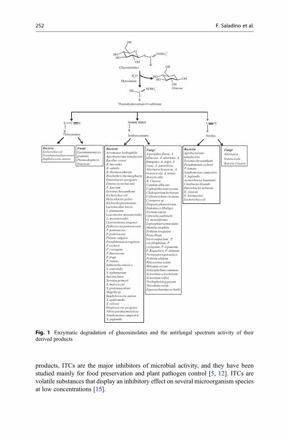
products, ITCs are the major inhibitors of microbial activity, and they have beenstudied mainly for food preservation and plant pathogen control [5, 12]. ITCs arevolatile substances that display an inhibitory effect on several microorganism speciesat low concentrations [15].
FungiAlternariabrassicicolaBotrytis Cinerea
FungiAspergillus flavus, A. alliaceus, A. alternata, A. fumigatus, A. niger, A. ryzae, A. parasiticusAlternaria brassicae, A. brassicicola, A. tenuisBotrytis alliiB. CinereaCandida albicansCephalothecium roseumCladosporium herbarumColletotrichum circinansCytospora sp.Diaporte phaseolorumEndomyces fibuligerErotium repensGiberella sanbinettiG. monoliformisLeptosphaeria maculansMonilia sitophilaPythium irregularePenicillium brevicompactum, P. corylilophilum, P. cyclopium, P. expansum, P. Roqueforti, P. ultimumPeronospora parasiticaPythium ultimimRhizoctonia solaniRhizopus oryzaeSchizophyllum communeSclerotinia sclerotiorumSclerotium rolfsiiTrichophyton gypseumTricodema virideZygosaccharomyces bailii
FungiGaeumannomyces graminisPlasmodiophora brassicae
Bacteria Escherichia coliPseudomonas fluorescensStaphylococcus aureus
BacteriaAeromonas hydrophiliaAgrobacterium tumefaciensBacillus cereusB. mycoidesB. subtilisB. thermoaciduransBrochothrix thermosphactaEnterobacter aerogenerEnterococcus faecalisE. faeciumErwinia chrysanthemiEscherichia coliHelycobacter pyloriKlebsiella pneumoniaeLactobacillus brevisL. plantarumLeuconostoc mesenteroidesL. mesenteroidesListeria monocytogenesPediococcus pentosaceousP. pentosaceusP. pentosaceusProteus vulgarisPseudomonas aeruginosaP. cichoriiP. corrugataP. fluorescensP. fragiP. tomatoSalmonella entericaS. enteritidisS. typhimuriumSarcina luteaSerratia grimesiiS. marcescensS. proteamaculansShigella sp. Staphylococcus aureusS. epidermidisS. xylosusStreptococcus pyogenesVibrio parahaemolyticusXanthomonas campestrisX. juglandis
BacteriaAgrobacteriumtumefaciensErwinia chrysanthemiPseudomonas cichoriiP. tomatoXanthomonas campestrisX. juglandisAcinetobacter baumaniiCitrobacter freundiiEnterobacter asburiaeE. cloacaeE. hormaecheiEscherichia coli
Glucosinolates
Myrosinase
Glucose
Thionohydroxamate-O-sulfonate
Thiocyanates Isothiocyanates Nitriles
S
R R
N C
R
C N
R
CHS NOSO3
─
NOSO3
─
OH
OH
OH
HO
HOHO
OH
O
OH
S
R
C
H2O
HO
SC N
Fig. 1 Enzymatic degradation of glucosinolates and the antifungal spectrum activity of theirderived products
252 F. Saladino et al.

Therefore, the objective of this chapter was to present results of studies onantimicrobial activity of GLS and their enzymatically degradation products andhighlight important aspects on the application.
2 Antibacterial Activity
GLS and, above all, their hydrolysis products elicit a wide spectrum of antimicrobialactivity against a variety of bacteria. The concentrations of these compoundsrequired to inhibit microorganisms are difficult to compare given differences inmethodologies, materials, and test strains employed. There are considerably moredata for the ITCs, and in particular for AITC, than others GLS products. Further-more, the mode of delivery to target microorganisms has a large impact on theantibacterial effect. Dissolution of these compounds in liquid media can result in aweak antimicrobial activity, while lower concentrations in the vapor phase aresufficient to inhibit microorganisms [10]. On Table 1 are summarized some studieswhich report the antibacterial potential of these compounds.
Horseradish vapors, containing GLS hydrolysis products, showed strongerantibacterial activities against several bacterial strains [16, 17]. Later, also studiedand compared were the bacteriostatic and bactericidal effects of AITC, EITC, andMITC against 10 strains. MITC was the most effective, in both solution and vaporphases, followed by AITC and EITC. Escherichia coli and Staphylococcus aureusappeared to be the more resistant strains, while the least resistant were Bacillussubtilis, Bacillus mycoides, and Serratia marcescens.
Virtanen [18] reported the antimicrobial activity of BITC and β-phenylethyl,m-methoxybenzyl, and methoxybenzyl ITCs against S. aureus. The activity ofthese ITCs was higher than the antimicrobial activity of a series of aliphatic ITCs.Zsolnai [19] demonstrated that the same concentration of AITC and PITC, used toseverely inhibit the growth of yeasts and fungi, was not effective against Strepto-coccus pyogenes, S. aureus, and other Gram-negative bacteria. BITC was effectiveagainst Staphylococci but not on other bacteria.
Kanemaru and Miyamoto [20] studied the antibacterial activity of brown mustardand its major pungent compound, AITC, on the growth of E. coli 3301, S. aureusIFO 3761, Proteus vulgaris IFO 3851, Pseudomonas fragi IFO 3458, and Pseudo-monas aeruginosa IFO 3755. To prepare the extract of black mustard extract wasprepared as 20% mustard in ethanol (70%) after myrosinase treatment. AITC wasalso dissolved in 70% ethanol to form an equivalent concentration. The nutrientbroth in which the bacteria were cultured contained the mustard extract or AITC andwas stored at 30 �C on a shaker. Turbidimetry was used to determine bacterialgrowth. The results obtained evidenced that the antibacterial effect of mustard wasmainly due to AITC. The concentrations of mustard in the medium that inhibitedbacterial growth for 24 h were 0.138%, 0.104%, 0.064%, 0.043%, and 0.089% andthose of AITC were 14.5, 12.3, 6.5, 3.6, and 7.2 ppm for S. aureus, E. coli,P. vulgaris, P. fragi, and P. aeruginosa, respectively. A bacteriostatic effect was
8 Antimicrobial Activity of the Glucosinolates 253
Table 1 Antibacterial potential of glucosinolate-derived products against several species on foodproducts
Glucosinolatederivative Bacterial strain Food product Reference
Allyl isothiocyanate Bacillus thermoacidurans Fresh applejuice
[44]
Allyl isothiocyanate Bacillus subtilis IFO-13722Bacillus cereus IFO-13494Staphylococcus aureus IFO-12732Staphylococcus epidermidis IFO-12993Escherichia coli JCM-1649Salmonella typhimurium ATCC-14028Salmonella enteritidis JCM-189Vibrio parahaemolyticus IFO-12711P. Pseudomonas aeruginosa IFO-13275
Fresh beefCured porkSliced rawtunaCheeseEgg sandwichNoodlesPasta
[45]
Allyl isothiocyanatePhenethylisothiocyanateAllyl thiocyanate
Staphylococcus aureus Cooked roastbeef
[46]
Escherichia coli O157:H7
Staphylococcus typhimurium
Listeria monocytogenes
Serratia grimesii
Lactobacillus sake
Allyl isothiocyanatePhenethylisothiocyanateAllyl thiocyanate1-Butaneisothiocyanate
Pseudomonas spp. EnterobacteriaceaeLactic acid bacteria
Precookedroast beefslices
[47]
Methylisothiocyanate,Allyl isothiocyanate
Rifampicin-resistant strain ofSalmonella, Montevideo streptomycin-resistant strains of Escherichia coliO157:H7 and Listeria monocytogenesScott A
Apples,tomatoes,Iceberg lettuce
[48]
Allyl isothiocyanate Escherichia coli O157:H7 Fresh groundbeef
[49]
Allyl isothiocyanate Pediococcus pentosaceusStaphylococcus carnosusEscherichia coli O157:H7
Dry fermentedsausage
[50]
Allyl isothiocyanate Lactobacillus algidusLeuconostoc mesenteroidesLeuconostoc carnosumCarnobacterium maltaromaticumCarnobacterium divergensBrochothrix thermosphactaSerratia proteamaculans
Marinatedpork
[51]
Allyl isothiocyanate Salmonella Freshcantaloupe
[52]
(continued)
254 F. Saladino et al.

shown by mustard on S. aureus and E. coli (0.8%), while the effect was bactericidalon P. aeruginosa at 0.2%
Shofran et al. [21] tested the antimicrobial activity of sinigrin and four sinigrinhydrolysis products, in broth culture, against different species of bacteria. Sinigrin isa GLS that, upon injury or mechanical disruption of plant tissue, is hydrolyzed bymyrosinase producing up to four distinct compounds: AITC, allyl TC (ATC), allylcyanide (AC), and 1-cyano-2,3-epithiopropane (CEPT). Sinigrin had little effectupon the growth of microorganisms [22], but its hydrolysis products were effectivein inhibition of growth. The species of bacteria studied in the experiment were E. coli33625, E. coli NC101, Pseudomonas fluorescens MD13, Aeromonas hydrophilia7966, S. aureus 4220, B. subtilis IS75, Pediococcus pentosaceus FFL48,Leuconostoc mesenteroides FFL44, Lactobacillus brevis MD42, and Lactobacillusplantarum MOP3. Sinigrin, AC, and CETP at 1000 ppm did not show inhibitoryeffects against any of the bacteria tested. ATC was inhibitory to the growth of3 strains of Gram-negative (E. coli 33625, E. coli NC101, P. fluorescens MD13)and 1 strain of Gram-positive bacteria (S. aureus 4220) with minimum inhibitoryconcentration (MIC) values ranged between 200 and 400 ppm. The antimicrobialactivity of ATC was due to its conversion to AITC, sinigrin hydrolysis products withthe highest antibacterial activity. AITC was effective against all the bacteria tested,except L. plantarum MOP3. The MIC of AITC against Gram-negative and Gram-positive non-lactic acid bacteria ranged between 100 and 200 ppm, while lactic acidbacteria were more resistant with MIC between 500 and 1000 ppm. It should behighlighted that the antimicrobial activity of AITC can be different if it is used ingaseous form or dissolved in broth culture. Furthermore, a lot of factors caninfluence the generation of AITC from sinigrin.
Kyung and Fleming [23] tested sinigrin and its derivate products against 15 spe-cies of bacteria: Pediococcus pentosaceus LA3, P. pentosaceus LA76,L. mesenteroides LA10, L. mesenteroides LA113, L. plantarum LA97,L. plantarum LA70, L. brevis LA25, L. brevis LA200, Listeria monocytogenesB67, L. monocytogenes B70, S. aureus B31, E. coli B34, Enterobacter aerogenes
Table 1 (continued)
Glucosinolatederivative Bacterial strain Food product Reference
Allyl isothiocyanate Leuconostoc mesenteroidesLactobacillus plantarum
Kimchi [53]
Allyl isothiocyanate Escherichia coliListeria monocytogenes
Fresh cutonions
[54]
Allyl isothiocyanate Listeria monocytogenesSalmonella typhimurium
Chicken breast [56]
4-Hydroxybenzylisothiocyanate
Salmonella Sauce withparticulates
[55]
8 Antimicrobial Activity of the Glucosinolates 255

B146, B. subtilis B96, and Salmonella typhimurium B38. Sinigrin itself was notantimicrobial because it did not inhibit growth up to 1000 ppm and microorganismsdid not degrade it to its antimicrobial aglycones. AITC is known to be antimicrobial,and the MICs found ranged from 50 to 500 ppm for bacteria, including Gram-positive, Gram-negative, pathogenic, and lactic acid bacteria.
Delaquis and Sholberg [24] evaluate the microbistatic and microbicidal propertiesof gaseous AITC against bacterial cells of S. Typhimurium (ATCC 14028),L. monocytogenes (strain 81–861), E. coli O157:H7 (ATCC 43895), and Pseudo-monas corrugata (isolated from lettuce). S. typhimurium, L. monocytogenes, andE. coli O157:H7 were inhibited when exposed to 1000 μg L�1 AITC. P. corrugatafailed to grow in the presence of 500 μg L�1. Variations at different incubationtemperatures were observed. Bactericidal activities varied with strain and increasedwith time of exposure. The most resistant bacterium was E. coli.
The antibacterial properties of the GLS and their hydrolysis products became ofbig interest and importance also in the eradication of pathogenic microorganismsthat is complicated by the development of resistance to conventional antimicrobialagents. Helicobacter pylori is one of the most prevalent human pathogens in theworld. Gastric infections withH. pylori are known to cause gastritis and peptic ulcersand dramatically enhance the risk of gastric cancer. Antibiotic therapy isrecommended for infected patients with gastric or duodenal ulcers or gastricmucosa-associated lymphoid tissue lymphoma, but this treatment is not universallysuccessful. Even with the combination of two or more antibiotics, H. pylori isdifficult to eradicate due to the development of resistance of this bacteria to theseantibiotics and the persistence of organisms within gastric epithelial cells and,furthermore, due to logistic, sociologic, and economic reasons. The ITC sulforaph-ane (SFN) appears to overcome all of these problems. SFN is abundant in certainvarieties of broccoli and broccoli sprouts in the form of its GLS precursor calledglucoraphanin. It has been demonstrated that SFN is a potent bacteriostatic agentagainst 3 reference strains and 45 clinical isolates of H. pylori. The MIC for 90% ofthe strains is<4 μg mL�1, irrespective of their resistance to conventional antibiotics.It is a potent bactericidal agent against both extra- and intracellular H. pylori in vitro.Further, brief exposure to SFN eliminated intracellular H. pylori from a humanepithelial cell line (HEp-2). Although higher concentrations are required to achievebactericidal activity for the intracellular forms, SFN accumulates intracellularly tohigh levels, as its glutathione conjugate. It can be safely administered to humansbecause it is present in high concentrations in edible cruciferous vegetables and canbe directly delivered to the stomach [25].
Haristoy et al. [26] evaluated the effect of SFN in vivo against H. pylori by usinghuman gastric xenografts in nude mice. H. pylori was completely eradicated in 8 ofthe 11 SFN-treated grafts, after short-term administration of SFN at a dose that canbe achieved in the human diet.
Haristoy et al. [27] analyzed the activities of 12 ITCs including sulforaphane on25 strains of H. pylori using an agar dilution assay. The ITCs tested were iberin,cheirolin, erucin, D,L-SFN, D-SFN, L-SFN, L-sulforaphane, erysolin, berteroin,alyssin, hirsutin, PEITC, BITC, and 4-(α-L-rhamnopyranosyloxy)benzyl ITC.
256 F. Saladino et al.

Furthermore, the bactericidal activities of the six ITCs (cheirolin, L-sulforaphane,erysolin, berteroin, hirsutin, and 4-(α-L-rhamnopyranosyloxy)benzyl ITC) thatshowed the lowest MICs were determined both directly and against intracellularbacteria in cultured human epithelial cells. The MIC90 values for these ITCs rangedbetween 4 and 32 μg mL�1. It has been demonstrated that, in addition to SFN, four(cheirolin, berteroin, hirsutin, and 4-(α-L-rhamnopyranosyloxy)benzyl ITC) of themost active compounds exhibited high bactericidal activity against both extra- andintracellular bacteria.
Ono et al. [28] screened, isolated, and identified antibacterial compounds occur-ring in some common foods for bactericidal use, against E. coli and S. aureus.Among the different foodstuffs studied, wasabi stems, banana fruits, corianderleaves, and mustard seeds showed antibacterial activity. In particular, the lowerminimal bactericidal concentration was obtained for wasabi stems, so their activitywas highest. The compound with the antibacterial activity was identified as the6-methyl-sulfinylhexyl ITC. The ethyl, butyl, hexyl, and octyl homologues of thisITC were determined in some Cruciferae plants. The main component contained inwasabi was the hexyl homologue, whereas horseradish contained the ethyl and hexylhomologues. Broccoli, Chinese cabbage, radish, and turnip almost exclusivelycontained the butyl homologue, and cabbage contained only the hexyl homologue.These homologues were also active against E. coli and S. aureus with minimalbactericidal concentration ranged between 0.1 and 2.0 mg mL�1.
Liu and Yang [29] studied the stability and the antimicrobial activity of AITC intwo medium-chain triglyceride (MCT) and soybean oil (SBO), dispersed in an oil-in-water system during long-term storage. It has been shown that the stability andantimicrobial activity were affected by the content, type, and oxidative stability ofthe oil. In particular, high oil content is favorable for AITC stability inthe emulsion. AITC with MCT were more effective than AITC with SBOin inhibiting Gram-negative bacteria E. coli O157:H7, Salmonella enterica,and Vibrio parahaemolyticus and Gram-positive bacteria S. aureus andL. monocytogenes.
Luciano and Holley [30] evaluated the antibacterial activity of AITC againstE. coli O157:H7 at different pH values and examined the inhibitory action of thiscompound against two enzymes important in the metabolism of this food-bornepathogen (thioredoxin reductase and acetate kinase). AITC showed greater antimi-crobial activity at low pH values (4.5 and 5.5). Decomposition products of this ITCwere also studied, and they did not show antibacterial activity toward E. coli O157:H7. Only AITC is antimicrobial in its original form. Furthermore, it has beendemonstrated that only 1 μL L�1 of AITC could decrease the activity of thioredoxinreductase and AITC at 10–100 μL L�1 was able to significantly inhibit boththioredoxin reductase and acetate kinase.
The antimicrobial properties of different GLS autolysis products of Hornungiapetraea were investigated against two isolates of S. aureus, Salmonella enteritidis,Klebsiella pneumoniae, Sarcina lutea, E. coli, Shigella sp., and Bacillus cereus. Thetested compounds were showed to be active against all tested microorganisms, withthe activity ranging from 1 to 1250 mg mL�1 for inhibitory and 1 to 5000 mg mL�1
8 Antimicrobial Activity of the Glucosinolates 257

for microbicidal activity. In particular, the assays showed a very high antibacterialactivity of the tested ITCs against S. lutea [31].
Olaimat and Holley [32] determined the minimum inhibitory and minimumbactericidal concentrations of AITC from mustard against five strains each ofSalmonella and L. monocytogenes individually and combined. The MIC and MBCvalues of AITC ranged from 60 to 100 ppm and 120 to 180 ppm, respectively, at37 �C and ranged from 10 to 40 ppm and 200 to 600 ppm, respectively, at 21 �Cagainst both pathogens. AITC had no antimicrobial activity at low temperatures(4 �C or 10 �C) and alkaline pH over 10, but at neutral pH, L. monocytogenes isreduced. At acidic pH, AITC was more effective against Salmonella. However,AITC was more effective at combinations of 21 �C and neutral pH againstL. monocytogenes and at combinations of higher temperature and acidic pH againstSalmonella.
A lot of data are available about the antimicrobial activity of ITCs, but the resultsare difficult to compare. Accordingly, Wilson et al. [33] studied the antibacterialactivity of a large number of ITCs on a wide range of microorganisms, using for allthe same experimental conditions. Ten ITCs were tested, and, among them, six wereinvestigated for the first time: SFN, iberin, AITC, BITC, MITC, PITC, PEITC-,propyl-, 3-methylthiophenyl-, and 3-methylthiopropyl-ITC. The bacteria tested werefourteen and included 8 Gram-positive species (B. cereus CIP 78.3, B. subtilis ATCC6633, Enterococcus faecalisG9h, Enterococcus faeciumATCC 19434, L. plantarumDSM 9843 [299v], L. monocytogenes LC 10, S. aureus ATCC 6538, and Staphylo-coccus xylosus LC 57) and 6 Gram-negative species (K. pneumoniae DSM681, E. coli ATCC 25922, P. aeruginosa DSM 1128, S. enteritidis LC216, S. typhimurium LC 443, and S. marcescens LC 448). A turbidimeter wasused to monitor the growth of bacteria, and the antimicrobial activity was expressedas antimicrobial efficacy index that is a function of the growth delay, the reduction inthe maximum population, and the reduction in maximum specific growth rate. Allthe ITCs tested displayed antimicrobial activity, depending on the target bacteria andthe structural features of the molecule considered. BITC showed the highest value ofantimicrobial efficacy index, followed by PEITC. Different from other studies, AITCwas the least active ITC, and not necessarily aromatic ITCs were more active thanaliphatic compound. For example, 3-methylthiopropyl-ITC was much more activethan PITC. Gram-negative bacteria were overall more sensitive to ITCs than Gram-positive bacteria, and considerable variations in sensitivity were evidenced betweenspecies even within the same Gram type.
AITC, BITC, and PEITC purified from cruciferous plants were evaluated against15 isolates of methicillin-resistant S. aureus (MRSA) isolated from diabetic footulcer patients. In general, the AITC always presented the higher MIC values and thuslower antimicrobial activity, while BITC and PEITC presented the lowest MIC.Therefore, these ITCs showed the highest antimicrobial activity. The AITC andPEITC were essentially bacteriostatic, whereas BITC was bactericidal in 11 isolatesof MRSA. Based on this, BITC is more effective in suppressing MRSA strains thanPEITC. The antibacterial effectiveness of these compounds depends on the dosetested and on the chemical structure [34].
258 F. Saladino et al.

GLS and their derivate products are useful also in inhibiting the growth ofpathogenic bacteria that can contaminate vegetable seeds. This contamination canoccur at any point, from the field to the sprouting process and during subsequenthandling of sprouts until they are consumed. Populations of E. coli O157:H7 havebeen reported to reach 106–107 cfu g�1 of sprouts produced from contaminatedseeds. E. coli O157:H7 causes life-threatening hemorrhagic colitis, hemolytic ure-mic syndrome, and thrombotic thrombocytopenic purpura in the young, old, andimmunocompromised. The efficacy of AITC in killing E. coli O157:H7 on dry andwet alfalfa seeds was investigated. AITC was lethal to E. coli inoculated onto agardisks, but, unfortunately, the enhanced effectiveness of AITC in killing the pathogenonto alfalfa seeds is offset by a dramatic reduction in seed viability. Nevertheless, theuse of AITC for the purpose of killing E. coli O157:H7 in other fields and, perhaps,other pathogens on alfalfa seed holds promising [35].
GLS hydrolysis products also displayed antimicrobial activity against plantpathogenic microorganisms, and this feature reinforces the potential for usingthem as alternatives to the traditional chemical control of phytopathogenic bacteria.Aires et al. [36] evaluated the antibacterial effects of GLS hydrolysis productsagainst six relevant plant pathogenic Gram-negative bacteria, using a disc diffusionassay: Agrobacterium tumefaciens, Erwinia chrysanthemi, Pseudomonas cichorii,Pseudomonas tomato, Xanthomonas campestris, and Xanthomonas juglandis. TheGLS hydrolysis products used in the in vitro assay were AITC, AC, SFN, BITC,benzyl cyanide (BC), PEITC, 2-phenylethyl cyanide (PEC), indole-3-acetonitrile(IAN), indole-3-carbinol (I3C), and ascorbigen (ASC). A mix of AITC, BITC, andPEITC also was tested. The strongest inhibitory effect was showed by PEITC andSFN. Among the different GLS hydrolysis products studied, the ITCs were moreefficient than the other products, and the antimicrobial effects were dose-dependent.
A transgenic Arabidopsis thaliana that overexpressed p-hydroxybenzyl GLS wasused to evaluate the capacity of GLS and their breakdown products to influence andmodify the natural rhizosphere community. It was showed that the proteobacteriaand also the fungal community in the rhizosphere of the transgenic plant weresignificantly affected. Modification of the GLS content of the plant could be analternative to the use of pesticides [37].
Aires et al. [38] evaluated the antimicrobial activity of intact GLS and theirhydrolysis products and microbial catabolites, against human pathogenic or gastro-intestinal tract bacteria: the Gram-positive E. faecalis, S. aureus, and Staphylococcussaprophyticus and the Gram-negative Acinetobacter baumannii, Citrobacterfreundii, Enterobacter asburiae, Enterobacter cloacae, Enterobacter hormaechei,E. coli (two strains), Hafnia alvei, Klebsiella oxytoca, K. pneumoniae, Morganellamorganii, Proteus mirabilis, P. aeruginosa, S. typhi, and Stenotrophomonasmaltophilia. The intact GLS examined were sinigrin, glucoraphanin,glucotropaeolin, gluconasturtiin, and indole glucobrassicin, while the enzymatichydrolysis products were AITC, SFN, BITC, PEITC, I3C, AC, BC, PEC, and3,30-di-indolylmethane (DIM). Allylamine (AAM), benzylamine (BAM), and2-phenylethylamine (PAM), which are microbial metabolites of GLS, were alsotested. Among the compounds tested, only ITCs were effective, but GLS, nitriles,
8 Antimicrobial Activity of the Glucosinolates 259

and amines were ineffective at all the doses used. The highest activity was shown bySFN and BITC. IAN had some inhibitory activity against the Gram-negative bacte-ria. I3C had some inhibitory effects against the Gram-positive bacteria but had noeffect, even at the highest dose, against the Gram-negative bacteria. The compound,the concentration used, and the microorganism tested influence the antimicrobialactivity of the GLS hydrolysis products. Some of these were more effective thanconventional antibiotics in inhibiting the growth of pathogenic microorganisms,such as ITCs. The data reported in this study demonstrate the potential for usingthese natural antimicrobials as an alternative or in combination with antibiotic-basedtherapies for treating infectious diseases.
Some ITCs display a synergy with conventional antibiotics. Tajima et al. [39]examined different hydroxy ITCs for antimicrobial synergism with various antibi-otics against E. coli and S. aureus. It was demonstrated that 2-(4-hydroxyphenyl)ethyl ITC displayed antimicrobial synergism with aminoglycosides, such as strep-tomycin, against E. coli and S. aureus grown in glucose-containing medium. How-ever, small changes in the concentrations of both ITC and streptomycin affect theircombined action from synergism to suppression of antimicrobial activity. Themechanism of synergism and suppression remains unclear [40].
Palaniappan et al. [41] examined the synergistic interaction between naturalantimicrobials and antibiotics to which the target bacteria were resistant. Amongthe agents studied, AITC was effective in reducing the MIC of erythromycin whentested against S. pyogenes.
The antibacterial effect in vitro of PEITC and its synergistic effect with antibioticsagainst different E. coli from human and animal were demonstrated by Freitaset al. [42].
Many of the older references about the antimicrobial properties of ITCs wereoften related to the use of these compounds as preservatives in foods. Tressler andJoslyn [43] suggested that the Romans added large quantities of mustard seed tocrushed grape for preservative purposes. The use of mustard oils to fruit juices andwines has apparently been practiced for generations in some parts of the world.Kosker et al. [44] showed the possibility of using AITC as preservative in fresh applecider at a concentration of 20 ppm. Furthermore, it was shown that the thermalresistance of Bacillus thermoacidurans can be greatly reduced using AITC 10 ppmin buffer and fruit juices. Kostova et al. [9] studied the use of AITC in thedisinfection of eggs. It was reported that AITC could control the growth of micro-organisms on the surface of goose and hen eggs by application of the solution or asvapor. This method was not pursued because the AITC was absorbed through theshell.
The major pungent component of black mustard (Brassica nigra) and brownmustard (B. juncea), which is the same as that of wasabi (Eutrema wasabi Maxim.),is AITC. The antimicrobial activity of brown mustard AITC vapor and the possibil-ity of its use as modified atmosphere packing were studied by Isshiki et al. [45]. Thebacteria used were B. subtilis IFO-13722, B. cereus IFO-13494, S. aureusIFO-12732, Staphylococcus epidermidis IFO-12993, E. coli JCM-1649,S. typhimurium A TCC-14028, S. enteritidis JCM-1891, V. parahaemolyticus
260 F. Saladino et al.

IFO-12711, and P. aeruginosa IFO-13275. First, the antibacterial activity of AITCvapor, against each microorganism, was evaluated in Petri dishes, and then applica-tion experiments were carried out with different foods. AITC vapor inhibited thegrowth of all microorganisms examined in the experiments. In the applicationexperiments, none of the tested samples were spoiled after 7 days, while the controlsgrew sufficiently after 2 days [45].
Ward et al. [46] evaluated the effectiveness of different concentrations of avolatile distillate extracted from fresh horseradish root against the growth of spoilageand pathogenic bacteria inoculated on agar and roast beef slices at 12 �C. Thedistillate was composed by about 90% AITC and 9% 2-phenethyl ITC, and thebacteria tested were S. aureus, E. coli O157:H7, S. typhimurium, L. monocytogenes,Serratia grimesii, and Lactobacillus sake. L. sake was the most resistant: 20000 nLdistillate L�1 air were required to completely inhibit growth on agar. On the otherside 4000 nL distillate L�1 air completely inhibited the growth of S. aureus, E. coliO157:H7, S. typhimurium, L. monocytogenes, and S. grimesii on agar for 7 days inaerobic storage at 12 �C. These bacteria were more resistant when inoculated onroast beef: 20,000 nL distillate L�1 were required to completely inhibit the growth,and L. sake was weakly inhibited at this concentration.
Delaquis et al. [47] determined the effect of vaporized horseradish essential oil(HEO) on microbial growth in precooked roast beef slices contaminated withPseudomonas spp. and Enterobacteriaceae and lactic acid bacteria. The sliceswere stored 28 days at 4 � 2 �C in air or 100% N2 with and without HEO. Theresults showed that 20 μL L�1 of HEO inhibited the growth of most spoilage bacteriaand Pseudomonas spp. And Enterobacteriaceae were strongly inhibited than lacticacid bacteria that were more resistant. The chemical changes and sensory propertiesof precooked roast beef treated with HEO were also evaluated and revealed that thedevelopment of off-flavors and odors derived from fat oxidation products wasdelayed by HEO.
The bactericidal activity of AITC and MITC was tested on iceberg lettuceinoculated with a rifampicin-resistant strain of Salmonella Montevideo andstreptomycin-resistant strains of E. coli O157:H7 and L. monocytogenes Scott A insealed containers at 4 �C for 4 days. MITC was more active againstL. monocytogenes than the other bacteria, while AITC showed stronger activityagainst E. coliO157:H7 and S. Montevideo. Furthermore in this study, the AITC wastested also on tomato stem scars and skin contaminated with S. Montevideo and onapple stem scars contaminated with E. coli O157:H7. S. Montevideo inoculated ontomato skin was more sensitive to AITC than that on stem scars. Treatment withvapor generated from 500 mL of AITC caused an 8-log reduction in bacteria ontomato skin but only a 5-log reduction on tomato stem scars. The bactericidalactivity of AITC was weaker for E. coli O157:H7 on apple stem scars; only a3-log reduction in bacteria occurred when 600 mL of AITC was used [48].
The incorporation of mustard flour (non-deheated) as an ingredient in packagedground beef to inactivate E. coli O157:H7 was tested by Nadarajah et al. [49]. Theresults showed that it is possible to use mustard flour at levels of between 5 and 10%to eliminate E. coli O157:H7 from fresh ground beef. The sensory evaluation of
8 Antimicrobial Activity of the Glucosinolates 261

cooked ground beef was carried out and showed that there were no significantdifferences between the overall sensory acceptability of ground beef formulatedwith 5% and 10% mustard [49].
Four sausage batters (17.59% beef, 60.67% pork, and 17.59% pork fat) wereinoculated with P. pentosaceus and Staphylococcus carnosus and a five-straincocktail of nonpathogenic variants of E. coli O157:H7. Microencapsulated AITCwas added to three batters at 500, 750, or 1000 ppm to determine its antimicrobialeffects. E. coli O157:H7 was reduced by 6.5 log10 CFU g�1 in sausages containing750 and 1000 ppm AITC after 21 and 16 days of processing, respectively. E. coliO157:H7 numbers were reduced by 4.75 log10 CFU g�1 after 28 days of processingin treatments with 500 ppm AITC, and the organism was not recovered from thistreatment beyond 40 days [50].
The antimicrobial activity of AITC against growth of typical spoilage bacteria(Lactobacillus algidus, L. mesenteroides, Leuconostoc carnosum, Carnobacteriummaltaromaticum, Carnobacterium divergens, Brochothrix thermosphacta, Serratiaproteamaculans) from marinated pork was also investigated in vacuum-packed porkmeat. MICs for AITC were difficult to determine because of the absence of gastightbarrier between the wells of a single plate used in the experiment. As AITC exertsantimicrobial activity in both liquid and gas phases, the addition of AITC to one wellaffected bacterial growth in adjacent wells. In fact, the addition of AITC completelyinhibited the growth of S. proteamaculans and B. thermosphacta even in controlwells containing no AITC. To determine the MIC for AITC in liquid phase,experiments with sealed wells would have to be carried out. The ability of AITCto exert antimicrobial effects in its gas phase even at low concentrations may make itmore useful for applications in modified atmosphere-packaged foods [51].
AITC was also incorporated into chitosan coatings to develop an antimicrobialapplication against Salmonella that would improve the safety and extend shelf life ofwhole fresh cantaloupe. It has been demonstrated that with AITC concentrationsincreasing from 10 to 60 μL mL�1, the antibacterial effects of coating treatmentsagainst Salmonella increased, and no visual changes in overall appearance and colorof cantaloupe rind and flesh due to coating treatments were observed [52].
AITC was encapsulated using gum Arabic and chitosan to overcome the problemof its high volatility to investigate the effect of microencapsulated AITC as a naturaladditive on the shelf life and quality of Kimchi, a traditional Korean fermentedvegetable food. Encapsulated AITC addition to Kimchi resulted in positive changesin pH, titratable acidity, and microbial analysis compared to that of control. Thenumber of Leuconostoc and Lactobacillus species in Kimchi decreased with anincrease in the concentration of AITC. However, with regard to sensory analysis,AITC concentrations of 0.10% or lower are recommended for manufacturingKimchi [53].
The antimicrobial effect of AITC entrapped in alpha and beta cyclodextrininclusion complexes (IC) against different target organisms, among themEscherichia coli and Listeria monocytogenes, was determined. AITC entrapped inbeta IC exhibited a significantly better antimicrobial effect compared to unentrappedAITC. The antimicrobial effect of beta IC was determined during aerobic storage of
262 F. Saladino et al.

packaged fresh-cut onions. This application of beta IC (200 mL L�1) to packagedfresh-cut onions effectively decreased numbers of L. monocytogenes [54]. ITCs areused in food active packaging to reduce, inhibit, or retard the growth of microor-ganisms on food products. White mustard essential oil (WMEO) showed antimicro-bial activity against Salmonella recovered from inoculated frozen vegetables andchicken particulates. The antibacterial property was due to the production of4-hydroxybenzyl ITC obtained by the hydrolysis of the GLS sinalbin, present inwhite mustard essential oil derived from white mustard seeds (Sinapis alba L.) [55].
AITC in combination with modified atmosphere packaging (MAP) was tested tocontrol the growth of L. monocytogenes and S. typhimurium on fresh chicken breastsduring refrigerated storage for 21 days. On day 21, the microbial counts in theproducts packaged with AITC and MAP were lower than ambient air and MAP, evenif AITC was less effective against L. monocytogenes than S. typhimurium. Further-more, vapor AITC has been found to be more effective than liquid AITC [56], but itsstrong odor can limit its use in food systems. The use of AITC as a flavoringsubstance has been evaluated by the Joint FAO/WHO Expert Committee on FoodAdditives (JECFA) and by the EFSA (European Food Safety Authority) Panel onFood Additives, Flavourings, Processing Aids and Materials in Contact with Food(AFC). This report concluded that there were no safety concerns from AITCconsumption at the estimated levels of intake [57].
3 Antifungal Activity
One of the first studies that demonstrated antifungal activity of cruciferous plant wascarried out in the 1930s, when these authors demonstrated in vitro toxicity of volatilecompounds (AITC, PITC, MITC, EITC, ethyl TC, allyl sulfide, ethyl sulfide, andsinigrin) toward certain fungi (Colletotrichum circinans, Botrytis allii, Aspergillusniger, A. alliaceus, and Gibberella saubinetti) [58]. The antifungal property wascorroborated by Hooker et al. [59], and after this, many others investigations werefollowed. In general, volatile sulfur compounds demonstrate more potent inhibitoryeffects toward fungi than bacteria [60].
Studies have shown that GLS did not present antimicrobial activity in their intactform, only after enzymatic hydrolysis. Therefore, sinigrin which is one of the mostimportant GLS present in oriental mustard presented no effect against Paecilomycesfumosoroseus [61]. Sinigrin also did not affect Alternaria brassicae (causative agentof black spot) in Czapek-Dox agar medium, as well as sclerotium formation ofSclerotinia sclerotiorum (causative agent of stem rot) [62]. Native GLS had nofungitoxic activity, whereas their hydrolytic products, in particular glucoiberin,glucoerucin, glucoheirolin, and glucotropaeolin, inhibited growth of Rhizoctoniasolani, S. sclerotiorum, Diaporthe phaseolorum, and Pythium irregulare with dif-ferent inhibitory responses depending upon the chemical nature of the hydrolyticproducts [3, 63].
The composition of hydrolysis products from GLS varies according to substrate,pH conditions, presence of ferrous ions, and specific protein factors. The chemical
8 Antimicrobial Activity of the Glucosinolates 263

nature of the breakdown products depends mainly on the structure of the GLS, plantspecies, and reaction conditions [64]. They are classified as nitriles, TCs,epithionitriles, oxazolidine-2-thiones, ITCs, and epithioalkanes with different anti-microbial activity [65].
From these groups, ITC is the major inhibitor of microbial populations, anddifferences in the potential are related to the nature of their side chain [66]. Assayswith ITCs have been conducted directly as a component of a growth medium and amodel food system and (generally more antimicrobial effective) in the gaseous form[49]. Volatiles released from GLS, predominantly 2-propenyl GLS, showed toxiceffects to the blackleg fungus, Leptosphaeria maculans, in vitro [67]. AITC gaseousat 0.1 mg L�1 for 4 h showed a fungistatic effect against Botrytis cinerea (gray mold)reducing by over 45% the incidence of the gray mold on strawberries [68]. AITC(2 ppm) inhibited the growth of Penicillium roqueforti, P. corylophilum, Eurotiumrepens, A. flavus, and Endomyces fibuliger on rye bread slices in airtightenvironment [69].
Sellam et al. [70] demonstrate that ITCs were effective in vitro in differentdevelopment stages of A. brassicicola and A. brassicae. Moreover, antifungalactivity of 57 substituted derivatives of PEITC was determined on A. niger, Peni-cillium cyclopium, Rhizopus oryzae, A. flavus, A. oryzae, A. fumigatus,P. brevicompactum, Cladosporium herbarum, Trichoderma viride, Alternaria ten-uis, Monilia sitophila, Cytospora sp., Schizophyllum commune, Fusarium sp.,Cephalothecium roseum, and Trichophyton gypseum in culture medium. The authorsdescribe that several PEITC derivatives, as well as the most active natural ITCanalogues, represent remarkable antifungal compounds; however, there are somedifferences in their antifungal potential [13].
Several studies have been conducted using glucosinolate-derived productsagainst molds and yeast (Table 2). These examples show the efficiency of ITCsagainst saprophytic and parasitic fungal species, usually applied at low levels inculture medium, food products, and plant defense. In agricultural sciences, ITCssuch as AITC have been effective fumigants on the control of insects and fungalspecies [71]. Among ITCs, allyl isothiocyanate (AITC) is one of the most studied.Beneficial biological effects have been reported including antibacterial, antifungal,anti-nematode, and anti-insect activities [72]. Its uses as natural preservative havebeen growing because of its food origin and low toxicity [30]. The antimicrobialactivity of AITC, as well as other ITCs, is related with the concentration of thecompound applied, time of exposure, strains, microbial loading, temperature, foodcomposition, pH conditions, water activity, and on diffusion of the vapor in foodpackaging systems [48, 71, 73]. However, its use on food products usually is limitedby the interference of organoleptic characteristics, its poor aqueous solubility,instability at high temperature, and intrinsic food compounds.
GLS-derived products have also been presenting antibiocidal potential againstyeast. Kyung and Fleming [23] reported that AITC showed antifungal effects againstfermentative yeasts on culture media with an MIC � 4 ppm. Candida albicans, afungus potentially pathogenic to human, was inhibited by fresh cauliflower juice(Brassica oleracea var. botrytis) [74]. In the use of BITC and 3- and
264 F. Saladino et al.

4-methoxybenzyl ITCs, antifungal effects against Aspergillus fumigatus andC. albicans were revealed with MIC of 1 μg mL�1 (Radulovic et al., 2012). Anessential oil obtained from white mustard seeds containing 25 mg L�1 of4-hydroxybenzyl isothiocyanate (4HBITC)/L was able to stabilize an acidifiedfruit drink against acid-tolerant bacteria (Gluconobacter species) and preservative-resistant yeast (Zygosaccharomyces bailii) for 28 days at ambient temperature [75].
Thus, the ability of ITCs to reduce mycotoxigenic molds and mycotoxins wasalso investigated. P. expansum (patulin producer) was inhibited with > 50 mg ofAITC, whereas A. parasiticus (aflatoxin producer) in culture medium was sensibleto doses > 5 [76]. Aspergillus parasiticus was inactivated in fresh pizza crust after30 days of AITC exposition and suppressed aflatoxin formation [77]. AITC, BITC,and PITC inhibited the growth of Gibberella moniliformis strains 2983, 5847, and5850 and reduced 2.1–89.7% of the mycelium size. ITCs also reacted with FB2 inbread reducing the levels by 73–100% [78]. Benzyl-ITC showed antifungal activityagainst Alternaria alternata on tomato [79] and ethyl-ITC against P. expansum onapple [71], both patulin producers. AITC gaseous at 0.1 μL L�1 was investigated toreduce aflatoxin produced by A. parasiticus and beauvericin and enniatins producedby Fusarium. The authors observed reduction of 6.9% to 23% mycotoxin levelswhile at 10 μL L�1; AITC completely inhibited the production of mycotoxins for30 days [80]. In a commercial packaging simulation, GLS present in yellow andoriental mustard flours reduced aflatoxin B1, B2, G1, and G2 in nuts (peanut, cashew,almonds, walnut, pistachio, and hazelnut). This reduction ranged from 83.1 to 87.2%
Table 2 Antifungal potential of glucosinolate-derived products against several species on foodproducts
Glucosinolate derivative Fungal strain Food product Reference
4-Hydroxybenzylisothiocyanate
Zygosaccharomyces bailii Acidified fruitdrink
[75]
Yellow and oriental mustard(based on allyl isothiocyanateand p-hydroxybenzylisothiocyanate)
Aspergillus parasiticusCECT 2681
Peanut, cashew,almonds,walnut,pistachio,hazelnut
[81]
Allyl isothiocyanate Penicillium roqueforti,P. corylophilum, Eurotiumrepens, A. flavus,Endomyces fibuliger
Rye bread slices [69]
Allyl benzyl phenylisothiocyanates
Gibberella moniliformisstrains 2983, 5847, 5850
Bread [78]
Allyl isothiocyanate Botrytis cinérea Strawberries [68]
Allyl isothiocyanate Aspergillus parasiticus Fresh pizzacrust
[77]
Allyl isothiocyanate Aspergillus parasiticus,Fusarium poae
Wheat flour [80]
Benzyl isothiocyanate Alternaria alternata Tomato [79]
Ethyl isothiocyanate Penicillium expansum Apple [71]
8 Antimicrobial Activity of the Glucosinolates 265
in the oriental mustard flour, whereas it was 27.0–32.5% in the yellow flour[81]. AITC reacted with beauvericin in solution, reducing from 20% to 100%, andin a food system, beauvericin was reduced from 10% to 65%, in a dose-dependentmanner [82]. AITC, BITC, and PITC diminished fumonisin B1 (FB1) and B2 (FB2)levels in solution from 42% to 100%, and on fumigation treatment (50, 100, and500 μL L�1), ITCs were able to reduce 53–96% of FB1 and 29–91% of FB2
contained in corn products, with four reaction products identified through thereaction [83].
3.1 Plant Protection
GLS-derived products have been recognized as antimicrobial agents, and severalstudies demonstrated the ability to control soil-borne plant pathogens [84–87]. TheGLS content in plant reaches about 1% (highly variable) of dry weight in sometissues of Brassica vegetables [88]. Plant species and age are the major determinantsof GLS composition [89], but also other factors such as nutritional status of the plant,fungal infection, and insect damage have significant effect on the content in growingplants [64].
Qualitative and quantitative differences of GLS composition vary also amongplant organs [89]. GLS are found mainly in seeds, siliques, and young leaves, whileintermediate contents are detected in leaves, stems, and roots [90]. Indole GLS andtheir hydrolysis products found in large amounts in roots may be related to theirhigher stability in the soil than air [91]. These compounds play a role in thedevelopment of root disease, caused by Plasmodiophora brassicae [92]. Volatilecompounds from macerated Brassicae root tissue inhibited the fungal pathogen ofwheat, Gaeumannomyces graminis [87]. Nevertheless, roots of a transgenicArabidopsis thaliana had altered the profile of GLS compared withnon-transgenic, with influence in the microbial community on roots and activepopulations in the rhizosphere [37]. The rhizospheric strains of Fusarium showeda protective effect on Lepidium sativum against Pythium ultimum. Accumulation ofITCs in roots not only increases resistance of the plant but also gives a competitiveadvantage to Fusarium strains [93].
Degradation products of GLS showed an inhibition of L. maculans at concentra-tions greater than 40 μg mL�1 [94]. Cauliflower plants (Brassica oleracea var.botrytis) infected by Peronospora parasitica resistant to downy mildew presentedhigher sinigrin content than the susceptible variety. The susceptible seedlingsexhibited a 12% decrease in glucobrassicin and a 25% increase in methoxygluco-brassicin when compared with healthy ones six days after treatment whereas nodifference in glucobrassicin and a 10% increase in methoxyglucobrassicin wereobserved in healthy and inoculated resistant seedlings [95].
The disease resistance may be dependent on fungal pathogen species and thecomposition of GLS-derived products present in the plant [91]. Arabidopsis thalianamutant extracts were investigated on B. cinerea and Alternaria brassicicola isolates.A. brassicicola was more affected by aliphatic GLS and ITCs, while B. cinerea
266 F. Saladino et al.

isolates showed variable composition-dependent sensitivity to GLS and their hydro-lysis products [96]. Propenyl ITC and EITC demonstrated fungistatic potential at0.3 μL, which inhibited mycelial growth and completely suppressed conidial andchlamydospore germination of four Fusarium oxysporum isolates. EITC, BITC, andPEITC were fungitoxic to F. oxysporum conidia and chlamydospores [97]. ITCsreleased from cabbage tissues were effective toward P. parasitica, P. ultimum, andSclerotium rolfsii [98]. PEITC inhibited the growth of a range of fungi, oomycetes,and bacteria [99]. Pedras and Sorensen [100] observed that 5-(methylsulfamyl)-pentyl-1-ITC, 6-(methylsulfamyl)-hexyl-1-ITC, and 6-(methylsulfinyl)-hexyl-1-ITCinhibited spore germination of Phoma lingam virulent isolate BJ 125 at a concen-tration of 5 � 10�4 M. Alternaria infection was positively correlated with GLScontent in 33 oilseed rape lines (Brassica napus L. ssp. oleifera) [101].
4 Structure Activity Relationships
The mechanism of ITC antimicrobial action is unclear, but some hypotheses havebeen proposed. The central electrophilic carbon of ITCs (R-N = C = S) undergoesrapid reaction with hydroxyls, amines, and thiols, generating products such ascarbamates, thiourea, and thiocarbamates, respectively [102, 103]. Thereby, AITCreacted with glutathione, amino acids, proteins, water, alcohol, and sulfites [104,105], and it was able to disintegrate the cysteine disulfide bond through an oxidativeprocess [104, 106].
Zsolnai [19] reported that thioglycolate and cysteine could diminish theantibacterial action of ITCs. The study also describes that the antimicrobial actionof ITCs may be linked to the inhibition of sulfhydryl enzymes. This finding isconsistent with those observations of Luciano et al. [106], who reported that AITCwas able to react with glutathione and cysteine naturally present in meat, whichinterfered on their antimicrobial activity. In addition, the presence of proteic sub-stances reduced genotoxic activity of AITC, PEITC [107], and MITC [108], onwhich the compounds were able to cause DNA damage in Salmonella, E. coli, andhuman cells (Hep G2) [109].
Kojima and Ogawa [110] suggested that ITCs act by inhibiting the oxygen uptakeby yeast through the uncoupler action of oxidative phosphorylation in the mitochon-dria of yeast, inhibiting the coupling between the electron transport and phosphor-ylation reactions, thus hindering the ATP synthesis. However, the levels to achieveboth enzymatic and oxygen uptake inhibitions used in the study were 200 timesgreater than the actual MIC of the ITCs for those organisms [30].
It is not clear if AITC crosses membranes and enters the cytoplasm of prokaryoticand eukaryotic cells or if it has an effect on cell membranes. Inside a cell, AITC canreact with glutathione, sulfites, amino acids, oligopeptides, proteins, and water[111]. Delaquis and Mazza [10] suggest that AITC might cause inactivation ofessential intracellular enzymes through oxidative cleavage of disulfide bonds. Linet al. [112] observed damages on the bacteria cell by exposition to AITC, creatingpores on cell membranes and inducing leaking of cellular substances. AITC was able
8 Antimicrobial Activity of the Glucosinolates 267

to modify the internal structure of L. monocytogenes when compared to non-treatedcells when analyzed by transmission electron microscopy [104]. On the other hand,Ahn et al. [113] observed no damage in cell wall or leakage of ATP when AITC wastested against L. monocytogenes. The reduction of ATP could be the result ofinhibition of enzymes related to ATP formation or depletion of proton motive force.
The mechanism of fungal death by ITCs was investigated by Calmeset al. [114]. Exposure of AITC, PEITC, and BITC in A. brassicicola decreasedoxygen consumption rate, intracellular accumulation of reactive oxygen species(ROS), and mitochondrial membrane depolarization. The two major regulators ofthe response to oxidative stress, MAP kinase AbHog1 and the transcription factorAbAP1, were activated in the presence of ITCs. Once activated by ITC-derivedROS, AbAP1 may promote the expression of different oxidative-response genes.Besides, fungal strains deficient in AbHog1 or AbAP1 were hypersensitive to ITCs,and it might be useful to understand the mechanism of fungal resistance. In otherstudies, the authors [13] suggest some differences on the mode of action of 57 ITCsand related compounds investigated against A. niger, P. cyclopium, Rhizopus oryzae,and other species. These variations occurred in compounds in which -NCS group isdirectly bound on the aromatic moiety compared with the bounds on aliphaticradical. Normally, aromatic ITCs are more toxic than aliphatic, and the fungaltoxicity of aliphatic ITCs decreased with the increasing length of the side chain [64].
Furthermore, it may be considered that AITC degraded in aqueous solution at37 �C, generating allyl dithiocarbamate, diallyl tetra- and pentasulfide, sulfur, and N,N’-diallylthiourea, dependent on temperature and pH conditions [104] However,there is no information relating this degradation to ITC’s antimicrobialpotential [30].
5 Conclusions
With the current trend, natural compounds are preferred and widely studied. Con-sidering the data from several studies carried out, it may be observed thatglucosinolates demonstrate a biocidal effect after their enzymatic hydrolysis.These breakdown products show a huge antibacterial and antifungal capacity, andthey may be used on food preservation as well as plant defense. Several studies havedemonstrated that the structure of glucosinolates and the microbial strain are respon-sible for their antimicrobial potential. Among the GLS hydrolysis products, ITCs arethe main group that demonstrated an efficiency to reduce microbial growth. Allylisothiocyanates are the most investigated ITCs against microorganisms, and its useas a fumigant agent on food preservation has been investigated.
There is not enough information regarding the mechanism behind the antimicro-bial activity of GLS. Studies indicated that the central electrophilic carbon of ITCsmay react with hydroxyls, amines, and thiols. However, it is not clear if ITC crossesthe membrane and enters the cytoplasm or if they have an effect on cell membranes.Thus, further studies are necessary to clarify the mechanism of these active
268 F. Saladino et al.

compounds on microorganisms and evaluate the feasibility application of GLSproducts as food preservative through fumigation treatment.
Acknowledgments This research was supported by the Economy and Competitiveness SpanishMinistry (AGL2013-43194-P), by the pre-PhD program of the University of Valencia “SantiagoGrisolia,” and by Conselho Nacional de Desenvolvimento Científico e Tecnológico (CNPq)(Process 400896/2014-1) from Brazil.
References
1. Lucera A, Costa C, Conte A, Del Nobile MA (2012) Food applications of natural antimicrobialcompounds. Front Microbiol 3(287):1–13
2. Dufour V, Stahl M, Baysse C (2015) The antibacterial properties of isothiocyanates. Microbiol161:229–243
3. Vig A, Rampal G, Thinda TH, Arora S (2009) Bio-protective effects of glucosinolates – areview. LWT – Food Sci Technol 42:1561–1572
4. Vaughn SF (1999) Glucosinolates as natural pesticides. In: Cutler HG, Cutler SJ (eds)Biologically active natural products. CRC Press, Boca Raton
5. Gyawali R, Ibrahim SA (2014) Natural products as antimicrobial agents. Food Control46:412–429
6. Hansen M, Møller P, Sørensen H, De Trejo MC (1995) Glucosinolates in broccoli stored undercontrolled atmosphere. J Am Soc Hortic Sci 120(6):1069–1074
7. Chen S, Andreasson E (2001) Update on glucosinolate metabolism and transport. PlantPhysiol Biochem 39:743–758
8. Cataldi TRI, Rubino A, Lelario F, Bufo SA (2007). Naturally occurring glucosinolates in plantextracts of rocket salad (Eruca sativa L.) identified by liquid chromatography coupled withnegative ion electrospray ionization and quadrupole ion-trap mass spectrometry. RapidCommun Mass Sp 21:2374–2388
9. Kostova Z, Rashev C, Lulov R (1983) Studies on the possibilities to disinfect goose eggs andhen eggs with allyl isothiocyanate (AITC). Nauch Tr Vissh Selskostop Inst Vasil KolarovPlodiv 28:133–141
10. Delaquis PJ, Mazza G (1995) Antimicrobial properties of isothiocyanates in food preservation.Food Technol 49:73–84
11. Mithen RF (2001) Glucosinolates and their degradation products. Adv Bot Res 35:214–26212. Chew FS (1988) Biological effects of glucosinolates. In: Cutler HG (ed) Biologically active
natural products: potential use in agriculture. American Chemical Society, Washington, DC13. Drobnica L, Zemanová M, Nemec P, Antos K, Kristian P, Stullerova A, Knoppová V, Nemec P
(1968) Antifungal activity of isothiocyanates and related compounds. Appl Microbiol15:701–709
14. Saavedra MJ, Borges A, Dias C, Aires A, Bennett RN, Rosa ES, Simoes M (2010) Antimi-crobial activity of phenolics and glucosinolate hydrolysis products and their synergy withstreptomycin against pathogenic bacteria. Med Chem 6:174–183
15. Lewis JA, Papavizas GC (1971) Effect of sulfur-containing volatile compounds and vaporsfrom cabbage decomposition on Aphanomyces euteiches. Phytopath 61:208–214
16. Foter MJ, Golik AM (1938) Inhibitory properties of horseradish vapors. Food Res 3:609–61317. Foter MJ (1940) Bactericidal properties of allyl isothiocyanate and related oils. Food Res
3:147–15118. Virtanen AI (1965) Studies on organic sulfur containing compounds and other labile sub-
stances in plants. Angew Chem Int Ed 1:207–22819. Zsolnai T (1966) Antimicrobial effect of thiocyanates and isothiocyanates. Arztl Forsch
16:870–876
8 Antimicrobial Activity of the Glucosinolates 269
20. Kanemaru K, Miyamoto T (1990) Inhibitory effects on the growth of several bacteria bybrown mustard and allyl isothiocyanate. Nippon Shokuhin Kogyo Gakkaishi 37:823–829
21. Shofran BG, Purrington ST, Breidt F, Fleming HP (1998) Antimicrobial properties of sinigrinand its hydrolysis products. J Food Sci 63:621–624
22. Brabban AD, Edwards C (1995) The effects of glucosinolates and their hydrolysis products onmicrobial growth. J Appl Bact 79:171–177
23. Kyung KH, Fleming HP (1997) Antimicrobial activity of sulfur compounds derived fromcabbage. J Food Prot 60:67–71
24. Delaquis PJ, Sholberg PL (1997) Antimicrobial activity of gaseous allyl isothiocyanate. J FoodProt 60:943–947
25. Fahey JW, Haristoy X, Dolan PM (2002) Sulforaphane inhibits extracellular, intracellular, andantibiotic-resistant strains of Helicobacter pylori and prevents benzo[a]pyrene-induced stom-ach tumors. Proc Natl Acad Sci U S A 99:7610–7615
26. Haristoy X, Angioi-Duprez K, Duprez A, Lozniewski A (2003) Efficacy of sulforaphane ineradicating Helicobacter pylori human gastric xenografts implanted in nude mice. AntimicrobAgents Chemother 47:3982–3984
27. Haristoy X, Fahey JW, Scholtus I, Lozeniewski A (2005) Evaluation of the antimicrobialeffects of several isothiocyanates on Helicobacter pylori. Planta Med 71:326–330
28. Ono H, Tesaki S, Tanabe S, Watanabe M (1998) 6-Methylsulfinylhexyle isothiocyanate and itshomologues as food originated compounds with antibacterial activity against Escherichia coliand Staphylococcus aureus. Biosci Biotechnol Biochem 62:363–365
29. Liu TT, Yang TS (2010) Stability and antimicrobial activity of Allyl Isothiocyanate duringlong-term storage in an oil-in-water emulsion. J Food Sci 75:C445–C451
30. Luciano FB, Holley RA (2009) Enzymatic inhibition by allyl isothiocyanate and factorsaffecting its antimicrobial action against Escherichia coli O157:H7. Int J Food Microbiol131:240–245
31. Radulovic NS, Dekic MS, Stojanovic-Radic ZZ (2012) Antimicrobial volatile glucosinolateautolysis products fromHornungia petraea (L.) Rchb. (Brassicae). Phytochem Lett 5:351–357
32. Olaimat AN, Holley RA (2013) Effects of changes in pH and temperature on the inhibition ofSalmonella and Listeria monocytogenes by Allyl isothiocyanate. Food Control 34:414–419
33. Wilson AE, Bergaentzlé M, Bindler F, Marchioni E, Lintz A, Ennahar S (2013) In vitroefficacies of various isothiocyanates from cruciferous vegetables as antimicrobial agentsagainst foodborne pathogens and spoilage bacteria. Food Control 30:318–324
34. Dias C, Aires A, Saavedra MJ (2014) Antimicrobial activity of isothiocyanates from crucif-erous plants against Methicillin-Resistant Staphylococcus aureus (MRSA). Int J Mol Sci15:19552–19561
35. Park CM, Taormina PJ, Beuchat LR (2000) Efficacy of allyl isothiocyanate in killingenterohemorrhagic Escherichia coli O157:H7 on alfalfa seeds. Int J Food Microbiol 56:13–20
36. Aires A, Mota VR, Saavedra MJ, Monteiro AA, Simoes M, Rosa EA, Bennett RN (2009)Initial in vitro evaluations of the antibacterial activities of glucosinolate enzymatic hydrolysisproducts against plant pathogenic bacteria. J Appl Microbiol 106:2096–2105
37. Bressan M, Roncato MA, Bellvert F, Comte G, Haichar FZ, Achouak W, Berge O (2009)Exogenous glucosinolate produced by Arabidopsis thaliana has an impact on microbes in therhizosphere and plant roots. ISME J 3:1243–1257
38. Aires A, Mota VR, Saavedra MJ, Rosa EA, Bennett RN (2009) The antimicrobial effects ofglucosinolates and their respective enzymatic hydrolysis products on bacteria isolated from thehuman intestinal tract. J Appl Microbiol 106:2086–2095
39. Tajima H, Kimoto H, Taketo A (2001) Specific antimicrobial synergism of synthetic hydroxyisothiocyanates with aminoglycoside antibiotics. Biosci Biotechnol Biochem 65:1886–1888
40. Tajima H, Kimoto H, Taketo A (2003) Paradoxical effect of synthetic hydroxy isothiocyanateson antimicrobial action of aminoglycosides. Biosci Biotechnol Biochem 67:1844–1846
41. Palaniappan K, Holley RA (2010) Use of natural antimicrobials to increase antibiotic suscep-tibility of drug resistant bacteria. Int J Food Microbiol 140:164–168
270 F. Saladino et al.

42. Freitas E, Aires A, Rosa E, Saavedra MJ (2013) Antibacterial activity and synergistic effectbetween watercress extracts, 2‐phenylethyl isothiocyanate and antibiotics against 11 isolates ofEscherichia coli from clinical and animal source. Lett Appl Microbiol 57(4):266–273
43. Tressler DK, Joslyn MA (1954) The chemistry and technology of fruit and vegetables juiceproduction. Avi Publ Co, New York
44. Kosker O, Esselen WB Jr, Fellers CR (1952) Effect of allylisothiocyanate and related sub-stances on the thermal resistance of Aspergillus niger, Saccharomyces ellipsoideus andBacillus thermoacidurans. Food Res 16:510–514
45. Isshiki K, Tokuoka K, Mori R, Chiba S (1992) Preliminary examination of allyl isothiocyanatevapor for food preservation. Biosci Biotechnol Biochem 56:1476–1477
46. Ward SM, Delaquis PJ, Holley RA, Mazza G (1998) Inhibition of spoilage and pathogenicbacteria on agar and pre-cooked roasted beef by volatile horseradish distillates. Food Res Int31:19–26
47. Delaquis PJ, Ward SM, Holley RA, Cliff MC, Mazza G (1999) Microbiological, chemical andsensory properties of pre-cooked roast beef preserved with horseradish essential oil. J Food Sci64:519–524
48. Lin CM, Kim J, Du WX, Wei CI (2000) Bactericidal activity of isothiocyanate againstpathogens on fresh produce. J Food Prot 63:25–30
49. Nadarajah D, Han JH, Holley RA (2005) Use of mustard flour to inactivate Escherichia coliO157:H7 in ground beef under nitrogen flushed packaging. Int J Food Microbiol 99:257–267
50. Chacon PA, Muthukumarasamy P, Holley RA (2006) Elimination of Escherichia coli O157:H7 from fermented dry sausages at an organoleptically acceptable level of microencapsulatedAllyl isothiocyanate. Appl Environ Microbiol 72:3096–3102
51. Schirmer BC, Langsrud S (2010) Evaluation of natural antimicrobials on typical meat spoilagebacteria in vitro and in vacuum-packed pork meat. J Food Sci 75:M98–M102
52. Chen W, Jin TJ, Gurtler JB, Geveke DJ, Fan X (2012) Inactivation of Salmonella on wholecantaloupe by application of an antimicrobial coating containing chitosan and allyl isothiocy-anate. Int J Food Microbiol 155:165–170
53. Ko JA, KimWY, Park HJ (2012) Effects of microencapsulated Allyl isothiocyanate (AITC) onthe extension of the shelf-life of Kimchi. Int J Food Microbiol 153:92–98
54. Piercey MJ, Mazzanti G, Budge SM, Delaquis PJ, Paulson AT, Truelstrup Hansen L (2012)Antimicrobial activity of cyclodextrin entrapped allyl isothiocyanate in a model system andpackaged fresh-cut onions. Food Microbiol 30:213–218
55. David JRD, Ekanayake A, Singh I, Farina B, Meyer M (2013) Effect of white mustardessential oil on inoculated Salmonella sp. in a sauce with particulates. J Food Prot 76:580–587
56. Shin J, Harte B, Ryser E, Selke S (2010) Active packaging of fresh chicken breast, with allylisothiocyanate (AITC) in combination with modified atmosphere packaging (MAP) to controlthe growth of pathogens. J Food Sci 75:M65–M71
57. EFSA (2010) EFSA panel on food additives and nutrient sources added to food (ANS):scientific opinion on the safety of allyl isothiocyanate for the proposed uses as a food additive.EFSA J 8:1943–1983
58. Walker JC, Morell S, Foster HH (1937) Toxicity of mustard oils and related sulfur compoundsto certain fungi. Am J Bot 24:536–541
59. Hooker WJ, Walker JC, Smith FG (1943) Toxicity of beta-phenethyl isothiocyanate to certainfungi. Am Soc Am 30:632–637
60. Kyung KH (2011) Antimicrobial activity of volatile sulfur compounds in foods. ACS Sym Ser1068(16):323–338
61. Vega FE, Dowd PF, McGuire MR, Jackson MA, Nelsen TC (1997) In vitro effects ofsecondary plant compounds on germination of blastospores of the entomopathogenic fungusPaecilomyces fumosoroseus (deuteromycotina: Hyphomycetes). J Investerbr Pathol70:209–213
62. Whitmore BB, Naidu AS (2000) Glucosinolates. In: Naidu AS (ed) Natural food antimicrobialsystems. CRC Press, Boca Raton
8 Antimicrobial Activity of the Glucosinolates 271

63. Manici LM, Lazzeri L, Palmieri S (1997) In vitro fungitoxic activity of some glucosinolatesand their enzyme-derived products toward plant pathogenic fungi. J Agric Food Chem45:2768–2773
64. Redovnikovic IR, Glivetic T, Delonga K, Vorkapic-Furac J (2008) Glucosinolates and theirpotential role in plant. Period Biol 110:297–309
65. Cartea ME, Velasco P (2008) Glucosinolates in Brassica foods: bioavailability in food andsignificance for human health. Phytochem Rev 7:213–229
66. Mayton HS, Oliver C, Vaughn SF, Loria R (1996) Correlation of fungicidal activity ofBrassica species with allyl isothiocyanate production in macerated leaf tissue. Phytopathology86:267–271
67. Sexton AC, Kirkegaard JA, Howlett BJ (1999) Glucosinolates in Brassica juncea and resis-tance to Australian isolates of Leptosphaeria maculans, the blackleg fungus. Australas PlantPathol 28:95–102
68. Ugolini L, Martini C, Lazzeri L, D’Alvino L, Mari M (2014) Control of postharvest greymould (Botrytis cinerea Per.: Fr.) on strawberries by glucosinolate-derived allyl isothiocyanatetreatments. Postharvest Biol Tec 90:34–39
69. Suhr KI, Nielsen PV (2005) Inhibition of fungal growth on wheat and rye bread by modifiedatmosphere packaging and active packaging using volatile mustard essential oil. J Food Sci70:37–44
70. Sellam A, Iacomi-Vasilescu B, Simoneau P (2007) In vitro antifungal activity of brassinin,camalexin and two isothiocyanates against the crucifer pathogens Alternaria brassicicola andAlternaria brassicae. Plant Pathol 56:296–301
71. Wu H, Zhang X, Zhang G, Zeng S, Kin K (2011) Antifungal vapour-phase activity of acombination of allyl isothiocyanate and ethyl isothiocyanate against Botrytis cinerea andPenicillium expansum infection on apples. J Phytopathol 159:4050–4455
72. Wang SY, Chen CT, Yin J (2010) Effect of allyl isothiocyanate on antioxidants and fruit decayof blueberries. Food Chem 120:199–204
73. Ma J (2012) Allyl isothiocyanate derived from oriental mustard meal as a natural antimicrobialto inhibit the growth of moulds on bread. Master thesis. Guelph
74. Sisti M, Amagliani G, Brandi G (2003) Antifungal activity of Brassica oleracea var. botrytisfresh aqueous juice. Fitoterapia 74:453
75. Ekanayake A, Kester JJ, Li JJ, Zehentbauer GN, Bunke PR, Zent JB (2006) Isogard TM: anatural anti-microbial agent derived from white mustard seed. Acta Hortic 709:101–108
76. Manyes L, Luciano FB, Manes J, Meca G (2015) In vitro antifungal activity of allylisothiocyanate (AITC) against Aspergillus parasiticus and Penicillium expansum and evalu-ation of the AITC estimated daily intake. Food Chem Toxicol 83:293–299
77. Quiles JM, Manyes L, Luciano F, Manes J, Meca G (2015) Influence of antimicrobialcompound allyl isothiocyanate against the Aspergillus parasiticus growth and its aflatoxinsproduction in pizza crust. Food Chem Toxicol 83:222–228
78. Azaiez I, Meca G, Manyes L, Fernández-Franzón M (2013) Antifungal activity of gaseousallyl, benzyl and phenyl isothiocyanate in vitro and their use for fumonisins reduction in bread.Food Control 32:428–434
79. Troncoso-Rojas R, Sanchez-Estrada A, Ruelas C, Grcia HS, Tiznado ME (2005) Effect ofbenzyl isothiocyanate on tomato fruit infection development by Alternaria alternate. J SciFood Agric 85:1427–1434
80. Nazareth TM, Bordin K, Manyes L, Meca G, Manes J, Luciano FB (2016) Gaseous allylisothiocyanate to inhibit the production of aflatoxins, beauvericin and enniatins by Aspergillusparasiticus and Fusarium poae in wheat flour. Food Control 62:317–321
81. Hontanaya C, Meca G, Luciano FB, Manes J, Font G (2015) Inhibition of aflatoxin B1, B2, G1and G2 production by Aspergillus parasiticus in nuts using yellow and oriental mustard flours.Food Control 47:154–160
82. Meca G, Luciano FB, Zhou T, Tsao R, Manes J (2012) Chemical reduction of the mycotoxinbeauvericin using allyl isothiocyanate. Food Chem Toxicol 50:1755–1762
272 F. Saladino et al.

83. Azaiez I, Meca G, Manyes L, Luciano FB, Fernández-Frazón M (2013) Study of the chemicalreduction of the fumonisins toxicity using allyl, benzyl and phenyl isothiocyanate in modelsolution and in food products. Toxicon 63:137–146
84. Chan MKY, Close RC (1987) Aphanomyces root rot of pears. 3. Control by use of cruciferousamendments. N Z J Agric Res 30:225–233
85. Vierheilig H, Ocampo JA (1990) Effect of isothiocyanates on germination of spores ofG. mosseae. Soil Biol Biochem 22:1161–1163
86. Gamliel A, Stapleton JJ (1993) Characterization of antifungal volatile compounds evolvedfrom solarized soil amended with cabbage residues. Phytopathology 83:899–905
87. Angus JF, Gardner PA, Kirkegaard JA, Desmarchelier JM (1994) Biofumigation:isothiocyanates released from Brassica roots inhibit growth of take-all fungus. Plant and Soil162:107–112
88. Rosa EAS (1997) Daily variation in glucosinolate concentrations on the leaves androots of cabbage seedlings in two constant temperature regimes. J Sci Food Agric73:364–368
89. Fahey JW, Zalcmann AT, Talalay P (2001) The chemical diversity and distribution ofglucosinolates and isothiocyanates among plants. Phytochemistry 56:5–51
90. Agneta R, Lelario F, De Maria S, Mollers C, Bufo SA, Rivelli AR (2014) Glucosinolate profileand distribution among plant tissue and phonological stages of field-grown horseradish.Phytochemistry 106:178–187
91. Aghajanzadehdivaei T (2015) Sulfur metabolism, glucosinolates and fungal resistance inBrassica. Doctoral thesis. University of Groningen, p 172
92. Ludwing-Mueller J, Bennett R, Kiddle G, Ihmig S, Ruppel M, Hilgenberg W (1999) The hostrange of Plasmodiophora brassicae and its relationship to endogenous glucosinolate content.New Phytol 141:443–458
93. Ishimoto H, Fukushi Y, Tahara S (2004) Non-pathogenic Fusarium strains protect the seed-lings of Lepidium sativum from Pythium ultimum. Soil Biol Biochem 36:409–414
94. Mithen RF, Lewis BG (1986) In vitro activity of glucosinolates and their products againstLeptosphaeria maculans. Trans Br Mycol Soc 87:433–440
95. Ménard R, Laure JP, Silué D, Thouvenot D (1999) Glucosinolates in cauliflower as biochem-ical markers for resistance against downy mildew. Phytochemistry 52:29–35
96. Buxdorf K, Yaffe H, Barda O, Levy M (2013) The effects of glucosinolates and theirbreakdown products on necrotrophic fungi. Effects of glucosinolate on fungi. 8:1–10. Avail-able on www.phosone.org
97. Smolinska U, Morra MJ, Knudsen GR, James RL (2003) Isothiocyanates produced byBrassicaceae species as inhibitors of Fusarium oxysporum. Plant Dis 87:407–412
98. Greenhalg JR, Mitchell ND (1976) The involvement of flavor volatiles in the resistanceto downy mildew of wild and cultivated forms of Brassica oleracea. New Phytol61:709–714
99. Smith BJ, Kikegaard JA (2002) In vitro inhibition of soil microorganisms by 2-phenylethylisothiocyanate. Plant Pathol 51:585–593
100. Pedras MS, Sorensen JL (1998) Phytoalexin accumulation and antifungal compounds from thecrucifer wasabi. Phytochemistry 49:1959–1965
101. Giamoustaris A, Mithen R (1997) Glucosinolates and disease resistance in oilseed rape(Brassica napus ssp. oleifera). Plant Pathol 46:271–275
102. Zhang Y, Talalay P (1994) Anticarcinogenic activities of organic isothiocyanates: chemistryand mechanisms. Cancer Res 54:1916–1930
103. Brown KK, Hampton MB (2011) Biological targets if isothiocyanates. Biochim Biophys Acta1810:888–894
104. Kawakishi S, Namiki M (1969) Decomposition of allyl isothiocyanate in aqueous solution.Agric Biol Chem 33:452–459
105. Cejpek K, Valusek J, Velisek J (2000) Reactions of allyl isothiocyanate with alanine, glycineand several peptides in model systems. J Agric Food Chem 48:3560–3565
8 Antimicrobial Activity of the Glucosinolates 273

106. Luciano FB, Hosseinian FS, Beta T, Holley RA (2008) Effect of free-SH containing com-pounds on allyl isothiocyanate antimicrobial activity against Escherichia coli O157:H7.J Food Sci 73:M214–M220
107. Kassie F, Knasmuller S (2000) Genotoxic effects of allyl isothiocyanate (AITC) and phenethylisothiocyanate (PEITC). Chem Biol Interact 127:163–180
108. Kassie F, Laky B, Nobis E, Kundi M, Knasmuller S (2001) Genotoxic effects of methylisothiocyanate. Mut Res Gen Toxicol Environ Mutagen 490:1–9
109. Luciano FB (2010) Mechanism of action and utilization of isothiocyanates from mustardagainst Escherichia coli O 157:H7. Doctoral thesis. The University of Manitoba
110. Kojima M, Oawa K (1971) Studies on the effect of isothiocyanates and their analogues onmicroorganisms. (I) Effects of isothiocyanates on the oxygen uptake of yeasts. J FermentTechnol 49:740–746
111. Hyldgaard M, Mygind T, Meyer RL (2012) Essential oils in food preservation: mode of action,synergies, and interactions with food matrix components. Front Microbiol 3:1–24
112. Lin CM, Preston JF, Wei CI (2000) Antibacterial mechanism of allyl isothiocyanate. J FoodProt 63:727–734
113. Ahn E, Kjim J, Shin D (2001) Antimicrobial effects of allyl isothiocyanate on severalmicroorganisms. Korean J Food Sci Technol 31:206–211
114. Calmes B, Guyen GN, Durmur J, Brisach CA, Campion C, Iacomi B, Pigné S, Dias E,Macherel D, Guillemette T, Simoneau P (2015) Glucosinolate-derived isothiocyanates impactmitochondrial function in fungal cells and elicit an oxidative stress response necessary forgrowth recovery. Front Plant Sci 6:1–14
274 F. Saladino et al.

Neuroprotective Effects of Glucosinolates 9Cristina Angeloni, Silvana Hrelia, and Marco Malaguti
AbstractOxidative stress, excitotoxicity, inflammation, misfolded proteins, and neuronalloss are common characteristics of a wide range of chronic neurodegenerativediseases, such as Alzheimer’s disease, Parkinson’s disease, multiple sclerosis, andamyotrophic lateral sclerosis. For these disorders, the current healthcare out-comes are considered inadequate; in fact these pathologies are treated afteronset of the disease, frequently at near end-stages, and pessimistic prognosisconsiders pandemic scenario for these disorders over the next 10–20 years.Phytochemicals have been regarded as an alternative and preventive therapeuticstrategy to control the occurrence and progression of neurodegenerative diseases.Recent research has shown that dietary phytochemicals have pleiotropic behav-iors, exerting antioxidant, anti-inflammatory, and cytoprotective effects in neuro-nal and glial cells. In particular, isothiocyanates, the activated form ofglucosinolates present in Brassica vegetables, have shown neuroprotective activ-ity in several experimental paradigms due to their peculiar ability to activate theNrf2/ARE pathway, playing a role in boosting the neuronal natural phase2 enzyme antioxidant defense system and functioning as a powerful indirectantioxidant. This chapter summarizes the preventive glucosinolate-derivedisothiocyanates effects in neurodegeneration and underscores the powerful pre-ventive role that these compounds play in assisting the body to help fend off avariety of neurodegenerative diseases.
C. Angeloni (*) • S. Hrelia • M. MalagutiDipartimento di Scienze per la Qualità della Vita, Alma Mater Studiorum, Università di Bologna,Rimini, Italye-mail: [email protected]; [email protected]; [email protected]
# Springer International Publishing Switzerland 2017J.-M. Mérillon, K.G. Ramawat (eds.), Glucosinolates, Reference Series inPhytochemistry, DOI 10.1007/978-3-319-25462-3_20
275

KeywordsGlucosinolates • Isothiocyanates • Sulforaphane • Oxidative stress •Neurodegeneration • Parkinson’s disease • Alzheimer’s disease • Multiple scle-rosis • Amyotrophic lateral sclerosis
Abbreviations4-IPITC 4-iodophenyl isothiocyanate6-OHDA 6-hydroxydopamineACh AcetylcholineAChE AcetylcholinesteraseAD Alzheimer’s diseaseAGEs Advanced glycation end productsALS Amyotrophic lateral sclerosisAPP Amyloid precursor proteinARE Antioxidant response elementAβ Amyloid-βBBB Blood brain barrierBDNF Brain-derived neurotropic factorChAT Choline acetyltransferaseCNS Central nervous systemCPK Creatine phosphokinaseCysDA 5-S-cysteinyl-dopamineDA DopaminergicEAE Autoimmune encephalomyelitisERK Extracellular signal-regulated kinaseγGCS γ-Glutamyl cysteine synthetaseGLRX GlutaredoxinGLSs GlucosinolatesGPX Glutathione peroxidaseGR Glutathione reductaseGSH GlutathioneGST Glutathione-S-transferaseHD Huntington’s diseaseHO1 Heme oxygenase 1Hsp27 Heat shock protein 27HSV-1 Herpes simplex virus 1iNOS Inducible nitric oxide synthaseITCs IsothiocyanatesJNK c-Jun N-terminal protein kinaseKeap1 Kelch-like-ECH-associated protein 1LC3 Protein 1 light chain 3MAO Monoamine oxidaseMIF Macrophage migration inhibitory factorMNCs Mixed neural cultures
276 C. Angeloni et al.

MPTP Methyl-4-phenyl-1,2,3,6-tetrahydropyridineMS Multiple sclerosisNFTs Neurofibrillary tanglesNGF Nerve growth factorNOX NADPH oxidaseNQO1 NADPH quinone oxidoreductase 1Nrf2 Nuclear factor NF-E2-related factor 2PD Parkinson’s diseasePKB Protein kinase BROS Reactive oxygen speciesSF SulforaphaneTR Thioredoxin reductaseTX Thioredoxin
Contents1 Introduction . . . . . . . . . . . . . . . . . . . . . . . . . . . . . . . . . . . . . . . . . . . . . . . . . . . . . . . . . . . . . . . . . . . . . . . . . . . . . . . . . . 2772 Glucosinolates . . . . . . . . . . . . . . . . . . . . . . . . . . . . . . . . . . . . . . . . . . . . . . . . . . . . . . . . . . . . . . . . . . . . . . . . . . . . . . . 2793 Role of GLS-Derived ITCs in Counteracting Oxidative Stress . . . . . . . . . . . . . . . . . . . . . . . . . . . . 2824 Role of GLS-Derived ITCs in Loss of Proteostasis . . . . . . . . . . . . . . . . . . . . . . . . . . . . . . . . . . . . . . . . 2845 Protective Effects of GLS-Derived ITCs Against Neurodegenerative Diseases . . . . . . . . . . . 286
5.1 Alzheimer’s Disease . . . . . . . . . . . . . . . . . . . . . . . . . . . . . . . . . . . . . . . . . . . . . . . . . . . . . . . . . . . . . . . . . . . 2865.2 Parkinson’s Disease . . . . . . . . . . . . . . . . . . . . . . . . . . . . . . . . . . . . . . . . . . . . . . . . . . . . . . . . . . . . . . . . . . . 2885.3 Multiple Sclerosis . . . . . . . . . . . . . . . . . . . . . . . . . . . . . . . . . . . . . . . . . . . . . . . . . . . . . . . . . . . . . . . . . . . . . 2895.4 Amyotrophic Lateral Sclerosis . . . . . . . . . . . . . . . . . . . . . . . . . . . . . . . . . . . . . . . . . . . . . . . . . . . . . . . . 290
6 Conclusions . . . . . . . . . . . . . . . . . . . . . . . . . . . . . . . . . . . . . . . . . . . . . . . . . . . . . . . . . . . . . . . . . . . . . . . . . . . . . . . . . . 291References . . . . . . . . . . . . . . . . . . . . . . . . . . . . . . . . . . . . . . . . . . . . . . . . . . . . . . . . . . . . . . . . . . . . . . . . . . . . . . . . . . . . . . . 291
1 Introduction
The continuous extend of human lifespan has led to a concomitant increase in theincidence of age-related disorders such as neurodegenerative diseases likeAlzheimer’s disease (AD), amyotrophic lateral sclerosis (ALS), multiple sclerosis(MS), and Parkinson’s disease (PD). These pathologies are progressive, devastating,and terminal and makes modern societies to face a big challenge in terms of social andfinancial management of this situation [1, 2]. For these disorders, the currenthealthcare outcomes are considered inadequate; in fact, chronic pathologies such asneurodegenerative diseases are treated after onset of the disease, frequently atnear end-stages, and pessimistic prognosis considers pandemic scenario for thesedisorders over the next 10–20 years. Although an intensive research is currentlyfocused on deciphering the cellular and molecular mechanisms underlyingneurodegeneration, the possibility to prevent, treat, or simply slowdown these disor-ders remains an elusive goal. The goal is to change from interventional medicine topreventive medicine and from disease to health. In the last years, the recognition ofsimilar and common characteristic between the different neurodegenerative diseasessuch as loss of proteostasis [3–5] and oxidative stress [6–8] has emerged.
9 Neuroprotective Effects of Glucosinolates 277

AD affects approximately 10% of people over 80 years and is the most commonform of dementia. AD pathophysiology is characterized by both deposition ofamyloid-β (Aβ) peptides, derived from amyloid precursor protein (APP) cleavageby γ-secretase and β-secretase, and intracellular neurofibrillary tangles (NFTs). NFTsare mainly constituted by insoluble form of hyperphosphorylated tau protein[9]. Even though Aβ plaques and NFTs are the main histological markers of AD,other biochemical factors are involved in AD such as mitochondrial dysfunction,loss of calcium regulation, oxidative damage to both proteins and lipids, glycationthat plays a role in both oxidative stress and protein cross-linking, and increasedcytokine concentration in blood circulation [9–12].
PD is a neurodegenerative disease characterized by progressive dopaminergic(DA) neuron death in the midbrain region known as substantia nigra pars compacta.Neuronal loss in this region is not the only hallmark of PD; histological analyseshave demonstrated that Lewy bodies, aggregated proteins into the neurons, arefrequently present in biopsies of PD suffering brains [13]. PD exhibits high inci-dence and is the second most common neurodegenerative disease in aged people.Motor symptoms, such as uncontrolled tremor, postural imbalance, slowness ofmovement, and rigidity, develop during the disease progression and characterizepatients suffering from PD [14]. The onset of PD has been related either to environ-mental risk factors [15, 16] or to a genetic origin [17], although the exact etiology isonly beginning to be elucidated. Genetic studies suggest that familial forms of PDare mainly linked to mutations in α-synuclein protein [18], a principal component ofLewy body inclusions [19], and parkin [17, 20]. Many factors are involved in thepathogenic mechanisms that lead to DA neuron loss like oxidative stress, mitochon-drial dysfunction, and environmental toxins [21]. In particular, in both idiopathic andgenetic cases of PD, dopamine-dependent oxidative stress is thought to be a keymechanism that leads to cellular dysfunction and demise [14]. Dopamine itself canbe responsible for oxidative stress to neurons and exerts a role in PA development. Itcan spontaneously undergo auto-oxidation at physiological pH resulting inthe production of toxic molecules such as hydrogen peroxide, superoxide radicals,and DA-quinone species [22]. Alternatively, it can be enzymatically deaminated bymonoamine oxidase (MAO) into the nontoxic metabolite 3,4-dihydroxyphenylaceticacid and hydrogen peroxide [23]. DA-quinone species have been involved inmitochondrial dysfunction [24] and α-synuclein-associated neurotoxicity in PD bycovalently modifying α-synuclein monomer [25] and by stabilizing the toxicprotofibrillar α-synuclein [26].
Another neurodegenerative disease whose incidence has increased from 2.1million in 2008 to 2.3 million in 2013 is MS [27], the first cause of disability aftertraumatic brain injury in young individuals [28]. MS is a demyelinating, inflamma-tory, and progressive disorder that affects the CNS. Awide set of symptoms such asfatigue, muscle weakness, sensory impairment, vertigo, mood disorder, and cogni-tive disabilities characterize patients suffering from MS [29]. MS etiology is multi-factorial, and neurodegenerative, neuroinflammatory, autoimmune processes arerelated to the disease; moreover, the immune system activation toward unrecognized“self” antigens is involved in MS onset [30].
278 C. Angeloni et al.

Among neurodegenerative disorders, ALS, also called Lou Gehrig’s disease, isknown for affecting, selectively, upper motor neurons and lower spinal motorneurons, causing their progressive degeneration and leading to paralysis of allskeletal muscles [31]. Disease onset is generally between 50 and 65 years, prognosisis unfavorable, and death occurs 3–5 years after diagnosis due to respiratory failure[32]. Two different forms of the disease are known: familial ALS (5–10%) andsporadic ALS (90–95%); both forms share initial symptoms, muscle weakness andcramping. Even though ALS pathophysiology still has to be clearly understood,numerous mechanisms are involved in its development, including oxidative stress,inflammation, protein misfolding, protein aggregation, altered RNA splicing andsignaling, defects in axonal transport, mitochondrial dysfunctions, glutamateexcitotoxicity, loss of proteostasis, and primary and secondary ion channel defects[33, 34]. Although enormous efforts have been made to develop agents that could beused to counteract neurodegenerative disorders, no drugs and therapies are availableto definitively halt the progression of these debilitating diseases. For these reasons,phytochemicals have been regarded as an alternative and preventive therapeuticstrategy to control the occurrence and progression of neurodegenerative diseases.Recent research has shown that dietary phytochemicals have pleiotropic behaviors,exerting antioxidant, antiallergic, anti-inflammatory, antiviral, antiproliferative, andanticarcinogenic effects [35–37]. In particular, isothiocyanates (ITCs), the activatedform of glucosinolates (GLSs) present in Brassica vegetables, have shownneuroprotective activity in several experimental paradigms.
This chapter focuses on the role of GLS-derived ITCs in preventing neurodegen-erative diseases based on the most recent scientific literature.
2 Glucosinolates
GLSs are a large family (about 200 GLSs have been identified) of plant secondmetabolites, rich in sulfur, particularly found in the order of Brassicales responsiblefor the characteristic taste and aroma of these vegetables [38]. Chemically, GLSs(Fig. 1) are characterized by β-thioglycoside N-hydroxysulfates substituted withdifferent R-groups (Table 1) and a sulfur-linked β-d-glucopyranose moiety [39].
As previously described, GLSs are mainly abundant in plants of Brassicales order:broccoli are particularly rich in glucoraphanin (4-methylsulphinylbutyl), watercress ingluconasturtiin (2-phenethyl), and cabbage in sinigrin (2-propenyl) [40, 41] (Table 1).These molecules share a common sulfur-linked b-D-glucopyranose structure, whileR-groups, aliphatic or aromatic, derive from specific plant biosynthetic pathwaysconverting amino acids in GLSs side chain [42]. Other GLSs found in vegetables areprogoitrin (2-hydroxy-3-butenyl), neoglucobrassicin (1-methoxy-3-indolylmethyl),glucoiberin (3-methylsulphinylpropyl), and 4-methoxygluco-brassicin (4-methoxy-indolylmethyl), glucobrassicin (3-indolylmethyl), 4-hydroxygluco-brassicin(4-hydroxy-3-indolylmethyl), and glucobrassicanapin (4-pentenyl) [43] (Table 1).
GLSs stored inside plant cells are stable, but as soon as they are exposed to theenzyme myrosinase (EC 3.2.1.147), for example, when plant cells are damaged by
9 Neuroprotective Effects of Glucosinolates 279

Fig. 1 Glucosinolatesgeneric structure
Table 1 R side chains ofdifferent GLSs
GLS trivial name R side chain
4-Hydroxygluco-brassicin 4-Hydroxyindol-3-ylmethyl
4-Methoxygluco-brassicin 4-Methoxyindol-3-ylmethyl
Epiprogoitrin (2S)-2-Hydroxy-3-butenyl
Glucoalyssin 5-Methylsulfinylpentyl
Glucoarabidopsithalianain 4-Hydroxylbutyl
Glucoarabishirsuin 8-Methylthiooctyl
Glucoarabishirsutain 7-Methylthioheptyl
Glucobarbarin (2S)-2-Hydroxy-2-phenethyl
Glucobrassicanapin Pent-4-enyl
Glucobrassicin Indol-3-ylmethyl
Glucocapparin Methyl
Glucocheirolin 3-Methylsulfonylpropyl
Glucocleomin 2-Hydroxy-2-methylbutyl
Glucoconringiin 2-Hydroxy-2-methylpropyl
Glucoerucin 4-Methylthiobutyl
Glucoerysimumhieracifolium 3-Hydroxypropyl
Glucohesperin 6-Methylsulfinylhexyl
Glucohirsutin 8-Methylsulfinyloctyl
Glucoibarin 7-Methylsulfinylheptyl
Glucoiberin 3-Methylsulfinylpropy l
Glucoibervirin 3-Methylthiopropyl
Glucomalcomiin 3-Benzoyloxypropyl
Gluconapin 3-Butenyl
Gluconapoleiferin 2-Hydroxy-pent-4-enyl
Gluconastrutiin 2-Phenylethyl
Glucoputranjivin 1-Methylethyl
Glucoraphanin 4-Methylsulfinylbutyl
Glucoraphasatin 4-Methylthio-3-butenyl
Glucoraphenin 4-Methylsulfinyl-3-butenyl
Glucosinalbin p-Hydroxybenzyl
Glucosisymbrin 2-Hydroxy-1-methylethyl
Glucosquerellin 6-Methylthiohexyl
Glucotropaeolin Benzyl
Neoglucobrassicin 1-Methoxyindol-3-ylmethyl
Progoitrin (2R)-2-Hydroxy-3-butenyl
Sinigrin 2-Propenyl
280 C. Angeloni et al.

cutting or chewing, GLSs are quickly hydrolyzed to glucose and aglycones (Fig. 2).Myrosinase, a β-thioglucosidase, is an enzyme involved in the plant defense systemthat catalyzes the initial step of a series of reactions responsible for plant defense[44]. Briefly, myrosinase hydrolyzes β-thioglucoside bond yielding to glucose andan aglycone that spontaneously loses a sulfate group. Later the aglycone isconverted, according to different physiological conditions, in ITCs at neutral pHor in nitriles at pH<3 [45].
Although myrosinases are exclusively a family of plant enzymes, GLSs hydro-lysis partially occurs also in humans, and it is catalyzed by thioglucosidase enzymespresent in the gut microbiota [46]. Even though GLSs do not exert any significantbiological properties, compounds produced by their hydrolysis are responsible foralmost all of the known health-related properties of Brassicales plants. Among thesecompounds, the ITC sulforaphane (1-isothiocyanato-4-methylsulphinylbutane)(SF), derived from the hydrolysis of the GLS glucoraphanin, has attracted greatattention for its health benefits (Fig. 3).
Absorption of ITCs and the other products of glucosinolate hydrolysis dependson plant myrosinase residual activity at gut level and on the gut microbiotathioglucosidase activity [47]. ITCs are lipophilic compounds often characterizedby low molecular weight, thanks to their physical chemical characteristics ITCs candiffuse through the plasma membrane into the enterocytes [48, 49]. Once into thecell, ITCs are quickly conjugated to glutathione (ITC-GSH) by the cytosolic enzymeglutathione-S-transferase (GST), driving their passive absorption into the cell[50]. Moreover, ITCs can be converted one into the other, for example, in vivo SFis partially reduced to erucin (4-methylthiobutyl isothiocyanate) [51]. Whether or notthe conversion of SF to erucin can play a role in the health-promoting effects of
Fig. 2 Glucosinolate hydrolysis by myrosinase
9 Neuroprotective Effects of Glucosinolates 281

GLSs is still debated, and some scientists suggest the possibility that these two ITCsexert different activities [52–54].
Once absorbed, ITCs are extensively metabolized through the mercapturic-acidpathway leading to the production of hydrophilic conjugates such as ITC-N-acetylcysteine, ITC-cysteine, and ITC-cysteinylglycine that are excreted in theurine [55].
Both animal and human studies have investigated the pharmacokinetics of thesecompounds, demonstrating that ITCs reach their plasma peak concentration between0.5 and 3 h after administration [56–61]; moreover, they are still detectable in plasmaafter 240 h in rats and 90 h in mice [62].
To act as neuroprotective agents and to maintain brain function, protectivecompounds need to cross the blood brain barrier (BBB) and to concentrate in thecentral nervous system (CNS). ITCs distribution in different tissues has beenstudied, demonstrating that ITCs are primarily accumulated in the liver, kidneys,and spleen and to a lesser extent in the brain [63–66].
In summary, although GLSs do not show health-promoting effects, they exertinteresting biological activities as soon as they are hydrolyzed by myrosinase whichrepresents the key step leading to ITCs formation. ITCs are bioavailable, absorbedand conjugated by the enterocytes, metabolized, distributed to different tissuesincluding the brain, and finally excreted by urines.
3 Role of GLS-Derived ITCs in Counteracting Oxidative Stress
The brain is particularly exposed to oxidative stress because it requires high amountof oxygen for its metabolism, has an elevated content of polyunsatured fatty acidsthat are prone to oxidation, and has low antioxidant defenses [67–69]. Oxidativestress occurs from an impairment in the balance between free radical production andscavenging that leads to an abnormal generation of reactive oxygen species (ROS).The involvement of oxidative stress in neurodegeneration has been widely demon-strated and is a common characteristic of many neurodegenerative diseases [70].
Several potential sources of oxidative stress are present in AD brain. Traceelements like iron, aluminum, mercury, and copper are increased and enhance freeradical generation [71]. Neuroinflammation, a typical characteristic of AD, providesa significant source of oxidative stress [72, 73]. Inducible nitric oxide synthase(iNOS) expression levels are elevated [74], resulting in excess release of nitricoxide and nitrative stress [75]. NADPH oxidase (NOX) subunit induction maytrigger the production of superoxide and the formation of hydrogen peroxide [76].MS is characterized by an increase in genes and proteins involved in ROS
Fig. 3 Sulforaphanechemical structure
282 C. Angeloni et al.

production such as NADPH oxidase subunits [77], iNOS [78], and myeloperoxidase[79]. MS brain tissue is characterized by abundant mitochondrial defects, whichrepresents another pathogenic process contributing to oxidative stress [80]. In PD,the primary source of ROS is the mitochondrial complex I together with elevatedfree iron levels [81, 82]. Mitochondria from ALS patients exhibit impairedCa2+ homeostasis and an increased production of ROS [83]; moreover, the presenceof mutant SOD has been linked to an increased production of free radicals such asperoxynitrite, hydrogen peroxide, and hydroxyl radicals in ALS [84].
To counteract the deleterious effects of ROS and control the delicate redoxbalance in the brain, cells are endowed with an efficient antioxidant defense systemconsisting of antioxidants and antioxidant enzymes. This system is regulated at thetranscriptional level by a common mechanism that involves two proteins: the nuclearfactor NF-E2-related factor 2 (Nrf2) and Kelch-like-ECH-associated protein1 (Keap1) [85, 86].
Nrf2 is a member of cap“n”collar family of basic leucine zipper transcriptionfactor whose functional activity is determined by the level of expression, degradationby the proteasome, and distribution between the nucleus and the cytoplasm[87]. Under homeostatic conditions, Nrf2 activity is inhibited by its negative regu-lator Keap1 [88]. In response to oxidative stress, Nrf2 dissociates from cytosolicKeap1, translocates to the nucleus where it binds to the antioxidant response element(ARE) [89, 90] in the promoter regions of many antioxidant genes inducing theirtranscription [91]. In the last years, the ability of various natural and syntheticisothiocyanates to activate Nrf2 has been investigated, and many reports describethe beneficial effects of Nrf2 activation in experimental models ofneurodegeneration.
Keap1 possesses many reactive cysteine residues that are a sort of sensors of theintracellular redox state, and it has been reported that ITCs activate Nrf2 by directreaction with these cysteine groups [92]. In particular, SF binds to 25 of the27 cysteines of human Keap1 in a concentration-dependent manner [93]. The roleof most of these bindings remains unknown, even if it has been reported that Cys273and Cys288, but not Cys77, Cys171, Cys257, and Cys297, are required for Keap1-dependent Nrf2 ubiquitination, whereas Cys151 is required for the escape of Nrf2from Keap1-mediated ubiquitination [94, 95].
Several reports confirm the ability of SF and other ITCs to upregulate many Nrf2-dependent antioxidant enzymes in different cell systems [96–100], like glutathionereductase (GR), glutaredoxin (GLRX), glutathione peroxidase (GPX), thioredoxin(TX), thioredoxin reductase (TR), heme oxygenase 1 (HO1), and NADPH quinoneoxidoreductase 1 (NQO1) (Fig. 4).
In vitro studies demonstrate that SF can reduce oxidative stress in BV2 cells, amicroglial-like cell line, by activating the Nrf2 antioxidant pathways [101] ; SFconfers neuroprotection to mixed neural cultures (MNCs) consisting of neurons,astrocytes, and microglia exposed to herpes simplex virus 1 (HSV-1) by activatingthe Nrf2/ARE-dependent transcription of antioxidant enzymes [102]; in Neuro2Acells, SF increased the expression of Nrf2 and of downstream targets HO1 and
9 Neuroprotective Effects of Glucosinolates 283

NQO1 [103]; SF counteracted ROS production induced by antipsychotic drugs inhuman neuroblastoma SK-N-SH cells, increasing GSH levels and inducing NQO1activity [104]; in rat striatal cultures, SF and 6-(methylsulfinyl) hexyl isothiocyanateupregulated γ-glutamyl cysteine synthetase (γGCS), the rate-limiting enzyme inGSH synthesis [105]; and SF protected PC12 cells against 6-hydroxydopamine(6-OHDA)-induced damage, activating Nrf2 and increasing HO1 expression [106].
Jazwa et al. [107] investigated the relevance of Nrf2 activation on brain protectionelicited by SF against the Parkinsonian toxin methyl-4-phenyl-1,2,3,6-tetrahydro-pyridine (MPTP) in a mice model of PD. SF increased Nrf2 protein levels in thebasal ganglia and led to the upregulation of HO1 and NQO1. In wild-type mice, butnot in Nrf2-knockout mice, SF counteracted MPTP-induced death of nigral dopa-minergic neurons.
In conclusion ITCs have been demonstrated to be potent indirect antioxidantsthanks to their ability to induce the antioxidant defense system activating the Nrf2/ARE pathway. On these basis, ITCs can be considered a natural and effectivetherapeutic strategy in counteracting oxidative stress that characterizesneurodegeneration and many other chronic diseases.
4 Role of GLS-Derived ITCs in Loss of Proteostasis
In the last years, many researches have been focused on the mechanisms behind theloss of proteostasis in neurons and its association with neurodegeneration. Theimpairment of the protein degradation system leads to an abnormal accumulationof toxic protein oligomers, which are considered the starting material for thedevelopment of neurodegenerative proteinopathy. The spectrum of CNS linked to
Fig. 4 Nrf2 pathway activation by sulforaphane
284 C. Angeloni et al.

proteinopathies is particularly broad and includes AD, PD, Lewy body dementia,Pick disease, frontotemporal dementia, Huntington’s disease (HD), ALS, and manyothers [82].
Unwanted proteins are safely eliminated via two major mechanisms: autophagicdegradation in the lysosome or by targeted breakdown in the proteasome. Even ifautophagy seems to be a less selective process compared to the proteasome, in recentyears, the role of autophagy impairment in neurodegenerative disease has beenwidely demonstrated [108, 109]. Autophagy pathway involves the de novo synthesisof vesicles called autophagosomes, which can embed organelles, protein aggregates,and invading pathogens. The autophagosomes fuse with endosomal compartmentsto form amphisomes before fusing with the lysosome, where their contents aredegraded and the resulting metabolites are recycled back to the cytoplasm [108].
On the other hand, proteasomal degradation in neurons is under the control of thepeptide ubiquitin. Ubiquitin covalently binds to the target proteins in a process calledconjugation, sealing the fate of proteins that are digested by the proteasome[110]. The amino acids obtained by protein degradation are then reused by the cell.
Kwak et al. [111] observed that SF increases the expression of the catalytic coresubunits of the proteasome, leading to enhanced proteasome activity in Neuro2Acells. They suggested that the protective effect of SF against H2O2-induced damageis, in part, mediated by proteasome induction as challenging the cells withproteasome inhibitors led to a significant attenuation of SF neuroprotection. More-over, the overexpression of the proteasome subunit PSMB5 by gene transfectionincreased resistance of the cells to H2O2-induced damage and protein oxidation.Both proteasome activation and induction of PSMB5 have been linked to theantioxidant response under the control of the Keap1-Nrf2 pathway [111, 112]. In asubsequent paper, the same authors demonstrated that SF protects neurons fromAβ1-42 toxicity in a proteasome-dependent manner [113]. They also observed thatAβ digestion can be mediated by isolated proteasome fraction, implying a potentialrole of the proteasome in the proteolysis of Aβ. A more recent study reported that SFmodulates the proteasome activity via the upregulation of heat shock protein27 (Hsp27) and that the phosphorylation of Hsp27 is irrelevant to SF-inducedproteasome activation [114]. Interestingly, unlike SF, benzyl ITC and phenethylITC did not induce Hsp27 protein accumulation or proteasome activation[115]. The ability of SF to enhance proteasome activity has also been demonstratedin vivo in a transgenic mouse model of HD [116].
Jo et al. [117] reported that SF induces autophagy in primary cortical neurons. Inparticular SF reduces the levels of phosphorylated tau by activating the autophagyadaptor protein NDP52. Another study demonstrated that SF induces autophagy viaextracellular signal-regulated kinase (ERK) activation, independent of Nrf2 activityin neuronal cells [118]. In vivo data obtained in a mouse model of HD indicated that,in the brain, SF is able to enhance autophagy, increasing both microtubule-associatedprotein 1 light chain 3 (LC3)-I and LC3-II levels [116].
The effect of SF on the ubiquitin-proteasome system and autophagy has beensummarized in Fig. 5.
9 Neuroprotective Effects of Glucosinolates 285

5 Protective Effects of GLS-Derived ITCs AgainstNeurodegenerative Diseases
5.1 Alzheimer’s Disease
In an attempt to characterize the etiopathogenesis of AD and identify effectivetherapies, an abundance of research has been carried out, primarily using transgenicanimal models. Beside them, use of nontransgenic animal models of AD has beenwidely employed, like iron overload [119] combined administration of D-galactoseand aluminum [120], injection of Aβ-peptides, and scopolamine-induced memoryimpairment [121] models. However to date, no single animal model has been able toexactly replicate neuropathological and behavioral symptoms of human AD [122],so the health benefits observed cannot be immediately translated to human.
The potential neuroprotective effects of SF against Aβ-induced cell death in vivohave been initially investigated in mice that underwent single intracerebroventricularinjections of Aβ aggregates [123]. A 6 days SF treatment ameliorated cognitivedysfunction induced by Aβ exposure. In particular, SF improved spatial working
Fig. 5 Schematic representation of the effect of SF on the proteasome and the autophagic systems
286 C. Angeloni et al.
memory and contextual memory in Aβ treated mice. In control mice, SF did notimprove or worsen animal behavior, suggesting that the compound doesn’t influencecognition/memory in healthy subjects [123]. One hypothesized mechanism by whichGLS-derived ITCs could counteract Aβ peptide-induced toxicity in the brain is theirdirect reaction with Aβ peptides. SF binds covalently and specifically at position16 and 28 to the free NH2 group of N-terminal aspartic acid and the ε-amino group oflysine and reduces the inclination of Aβ to aggregate [82]. The neuroprotective effectof SF has also been investigated in an AD mice model, in which AD-like lesionswere induced by combined administration of aluminum and D-galactose [124]. SFimproved cognitive impairment by reducing cholinergic neuron loss in the medialseptal and hippocampal CA1 regions through a hypothesized mechanism associatedwith neurogenesis and aluminum load reduction. In a scopolamine-induced memoryimpairment model, SF significantly ameliorated the cholinergic system reactivity,increasing acetylcholine (ACh) level, decreasing acetylcholinesterase (AChE) activ-ity, and increasing choline acetyltransferase (ChAT) expression in the hippocampusand frontal cortex of mice [125]. The ability of SF to positively affect the cholinergicsystem was also shown in vitro in scopolamine-treated primary cortical neurons[125]. These findings are not in agreement with the results of Zhang et al. [126] thatobserved that SF treatment did not affect acetylcholine level or the activity of cholineacetyltransferase or acetylcholinesterase in the cerebral cortex. Also the ITCkaempferol has been demonstrated to be effective in counteracting Aβ peptide-induced neurotoxicity in mice. Kaempferol significantly protected against Aβpeptide-induced neurotoxicity, reducing lipid peroxidation in brain tissues andameliorating impairments in short-term memory [127].
An extract of M. oleifera, rich in ITCs, improved cognitive performance of ratstreated with colchicine, as a model of AD, counteracting both the reduction of brainmonoamine levels and the impairment in electrical activity [128]. SF exhibitedneuroprotective properties also in an iron-induced model of memory impairmentby counteracting mitochondrial and synaptic alterations. In particular, SF was able toincrease both the mitochondrial fission protein, DNM1L, and synaptophysin in thehippocampus leading to a recovery of recognition memory impairment induced byiron [129]. Advanced glycation end products (AGEs) are emerging as importantplayers in the onset and progression of AD. AGEs are complex, heterogeneousmolecules formed by the reaction of glucose molecules or toxic aldehydes, likemethylglyoxal, with amino groups in proteins and cause extensive protein cross-linking and oxidative stress [130, 131, 132]. Their pathological role in AD has beensuggested because they accumulate in the aging brain [133] and in the brains of ADindividuals [134, 135]. Recently, it has been observed that SF protects SH-SY5Ycells from cell death induced by methylglyoxal by different mechanisms: modulatingERK1/2, p38 MAPK, c-Jun N-terminal protein kinase (JNK), and brain-derivedneurotropic factor (BDNF)-mediated pathways, upregulating glyoxalase 1, the mainmethylglyoxal detoxifying enzyme, and reverting the reduction of glucose uptakeinduced by methylglyoxal [130].
9 Neuroprotective Effects of Glucosinolates 287

5.2 Parkinson’s Disease
As previously reported, dopamine itself can spontaneously undergo auto-oxidationleading to the formation of toxic DA-quinone species, superoxide radicals, andhydrogen peroxide [22]. For these reasons, many studies investigating PD havebeen carried out using oxidized DA derivatives like the MPTP and the 6-OHDA bothin vivo and in vitro [136, 137].
The ITC SF demonstrated protective effects in different animal models of PD. SFsignificantly counteracted nigrostriatal neurodegeneration induced by MPTP in micethanks to its ability to enhance the endogenous antioxidant defense system and tocounteract neuroinflammation [107]. In particular, SF activated the Nrf2-mediatedpathway inducing the overexpression of HO1 and NQO1 and reduced astrogliosis,microglia activation, and the release of two pro-inflammatory cytokines such as IL-6and TNF-α [107]. In a 6-OHDA mouse model of PD, SF ameliorated behavioralimpairments such as motor coordination and rotational behavior and counteracted6-OHDA-induced apoptosis via blocking DNA fragmentation and caspase-3 activa-tion [138]. The authors hypothesized that SF neuroprotection may be wholly or inpart due to its ability to enhance GSH and its related enzymes and also to counteractERK1/2 activation induced by 6-OHDA. SF neuroprotective activity has been alsoproved in α-synuclein-expressing flies and Drosophila parkin mutants [139]. SFprotective ability to counteract different deleterious molecular mechanisms charac-teristic of PD has also been investigated in dopaminergic cell lines. In particular, SFwas able to protect CATH.a and SK-N-BE(2)C cells, as well as mesencephalicdopaminergic neurons exposed to 6-OHDA and BH4 [140], by increasing NQO1mRNA level and activity, reducing quinone-modified proteins, and attenuating BH4-induced ROS production, DNA fragmentation, and membrane breakage. Analogousresults have been obtained by other authors. Siebert et al. [141] demonstrated that SFprotects organotypic nigrostriatal co-cultures against 6-OHDA-induced damage byan increase in antioxidant activity mediated by Nrf2 upregulation. SF counteractscell death of dopaminergic-like neuroblastoma SH-SY5Y cells, induced by H2O2
and 6-OHDA, increasing endogenous GSH levels, upregulating enzymes involvedin GSH metabolism including GST and GR, and normalizing the intracellular redoxstatus [99]. SF protective mechanism against 6-OHDA-induced cytotoxicity wasfurther characterized by Deng et al. that confirmed SF ability to increase Nrf2nuclear translocation [142] and upregulate HO1 expression in a PI3K/Akt-dependentmanner [106]. Moreover, SF protects cortical neurons against 5-S-cysteinyl-dopa-mine (CysDA) [143], a neurotoxin produced in vivo by the reaction of DA-quinonewith cellular thiols [144–146]. In particular, SF enhanced the cellular antioxidantdefense system-activating Nrf2 pathway and upregulating a battery of antioxidant/detoxifying phase II enzymes such as GST, GR, TR, and NQO1. Moreover, a keyrole of ERK1/2 and Akt/protein kinase B (PKB) in SF modulation of the Nrf-2pathway has been suggested [143].
Other isothiocyanates demonstrated neuroprotective activities against PD bothin vitro and in vivo. In a 6-OHDA mouse model of PD, 6-(methylsulfinyl)hexylisothiocyanate, one of the major bioactive component in wasabi roots, led to a
288 C. Angeloni et al.
significant decrease in oxidative stress and apoptotic cell death, improving behav-ioral impairments, in particular motor deficits [138, 147]. A new formulation ofglucoraphanin, (RS)-4-methylsulfinylbutyl glucosinolate, mainly present in Tuscanblack kale seeds and hydrolyzed with myrosinase, demonstrated protective effect ina MPTP mice model of PD ameliorating motor deficits such as movement coordi-nation and tremors [148]. It has been suggested that the molecular mechanismsbehind this protection are related to the reduction of dopamine transporter degrada-tion, tyrosine hydroxylase expression, and oxidative stress by Nrf2-mediated path-way activation, the inhibition of apoptotic cell death, and the increased level ofneurotrophic factors, like BDNF, GAP-43, and nerve growth factor (NGF). Thetreatment of SH-SY5Y cells with erucin, produced by bioconversion of SF,prevented oxidative stress induced by 6-OHDA, reducing intracellular ROS produc-tion and loss of mitochondrial membrane potential [100].
These promising results obtained with natural ITCs in different model of PD ledto the production of synthetic ITCs to be investigated in this context ofneurodegeneration. Lee et al. [149] reported the synthesis of three novel ITCs(ITC-1, ITC-2, and ITC- 3) and investigated their protective effects both in vitro,using CATH.a neuronal cells and microglial BV-2 cells, and in vivo in an animalmodel of PD. ITC-3 demonstrated the highest performance thanks to its lowcytotoxicity compared to ITC-1, ITC-2, and SF. ITC-3 exerted its neuroprotectiveactivity through the modulation of Nrf2 pathway, enhancing the antioxidant defensesystem, inhibiting pro-inflammatory mediators in activated microglia, and alleviat-ing PD-associated motor deficits. Another synthetic ITC is 4-iodophenyl isothiocy-anate (4-IPITC) that elicited neuroprotective and neurotrophic activities both in vitroand in vivo [150]. 4-IPITC protected cortical neurons from different damages suchas excessive glutamate exposure, oxygen-glucose deprivation, oxidative stress, and1-methyl-phenylpyridinium; in a MPTP mouse model of PD, 4-IPITC reducedMPTP toxicity. Interestingly, 4-IPITC binds to both monoamino oxidases A and Band to the dopamine transporter.
5.3 Multiple Sclerosis
The protective effect of ITCs on MS have been mainly investigated using theexperimental model of autoimmune encephalomyelitis (EAE) that reproducesmany clinical and pathological characteristic of humanMS, such as paralysis, weightloss, demyelination, CNS inflammation, and BBB disruption [151]. Based on thisexperimental model, Giacoppo et al. [152] demonstrated that one of the two enan-tiomers of SF (R-SF) attenuates the development of EAE. In particular they observedthat R-SF modulates inflammatory pathway during EAE by inhibiting the IkB-αdegradation as well as the NF-kB activation, leading to a reduction of IL-1β, JNKexpression, and apoptosis. These results have been confirmed and expanded by thedata of Li et al. [153] that suggested that SF may inhibit the development andseverity of EAE by its potent antioxidant and anti-inflammatory activity. SF treat-ment reduced the loss of the BBB integrity induced by inflammation, accompanied
9 Neuroprotective Effects of Glucosinolates 289
by reducing inflammatory infiltrates and demyelination in mice. Furthermore, SFtreatment reduced oxidative stress by activating the Nrf2 pathway and upregulatingHO1 and NQO1 expression, reducing antigen-specific Th17 responses, and enhanc-ing IL-10 responses. Another suggested mechanism by which SF could counteractMS is its ability to modulate the pro-inflammatory cytokine macrophage migrationinhibitory factor (MIF) that is strongly involved in MS [154], in fact its levels areelevated in the cerebrospinal fluid of MS patients during relapses [155]. Chemicalinhibition of MIF tautomerase activity slows the progression of EAE, reducingmigration of leukocytes into the brain and spinal cord of treated animals [154]. Inter-estingly, ITCs [156–158] are able to inhibit MIF tautomerase activity. Moreover, SFnot only inhibits MIF tautomerase activity but induces MIF protein levels, possiblyincreasing its protective oxidoreductase activity [159].
Other ITCs, in addition to SF, have been investigated in relation to MS. In amouse model of EAE, 4-(α-l-rhamnosyloxy) benzyl isothiocyanate, an ITC obtainedby myrosinase hydrolysis of the GLS glucomoringin, counteracted the severity ofEAE [160]. This ITC inhibited the inflammatory cascade that underlies the processesleading to severe MS, in particular, reducing TNF-α, inhibiting the activation ofERK1/2 signaling pathway, downregulating iNOS expression, and counteractingROS and apoptosis. The synthetic 4-IPITC, thanks to its ability to modulate theantioxidant defense system by activating Nrf2 pathway, delayed EAE onset anddecreased clinical score [150].
Taken together, these studies add new interesting properties and applicability ofITCs, suggesting their potential role in the treatment or prevention of MS, at least inassociation with current conventional therapy.
5.4 Amyotrophic Lateral Sclerosis
The effects of GLS-derived ITCs in ALS prevention/counteraction have been inves-tigated both in vivo and in vitro. Galuppo et al. [161] treated rats overexpressing themutated human gene SOD1G93A, which represents a transgenic model of ALS, oncea day with 4-(α-L-rhamnosyloxy)-benzyl isothiocyanate via intraperitoneal (i.p.)injection for two weeks. Their data showed that rats treated with isothiocyanatehad better performance in the open field test and showed a stronger grip capabilityand a higher muscle strength in the Hanging Wire Test than untreated rats.
A phenotypic characteristic of ALS is the atrophy of skeletal muscle fibers whichbecome denervated as their corresponding motor neurons degenerate [162]. Malagutiet al. [163] demonstrated that SF treatment is able to counteract muscle damageinduced by acute exercise in rats through the modulation of a scope of detoxifyingphase II enzymes. In particular, SF reduced plasma LDH and creatine phosphokinase(CPK) activities, two well-known biomarkers of tissue damage, and counteractedmuscle oxidative stress through the upregulation of GR, GST, and NQO1 expressionand activity.
In an in vitro study, Duan et al. [164] investigated the protective effect of the ITCSF against the toxic effects of wild-type and mutant TDP-43 protein on NSC-34 cell
290 C. Angeloni et al.

line. Mutant TDP-43 has been identified in familial ALS patients [165], and TDP-43inclusions are now recognized as a common characteristic of most ALS patients[166]. SF protected mutant TDP-43 cells counteracting mitochondria dysfunctionand oxidative injury. Interestingly, this protection was not afforded by the expectedmodulation of the Nrf2-ARE pathway but by mechanisms that must be still eluci-dated. Chan et al. [167] investigated the effect of SF against motor neuron degen-eration in organotypic spinal cord cultures exposed to threohydroxyaspartate toinduce glutamate excitotoxicity. SF was able to increase the protective activity ofriluzole, the only drug proven to slow the disease process in humans and approvedfor management of ALS against excitotoxicity. SF enhanced riluzole efficacy bypromoting the activation of the Nrf2 signaling pathway, inducing NQO1 and HOprotein expression, and reducing glutamate accumulation in the extracellular space.
6 Conclusions
Researches described above suggest that GLS-derived ITCs exhibit a wide pleiotro-pic activity against the onset and progression of several neurodegenerative diseasessuch as AD, PD, MS, and ALS. ITCs show both common mechanisms of action andspecific activities peculiar to the single neurodegenerative disorder. Besides the well-characterized ITC mechanism of action as inducers of cytoprotective genes throughthe activation of the Nrf2 pathway, the ITC ability to enhance the endogenousdegradation protein systems has been emerging as a key protective mechanism tofight the abnormal accumulation of toxic protein oligomers that characterized mostof the neurodegenerative diseases. Because at present there are no adequate therapiesto counteract the onset and the inexorable progression of these disorders whoseincidences are exponentially growing, the possibility to use ITCs, safe and cheapcompounds, to counteract neurodegeneration appears as a promising avenue thatabsolutely need to be explored.
Acknowledgments This work was supported by MIUR-FIRB (project RBAP11HSZS) and“Fondazione del Monte di Bologna e Ravenna (Italy).”
References
1. Hung CW, Chen YC, Hsieh WL, Chiou SH, Kao CL (2010) Ageing and neurodegenerativediseases. Ageing Res Rev 9(Suppl 1):S36–S46
2. Olesen J, Gustavsson A, Svensson M et al (2012) The economic cost of brain disorders inEurope. Eur J Neurol 19:155–162
3. McKinnon C, Tabrizi SJ (2014) The ubiquitin-proteasome system in neurodegeneration.Antioxid Redox Signal 21:2302–2321
4. Motori E, Puyal J, Toni N et al (2013) Inflammation-induced alteration of astrocyte mitochon-drial dynamics requires autophagy for mitochondrial network maintenance. Cell Metab18:844–859
5. Deger JM, Gerson JE, Kayed R (2015) The interrelationship of proteasome impairment andoligomeric intermediates in neurodegeneration. Aging Cell 14:715–724
9 Neuroprotective Effects of Glucosinolates 291

6. Cobb CA, Cole MP (2015) Oxidative and nitrative stress in neurodegeneration. Neurobiol Dis84:4–21
7. Santos R, de Almodóvar CR, Bulteau AL, Gomes CM (2013) Neurodegeneration,neurogenesis, and oxidative stress. Oxid Med Cell Longev 2013:730581
8. White A, Culmsee C, Beart P (2013) Oxidative stress and neurodegeneration. Neurochem Int62:521
9. Querfurth HW, LaFerla FM (2010) Alzheimer’s disease. N Engl J Med 362:329–34410. Markesbery WR, Carney JM (1999) Oxidative alterations in Alzheimer's disease. Brain Pathol
9:133–14611. Angeloni C, Zambonin L, Hrelia S (2014) Role of methylglyoxal in Alzheimer’s disease.
BioMed Res Intl 2014:238485–23848512. Saleem M, Herrmann N, Swardfager W, Eisen R, Lanctôt KL (2015) Inflammatory markers in
mild cognitive impairment: a meta-analysis. J Alzheimers Dis 47:669–67913. Dawson TM, Dawson VL (2003) Molecular pathways of neurodegeneration in Parkinson's
disease. Science 302:819–82214. Lotharius J, Brundin P (2002) Pathogenesis of Parkinson’s disease: dopamine, vesicles and
alpha-synuclein. Nat Rev Neurosci 3:932–94215. Calne S, Schoenberg B, Martin W, Uitti RJ, Spencer P, Calne DB (1987) Familial Parkinson’s
disease: possible role of environmental factors. Can J Neurol Sci 14:303–30516. Schoenberg BS (1987) Environmental risk factors for Parkinson’s disease: the epidemiologic
evidence. Can J Neurol Sci 14:407–41317. Kitada T, Asakawa S, Hattori N et al (1998) Mutations in the parkin gene cause autosomal
recessive juvenile parkinsonism. Nature 392:605–60818. Polymeropoulos MH, Lavedan C, Leroy E et al (1997) Mutation in the alpha-synuclein gene
identified in families with Parkinson’s disease. Science 276:2045–204719. Spillantini MG, Schmidt ML, Lee VM, Trojanowski JQ, Jakes R, Goedert M (1997) Alpha-
synuclein in Lewy bodies. Nature 388:839–84020. Valente EM, Abou-Sleiman PM, Caputo V et al (2004) Hereditary early-onset Parkinson’s
disease caused by mutations in PINK1. Science 304:1158–116021. Sherer TB, Betarbet R, Greenamyre JT (2001) Pathogenesis of Parkinson’s disease. Curr Opin
Investig Drugs 2:657–66222. Jenner P (2003) Oxidative stress in Parkinson’s disease. Ann Neurol 53(Suppl 3):S26–S36,
discussion S36–S2823. Maker HS, Weiss C, Silides DJ, Cohen G (1981) Coupling of dopamine oxidation (mono-
amine oxidase activity) to glutathione oxidation via the generation of hydrogen peroxide in ratbrain homogenates. J Neurochem 36:589–593
24. Lee SY, Moon Y, Hee Choi D, Jin Choi H, Hwang O (2007) Particular vulnerability of ratmesencephalic dopaminergic neurons to tetrahydrobiopterin: relevance to Parkinson’s disease.Neurobiol Dis 25:112–120
25. Dunnett SB, Bjorklund A (1999) Prospects for new restorative and neuroprotective treatmentsin Parkinson’s disease. Nature 399:A32–A39
26. Conway KA, Rochet JC, Bieganski RM, Lansbury PT Jr (2001) Kinetic stabilization of thealpha-synuclein protofibril by a dopamine-alpha-synuclein adduct. Science 294:1346–1349
27. Browne P, Chandraratna D, Angood C et al (2014) Atlas of multiple sclerosis 2013: a growingglobal problem with widespread inequity. Neurology 83:1022–1024
28. Sospedra M, Martin R (2005) Immunology of multiple sclerosis. Annu Rev Immunol23:683–747
29. Awad AM, St€uve O (2010) Immunopathogenesis of multiple sclerosis: new insights andtherapeutic implications. Continuum (Minneap Minn) 16:166–180
30. Simpson S, Taylor BV, van der Mei I (2015) The role of epidemiology in MS research: pastsuccesses, current challenges and future potential. Mult Scler 21:969–977
31. Kiernan MC, Vucic S, Cheah BC et al (2011) Amyotrophic lateral sclerosis. Lancet377:942–955
292 C. Angeloni et al.

32. de Paula CZ, Gonçalves BD, Vieira LB (2015) An overview of potential targets fortreating amyotrophic lateral sclerosis and huntington’s disease. Biomed Res Int 73(12):1026–1037
33. Souza PV, Pinto WB, Chieia MA, Oliveira AS (2015) Clinical and genetic basis of familialamyotrophic lateral sclerosis. Arq Neuropsiquiatr 73:1026–1037
34. Zarei S, Carr K, Reiley L et al (2015) A comprehensive review of amyotrophic lateralsclerosis. Surg Neurol Int 6:171
35. Middleton E Jr (1998) Effect of plant flavonoids on immune and inflammatory cell function.Adv Exp Med Biol 439:175–182
36. Hollman PC, Katan MB (1999) Health effects and bioavailability of dietary flavonols. FreeRadic Res 31(Suppl):S75–S80
37. Eastwood MA (1999) Interaction of dietary antioxidants in vivo: how fruit and vegetablesprevent disease? QJM 92:527–530
38. Clarke DB (2010) Glucosinolates, structures and analysis in food. Anal Methods 2:1539. James D, Devaraj S, Bellur P, Lakkanna S, Vicini J, Boddupalli S (2012) Novel concepts of
broccoli sulforaphanes and disease: induction of phase II antioxidant and detoxificationenzymes by enhanced-glucoraphanin broccoli. Nutr Rev 70:654–665
40. Navarro A, Boveris A (2009) Brain mitochondrial dysfunction and oxidative damage inParkinson's disease. J Bioenerg Biomembr 41:517–521
41. Navarro SL, Li F, Lampe JW (2011) Mechanisms of action of isothiocyanates in cancerchemoprevention: an update. Food Funct 2:579–587
42. Fahey JW, Zalcmann AT, Talalay P (2001) The chemical diversity and distribution ofglucosinolates and isothiocyanates among plants. Phytochemistry 56:5–51
43. Shapiro TA, Fahey JW, Wade KL, Stephenson KK, Talalay P (1998) Human metabolism andexcretion of cancer chemoprotective glucosinolates and isothiocyanates of cruciferous vege-tables. Cancer Epidemiol Biomarkers Prev 7:1091–1100
44. Bones AM, Rossiter JT (2006) The enzymic and chemically induced decomposition ofglucosinolates. Phytochemistry 67:1053–1067
45. Halkier BA, Gershenzon J (2006) Biology and biochemistry of glucosinolates. Annu RevPlant Biol 57:303–333
46. Matusheski NV, Juvik JA, Jeffery EH (2004) Heating decreases epithiospecifier proteinactivity and increases sulforaphane formation in broccoli. Phytochemistry 65:1273–1281
47. Verkerk R, Schreiner M, Krumbein A et al (2009) Glucosinolates in Brassica vegetables: theinfluence of the food supply chain on intake, bioavailability and human health. Mol Nutr FoodRes 53(Suppl 2):S219
48. Cooper DA, Webb DR, Peters JC (1997) Evaluation of the potential for olestra to affect theavailability of dietary phytochemicals. J Nutr 127:1699S–1709S
49. Winiwarter S, Bonham NM, Ax F, Hallberg A, Lennernas H, Karlen A (1998) Correla-tion of human jejunal permeability (in vivo) of drugs with experimentally and theoret-ically derived parameters. A multivariate data analysis approach. J Med Chem41:4939–4949
50. Zhang Y, Callaway EC (2002) High cellular accumulation of sulphoraphane, a dietaryanticarcinogen, is followed by rapid transporter-mediated export as a glutathione conjugate.Biochem J 364:301–307
51. Clarke JD, Riedl K, Bella D, Schwartz SJ, Stevens JF, Ho E (2011) Comparison of isothio-cyanate metabolite levels and histone deacetylase activity in human subjects consumingbroccoli sprouts or broccoli supplement. J Agric Food Chem 59:10955–10963
52. Jakubikova J, Sedlak J, Mithen R, Bao Y (2005) Role of PI3K/Akt and MEK/ERK signalingpathways in sulforaphane- and erucin-induced phase II enzymes and MRP2 transcription,G2/M arrest and cell death in Caco-2 cells. Biochem Pharmacol 69:1543–1552
53. Hanlon N, Coldham N, Sauer MJ, Ioannides C (2009) Modulation of rat pulmonarycarcinogen-metabolising enzyme systems by the isothiocyanates erucin and sulforaphane.Chem Biol Interact 177:115–120
9 Neuroprotective Effects of Glucosinolates 293

54. Melchini A, Costa C, Traka M et al (2009) Erucin, a new promising cancer chemopreventiveagent from rocket salads, shows anti-proliferative activity on human lung carcinoma A549cells. Food Chem Toxicol 47:1430–1436
55. Kassahun K, Davis M, Hu P, Martin B, Baillie T (1997) Biotransformation of the naturallyoccurring isothiocyanate sulforaphane in the rat: identification of phase I metabolites andglutathione conjugates. Chem Res Toxicol 10:1228–1233
56. Gasper AV, Al-Janobi A, Smith JA et al (2005) Glutathione S-transferase M1 polymorphismand metabolism of sulforaphane from standard and high-glucosinolate broccoli. Am J ClinNutr 82:1283–1291
57. Shapiro TA, Fahey JW, Wade KL, Stephenson KK, Talalay P (2001) Chemoprotectiveglucosinolates and isothiocyanates of broccoli sprouts: metabolism and excretion in humans.Cancer Epidemiol Biomarkers Prev 10:501–508
58. Egner PA, Chen JG, Wang JB et al (2011) Bioavailability of Sulforaphane from two broccolisprout beverages: results of a short-term, cross-over clinical trial in Qidong, China. CancerPrevent Res 4:384–395
59. Clarke JD, Hsu A, Williams DE et al (2011) Metabolism and tissue distribution of sulforaph-ane in Nrf2 knockout and wild-type mice. Pharm Res 28:3171–3179
60. Dinkova-Kostova AT, Kostov RV (2012) Glucosinolates and isothiocyanates in health anddisease. Trends Mol Med 18:337–347
61. Veeranki OL, Bhattacharya A, Marshall JR, Zhang Y (2013) Organ-specific exposure andresponse to sulforaphane, a key chemopreventive ingredient in broccoli: implications forcancer prevention. Br J Nutr 109:25–32
62. Bollard M, Stribbling S, Mitchell S, Caldwell J (1997) The disposition of allyl isothiocyanatein the rat and mouse. Food Chem Toxicol 35:933–943
63. Conaway CC, Jiao D, Kohri T, Liebes L, Chung FL (1999) Disposition and pharmacokineticsof phenethyl isothiocyanate and 6-phenylhexyl isothiocyanate in F344 rats. Drug MetabDispos 27:13–20
64. Munday R, Mhawech-Fauceglia P, Munday CM et al (2008) Inhibition of urinary bladdercarcinogenesis by broccoli sprouts. Cancer Res 68:1593–1600
65. Eklind KI, Morse MA, Chung FL (1990) Distribution and metabolism of the naturalanticarcinogen phenethyl isothiocyanate in A/J mice. Carcinogenesis 11:2033–2036
66. Ioannou YM, Burka LT, Matthews HB (1984) Allyl isothiocyanate: comparative disposition inrats and mice. Toxicol Appl Pharmacol 75:173–181
67. Halliwell B (1992) Reactive oxygen species and the central nervous system. J Neurochem59:1609–1623
68. Hamilton ML, Van Remmen H, Drake JA et al (2001) Does oxidative damage to DNAincrease with age? Proc Natl Acad Sci U S A 98:10469–10474
69. Aruoma OI (2002) Neuroprotection by dietary antioxidants: new age of research. Nahrung46:381–382
70. Niedzielska E, Smaga I, Gawlik M, et al (2015) Oxidative stress in neurodegenerativediseases. Mol Neurobiol pp. 1–32. doi:10.1007/s12035-015-9337-5
71. Shcherbatykh I, Carpenter DO (2007) The role of metals in the etiology of Alzheimer’sdisease. J Alzheimers Dis 11:191–205
72. Eckert A, Keil U, Marques CA et al (2003) Mitochondrial dysfunction, apoptotic cell death,and Alzheimer’s disease. Biochem Pharmacol 66:1627–1634
73. Colton CA, Chernyshev ON, Gilbert DL, Vitek MP (2000) Microglial contribution to oxida-tive stress in Alzheimer’s disease. Ann N YAcad Sci 899:292–307
74. Shimohama S, Tanino H, Kawakami N et al (2000) Activation of NADPH oxidase inAlzheimer’s disease brains. Biochem Biophys Res Commun 273:5–9
75. Wallace MN, Geddes JG, Farquhar DA, Masson MR (1997) Nitric oxide synthase in reactiveastrocytes adjacent to beta-amyloid plaques. Exp Neurol 144:266–272
76. Lambeth JD (2004) NOX enzymes and the biology of reactive oxygen. Nat Rev Immunol4:181–189
294 C. Angeloni et al.

77. Fischer MT, Sharma R, Lim JL et al (2012) NADPH oxidase expression in active multiplesclerosis lesions in relation to oxidative tissue damage and mitochondrial injury. Brain135:886–899
78. Liu JS, Zhao ML, Brosnan CF, Lee SC (2001) Expression of inducible nitric oxide synthaseand nitrotyrosine in multiple sclerosis lesions. Am J Pathol 158:2057–2066
79. Chen JW, Breckwoldt MO, Aikawa E, Chiang G, Weissleder R (2008) Myeloperoxidase-targeted imaging of active inflammatory lesions in murine experimental autoimmune enceph-alomyelitis. Brain 131:1123–1133
80. Lu F, Selak M, O'Connor J et al (2000) Oxidative damage to mitochondrial DNA and activityof mitochondrial enzymes in chronic active lesions of multiple sclerosis. J Neurol Sci177:95–103
81. Yoritaka A, Hattori N, Uchida K, Tanaka M, Stadtman ER, Mizuno Y (1996) Immunohisto-chemical detection of 4-hydroxynonenal protein adducts in Parkinson disease. Proc Natl AcadSci U S A 93:2696–2701
82. Yasuhara T, Hara K, Sethi KD, Morgan JC, Borlongan CV (2007) Increased 8-OHdG levels inthe urine, serum, and substantia nigra of hemiparkinsonian rats. Brain Res 1133:49–52
83. Beal MF, Ferrante RJ, Browne SE, Matthews RT, Kowall NW, Brown RH (1997) Increased3-nitrotyrosine in both sporadic and familial amyotrophic lateral sclerosis. Ann Neurol42:644–654
84. Rizzardini M, Mangolini A, Lupi M, Ubezio P, Bendotti C, Cantoni L (2005) Low levels ofALS-linked Cu/Zn superoxide dismutase increase the production of reactive oxygen speciesand cause mitochondrial damage and death in motor neuron-like cells. J Neurol Sci232:95–103
85. Talalay P, De Long MJ, Prochaska HJ (1988) Identification of a common chemical signalregulating the induction of enzymes that protect against chemical carcinogenesis. Proc NatlAcad Sci U S A 85:8261–8265
86. Itoh K, Wakabayashi N, Katoh Yet al (1999) Keap1 represses nuclear activation of antioxidantresponsive elements by Nrf2 through binding to the amino-terminal Neh2 domain. Genes Dev13:76–86
87. Turpaev KT (2013) Keap1-Nrf2 signaling pathway: mechanisms of regulation and role inprotection of cells against toxicity caused by xenobiotics and electrophiles. Biochemistry(Mosc) 78:111–126
88. Baird L, Dinkova-Kostova AT (2011) The cytoprotective role of the Keap1-Nrf2 pathway.Arch Toxicol 85:241–272
89. Marini MG, Chan K, Casula L, Kan YW, Cao A, Moi P (1997) hMAF, a small humantranscription factor that heterodimerizes specifically with Nrf1 and Nrf2. J Biol Chem272:16490–16497
90. Nguyen T, Huang HC, Pickett CB (2000) Transcriptional regulation of the antioxidantresponse element. Activation by Nrf2 and repression by MafK. J Biol Chem275:15466–15473
91. Itoh K, Chiba T, Takahashi S et al (1997) An Nrf2/small Maf heterodimer mediates theinduction of phase II detoxifying enzyme genes through antioxidant response elements.Biochem Biophys Res Commun 236:313–322
92. Dinkova-Kostova AT, Holtzclaw WD, Kensler TW (2005) The role of Keap1 in cellularprotective responses. Chem Res Toxicol 18:1779–1791
93. Hong F, Freeman ML, Liebler DC (2005) Identification of sensor cysteines in human Keap1modified by the cancer chemopreventive agent sulforaphane. Chem Res Toxicol18:1917–1926
94. Zhang DD, Hannink M (2003) Distinct cysteine residues in Keap1 are required for Keap1-dependent ubiquitination of Nrf2 and for stabilization of Nrf2 by chemopreventive agents andoxidative stress. Mol Cell Biol 23:8137–8151
95. Hu C, Eggler AL, Mesecar AD, van Breemen RB (2011) Modification of keap1 cysteineresidues by sulforaphane. Chem Res Toxicol 24:515–521
9 Neuroprotective Effects of Glucosinolates 295

96. Juge N, Mithen RF, Traka M (2007) Molecular basis for chemoprevention by sulforaphane: acomprehensive review. Cell Mol Life Sci 64:1105–1127
97. Dinkova-Kostova AT, Talalay P (2008) Direct and indirect antioxidant properties of inducersof cytoprotective proteins. Mol Nutr Food Res 52(Suppl 1):S128–S138
98. Angeloni C, Leoncini E, Malaguti M, Angelini S, Hrelia P, Hrelia S (2009) Modulation ofphase II enzymes by sulforaphane: implications for its cardioprotective potential. J Agric FoodChem 57:5615–5622
99. Tarozzi A, Morroni F, Merlicco A et al (2009) Sulforaphane as an inducer of glutathioneprevents oxidative stress-induced cell death in a dopaminergic-like neuroblastoma cell line. JNeurochem 111:1161–1171
100. Tarozzi A, Morroni F, Bolondi C et al (2012) Neuroprotective Effects of Erucin against6-Hydroxydopamine-Induced Oxidative Damage in a Dopaminergic-like NeuroblastomaCell Line. Int J Mol Sci 13:10899–10910
101. Innamorato NG, Rojo AI, Garcia-Yague AJ, Yamamoto M, de Ceballos ML, Cuadrado A(2008) The transcription factor Nrf2 is a therapeutic target against brain inflammation. JImmunol 181:680–689
102. Schachtele SJ, Hu S, Lokensgard JR (2012) Modulation of experimental herpes encephalitis-associated neurotoxicity through sulforaphane treatment. PLoS One 7:e36216
103. Negi G, Kumar A, Sharma SS (2011) Nrf2 and NF-kappaB modulation by sulforaphanecounteracts multiple manifestations of diabetic neuropathy in rats and high glucose-inducedchanges. Curr Neurovasc Res 8:294–304
104. Mas S, Gasso P, Trias G, Bernardo M, Lafuente A (2012) Sulforaphane protects SK-N-SHcells against antipsychotic-induced oxidative stress. Fundam Clin Pharmacol 26:712–721
105. Mizuno K, Kume T, Muto C et al (2011) Glutathione biosynthesis via activation of the nuclearfactor E2-related factor 2 (Nrf2)–antioxidant-response element (ARE) pathway is essential forneuroprotective effects of sulforaphane and 6-(methylsulfinyl) hexyl isothiocyanate. JPharmacol Sci 115:320–328
106. Deng C, Tao R, Yu SZ, Jin H (2012) Sulforaphane protects against 6-hydroxydopamine-induced cytotoxicity by increasing expression of heme oxygenase-1 in a PI3K/Akt-dependentmanner. Mol Med Rep 5:847–851
107. Jazwa A, Rojo AI, Innamorato NG, Hesse M, Fernandez-Ruiz J, Cuadrado A (2011) Pharma-cological targeting of the transcription factor Nrf2 at the basal ganglia provides diseasemodifying therapy for experimental parkinsonism. Antioxid Redox Signal 14:2347–2360
108. Reggiori F, Komatsu M, Finley K, Simonsen A (2012) Autophagy: more than a nonselectivepathway. Int J Cell Biol 2012:219625
109. Vidal RL, Matus S, Bargsted L, Hetz C (2014) Targeting autophagy in neurodegenerativediseases. Trends Pharmacol Sci 35:583–591
110. Hochstrasser M (1992) Ubiquitin and intracellular protein degradation. Curr Opin Cell Biol4:1024–1031
111. Kwak MK, Cho JM, Huang B, Shin S, Kensler TW (2007) Role of increased expression of theproteasome in the protective effects of sulforaphane against hydrogen peroxide-mediatedcytotoxicity in murine neuroblastoma cells. Free Radic Biol Med 43:809–817
112. Kwak MK, Wakabayashi N, Greenlaw JL, Yamamoto M, Kensler TW (2003) Antioxidantsenhance mammalian proteasome expression through the Keap1-Nrf2 signaling pathway. MolCell Biol 23:8786–8794
113. Park HM, Kim JA, Kwak MK (2009) Protection against amyloid beta cytotoxicity bysulforaphane: role of the proteasome. Arch Pharm Res 32:109–115
114. Gan N, Wu YC, Brunet M et al (2010) Sulforaphane activates heat shock responseand enhances proteasome activity through up-regulation of Hsp27. J Biol Chem285:35528–35536
115. Mi L, Gan N, Chung FL (2011) Isothiocyanates inhibit proteasome activity and proliferation ofmultiple myeloma cells. Carcinogenesis 32:216–223
296 C. Angeloni et al.

116. Liu Y, Hettinger CL, Zhang D, Rezvani K, Wang X, Wang H (2014) Sulforaphane enhancesproteasomal and autophagic activities in mice and is a potential therapeutic reagent forHuntington’s disease. J Neurochem 129:539–547
117. Jo C, Gundemir S, Pritchard S, Jin YN, Rahman I, Johnson GV (2014) Nrf2 reduces levels ofphosphorylated tau protein by inducing autophagy adaptor protein NDP52. Nat Commun5:3496
118. Jo C, Kim S, Cho SJ et al (2014) Sulforaphane induces autophagy through ERK activation inneuronal cells. FEBS Lett 588:3081–3088
119. Lee DW, Andersen JK, Kaur D (2006) Iron dysregulation and neurodegeneration: the molec-ular connection. Mol Interv 6:89–97
120. Xiao F, Li XG, Zhang XYet al (2011) Combined administration of D-galactose and aluminiuminduces Alzheimer-like lesions in brain. Neurosci Bull 27:143–155
121. Dawson GR, Heyes CM, Iversen SD (1992) Pharmacological mechanisms and animal modelsof cognition. Behav Pharmacol 3:285–297
122. Cavanaugh SE, Pippin JJ, Barnard ND (2014) Animal models of Alzheimer disease: historicalpitfalls and a path forward. ALTEX 31:279–302
123. Kim HV, Kim HY, Ehrlich HY, Choi SY, Kim DJ, Kim Y (2013) Amelioration of Alzheimer’sdisease by neuroprotective effect of sulforaphane in animal model. Amyloid 20:7–12
124. Chen G, Fang Q, Zhang J, Zhou D, Wang Z (2011) Role of the Nrf2-ARE pathway in earlybrain injury after experimental subarachnoid hemorrhage. J Neurosci Res 89:515–523
125. Lee S, Kim J, Seo SG, Choi BR, Han JS, Lee KW (2014) Sulforaphane alleviatesscopolamine-induced memory impairment in mice. Pharmacol Res 85:23–32
126. Zhang R, Zhang J, Fang L et al (2014) Neuroprotective effects of sulforaphane on cholinergicneurons in mice with Alzheimer's disease-like lesions. Int J Mol Sci 15:14396–14410
127. Kim JK, Shin EC, Kim CR et al (2013) Effects of brussels sprouts and their phytochemicalcomponents on oxidative stress-induced neuronal damages in PC12 cells and ICR mice. J MedFood 16:1057–1061
128. Ganguly R, Guha D (2008) Alteration of brain monoamines & EEG wave pattern in rat modelof Alzheimer’s disease & protection by Moringa oleifera. Indian J Med Res 128:744–751
129. Lavich IC, de Freitas BS, Kist LWet al (2015) Sulforaphane rescues memory dysfunction andsynaptic and mitochondrial alterations induced by brain iron accumulation. Neuroscience301:542–552
130. Angeloni C, Zambonin L, Hrelia S (2014) Role of methylglyoxal in Alzheimer’s disease.BioMed Res Int 2014:238485
131. Rahmadi A, Steiner N, Munch G (2011) Advanced glycation end products as gerontotoxinsand biomarkers for carbonyl-based degenerative processes in Alzheimer’s disease. Clin ChemLab Med: CCLM/FESCC 49:385–391
132. Chellan P, Nagaraj RH (1999) Protein crosslinking by the Maillard reaction: dicarbonyl-derived imidazolium crosslinks in aging and diabetes. Arch Biochem Biophys 368:98–104
133. Edison P, Archer HA, Gerhard A et al (2008) Microglia, amyloid, and cognition in Alzheimer'sdisease: an [11C](R)PK11195-PET and [11C]PIB-PET study. Neurobiol Dis 32:412–419
134. Hickman SE, Allison EK, El Khoury J (2008) Microglial dysfunction and defective beta-amyloid clearance pathways in aging Alzheimer’s disease mice. J Neurosci 28:8354–8360
135. Krautwald M, Leech D, Horne S et al (2011) The advanced glycation end product-loweringagent ALT-711 is a low-affinity inhibitor of thiamine diphosphokinase. Rejuvenation Res14:383–391
136. Gubellini P, Kachidian P (2015) Animal models of Parkinson’s disease: An updated overview.Rev Neurol (Paris) 171(11):750–761
137. Meredith GE, Sonsalla PK, Chesselet MF (2008) Animal models of Parkinson’s diseaseprogression. Acta Neuropathol 115:385–398
138. Morroni F, Tarozzi A, Sita G et al (2013) Neuroprotective effect of sulforaphane in6-hydroxydopamine-lesioned mouse model of Parkinson’s disease. Neurotoxicology 36C:63–71
9 Neuroprotective Effects of Glucosinolates 297

139. Trinh K, Moore K, Wes PD et al (2008) Induction of the phase II detoxification pathwaysuppresses neuron loss in Drosophila models of Parkinson’s disease. J NeuroScience28:465–472
140. Han JM, Lee YJ, Lee SY et al (2007) Protective effect of sulforaphane against dopaminergiccell death. J Pharmacol Exp Ther 321:249–256
141. Siebert A, Desai V, Chandrasekaran K, Fiskum G, Jafri MS (2009) Nrf2 activators provideneuroprotection against 6-hydroxydopamine toxicity in rat organotypic nigrostriatal cocul-tures. J Neurosci Res 87:1659–1669
142. Deng C, Tao R, Yu SZ, Jin H (2012) Inhibition of 6-hydroxydopamine-induced endoplasmicreticulum stress by sulforaphane through the activation of Nrf2 nuclear translocation. Mol MedRep 6:215–219
143. Vauzour D, Buonfiglio M, Corona G et al (2010) Sulforaphane protects cortical neuronsagainst 5-S-cysteinyl-dopamine-induced toxicity through the activation of ERK1/2, Nrf-2and the upregulation of detoxification enzymes. Mol Nutr Food Res 54:532–542
144. Spencer JP, Whiteman M, Jenner P, Halliwell B (2002) 5-s-Cysteinyl-conjugates of catechol-amines induce cell damage, extensive DNA base modification and increases in caspase-3activity in neurons. J Neurochem 81:122–129
145. Vauzour D, Ravaioli G, Vafeiadou K, Rodriguez-Mateos A, Angeloni C, Spencer JP (2008)Peroxynitrite induced formation of the neurotoxins 5-S-cysteinyl-dopamine and DHBT-1:implications for Parkinson’s disease and protection by polyphenols. Arch Biochem Biophys476:145–151
146. Vauzour D, Vafeiadou K, Spencer JP (2007) Inhibition of the formation of the neurotoxin 5-S-cysteinyl-dopamine by polyphenols. Biochem Biophys Res Commun 362:340–346
147. Morroni F, Sita G, Tarozzi A, Cantelli-Forti G, Hrelia P (2014) Neuroprotection by6-(methylsulfinyl)hexyl isothiocyanate in a 6-hydroxydopamine mouse model of Parkinson’sdisease. Brain Res 1589:93–104
148. Galuppo M, Iori R, De Nicola GR, Bramanti P, Mazzon E (2013) Anti-inflammatory and anti-apoptotic effects of (RS)-glucoraphanin bioactivated with myrosinase in murine sub-acute andacute MPTP-induced Parkinson’s disease. Bioorg Med Chem 21:5532–5547
149. Lee JA, Son HJ, Park KD et al (2015) A novel compound ITC-3 activates the Nrf2 signalingand provides neuroprotection in Parkinson’s disease models. Neurotox Res 28:332–345
150. Wellejus A, Elbrønd-Bek H, Kelly NM, Weidner MS, Jørgensen SH (2012) 4-iodophenylisothiocyanate: a neuroprotective compound. Restor Neurol Neurosci 30:21–38
151. Fletcher JM, Lalor SJ, Sweeney CM, Tubridy N, Mills KH (2010) T cells in multiple sclerosisand experimental autoimmune encephalomyelitis. Clin Exp Immunol 162:1–11
152. Giacoppo S, Galuppo M, Iori R et al (2013) Protective role of (RS)-glucoraphanin bioactivatedwith myrosinase in an experimental model of multiple sclerosis. CNS Neurosci Ther19:577–584
153. Li B, Cui W, Liu J et al (2013) Sulforaphane ameliorates the development of experimentalautoimmune encephalomyelitis by antagonizing oxidative stress and Th17-related inflamma-tion in mice. Exp Neurol 250:239–249
154. Kithcart AP, Cox GM, Sielecki T et al (2010) A small-molecule inhibitor of macrophagemigration inhibitory factor for the treatment of inflammatory disease. FASEB J 24:4459–4466
155. Niino M, Ogata A, Kikuchi S, Tashiro K, Nishihira J (2000) Macrophage migration inhibitoryfactor in the cerebrospinal fluid of patients with conventional and optic-spinal forms ofmultiple sclerosis and neuro-Behçet’s disease. J Neurol Sci 179:127–131
156. Cross JV, Rady JM, Foss FW, Lyons CE, Macdonald TL, Templeton DJ (2009) Nutrientisothiocyanates covalently modify and inhibit the inflammatory cytokine macrophage migra-tion inhibitory factor (MIF). Biochem J 423:315–321
157. Ouertatani-Sakouhi H, El-Turk F, Fauvet B et al (2009) A new class of isothiocyanate-basedirreversible inhibitors of macrophage migration inhibitory factor. Biochemistry 48:9858–9870
298 C. Angeloni et al.

158. Brown KK, Blaikie FH, Smith RA et al (2009) Direct modification of the proinflammatorycytokine macrophage migration inhibitory factor by dietary isothiocyanates. J Biol Chem284:32425–32433
159. Angeloni C, Turroni S, Bianchi L et al (2013) Novel targets of sulforaphane in primarycardiomyocytes identified by proteomic analysis. PLoS One 8:e83283
160. Galuppo M, Giacoppo S, De Nicola GR et al (2014) Antiinflammatory activity ofglucomoringin isothiocyanate in a mouse model of experimental autoimmune encephalomy-elitis. Fitoterapia 95:160–174
161. Galuppo M, Giacoppo S, Iori R, De Nicola GR, Bramanti P, Mazzon E (2015) Administrationof 4-(α-L-rhamnosyloxy)-benzyl isothiocyanate delays disease phenotype in SOD1(G93A)rats: a transgenic model of amyotrophic lateral sclerosis. Biomed Res Int 2015: 259417
162. Rowland LP, Shneider NA (2001) Amyotrophic lateral sclerosis. N Engl J Med344:1688–1700
163. Malaguti M, Angeloni C, Garatachea N et al (2009) Sulforaphane treatment protects skeletalmuscle against damage induced by exhaustive exercise in rats. J Appl Physiol (1985)107:1028–1036
164. Duan W, Li X, Shi J, Guo Y, Li Z, Li C (2010) Mutant TAR DNA-binding protein-43 inducesoxidative injury in motor neuron-like cell. Neuroscience 169:1621–1629
165. Wijesekera LC, Leigh PN (2009) Amyotrophic lateral sclerosis. Orphanet J Rare Dis 4:3166. Yokoseki A, Shiga A, Tan CF et al (2008) TDP-43 mutation in familial amyotrophic lateral
sclerosis. Ann Neurol 63:538–542167. Chang G, Guo Y, Jia Y et al (2010) Protective effect of combination of sulforaphane and
riluzole on glutamate-mediated excitotoxicity. Biol Pharm Bull 33:1477–1483
9 Neuroprotective Effects of Glucosinolates 299

Antileukemic Activity of Sulforaphane 10Elena Catanzaro and Carmela Fimognari
AbstractThe biological complexity of cancer offers numerous targets for antineoplastictherapies. Diet is a common source of biologically active compounds. Manystudies demonstrated an inverse relation between the consumption of vegetablesand the risk of cancer. In this context, the interest is currently focused toward thestudy of certain diet-derived molecules to prevent, delay, or treat cancer by actingsimultaneously on different critical pathways. Among phytochemicals, sulforaph-ane, an isothiocyanate derived from the hydrolysis of the glucosinolateglucoraphanin mainly present in Brassica vegetables, is one of the most promis-ing diet-derived molecules. In this chapter, we will present the antileukemicactivity of sulforaphane and discuss its therapeutic potential in terms of mecha-nisms of action, safety, and limits.
KeywordsLeukemia • Sulforaphane • Brassica vegetables • Dietary phytochemicals • Apo-ptosis • Cell cycle
AbbreviationsAKT Plasmatic protein kinase BALL Acute lymphoblastic leukemiaAML Acute myeloid leukemiaAPL Acute promyelocytic leukemiaB-ALL Acute B-lymphoblastic leukemiaCDK Cyclin-dependent kinase
E. Catanzaro (*) • C. FimognariDepartment for Life Quality Studies, Alma Mater Studiorum-University of Bologna, Rimini, Italye-mail: [email protected]; [email protected]
# Springer International Publishing Switzerland 2017J.-M. Mérillon, K.G. Ramawat (eds.), Glucosinolates, Reference Series inPhytochemistry, DOI 10.1007/978-3-319-25462-3_14
301
CLL Chronic lymphoblastic leukemiaCML Chronic myeloid leukemiaCYC CyclinCYP450 Cytochrome P450DSB Double-strand breakGCLC Glutamate-cysteine ligase catalyticGCLM Glutamate-cysteine ligase modifierGLS GlucosinolateGSH GlutathioneHRR Homologous recombination repairITC IsothiocyanatemTOR Mammalian target of rapamycinPARP Poly (ADP-ribose) polymerasePIP3 Phosphatidylinositol 3-kinaseQR NAD(P)H OxidoreductaseROS Reactive oxygen speciesSFN SulforaphaneSTAT Signal transducers and activators of transcriptionT-ALL Acute T-lymphoblastic leukemia
Contents1 Introduction . . . . . . . . . . . . . . . . . . . . . . . . . . . . . . . . . . . . . . . . . . . . . . . . . . . . . . . . . . . . . . . . . . . . . . . . . . . . . . . . . . 3022 Sulforaphane . . . . . . . . . . . . . . . . . . . . . . . . . . . . . . . . . . . . . . . . . . . . . . . . . . . . . . . . . . . . . . . . . . . . . . . . . . . . . . . . . 3053 Sulforaphane and Leukemia . . . . . . . . . . . . . . . . . . . . . . . . . . . . . . . . . . . . . . . . . . . . . . . . . . . . . . . . . . . . . . . . 3064 Antileukemic Mechanisms of SFN . . . . . . . . . . . . . . . . . . . . . . . . . . . . . . . . . . . . . . . . . . . . . . . . . . . . . . . . . 306
4.1 Modulation of Phase I and Phase II Enzymes . . . . . . . . . . . . . . . . . . . . . . . . . . . . . . . . . . . . . . . . 3064.2 Apoptosis Induction . . . . . . . . . . . . . . . . . . . . . . . . . . . . . . . . . . . . . . . . . . . . . . . . . . . . . . . . . . . . . . . . . . . 3074.3 Cell-Cycle Modulation . . . . . . . . . . . . . . . . . . . . . . . . . . . . . . . . . . . . . . . . . . . . . . . . . . . . . . . . . . . . . . . . 3094.4 Other Mechanisms Involved in the Antileukemic Activity of SFN . . . . . . . . . . . . . . . . . 311
5 Bioavailability and Safety Issues of SFN . . . . . . . . . . . . . . . . . . . . . . . . . . . . . . . . . . . . . . . . . . . . . . . . . . . 3116 Conclusions . . . . . . . . . . . . . . . . . . . . . . . . . . . . . . . . . . . . . . . . . . . . . . . . . . . . . . . . . . . . . . . . . . . . . . . . . . . . . . . . . . 313References . . . . . . . . . . . . . . . . . . . . . . . . . . . . . . . . . . . . . . . . . . . . . . . . . . . . . . . . . . . . . . . . . . . . . . . . . . . . . . . . . . . . . . . 314
1 Introduction
Cancer is the second leading cause of death among chronic diseases. During hisentire life, one out of two people will be diagnosed with some types of cancer [1],and about 1% of blown cases will occur on children between 0 and 14 years old[2]. In 2015 leukemia counts about 54,000 new cases, and despite all the improve-ments in the disease understanding and treatment strategies, it persists to be fatal in45% adults and 30% of children so far [2].
Cancer pathogenesis is a multistep process that involves a progressive transfor-mation of healthy cells to neoplastic ones, due to subsequent molecular alterations incritical genes. These modifications are expressed by the acquisition of specific
302 E. Catanzaro and C. Fimognari

common biological properties, which allow cancer cells to sustain proliferativesignaling, resist cell death, and evade growth suppressors [3].
Carcinogenesis is divided into three sequential stages: initiation, promotion, andprogression. Initiation is the first and irreversible step where genetic or epigeneticevents affect cellular genome, producing the potential for tumor development.Genetic errors at molecular DNA structure can lead to mutations [4]. In particular,mutations involve one or more genes that control key regulatory cell pathways andtarget heritable DNA sequences. These alterations can arise spontaneously or can beinduced by the exposure to a carcinogen [5]. Some xenobiotics are toxic in a directway, but often carcinogens are not active per se. Most of them need metabolicactivation to acquire their initiating potential. The activation is catalyzed by phaseI enzymes, mostly belonging to the cytochrome P450 (CYP450) family. Phase Ienzymes enhance the polarity of a xenobiotic through the reactions of oxidation,reductions, or hydrolysis [6]. Reactive intermediates such as electrophiles, nucleo-philes, reactive oxygen species (ROS), or redox intermediates are generated, and theactivated species, called ultimate carcinogens, interact with DNA, thus increasingthe risk of DNA damage and inducing cancer initiation. Phase II enzymes catalyzethe detoxification of carcinogens through the conjugation of the metabolites pro-duced by phase I enzymes. In order to deactivate the reactive metabolites producedby phase I enzymes, conjugating enzymes, such as NAD(P)H-quinone oxidoreduc-tase (QR) or glutathione S-transferases, allow the binding of more polar functionalgroups, such as glutathione, sulfate, or glucuronate, to the metabolite [6].
The initiated cell can persist in its silent potential state for long time, untilchemical signals or other events promote its expansion into a clone of neoplasticcells during the phase of promotion [7].
Promotion is the second step of carcinogenesis. It is a reversible process that doesnot involve the genome structure, but rather its expression.
The last phase of carcinogenesis is the progression, where cells develop themalignant phenotype. Cells acquire aggressive characteristics, such as the abilityto invade local tissues, metastasize, and promote angiogenesis. The tumor sizeincreases and cells may accumulate further mutations that lead to a heterogeneouscell population [4].
Leukemia is a diversified group of hematological malignancies and includesnumerous different biological subgroups with distinctive characteristics, butconnected by the same origin: the hematopoietic precursor cells. The bone marrowis the groundwork of all leukemias, and during the carcinogenesis abnormallyproliferating cancer cells displace bone marrow stem cells. The subsequent spreadof a large number of cancer cells in the bloodstream makes the disease full blown[8]. Commonly, leukemia is classified as acute myeloid leukemia (AML), chronicmyeloid leukemia (CML), T-cell acute lymphoblastic leukemia (T-ALL), B-cellacute lymphoblastic leukemia (B-ALL), and chronic lymphoblastic leukemia(CLL) [9]. Today, chemotherapy remains the most effective way to induce theremission of hematological malignancies. For example, the gold standard therapy
10 Antileukemic Activity of Sulforaphane 303
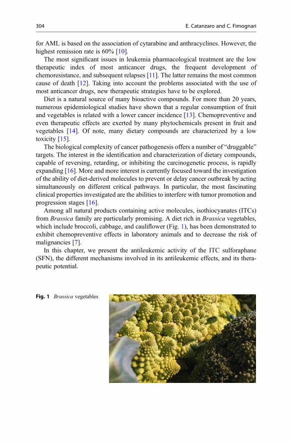
for AML is based on the association of cytarabine and anthracyclines. However, thehighest remission rate is 60% [10].
The most significant issues in leukemia pharmacological treatment are the lowtherapeutic index of most anticancer drugs, the frequent development ofchemoresistance, and subsequent relapses [11]. The latter remains the most commoncause of death [12]. Taking into account the problems associated with the use ofmost anticancer drugs, new therapeutic strategies have to be explored.
Diet is a natural source of many bioactive compounds. For more than 20 years,numerous epidemiological studies have shown that a regular consumption of fruitand vegetables is related with a lower cancer incidence [13]. Chemopreventive andeven therapeutic effects are exerted by many phytochemicals present in fruit andvegetables [14]. Of note, many dietary compounds are characterized by a lowtoxicity [15].
The biological complexity of cancer pathogenesis offers a number of “druggable”targets. The interest in the identification and characterization of dietary compounds,capable of reversing, retarding, or inhibiting the carcinogenetic process, is rapidlyexpanding [16]. More and more interest is currently focused toward the investigationof the ability of diet-derived molecules to prevent or delay cancer outbreak by actingsimultaneously on different critical pathways. In particular, the most fascinatingclinical properties investigated are the abilities to interfere with tumor promotion andprogression stages [16].
Among all natural products containing active molecules, isothiocyanates (ITCs)from Brassica family are particularly promising. A diet rich in Brassica vegetables,which include broccoli, cabbage, and cauliflower (Fig. 1), has been demonstrated toexhibit chemopreventive effects in laboratory animals and to decrease the risk ofmalignancies [7].
In this chapter, we present the antileukemic activity of the ITC sulforaphane(SFN), the different mechanisms involved in its antileukemic effects, and its thera-peutic potential.
Fig. 1 Brassica vegetables
304 E. Catanzaro and C. Fimognari
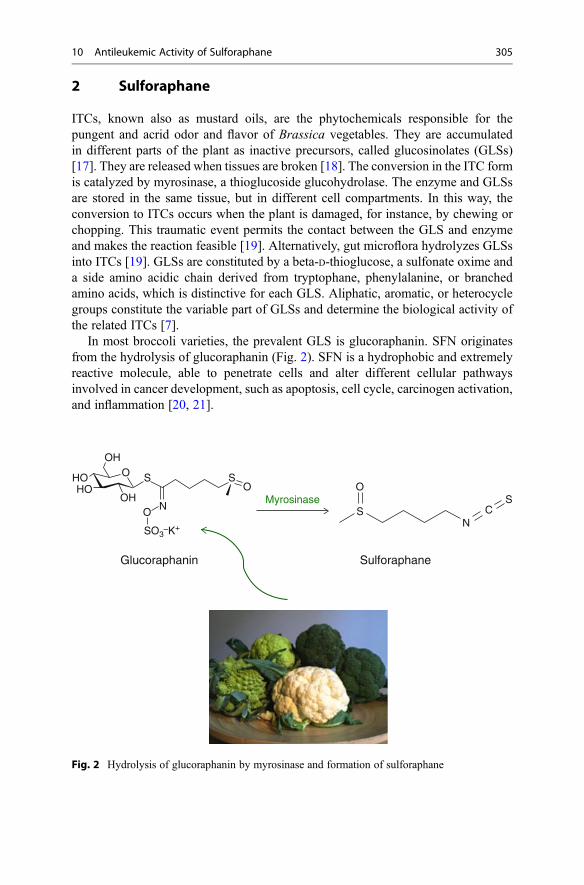
2 Sulforaphane
ITCs, known also as mustard oils, are the phytochemicals responsible for thepungent and acrid odor and flavor of Brassica vegetables. They are accumulatedin different parts of the plant as inactive precursors, called glucosinolates (GLSs)[17]. They are released when tissues are broken [18]. The conversion in the ITC formis catalyzed by myrosinase, a thioglucoside glucohydrolase. The enzyme and GLSsare stored in the same tissue, but in different cell compartments. In this way, theconversion to ITCs occurs when the plant is damaged, for instance, by chewing orchopping. This traumatic event permits the contact between the GLS and enzymeand makes the reaction feasible [19]. Alternatively, gut microflora hydrolyzes GLSsinto ITCs [19]. GLSs are constituted by a beta-D-thioglucose, a sulfonate oxime anda side amino acidic chain derived from tryptophane, phenylalanine, or branchedamino acids, which is distinctive for each GLS. Aliphatic, aromatic, or heterocyclegroups constitute the variable part of GLSs and determine the biological activity ofthe related ITCs [7].
In most broccoli varieties, the prevalent GLS is glucoraphanin. SFN originatesfrom the hydrolysis of glucoraphanin (Fig. 2). SFN is a hydrophobic and extremelyreactive molecule, able to penetrate cells and alter different cellular pathwaysinvolved in cancer development, such as apoptosis, cell cycle, carcinogen activation,and inflammation [20, 21].
Glucoraphanin
O
S
SN
CS
OS
NOH
OHOHO
OH
O
SO3–K+
Myrosinase
Sulforaphane
Fig. 2 Hydrolysis of glucoraphanin by myrosinase and formation of sulforaphane
10 Antileukemic Activity of Sulforaphane 305
3 Sulforaphane and Leukemia
The chemopreventive activity of SFN has been investigated for decades. Morerecently, its role as a therapeutic agent in hematological malignancies has beenexplored, and very interesting results have been obtained.
The antileukemic activity of SFN can be exploited in two different ways,depending on the treatment doses: at nontoxic concentrations (concentrations thatdon’t induce a decrease in cell viability), it acts as a chemopreventive agent able toblock cancer formation; at higher doses, SFN exhibits a therapeutic activity [22]. Inthis context, SFN has been tested as a single drug or in combination with differentanticancer drugs.
4 Antileukemic Mechanisms of SFN
The antileukemic activity of SFN has been tested by different experimentalapproaches, such as a number of leukemia cell lines, animal models, and blastfrom leukemia patients.
4.1 Modulation of Phase I and Phase II Enzymes
As previously described, the bioactivation of chemical carcinogens is catalyzed byphase I enzymes, in particular the CYP450 family. The regulation of these proteins interms of expression and function is crucial to determine the activity of a chemicalcarcinogen. SFN modulates the gene expression and function of specific CYP450isoforms [23]. Several studies conducted in rats or humans showed its ability tointeract with CYP2B, CYP3A, CYP1A2, and CYP2E1 [24, 25]. CYP3A catalyzesthe conversion of aflatoxin B into its epoxide active form, which interacts with DNAcreating the mutagenic adduct [26]. In rats, SFN decreases the synthesis or increasesthe degradation of CYP2B and CYP3A [24]. As well, SFN reduces mRNA levels ofCYP3A4 in humans’ hepatocytes [25]. CYP1A2 catalyzes the activation of hetero-cyclic amines [27], such as the conversion of 2-amino-3-methylimidazo-[4,5-f]quinoline to its mutagenic intermediates. Although SFN induces no alteration inCYP1A2 hepatic levels in rats, it inhibits its metabolic activity and makes theenzyme not catalytic competent [24]. At last, SFN inhibits the genotoxicity ofN-nitrosodimethylamine by acting as a competitive inhibitor of CYP2E1 inacetone-treated liver rat microsomes [28]. On the whole, the above-presented studiesdemonstrate that, although in animal models, SFN interferes with the activation ofmany carcinogens.
Phase II enzymes inactivate carcinogenic intermediates before their interactionwith nucleic acids. Nuclear factor (erythroid-derived 2)-related factor 2 (Nrf2) is acytoplasmatic transcription factor expressed by nearly all cell types. Its function isblocked by the binding with its suppressor, i.e., kelch-like ECH-associated protein1, which prevents Nrf2 nuclear translocation and activation of Nrf2-target genes
306 E. Catanzaro and C. Fimognari
through binding to the antioxidant response element. Activated Nrf2 translocates tothe nucleus and upregulates target genes such as glutamate-cysteine ligase catalytic(GCLC), modifier (GCLM) subunits, and NAD(P)H-quinone oxidoreductase(QR) [22].
In humans, oral administration of a dietary extract of broccoli sprouts increasesthe mRNA levels of Nrf2-dependent enzymes, such as glutathione transferase,versus a placebo-controlled group [29]. In addition, differentiated humanpromyelocytic cells (HL-60) used as a model of neutrophils and treated with 5 μMSFN for 16 h show an increase in glutathione (GSH) levels [30]. This is aninteresting finding because GSH plays a pivotal role in preventing the uncontrolledformation of free radicals and ROS. The same study reports an increased expressionof GCLC and GCLM and a better intracellular GSH/oxidized glutathione ratio[30]. Glutamate-cysteine ligase is constituted by a catalytic subunit (GCLC) and aregulatory subunit (GCLM) and represents the rate-limiting enzyme in the GSHbiosynthesis pathway. Very similar effects were found on human lymphoblasts,where SFN promotes the nuclear translocation of Nrf2 through the release fromkelch-like ECH-associated protein 1. In the nucleus, Nrf2 activates the antioxidantresponse element transcription system, which in turn increases the expression of theγ-glutamylcysteine synthetase. This enzyme catalyzes the reaction between L-gluta-mate and cysteine to synthesize glutamylcysteine, which is involved in GSH syn-thesis [31]. On the whole, the above-reported studies suggest that SFN is able torestore the intracellular redox state.
One of the most representative phase II enzymes is QR. QR blocks the productionof ROS by starting from quinones and the subsequent depletion of GSH levels[16]. SFN induces QR activity in lymphoblastic cells at very low concentrationscompared to those required to decrease cell viability (0.35 μM versus 1 μM) [22].
The combination of the above-described activities of SFN leads to the detoxifi-cation of carcinogens and thus improves the cellular defense mechanisms frompotential mutagenic events [7].
Of note, the ability of SFN of modulating metabolic enzymes can be reached bythe consumption of 200 μM of SFN-containing broccoli sprouts, which lead toplasma concentrations less than 3 μM [32].
4.2 Apoptosis Induction
Apoptosis, also called programmed cell death, is a mechanism involved in cancerdevelopment [33]. Healthy cells are able to detect and integrate intra- and extracel-lular signals and induce, when necessary, apoptosis through the intrinsic or theextrinsic pathway [33]. The two pathways have the same downstream effectorsand initiate a proteolytic pathway involving effector caspases that directly promoteapoptosis. As an example, high levels of mitochondrial calcium and ROS damagethe mitochondrial membrane and allow the release of cytochrome c that initiates thecaspases’ cascade and triggers the intrinsic apoptotic pathway [34]. Several proteinsare target of caspases and used as markers of apoptosis. PARP [poly (ADP-ribose)
10 Antileukemic Activity of Sulforaphane 307

polymerase] is the substrate of caspase 3 and high levels of cleaved PARP are used asmarker of apoptosis [35].
Cancer cells develop numerous strategies to limit or evade apoptosis, mostlyacting on key actors involved in the programmed cell death control, such as thetumor suppressor gene p53 [36]. p53 detects DNA damage and triggers apoptosisthrough the upregulation of BH3-only proteins, such as Bax, or by an unbalancebetween pro- and anti-apoptotic proteins such as Bax and Bcl-2 [33]. Bcl-2 is ananti-apoptotic protein; it acts by inhibiting proapoptotic proteins, such as Bax andBak. Bcl-2 is located on the cytoplasmic surface of the mitochondrial outer mem-brane, the endoplasmic reticulum, and the nuclear envelope. It detects the damagesof these organelles and modulates their activity by modifying proteins’ and mole-cules’ flux. When located on the cytoplasmic mitochondrial membrane, Bcl-2 formsheterodimers with proapoptotic proteins such as Bax and inhibits their activity. Itblocks apoptosis by inhibiting both the formation of mitochondrial transition poresand the release of cytochrome c, with subsequent inactivation of the caspases’cascade [37]. When Bax is not bound, it breaks the membrane allowing the releaseof proapoptotic proteins. Among these, the most important one is cytochrome c,which directly activates the intrinsic apoptotic pathway through the caspases’cascade [33].
The mechanisms responsible for the proapoptotic activity of SFN are complex.ROS production is a key event in SFN-induced apoptosis. In promonocytic leukemiacells (U937), SFN causes an increase in ROS levels that triggers the loss of themitochondrial membrane potential and activates the intrinsic apoptotic pathway [38,39]. The rise levels of caspase 3, 8, and 9 and the cleaved PARP observed after SFNtreatment on T- and B-leukemia cells [20] confirm these results.
On leukemia T cells, SFN modulates the expression of some proteins that play acritical role in the control of cell life or death: p53, Bax, and Bcl-2. p53 is a tumorsuppressor gene that detects DNA damage and arrests the cell cycle at the G1/Sphase. In this way, DNA damage can be repaired before DNA replication [36]. If thedamage cannot be repaired, p53 induces apoptosis. SFN increases Bax and p53protein levels, while it doesn’t affect Bcl-2 protein levels in leukemia T-cell lines[19]. Although there isn’t a modulation of Bcl-2, the proapoptotic effect of SFNresults from a perturbed Bax/Bcl-2 ratio. This is due to the stimulation of Bax geneexpression caused by the increased p53 protein levels induced by SFN [40].
SFN induces apoptosis in each phase of the cell cycle, but with different inten-sities. The highest proapoptotic activity is exerted on the G1-phase cells, followed bythose that are in the S phase, and lastly by those in the G2/M phase. This differentactivity is probably due to the different expressions of p53 in the different phases ofthe cell cycle. The highest expression of p53 is reported during the G1 phase,whereas it decreases from the S throughout the subsequent cell-cycle phases.These data are consistent with the notion that p53 seems to play a role in the SFNproapoptotic activity [41].
SFN causes a dose- and time-dependent reduction of viability on several leuke-mia cell lines such as REH (ALL cell line), HL-60 (promyelocytic leukemia), andJurkat (T-ALL) [8]. Depending on experimental conditions, it induces apoptosis or
308 E. Catanzaro and C. Fimognari

necrosis. After prolonged treatment with high concentrations (30 μM) of SFN,necrosis of T-cell leukemia becomes the predominant mechanism of cell death[19]. These results suggest that very high levels of this molecule can be toxic.However, a 30 μM concentration of SFN cannot be reached by dietary ingestion ofBrassica vegetables. At concentrations between 3 and 10 μM, apoptosis is the mainmechanism of cellular death induced by SFN in all tested cell lines [19, 20].
To mimic the influence of tumor microenvironment and explore the SFN effectsin a more representative model of the in vivo condition, some studies have beenperformed on ALL cell lines cultured in hypoxia. Hypoxic areas develop in manysolid tumors and the bone marrow environment [8]. In hypoxia, cell metabolismchanges as well as the characteristics of the microenvironment such as pH. Thesefactors influence the cancer response to the antineoplastic drugs. After SFN treat-ment in hypoxic conditions, ROS production was confirmed, as well as itsproapoptotic potential, even though less marked if compared to normoxic operatingconditions. The pathways triggered by SFN in hypoxic and normoxic conditions areprobably the same. For example, after prolonged treatment times, necrosis representsthe predominant mechanism of cell death induced by SFN in hypoxic conditions. Onthe while, these data suggest that the SFN efficacy is comparatively conserved inhypoxia [8].
Ex vivo models are highly predictive of patients’ responses and clinical out-comes, as compared to in vitro cell lines. This is mainly due to the ability of ex vivomodels in maintaining the heterogeneity of in vivo tumors, as well as the pharma-cological determinants and the growth kinetic factors. Cellular populations obtainedfrom patients are largely characterized by the same in vivo environment. Thesefeatures mimic the biological complexity of tumors in clinical practice, with theadvantage of controlled conditions [42]. Ex vivo experiments performed on blastsfrom leukemia patients show an interesting proapoptotic potential of SFN in patientssuffering from AML, pre-B-ALL, B-ALL, and T-ALL, but not in those sufferingfrom CLL [8]. The IC50 (concentration able to decrease cell viability by 50%following one cell-cycle exposure) on patient lymphoblasts was in the 7–13 μMrange [20].
Taken together, SFN controls the spread of leukemic cells, but apparently withdifferent sensitivities for the diverse types of leukemia.
4.3 Cell-Cycle Modulation
Cell cycle is a finely regulated process that allows to control cell growth andmaintain a balance between cell death and proliferation [43]. It is divided into foursubsequent phases: G1, S, G2, and M phase. During the G1 phase, quiescent cells (G0
phase) start synthesizing mRNA and proteins necessary for DNA synthesis(S phase). After DNA synthesis, cells go through the G2 phase where more biosyn-thetic activity occurs and finally reach M phase where cell division takes place[44]. Cyclin (cyc) and cyclin-dependent kinases (cdk) regulate each phase of thisprocess [43]. Kinase complexes are formed by a catalytic unit (cdk) and a regulatory
10 Antileukemic Activity of Sulforaphane 309

subunit (cyc). Cycs are substrate and phase specific. They modulate cdk activity andtheir levels oscillate during the different phases of the cell cycle. Cdk/cyclin isregulated, both negatively and positively, by reversible phosphorylation or byinhibitory molecules such as p21 [45].
SFN induces a block of the cell cycle on T-ALL and pre-B-ALL cells, whichcontributes to its ability to inhibit leukemia cell growth [20]. The inhibition wastime dependent and mediated by an accumulation of cells in the G2/M phase. Theblock was evident after treatment with SFN at concentrations of 10 and 30 μM[19]. In the same experimental conditions, SFN induced apoptosis of proliferatingcells. Taken together, these data show that the block of the cell cycle and apoptosisare two independent events induced by SFN and that apoptosis is not a conse-quence of cells’ inability to proceed through the cell cycle and overcome growtharrest [19].
The antiproliferative effects of SFN are imputable to its capability of modulatingcell-cycle regulatory proteins [20]. The G2/M block was due to the inactivation ofcdc-2, operated by a phosphatase that removes the inhibitory phosphates on thecyc/cdk complex, and to the increase in p21 protein levels. The overexpression ofp21 seems to have an important role. This protein modulates cell cycle and otherbiological activities mainly by binding and inhibiting cdks such as cdc-2 and leadingto growth arrest at specific cell-cycle stages. SFN inactivates the maturation-promoting factor cdc-2/cyclin B1 and blocks the cell cycle in the G2/M phase bythe inhibitory phosphorylation of cdc-2 [20]. Cyclin B1 was unchanged after SFNtreatment [20]. Cdc-2/cyclin B1 complex modulates the cell transition from the G2 tothe M phase. When cdc-2 is phosphorylated, it cannot activate cyclin B and cdc-25C,responsible for the phosphorylation/activation of cdc-2 itself. In this way, cell cyclecannot proceed through the M phase [46, 47].
Another mechanism involved in the antiproliferative activity of SFN is theinhibition of plasmatic protein kinase B (AKT)/mammalian target of rapamycin(mTOR). Phosphatidylinositol 3-kinase (PI3K)/AKT/mTOR is a pathway involvedin cellular proliferation. Among various functions, it controls cell-cycle progressionand is constitutively activated in different kinds of tumors. In normal cells, whenactivated, PI3K phosphorylates and activates AKT, which starts the signaling cas-cade. In turn, AKT activates mTOR that induces the transcription of many proteins,such as p70, a protein that promotes cell proliferation [48]. About 30% of all pre-B-ALL is characterized by the constitutive activation of PI3K/AKT/mTOR, whichinduces cancer cell survival and permanent cell growth state. Taken together, thesefindings support the role of PI3K/AKT/mTOR as a potential target to develop newantileukemic therapeutic strategies [49]. It has been demonstrated that SFNdecreases the levels of both total and phosphorylated AKT and mTOR [20,50]. mTOR, in turn, leads to the block of cells in the G1 phase [50]. These resultscan be very useful if translated to the clinical practice, where SFN could be effectivefor the treatment of ALL with activated AKT.
A role for DNA damage in the antiproliferative effects of SFN was excluded. SFNdid not induce double-strand breaks (DSBs) on pre-B leukocytes [20].
310 E. Catanzaro and C. Fimognari
4.4 Other Mechanisms Involved in the Antileukemic Activityof SFN
Despite the promising antileukemic activity of SFN, the molecular mechanismsunderlying its effects are not completely defined. Recent studies have found a roleof SFN in the modulation of the signal transducers and activators of transcription(STAT). STAT is a group of cytoplasmic silent transcription factors. They have acritical role in different biologic processes such as cellular growth, apoptosis,differentiation, and proliferation and are finely regulated in normal cells. Hemato-logical malignancies including leukemias are characterized by constitutively activeforms of some STAT transduction pathways [51]. For example, CML is character-ized by an aberrant STAT5 pathway [51]. SFN has been shown to inhibit STAT5 andthus its target genes in a mouse IL-3-dependent pre-B-cell line and in a human CMLcell line [52].
Another important molecular target of SFN is NF-kB. It has a central role in thesurvival and differentiation pathways of B cells. It protects cells from the damageinduced by chemotherapy or ionizing radiations and blocks apoptosis [53]. SFNblocks the DNA-binding activity of NF-kB through its interaction with the essentialcysteine residues of NF-kB subunits [54].
5 Bioavailability and Safety Issues of SFN
An in vivo study in a mouse model demonstrated the SFN efficacy in controllinghuman leukemic cell expansion. After 4 days of oral administration of 4 mg/die ofSFN, a significant reduction of tumor growth was observed when SFN was admin-istrated in subcutaneous xenograft tumor models. However, the oral administrationof SFN just reduced leukemia progression in the first 2 weeks, but it did not changethe survival rate [20]. This incomplete response is probably due to the inability ofSFN to reach pharmacologically relevant concentrations in some tissues, like thespine or bone marrow, where leukemia cells start their expansion since the veryinitial phases [20]. Furthermore, the metabolism of SFN generates some species,including SFN-cysteine, SFN-glutathione, and SFN-N-acetylcysteine, that are muchless effective on ALL cells than SFN [20]. This suggests a limited toxic potential ofSFN and a time-limited action. Therefore, further studies need to be done in order tofind an administration way that allows maintaining the therapeutic concentrations ofSFN in vivo.
The bioavailability of SFN depends on many factors. Its pharmacokinetics hasbeen elucidated in some human studies. In particular, dietary SFN bioavailabilitydepends on the vegetal source, the food preparation process, the pH during theconversion of GLS to ITCs, and many other variables. For example, 150 g ofbroccoli contain between 56 and 112 mg of SFN that correspond to an intakebetween 0.75 and 1.5 mg/kg b.w. [55]. After oral administration, ITCs are absorbed,then conjugated to GSH, and finally metabolized to mercapturic acid and eliminated
10 Antileukemic Activity of Sulforaphane 311

through the urine. Some in vivo studies demonstrated that the peak plasma concen-tration of SFN after oral administration is reached in about 1 h. The lipophilic natureand small size of the molecule favor its rapid absorption [55]. Likewise, some studiesin humans showed that SFN is rapidly absorbed, reaching the peak plasma concen-tration after 1 h after broccoli sprouts or broccoli soup consumption [32]. Further-more, it was demonstrated in humans that the peak plasma concentration can rangefrom 3.4% (cooked broccoli) to 37% (raw broccoli) of the ingested dose [56]. There-fore, taking into account that broccoli sprouts contain 200 μM SFN and that SFNhalf life is about 1.75 h, after 1 h the blood concentration will range between 0.943and 2.27 μM/L, implying a potential chemopreventive significance [32]. Theseresults indicate that a dietary consumption of Brassica vegetables is not enough tolead to an antileukemic activity or to reach inhibitory cell growth concentrations.
Clinical studies on prostate cancer patients have been performed to test thetoxicity of a daily consumption of a 200 μM lyophilized SFN prepared startingfrom an aqueous extract. No adverse effects were reported [57].
An ideal anticancer agent should be selective for neoplastic cells. This underlinesthe need to know its effects on healthy non-transformed cells. Since SFN inducesapoptosis, decreases cell viability, and blocks the cell cycle of T-ALL cells, itsactivity has been investigated on non-transformed T lymphocytes as a normalcounterpart of leukemia cells. One study showed that 72 h treatment with 30 μMSFN inhibits cell cycle and increases apoptosis in a dose-dependent manner innormal T cells. As well, it induces an increased expression of some proapoptoticproteins such as p53 and Bax. However, although SFN is not completely selectivefor cancer cells, the activity was less pronounced than in T-ALL cells [58]. Otherstudies have shown different outcomes on different cell types: SFN did not induceany significant cytotoxic or proapoptotic effect on normal peripheral blood mono-nuclear cells, thus suggesting a selective activity for transformed cells [20].
Of note, the above-presented studies were performed in vitro. In vivo studies haveto be done in order to better evaluate its toxicological profile. In this context, somerecent studies explored the genotoxicity of SFN. Phospho-H2AX is the principalbiomarker of DNA DSBs. SFN was able to induce DSBs in adenocarcinoma cells,although DNA repair is achieved through the homologous recombination repairpathway (HRR) [59]. HRR repairs DSBs through the exchange of the damagedDNA strand with a similar or identical nucleotide sequence; this information isretrieved from undamaged sequences of homologue chromosomes or sister chroma-tids [60]. The key effector of this pathway is Rad51 protein, which promotes strandexchange with a regular nucleotide filament and facilitates the homologous pairing[61]. In particular, it was demonstrated that DSBs induced by SFN were associatedwith Rad51 foci. These data confirm the hypothesis that DSBs repair is mediated byHRR. Moreover, HRR activation is dependent on the checkpoint kinase 1 (Chk-1), akinase that controls a checkpoint in the G2 phase of the cell cycle and induces Rad51activation. Therefore, cell-cycle block induced by SFN in the G2/M phase corrob-orates the hypothesis that SFN induces DSBs that are repairable through the HRRpathway [59, 62]. In 2012, Suppipat et al. [20] demonstrated that, compared to afrank genotoxic compound like etoposide, 7.5 μM SFN did not induce a significant
312 E. Catanzaro and C. Fimognari

increase in phosphorylated H2AX in human pre-B cells and T-lymphoblastoid cells.Similar results were reported on human lymphocyte, where SFN did not increase thenumber of micronuclei, a biomarker of DNA mutations [63]. Taken together, theabove-reported studies suggest that SFN is able to induce genotoxic lesions, whichare repairable and do not evolve in a frank mutagenic effect.
6 Conclusions
Carcinogenesis is a multistep process in which normal cells accumulate sequentialalterations in critical genes through which cells gain the peculiar tumorigenicphenotype. These new developed biological capabilities, called “hallmarks of can-cer” by Hanahan and Weinberg, allow the transformed cells to survive, proliferate,and differentiate [3]. The multifactorial and complex nature of carcinogenesis led tofocus on multi-target or pleiotropic therapy able to affect different pathwayssimultaneously.
Diet is a common source of biologically active compounds. Anticancer activityhas been reported for many natural compounds, such as curcumin, resveratrol,genistein, etc. SFN is an ITC stored in edible Brassica vegetables in the form ofinactive precursors (GLS). SFN exhibits chemopreventive and anticancer activitieson many different tumors, imputable to its capacity to interfere with different steps ofthe carcinogenetic process [64]. It inhibits phase I enzymes involved in the activationof xenobiotics and stimulates phase II enzymes, thus promoting the inactivation andelimination of carcinogens. Furthermore, it modulates different critical pathways forthe neoplastic process, such as apoptosis and cell cycle [41].
The antileukemic activity of SFN was not completely elucidated, but recentstudies evidenced a very interesting profile [20]. SFN exerts antileukemic effectsin different leukemia cell lines and blasts from pediatric and adult ALL, T-ALL,B-ALL, and AML patients [8, 20]. SFN acts mostly by inducing apoptosis and cell-cycle arrest and modulating diverse signaling pathways. On human leukemias,SFN-induced apoptosis is triggered by ROS, which trigger the activation of cancercell growth signaling pathways. Apoptosis develops typically through p53- andBcl-2-dependent pathways. Caspases 3, 8, and 9 are implicated as well. SFN blocksthe cell cycle in the G2/M phase, and AKT/mTOR pathway, a B-ALL leukemiapreferential target, is implicated.
Besides the promising pharmacological profile, SFN shows an equally favorabletoxicological profile. Currently, one of the most important problems of antineoplastictherapy is its high toxicity that often limits the efficacy of the treatment [7]. Thedietary origin of SFN lets presume a good tolerability. Several studies have con-firmed the safety in humans and in vitro studies have excluded a genotoxiceffect [63].
Drug resistance is the second most limiting effect of antineoplastic chemotherapy.SFN could be used on resistant cancer cells. As an example, SFN increased theefficacy of anthracyclines in cell models characterized by p53 dysregulations. Sincep53 mutations can be detected in many types of leukemia, such as T-ALL, B-cell
10 Antileukemic Activity of Sulforaphane 313

precursor-ALL, and AML [65], the above-reported evidence would suggest itsactivity in a variety of hematological malignancies resistant to conventional chemo-therapy. SFN restored chemosensitivity of doxorubicin-resistant cells and increasedits potency. In particular, co-treatment with SFN decreased the effective dose ofdoxorubicin required to achieve the therapeutic effect within the range tolerated inclinical practice [66].
In conclusion, many studies suggest a role of SFN as candidate for the treatmentof many leukemia types. Further efforts will be made in order to better define itspharmacotoxicological profile.
References
1. Ahmad A, Ormiston-Smith N, Sasieni P (2015) Trends in the lifetime risk of developing cancerin great Britain: comparison of risk for those born from 1930 to 1960. Br J Cancer 112:943–947
2. Society AA http://www.cancer.org/acs/groups/content/@editorial/documents/document/acspc-044552.pdf. Accessed 9 Sept 2015
3. Hanahan D, Weinberg RA (2011) Hallmarks of cancer: the next generation. Cell 144:646–6744. Kufe DW, Pollock RE, Weichselbaum RR, Bast RC, Gansler TS, Holland JF, Frei E (2003)
Holland-Frei cancer medicine. BC Decker, Hamilton5. Devi PU (2004) Basics of carcinogenesis. Health Admin 17:16–246. Iyanagi T (2007) Molecular mechanism of phase I and phase Ii drug‐metabolizing enzymes:
implications for detoxification. Int Rev Cytol 260:35–1127. Fimognari C, Hrelia P (2007) Sulforaphane as a promising molecule for fighting cancer. Mutat
Res-Rev Mutat 635:90–1048. Fimognari C, Turrini E, Sestili P, Calcabrini C, Carulli G, Fontanelli G, Rousseau M, Cantelli-
Forti G, Hrelia P (2014) Antileukemic activity of sulforaphane in primary blasts from patientsaffected by myelo- and lympho-proliferative disorders and in hypoxic conditions. PLoS One 9(7):e101991
9. Vardiman JW, Thiele J, Arber DA, Brunning RD, Borowitz MJ, Porwit A, Harris NL, Le BeauMM, Hellström-Lindberg E, Tefferi A (2009) The 2008 revision of the World Health Organi-zation (WHO) classification of myeloid neoplasms and acute leukemia: rationale and importantchanges. Blood 114:937–951
10. Fathi AT, Karp JE (2009) New agents in acute myeloid leukemia: beyond cytarabine andanthracyclines. Curr Oncol Rep 11:346–352
11. Arcangeli A, Pillozzi S, Becchetti A (2012) Targeting Ion channels in leukemias: a newchallenge for treatment. Curr Med Chem 19:683–696
12. Karp JE, Ross DD, Yang W, Tidwell ML, Wei Y, Greer J, Mann DL, Nakanishi T, Wright JJ,Colevas AD (2003) Timed sequential therapy of acute leukemia with flavopiridol in vitro modelfor a phase i clinical trial. Clin Cancer Res 9:307–315
13. Danaei G, Vander Hoorn S, Lopez AD, Murray CJ, Ezzati M, group CRAc (2005) Causes ofcancer in the World: comparative risk assessment of nine behavioural and environmental riskfactors. Lancet 366:1784–1793
14. Si H, Liu D (2014) Dietary antiaging phytochemicals and mechanisms associated withprolonged survival. J Nutr Biochem 25:581–591
15. Keum Y-S, Jeong W-S, Kong A (2005) Chemopreventive functions of isothiocyanates. DrugNews Perspect 18:445–451
16. Turrini E, Ferruzzi L, Fimognari C (2014) Natural compounds to overcome cancerchemoresistance: toxicological and clinical issues. Expert Opin Drug Metab Toxicol10:1677–1690
314 E. Catanzaro and C. Fimognari

17. de Figueiredo SM, Filho SA, Nogueira-Machado JA, Caligiorne RB (2013) The anti-oxidantproperties of isothiocyanates: a review. Recent Pat Endocr Metab Immune Drug Discov 7:213–225
18. Zhang Y, Tang L (2007) Discovery and development of sulforaphane as a cancer chemopre-ventive phytochemical. Acta Pharmacol Sin 28:1343–1354
19. Fimognari C, N€usse M, Cesari R, Iori R, Cantelli-Forti G, Hrelia P (2002) Growth inhibition,cell-cycle arrest and apoptosis in human T-cell leukemia by the isothiocyanate sulforaphane.Carcinogenesis 23:581–586
20. Suppipat K, Park CS, Shen Y, Zhu X, Lacorazza HD (2012) Sulforaphane induces cell cyclearrest and apoptosis in acute lymphoblastic leukemia cells. PLoS One 7(12):e51251
21. Fimognari C, Lenzi M, Cantelli-Forti G, Hrelia P (2008) Induction of differentiation in humanpromyelocytic cells by the isothiocyanate sulforaphane. In Vivo 22:317–320
22. Misiewicz I, Skupinska K, Kowalska E, Lubinski J, Kasprzycka-Guttman T (2004)Sulforaphane-mediated induction of a phase 2 detoxifying enzyme Nad (P) H: quinone reduc-tase and apoptosis in human lymphoblastoid cells. Acta Biochim Pol 51(3):711–722
23. Jancova P, Anzenbacher P, Anzenbacherova E (2010) Phase II drug metabolizing enzymes.Biomed Pap 154:103–116
24. Yoxall V, Kentish P, Coldham N, Kuhnert N, Sauer MJ, Ioannides C (2005) Modulation ofhepatic cytochromes P450 and phase II enzymes by dietary doses of sulforaphane in rats:implications for its chemopreventive activity. Int J Cancer 117:356–362
25. Mahéo K, Morel F, Langouët S, Kramer H, Le Ferrec E, Ketterer B, Guillouzo A (1997)Inhibition of cytochromes P-450 and induction of glutathione S-transferases by sulforaphane inprimary human and rat hepatocytes. Cancer Res 57:3649–3652
26. Kamdem LK, Meineke I, Gödtel-Armbrust U, Brockmöller J, Wojnowski L (2006) Dominantcontribution of P450 3a4 to the hepatic carcinogenic activation of aflatoxin B1. Chem ResToxicol 19:577–586
27. Dashwood RH (2002) Modulation of heterocyclic amine-induced mutagenicity and carcinoge-nicity: an ‘a-to-Z’guide to chemopreventive agents, promoters, and transgenic models. MutatRes 511:89–112
28. Juge N, Mithen R, Traka M (2007) Molecular basis for chemoprevention by sulforaphane: acomprehensive review. Cell Mol Life Sci 64:1105–1127
29. Riedl MA, Saxon A, Diaz-Sanchez D (2009) Oral sulforaphane increases phase II antioxidantenzymes in the human upper airway. Clin Immunol 130:244–251
30. Dias I, Chapple I, Milward M, Grant MM, Hill E, Brown J, Griffiths HR (2013) Sulforaphanerestores cellular glutathione levels and reduces chronic periodontitis neutrophil hyperactivityin vitro. PLoS One 8:e66407
31. Mizuno K, Kume T, Muto C, Takada-Takatori Y, Izumi Y, Sugimoto H, Akaike A (2011)Glutathione biosynthesis via activation of the nuclear factor E2-related factor 2 (Nrf2)-antiox-idant-response element (are) pathway is essential for neuroprotective effects of Sulforaphaneand 6-(methylsulfinyl) hexyl isothiocyanate. J Pharmacol Sc 115:320–328
32. Ye L, Dinkova-Kostova AT, Wade KL, Zhang Y, Shapiro TA, Talalay P (2002) Quantitativedetermination of dithiocarbamates in human plasma, serum, erythrocytes and urine: pharmaco-kinetics of broccoli sprout isothiocyanates in humans. Clin Chim Acta 316:43–53
33. Adams J, Cory S (2007) The Bcl-2 apoptotic switch in cancer development and therapy.Oncogene 26:1324–1337
34. Fimognari C, Lenzi M, Ferruzzi L, Turrini E, Scartezzini P, Poli F, Gotti R, Guerrini A,Carulli G, Ottaviano V (2011) Mitochondrial pathway mediates the antileukemic effects ofhemidesmus indicus, a promising botanical drug. PLoS One 6:e21544
35. Khan N, Afaq F, Mukhtar H (2006) Apoptosis by dietary factors: the suicide solution fordelaying cancer growth. Carcinogenesis 28:233–239
36. Junttila MR, Evan GI (2009) P53 – a jack of all trades but master of none. Nat Rev Cancer9:821–829
37. Portier BP, Taglialatela G (2006) Bcl-2 localized at the nuclear compartment induces apoptosisafter transient overexpression. J Biol Chem 281:40493–40502
10 Antileukemic Activity of Sulforaphane 315

38. Sestili P, Fimognari C (2015) Cytotoxic and antitumor activity of sulforaphane: the role ofreactive oxygen species. Biomed Res Int 2015:402386
39. Choi WY, Choi BT, Lee WH, Choi YH (2008) Sulforaphane generates reactive oxygen speciesleading to mitochondrial perturbation for apoptosis in human leukemia U937 Cells. BiomedPharmacother 62:637–644
40. Xu J, Ji L-D, Xu L-H (2006) Lead-induced apoptosis in Pc 12 Cells: involvement of P53, Bcl-2family and caspase-3. Toxicol Lett 166:160–167
41. Fimognari C, Lenzi M, Sciuscio D, Cantelli-Forti G, Hrelia P (2007) Cell-cycle specificity ofsulforaphane-mediated apoptosis in jurkat t-leukemia cells. In Vivo 21:377–380
42. Bosanquet AG, Bell PB (2004) Ex vivo therapeutic index by drug sensitivity assay using freshhuman normal and tumor cells. J Exp Ther Oncol 4:145–154
43. Sandal T (2002) Molecular aspects of the mammalian cell cycle and cancer. Oncologist 7:73–8144. Morgan DO (2007) The cell cycle: principles of control. New Science Press, London45. Clarke JD, Dashwood RH, Ho E (2008) Multi-targeted prevention of cancer by sulforaphane.
Cancer Lette 269:291–30446. Singh SV, Herman-Antosiewicz A, Singh AV, Lew KL, Srivastava SK, Kamath R, Brown KD,
Zhang L, Baskaran R (2004) Sulforaphane-induced G2/M phase cell cycle arrest involvescheckpoint kinase 2-mediated phosphorylation of cell division cycle 25c. J Biol Chem279:25813–25822
47. Guidet S, Meijerink S, Tung HL (2012) Progress in cell cycle research. Springer, New York48. Rafalski VA, Brunet A (2011) Energy metabolism in adult neural stem cell fate. Prog Neurobiol
93:182–20349. Neri L, Cani A, Martelli A, Simioni C, Junghanss C, Tabellini G, Ricci F, Tazzari P, Pagliaro P,
McCubrey J (2014) Targeting the Pi3k/Akt/Mtor signaling pathway in B-precursor acutelymphoblastic leukemia and its therapeutic potential. Leukemia 28:739–748
50. Feldman ME, Apsel B, Uotila A, Loewith R, Knight ZA, Ruggero D, Shokat KM (2009)Active-site inhibitors of Mtor target rapamycin-resistant outputs of Mtorc1 and Mtorc2. PLoSBiol 7:371
51. Bowman T, Garcia R, Turkson J, Jove R (2000) Stats in oncogenesis. Oncogene 19:2474–248852. Pinz S, Unser S, Rascle A (2014) The natural chemopreventive agent sulforaphane inhibits
Stat5 activity. PLoS One 9(6):e9939153. Dolcet X, Llobet D, Pallares J, Matias-Guiu X (2005) Nf-Kb in development and progression of
human cancer. Virchows Arch 446:475–48254. Cheung KL, Kong A-N (2010) Molecular targets of dietary phenethyl isothiocyanate and
sulforaphane for cancer chemoprevention. AAPS J 12:87–9755. Hanlon N, Coldham N, Gielbert A, Kuhnert N, Sauer MJ, King LJ, Ioannides C (2008)
Absolute bioavailability and dose-dependent pharmacokinetic behaviour of dietary doses ofthe chemopreventive isothiocyanate sulforaphane in rat. Br J Nutr 99:559–564
56. Egner PA, Chen JG, Wang JB, Wu Y, Sun Y, Lu JH, Zhu J, Zhang YH, Chen YS, Friesen MD(2011) Bioavailability of sulforaphane from two broccoli sprout beverages: results of a short-term, cross-over clinical trial in Qidong, China. Cancer Prev Res 4:384–395
57. Alumkal JJ, Slottke R, Schwartzman J, Cherala G, Munar M, Graff JN, Beer TM, Ryan CW,Koop DR, Gibbs A (2014) A phase II study of sulforaphane-rich broccoli sprout extracts in menwith recurrent Prostate cancer. Invest New Drugs 33:480–489
58. Fimognari C, N€usse M, Berti F, Iori R, Cantelli‐Forti G, Hrelia P (2003) Sulforaphanemodulates cell cycle and apoptosis in transformed and non-transformed human T lymphocytes.Ann NYAcad Sci 1010:393–398
59. Sekine-Suzuki E, Yu D, Kubota N, Okayasu R, Anzai K (2008) Sulforaphane induces DNAdouble strand breaks predominantly repaired by homologous recombination pathway in humancancer cells. Biochem Biophys Res Commun 377:341–345
60. Dudáš A, Chovanec M (2004) DNA double-strand break repair by homologous recombination.Mutat Res 566:131–167
316 E. Catanzaro and C. Fimognari

61. West SC (2003) Molecular views of recombination proteins and their control. Nat Rev Mol CellBiol 4:435–445
62. Bahassi E, Ovesen J, Riesenberg A, Bernstein W, Hasty P, Stambrook P (2008) The checkpointkinases Chk1 and Chk2 regulate the functional associations between Hbrca2 and Rad51 inresponse to DNA damage. Oncogene 27:3977–3985
63. Fimognari C, Berti F, Iori R, Cantelli-Forti G, Hrelia P (2005) Micronucleus formation andinduction of apoptosis by different isothiocyanates and a mixture of isothiocyanates in humanlymphocyte cultures. Mutat Res 582:1–10
64. TortorellaStephanie M, RoyceSimon G, LicciardiPaul V, KaragiannisTom C (2015) Dietarysulforaphane in cancer chemoprevention: the role of epigenetic regulation and Hdac inhibition.Antioxid Redox Signal 22(16):1382–1424
65. Drexler H, Fombonne S, Matsuo Y, Hu Z, Hamaguchi H, Uphoff C (2000) P53 alterations inhuman leukemia-lymphoma cell lines: in vitro artifact or prerequisite for cell immortalization?Leukemia 14:198–206
66. Fimognari C, N€usse M, Lenzi M, Sciuscio D, Cantelli-Forti G, Hrelia P (2006) Sulforaphaneincreases the efficacy of doxorubicin in mouse fibroblasts characterized by P53 mutations.Mutat Res 601:92–101
10 Antileukemic Activity of Sulforaphane 317

Sulforaphane and Atherosclerosis 11Pon Velayutham Anandh Babu, Chrissa Petersen, andZhenquan Jia
AbstractEpidemiological studies have found associations between cruciferous vegetableconsumption and reduced risk of chronic conditions, such as atherosclerosis,diabetes, and cancer. Sulforaphane, a molecule found in its precursor state incruciferous and other vegetables, is the focus of much current nutritional interest.Others and we have recently reported the beneficial cardiovascular effects ofsulforaphane and the possible molecular mechanisms involved. Sulforaphaneimproved cardiovascular complications such as vascular inflammation, hyperten-sion, and atherosclerosis in animal models. Evidence shows that sulforaphanemay exert the beneficial cardiovascular effects by acting on multiple targets suchas (i) activating Kelch-like ECH-associated protein 1 (Keap1)/nuclear factor[erythroid-derived 2]-like 2 (Nrf2) signaling pathway, (ii) inhibiting inflamma-tory pathways, and (iii) regulating lipid metabolism. However, human studiesrelated to the vascular effects of sulforaphane and cruciferous vegetables arelacking. Hence, well-designed human trials may be needed to evaluate thecardiovascular effects of sulforaphane and cruciferous vegetables and to recom-mend cruciferous vegetables, to improve cardiovascular health. This chapterprovides an overview of recent developments toward the understanding ofcardioprotective effects of sulforaphane and the molecular mechanisms involved.
P.V.A. Babu (*) • C. Petersen (*)Department of Nutrition and Integrative Physiology, College of Health, University of Utah, SaltLake City, UT, USAe-mail: [email protected]; [email protected]
Z. Jia (*)Department of Biology, University of North Carolina at Greensboro, Greensboro, NC, USAe-mail: [email protected]
# Springer International Publishing Switzerland 2017J.-M. Mérillon, K.G. Ramawat (eds.), Glucosinolates, Reference Series inPhytochemistry, DOI 10.1007/978-3-319-25462-3_11
319
KeywordsSulforaphane • Vascular disease • Nrf2 • Atherosclerosis • Cruciferous vegeta-bles • Endothelium • Cardioprotective
AbbreviationsARE Antioxidant response elementC/EBP CCAAT/enhancer-binding proteinCVD Cardiovascular diseaseGCL Glutamate-cysteine ligaseGCS γ-Glutamyl cysteine synthetaseGCLC Glutamate-cysteine ligase catalytic subunitGCLM Glutamate-cysteine ligase modifier subunitGPX Glutathione peroxidaseGR Glutathione reductaseGSH Glutathione-S-transferaseHDL High-density lipoproteinHO-1 Heme oxygenase-1ICAM-1 Intracellular adhesion molecule-1IĸBα Inhibitor of NFĸBIĸKβ IĸB kinaseIL-8 Interleukin-8Keap1 Kelch-like ECH-associated protein 1LDL Low-density lipoproteinLPS LipopolysaccharideMAPK Mitogen-activated protein kinaseMCP-1 Monocyte chemotactic protein-1NFĸB Nuclear factor ĸBNQO1 NADPH quinone oxidoreductaseNrf2 Nuclear factor [erythroid-derived 2]-like 2PPARγ Peroxisome proliferator-activated receptor γROS Reactive oxygen speciesSHRs Spontaneously hypertensive ratsSHRSP Spontaneously hypertensive stroke-proneSMC Smooth muscle cellsSOD Superoxide dismutaseTLR Toll-like receptorTNF-α Tumor necrosis factor-αVCAM-1 Vascular cell adhesion molecule-1VSMCs Vascular smooth muscle cellsWKY rats Wistar Kyoto rats
Contents1 Introduction . . . . . . . . . . . . . . . . . . . . . . . . . . . . . . . . . . . . . . . . . . . . . . . . . . . . . . . . . . . . . . . . . . . . . . . . . . . . . . . . . . 3212 Pathophysiology of Atherosclerosis . . . . . . . . . . . . . . . . . . . . . . . . . . . . . . . . . . . . . . . . . . . . . . . . . . . . . . . . 3213 Sulforaphane: Chemistry, Dietary Sources, and Bioavailability . . . . . . . . . . . . . . . . . . . . . . . . . . . 322
320 P.V.A. Babu et al.

4 Cardiovascular Beneficial Effects of Sulforaphane . . . . . . . . . . . . . . . . . . . . . . . . . . . . . . . . . . . . . . . . . 3235 Molecular Mechanisms Involved in the Cardioprotective Effects of Sulforaphane . . . . . . . 324
5.1 Activation of Nrf2/ARE Signaling Pathway . . . . . . . . . . . . . . . . . . . . . . . . . . . . . . . . . . . . . . . . . 3245.2 Inhibition of Inflammatory Pathways . . . . . . . . . . . . . . . . . . . . . . . . . . . . . . . . . . . . . . . . . . . . . . . . . 3295.3 Modulation of Lipid Metabolism . . . . . . . . . . . . . . . . . . . . . . . . . . . . . . . . . . . . . . . . . . . . . . . . . . . . . 332
6 Conclusions . . . . . . . . . . . . . . . . . . . . . . . . . . . . . . . . . . . . . . . . . . . . . . . . . . . . . . . . . . . . . . . . . . . . . . . . . . . . . . . . . . 333References . . . . . . . . . . . . . . . . . . . . . . . . . . . . . . . . . . . . . . . . . . . . . . . . . . . . . . . . . . . . . . . . . . . . . . . . . . . . . . . . . . . . . . . 333
1 Introduction
Cardiovascular disease (CVD) is the leading cause of death for both men and womenin the United States. Each year approximately 1.5 million American adults have aheart attack or stroke [1]. In 2010 alone, CVD accounted for 31.9% of all deaths, orabout one in every three deaths [2]. Lifestyle factors such as smoking, lack ofphysical activity, and a nutrient-poor diet play a major role in the development ofCVD, and it is therefore mostly preventable [3]. Most of the time, CVD is initiatedthrough a pathological state called atherosclerosis. Atherosclerosis is the buildup ofplaques within the arterial walls that narrows the arterial lumen and restricts theamount of blood that flows through it. The plaques are formed between the tunicaintima and tunica media and are comprised of an accumulation of lipids, cells,debris, and fibrous tissues [4]. In severe cases, the plaques become weak and rupture,attracting clotting factors in the blood to be attracted to and stick to the rupture site[4]. With limited blood flow through these clogged arteries, atherosclerosis leads tomajor complications like heart attack and stroke [4].
Evidence from epidemiological and clinical studies support that a diet containinga variety of fruits and vegetables can prevent the development of CVD [5–9]. Sulfo-raphane, which belongs to a group of molecules called isothiocyanates, is the focusof much current nutritional interest. Glucoraphanin is the precursor for sulforaphaneand is widely present in cruciferous vegetables such as broccoli, cabbage, Brusselssprouts, and non-cruciferous vegetables in the mustard family such as radishes[10]. Epidemiological studies have found associations between cruciferous vegeta-ble consumption and reduced risk of chronic conditions, such as atherosclerosis,diabetes, and cancer [10–13]. This chapter provides an overview of recent develop-ments toward the understanding of cardioprotective effects of sulforaphane and themolecular mechanisms involved. This chapter will specifically focus on the effectsof sulforaphane in the vasculature.
2 Pathophysiology of Atherosclerosis
The endothelium is a monolayer of endothelial cells that line the interior walls of theentire vascular system. Endothelium is essential for maintaining vascular homeosta-sis [14]. It forms a unique barrier for regulating the exchange of biomoleculesbetween blood and tissues and preventing infiltration by leukocytes or other cellsinto the vessel wall [15]. Indeed, a healthy vascular system is maintained by the
11 Sulforaphane and Atherosclerosis 321

proper functioning of the endothelium, and endothelial dysfunction contributessignificantly to vascular dysfunction [16]. Endothelial dysfunction is one of thewarning signs of CVD and is involved in disease states where the normal functionof endothelium is severely affected such as atherosclerosis and hypertension [15,16]. Extensive studies have confirmed that atherosclerosis is initiated due the effectsof endothelial dysfunction [14–16].
Cardiovascular risk factors induce endothelial dysfunctional which can be char-acterized by the enhanced expression of adhesion molecules, binding of bloodmonocytes to endothelium, and increased endothelial permeability and accumulationof circulating lipids [14, 17–21]. Accumulating evidence has shown that leukocytebinding, facilitated by adhesion molecules and chemokines, is a key step in thedevelopment of atherosclerosis [14, 17–20]. The normal endothelial monolayerresists prolonged contact and adhesion of circulating blood monocytes. However,when endothelial cells undergo inflammatory activation, they increase the expres-sion of leukocyte adhesion molecules [22]. Various pro-inflammatory mediators andcell adhesion molecules have been found to be secreted by injured endothelial cells,which include thrombin, tumor necrosis factor-α (TNF-α), monocyte chemotacticprotein-1 (MCP-1), interleukin (IL)-8 (IL-8), vascular cell adhesion molecule-1(VCAM-1), intracellular adhesion molecule-1 (ICAM-1), and E-selectin[17–20]. Increased production of these inflammatory molecules serves as a molec-ular marker for endothelial inflammation. The adhesion molecules facilitate therecruitment of blood monocytes from the circulation to the endothelium followedby transmigration of the leukocytes to the subendothelium [21–23]. Furthermore,when blood monocytes migrate to the innermost layer of the arterial wall, they takeup lipids and lipoprotein particles and they differentiate to macrophage foam cells[22]. These foam cells secrete large amounts of cytokines such as TNF-α, IL-8, andinterleukin-1 (IL-1), which in turn amplify the inflammatory response and enhancevascular complications [22, 23].
3 Sulforaphane: Chemistry, Dietary Sources,and Bioavailability
Sulforaphane [1-isothiocyanate-(4R)-(methylsulfinyl)butane] is a small lipophilicmember of the isothiocyanate family [10]. The isothiocyanates are a group ofnaturally occurring compounds that are usually formed by the hydrolysis ofglucosinolates. Sulforaphane is formed from the precursor glucoraphanin which isa glycoside [10]. Sulforaphane is produced from glucoraphanin when cruciferousvegetables such as broccoli, cabbage, Brussels sprouts, or some non-cruciferousvegetables in the mustard family such as radishes are mechanically broken down,commonly by being chewed. These plants contain the precursor (glucoraphanin) andthe enzyme (myrosinase) to perform the hydrolysis reaction. Glucoraphanin is stableand inert and the plants sequester glucoraphanin from the enzyme myrosinase. Oncethe barrier is broken down, myrosinase hydrolizes the glucosinolate into sulforaph-ane [10] (Fig. 1).
322 P.V.A. Babu et al.
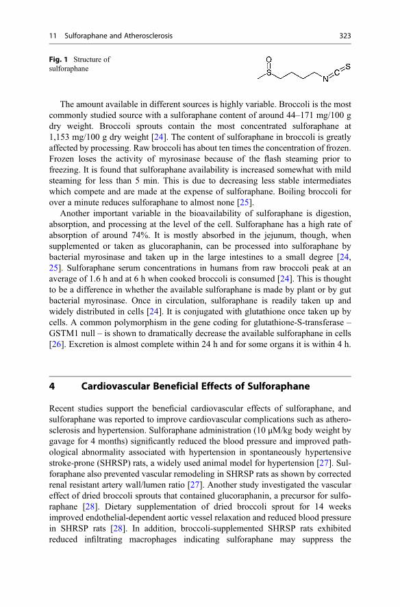
The amount available in different sources is highly variable. Broccoli is the mostcommonly studied source with a sulforaphane content of around 44–171 mg/100 gdry weight. Broccoli sprouts contain the most concentrated sulforaphane at1,153 mg/100 g dry weight [24]. The content of sulforaphane in broccoli is greatlyaffected by processing. Raw broccoli has about ten times the concentration of frozen.Frozen loses the activity of myrosinase because of the flash steaming prior tofreezing. It is found that sulforaphane availability is increased somewhat with mildsteaming for less than 5 min. This is due to decreasing less stable intermediateswhich compete and are made at the expense of sulforaphane. Boiling broccoli forover a minute reduces sulforaphane to almost none [25].
Another important variable in the bioavailability of sulforaphane is digestion,absorption, and processing at the level of the cell. Sulforaphane has a high rate ofabsorption of around 74%. It is mostly absorbed in the jejunum, though, whensupplemented or taken as glucoraphanin, can be processed into sulforaphane bybacterial myrosinase and taken up in the large intestines to a small degree [24,25]. Sulforaphane serum concentrations in humans from raw broccoli peak at anaverage of 1.6 h and at 6 h when cooked broccoli is consumed [24]. This is thoughtto be a difference in whether the available sulforaphane is made by plant or by gutbacterial myrosinase. Once in circulation, sulforaphane is readily taken up andwidely distributed in cells [24]. It is conjugated with glutathione once taken up bycells. A common polymorphism in the gene coding for glutathione-S-transferase –GSTM1 null – is shown to dramatically decrease the available sulforaphane in cells[26]. Excretion is almost complete within 24 h and for some organs it is within 4 h.
4 Cardiovascular Beneficial Effects of Sulforaphane
Recent studies support the beneficial cardiovascular effects of sulforaphane, andsulforaphane was reported to improve cardiovascular complications such as athero-sclerosis and hypertension. Sulforaphane administration (10 μM/kg body weight bygavage for 4 months) significantly reduced the blood pressure and improved path-ological abnormality associated with hypertension in spontaneously hypertensivestroke-prone (SHRSP) rats, a widely used animal model for hypertension [27]. Sul-foraphane also prevented vascular remodeling in SHRSP rats as shown by correctedrenal resistant artery wall/lumen ratio [27]. Another study investigated the vasculareffect of dried broccoli sprouts that contained glucoraphanin, a precursor for sulfo-raphane [28]. Dietary supplementation of dried broccoli sprout for 14 weeksimproved endothelial-dependent aortic vessel relaxation and reduced blood pressurein SHRSP rats [28]. In addition, broccoli-supplemented SHRSP rats exhibitedreduced infiltrating macrophages indicating sulforaphane may suppress the
Fig. 1 Structure ofsulforaphane
11 Sulforaphane and Atherosclerosis 323

development of atherosclerosis. The effect of sulforaphane treatment on diabeticcardiomyopathy was also documented both in type 1 and type 2 diabetic animalmodels [29, 30]. Sulforaphane treatment for 3–6 months (0.5 mg/kg body weight)prevented diabetes-induced high blood pressure, cardiac inflammation, and cardiachypertrophy in the type 1 diabetic animal model [29]. Cardiomyopathy induced bytype 2 diabetes is associated with cardiac inflammation, oxidative stress, andremodeling [30]. Male C57BL/6J mice fed with high fat for 3 months followed bystreptozotocin injection for induction of insulin resistance and hyperglycemia wereused as type 2 diabetic animal model. Treatment with sulforaphane for 4 months(0.5 mg/kg body weight) significantly inhibited cardiac lipid accumulation andimproved cardiac inflammation, oxidative stress, and fibrosis in this model[30]. We have recently shown that dietary supplementation of sulforaphane(300 ppm) for 8 weeks improved TNF-α-induced vascular inflammation in C57BLmice [23].
5 Molecular Mechanisms Involved in the CardioprotectiveEffects of Sulforaphane
Evidence shows that sulforaphane may exert the beneficial cardiovascular effects byacting on multiple targets such as (i) activating Kelch-like ECH-associated protein1 (Keap1)/nuclear factor [erythroid-derived 2]-like 2 (Nrf2) signaling pathway,(ii) inhibiting inflammatory pathways, and (iii) regulating lipid metabolism (Table 1).
5.1 Activation of Nrf2/ARE Signaling Pathway
Oxidative stress, which is caused by an imbalance between reactive oxygen species(ROS) and antioxidant defense system, plays a major role in the development andprogression of cardiovascular diseases. Indeed, enhanced oxidative stress is associ-ated with various cardiovascular complications such as atherosclerosis, hyperten-sion, cardiomyopathy, cardiac hypertrophy, and congestive heart failure[31–33]. ROS activate endothelium, which leads to the attraction of monocytes tothe intimal layer of the vessel, a key initial event involved in atherosclerosis. Inaddition, ROS mediates the oxidative modification of low-density lipoprotein (LDL)to oxidized LDL. Oxidized LDL easily infiltrates endothelium to the subendothelialspace where it mediates the formation of macrophage-derived foam cells. Further,oxidative stress plays a major role in the progression from endothelial dysfunction toatherosclerosis [32]. Thus, reducing oxidative stress is one of the key strategies forthe prevention of atherosclerosis.
Cells have antioxidant mechanisms to protect against oxidative stress. Manyantioxidant enzymes in a cell are induced and activated when the insult by ROSreaches levels beyond the ability of the cell to neutralize them. Many of theantioxidant enzymes are regulated at the genetic level, and there are three importantcellular components involved in this adaptive signaling mechanism: Kelch-like
324 P.V.A. Babu et al.

Table 1 Mechanisms involved in the vascular effects of sulforaphane
Model TreatmentVascular effects andmechanisms Ref.
Spontaneously hypertensivestroke-prone rats
Sulforaphane (10 μM/kg body weight) for4 months
# Blood pressure [27]
Prevented vascularremodeling
Spontaneously hypertensivestroke-prone rats
Dietary broccolisprouts for 14 weeks
# Blood pressure [28]
# Infiltratingmacrophages
# Oxidized glutathione
# Protein nitrosylation
" Vessel relaxation
" Glutathione, GR,GPX
Type 1 diabetic mice Sulforaphane (0.5 mg/kg body weight) for3–6 months
# Blood pressure [29]
# Cardiacinflammation
# Cardiac hypertrophy
Type 2 diabetic mice Sulforaphane (0.5 mg/kg body weight) for4 months
# Cardiacinflammation
[30]
# Oxidative stress
# Lipid accumulation
C57BL/6 treated with TNF-α Dietarysupplementation ofsulforaphane(300 ppm) for 8 weeks
# Monocyte adhesionto vessel
[23]
# Circulating adhesionmolecules andchemokines
# F4/80-positivemacrophages
Human umbilical vein endothelialcells exposed to oxidized LDL
Sulforaphane(2.5 μM–10 μM)
" GCLC, GCLM,glutathione, HO-1
[46]
" ARE activity
# NFκB nucleartranslocation
# NFκB activity
# Adhesion molecules
# Binding ofmonocytes toendothelial cells
Vascular smooth muscle cells Sulforaphane (5 μM) " HO-1 [47]
" Peroxiredoxin
" Glutathione
Smooth muscle cells fromspontaneously hypertensive rats
Sulforaphane(0.05–1 μM)
" HO-1 [48]
" GPX
" Glutathione
Spontaneously hypertensivestroke-prone rats
Sulforaphane " GCLC [27]
# Nitrated proteins
(continued)
11 Sulforaphane and Atherosclerosis 325
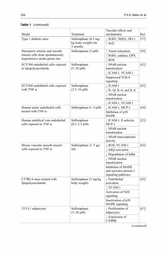
Table 1 (continued)
Model TreatmentVascular effects andmechanisms Ref.
Type 1 diabetic mice Sulforaphane (0.5 mg/kg body weight) for3 months
# SOD1, NQO1, HO-1 [37]
" Nrf2
Mesenteric arteries and smoothmuscle cells from spontaneouslyhypertensive stroke-prone rats
Sulforaphane (5 μM) " Vessel relaxation [50]
" SOD1, catalase, GPX# ROS
ECV304 endothelial cells exposedto lipopolysaccharide
Sulforaphane(5–20 μM)
# NFκB nucleartranslocation
[62]
# ICAM-1, VCAM-1
Suppressed TLR-4signaling
ECV304 endothelial cells exposedwith TNF-α
Sulforaphane(2.5–10 μM)
# ICAM-1 [63]
# IL-1β, IL-6, and IL-8
# NFκB nucleartranslocation
# ICAM-1, VCAM-1
Human aortic endothelial cellstreated with TNF-α
Sulforaphane (1–4 μM) # ICAM-1, MCP-1 [64]
Inhibition of p38MAPK
Human umbilical vein endothelialcells exposed to TNF-α
Sulforaphane(0.5–2.5 μM)
# ICAM-1, E-selectin,MCP-1
[23]
# NFκB nucleartranslocation
# NFκB transcriptionalactivity
Mouse vascular smooth musclecells exposed to TNF-α
Sulforaphane (1–5 μg/ml)
# ROS, VCAM-1 [65]
# IĸKβ activation
# Degradation of IκBα# NFκB nucleartranslocation
Inhibition of MAPKand activator protein-1signaling pathways
C57BL/6 mice treated withlipopolysaccharide
Sulforaphane (5 mg/kgbody weight)
# Endothelialactivation
[45]
# VCAM-1
Activation of Nrf2signaling
Inactivation of p38MAPK signaling
3T3-L1 adipocytes Sulforaphane(5–20 μM)
# Proliferation ofadipocytes
[67]
# Expression ofC/EBPβ
(continued)
326 P.V.A. Babu et al.

ECH-associated protein 1 (Keap1), nuclear factor [erythroid-derived 2]-like2 (Nrf2), and antioxidant response element (ARE) [34, 35]. Nrf2 is a transcriptionfactor that plays a major role in regulating the antioxidant response and maintainingthe redox homeostasis [36]. Keap1 is a cysteine-rich negative regulator of Nrf2 thatregulates Nrf2 by controlling its subcellular location. Under physiological condi-tions, the Nrf2 pathway is suppressed by the sequestration of Nrf2 in the cytoplasmwhich is mediated through the binding of Nrf2 to the inhibitor Keap1. Further,Keap1 interaction with Nrf2 induces rapid ubiquitination and rapid degradation ofNrf2 by proteasome leading to low expression of ARE-driven genes [37]. Uponexposure of cells to oxidative stress or electrophilic compounds, a signal thatinvolves phosphorylation and/or modification interferes with the Keap1-Nrf2 com-plex leading to dissociation of Nrf2 and translocation to the nucleus and binding tothe ARE. ARE is a cis-acting regulatory region of the genome with the consensussequence 50-TGACnnnGC-30 that regulates the basal and inducible expression ofmore than 200 genes [37]. Binding of Nrf2 to ARE induces the transcriptionalactivation of ARE-mediated genes that play a role in detoxification, antioxidant,and anti-inflammatory activities [37].
ARE is important in cellular protection from oxidative species, and it regulatesboth direct antioxidant enzymes (such as superoxide dismutase) and indirect anti-oxidant enzymes (such as rate-limiting enzymes of glutathione synthesis andxenobiotic-detoxifying enzymes) [38]. This includes heme oxygenase-1 (HO-1),glutathione-S-transferase (GST), superoxide dismutase (SOD), catalase,
Table 1 (continued)
Model TreatmentVascular effects andmechanisms Ref.
C57BL/6 N mice fed with high-fatdiet
0.1% Sulforaphane indiet for 6 weeks
# Expressions ofPPARγ, C/EBPα
[68]
# Fat accumulation
# Serum cholesterol,triglycerides
# Lipogenesis
Human umbilical vein endothelialcells exposed to TNF-α
Sulforaphane (10 μM) # Endothelial lipase [69]
# IĸKβphosphorylation
# IĸBαphosphorylation
"Increase, #decrease, ARE antioxidant response element, C/EBP CCAAT/enhancer-binding pro-tein, GCL glutamate-cysteine ligase, GCLC glutamate-cysteine ligase catalytic subunit, GCLMglutamate-cysteine ligase modifier subunit, GPX glutathione peroxidase, GR glutathione reductase,HO-1 heme oxygenase-1, ICAM-1 intracellular adhesion molecule-1, IĸBα inhibitor of NFĸB, IĸKβIĸB kinase, LDL low-density lipoprotein, MAPK mitogen-activated protein kinase, MCP-1 mono-cyte chemotactic protein-1, NFĸB nuclear factor ĸB, NQO1 NADPH quinone oxidoreductase, Nrf2nuclear factor [erythroid-derived 2]-like 2, PPARγ peroxisome proliferator-activated receptor-γ,ROS reactive oxygen species, SOD superoxide dismutase, TLR Toll-like receptor, TNF-α tumornecrosis factor-α, VCAM-1 vascular cell adhesion molecule-1
11 Sulforaphane and Atherosclerosis 327

γ-glutamyl cysteine synthetase (GCS), and NADPH quinone oxidoreductase(NQO1) [37]. Glutathione is the most abundant endogenous antioxidants used forthe day-to-day protection of cells against oxidative stress. Glutathione is a tripeptidemade up of the amino acids glutamate, glycine, and cysteine. The rate-limitingenzyme in the production of glutathione is glutamate-cysteine ligase (GCL). GCLhas two subunits, glutamate-cysteine ligase catalytic subunit (GCLC) and glutamate-cysteine ligase modifier subunit (GCLM). Activation of the Nrf/ARE pathwaysubstantially increases the concentration of active glutathione by producing enzymesinvolved in the production as well as the restoration of glutathione [39]. Thus, theKeap1-Nrf2-ARE signaling pathway is responsible for turning on an adaptiveresponse against oxidative stress and maintaining the redox homeostasis [36].
ARE activators induce a conformation rearrangement of Keap1 which facilitatesNrf2 nuclear translocation and subsequent activation of ARE-mediated transcriptionof protective genes [40]. Evidence indicate that Nrf2 activators such asisothiocyanates alter Keap1 conformation by interacting with the thiol groups ofthe specific cysteine residues within Keap1 and this will prevent the ubiquitinationand degradation of Nrf2. This will also facilitate Nrf2 nuclear translocation andsubsequent binding of Nrf2 to ARE [40]. Once bound, they initiate transcription ofmany genes that are regulated by ARE. The sulforaphane activation of Nrf2 isthrough the interaction of the thiol groups of Keap1 followed by nuclear transloca-tion of Nrf2 [41]. Indeed, a study showed a 1.4� increase in the amount of Nrf2upon sulforaphane exposure at 10 μM concentration [42]. The partial as well as thefull release of Nrf2 by sulforaphane was proposed for the long-lasting and conse-quential actions of sulforaphane to protect against cardiovascular disease [36,43–45].
The protective effect of sulforaphane on oxidized LDL-induced endothelialdysfunction was reported recently. In this study, sulforaphane (2.5–10 μM)suppressed oxidized LDL-induced ROS production in human umbilical vein endo-thelial cells (HUVEC) by inducing dose-dependent increases in the expression ofGCL catalytic and modifier subunits, intracellular glutathione level, HO-1 levels,and ARE activity [46]. Sulforaphane treatment (5 μM for 12 and 24 h) was alsoshown to increase the expressions of antioxidant enzymes (HO-1 and peroxiredoxin)and glutathione content in vascular smooth muscle cells (VSMCs) [47]. This studysuggests that sulforaphane may protect against vascular SMC dysfunction inage-related cardiovascular disease.
Recent animal studies also support that the vascular protective effect of sulfo-raphane may be mediated through the activation of Nrf2 signaling. The glutathionesystem consists of glutathione, GCS, GST, glutathione peroxidase (GPX), andglutathione reductase (GR) [48]. An impaired glutathione system is associatedwith hypertension [48]. Smooth muscle cells (SMC) from spontaneously hyperten-sive rats (SHRs) exhibited enhanced oxidative stress that is associated with lowerbasal levels of glutathione as compared to the SMC from control Wistar Kyoto(WKY) rats [48]. In this study, sulforaphane (0.05–1 μM) induced a dose-dependentincrease in cellular glutathione levels, HO-1 protein content, and GPX activity inboth the strains. In addition, sulforaphane upregulated the impaired glutathione
328 P.V.A. Babu et al.

system in vascular smooth muscle cells from SHR [48]. This could be due to theactivation of GCL, as sulforaphane is also a potent inducer of GCL [49]. Oxidativestress in kidneys mediates hypertension in SHRSP rats [27]. Sulforaphane adminis-tration improved blood pressure in SHRSP rats, which is associated with significantincrease in GCLC in SHRSP rat kidneys [27]. Further, this study showed a reductionin nitrated proteins with sulforaphane treatment, which is indirect evidence fordecreased oxidative stress.
Treatment with sulforaphane (0.5 mg/kg for 3 months) prevented diabetes-induced vascular inflammation and aortic damage in streptozotocin-induced type1 diabetic mice [37]. This was associated with the upregulation of Nrf2 and itsdownstream antioxidants such as SOD1, NQO1, and HO-1 in the aorta of diabeticmice [37]. The effect of sulforaphane on Nrf2-induced redox signaling in smallarteries and smooth cells from SHRSP rats was also reported [50]. In this study,sulforaphane improved vascular function as shown by improved contractility andendothelial-dependent vessel relaxation, which was mediated through activation ofNrf2-mediated redox signaling. There was an increased ROS production andreduced Nr2 activity in arteries and VSMC in SHRSP rats. In addition, the expres-sions of antioxidant enzymes such as SOD1, catalase, and GPX were reduced inSHRSP rats [50]. Sulforaphane treatment suppressed ROS production and increasedthese antioxidant enzymes in SHRSP rats. Sulforaphane also blocked angiotensinII-induced ROS production in SHRSP rats [50].
In another study, dietary supplementation of broccoli sprouts improvedendothelial-dependent aortic vessel relaxation and reduced blood pressure inSHRSP rats [28]. This was associated with improved oxidative stress as shown byincreased glutathione, decreased oxidized glutathione, decreased proteinnitrosylation, and increased GR and GPX activities [28]. In this study the authorsdidn’t measure the expression of Nrf2 but the effect is possibly mediated throughsulforaphane-mediated upregulation of Nrf2.
Together, these studies indicate that sulforaphane exerts vascular protectiveeffects by activating Nrf2/ARE signaling. By activating Nrf2, sulforaphane inducesthe expression of antioxidant enzymes, maintains the cell glutathione status, andreduces the oxidative stress that ultimately improves the vascular complications anddysfunction.
5.2 Inhibition of Inflammatory Pathways
Atherosclerosis is an inflammatory disease, and endothelial inflammation-inducedendothelial dysfunction plays a pivotal role in the development of atherosclerosis.Many of the risk factors for cardiovascular disease such as diabetes, hypertension,and smoking are potential activators of an inflammatory response in endotheliumthat induces the adhesion of monocytes to the vascular endothelial cells, an initialevent in the development of atherosclerosis [51–53]. Oxidative stress can triggerseveral intracellular signaling events that ultimately induce vascular inflammation byupregulating the expression of a number of pro-inflammatory molecules such as
11 Sulforaphane and Atherosclerosis 329

chemokines and adhesion molecules [54]. This includes IL-8, MCP-1, TNF-α,ICAM-1, VCAM-1, and E-selectin [17, 54, 55]. These inflammatory moleculesthen induce the binding of monocytes to the endothelial cells, and their subsequenttransendothelial migration into the vessels, leading to the pathogenesis of athero-sclerosis [54, 56].
Nuclear factor ĸB (NFĸB) is a pro-inflammatory transcription factor that plays amajor role in upregulating inflammatory molecules such as chemokines and adhe-sion molecules [57]. p50/p65 is the most abundant form of NFκB. Under homeo-static conditions, NFĸB is made inactive by sequestration by inhibitor of NFĸB(IĸBα). Inflammatory conditions activate IĸB kinase (IĸKβ), a multienzyme kinasecomplex, which phosphorylates IĸBα [58]. Phosphorylated IĸBα is degraded by aubiquitination process targeting IĸBα to proteasomal degradation [58]. This leadsto the nuclear translocation of p50/p65 [59]. In the nucleus, p50/p65 dimers bind tothe promoters of NFκB-dependent inflammatory genes and regulate inflammatorygenes involved in atherosclerosis [60, 61]. IĸKβ has an ETGE-binding motif whichis the stronger of two Keap1-binding motifs [35, 58]. When Nrf2 is released fromKeap1 and the complementary motif is exposed, Keap1 will sequester IĸKβdecreasing IĸBα phosphorylation and degradation and decreasing the translocationof NFĸB [35]. Hence Nrf2 negatively regulates the activation of the NFĸBpathway.
Others and we have reported the protective effects of sulforaphane on endothelialinflammation and the possible molecular mechanisms involved in the vasculareffects of sulforaphane. A recent study reported that sulforaphane inhibited thelipopolysaccharide (LPS)-induced expression of ICAM-1 and VCAM-1 inECV304 endothelial cells by inhibiting LPS-induced nuclear translocation ofNFкB [62]. In this study, sulforaphane suppressed Toll-like receptor (TLR)-4(TLR-4, a membrane receptor of LPS), MyD88 (an effector downstream of TLRsignaling), and downstream factors such as p38 mitogen-activated protein kinase(MAPK) and JNK that ultimately blocked NFкB translocation and the subsequentexpression of adhesion molecules [62]. The anti-inflammatory effect of sulforaphanewas also reported in oxidized LDL-induced HUVEC [46]. Treatment with sulfo-raphane dose-dependently suppressed NFκB nuclear translocation, NFκB activity,adhesion molecules, and binding of monocytes to EC.
TNF-α is an inflammatory cytokine that amplifies several signaling pathways thattrigger the secretion of various inflammatory mediators in endothelium leading tovascular inflammation [63]. Sulforaphane treatment (2.5–10 μM) attenuatedTNF-α-induced expression of ICAM-1 in ECV304 endothelial cells [63]. In addi-tion, sulforaphane dose-dependently inhibited the secretion of pro-inflammatorycytokines such as IL-1β, IL-6, and IL-8. TNF-α activates the NFκB pathway inendothelial cells as shown by increased NFκB DNA-binding activity and increasedphosphorylation of Iκκβ and IκBα [63]. Sulforaphane inhibited NFκB DNA-bindingactivity and downregulated RhoA/Rho-associated protein kinase and NFκB inflam-matory signaling pathways in endothelial cells [63]. In another study, sulforaphane(1–4 μM) pretreatment for 1 h suppressed TNF-α-induced enhanced MCP-1 andICAM-1 expressions in human aortic endothelial cells. This study suggests the effect
330 P.V.A. Babu et al.

of sulforaphane on endothelial inflammation may be mediated through inhibition ofp38 MAPK [64].
We have recently shown that sulforaphane, at a physiological relevant concen-tration, prevents TNF-α-induced endothelial inflammation in human umbilical veinendothelial cells (HUVEC) and improved TNF-α-induced vascular inflammation inC57BL/6 mice [23]. In our study, sulforaphane at a very low concentration (0.5 μM)significantly inhibited TNF-α-induced binding of monocytes to HUVEC both instatic and flow conditions. In addition, sulforaphane suppressed TNF-α-inducedproduction of inflammatory molecules such as MCP-1, VCAM-1, and E-selectin.In our animal study, dietary supplementation of sulforaphane (300 ppm) for 8 weekssignificantly abolished TNF-α increased ex vivo monocyte adhesion and circulatingadhesion molecules and chemokines in C57BL/6 mice [23]. We also showed thatdietary sulforaphane prevented the eruption of endothelial lining in the intimal layer,preserved elastin fibers’ delicate organization, and reduced monocyte-derivedF4/80-positive macrophages in the aorta of TNF-α-treated C57BL/6 mice. Further,we investigated the underlying mechanisms involved in the vascular protectiveeffects of sulforaphane [23]. Sulforaphane inhibited TNF-α-induced NFκB tran-scriptional activity and NFκB p65 nuclear translocation in HUVEC indicating theeffect of sulforaphane may be associated with suppression of NFκB signaling.
Enhanced ICAM-1 and VCAM-1 are also associated with increased VSMCproliferation leading to increased neointima and atherosclerotic lesion formation[65]. In a recent study, the protective effect of sulforaphane (1–5 μg/mL for 2 h) onmouse VSMC exposed to TNF-α was reported [65]. Sulforaphane treatmentsuppressed TNF-α-induced increased production of intracellular ROS andVCAM-1 protein expression in VSMC, and this protective effect was mediatedthrough inhibition of MAPK and activator protein-1 signaling pathway [65]. Further,sulforaphane inhibited TNF-α-induced IĸKβ activation, prevented the degradationof IκBα, and reduced the nuclear translocation of p65 in VSMC indicating the anti-inflammatory effects of sulforaphane. This study suggests that sulforaphane mayinhibit the adhesive capacity of VSMC and suppress inflammation within theatherosclerotic lesion.
Vascular inflammation and atherosclerosis develop predominantly in the sitesof the arterial tree that are exposed to nonuniform blood flow, e.g., near thebranches and bends of the arterial tree, which exerts relatively low shear stress onvascular endothelial cells activating inflammatory signals [45]. Unidirectionalhigh shear stress suppresses vascular inflammation, whereas low shear stressactivates inflammatory signals. C57BL/6 mice were treated with sulforaphane(5 mg/kg body weight by intraperitoneal injection) followed by treatment withlipopolysaccharide [45]. In this study sulforaphane treatment suppressed endo-thelial activation which is mediated through activation of Nrf2 and inactivatingp38 MAPK and suppressing VCAM-1 signaling at the susceptible site in themurine aorta [45].
Collectively, the anti-inflammatory effect of sulforaphane is well documented inin vitro and animal studies. Sulforaphane targets multiple inflammatory signals in thevasculature to exert its anti-inflammatory effects.
11 Sulforaphane and Atherosclerosis 331
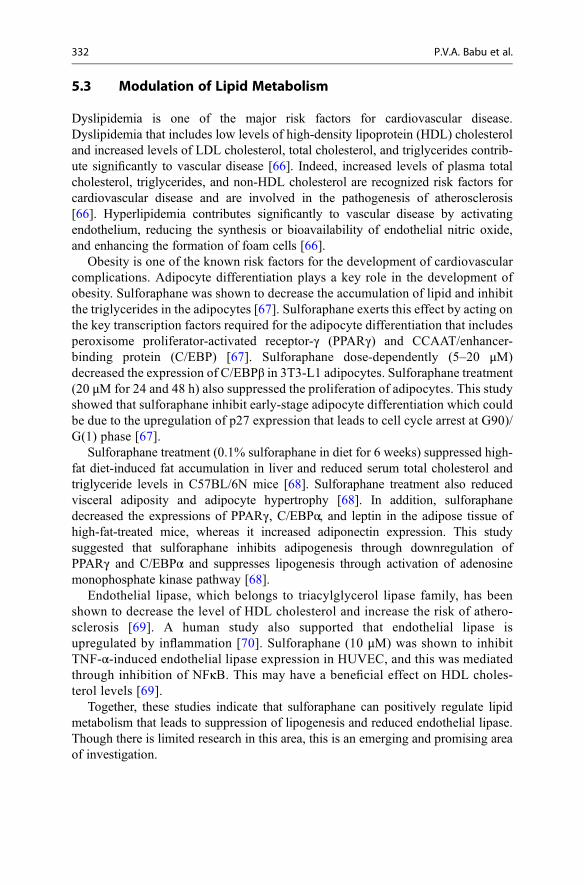
5.3 Modulation of Lipid Metabolism
Dyslipidemia is one of the major risk factors for cardiovascular disease.Dyslipidemia that includes low levels of high-density lipoprotein (HDL) cholesteroland increased levels of LDL cholesterol, total cholesterol, and triglycerides contrib-ute significantly to vascular disease [66]. Indeed, increased levels of plasma totalcholesterol, triglycerides, and non-HDL cholesterol are recognized risk factors forcardiovascular disease and are involved in the pathogenesis of atherosclerosis[66]. Hyperlipidemia contributes significantly to vascular disease by activatingendothelium, reducing the synthesis or bioavailability of endothelial nitric oxide,and enhancing the formation of foam cells [66].
Obesity is one of the known risk factors for the development of cardiovascularcomplications. Adipocyte differentiation plays a key role in the development ofobesity. Sulforaphane was shown to decrease the accumulation of lipid and inhibitthe triglycerides in the adipocytes [67]. Sulforaphane exerts this effect by acting onthe key transcription factors required for the adipocyte differentiation that includesperoxisome proliferator-activated receptor-γ (PPARγ) and CCAAT/enhancer-binding protein (C/EBP) [67]. Sulforaphane dose-dependently (5–20 μM)decreased the expression of C/EBPβ in 3T3-L1 adipocytes. Sulforaphane treatment(20 μM for 24 and 48 h) also suppressed the proliferation of adipocytes. This studyshowed that sulforaphane inhibit early-stage adipocyte differentiation which couldbe due to the upregulation of p27 expression that leads to cell cycle arrest at G90)/G(1) phase [67].
Sulforaphane treatment (0.1% sulforaphane in diet for 6 weeks) suppressed high-fat diet-induced fat accumulation in liver and reduced serum total cholesterol andtriglyceride levels in C57BL/6N mice [68]. Sulforaphane treatment also reducedvisceral adiposity and adipocyte hypertrophy [68]. In addition, sulforaphanedecreased the expressions of PPARγ, C/EBPα, and leptin in the adipose tissue ofhigh-fat-treated mice, whereas it increased adiponectin expression. This studysuggested that sulforaphane inhibits adipogenesis through downregulation ofPPARγ and C/EBPα and suppresses lipogenesis through activation of adenosinemonophosphate kinase pathway [68].
Endothelial lipase, which belongs to triacylglycerol lipase family, has beenshown to decrease the level of HDL cholesterol and increase the risk of athero-sclerosis [69]. A human study also supported that endothelial lipase isupregulated by inflammation [70]. Sulforaphane (10 μM) was shown to inhibitTNF-α-induced endothelial lipase expression in HUVEC, and this was mediatedthrough inhibition of NFκB. This may have a beneficial effect on HDL choles-terol levels [69].
Together, these studies indicate that sulforaphane can positively regulate lipidmetabolism that leads to suppression of lipogenesis and reduced endothelial lipase.Though there is limited research in this area, this is an emerging and promising areaof investigation.
332 P.V.A. Babu et al.

6 Conclusions
Epidemiological studies have found associations between cruciferous vegetableconsumption and reduced risk of chronic conditions, such as atherosclerosis, diabe-tes, and cancer [10–13]. In addition, as discussed above, in vitro and animal studiessupport the beneficial cardiovascular effects of sulforaphane, a molecule found in itsprecursor state in cruciferous and other vegetables. Sulforaphane acts on multipletargets in the vascular system, including activating Nrf2/ARE signaling, improvinginflammatory pathways, and positively regulating lipid metabolism. However,human studies related to the vascular effects of sulforaphane and cruciferous vege-tables are lacking. In addition, studies on the structure-activity relationships ofsulforaphane are needed to understand how sulforaphane interacts with variousbiomolecules in the cardiovascular system to exert its beneficial cardiovasculareffects. The consumption of sulforaphane-containing foods is safe, but there issome research that suggests that too much might increase the risk of some types ofcancer [25]. Hence, well-designed human trials may be needed to evaluate thecardiovascular effects of sulforaphane and cruciferous vegetables and to recommendcruciferous vegetables, such as broccoli, to improve cardiovascular health.
References
1. Ritchey MD, Wall HK, Gillespie C, George MG, Jamal A, Division for Heart D, StrokePrevention CDC (2014) Million hearts: prevalence of leading cardiovascular disease risk factors– United States, 2005–2012. MMWR Morb Mortal Wkly Rep 63:462–467
2. Go AS, Mozaffarian D, Roger VL, Benjamin EJ, Berry JD, Blaha MJ, Dai S, Ford ES, Fox CSet al (2014) Heart disease and stroke statistics – 2014 update: a report from the American HeartAssociation. Circulation 129:e28–e292
3. Luepker RV (2011) Cardiovascular disease: rise, fall, and future prospects. Annu Rev PublicHealth 32:1–3
4. Galkina E, Ley K (2009) Immune and inflammatory mechanisms of atherosclerosis. Annu RevImmunol 27:165–197
5. Hooper L, Kroon PA, Rimm EB, Cohn JS, Harvey I, Le Cornu KA, Ryder JJ, Hall WL, CassidyA (2008) Flavonoids, flavonoid-rich foods, and cardiovascular risk: a meta-analysis of random-ized controlled trials. Am J Clin Nutr 88:38–50
6. Mulvihill EE, Huff MW (2010) Antiatherogenic properties of flavonoids: implications forcardiovascular health. Can J Cardiol 26(Suppl A):17A–21A
7. Erdman JW Jr, Balentine D, Arab L, Beecher G, Dwyer JT, Folts J, Harnly J, Hollman P, KeenCL et al (2007) Flavonoids and heart health: proceedings of the ILSI North America flavonoidsworkshop, May 31–June 1, 2005, Washington, DC. J Nutr 137:718S–737S
8. Beecher GR (2003) Overview of dietary flavonoids: nomenclature, occurrence and intake.J Nutr 133:3248S–3254S
9. Del Rio D, Borges G, Crozier A (2010) Berry flavonoids and phenolics: bioavailability andevidence of protective effects. Br J Nutr 104(Suppl 3):S67–S90
10. Houghton CA, Fassett RG, Coombes JS (2013) Sulforaphane: translational research fromlaboratory bench to clinic. Nutr Rev 71:709–726
11 Sulforaphane and Atherosclerosis 333

11. Genkinger JM, Platz EA, Hoffman SC, Comstock GW, Helzlsouer KJ (2004) Fruit, vegetable,and antioxidant intake and all-cause, cancer, and cardiovascular disease mortality in acommunity-dwelling population in Washington County, Maryland. Am J Epidemiol160:1223–1233
12. Lockheart MS, Steffen LM, Rebnord HM, Fimreite RL, Ringstad J, Thelle DS, Pedersen JI,Jacobs DR Jr (2007) Dietary patterns, food groups and myocardial infarction: a case-controlstudy. Br J Nutr 98:380–387
13. Zhang X, Shu XO, Xiang YB, Yang G, Li H, Gao J, Cai H, Gao YT, Zheng W (2011)Cruciferous vegetable consumption is associated with a reduced risk of total and cardiovasculardisease mortality. Am J Clin Nutr 94:240–246
14. Grover-Paez F, Zavalza-Gomez AB (2009) Endothelial dysfunction and cardiovascular riskfactors. Diabetes Res Clin Pract 84:1–10
15. Su JB (2015) Vascular endothelial dysfunction and pharmacological treatment. World J Cardiol7:719–741
16. Chistiakov DA, Orekhov AN, Bobryshev YV (2015) Endothelial barrier and its abnormalitiesin cardiovascular disease. Front Physiol 6:365
17. Pearson TA, Mensah GA, Alexander RW, Anderson JL, Cannon RO 3rd, Criqui M, Fadl YY,Fortmann SP, Hong Y et al (2003) Markers of inflammation and cardiovascular disease:application to clinical and public health practice: a statement for healthcare professionalsfrom the centers for disease control and prevention and the American Heart Association.Circulation 107:499–511
18. Thompson SG, Kienast J, Pyke SD, Haverkate F, van de Loo JC (1995) Hemostatic factors andthe risk of myocardial infarction or sudden death in patients with angina pectoris. EuropeanConcerted Action on Thrombosis and Disabilities Angina Pectoris Study Group. N Engl J Med332:635–641
19. Danesh J, Wheeler JG, Hirschfield GM, Eda S, Eiriksdottir G, Rumley A, Lowe GD, Pepys MB,Gudnason V (2004) C-reactive protein and other circulating markers of inflammation in theprediction of coronary heart disease. N Engl J Med 50:1387–1397
20. Ridker PM, Hennekens CH, Buring JE, Rifai N (2000) C-reactive protein and other markers ofinflammation in the prediction of cardiovascular disease in women. N Engl J Med 342:836–843
21. Yin G, Yang X, Li B, Yang M, Ren M (2014) Connexin43 siRNA promotes HUVECproliferation and inhibits apoptosis induced by ox-LDL: an involvement of ERK signalingpathway. Mol Cell Biochem 394:101–107
22. Libby P (2002) Inflammation in atherosclerosis. Nature 420:868–87423. Nallasamy P, Si H, Babu PV, Pan D, Fu Y, Brooke EA, Shah H, Zhen W, Zhu H et al (2014)
Sulforaphane reduces vascular inflammation in mice and prevents TNF-alpha-induced mono-cyte adhesion to primary endothelial cells through interfering with the NF-kappaB pathway. JNutr Biochem 25:824–833
24. Ye L, Dinkova-Kostova AT, Wade KL, Zhang Y, Shapiro TA, Talalay P (2002) Quantitativedetermination of dithiocarbamates in human plasma, serum, erythrocytes and urine: pharmaco-kinetics of broccoli sprout isothiocyanates in humans. Clin Chim Acta 316:43–53
25. Latte KP, Appel KE, Lampen A (2011) Health benefits and possible risks of broccoli – anoverview. Food Chem Toxicol 49:3287–3309
26. Cornelis MC, El-Sohemy A, Campos H (2007) GSTT1 genotype modifies the associationbetween cruciferous vegetable intake and the risk of myocardial infarction. Am J Clin Nutr86:752–758
27. Senanayake GV, Banigesh A, Wu L, Lee P, Juurlink BH (2012) The dietary phase 2 proteininducer sulforaphane can normalize the kidney epigenome and improve blood pressure inhypertensive rats. Am J Hypertens 25:229–235
28. Wu L, Noyan Ashraf MH, Facci M,Wang R, Paterson PG, Ferrie A, Juurlink BH (2004) Dietaryapproach to attenuate oxidative stress, hypertension, and inflammation in the cardiovascularsystem. Proc Natl Acad Sci U S A 101:7094–7099
334 P.V.A. Babu et al.

29. Bai Y, Cui W, Xin Y, Miao X, Barati MT, Zhang C, Chen Q, Tan Y, Cui Tet al (2013) Preventionby sulforaphane of diabetic cardiomyopathy is associated with up-regulation of Nrf2 expressionand transcription activation. J Mol Cell Cardiol 57:82–95
30. Zhang Z, Wang S, Zhou S, Yan X, Wang Y, Chen J, Mellen N, Kong M, Gu J et al (2014)Sulforaphane prevents the development of cardiomyopathy in type 2 diabetic mice probably byreversing oxidative stress-induced inhibition of LKB1/AMPK pathway. J Mol Cell Cardiol77:42–52
31. Dhalla NS, Temsah RM, Netticadan T (2000) Role of oxidative stress in cardiovasculardiseases. J Hypertens 18:655–673
32. Park KH, Park WJ (2015) Endothelial dysfunction: clinical implications in cardiovasculardisease and therapeutic approaches. J Korean Med Sci 30:1213–1225
33. Babu PV, Liu D (2008) Green tea catechins and cardiovascular health: an update. Curr MedChem 15:1840–1850
34. Villeneuve NF, Lau A, Zhang DD (2010) Regulation of the Nrf2-Keap1 antioxidant response bythe ubiquitin proteasome system: an insight into cullin-ring ubiquitin ligases. Antioxid RedoxSignal 13:1699–1712
35. Kensler TW, Egner PA, Agyeman AS, Visvanathan K, Groopman JD, Chen JG, Chen TY,Fahey JW, Talalay P (2013) Keap1-nrf2 signaling: a target for cancer prevention by sulforaph-ane. Top Curr Chem 329:163–177
36. Kwon JS, Joung H, Kim YS, Shim YS, Ahn Y, Jeong MH, Kee HJ (2012) Sulforaphane inhibitsrestenosis by suppressing inflammation and the proliferation of vascular smooth muscle cells.Atherosclerosis 225:41–49
37. Miao X, Bai Y, Sun W, Cui W, Xin Y, Wang Y, Tan Y, Miao L, Fu Y et al (2012) Sulforaphaneprevention of diabetes-induced aortic damage was associated with the up-regulation of Nrf2 andits down-stream antioxidants. Nutr Metabol 9:84
38. Kwak MK, Wakabayashi N, Itoh K, Motohashi H, Yamamoto M, Kensler TW (2003)Modulation of gene expression by cancer chemopreventive dithiolethiones through theKeap1-Nrf2 pathway. Identification of novel gene clusters for cell survival. J Biol Chem278:8135–8145
39. Yang YM, Noh K, Han CY, Kim SG (2010) Transactivation of genes encoding for phase IIenzymes and phase III transporters by phytochemical antioxidants. Molecules 15:6332–6348
40. Stefanson AL, Bakovic M (2014) Dietary regulation of Keap1/Nrf2/ARE pathway: focus onplant-derived compounds and trace minerals. Nutrients 6:3777–3801
41. Velmurugan GV, Sundaresan NR, Gupta MP, White C (2013) Defective Nrf2-dependent redoxsignalling contributes to microvascular dysfunction in type 2 diabetes. Cardiovasc Res100:143–150
42. Thimmulappa RK, Mai KH, Srisuma S, Kensler TW, Yamamoto M, Biswal S (2002) Identifi-cation of Nrf2-regulated genes induced by the chemopreventive agent sulforaphane by oligo-nucleotide microarray. Cancer Res 62:5196–5203
43. Balligand JL (2013) Reducing damage through Nrf-2. Cardiovasc Res 100:1–344. Chapple SJ, Siow RC, Mann GE (2012) Crosstalk between Nrf2 and the proteasome: thera-
peutic potential of Nrf2 inducers in vascular disease and aging. Int J Biochem Cell Biol44:1315–1320
45. Zakkar M, Van der Heiden K, le Luong A, Chaudhury H, Cuhlmann S, Hamdulay SS, Krams R,Edirisinghe I, Rahman I et al (2009) Activation of Nrf2 in endothelial cells protects arteries fromexhibiting a proinflammatory state. Arterioscler Thromb Vasc Biol 29:1851–1857
46. Huang CS, Lin AH, Liu CT, Tsai CW, Chang IS, Chen HW, Lii CK (2013) Isothiocyanatesprotect against oxidized LDL-induced endothelial dysfunction by upregulating Nrf2-dependentantioxidation and suppressing NFκB activation. Mol Nutr Food Res 57:1918–1930
47. Rizzo B, Maltese G, Paraskevi MP, Hrelia S, Mann G, Siow R (2014) Induction of antioxidantgenes by sulforaphane and klotho in human aortic smooth muscle cells. Free Radic Biol Med75(Suppl 1):S14–S15
11 Sulforaphane and Atherosclerosis 335
48. Wu L, Juurlink BH (2001) The impaired glutathione system and its up-regulation by sulforaph-ane in vascular smooth muscle cells from spontaneously hypertensive rats. J Hypertens19:1819–1825
49. Juurlink BH (2001) Therapeutic potential of dietary phase 2 enzyme inducers in amelioratingdiseases that have an underlying inflammatory component. Can J Physiol Pharmacol79:266–282
50. Lopes RA, Neves KB, Tostes RC, Montezano AC, Touyz RM (2015) Downregulation ofnuclear factor erythroid 2-related factor and associated antioxidant genes contributes toredox-sensitive vascular dysfunction in hypertension. Hypertension 66:1240–1250
51. Menzaghi C, Bacci S, Salvemini L, Mendonca C, Palladino G, Fontana A, De Bonis C,Marucci A, Goheen E et al (2013) Serum resistin, cardiovascular disease and all-cause mortalityin patients with type 2 diabetes. PLoS One 8:e64729
52. Whelton A (2006) Clinical implications of nonopioid analgesia for relief of mild-to-moderatepain in patients with or at risk for cardiovascular disease. Am J Cardiol 97:3–9
53. Pearson TA, Mensah GA, Hong Y, Smith SC Jr (2004) CDC/AHA workshop on markers ofinflammation and cardiovascular disease: application to clinical and public health practice:overview. Circulation 110:e543–e544
54. Paneni F, Beckman JA, Creager MA, Cosentino F (2013) Diabetes and vascular disease:pathophysiology, clinical consequences, and medical therapy: part I. Eur Heart J 34:2436–2443
55. Shirwany NA, Zou MH (2012) Vascular inflammation is a missing link for diabetes-enhancedatherosclerotic cardiovascular diseases. Front Biosci 17:1140–1164
56. Frangogiannis NG (2008) The immune system and cardiac repair. Pharmacol Res 58:88–11157. Savoia C, Schiffrin EL (2007) Vascular inflammation in hypertension and diabetes: molecular
mechanisms and therapeutic interventions. Clin Sci 112:375–38458. Bouwmeester T, Bauch A, Ruffner H, Angrand PO, Bergamini G, Croughton K,
Cruciat C, Eberhard D, Gagneur J et al (2004) A physical and functional map of thehuman TNF-alpha/NFκ B signal transduction pathway. Nat Cell Biol 6:97–105
59. Kim F, Tysseling KA, Rice J, Gallis B, Haji L, Giachelli CM, Raines EW, Corson MA,Schwartz MW (2005) Activation of Iκbeta by glucose is necessary and sufficient to impairinsulin signaling and nitric oxide production in endothelial cells. J Mol Cell Cardiol 39:327–334
60. Yerneni KK, Bai W, Khan BV, Medford RM, Natarajan R (1999) Hyperglycemia-inducedactivation of nuclear transcription factor kappaB in vascular smooth muscle cells. Diabetes48:855–864
61. Bartchewsky W Jr, Martini MR, Masiero M, Squassoni AC, Alvarez MC, Ladeira MS,Salvatore D, Trevisan M, Pedrazzoli J Jr, Ribeiro ML (2009) Effect of Helicobacter pyloriinfection on IL-8, IL-1beta and COX-2 expression in patients with chronic gastritis and gastriccancer. Scand J Gastroenterol 44:153–161
62. Shan Y, Lin N, Yang X, Tan J, Zhao R, Dong S, Wang S (2012) Sulphoraphane inhibited theexpressions of intercellular adhesion molecule-1 and vascular cell adhesion molecule-1 throughMyD88-dependent toll-like receptor-4 pathway in cultured endothelial cells. Nutr MetabCardiovasc Dis 22:215–222
63. Hung CN, Huang HP, Wang CJ, Liu KL, Lii CK (2014) Sulforaphane inhibits TNF-alpha-induced adhesion molecule expression through the Rho A/ROCK/NF-kappaB signaling path-way. J Med Food 17:1095–1102
64. Chen XL, Dodd G, Kunsch C (2009) Sulforaphane inhibits TNF-alpha-induced activation ofp38 MAP kinase and VCAM-1 and MCP-1 expression in endothelial cells. Inflamm Res58:513–521
65. Kim JY, Park HJ, Um SH, Sohn EH, Kim BO, Moon EY, Rhee DK, Pyo S (2012) Sulforaphanesuppresses vascular adhesion molecule-1 expression in TNF-alpha-stimulated mouse vascularsmooth muscle cells: involvement of the MAPK, NF-kappaB and AP-1 signaling pathways.Vasc Pharmacol 56:131–141
66. Hopkins PN (2013) Molecular biology of atherosclerosis. Physiol Rev 93:1317–1542
336 P.V.A. Babu et al.

67. Choi KM, Lee YS, Sin DM, Lee S, Lee MK, Lee YM, Hong JT, Yun YP, Yoo HS (2012)Sulforaphane inhibits mitotic clonal expansion during adipogenesis through cell cycle arrest.Obesity 20:1365–1371
68. Choi KM, Lee YS, Kim W, Kim SJ, Shin KO, Yu JY, Lee MK, Lee YM, Hong JT et al (2014)Sulforaphane attenuates obesity by inhibiting adipogenesis and activating the AMPK pathwayin obese mice. J Nutr Biochem 25:201–207
69. Kivela AM, Makinen PI, Jyrkkanen HK, Mella-Aho E, Xia Y, Kansanen E, Leinonen H, VermaIM, Yla-Herttuala S, Levonen AL (2010) Sulforaphane inhibits endothelial lipase expressionthrough NF-kappaB in endothelial cells. Atherosclerosis 213:122–128
70. Badellino KO, Wolfe ML, Reilly MP, Rader DJ (2008) Endothelial lipase is increased in vivo byinflammation in humans. Circulation 117:678–685
11 Sulforaphane and Atherosclerosis 337

Therapeutic Paradigm UnderscoringGlucosinolate Sulforaphane inChemo- and Radiosensitization ofCancer: Preclinical and Clinical Perspective
12
Sanjeev Banerjee and Shivani B. Paruthy
AbstractFruits and vegetables harbor innocuous bioactive compounds which after absorp-tion and distribution tend to have an effect on general defense mechanism of thebody including cancer prevention and therapeutic effects. Emerging knowledgefrom clinical and laboratory studies reveal an important insight regarding theirmechanism of action orchestrating therapeutic paradigm with conventional can-cer treatment modalities to enhance the curative index of cancer treatment.However, unlike conventional cancer therapeutics, natural bioactive compoundsrarely develop resistance undermining their chemopreventive actions. One suchbioactive natural compound – sulforaphane, a cognate isothiocyanate limitedmostly to vegetables of Brassica family and enriched in broccoli – is considereda promising chemopreventive agent against cancer. Sulforaphane is released fromhydrolysis of glucoraphanin isothiocyanate by action of myrosinase enzymewhich is also found localized inside vegetal tissues. Overwhelming evidencepoints to sulforaphane’s multitargeted actions operationally targeting core cellsurvival signaling pathways in tumor cells and enzyme induction mediated by thenuclear factor erythroid 2-related factor 2 (Nrf2)-regulated transcriptions of genesencoding carcinogen detoxification, antioxidant enzymes, and other effectsincluding reversal of resistance and reduction in the systemic toxicity of drug.This chapter presents a broad perspective on the role of sulforaphane inaugmenting multimodal cancer therapy including putative mechanismcomplementing the efficacy of chemo- and radiotherapy with presumptive notion
S. Banerjee (*)Department of Pathology (Past), Barbara Ann Karmanos Cancer Institute, Wayne State UniversitySchool of Medicine, Detroit, MI, USAe-mail: [email protected]
S.B. ParuthyDepartment of Surgery, Vardhman Mahavir Medical College and Safdarjung Hospital, New Delhi,Delhi, India
# Springer International Publishing Switzerland 2017J.-M. Mérillon, K.G. Ramawat (eds.), Glucosinolates, Reference Series inPhytochemistry, DOI 10.1007/978-3-319-25462-3_19
339
of its future use in clinics in fight against cancer and patient’s benefit. Clinicaltrials have also been reviewed to ensure clinical safety and efficacy of sulforaph-ane in patients diagnosed with cancer.
KeywordsGlucosinolate • Isothiocyanates • Brassicaceae • Glucoraphanin • Sulforaphane •Chemosensitization • Chemoresistance • Radiosensitization
AbbreviationsALDH-1 Aldehyde dehydrogenase 1ARE Antioxidant response elementBCl2 B-cell lymphoma 2COX-2 Cycloxygenase-2GSC Genomic standards consortiumGTC Green tea catechinsHCG Human chorionic gonadotropinHNF-3β Hepatocyte nuclear factor 3βHRR Homologous recombination repairIL-1β Interleukin-1βKeap-1 Kelch-like ECH-associated protein 1MAPK Mitogen-activated protein kinasesNF-κB Nuclear factor- kappa BNHEJ Nonhomologous end joiningNOD/SCID Nonobese diabetic/severe combined immunodeficiencyNrf2 Nuclear factor erythroid 2-related factor 2Oct 2/3 Octamer transcription factor-2/3OTX-2 Orthodenticle homeobox 2PDX-1 Pancreatic and duodenal homeobox 1PET Positron emission tomographyTP63 Tumor protein p63UV UltravioletVEGFR-2 Vascular endothelial growth factor receptor-2
Contents1 Introduction . . . . . . . . . . . . . . . . . . . . . . . . . . . . . . . . . . . . . . . . . . . . . . . . . . . . . . . . . . . . . . . . . . . . . . . . . . . . . . . . . . 3412 Sulforaphane: Its Bioavailability and Mechanism of Action . . . . . . . . . . . . . . . . . . . . . . . . . . . . . . . 3423 Sulforaphane and Chemo- and Radiosensitization of Site-Specific Cancers . . . . . . . . . . . . . . 345
3.1 SFN and Breast Cancer . . . . . . . . . . . . . . . . . . . . . . . . . . . . . . . . . . . . . . . . . . . . . . . . . . . . . . . . . . . . . . 3453.2 SFN and Glioblastoma . . . . . . . . . . . . . . . . . . . . . . . . . . . . . . . . . . . . . . . . . . . . . . . . . . . . . . . . . . . . . . . 3483.3 SFN and Head and Neck Cancer . . . . . . . . . . . . . . . . . . . . . . . . . . . . . . . . . . . . . . . . . . . . . . . . . . . . 3493.4 SFN and Gynecologic Cancers . . . . . . . . . . . . . . . . . . . . . . . . . . . . . . . . . . . . . . . . . . . . . . . . . . . . . . 3513.5 SFN and Nonmelanoma Cutaneous Carcinoma . . . . . . . . . . . . . . . . . . . . . . . . . . . . . . . . . . . . . 3523.6 SFN and Barett’s Adenocarcinoma . . . . . . . . . . . . . . . . . . . . . . . . . . . . . . . . . . . . . . . . . . . . . . . . . . 3533.7 SFN and Bronchial Carcinoid . . . . . . . . . . . . . . . . . . . . . . . . . . . . . . . . . . . . . . . . . . . . . . . . . . . . . . . 3543.8 SFN and Hematological Malignancies . . . . . . . . . . . . . . . . . . . . . . . . . . . . . . . . . . . . . . . . . . . . . . 3543.9 SFN and Colon Carcinoma . . . . . . . . . . . . . . . . . . . . . . . . . . . . . . . . . . . . . . . . . . . . . . . . . . . . . . . . . . 3583.10 SFN and Pancreatic Cancer . . . . . . . . . . . . . . . . . . . . . . . . . . . . . . . . . . . . . . . . . . . . . . . . . . . . . . . . . . 359
340 S. Banerjee and S.B. Paruthy
4 SFN and TRAIL Therapy . . . . . . . . . . . . . . . . . . . . . . . . . . . . . . . . . . . . . . . . . . . . . . . . . . . . . . . . . . . . . . . . . . . 3615 SFN, TRAIL, and Cancer Stem Cells . . . . . . . . . . . . . . . . . . . . . . . . . . . . . . . . . . . . . . . . . . . . . . . . . . . . . . 364
5.1 Prostate Cancer . . . . . . . . . . . . . . . . . . . . . . . . . . . . . . . . . . . . . . . . . . . . . . . . . . . . . . . . . . . . . . . . . . . . . . . . 3645.2 Pancreatic Cancer . . . . . . . . . . . . . . . . . . . . . . . . . . . . . . . . . . . . . . . . . . . . . . . . . . . . . . . . . . . . . . . . . . . . . 365
6 Sulforaphane Combination with Other Natural Agents in Cancer Therapy . . . . . . . . . . . . . . . 3657 Sulforaphane and Clinical Trials . . . . . . . . . . . . . . . . . . . . . . . . . . . . . . . . . . . . . . . . . . . . . . . . . . . . . . . . . . . . 3698 Conclusions and Perspective . . . . . . . . . . . . . . . . . . . . . . . . . . . . . . . . . . . . . . . . . . . . . . . . . . . . . . . . . . . . . . . . 371References . . . . . . . . . . . . . . . . . . . . . . . . . . . . . . . . . . . . . . . . . . . . . . . . . . . . . . . . . . . . . . . . . . . . . . . . . . . . . . . . . . . . . . . 371
1 Introduction
Cancer is a disease that seems to be preexisting since the time of Egyptian legend –Pharaohs, with anecdotal imagination by the Greek physician Hippocrates (460–370BC) emulating giant crab of Greek mythology-Karkinos, symbolizing present-daycarcinomatous tumors. It is a major public health problem in the USA and othercountries of the world with severe health consequences. Recent report compiled bythe American Cancer Society projects approximately 1,685,210 new cancer cases in2016, with a likelihood of 595,690 people succumbing to this disease in the USA[1]. According to theWorld Health Organization [WHO; http://www.who.int/cancer/en/], cancer is uncontrolled growth of cells beyond their usual boundaries invadinginto tissues and metastasizing to distant sites of the body, causing significantmorbidity and, if untreated, death of the host. With advancement in knowledgeabout this disease over the years, additional critical hallmarks associated with itsprogression have been recognized which has been compiled and concisely publishedby Hanahan and Weinberg recently [2]. Standard treatment options for the majorityof cancers include surgery and/or chemo- and radiotherapy to enhance therapeuticefficacy depending on tumor stage. This have undoubtedly helped in increasingcancer survival, but still the high mortality rate associated with this disease under-scores current therapeutic interventions in curtailing this disease-related death inci-dence. Alongside advancements relating to understanding pathobiology andimprovements in cancer therapy over the past few decades, meta-analysis of epide-miological studies emerged in parallel in literature highlighting intake of fruits andgreen vegetables is inversely associated with cancer incidence and mortality[3–6]. This prompted research efforts to isolate and gain insight into mechanismand efficacy of bioactive compounds present in edible fruits and vegetables that mayeffectively intervene and modulate the ontogeny of carcinogenesis and influence ourknowledge relating to prevention of cancer. Contextually, laboratory findings tilldate provide a strong rationale and perspective on several food-derived bioactivecompounds multi-targeting core regulatory pathways in etiology and pathobiologyof the tumorigenic process and, in addition, demonstrate putative therapeutic advan-tage as potential drugs since the molecular mechanisms are common to bothchemoprevention and cancer therapy [7, 8].
Food crops of the genus Brassica which include cruciferous vegetables such asbroccoli, cabbage, cauliflower, kale, watercress, etc. are largely cultivated in bothtropical and temperate regions of the world and constitute an important serving ofdifferent ethnic cuisines including raw intake. In context of cancer epidemiology,
12 Therapeutic Paradigm Underscoring Glucosinolate Sulforaphane in Chemo- and. . . 341

Verhoeven et al. summarizing the results of 7 cohort and 87 case-control studiesconcluded an inverse association between intake of some types of Brassicaceae familyvegetable and cancer risk [9]. Recently, in another large series of studies, additionalevidence of favorable effect of cruciferous vegetables on several common cancers hasemerged [10]. Cruciferous vegetables, like other vegetables, are also enriched innutrients including folate, fibers, carotenoids, and chlorophyll compounds but inaddition contain well-characterized sulfur-rich secondary metabolite compounds spe-cific to this crop of vegetables that impart a characteristic pungent astringent flavor; thegeneration and retention of this distinctive class of metabolic product ostensibly wardsoff plant pathogens and defend against pests and grazing predators facilitating theirsurvival in nature. Additional chemical characterization of the compounds present inBrassicaceae family vegetables led to their identity as belonging to a distinct class ofchemicals called glucosinolates (β-thioglucoside-N-hydroxy-sulfates), whose degra-dation products include biologically active compounds: the isothiocyanates, indoles,and nitriles; all these presumptively offer chemoprotection against human cancer riskand impede tumor growth with accompanying therapeutic benefits as well [11]. Tilldate, over 150 glucosinolates have been identified, all sharing a common sulfur-linkedβ-D-glucopyranose structure differing in side chain [12]. Noteworthy, glucosinolatecomposition fluctuates among different cruciferous vegetables, e.g., the predominantglucosinolate present in broccoli is glucoraphanin (the precursor to sulforaphane),whereas sinigrin is predominant in cabbage, and watercresses are high ingluconasturtiin. Correspondingly, in intact plant tissues, an endogenous plant enzymecalled myrosinase is physically separated in membranous vesicles that preempt itshydrolytic action on glucosinolates. Upon chewing, the released myrosinase enzymeaction hydrolyzes glucosinolates to highly unstable aglycone intermediate; the latterundergoes rearrangement to form nitriles, isothiocyanates, or indole depending on thestructure of the aglycone, temperature, and pH; without hydrolysis, glucosinolates perse are inert compounds and lack any valuable biological and therapeutic activity. Themost extensively studied cognate isothiocyanates (ITCs) derived from metabolism ofglucosinolates are allyl isothiocyanates (from sinigrin), benzyl isothiocyanate andphenethyl isothiocyanate (from gluconasturtiin), and sulforaphane (fromglucoraphanin). With emerging recognition that chemopreventive bioactive phyto-chemicals may also overcome conventional drug resistance, this chapter embodies asuccinct overview on therapeutic efficacy of one promising glucosinolate – sulforaph-ane, the cognate isothiocyanate of glucoraphanin, specifically in combination withchemotherapy (chemosensitization) and radiosensitization of different site-specificcancers reported to date.
2 Sulforaphane: Its Bioavailability and Mechanism of Action
Sulforaphane (SFN; 1-isothiocynato-4-(methyl-sulfinyl) butane) is generally foundin high concentrations in broccoli sprouts (0.8–21.7 μmol/g of dry weight)with levels 20–50 times than those found in mature market-stage plants (Fig. 1)[13, 14]. It has been shown in human subjects that plasma concentrations of SFN and
342 S. Banerjee and S.B. Paruthy

its metabolites can reach between 2.2 and 7.3 μmol after consumption of 100 g ofstandard and high glucosinolate (super) broccoli, respectively [15]. Moreover, highconcentrations of SFN are also achieved in tissues, and it has been reported usingLC-MS that SFN and its metabolites (SFN-GSH) reach a concentration in the smallintestine equivalent to roughly 3–30 μM of total SFN [16]. Strikingly, in a propor-tional dose-exposure relationship with increasing concentration of glucosinolates inbroccoli, greater exposure to SFN has also been ascribed after its consumption. Afterabsorption and entry into the enterocytes, SFN is predominantly metabolized via themercapturic acid pathway with glutathione (GSH) conjugation by glutathioneS-transferase followed by sequential metabolism leading to the generation of sulfo-raphane-cysteine (SFN-Cys) and sulforaphane-N-acetylcysteine (SFN-NAC) [17,18]. When metabolism of glucoraphanin efficiently occurs, SFN-NAC is the primarySFN metabolite excreted in the urine. Once SFN is distributed, it accumulates in thetissues and sustains therein to achieve the antitumor actions [18]. Observations froma pilot study point that sulforaphane metabolites could be detected within measur-able limits in enriched breast epithelial cells following oral dosing of broccoli sproutpreparations (containing 200 μmol of sulforaphane) to healthy women undergoingreduction mammoplasty, conveying a strong rationale of its bioavailability withprospect of its protective effects in clinical trials and in women at risk for breastcancer [19]. In laboratory setting using cultured cells and preclinical animal modelsof cancer, SFN has received considerable attention and has been thoroughlyinvestigated for its promising antitumor and chemopreventive activity. Severalpublished reviews describe in-depth molecular mechanism of SFN action intumor cells [12, 20–23]. Collectively, all mechanistic studies till date report SFN
Fig. 1 Principle sources of Sulforaphane (Broccoli and brocolli sprouts), its release from planttissues and some important information regarding its chemistry
12 Therapeutic Paradigm Underscoring Glucosinolate Sulforaphane in Chemo- and. . . 343

as a promising pleiotropic bioactive molecule operating through various cellularand molecular mechanisms that interferes with multiple oncogenic pathways. Ittargets precancerous and cancer cells thru Nrf2/Keap1/ARE-signaling pathwayconsidered as a major regulator of cytoprotective phase II detoxification enzymesthat reduce susceptibility to carcinogens and protect against DNA adduct genera-tion and oxidative and electrophilic stress (Fig. 2a) [24, 25]. Other studies reportthat SFN mediates the inhibition of cell proliferation and anti-inflammatory effectthrough significant downregulation of cyclooxygenase-2 (COX-2) expression bypreventing the binding of NF-κB to its consensus DNA sequence and encouragesapoptotic pathway by downregulation of Bcl-2 and Bcl-xL proteins renderingtumor cells to undergo apoptosis resulting in the elimination of clonal expansionof initiated and/or transformed tumor cells (Fig. 2b). SFN reportedly also modu-lates signal transduction pathways such as MAP kinases and CDK’s mediated cellcycle progression and arrest and inhibits angiogenesis, invasion, and metastasis of
TRAIL
Death receptor 4/5M
embr
ane
depo
lariza
tion
p53
PI-3K
P P
PDK1/2
CULIN-3
p-Akt
p-IKK
p-p65-IκBα
p-p65nucleartranslocated
nucleartranslocated
Proteasomaldegradation
IκBα
ROS
Activated receptortyrosine kinase
Apoptotic /Chk2
stimuli
Oxidative stressNrf-2 inducing agentsSFN
FADDSFN
SFN
SFN
SFN
SFN
Cell Membrane
SFN
SFN
SFN
SFNSFN
SFNSFN
SFN
Nucleus
SFN
SFN
SFN
SFN
SFN
FLIP
Bid Bcl-2
t-Bid Bax/Bak
Cyt C
Apaf-1
BaxCaspase-8/10(cleaved)
Caspase-3(cleaved)
Caspase-9
(cleaved)
Caspase-3
(cleaved)IAPs
Cellular targets
ApoptosisApoptosis CDc25C
DNAdamage Target
genes
Chemotherapeuticdrugs
Bcl-2Cyclin D1VEGFMMPs
SP1
s-MAFCBP/p300
p-p65 ARE
Nrf-2
Nrf-2
Nrf-2
KEAP-1
KEAP-1
Nrf-2
UbUb
Ub
p21
Chk-2
ATM
G1 arrest
Thiol homeostasisAntioxidant activityDetoxification genesProteasome functionNADPH synthesis
Fig. 2 Generalized schematic overview representing signaling pathways and cellular targetsaltered by Sulforaphane
344 S. Banerjee and S.B. Paruthy

cancer [12, 21, 26]. More recently SFN has been shown to inhibit histonedeacetylase (HDAC) activity leading to the reactivation of tumor suppressorgenes and silencing oncogenes [27–29]. Based on accumulating evidence, SFNappears to be a promising agent for cancer therapy presumptively associated withmultitargeted therapeutic effects. In line with this notion, a growing body ofpreclinical data on use of SF in combination with conventional therapies hasbeen investigated and reported.
3 Sulforaphane and Chemo- and Radiosensitization ofSite-Specific Cancers
In pursuit of novel therapeutic strategies to enhance the efficacy of chemotherapy,radiotherapy, and/or other conventional therapies for the treatment of humancancers, a growing body of emerging preclinical data on SFN combination pointsto an improvement in clinical outcome. Much of this anticipated success intherapeutic arena owes to the bioavailability of SFN needed for targeted inactiva-tion of signaling pathways that allegedly contribute to therapeutic resistance. Anumber of phase I and II clinical trials have been conducted to investigate thetoxicity of SFN in healthy men and women and patients with cancer. Because ofthe lack of evidence for toxic effects in humans alongside increased bioavailabilityand a growing understanding of its mechanism of action, SFN seems to be usefuland holds promise when combined with conventional therapeutics in clinics.Preclinical cognizance and novel therapeutic viewpoints to improve therapeuticefficacy deem vital for cancer treatment. Interestingly, combination therapy has theadvantage that each agent targets different levels and/or specific signaling path-ways contributing to tumor cell survival and growth, and stalling these survivalsignaling targets will more effectively and efficaciously inhibit the tumor cellproliferation, tumor growth, progression, and its recurrence pragmatically withsuperior therapeutic outcome than individual inhibitors. Additionally, it permitsreducing the therapeutic dose and thereby restricting problems of side effects ofhigh dosages of drugs during monotherapy, alongside inducing and augmentingapoptosis of drug-resistant and tumor-initiating cells/cancer stem cells (TIC/CSC)that may have innocuously escaped the monotherapy treatment course. We sum-marize below current state of knowledge relating to the addition of SFN tostrengthen and boost treatment armamentarium based on preclinical cognizanceand clinical trials.
3.1 SFN and Breast Cancer
Breast cancer is the most common single cause of cancer-related death in women.Results from several case-controlled studies conducted in the USA, Sweden, andChina came to a unanimous conclusion of significantly lower intake of cruciferous
12 Therapeutic Paradigm Underscoring Glucosinolate Sulforaphane in Chemo- and. . . 345

vegetables in women diagnosed with BC than in cancer-free control group[30–32]. Oral administration of SFN inhibits mammary carcinogenesis in rats treatedwith 7,12-dimethylbenz[a]anthracene [13]. Sulforaphane exerts a direct chemopre-ventive action on animal and human mammary tissue. A single 150 μM oral dose ofsulforaphane elevates cytoprotective NAD(P)H:quinone oxidoreductase (NQO1)and heme oxygenase-1 (HO-1) gene transcripts in a rat mammary epithelium,which in itself provides strong evidence of a pronounced pharmacodynamic actionof sulforaphane [19]. Moreover, as pointed earlier, detection of measurable quanti-ties of sulforaphane metabolites in human breast tissue provides a strong rationalefor evaluating the protective effects of a broccoli sprout preparation in clinical trialsof women at risk for breast cancer. From a clinical pathologist viewpoint, breastcancer is a heterogeneous disease with distinct histologic subtypes. Approximately75% of current diagnosed breast cancer are estrogen receptor (ER) positive, puta-tively implying the role of estrogen and ER contribution in expression of genesinvolved in the etiopathogenesis and progression of ER+ve breast cancer includingwomen who are at elevated risk of breast cancer. Tamoxifen and its active metab-olite, 4-hydroxy tamoxifen, are the first-line conventional adjuvant treatment forpremenopausal BC patients with ER+ve tumors because of its ability to bind toendogenous natural ER sites depriving estrogen-mediated growth stimulus for cellsto proliferate and survive. Paradoxically, tamoxifen is both an antagonist (in breasttissues) and an agonist (in the endometrium and bone) of ER, but in clinical setup,the net benefits outweigh the risk for treatment of premenopausal breast cancerpatients with ER+ve tumors. However, despite the benefits, approximately30–35% of women with ER+ve breast cancer develops de novo or intrinsic endo-crine therapy resistance to tamoxifen and makes clinical management a challengewith tumor recurrence after drug therapy. Thus, to overcome this limitation, Pawliket al. examined sensitization of ER+ve breast cancer cell lines to4-hydroxytamoxifen by SFN. The investigators used three ER-positive BC celllines – T47D, MCF-7, and BT-474 – as representative models to compare thesensitivity to combination therapy using SFN and 4-hydroxytamoxifen [33]. Theresults reveal a synergistic effect in that the combination of SFN and4-hydroxytamoxifen inhibited cell viability more efficiently than both compoundsused alone at concentrations lower than their IC50 values, and the decreased viabilityof cancer cells ensued from apoptosis largely due to the downregulation of anti-apoptotic proteins – Bcl-2 and survivin. Additionally, the combined treatmentreduced the clonogenicity of cells as well compared to controls by about 60%(MCF-7 and BT-474 cells) and 70% (T47D cells). Further outspreading their studyemulating clinical situation, T47D and MCF-7 derivatives (T47D tamR and MCF-7tamR, respectively) which could grow in media containing 500 nM of4-hydroxytamoxifen (tamoxifen-resistant) with retention of ER expression wereestablished. Importantly, SFN could even sensitize these tamoxifen-resistant cellsto 4-hydroxytamoxifen causing a statistically significant drop in viability of thesecells as compared to the treatment with single compound. In essence, this studyimplies an approach that allows the use of lower doses of tamoxifen, hence decreas-ing the level of its toxicity and concomitantly improving the risk-benefit profile of
346 S. Banerjee and S.B. Paruthy

the drug. Moreover, such strategy might even protect against acquisition by cancercells the drug-resistant phenotypes during therapy.
Another subtype of BC classified as HER-2-positive BC comprises about20–25% of cases over expressing human epidermal growth factor receptor-2(HER-2/ErbB-2/neu) associated with poor clinical outcome. HER-2 is a memberof the epidermal growth factor receptor family which consists of HER-1 (EGFR/Erb-1), HER-2 (Erb-2/neu, HER-3 (Erb3), and HER-4 (Erb-4) and accepted as a potentialtarget for anticancer therapies mainly targeted therapy. Overamplification of HER-2/neu receptor in tumor cells perpetuate constant signaling that proceeds throughphosphatidylinositol-3-kinase (PI3/Akt)-mTOR-S6 kinase axis pathway and resultsin enhanced cell proliferation and survival of tumor cells mainly due to evasion ofapoptosis, neoangiogenesis and a predilection to central nervous system metastasismaking it difficult to treat the disease. Lapatinib is a relatively new specific inhibitorof EGFR and HER-2 and reportedly has some activity against brain metastasis, butits efficacy becomes limited by the emergence of lapatinib-resistant cells possiblydue to hyperactivity of the signaling network downstream of Her-2 [34]. Of contex-tual relevance, SFN reportedly lowers the protein and phosphorylation levels ofpro-survival Akt, mTOR, and S6 kinase pathway in phenotypically different breastcancer cell lines [35]. Thus, in in vitro cell culture model system with breast cancercells overexpressing HER-2, it has been credibly shown that combination treatmentof SFN and lapatinib decreases cell survival more efficiently than each of the agentsused alone [36]. Possible mechanism relating to synergistic effect of the combinedtreatment has been reviewed by Kaczynska et al. which expectedly include theinhibition of the Akt-mTOR-S6 signaling axis [36]. More logistically, lapatinib-resistant HER-2-overexpressing breast cancer cells have been developed aftertreating HER2-overproducing SKBr3 cells cultured for a few months in the presenceof increasing drug concentrations and mixed with isogenic lapatinib-sensitive cellsin different proportions mimicking heterogeneous population of cancer cells as intumor [37]. Simulating this as a chemoresistant model, the cells were next investi-gated for viability following combination treatment for 48 h with SFN (5 μM) andlapatinib (100 nM) to test the relevance of drug combination to overcome lapatinibdrug resistance [37]. Additional effects of the combination of SFN and lapatinib ontumor cell migration were evaluated using transwell matrigel invasion assay. Theassay results undeniably revealed that with increasing percentage of resistant cells,lapatinib alone was less effective, but a combination of lapatinib and SFN signifi-cantly and efficiently inhibited the viability and migration of the resistant cellscompared to the activity of each agent alone as monotherapy [37].
Other significant discoveries with SFN as sensitization partner in breast cancertherapy have been reported in literature [38–41]. Clofarabine is a novel adenosineanalogue that reactivates DNA methylation-silenced tumor suppressor genes (suchas adenomatous polyposis coli (APC), phosphatase and tensin homologue (PTEN),and retinoic acid receptor beta 2 (RARβ2) as well as, surge p21 expression [38]. Theapoptotic and cytotoxic effects of clofarabine have been investigated on breastcancer cell lines [42]. In breast cancer cells- MCF-7 and MDA-MB-231, SFN atphysiologically relevant (10 μM) concentration leads to hypomethylation of PTEN
12 Therapeutic Paradigm Underscoring Glucosinolate Sulforaphane in Chemo- and. . . 347

and RARβ2 promoters with concomitant gene upregulation [43]. It has been foundthat SFN alone and in combination with clofarabine epigenetically normalize theexpression of DNA methylation-silenced tumor suppressor genes in invasive andnoninvasive human breast cancer cells resulting in increase in cell growth arrest andapoptosis at noninvasive cancer stage and may probably contribute as a valuabletherapeutic support in other solid tumors as well [43]. Sulforaphane preconditioningalso sensitizes breast cancer cells to mitomycin C; the activation of NAD(P)H:quinone oxidoreductase-1(NQO1) by SFN acts as an activator of mitomycin C intumor cells and makes them sensitive to the drug [41]. In addition, combinationaltreatment of SFN and gemcitabine on MCF-7 breast cancer cells shows that SFNenhances the growth inhibitory effects of gemcitabine in a synergistic manner[combination index (CI) <1] [39]. Notably, SFN induces apoptosis and anti-inflammatory effects on MCF-7 cells via downregulation of Bcl-2 and COX-2respectively, minimizing toxicity to normal cells. Taken together, SFN might be apotent combination partner in anticancer armamentarium for breast cancer treatment.
3.2 SFN and Glioblastoma
According to the National Brain Tumor Society (http://www.braintumor.org), glio-blastoma multiforme (GBM) accounts 12–15% of all intracranial tumors with amean survival rate of ~15 months and 5-year survival rate of ~4% despite multi-modal treatment strategies. Its etiology is unknown, but increasing research atmolecular level points to epigenetic silencing of the MGMT (O-6-methylguanine-DNA methyltransferase) DNA repair gene by promoter methylation that compro-mises the DNA repair [44]. In 2005, FDA approved the alkylating agent,temozolomide (TMZ), for the treatment of adult patients with newly diagnosedand recurrent malignant gliomas based on survival benefit results and relativelylow toxicity [45]. Therefore, current standard protocol adopted for GB treatment isconcomitant radiotherapy combined with oral TMZ after surgery, although inherentand acquired TMZ resistance develops quickly and results in dismal prognosis. Pilotstudies emphasizing SFN alone induce apoptosis and inhibits the migration andinvasion of human glioblastoma malignant cells have been reported [46–48]. Thishas been confirmed by a multitude of assays including annexin V binding capacitywith flow cytometric analysis, Wright staining, and ApopTag assay for apoptosis,while morphological scratch assay, transwell invasion assay, and microscopic obser-vations of remarkably reduced formation of cell pseudopodia cumulatively supportSFN efficacy in inhibiting the invasion and migration of glioblastoma cells[46–48]. Mechanistically, SFN increases Bax:Bcl-2 ratio and caspase-3 activityand upregulates calpain – a Ca2+-dependent cysteine protease – and subsequentrelease of cytochrome c from mitochondria indicate execution of apoptosis withinglioblastoma cells [47]. Furthermore, the apoptosis phenomenon by SFN is favoredby the downregulation of NF-κB and an upsurge in inhibitor of NF-κB-IκBαexpression complimenting the process of apoptosis [47]. SFN inhibits the invasion
348 S. Banerjee and S.B. Paruthy

of glioblastoma U251MG cells via the upregulation of E-cadherin and otherinvasion-related proteins – phosphorylated ERK1/2 and CD44v6, and down-regulation of MMP-2, MMP-9, and galectin-3 [46, 48].
Additional, improvements in clinical efficacy of the combination of SFN andtemozolomide (TMZ)-based chemotherapy in TMZ-resistant GBM cells have beenreported [49, 50]. In one such study, TMZ-resistant cell lines were generated by astepwise 6-month exposure of parental GBM cells to TMZ. Further characterizationof these cells reveals the presence of constitutively active NF-κB and increasesMGMT expression with expected high IC50 for TMZ relative to isogenic controlcell lines. Of translational interest, SFN efficiently inhibits the constitutively activeNF-κB signaling and reduces MGMTexpression to reverse chemoresistance to TMZin these isogenic cells resistant to TMZ [49]. Moreover, sequential combinationtreatment with SFN and TMZ synergistically inhibited survival proficiency along-side escalation of apoptosis in the treated TMZ-resistant GBM cells. Of greaterinterest was the recapitulation of the above findings with SFN in chemoresistantxenografts subcutaneously generated in the nude mouse model by injectingTMZ-resistant GBM cells, showing that SFN remarkably suppresses tumor cellgrowth and enhances cell death. These observations clearly attest the fact thatclinical efficacy of TMZ-based chemotherapy in TMZ-resistant GBM may beimproved by combination with SFN [49]. For mechanistic understanding and toexplore the mechanism of resistance, several miRNAs have recently been implicatedin glioma development, such as microRNA-21 (miR-21) which has been found tofunction as an oncogene in cultured GBM cells. miR-21 protects human glioblas-toma U87MG cells from chemotherapeutic drug TMZ-induced apoptosis bydecreasing Bax/Bcl-2 ratio and caspase-3 activity [51]. Further, mechanistic view-point investigations for its clinical application reveal that SFN effectively enhancesTMZ-induced apoptosis by inhibiting miR-21 via Wnt/β-catenin signaling in GBMcells [50]. Collectively, these findings support the contention for including SFN as apotential adjuvant therapeutic agent in treating GB patients combined with TMZ inthe future to overcome TMZ resistance and to avoid resistance emergence. Furtherexplorations for the efficacious clinical application of SFN in GBM patients areclearly warranted.
3.3 SFN and Head and Neck Cancer
Head and neck cancer is a term collectively used to describe malignant tumors thatdevelop in the thin layer of squamous cells that lines the moist mucosal surfaces in oraround the throat, larynx, nose, sinuses, and mouth and often referred to as squamouscell carcinomas of the head and neck. Oncologists further classify these tumorseither as carcinoma in situ or invasive squamous cell carcinoma depending onwhether cancer is limited to the squamous layer of cells or has grown beyond thiscell layer and moved into the deeper tissue (www.cancer.gov/types/head-and-neck).According to the American Academy of Otolaryngology, each year more than
12 Therapeutic Paradigm Underscoring Glucosinolate Sulforaphane in Chemo- and. . . 349

55,000 Americans develop cancer of the head and neck; nearly 13,000 of them willdie from it. Wisdom states abstaining from tobacco use is the most preventable causefrom these deaths. Treatments include surgery, radiation, and chemotherapy butnevertheless have the disadvantage of significant side effects and developing radio-resistance if undergoing radiotherapy. Kotowski et al. endeavored to investigatewhether SFN could act as a radiosensitizer in four investigative head and necksquamous cancer cell lines – SCC9, SCC25, CAL27, and FADU [52]. The authorsfound SFN and radiation in combination lead to stronger inhibition of cell prolifer-ation and clonogenic survival than each treatment method alone, leading to theconclusion that SFN may possibly be a promising agent as radiosensitizer duringhead and neck radiotherapeutic strategy. Besides, several clinical trials have dem-onstrated the efficacy of minimally invasive photodynamic treatment therapy (PDT)in the treatment of early oropharyngeal primary and recurrent cancers, as well as inpalliative treatment of refractory head and neck cancers [53]. PDT is also used as apromising therapy in association with surgery to increase tumor-free margins toincrease cure rates. Photofrin is one of the most commonly used photosensitizer withmajor side effects being skin sensitivity lasting up to 6 weeks after infusion. It hasbeen shown that PDT combined with SFN enhances the cytotoxic effect onAMC-HN3 human head and neck cancer cells compared to PDTalone, which resultsfrom surge of reactive oxygen species (ROS) generation leading to apoptosis. Thepath for cell death ensues from activation of caspases by ROS driving both intrinsicand extrinsic caspase pathways of apoptosis [54].
Cancer in the salivary glands usually classified as either adenocarcinoma, adenoidcystic carcinoma (ACC), or mucoepidermoid carcinoma is uncommon, accountingfor less than 5% of all cancers of the head and neck [55]. Interestingly, in a highmetastatic cell line of the salivary gland, adenoid cystic carcinoma (ACC-M),sulforaphane induces G2-M arrest and apoptosis [56]. Also, the levels of NF-κBp65 in both the cytoplasm and the nucleus have been reportedly found tobe markedly suppressed by SFN in a time-dependent manner in this cell line [56].5-Fluorouracil [5FU] appears to be active in ACC as a single agent. Based onprevious reports of SFN as a chemosensitizer, prompted researchers to test whetherSFN-induced suppression of NF-κB may possibly improve the effectiveness oftraditional anti-ACC drug 5FU. To this, Wang et al. pursued with the investigationand found synergistic inhibitory effect of SFN and 5-fluorouracil in high (ACC-M)and low (ACC-2) metastatic cell lines of salivary gland adenoid cystic carcinomawhich are relatively resistant to 5FU chemotherapy. Treatment of these cells withSFN and 5-FU in combination led to synergistic inhibition in cell growth and adecreased expression in nuclear NF-κB p65 protein; the synergistic inhibitory effectwas more significant in ACC-M cells complying with the greatly decreased expres-sion of NF-κB p65 (almost fivefold) after the combination treatment. The findingsdemonstrate synergism between SFN and 5-FU at higher doses against the ACC-Mand ACC-2 cells, which could be related with the decreased expression of nuclearNF-κB p65 protein [57]. Clearly, more studies are warranted before SFN translatesinto clinics as chemo- and radiosensitizing agent for use in head and neck cancers.
350 S. Banerjee and S.B. Paruthy

3.4 SFN and Gynecologic Cancers
3.4.1 Ovarian Cancer and SFAmong gynecological disorders, ovarian cancer ranks as single most common causeof high mortality due mainly to detection at advanced stage and development ofresistance to conventional chemotherapy. Multiple studies have confirmed that SFNinduces growth arrest and apoptosis in multiple ovarian cancer cells (PA-1, OVCAR-3, and SKOV-3) [58, 59]. Additional studies with SFN to define downstreammolecular events at the level of cell cycle control in ovarian cancer cells revealedincreased levels of tumor suppressor retinoblastoma protein (RB) and decreasedlevels of E2F-1 transcription factor [60]. Furthermore, ovarian cancer cells treatedwith cytotoxic concentrations of SFN display reduced cell migration and increasedapoptotic cell death due to an increase in Bak/Bcl-2 ratio accompanied by cleavageof procaspase-9 and poly(ADP-ribose)-polymerase (PARP) [60]. Recommendedchemotherapy for ovarian cancer therapy is paclitaxel alone or in combinationwith platinum-based drugs, but emergence of resistant phenotype poses a challengeto successful chemotherapy. Thus, any alleged beneficial effect of SFN as combi-nation partner with paclitaxel in treating ovarian cancer was investigated usingMDAH-2774 cell line as model system. It was concluded that SFN (8 μM) withpaclitaxel (2 μM) for 3 days of treatment drives the tumor cells to undergo syner-gistic cell death resulting in ~70% cell death [60]. In another unconnected study,modulation of cisplatin sensitivity in human ovarian carcinoma A2780 and SKOV3cell lines by sulforaphane has been reported [61]. Findings by the authors reveal adual effect as SFN significantly potentiated cisplatin-induced DNA damage inA2780 cells, while it protected SKOV3 cells against cisplatin cross-linking toDNA preventing cell death, the difference attributed to the differential inducibilityof Nrf2 by SFN in these cells [61]. Clearly, more work including robust preclinicalanimal tumor model study is necessary before initiation of SFN as combinationpartner in ovarian cancer.
3.4.2 Cervical Cancer and SFNCervical cancer is one of the common cancers in women worldwide except the USAand other developed countries where Pap test screening is routine. Yet over 4,000women in the USA die from cervical cancer each year. It can be treated successfully,if detected early, and usually is associated with long survival and good quality of life(http://www.webmd.com/cancer/cervical-cancer). HPV is the most common causeof cervical cancer, and the risk can be prevented by appropriate measures includingvaccination. The anticancer effect of sulforaphane is associated with its apoptosis-inducing and anti-inflammatory actions in human cervical cancer cells [62]. Addi-tionally, the combination of SFN and gemcitabine improves the growth inhibitionpotential in a synergistic manner in HeLa cells compared to individual drugs.Expression analysis of genes involved in apoptosis and inflammation reveals signif-icant downregulation of Bcl-2, COX-2, and IL-1β upon treatment with sulforaphaneconfirming “double-edge” actions of SFN as an anti-inflammatory and proapoptotic
12 Therapeutic Paradigm Underscoring Glucosinolate Sulforaphane in Chemo- and. . . 351

mediator overlaying combination strategy benefits [62]. SFN also sensitizes HeLacells to apoptosis induced by cisplatin [63]. It causes a time-dependent cleavage ofPARP protein in HeLa cells; however, a combination of 20 μM SFN and 10 μMcisplatin resulted in a large amount of cleaved PARP relative to either of the singleagent implying that SFN also synergizes with cisplatin to induce apoptosis in HeLacells [63].
Enhancement of radiosensitivity by SFN in human cervical cancer cells has alsobeen documented [64]. Clonogenic cell survival in HeLa cells pretreated with SFN(20 μM) for 24 h and exposed to 0–6 Gy X-rays was significantly low compared tocells treated with radiation only. Compared to the cell survival rate of 0.1 (10%) forX-ray treatment alone, the relative biological effectiveness for the combined treat-ment was 1.5. Mechanistically, SFN augments radiosensitivity due to interferencewith repair of DNA double-strand breaks (DSB) induced by radiation together withdownregulation of DNA damage repair proteins (phosphorylated DNA-dependentprotein kinase catalytic subunit (DNA-PKcs) (Thr2609), Ku70, Ku80 expressions)and Rad51 foci implying the impairment of the NHEJ and HRR pathways by SFN.The repair inhibition by SFN partially contributed to the enhanced apoptosis inducedby the combined treatment. Interestingly, combined treatment with radiation andSFN also significantly inhibits tumor growth in vivo in a tumor xenograft model[64]. Although stand-alone radiation and SFN treatments controlled the tumorgrowth to a certain extent, the combined treatment exhibited more effective tumorcontrol which clearly points the usefulness of the combined regimen for improvedtumor therapy.
3.5 SFN and Nonmelanoma Cutaneous Carcinoma
Nonmelanoma skin cancer is the most common form of skin cancer, and squamouscell carcinoma accounting ~16% is the most common form of nonmelanoma skincancers (NMSC). Higher incidence of NMSCs is reported in lighter-skinnedpopulations, inhabitants closer to the equator, and occupations compelling greateroutdoor exposure. There is also a significantly greater risk of cancer in patients withgenetic disorders or mutations that lead to greater UV sensitivity. Moreover, photo-therapy which is employed in the treatment of various skin diseases is also associatedwith increased risk of NMSC; this association is stronger with psoralen photo-chemotherapy than with narrowband UV-B [65]. Interestingly, in healthy humansubjects, topical applications of extracts delivering the Nrf2 activator sulforaphanehave been recorded to reduce the degree of solar-simulated UV radiation-inducedskin erythema, a quantifiable surrogate endpoint for cutaneous damage and skincancer risk [66].
Intriguingly, population of solid organ transplant recipients receiving long-termazathioprine therapy are associated with a profoundly increased risk (in certain casesby more than 100-fold) of developing squamous cell carcinoma of the skin [67].
352 S. Banerjee and S.B. Paruthy

Azathioprine is a prodrug that is metabolized and ultimately converted tothioguanine nucleotide and results in partial substitution of guanine with6-thioguanine in DNA. Subsequent studies revealed that 6-thioguanine DNA andUV-A radiation from exposure to sun cause mutagenic damage to DNA and syner-gistically increase the susceptibility to develop squamous cell carcinoma. Contex-tually, it has been shown that pharmacological upregulation of the Keap1/Nrf2/AREpathway by sulforaphane results in lower 6-thioguanine incorporation in DNA andprotects 6-thioguanine-treated cells against photo-oxidative stress following expo-sure to UV-A radiation [66, 68, 69]. Protection by SFN is essentially afforded by theincreased levels of glutathione and induction of multidrug resistance-associatedprotein 4, an organic anion efflux pump that exports nucleoside monophosphateanalogs [69]. Concludingly, SFN carries significant therapeutic advantage in non-melanoma cutaneous cancer.
3.6 SFN and Barett’s Adenocarcinoma
Barrett esophageal adenocarcinoma (BEAC) develops within precursor lesion –Barrett esophagus – in which metaplasia of the normal squamous lining of the distalesophagus with columnar epithelium has developed in response to chronic gastro-esophageal reflux. The incidence of this cancer is rapidly increasing in Europe andthe USA, and the recommended treatment option is to undergo esophagectomy/surgical resection and other interventions [70]. The potential use of SFN in Barrettadenocarcinoma chemoprevention and as adjuvant in chemotherapy has been inves-tigated and reported using two BEAC cell lines (FLO-1 and OE33) [71]. Of consid-eration, SFN induces cell cycle arrest and apoptosis and suppresses multidrugresistance protein (MRP) directed at reducing drug efflux in treated BEAS celllines at concentration range between 3 and 7 μM, which is lower than the concen-tration of SFN (10–20 μM) required to kill other cancer cell lines. Additionally,treatment with SFN was also associated with induction of caspase 8 and p21 andsuppression of HSP90, a molecular chaperone required for activity of severalproliferation-related proteins. Significant anticancer activity of SFN has also beenrecorded in subcutaneously growing tumor model in vivo induced by BEAS celllines [72]. Of clinical interest, neoadjuvant chemotherapy inducing side effects andresistance due to overexpression of multidrug resistance proteins compromisingtreatment options is a well-known phenomenon. Given the drug, paclitaxel, combi-nation of SFN and paclitaxel enhanced sensitization to the drug and lead to asignificant improvement in anticancer and antiproliferative activity of treated cellsin part due to apoptosis and induction of caspase-8 and p21, and suppression ofHSP90 and MRP reducing drug efflux [71]. Collectively, despite availability oflimited data on marked anticancer activity of SFN in BEAC, rationale for its clinicalevaluation in the future can be planned with more preclinical research data forth-coming in future.
12 Therapeutic Paradigm Underscoring Glucosinolate Sulforaphane in Chemo- and. . . 353

3.7 SFN and Bronchial Carcinoid
Bronchial carcinoids are an uncommon group of slow-growing neuroendocrinetumors derived from peptide- and amine-producing neuroendocrine cells residingin the bronchial mucosa. These often display an indolent clinical behavior and affectpatients in their forties to sixties. In neuroendocrine tumors (NET) and carcinoids,tumor-derived 5-hydroxytryptamine (5-HT) functions as an autocrine growth factor;this and the overexpression of carbonic anhydrases (CAs) together constitute a majorcomponent of the survival mechanism of tumor cells. Conventional cytotoxic agentssuch as 5-fluorouracil, doxorubicin, and cyclophosphamide which are effective inthe treatment of other neoplasms are ineffective against carcinoids [73]. However,inhibition of CAs which regulates intracellular and extracellular pH can severelyabrogate homeostatic and neuroendocrine functions, and therefore inhibition ofenzymatic activity of CAs has been studied extensively as a therapeutic strategyagainst this cancer. Acetazolamide (AZ) is a classic pan-carbonic anhydrase(CA) inhibitor with prior inhibitory effect established on proliferation of tumorcells [74, 75]. Moreover, SFN has been reported to downregulate the expression of5-HT receptors [76]. Thus, conceptually SFN can potentially demonstrate an anti-tumor additive or synergistic effect with AZ in pulmonary carcinoids potentiallyupsetting the survival mechanism of carcinoids by multiple mechanism of action.With two lung carcinoid cell lines-well-differentiated H-727 (TC) and poorly dif-ferentiated H-720 (AC) – the effects of acetazolamide (AZ) and sulforaphane on cellviability, clonogenicity, serotonin-induced growth effect and serotonin content, andgrowth effect on NOD/SCID mice subcutaneous xenografts have been reported[77]. Both compounds significantly reduced cell viability and colony formation ina dose-dependent manner in H-727 and H-720 cell lines. Of significant interest is thefact that treatment of H-727 and H-720 subcutaneous xenografts in NOD/SCID micewith the combination of AZ + SFN for 2 weeks demonstrated highly significantgrowth inhibition and reduction of 5-HT content, compromising the invasive capa-bility of H-727 tumor cells [77]. Further, both in vivo and in vitro models, markedreduction in secretory vesicles correlated with decrease in 5-HT content. Noticeably,the combination of AZ and SFN displayed a more marked and efficacious effect thandid any single agent with therapeutic effective dose within the window of clinicalrange and bioavailability. From these results a new potential therapeutic strategy forthe treatment of bronchial carcinoids may be expected.
3.8 SFN and Hematological Malignancies
3.8.1 Multiple MyelomaMultiple myeloma (MM) first recognized in 1848 is characterized by the prolifera-tion of malignant plasma cells outnumbering normal plasma cells that help to fightinfections and lead to overabundance of monoclonal paraprotein (M protein) in thebone marrow. It is so-called because tumors develop inside the bone in multiple partsof the body and behold as the second most common debilitating malignancy within
354 S. Banerjee and S.B. Paruthy

spectrum of hematological malignancies. Bortezomib, a proteasome inhibitorapproved by the Food and Drug Administration in 2003, is taken as a standard fortreatment of patients with relapsed and resistant MM, but the drug still poses achallenge notably to drug resistance and adverse side effects [78–80]. Thus, inpursuit of novel nature derived bioactive agents for use in combination with con-ventional therapies to improve the clinical outcome of MM patients, Jakubikovaet al. attempted exploring antitumor activity and therapeutically relevant signalingevents that can allegedly be reformed by isothiocyanates including SFN in MM[81]. The authors report cytotoxic effects of SFN monotherapy against a panel ofprimary human MM cell lines and cells resistant to conventional and/or novel anti-MM agents (MM.1S, OPM1, RPMI-S, RPMI-Dox40, RPMI-MR20, RPMI-LR5,MM.1R, and OPM2 cells) and observed an almost similar concentration-dependentanti-MM effect. Additionally, the effect of ITC on affinity-purified CD138+ primaryMM cells from bone marrow samples from eight MM patients was treated for 48 hwith SFN, and a dose-dependent response with IC50 values in the range of 5–72 μMwas observed. Further, to rule out any toxicity due to SFN, purified CD138+ cellsfrom bone marrow of healthy donors as well as peripheral mononuclear cells fromhealthy donors were treated with SFN [81]. Alongside, the molecular sequelae ofSFN treatment in multiple myeloma cells were reviewed by running multiplexanalyses using bead arrays and Western immunoblotting and found hallmarked bythe induction of apoptotic cell death, modulation of mitochondrial transmembranepotential, cleavage of PARP and caspase-3 and caspase-9, as well as downregulationof antiapoptotic proteins including Mcl-1, X-IAP, c-IAP, and survivin. SFN-inducedG2/M cell cycle arrest is accompanied by mitotic phosphorylation of histone H3.Further multiplex analysis of phosphorylation of diverse components of signalingcascades pointed to changes in MAPK activation, increased phosphorylation ofc-jun and HSP27, as well as changes in the phosphorylation of Akt and GSK3α/βand p53 [81]. Additionally, SFN suppressed the proliferation of myeloma cells whencocultured with HS-5 stromal cells enhancing the in vitro anti-myeloma activity ofseveral conventional (dexamethasone, doxorubicin, andmelphalan) and novel anti-MMtherapies (bortezomib and lenalidomide) in MM.1S cells treated for 48 h with combi-nations of SFN and the drugs; additional evidence for synergistic effects was evaluatedusing CalcuSyn software. From the analyses, it was apparent that SFN executes asynergistic effect when combined with bortezomib and showed slight to moderatesynergism (defined according to criteria detailed by Chou-Talalay) with lenalidomide[81, 82]. All investigated doses of SFN had synergistic effects when combined withconventional drugs, including dexamethasone, doxorubicin, and melphalan. The appar-ent synergy upholds SFN to be combined with lower doses of other anti-MM agentsperceivably decreasing the severity of side effects while maintaining potent anti-MMactivity and providing preclinical framework for future clinical studies of ITC in MM.
The clinical efficacy of arsenic trioxide (ATO) in the treatment of relapsed orrefractory MM patients has been reported [83–85]. However, like most drugs used inthe treatment of MM, >50% of patients with refractory or relapsed disease eventu-ally present with resistance to ATOwhen it is used as a single agent [86]. Thus, basedon the validated SFN ability to inhibit proteasomal-mediated protein degradation,
12 Therapeutic Paradigm Underscoring Glucosinolate Sulforaphane in Chemo- and. . . 355
presumptive therapeutic potential of SFN to increase sensitivity of ATO was eval-uated in a panel of MM cell lines (PCNY-1, KMS-11, APR-1, MM1.S, and MM.1Rcells) in vitro [87]. ATO causes modest growth inhibition in three MM cell lines(PCNY-1, KMS-11, and ARP-1) when engaged as a single agent. Similarly, con-centrations up to 5 μM sulforaphane had a minimal effect on the proliferation of MMcells. However, using combination protocol, the ability of these compounds toreduce proliferative capacity was dramatically enhanced with combination indexanalysis revealing the relationship between 0.5 μM ATO and 3 μM sulforaphanestrongly synergistic in four out of five MM cell lines examined. Intriguingly, thesynergistic relationship was not observed in MM1.R cells but instead found antag-onistic which points to additional mechanism rather than ER stress. MM1.R is asubclone of the MM.1 human MM cell line selected for resistance to glucocorticoidtherapy and lacks glucocorticoid receptors [88]. Mechanistically, ATO and sulfo-raphane co-treatment augmented apoptotic induction as demonstrated by cleavage ofcaspase-3, caspase-4, and PARP, being dependent upon the production of reactiveoxygen species (ROS) as validated by glutathione depletion and partial inhibition ofthe apoptotic cascade after pretreatment with the radical scavenger N-acetyl-cysteine(NAC). Furthermore, the combination regimen results in enhanced ER stress sig-naling and activation of the unfolded protein response (UPR) signifying perturbationin protein processing (proteostasis). Consistent with this paradigm, upregulation ofHSP90 – a general marker for ER stress, and activation of the PERK (protein kinaseRNA-like endoplasmic reticulum kinase) pathway, eIF2α phosphorylation, andXBP1 (X-box binding protein 1) splicing-a key component of the unfolded proteinresponse (UPR), all tend to become enhanced upon co-treatment [87]. Of notewor-thy, the concentrations used in the study are clinically achievable and deserve furtherinvestigation in clinical treatment of MM.
3.8.2 Acute Myeloid LeukemiaSince the first successful clinical application of ATO given as a single agent fortreatment of adult acute promyelocytic leukemia (APL; a rare subtype of acutemyeloid leukemia characterized by an accumulation of abnormal promyelocytes),clinical trials with ATO have shown a remarkable rate of remission compromisinglong-term survival benefits in APL patients. Unfortunately, ATO has not demon-strated significant benefit in non-APL hematological malignancies [89]. Thus,narrowing down from screening a library of over 2,000 marketed drugs and naturalagents, the efficacy of SFN as ATO sensitizing agent was examined in a panel ofleukemic cell lines representing a variety of different myeloid malignancies [K-562,erythroblastic chronic myelogenous leukemia; U937, promonocytic leukemia; andHL60, acute myeloid leukemia] for any promising therapeutic benefit [90]. Interest-ingly, sulforaphane significantly increased the effectiveness of ATO-mediated cyto-toxicity through enhanced growth inhibition and increased apoptotic induction viathe intrinsic mitochondrial-mediated pathway. Also when leukemic cells weretreated in combination, a significant increase in the cellular level of reactive oxygen
356 S. Banerjee and S.B. Paruthy

species (ROS) levels was detectable suggestive of sulforaphane as an ATO potenti-ator through ROS-dependent mechanism which could be reversed upon addition ofthe free radical scavenger N-acetyl-l-cysteine (NAC). Importantly, this study choseto use the more conservative dose of SFN that is clinically relevant and achievablethrough diet alone, and the dose of ATO is also clinically feasible [91]. Of relevance,isothiocyanates failed to enhance ATO-mediated growth inhibition in NB-4 (acutepromyelocytic leukemia) cells which are significantly more sensitive to ATO due tothe presence of the t(15;17) translocation [92]. The leukemic cell lines used in thestudy described above do not contain the resulting PML-RARα fusion protein whichis rapidly degraded by ATO and shown to be a key mediator in APL sensitivity toATO. Thus, combinatorial treatment with SFN isothiocyanate and ATO mightprovide a promising therapeutic approach in select group of myeloid malignancies.
3.8.3 Chronic Myeloid LeukemiaChronic myeloid leukemia (CML; also called chronic myelogenous leukemia,chronic granulocytic leukemia, and chronic myelocytic leukemia) is a slow-growingmyeloproliferative disorder that occurs during or after middle age and rarely affectschildren and is associated with specific genetic abnormality in the leukemia cell,called Philadelphia chromosome. This abnormal gene is produced when geneticmaterial called ABL is displaced from chromosome 9 and replaces the normal part ofchromosome 22 next to the region called BCR (reciprocal translocation). Theresulting fusion gene – BCR-ABL – causes abnormal function of the ABL gene,which leads to leukemia [http://www.ucsf.health.org]. It is characterized by threephases: chronic phase, subsequently transforming into accelerated phase whichprogresses into blast crisis phase. Current first-line therapy for CML is BCR-ABLkinase inhibitor- imatinib mesylate (Gleevac
®
), which shows efficacy in mostpatients in controlling chronic phase of the disease [93]. CD34+/CD38¯-enrichedcells isolated from human leukemia KU812 cells exhibit resistance to imatinibtreatment because of higher endogenous expression of β-catenin and MDR-1[94]. Interestingly, SFN treatment increases imatinib sensitivity in CD34+/CD38¯leukemia cells; the addition of 30 μM SFN in the presence of 1 μM imatinib led toelimination of approximately 88% of CD34+/CD38¯ leukemia cells compared tocells without SFN treatment, with accompanying reduction in the expression ofBCR-ABL. In addition, the level of phosphorylated CrkL-the substrate of BCR-ABLkinase - is severely suppressed by the combination treatment. Furthermore, SFNtreatment decreases the expression of β-catenin and MDR-1 in imatinib-resistantCD34+/CD38¯ cells implying reduced drug resistance in these cells. Mechanisti-cally, SFN sensitizes these imatinib-resistant cells to undergo apoptosis imposingoxidative stress by depleting intracellular glutathione. In summary, this study pro-vides evidence that combination of imatinib and SFN could target and eliminateimatinib-resistant CML cells and SFN might be a good candidate as adjuvant therapyfor refractory CML patients.
12 Therapeutic Paradigm Underscoring Glucosinolate Sulforaphane in Chemo- and. . . 357

3.9 SFN and Colon Carcinoma
Colon cancer begins as a small polyp and diagnosed in more than 130,000 people inthe USA alone despite screening drive accounting for a 30% drop in colon cancerrate and increasing 5-year survival rate by 65%. Current treatment options for coloncancer include combination of a variety of chemotherapeutic drugs, especially thethird generation of platinum-based drug, oxaliplatin, plus 5-fluorouracil andleucovorin (referred to as FOLFOX). Other options include oxaliplatin pluscapecitabine (Xelox) or oxaliplatin plus cetuximab (CAPOX). Such oxaliplatin-based chemotherapeutic regimens show improved clinical efficacy as comprehendedfrom overall survival rate, time to tumor progression, and median overall survival inpatients with metastatic colorectal cancer and offer an alternative treatment optionagainst cisplatin-resistant tumors [95, 96]. However, further drug development andnovel combination regimen with higher efficacy and minimizing unwanted sideeffects which can possibly significantly improve patient’s outcome are warranted.Kaminski et al. touched base on the issue whether SFN pretreatment can enhance theanticarcinogenic actions of the chemotherapeutic drug, oxaliplatin, in a cell culturemodel of colorectal cancer cells (CaCo-2) by pretreatment with SFN [97]. Usingroutine standard techniques to analyze cell growth, drug interactions using combi-nation index method, and apoptotic events further complimented by Western blotresults and FACS analysis, the authors came to the conclusion that SFN andoxaliplatin co-incubation enhances the inhibition of CaCo-2 cell growth displayingalongside distinctive morphological changes such as cell surface exposure ofphosphatidylserine, membrane blebbing, as well as the occurrence of cytoplasmichistone-associated DNA fragments characteristic of apoptotic phenomenon by theactivation of both extrinsic and intrinsic pathways. With increasing concentrationsand treatment duration, a shift from apoptotic to necrotic cell death could beobserved. Collectively, the data suggest that SFN sensitizes colon cancer cells tooxaliplatin-induced cell growth inhibition thru induction of different modes of celldeath [97].
Another study endorses the possibility of enhancing the cytotoxicity ofbioreductive anticancer prodrugs with SFN to promote therapeutic sensitivity[40]. According to published reports, SFN preconditioning sensitizes human coloncancer cells toward bioreductive anticancer prodrug PR-104A – a metabolite of thedinitrobenzamide mustard pre-prodrug PR104 [98, 99]. The enzyme aldo-ketoreductase (AKR) 1C3 – a member of the AKR enzyme superfamily – mediates theaerobic activation of the DNA cross-linking prodrug PR-104A [98, 100]. A numberof clinical trials for cancer therapy using PR-104 are on record in literature[101–104], sans potential beneficial effect of SF on the efficacy especiallyPR-104A cytotoxicity. Because of the established effect of SFN on drug-metabolizing enzymes, proteome-wide changes in human HT-29 colon cancer cellsusing stable isotope labeling with amino acids in cell culture (SILAC) demonstrateAKR1C3 protein upregulation, but its activity in cells exposed to (low) concentra-tions of SFN is ineffective in growth inhibition [40]. Of interest, preconditioningHT-29 cells with SFN reduces the EC50 of PR-104A 3.6-fold which is associated to
358 S. Banerjee and S.B. Paruthy

AKR1C3 abundance and activity induced by SFN in a dose-dependent manner. Theeffect was reproducible in another colon cancer cell line, SW620 wherein similar toHT29, SW620 cells become sensitized to treatment with PR-104A by SFN pre-treatment, but not in other colon cancer cell lines where AKR1C3 is either absent orbarely detectable and cannot be induced by SFN [40]. Moreover, when theSFN-induced AKR1C3 overexpression was downregulated using the technique ofRNA interference, cytotoxicity of PR-104A was reduced indicating that SFN pre-conditioning is essential for inducing AKR1C3 abundance levels attesting previ-ously established link between AKR1C3 abundance and PR-104A efficacy [98,105]. Cumulatively, the results imply that susceptibility to induction of AKR1C3basal levels and activity by SFN influence the ability of cells to sustainbiotransformation-linked PR-104A therapeutic potential. Interestingly, SFN had nosignificant influence on PR-104A cytotoxicity in noncancerous, immortalizedhuman colonic epithelial cell line (HCEC) as a model for noncancerous tissue[106]. Summing up, dietary bioactive food components such as SFN should beconsidered for their potential to alter and possibly benefit bioreductive cancer drugactivity with no harm to normal tissues.
3.10 SFN and Pancreatic Cancer
Pancreatic cancer (PaCa) is among the most leading causes of cancer-related deathswith an average overall 5-year survival of <5%. Conventional chemotherapy israrely beneficial because of extensive local invasion, early systemic dissemination,and pronounced resistance to chemo- and radiotherapy. Emerging evidence pointsthat PaCa and other solid tumors contain a subset of a rare population of cells definedby their capability to undergo extensive self-renewal, multipotency, and high tumor-igenic capacity. Due to intrinsic stem cell-like properties, these cancer stem cells(CSC) are believed to underlie resistance to chemotherapy and disease relapseencouraging search for innovative therapies directed to the elimination of CSCs.Almost all pancreatic CSC, cell lines rapidly grow upon injection into nude micewith very invasive growth pattern along with self-renewal potential, express ALDH1(a marker associated with CSCs) activity, differentiation capability, high apoptosisresistance, and a characteristic surface expression of CSC markers (CD44+/CD24�,EpCAM+, CD133+, CXCR4+) and no E-cadherin expression [107, 108].
With significant impetus toward identifying natural bioactive compounds withpotential to sensitize CSCs, SFN has shown capability to sensitize pancreatic CSCsby downregulation of NF-κB along with inhibition of CSC properties (e.g., ALDH1and self-renewal activity, apoptosis resistance, and growth in immunodeficient mice)[109]. An extension to this study, Rausch et al. reported a strategy showing thatco-treatment with SFN and sorafenib – a multikinase inhibitor – augments theelimination of pancreatic CSC and overcomes apoptotic resistance [110]. This wasexperimentally apparent from the fact that a combination of sorafenib with SFNconsiderably reduced the clonogenicity and self-renewal potential of CSCs andintensified apoptosis in vitro. Mechanistically, SFN abolishes sorafenib-induced
12 Therapeutic Paradigm Underscoring Glucosinolate Sulforaphane in Chemo- and. . . 359

NF-κB activity in CSCs despite a strong increase of DNA binding induced bysorafenib as noticed in the investigated cell lines representing low and high CSCs.Complementing the in vitro findings, in vivo results show that treatment of miceharboring CSChigh xenografts with sorafenib reduced the tumor growth in a dose-dependent manner without obvious side effects to mice, as the general state of health,body weight, and liver parameters were constant during and after treatment [110]. Offurther interest was evidence that the reduction of pancreatic tumor xenografts wasdue to inhibition of proliferation and angiogenesis as concluded from Ki-67immunostaining and reduced vascularity observed in tissue samples analyzed byimmunohistochemistry and evidence indicative of apoptosis induction. Other molec-ular entities corresponding to EMT proteins such as Zeb-1 and Twist2 and vimentin(a mesenchymal marker) were investigated, and their expressions were found dimin-ished after co-treatment with SFN and sorafenib. Summarily, the abovementionedabridged data substantiates that combination with an NF-κB inhibitor such as SFNmay be a valuable therapeutic option to improve the therapeutic efficacy of sorafenibin pancreatic cancer.
In addition to the above study, SF combinations with other common cytotoxicchemotherapeutics – cisplatin, gemcitabine, doxorubicin, and 5-flurouracil – againstPaCa have also been reported [72]. It has been found that though cisplatin,gemcitabine, doxorubicin, 5-flurouracil, or SFN effectively induces apoptosis andthwarts cell viability, a combination of these agents with SFN escalates the toxicityof chemotherapeutic agent, 5FU being most effective targeting 80% of the cellpopulation. As gemcitabine is the standard chemotherapy for treatment of pancreaticcancer, the therapeutic potential of gemcitabine and SFN co-treatment was picked upfor further evaluation in detail. Based on short- and long-term assays, the combinedtreatment intensified the inhibition of clonogenicity, spheroid formation, and alde-hyde dehydrogenase-1 (ALDH1) activity supporting the investigators hypothesis.Furthermore, because Notch signaling and the transactivation subunit of the NF-κBtranscription factor – c-Rel are considered as a key player in stem cell self-renewal,these entities were further explored from mechanistic viewpoint whether CSCcharacteristics are targeted. Results indicate that treatment of CSChigh cells for48 h with SFN downregulated basal Notch-1 expression, while gemcitabine led toa strong induction of Notch-1, and downregulation of Notch-1 was associated withdownregulation of c-Rel expression. Of therapeutic interest, the downregulation ofNotch1 and c-Rel was strongest for the combination treatment indicating that CSCcharacteristics are targeted. Further to address whether SFN sensitizes CSC tocytotoxic therapy of gemcitabine in vivo, CSChigh pancreatic cancer cells weresubcutaneously xenografted into nude mice. Mice were left untreated or treatedwith SFN, gemcitabine, or both agents together, and tumor growth was measuredextending over a period of 10 days. Empirically, the group with combinationtreatment was found most beneficially affected from therapeutic viewpoint despitea 15% drop in body weight. Additionally, based on a modified protocol suited toaddress whether the combination treatment affects the tumor initiating the potentialof CSChigh pancreatic cancer cells, it was found the combination regimen totallyeradicated the growth of CSC xenografts and the tumor-initiating potential
360 S. Banerjee and S.B. Paruthy

suggesting that gemcitabine delays tumor growth by diminishing the number ofdifferentiated tumor cells while sparing the highly resistant CSCs. In contrast, SFNtargets the highly therapy-resistant CSC population and completes the therapeuticeffect of gemcitabine. No pronounced side effects were observed in normal cells orgeneral health conditions of mouse. Collectively, these data suggest that SFNincreases the effectiveness of various cytotoxic drugs against CSCs without inducingadditional toxicity in normal cells.
Another study relating to SFN and PaCa but directed against an essential molec-ular chaperone – heat shock protein 90 (Hsp90) has been described [111]. Theauthors contemplate that SFN potentiates the efficacy of 17-allylamino-17-demethoxygeldanamycin (17-AAG) against PaCa through enhanced abrogation ofHsp90 chaperone function. Given that the stability and maturation of a wide range ofoncogenic client proteins linked with survival signaling pathways including PI3K/Akt kinase, k-Ras mutation, and overexpression or activation of mutant p53 areintimately coupled with pancreatic carcinogenesis, Hsp90 has emerged as a prom-ising target in pancreatic cancer therapeutics. Thus, inhibition of Hsp90 by the well-known inhibitor, 17-AAG, provides a sensible strategy for the simultaneous inhibi-tion of multiple oncogenic client proteins [112, 113]. In line with the hypothesis,SFN has been found to sensitize pancreatic cancer cells to 17-AAG by significantlyenhancing the antiproliferative effect of 17-AAG by inducing apoptosis [111]. Mech-anistically, at cellular level, SFN causes disruption in the Hsp90-p50Cdc37 interactionaffecting Hsp chaperone function with oncogenic client proteins; the effect on thedestabilization of some proteins relative to control was 29%, 43%, 14% and 14% forAkt, p53 mutant, Raf-1 and Cdk4, respectively. Further, the combination anticancerefficacy of sulforaphane and 17-AAG was evaluated for validation in vivo in apancreatic cancer xenograft model [111]. Individual treatment with sulforaphane and17-AAG suppressed the tumor growth by approximately 45% and 50%, respectively,compared to control group. In contrast, combination treatment with sulforaphaneand 17-AAG led to about 70% inhibition of the tumor growth compared to controlgroup, and the final tumor volume in the combination group was significantlyreduced than that of the individual treatment groups. Meanwhile, neither sulforaph-ane nor 17-AAG at the administered dose regimen displayed any apparent toxicity asdetermined by body weight measurement. Collectively, these findings provide arationale for further preclinical and clinical investigation of sulforaphane or broccoli/broccoli sprout preparations as a supplement to 17-AAG to sensitize pancreaticcancer eliciting a more prominent clinical response.
4 SFN and TRAIL Therapy
As a member of tumor necrosis factor (TNF) receptor superfamily, TNF-relatedapoptosis-inducing ligand (TRAIL) is a therapeutically promising novel ligand-based biological agent for cancer therapy. It selectively induces apoptosis in manycancer and transformed cells in vitro and in vivo with little or no toxicity to normalcells. Upregulation of DR4 and DR5, the two agonistic death receptors, enhances the
12 Therapeutic Paradigm Underscoring Glucosinolate Sulforaphane in Chemo- and. . . 361

sensitivity of tumor cells to TRAIL; upon ligand binding, TRAIL transmits apoptoticsignaling through the cleavage and activation of caspases and Bid following inter-action. Surprisingly, an increasing number of publications report evidence of TRAILresistance in primary human tumor cells necessitating sensitization for TRAIL-induced apoptosis [114, 115]. Possible mechanisms relating to cellular resistanceto TRAIL-induced apoptosis and strategies to overcome resistance to TRAIL forclinical applications have been reviewed and published [116–119]. Interestingly,SFN apparently sensitizes the killing of tumor cells through increased expression ofcell surface death receptors – DR4 and DR5 that transmit apoptotic signal uponTRAIL binding. In human osteosarcoma, the most common form of primary malig-nant bone tumor, SFN reportedly enhances TRAIL-induced apoptosis in osteosar-coma cells (Saos2 and MG63 cells) through induction of DR5 at mRNA and proteinlevel in a dose-responsive manner [120]. Of greater clinical relevance is the fact thatSFN sensitizes TRAIL-induced apoptosis in a p53-independent manner which iscontextually important because conventional anti-osteosarcoma agents such asdoxorubicin, cisplatin, and etoposide mainly induce apoptosis in p53-dependentmanner [121, 122]. Logistically therefore, a combination treatment of SFN andTRAIL may be therapeutically beneficial in osteosarcoma with resistance to con-ventional agents caused by inactivated p53 [120].
Non-small cell lung cancer (NSCLC)-derived cells such as A549 lung adenocar-cinoma cells are known to be resistant to TRAIL-induced apoptosis through activa-tion of the PI3K/Akt pathway, and, thus, any TRAIL-based combination regimenthat can sensitize TRAIL-resistant NSCLC to undergo apoptosis is important forclinical management of the disease [123]. Jin et al. demonstrated that combinednontoxic doses of SFN and TRAIL inhibit proliferation and induce apoptosis inTRAIL-resistant A549 cells [124]. Moreover, the combined treatment was con-firmed to induce apoptosis by upregulation of apoptotic proteins including Bax,Bid, and caspases and PARP cleavage. Further, the expression and activation ofthese proteins in combination treatment group were found mediated by the down-regulation of pro-survival ERK and Akt signaling in the cells [124]. Thus, inconclusion, the use of TRAIL in combination with subtoxic doses of SFN mayprovide an effective therapeutic strategy for safe treatment of some resistantNSCLCs.
Rhabdomyosarcoma (RMS) is pathologic soft tissue sarcoma that affects mostlychildren and express skeletal muscle-specific markers. Two subtypes of RMS havebeen identified based on histopathologic features – embryonal (ERMS) and alveolar(ARMS), the latter associated with worst prognosis. The majority of ARMS tumorsare associated with t(2;13) or t(1;13) chromosomal translocations, which generatePAX3-FKHR and PAX7-FKHR fusion products, respectively [125]. SFN causesdose- and time-dependent growth inhibition and apoptosis in both ERMS andARMS subtypes with a remarkably stronger effect in the aggressive ARMS subtype[126]. Nevertheless, in ARMS cells, it was noticed that in addition to SFN-mediateddecrease in mRNA levels of highly relevant RMS-correlated oncogenes such asPAX3-FKHR, MYCN, and MET, the mRNA levels of p21 and TRAIL receptor DR5(but not DR4) also become elevated in these cells. Thus, from a rationalistic
362 S. Banerjee and S.B. Paruthy

viewpoint, since SFN enhances the levels of DR5 mRNA and protein only in theaggressive alveolar subtype (ARMS), the question whether SFN specifically canrestore the sensitivity to TRAIL in ARMS cells was further investigated by treatingthe cells with TRAIL, SFN, or their combination. Though ARMS cells (RH30 andRH4) proved to be TRAIL-resistant, SFN interestingly restores their sensitivity toTRAIL-induced cell growth inhibition leading to a robust effect seen in the combi-nation with TRAIL. By well-designed experiments, it was confirmed thatSFN-induced DR5 acts as a key regulator, being directly related to the TRAIL-induced cell growth inhibition. Further, the in vivo antitumor activity of SFN andTRAIL was evaluated in a xenograft murine model of ARMS by real-time molecularimaging using microPET during a 21-day treatment schedule. The results show thatthe systemic treatment (3 weeks) of mice with SFN or TRAIL as single agents onlydelayed tumor evolution, while the combined treatment (daily, intraperitoneal) withTRAIL (1.6 mg/kg) plus SFN (50 mg/kg) led to a progressive reduction of the tumor,culminating in its elimination which was further confirmed by the absence of anymetabolic signal from tumor cells as understood from semiquantitative analysisperformed on each identified tumor using the target-to-background ratio(TBR) [126].
With emerging trend of increasing incidence of hepatocellular carcinoma (HCC)and hepatoma cells being resistant to the apoptotic effects of TRAIL [127, 128], itdeems essential to seek a potent sensitizer for TRAIL-induced apoptosis in TRAIL-resistant HCC cells overexpressing Bcl-xL or Bcl-2. Therefore, the ability of SFN toenhance TRAIL-mediated apoptosis in human hepatoma cells was explored by Kimet al. [129]. They report first evidence that treatment with subtoxic doses of SFN incombination with TRAIL selectively sensitizes hepatoma cells to undergo apoptosis.It was established that SFN treatment significantly upregulated mRNA and proteinlevels of death receptor of TRAIL-DR5, accompanied by an increase in the gener-ation of reactive oxygen species (ROS). Of greater clinical interest is the fact thatcombinatorial treatment with SFN and TRAIL effectively induces irreversible celldeath not only in human hepatoma cells with wild-type p53 (HepG2 cells) but also inthose with mutant p53 [Huh-7 cells, Hep3B (p53 deleted), SNU-398, and SNU-449]commonly through DR5 upregulation. Based upon such unique findings, the authorscontemplate that this novel combined treatment could also be useful for hepatomacells wherein p53 function has been compromised by aflatoxin B-mediated muta-tions or by binding of the X-protein of hepatitis B virus. In conclusion, the use ofTRAIL in combination with subtoxic doses of sulforaphane may provide an effectivetherapeutic strategy for safe treatment of resistant hepatomas [129].
The molecular mechanisms by which sulforaphane enhances the therapeuticpotential of TRAIL in androgen-independent PC-3 cells and androgen-dependentLNCaP cells in vitro and in vivo models of prostate cancer have been described[130]. Findings indicate that sulforaphane inhibits cell viability in both PC-3 andLNCaP cells. PC-3 cells are recorded as sensitive to TRAIL, whereas LNCP cellsare resistant to TRAIL. Sulforaphane enhances the apoptosis-inducing potential ofTRAIL in PC-3 cells and interestingly sensitizes TRAIL-resistant LNCaP cells.The enhanced apoptosis-inducing potential of TRAIL has been reported mainly
12 Therapeutic Paradigm Underscoring Glucosinolate Sulforaphane in Chemo- and. . . 363

due to multiple mechanisms which include the induction of death receptors andproapoptotic members of Bcl-2 family and inhibition of antiapoptotic Bcl-2 pro-teins and activation of caspase(s). Also, by sulforaphane pretreatment, enhance-ment of the inhibitory effect of TRAIL on PC-3 colony formation and sensitizationof TRAIL-resistant LNCaP colonies convey the message that sulforaphane aug-ments the ability of TRAIL to inhibit colony growth. Additional data suggest thatupregulation of death receptors DR4 and DR5 by sulforaphane may be one of themechanisms that reinforce the proapoptotic effects of TRAIL in PC-3 cells andsensitize TRAIL-resistant LNCaP cells. In vivo, sulforaphane inhibits the growthof PC-3 cells orthotopically implanted in nude mice by inducing apoptosis andinhibiting tumor cell proliferation. It has been recorded to inhibit the expression ofproteins related to invasion, metastasis, and angiogenesis by inhibiting phosphor-ylation of AKT, ERK1/2, and FOXO3a and activation of NF-κB [130]. Theseprovide strong and compelling preclinical evidence that sulforaphane either aloneor in combination with TRAIL can be used to prevent and/or treat prostate cancer.
5 SFN, TRAIL, and Cancer Stem Cells
5.1 Prostate Cancer
An extension to the above report in support of SFN-sensitizing prostate cancer cellsto TRAIL-induced apoptosis, apoptosis-resistant cancer stem cells (CSCs) have beenidentified in advanced androgen-independent prostate cancer (AIPC) that are noteliminated by currently available therapeutics. Thus, the disease relapses due to theability of CSCs to self-renew and differentiate into multiple cell types populatingwith all the cellular subtypes of the original tumor. In particular, these cells over-express CSCmarkers including ALDH1, CD44, CD133, and c-Met [131]. Therefore,numerous investigators pursued investigating innovative approach combiningTRAIL and bioactive natural compounds aimed to eliminate CSCs. In line withsuch objective and utilizing advanced androgen-independent prostate cancer (AIPC)cell lines – DU145 and PC3 – with highly enriched CSC characteristics and primarypatient-derived prostate CSCs, Labsch et al. revealed a synergistic action betweenSFN and recombinant soluble TRAIL when used in combination [132]. The authorsbuilt their conclusion from results comparing the effects of the drugs either asmonotherapy or in combination using multitude of in vitro assays includingNF-κB activity, self-renewal and differentiation potential, stem cell signaling viaspheroid, colony-forming assays, and antibody protein array. Mechanistically, thedominance of a therapeutically relevant effect associated with the combinationregimen is a result of the inhibition of TRAIL-induced NF-κB activity by SFNand reduction in the levels of Oct-3/4, HNF-3β, PDX-1, Otx2, TP63, GSC, Snail,VEGFR-2, and HCG relative to monotherapy. These proteins relate to requirementfor self-renewal, differentiation, cell migration, and epithelial-mesenchymal transi-tion (EMT) and tumorigenesis. Additionally, it has been noted that the expression ofCXCR4 receptor which is involved in migration and metastasis becomes inhibited
364 S. Banerjee and S.B. Paruthy

following sulforaphane-only treatment and the combination with TRAIL furtherreduces the expression of this receptor. Similar results were noticed for the Notch-1 receptor and its ligand, Jagged-1, which are regulators of asymmetric and sym-metric division, progression, and metastasis in prostate cancer. Correspondingly, theinhibition of SOX 2 and Nanog expression which are important regulators of self-renewal potential was much stronger in the combination group. ALDH1 activitywhich represents differentiation and self-renewal potential was also significantlyinhibited by the combination regimen attesting strong inhibition of stem cell signal-ing potential. Furthermore, in an in vivo tumor engraftment model, TRAIL and SFNtogether inhibited tumor growth, reduced CSC marker expression, and eliminatedprimary prostate CSCs synergistically via the induction of apoptosis and inhibitedthe proliferation and CSC marker expression. Conclusively, these findings suggestthat sulforaphane shifts the balance from (TRAIL-induced) survival signaling toapoptosis and explains the observed synergistic effect.
5.2 Pancreatic Cancer
To address the need to eliminate oncogenically transformed CSC in pancreaticcancer (PaCa), Kallifatidis et al. pointed that pancreatic tumors harbor tumor-initi-ating cells (TICs; aka CSC), wherein TRAIL-activated NF-κB signaling acts as acontributor to resistance to apoptosis induction by TRAIL [109]. The reportedanticancer activity of SFN in pancreatic cancer stem cells has been detailed in thepreceding section [109]. Of interest, sulforaphane allegedly prevents NF-κB bindingand thus is able to arrogate resistance to chemotherapy without cytotoxicity tonormal cells. In a xenograft model, sulforaphane strongly blocked tumor growthand angiogenesis, while combination with TRAIL levied an additive effect withoutany obvious cytotoxicity to normal cells [109]. Further, primary tumor cells isolatedfrom tumors freshly resected from patients with pancreatic cancer expressingmarkers for TICs could also be sensitized by sulforaphane for TRAIL-inducedcytotoxicity; together these findings provide new insights into resistance mecha-nisms of TICs and suggest the combination of sulforaphane with TRAIL as apromising strategy for eliminating pancreatic TICs.
6 Sulforaphane Combination with Other Natural Agentsin Cancer Therapy
Against the backdrop of foregoing thought-provoking reports from laboratory inves-tigations confirming that SFN intercedes in sensitizing different treatment modalitiesincluding TRAIL therapy, anticipations from combination of SFN and other well-characterized chemopreventive bioactive natural compounds aiming to increase theantitumor response became the focus of several reports. Based on scientific rationalethat health benefits of fruits and vegetables are from additive and synergisticcombination of phytochemicals, SFN when combined with other bioactive
12 Therapeutic Paradigm Underscoring Glucosinolate Sulforaphane in Chemo- and. . . 365
phyto-compounds also displays promising results against various cancers. Such effectmay conceivably be due mainly because of the induction of a broad spectrum of anti-cancer pathways. This concept is well illustrated in study reporting chemopreventionof familial adenomatous polyposis (FAP) by combination of sulforaphane anddibenzoylmethane in ApcMin/+ mouse colorectal cancer model that mimics thehuman familial adenomatous polyposis (FAP) that progresses sporadically to colo-rectal cancer with early Apc mutation [133]. In a prior study on human coloncarcinoma HT-29 cells, SFN shows growth inhibition by inducing G1 cell cyclearrest and apoptosis, probably through the regulation of mitogen-activated proteinkinase (MAPK) without affecting the Akt pathway [134]. On the other hand,dibenzoylmethane (DBM) – a minor constituent of licorice, inhibits cancer cellgrowth by inhibiting Akt pathway activity. Thus, a study that included a combinationtreatment regimen of SFN and DBM at half of the single dose was designed toinvestigate their potential synergistic effect on colon cancer chemoprevention inApcMin/+ mouse model [133]. The results showed that dietary administrations ofSFN and DBM combination significantly inhibits the development of intestinaladenomas and block the colon tumor development by inhibition of cell survivaland growth-related cell signaling pathways (such as Akt and extracellular signal-regulated kinase) and induction of apoptosis and other related biomarkers (includingarachidonic acid metabolism, proliferating cell nuclear antigen, cleaved caspases,cyclin D1, and p21) conceptually supporting the hypothesis that both SFN and DBMin combination are potent natural dietary compounds in chemoprevention of gastro-intestinal cancers. Moreover, the protective ability of glucosinolates due to syner-gistic effect with other constituents in the diet cannot be ignored. In this direction,SFN in combination with tea polyphenol – epigallocatechin (EGCG) – has beentestified in ovarian, prostate, and colon cancers [135–137]. Previous publishedepidemiological studies indicate that consumption of green tea and cruciferousvegetables is inversely associated with occurrence of ovarian cancer[138–141]. Therefore, the combinatorial effect and underlying mechanism ofmajor components of green tea (epigallocatechin gallate – EGCG) and cruciferousvegetables (sulforaphane) on ovarian and colon cancer cells as basis for innovativetherapeutic approach befitted as subject for further investigation [135]. Encouragingdata interestingly depicts that EGCG potentiates the inhibiting effect of SFN onpaclitaxel-sensitive and paclitaxel-resistant ovarian cancer cell lines in time- anddose-dependent manner. Further examination points to SFN action in arrestingovarian cancer cells in G2/M phase, while EGCG and SFN co-treatment causescell cycle arrest in both G2/M and S phases resulting in a significant escalation inapoptosis. The combination also strongly inhibits hTERT (the main regulatorysubunit of telomerase) and Bcl-2 and alters the expression of phosphorylatedH2AX that promotes DNA damage response specifically in paclitaxel-resistantovarian cancer cell lines proposing the use of these compounds for overcomingpaclitaxel resistance in ovarian cancer treatment [135]. Furthermore, a synergisticcombination effect between sulforaphane and (-) epigallocatechin-3-gallate inhuman colon carcinoma cells (HT-29 AP-1) has also been reported [136]. Thesame group of investigators examined in prostate of Nrf2-knockout mice and
366 S. Banerjee and S.B. Paruthy

human prostate cancer cells the combination of EGCG and SFN with evidence ofregulatory cross talks between the transcription factors Nrf2 and AP-1 in prostatetumors as several key Nrf2-dependent genes were found downregulated (three- to35-fold) after in vivo administration of the combination of EGCG (100 mg/kg) andSFN (45 mg/kg) [137]. In silico bioinformatics data revealed conserved transcriptionfactor binding site (TFBS) signatures in the promoter regions of Nrf2 and AP-1suggesting the combination effect of SFN+EGCG is mediated via a concertedmodulation of Nrf2 and AP-1 pathways in the prostate [137].
Interestingly, SFN as a supportive partner with resveratrol and eugenol in induc-ing apoptosis of tumor cells has also been reported [142, 143]. In glioma cells,combination treatment with resveratrol inhibits cell proliferation and migration,reduces cell viability, induces lactate dehydrogenase release, decreasespro-survival Akt phosphorylation and increases caspase-3 activation [142]. Thispoints the worth of combining resveratrol and sulforaphane for the treatment ofglioma. However, more critical studies before translational implication of this studyare warranted. In human cervical cancer cells, concurrent sublethal doses of SFN andeugenol act in a synergistic manner with gemcitabine culminating in loss of viablecells by stimulation of apoptosis elicited by downregulation in the expression ofBcl-2, COX-2, and IL-β [143].
Based on the notion that additional distinctive isothiocyanate constituents ofcruciferous vegetables such as indole-3-carbinol (I3C) or its condensation product3,30-diindolylmethane and SFN underscore any prognostic interactive influence,their role in prostate and colon cancer models has been examined. In human coloncancer cell lines, ITCs like sulforaphane (SFN) are cytotoxic, whereas indolesincluding indole-3-carbinol and its condensation product 3,30-diindolylmethane actby cytostatic mechanisms. As a result, using defined combinations of SFN and3,30-diindolylmethane, the investigators interestingly found that at a total drugconcentration of 2.5 μM, all combinations of SFN and 3,30-diindolylmethane wereantagonistic, but consecutively with increasing concentrations, the antagonisticeffect gradually turned into a synergistic interaction at the highest combined cyto-toxic concentration of 40 μM [144]. Furthermore, SFN and DIM combination(at 10 μM concentrations of each) results in strong G2/M cell cycle arrest, whichwas not observed with either compound alone. Overall, the results suggest that atlow total concentrations (below 20 μM), which is physiologically more relevant, thecombined broccoli compounds display antagonistic interactions in terms of cellgrowth inhibition. Building on their findings, further elucidation of mechanisticinteractions between bioactive food components is warranted for much better pre-diction of beneficial health effects. With epidemiological findings weighing in favorof high intake of cruciferous vegetables reducing the risk of prostate cancer[145–147], the effect of the combination of glucosinolate hydrolysis products suchas I3C and SFN which are constituents of cruciferous vegetables seems logical andtherefore was examined mainly with reference to cell proliferation in a prostatecancer cell line (PC-3) model. Cell proliferation in PC-3 prostate cancer cells wassignificantly inhibited by I3C and sulforaphane at media concentrations of 0.2 mmol/L and 0.02 mmol/L, respectively, which explicitly explains the observed protective
12 Therapeutic Paradigm Underscoring Glucosinolate Sulforaphane in Chemo- and. . . 367

effect of cruciferous vegetables in relation to the risk of developing prostatecancer [148].
At present time, there is no strong evidence for an inverse association betweenconsumption of cruciferous vegetables and pancreatic cancer risk, except a prospec-tive study reporting cabbage consumption and statistically significant lower risk ofpancreatic cancer (one or more servings/week versus never consumption)[149]. However, in support of the notion that combination of chemopreventivebioactive compounds may afford protection against pancreatic cancer, few studieshave been conducted as presented herein. In related context, Hutzen et al. appraisedand reported bioactive isothiocyanates from cruciferous vegetables such as benzylisothiocyanate (BITC) and sulforaphane, both being capable of inhibiting cellviability and inducing apoptosis in PANC-1 by mechanistically targeting inhibitionof tyrosine phosphorylation of signal transducer and activator of transcription factor-3 (STAT-3) [150]. The STAT proteins comprise a family of latent transcription factorswith well-established roles in cell proliferation, growth, and survival and are consti-tutively active in many human cancers; thus, inactivation of STAT-3 signaling hasemerged as an attractive target in cancer therapeutics. Interestingly, SFN has minimaleffect on the direct inhibition of STAT-3 tyrosine phosphorylation; however, combi-nations of BITC and SFN inhibited cell viability and STAT-3 phosphorylation moreradically than either agent alone signifying that the combination of these bioactiveagents, BITC and sulforaphane, carries immense translational potential as a thera-peutic and interventional agent. Moreover, overlapping with this report, in a 16-weekmedium-term pancreatic carcinogenesis model in hamsters, the protective effects ofbenzyl isothiocyanate and sulforaphane against initiation of pancreatic carcinogen-esis have been acknowledged [151]. The multiplicity of combined pancreatic lesionsincluding atypical hyperplasias and adenocarcinomas was significantly decreased byfeeding a diet supplemented with 80 ppm BITC and 80 ppm SFN in the initiation butnot the post-initiation stage underpinning the fact that isothiocyanates BITC and SFNtogether can block benzo(a)pyrene initiation of hamster pancreatic carcinogenesis.Relating to pancreatic cancer therapy, another published study in literature docu-ments that low doses of aspirin (ASP, 1 mM), curcumin (CUR,10 μM), and sulfo-raphane (SFN, 5 μM) (ACS) combination statistically reduce cell viability andconcurrently induce apoptosis facilitated by activation of caspase-3 and poly(ADP-ribose) polymerase (PARP) cleavage, considered as hallmark of apoptosis[152]. Further mechanistic probing reveals that ACS inhibits NF-κB DNA bindingactivity and promotes sustained activation of phosphor-extracellular signal-regulatedkinase 1/2 (p-ERK1/2), c-Jun, p38 MAPK signaling, and p53 proteins as probablemechanism [152]. In addition, the feasibility of using solid lipid nanoparticulatesystem to deliver this combination chemopreventive regimen has been investigatedfor pancreatic cancer chemoprevention [153]. Referencing previously describedconcept of cancer stem cells backed by in vitro data that SFN can eliminatepancreatic CSC by inhibiting NF-κB activity alongside self-renewal capabilitysensitizes the cells to undergo apoptosis [72, 109, 110]. An extension to thisobservation, SFN, quercetin, and a natural mixture of a complete set of green teacatechins (combination of green tea-derived catechins – epicatechin-2-gallate (ECG)
368 S. Banerjee and S.B. Paruthy

and catechin gallate (CG)) have been found to complement each other in theelimination of advanced pancreatic cancer by miR-let-7 induction and K-ras inhibi-tion [154]. Additionally, this novel combination of quercetin, sulforaphane, and GTCalso inhibits the self-renewal potential, apoptosis resistance, and migratory potentialof tumor cells; of significant interest is the fact that the combination of bioactiveagents is more effective than each single agent despite current therapeutics do notadequately target the CSCs and novel therapeutic options are critically warranted.Additional citation in the literature opinions sulforaphane synergizing with quercetinto inhibit self-renewal capacity of pancreatic cancer stem cells by inhibiting stem cellpluripotent transcription factor Nanog which could be a novel strategy to eliminatecancer stem cell characteristics and destroy CSCs [155].
7 Sulforaphane and Clinical Trials
Given the exceptional inciting laboratory research conclusions projecting SFN as amultifaceted bioactive natural compound that targets multiple processes associatedwith tumor growth and survival and further outcome of experimental laboratorystudies in context of combined novel therapies, clinical trials are expected to beinitiated in the future to confirm therapeutic benefit of its combination with standardchemotherapeutics for chemo- and radiosensitization of cancer. The toxicity andeffects of SFN in healthy men and patients with cancer have been recorded. In aphase I clinical trial, SFN from broccoli sprouts has been evaluated for safety,tolerance, and metabolism and conveyed a good safety profile [156]. In this protocol,following a 5-day acclimatization period on a crucifer-free diet, broccoli sproutextracts were administered orally at 8-h intervals for 7 days (21 doses). Most clinicaltrials utilize broccoli sprouts or their hot water infusion as a rich and consistentsource of glucoraphanin because the young florets reportedly contain glucoraphaninlevels 20–50 times more than mature market-stage broccoli [13]. Current search forsulforaphane and cancer at http://www.clinicaltrials.gov indicates a total of 13 trialseither ongoing, complete, or currently recruiting (Table 1). Phase II clinical trial fortreating patients with recurrent prostate cancer is currently being performed(ClinicalTrials.gov, NCT01228084). Unfortunately, contextually relating to focusof this chapter, none of the projected trials is currently inclined to sensitizationaspect of cancer therapy. However, the POUDER study objective which is currentlyrecruiting participants will provide data on the clinical feasibility and acceptabilityof an supportive treatment option accompanying palliative chemotherapy[NCT01879878]. This pilot study, POUDER trial, determines the feasibility of arandomized controlled trial regarding the application of freeze-dried broccoli sproutsrich in sulforaphane and quercetin in patients with advanced surgicallynon-resectable pancreatic ductal adenocarcinoma (PDA) under palliative chemother-apy. The patients in the experimental arm will receive 15 capsules containing a totalof 90 mg of active sulforaphane during the chemotherapy treatment course as anutrition supplement, whereas patients assigned to the placebo group will receiveinactive substance (methylcellulose) with identical capsule and potion distribution.
12 Therapeutic Paradigm Underscoring Glucosinolate Sulforaphane in Chemo- and. . . 369

The outcome of this study will provide data on the clinical feasibility and accept-ability of supportive treatment option accompanying palliative chemotherapy.Importantly, from the outcome of the results, future clinical studies to create furtherawareness for therapeutic benefit of sulforaphane in combination with chemothera-peutic agents and radiotherapy in the field of oncology are anticipated.
Table 1 Clinical trials with sulforaphane in cancer (http://www.clinicaltrials.gov)
Official titleIdentifier# Study design Status
Pilot study evaluating broccoli sprouts inadvanced pancreatic cancer [POUDER trial]
NCT01879878
Intervention Recruitingparticipants
The effects of sulforaphane in patients withbiochemical recurrence of prostate cancer
NCT01228084
Interventional Study completed
Chemoprevention of prostate cancer, HDACinhibition, and DNA methylation
NCT01265953
Interventional Not known
Evaluating the effect of broccoli sprouts(sulforaphane) on cellular proliferation, anintermediate marker of breast cancer risk
NCT00982319
Interventional Study completed
A human dietary intervention study toinvestigate the effect of sulforaphane onprostate cancer interception
NCT01950143
Interventional Ongoing, but notrecruiting
Effect of topical application of sulforaphanecontaining broccoli sprout extracts onradiation dermatitis during external beamradiation therapy for breast cancer
NCT00894712
Observational Ongoing, but notrecruitingparticipants
Sulforaphane: a dietary histone deacetylase(HDAC) inhibitor in ductal carcinoma in situ(DCIS)
NCT00843167
Interventional Study completed
Broccoli sprout extracts in healthyvolunteers: a pilot study of Nrf2 pathwaymodulation in oral mucosa
NCT02023931
Interventional Study completed
An intervention study to assessbioavailability of sulforaphane delivered byglucoraphanin-enriched broccoli soups inhealthy subjects
NCT02300324
Interventional Completed
A pilot study evaluation of sulforaphane inatypical nevi-precursor lesions: assessmentof STAT1 and STAT3 risk markers ofmelanoma
NCT01568996
Interventional Ongoing, but notrecruitingparticipants
Broccoli sprouts intervention in Qidong, PR,China
NCT01437501
Interventional Completed
Cruciferous vegetable intake and histonestatus in screening colonoscopy patients
NCT01344330
Observational Recruitingparticipants
A randomized, double-blind, placebo-controlled, multiple ascending dose study toevaluate the safety, tolerance,pharmacokinetics, and pharmacodynamicsof Sulforadex
®
in healthy male subjectsfollowing daily dosing for 7 days
NCT02055716
Interventional Study completed
370 S. Banerjee and S.B. Paruthy

8 Conclusions and Perspective
Based on the above overwhelming evidence supporting preclinical cognizance ofSFN as an adjuvant combination partner in a spectrum of organ specific antitumortreatment modalities (including targeted therapy) currently in use, emerging per-spective justifies practical clinical application of SFN on cancer patients in future.However, more robust preclinical data using SFN or its analogs using availablegenetically modified animal models are warranted. This will add value to combinednovel therapies and additionally prove an indisputable multipronged broad research-based strategy toward stronger and efficacious therapeutic outcome in the field ofcancer treatment. Secondly, it is critical to explore and synthesize novel syntheticanalogs with improved bioavailability and extending their retention time in tumortissue with added benefit of capitalizing as innovative research strategy-basedprescription drug for prognostic anticancer treatment benefits.
References
1. Siegel RL, Miller KD, Jemal A (2016) Cancer statistics, 2016. CA Cancer J Clin 66:7–302. Hanahan D, Weinberg RA (2011) Hallmarks of cancer: the next generation. Cell 144:646–6743. Steinmetz KA, Potter JD (1991) Vegetables, fruit, and cancer. I. Epidemiology. Cancer Causes
Control 2:325–3574. Potter JD, Steinmetz K (1996) Vegetables, fruit and phytoestrogens as preventive agents.
IARC Sci Publ 139:61–905. Liu RH (2003) Health benefits of fruit and vegetables are from additive and synergistic
combinations of phytochemicals. Am J Clin Nutr 78:517S–520S6. Block G, Patterson B, Subar A (1992) Fruit, vegetables, and cancer prevention: a review of the
epidemiological evidence. Nutr Cancer 18:1–297. D’Incalci M, Steward WP, Gescher AJ (2005) Use of cancer chemopreventive phytochemicals
as antineoplastic agents. Lancet Oncol 6:899–9048. Sarkar FH, Li Y (2006) Using chemopreventive agents to enhance the efficacy of cancer
therapy. Cancer Res 66:3347–33509. van Poppel G, Verhoeven DT, Verhagen H, Goldbohm RA (1999) Brassica vegetables and
cancer prevention. Epidemiology and mechanisms. Adv Exp Med Biol 472:159–16810. Bosetti C, Filomeno M, Riso P, Polesel J, Levi F, Talamini R, Montella M, Negri E,
Franceschi S, La Vecchia C (2012) Cruciferous vegetables and cancer risk in a network ofcase-control studies. Ann Oncol 23:2198–2203
11. IARC (2004) Cruciferous vegetables, isothiocyanates and indoles. International Agency forResearch on Cancer, Lyon
12. Navarro SL, Li F, Lampe JW (2011) Mechanisms of action of isothiocyanates in cancerchemoprevention: an update. Food Funct 2:579–587
13. Fahey JW, Zhang Y, Talalay P (1997) Broccoli sprouts: an exceptionally rich source ofinducers of enzymes that protect against chemical carcinogens. Proc Natl Acad Sci U S A94:10367–10372
14. Kushad MM, Brown AF, Kurilich AC, Juvik JA, Klein BP, Wallig MA, Jeffery EH (1999)Variation of glucosinolates in vegetable crops of Brassica oleracea. J Agric Food Chem47:1541–1548
15. Gasper AV, Al-Janobi A, Smith JA, Bacon JR, Fortun P, Atherton C, Taylor MA, HawkeyCJ, Barrett DA, Mithen RF (2005) Glutathione S-transferase M1 polymorphism and
12 Therapeutic Paradigm Underscoring Glucosinolate Sulforaphane in Chemo- and. . . 371

metabolism of sulforaphane from standard and high-glucosinolate broccoli. Am J Clin Nutr82:1283–1291
16. Hu R, Khor TO, Shen G, Jeong WS, Hebbar V, Chen C, Xu C, Reddy B, Chada K, Kong AN(2006) Cancer chemoprevention of intestinal polyposis in ApcMin/+ mice by sulforaphane, anatural product derived from cruciferous vegetable. Carcinogenesis 27:2038–2046
17. Kassahun K, Davis M, Hu P, Martin B, Baillie T (1997) Biotransformation of the naturallyoccurring isothiocyanate sulforaphane in the rat: identification of phase I metabolites andglutathione conjugates. Chem Res Toxicol 10:1228–1233
18. Clarke JD, Dashwood RH, Ho E (2008) Multi-targeted prevention of cancer by sulforaphane.Cancer Lett 269:291–304
19. Cornblatt BS, Ye L, Dinkova-Kostova AT, Erb M, Fahey JW, Singh NK, Chen MS, Stierer T,Garrett-Mayer E, Argani P et al (2007) Preclinical and clinical evaluation of sulforaphane forchemoprevention in the breast. Carcinogenesis 28:1485–1490
20. Zhang Y, Tang L (2007) Discovery and development of sulforaphane as a cancer chemopre-ventive phytochemical. Acta Pharmacol Sin 28:1343–1354
21. Juge N, Mithen RF, Traka M (2007) Molecular basis for chemoprevention by sulforaphane: acomprehensive review. Cell Mol Life Sci 64:1105–1127
22. Fimognari C, Hrelia P (2007) Sulforaphane as a promising molecule for fighting cancer. MutatRes 635:90–104
23. Awasthi YC, Jaiswal S, Sahu M, Sharma A (2014) Mechanisms of chemopreventive activity ofSulforaphane. Springer, India
24. Fahey JW, Talalay P (1999) Antioxidant functions of sulforaphane: a potent inducer of Phase IIdetoxication enzymes. Food Chem Toxicol 37:973–979
25. Myzak MC, Dashwood RH (2006) Chemoprotection by sulforaphane: keep one eye beyondKeap1. Cancer Lett 233:208–218
26. Jakubikova J, Sedlak J, Mithen R, Bao Y (2005) Role of PI3K/Akt and MEK/ERK signalingpathways in sulforaphane- and erucin-induced phase II enzymes and MRP2 transcription,G2/M arrest and cell death in Caco-2 cells. Biochem Pharmacol 69:1543–1552
27. Tortorella SM, Royce SG, Licciardi PV, Karagiannis TC (2015) Dietary sulforaphane in cancerchemoprevention: the role of epigenetic regulation and HDAC inhibition. Antioxid RedoxSignal 22:1382–1424
28. Rajendran P, Delage B, Dashwood WM, Yu TW, Wuth B, Williams DE, Ho E, Dashwood RH(2011) Histone deacetylase turnover and recovery in sulforaphane-treated colon cancer cells:competing actions of 14-3-3 and Pin1 in HDAC3/SMRT corepressor complex dissociation/reassembly. Mol Cancer 10:68
29. Myzak MC, Karplus PA, Chung FL, Dashwood RH (2004) A novel mechanism ofchemoprotection by sulforaphane: inhibition of histone deacetylase. Cancer Res64:5767–5774
30. Ambrosone CB, McCann SE, Freudenheim JL, Marshall JR, Zhang Y, Shields PG (2004)Breast cancer risk in premenopausal women is inversely associated with consumption ofbroccoli, a source of isothiocyanates, but is not modified by GST genotype. J Nutr134:1134–1138
31. Fowke JH, Chung FL, Jin F, Qi D, Cai Q, Conaway C, Cheng JR, Shu XO, Gao YT, Zheng W(2003) Urinary isothiocyanate levels, brassica, and human breast cancer. Cancer Res63:3980–3986
32. Terry P, Wolk A, Persson I, Magnusson C (2001) Brassica vegetables and breast cancer risk.JAMA 285:2975–2977
33. Pawlik A, Slominska-Wojewodzka M, Herman-Antosiewicz A (2016) Sensitization of estro-gen receptor-positive breast cancer cell lines to 4-hydroxytamoxifen by isothiocyanatespresent in cruciferous plants. Eur J Nutr 55:1165–1180
34. O’Brien NA, Browne BC, Chow L, Wang Y, Ginther C, Arboleda J, Duffy MJ, Crown J,O’Donovan N, Slamon DJ (2010) Activated phosphoinositide 3-kinase/AKT signaling confersresistance to trastuzumab but not lapatinib. Mol Cancer Ther 9:1489–1502
372 S. Banerjee and S.B. Paruthy

35. Pawlik A, Wiczk A, Kaczynska A, Antosiewicz J, Herman-Antosiewicz A (2013)Sulforaphane inhibits growth of phenotypically different breast cancer cells. Eur J Nutr52:1949–1958
36. Kaczynska A, Swierczynska J, Herman-Antosiewicz A (2015) Sensitization of HER2positive breast cancer cells to Lapatinib using plants-derived isothiocyanates. Nutr Cancer67:976–986
37. Kaczynska A, Herman-Antosiewicz A (2016). Combination of lapatinib with isothiocyanatesovercomes drug resistance and inhibits migration of HER2 positive breast cancer cells. BreastCancer
38. Lubecka-Pietruszewska K, Kaufman-Szymczyk A, Stefanska B, Cebula-Obrzut B,Smolewski P, Fabianowska-Majewska K (2014) Clofarabine, a novel adenosine analogue,reactivates DNA methylation-silenced tumour suppressor genes and inhibits cell growth inbreast cancer cells. Eur J Pharmacol 723:276–287
39. Hussain A, Mohsin J, Prabhu SA, Begum S, Nusri Qel A, Harish G, Javed E, Khan MA,Sharma C (2013) Sulforaphane inhibits growth of human breast cancer cells and augments thetherapeutic index of the chemotherapeutic drug, gemcitabine. Asian Pac J Cancer Prev14:5855–5860
40. Erzinger MM, Bovet C, Hecht KM, Senger S, Winiker P, Sobotzki N, Cristea S,Beerenwinkel N, Shay JW, Marra G et al (2016) Sulforaphane preconditioning sensitizeshuman colon cancer cells towards the bioreductive anticancer prodrug PR-104A. PLoS One11:e0150219
41. Wang X, Doherty GP, Leith MK, Curphey TJ, Begleiter A (1999) Enhanced cytotoxicity ofmitomycin C in human tumour cells with inducers of DT-diaphorase. Br J Cancer80:1223–1230
42. Rahmati-Yamchi M, Zarghami N, Nozad Charoudeh H, Ahmadi Y, Baradaran B, Khalaj-Kondori M, Milani M, Akbarzadeh A, Shaker M, Pourhassan-Moghaddam M (2015)Clofarabine has apoptotic effect on T47D breast cancer cell line via P53R2 gene expression.Adv Pharm Bull 5:471–476
43. Lubecka-Pietruszewska K, Kaufman-Szymczyk A, Stefanska B, Cebula-Obrzut B,Smolewski P, Fabianowska-Majewska K (2015) Sulforaphane alone and in combinationwith clofarabine epigenetically regulates the expression of DNA methylation-silencedtumour suppressor genes in human breast cancer cells. J Nutrigenet Nutrigenomics 8:91–101
44. Hegi ME, Diserens AC, Gorlia T, Hamou MF, de Tribolet N, Weller M, Kros JM, HainfellnerJA, MasonW, Mariani L et al (2005) MGMT gene silencing and benefit from temozolomide inglioblastoma. N Engl J Med 352:997–1003
45. Stedt H, Samaranayake H, Pikkarainen J, Maatta AM, Alasaarela L, Airenne K, Yla-HerttualaS (2013) Improved therapeutic effect on malignant glioma with adenoviral suicide genetherapy combined with temozolomide. Gene Ther 20:1165–1171
46. Zhang Z, Li C, Shang L, Zhang Y, Zou R, Zhan Y, Bi B (2016) Sulforaphane induces apoptosisand inhibits invasion in U251MG glioblastoma cells. SpringerPlus 5:235
47. Karmakar S, Weinberg MS, Banik NL, Patel SJ, Ray SK (2006) Activation of multiplemolecular mechanisms for apoptosis in human malignant glioblastoma T98G and U87MGcells treated with sulforaphane. Neuroscience 141:1265–1280
48. Li C, Zhou Y, Peng X, Du L, Tian H, Yang G, Niu J, Wu W (2014) Sulforaphane inhibitsinvasion via activating ERK1/2 signaling in human glioblastoma U87MG and U373MG cells.PLoS One 9:e90520
49. Lan F, Yang Y, Han J, Wu Q, Yu H, Yue X (2016) Sulforaphane reverses chemo-resistance totemozolomide in glioblastoma cells by NF-kappaB-dependent pathway downregulatingMGMT expression. Int J Oncol 48:559–568
50. Lan F, Pan Q, Yu H, Yue X (2015) Sulforaphane enhances temozolomide-induced apoptosisbecause of down-regulation of miR-21 via Wnt/beta-catenin signaling in glioblastoma.J Neurochem 134:811–818
12 Therapeutic Paradigm Underscoring Glucosinolate Sulforaphane in Chemo- and. . . 373

51. Shi L, Chen J, Yang J, Pan T, Zhang S, Wang Z (2010) MiR-21 protected human glioblastomaU87MG cells from chemotherapeutic drug temozolomide induced apoptosis by decreasingBax/Bcl-2 ratio and caspase-3 activity. Brain Res 1352:255–264
52. Kotowski U, Heiduschka G, Brunner M, Czembirek C, Eder-Czembirek C, Schmidt R,Fahim T, Thurnher D (2011) Radiosensitization of head and neck cancer cells by the phyto-chemical agent sulforaphane. Strahlenther Onkol 187:575–580
53. Biel MA (2010) Photodynamic therapy of head and neck cancers. Methods Mol Biol635:281–293
54. Lee SJ, Hwang HJ, Shin JI, Ahn JC, Chung PS (2015) Enhancement of cytotoxic effect onhuman head and neck cancer cells by combination of photodynamic therapy and sulforaphane.Gen Physiol Biophys 34:13–21
55. Laurie SA, Licitra L (2006) Systemic therapy in the palliative management of advancedsalivary gland cancers. J Clin Oncol 24:2673–2678
56. Chu WF, Wu DM, Liu W, Wu LJ, Li DZ, Xu DY, Wang XF (2009) Sulforaphane inducesG2-M arrest and apoptosis in high metastasis cell line of salivary gland adenoid cysticcarcinoma. Oral Oncol 45:998–1004
57. Wang XF, Wu DM, Li BX, Lu YJ, Yang BF (2009) Synergistic inhibitory effect of sulforaph-ane and 5-fluorouracil in high and low metastasis cell lines of salivary gland adenoid cysticcarcinoma. Phytother Res 23:303–307
58. Chang CC, Hung CM, Yang YR, Lee MJ, Hsu YC (2013) Sulforaphane induced cell cyclearrest in the G2/M phase via the blockade of cyclin B1/CDC2 in human ovarian cancer cells.J Ovarian Res 6:41
59. Chuang LT, Moqattash ST, Gretz HF, Nezhat F, Rahaman J, Chiao JW (2007) Sulforaphaneinduces growth arrest and apoptosis in human ovarian cancer cells. Acta Obstet Gynecol Scand86:1263–1268
60. Bryant CS, Kumar S, Chamala S, Shah J, Pal J, Haider M, Seward S, Qazi AM, Morris R,Semaan A et al (2010) Sulforaphane induces cell cycle arrest by protecting RB-E2F-1 complexin epithelial ovarian cancer cells. Mol Cancer 9:47
61. Hunakova L, Gronesova P, Horvathova E, Chalupa I, Cholujova D, Duraj J, Sedlak J (2014)Modulation of cisplatin sensitivity in human ovarian carcinoma A2780 and SKOV3 cell linesby sulforaphane. Toxicol Lett 230:479–486
62. Sharma C, Sadrieh L, Priyani A, Ahmed M, Hassan AH, Hussain A (2011) Anti-carcinogeniceffects of sulforaphane in association with its apoptosis-inducing and anti-inflammatoryproperties in human cervical cancer cells. Cancer Epidemiol 35:272–278
63. Wang X, Govind S, Sajankila SP, Mi L, Roy R, Chung FL (2011) Phenethyl isothiocyanatesensitizes human cervical cancer cells to apoptosis induced by cisplatin. Mol Nutr Food Res55:1572–1581
64. Yu D, Sekine-Suzuki E, Xue L, Fujimori A, Kubota N, Okayasu R (2009) Chemopreventiveagent sulforaphane enhances radiosensitivity in human tumor cells. Int J Cancer 125:1205–1211
65. Zak-Prelich M, Narbutt J, Sysa-Jedrzejowska A (2004) Environmental risk factors pre-disposing to the development of basal cell carcinoma. Dermatol Surg 30:248–252
66. Knatko EV, Ibbotson SH, Zhang Y, Higgins M, Fahey JW, Talalay P, Dawe RS, Ferguson J,Huang JT, Clarke R et al (2015) Nrf2 activation protects against solar-simulated ultravioletradiation in mice and humans. Cancer Prev Res 8:475–486
67. Berg D, Otley CC (2002) Skin cancer in organ transplant recipients: epidemiology, pathogen-esis, and management. J Am Acad Dermatol 47:1–17, quiz 18-20
68. Benedict AL, Knatko EV, Dinkova-Kostova AT (2012) The indirect antioxidant sulforaph-ane protects against thiopurine-mediated photooxidative stress. Carcinogenesis33:2457–2466
69. Kalra S, Zhang Y, Knatko EV, Finlayson S, Yamamoto M, Dinkova-Kostova AT (2011) Oralazathioprine leads to higher incorporation of 6-thioguanine in DNA of skin than liver: theprotective role of the Keap1/Nrf2/ARE pathway. Cancer Prev Res 4:1665–1674
374 S. Banerjee and S.B. Paruthy

70. Devesa SS, Blot WJ, Fraumeni JF Jr (1998) Changing patterns in the incidence of esophagealand gastric carcinoma in the United States. Cancer 83:2049–2053
71. Qazi A, Pal J, Maitah M, Fulciniti M, Pelluru D, Nanjappa P, Lee S, Batchu RB, Prasad M,Bryant CS et al (2010) Anticancer activity of a broccoli derivative, sulforaphane, in barrettadenocarcinoma: potential use in chemoprevention and as adjuvant in chemotherapy. TranslOncol 3:389–399
72. Kallifatidis G, Labsch S, Rausch V, Mattern J, Gladkich J, Moldenhauer G, Buchler MW,Salnikov AV, Herr I (2011) Sulforaphane increases drug-mediated cytotoxicity toward cancerstem-like cells of pancreas and prostate. Mol Ther 19:188–195
73. Walther DJ, Peter JU, Bader M (2002) 7-Hydroxytryptophan, a novel, specific, cytotoxic agentfor carcinoids and other serotonin-producing tumors. Cancer 94:3135–3140
74. Cianchi F, Vinci MC, Supuran CT, Peruzzi B, De Giuli P, Fasolis G, Perigli G, Pastorekova S,Papucci L, Pini A et al (2010) Selective inhibition of carbonic anhydrase IX decreases cellproliferation and induces ceramide-mediated apoptosis in human cancer cells. J PharmacolExp Ther 334:710–719
75. Potter C, Harris AL (2004) Hypoxia inducible carbonic anhydrase IX, marker of tumourhypoxia, survival pathway and therapy target. Cell Cycle 3:164–167
76. Mastrangelo L, Cassidy A, Mulholland F, Wang W, Bao Y (2008) Serotonin receptors, noveltargets of sulforaphane identified by proteomic analysis in Caco-2 cells. Cancer Res68:5487–5491
77. Mokhtari RB, Kumar S, Islam SS, Yazdanpanah M, Adeli K, Cutz E, Yeger H (2013)Combination of carbonic anhydrase inhibitor, acetazolamide, and sulforaphane, reduces theviability and growth of bronchial carcinoid cell lines. BMC Cancer 13:378
78. Chauhan D, Hideshima T, Anderson KC (2008) Targeting proteasomes as therapy in multiplemyeloma. Adv Exp Med Biol 615:251–260
79. Orlowski RZ, Kuhn DJ (2008) Proteasome inhibitors in cancer therapy: lessons from the firstdecade. Clin Cancer Res 14:1649–1657
80. Oakervee HE, Popat R, Curry N, Smith P, Morris C, Drake M, Agrawal S, Stec J, Schenkein D,Esseltine DL et al (2005) PAD combination therapy (PS-341/bortezomib, doxorubicin anddexamethasone) for previously untreated patients with multiple myeloma. Br J Haematol129:755–762
81. Jakubikova J, Cervi D, Ooi M, Kim K, Nahar S, Klippel S, Cholujova D, Leiba M, Daley JF,Delmore J et al (2011) Anti-tumor activity and signaling events triggered by the iso-thiocyanates, sulforaphane and phenethyl isothiocyanate, in multiple myeloma.Haematologica 96:1170–1179
82. Chou TC (2006) Theoretical basis, experimental design, and computerized simulation ofsynergism and antagonism in drug combination studies. Pharmacol Rev 58:621–681
83. He X, Yang K, Chen P, Liu B, Zhang Y, Wang F, Guo Z, Liu X, Lou J, Chen H (2014) Arsenictrioxide-based therapy in relapsed/refractory multiple myeloma patients: a meta-analysis andsystematic review. OncoTargets Ther 7:1593–1599
84. Takahashi S (2010) Combination therapy with arsenic trioxide for hematological malignan-cies. Anticancer Agents Med Chem 10:504–510
85. Rollig C, Illmer T (2009) The efficacy of arsenic trioxide for the treatment of relapsed andrefractory multiple myeloma: a systematic review. Cancer Treat Rev 35:425–430
86. Munshi NC, Tricot G, Desikan R, Badros A, Zangari M, Toor A, Morris C, Anaissie E,Barlogie B (2002) Clinical activity of arsenic trioxide for the treatment of multiple myeloma.Leukemia 16:1835–1837
87. Doudican NA, Wen SY, Mazumder A, Orlow SJ (2012) Sulforaphane synergistically enhancesthe cytotoxicity of arsenic trioxide in multiple myeloma cells via stress-mediated pathways.Oncol Rep 28:1851–1858
88. Greenstein S, Krett NL, Kurosawa Y, Ma C, Chauhan D, Hideshima T, Anderson KC, RosenST (2003) Characterization of the MM.1 human multiple myeloma (MM) cell lines: a model
12 Therapeutic Paradigm Underscoring Glucosinolate Sulforaphane in Chemo- and. . . 375

system to elucidate the characteristics, behavior, and signaling of steroid-sensitive and -resis-tant MM cells. Exp Hematol 31:271–282
89. Fox E, Razzouk BI, Widemann BC, Xiao S, O’Brien M, Goodspeed W, Reaman GH, BlaneySM, Murgo AJ, Balis FM et al (2008) Phase 1 trial and pharmacokinetic study of arsenictrioxide in children and adolescents with refractory or relapsed acute leukemia, including acutepromyelocytic leukemia or lymphoma. Blood 111:566–573
90. Doudican NA, Bowling B, Orlow SJ (2010) Enhancement of arsenic trioxide cytotoxicity bydietary isothiocyanates in human leukemic cells via a reactive oxygen species-dependentmechanism. Leuk Res 34:229–234
91. Miller WH Jr, Schipper HM, Lee JS, Singer J, Waxman S (2002) Mechanisms of action ofarsenic trioxide. Cancer Res 62:3893–3903
92. Lunghi P, Tabilio A, Lo-Coco F, Pelicci PG, Bonati A (2005) Arsenic trioxide (ATO) andMEK1 inhibition synergize to induce apoptosis in acute promyelocytic leukemia cells. Leu-kemia 19:234–244
93. Deininger MW, Druker BJ (2003) Specific targeted therapy of chronic myelogenous leukemiawith imatinib. Pharmacol Rev 55:401–423
94. Lin LC, Yeh CT, Kuo CC, Lee CM, Yen GC, Wang LS, Wu CH, Yang WC, Wu AT (2012)Sulforaphane potentiates the efficacy of imatinib against chronic leukemia cancer stemcells through enhanced abrogation of Wnt/beta-catenin function. J Agric Food Chem60:7031–7039
95. Capdevila J, Elez E, Peralta S, Macarulla T, Ramos FJ, Tabernero J (2008) Oxaliplatin-basedchemotherapy in the management of colorectal cancer. Expert Rev Anticancer Ther8:1223–1236
96. Rixe O, Ortuzar W, Alvarez M, Parker R, Reed E, Paull K, Fojo T (1996) Oxaliplatin,tetraplatin, cisplatin, and carboplatin: spectrum of activity in drug-resistant cell lines and inthe cell lines of the National Cancer Institute’s Anticancer Drug Screen panel. BiochemPharmacol 52:1855–1865
97. Kaminski BM,Weigert A, Brune B, Schumacher M,Wenzel U, Steinhilber D, Stein J, Ulrich S(2011) Sulforaphane potentiates oxaliplatin-induced cell growth inhibition in colorectal cancercells via induction of different modes of cell death. Cancer Chemother Pharmacol67:1167–1178
98. Guise CP, Abbattista MR, Singleton RS, Holford SD, Connolly J, Dachs GU, Fox SB,Pollock R, Harvey J, Guilford P et al (2010) The bioreductive prodrug PR-104A is activatedunder aerobic conditions by human aldo-keto reductase 1C3. Cancer Res 70:1573–1584
99. Singleton RS, Guise CP, Ferry DM, Pullen SM, Dorie MJ, Brown JM, Patterson AV, WilsonWR (2009) DNA cross-links in human tumor cells exposed to the prodrug PR-104A:relationships to hypoxia, bioreductive metabolism, and cytotoxicity. Cancer Res69:3884–3891
100. Gu Y, Patterson AV, Atwell GJ, Chernikova SB, Brown JM, Thompson LH, Wilson WR(2009) Roles of DNA repair and reductase activity in the cytotoxicity of the hypoxia-activateddinitrobenzamide mustard PR-104A. Mol Cancer Ther 8:1714–1723
101. Jameson MB, Rischin D, Pegram M, Gutheil J, Patterson AV, Denny WA, Wilson WR(2010) A phase I trial of PR-104, a nitrogen mustard prodrug activated by both hypoxia andaldo-keto reductase 1C3, in patients with solid tumors. Cancer Chemother Pharmacol65:791–801
102. Abou-Alfa GK, Chan SL, Lin CC, Chiorean EG, Holcombe RF, Mulcahy MF, Carter WD,Patel K, Wilson WR, Melink TJ et al (2011) PR-104 plus sorafenib in patients with advancedhepatocellular carcinoma. Cancer Chemother Pharmacol 68:539–545
103. McKeage MJ, Gu Y, Wilson WR, Hill A, Amies K, Melink TJ, Jameson MB (2011) A phase Itrial of PR-104, a pre-prodrug of the bioreductive prodrug PR-104A, given weekly to solidtumour patients. BMC Cancer 11:432
104. McKeage MJ, Jameson MB, Ramanathan RK, Rajendran J, Gu Y, Wilson WR, Melink TJ,Tchekmedyian NS (2012) PR-104 a bioreductive pre-prodrug combined with gemcitabine
376 S. Banerjee and S.B. Paruthy

or docetaxel in a phase Ib study of patients with advanced solid tumours. BMC Cancer12:496
105. Jamieson SM, Gu Y, Manesh DM, El-Hoss J, Jing D, Mackenzie KL, Guise CP,Foehrenbacher A, Pullen SM, Benito J et al (2014) A novel fluorometric assay for aldo-ketoreductase 1C3 predicts metabolic activation of the nitrogen mustard prodrug PR-104A inhuman leukaemia cells. Biochem Pharmacol 88:36–45
106. Roig AI, Eskiocak U, Hight SK, Kim SB, Delgado O, Souza RF, Spechler SJ, Wright WE,Shay JW (2010) Immortalized epithelial cells derived from human colon biopsies express stemcell markers and differentiate in vitro. Gastroenterology 138(1012–1021):e1011–e1015
107. Hermann PC, Huber SL, Herrler T, Aicher A, Ellwart JW, Guba M, Bruns CJ, Heeschen C(2007) Distinct populations of cancer stem cells determine tumor growth and metastaticactivity in human pancreatic cancer. Cell Stem Cell 1:313–323
108. Li C, Heidt DG, Dalerba P, Burant CF, Zhang L, Adsay V, Wicha M, Clarke MF, Simeone DM(2007) Identification of pancreatic cancer stem cells. Cancer Res 67:1030–1037
109. Kallifatidis G, Rausch V, Baumann B, Apel A, Beckermann BM, Groth A, Mattern J, Li Z,Kolb A, Moldenhauer G et al (2009) Sulforaphane targets pancreatic tumour-initiating cells byNF-kappaB-induced antiapoptotic signalling. Gut 58:949–963
110. Rausch V, Liu L, Kallifatidis G, Baumann B, Mattern J, Gladkich J, Wirth T, Schemmer P,Buchler MW, Zoller M et al (2010) Synergistic activity of sorafenib and sulforaphaneabolishes pancreatic cancer stem cell characteristics. Cancer Res 70:5004–5013
111. Li Y, Zhang T, Schwartz SJ, Sun D (2011) Sulforaphane potentiates the efficacy of17-allylamino 17-demethoxygeldanamycin against pancreatic cancer through enhanced abro-gation of Hsp90 chaperone function. Nutr Cancer 63:1151–1159
112. Kamal A, Boehm MF, Burrows FJ (2004) Therapeutic and diagnostic implications of Hsp90activation. Trends Mol Med 10:283–290
113. Whitesell L, Lindquist SL (2005) HSP90 and the chaperoning of cancer. Nature reviews.Cancer 5:761–772
114. Dai X, Zhang J, Arfuso F, Chinnathambi A, Zayed ME, Alharbi SA, Kumar AP, Ahn KS, SethiG (2015) Targeting TNF-related apoptosis-inducing ligand (TRAIL) receptor by naturalproducts as a potential therapeutic approach for cancer therapy. Exp Biol Med 240:760–773
115. Maksimovic-Ivanic D, Stosic-Grujicic S, Nicoletti F, Mijatovic S (2012) Resistance to TRAILand how to surmount it. Immunol Res 52:157–168
116. Wang F, Lin J, Xu R (2014) The molecular mechanisms of TRAIL resistance in cancer cells:help in designing new drugs. Curr Pharm Des 20:6714–6722
117. Lim B, Allen JE, Prabhu VV, Talekar MK, Finnberg NK, El-Deiry WS (2015) TargetingTRAIL in the treatment of cancer: new developments. Expert Opin Ther Targets19:1171–1185
118. Trivedi R, Mishra DP (2015) Trailing TRAIL resistance: novel targets for TRAIL sensitizationin cancer cells. Front Oncol 5:69
119. Zhang L, Fang B (2005) Mechanisms of resistance to TRAIL-induced apoptosis in cancer.Cancer Gene Ther 12:228–237
120. Matsui TA, Sowa Y, Yoshida T, Murata H, Horinaka M, Wakada M, Nakanishi R, Sakabe T,Kubo T, Sakai T (2006) Sulforaphane enhances TRAIL-induced apoptosis through the induc-tion of DR5 expression in human osteosarcoma cells. Carcinogenesis 27:1768–1777
121. Lowe SW, Ruley HE, Jacks T, Housman DE (1993) p53-dependent apoptosis modulates thecytotoxicity of anticancer agents. Cell 74:957–967
122. Wang LH, Okaichi K, Ihara M, Okumura Y (1998) Sensitivity of anticancer drugs in Saos-2cells transfected with mutant p53 varied with mutation point. Anticancer Res 18:321–325
123. Kandasamy K, Srivastava RK (2002) Role of the phosphatidylinositol 30-kinase/PTEN/Aktkinase pathway in tumor necrosis factor-related apoptosis-inducing ligand-induced apoptosisin non-small cell lung cancer cells. Cancer Res 62:4929–4937
124. Jin CY, Moon DO, Lee JD, Heo MS, Choi YH, Lee CM, Park YM, Kim GY (2007)Sulforaphane sensitizes tumor necrosis factor-related apoptosis-inducing ligand-mediated
12 Therapeutic Paradigm Underscoring Glucosinolate Sulforaphane in Chemo- and. . . 377

apoptosis through downregulation of ERK and Akt in lung adenocarcinoma A549 cells.Carcinogenesis 28:1058–1066
125. Sorensen PH, Lynch JC, Qualman SJ, Tirabosco R, Lim JF, Maurer HM, Bridge JA, CristWM, Triche TJ, Barr FG (2002) PAX3-FKHR and PAX7-FKHR gene fusions are prognosticindicators in alveolar rhabdomyosarcoma: a report from the children’s oncology group. J ClinOncol 20:2672–2679
126. Bergantin E, Quarta C, Nanni C, Fanti S, Pession A, Cantelli-Forti G, Tonelli R, Hrelia P(2014) Sulforaphane induces apoptosis in rhabdomyosarcoma and restores TRAIL-sensitivityin the aggressive alveolar subtype leading to tumor elimination in mice. Cancer Biol Ther15:1219–1225
127. Yamanaka T, Shiraki K, Sugimoto K, Ito T, Fujikawa K, Ito M, Takase K, Moriyama M,Nakano T, Suzuki A (2000) Chemotherapeutic agents augment TRAIL-induced apoptosis inhuman hepatocellular carcinoma cell lines. Hepatology 32:482–490
128. Shankar S, Srivastava RK (2004) Enhancement of therapeutic potential of TRAIL by cancerchemotherapy and irradiation: mechanisms and clinical implications. Drug Resist Updat7:139–156
129. Kim H, Kim EH, Eom YW, Kim WH, Kwon TK, Lee SJ, Choi KS (2006) Sulforaphanesensitizes tumor necrosis factor-related apoptosis-inducing ligand (TRAIL)-resistant hepatomacells to TRAIL-induced apoptosis through reactive oxygen species-mediated up-regulation ofDR5. Cancer Res 66:1740–1750
130. Shankar S, Ganapathy S, Srivastava RK (2008) Sulforaphane enhances the therapeuticpotential of TRAIL in prostate cancer orthotopic model through regulation of apoptosis,metastasis, and angiogenesis. Clin Cancer Res 14:6855–6866
131. Sharpe B, Beresford M, Bowen R, Mitchard J, Chalmers AD (2013) Searching for prostatecancer stem cells: markers and methods. Stem Cell Rev 9:721–730
132. Labsch S, Liu L, Bauer N, Zhang Y, Aleksandrowicz E, Gladkich J, Schonsiegel F, Herr I(2014) Sulforaphane and TRAIL induce a synergistic elimination of advanced prostate cancerstem-like cells. Int J Oncol 44:1470–1480
133. Shen G, Khor TO, Hu R, Yu S, Nair S, Ho CT, Reddy BS, Huang MT, Newmark HL, KongAN (2007) Chemoprevention of familial adenomatous polyposis by natural dietary com-pounds sulforaphane and dibenzoylmethane alone and in combination in ApcMin/+ mouse.Cancer Res 67:9937–9944
134. Shen G, Xu C, Chen C, Hebbar V, Kong AN (2006) p53-independent G1 cell cycle arrest ofhuman colon carcinoma cells HT-29 by sulforaphane is associated with induction of p21CIP1and inhibition of expression of cyclin D1. Cancer Chemother Pharmacol 57:317–327
135. Chen H, Landen CN, Li Y, Alvarez RD, Tollefsbol TO (2013) Epigallocatechin gallate andsulforaphane combination treatment induce apoptosis in paclitaxel-resistant ovarian cancercells through hTERT and Bcl-2 down-regulation. Exp Cell Res 319:697–706
136. Nair S, Hebbar V, Shen G, Gopalakrishnan A, Khor TO, Yu S, Xu C, Kong AN (2008)Synergistic effects of a combination of dietary factors sulforaphane and (-) epigallocatechin-3-gallate in HT-29 AP-1 human colon carcinoma cells. Pharm Res 25:387–399
137. Nair S, Barve A, Khor TO, Shen GX, Lin W, Chan JY, Cai L, Kong AN (2010) Regulation ofNrf2- and AP-1-mediated gene expression by epigallocatechin-3-gallate and sulforaphane inprostate of Nrf2-knockout or C57BL/6J mice and PC-3 AP-1 human prostate cancer cells.Acta Pharmacol Sin 31:1223–1240
138. Zhang M, Binns CW, Lee AH (2002) Tea consumption and ovarian cancer risk: a case-controlstudy in China. Cancer Epid Biomark Prev 11:713–718
139. Lee AH, Su D, Pasalich M, Binns CW (2013) Tea consumption reduces ovarian cancer risk.Cancer Epidemiol 37:54–59
140. Hu J, Hu Y, Hu Y, Zheng S (2015) Intake of cruciferous vegetables is associated with reducedrisk of ovarian cancer: a meta-analysis. Asia Pac J Clin Nutr 24:101–109
141. Nagle CM, Olsen CM, Bain CJ, Whiteman DC, Green AC, Webb PM (2010) Tea consumptionand risk of ovarian cancer. Cancer Causes Control 21:1485–1491
378 S. Banerjee and S.B. Paruthy
142. Jiang H, Shang X, Wu H, Huang G, Wang Y, Al-Holou S, Gautam SC, Chopp M (2010)Combination treatment with resveratrol and sulforaphane induces apoptosis in human U251glioma cells. Neurochem Res 35:152–161
143. Hussain A, Priyani A, Sadrieh L, Brahmbhatt K, Ahmed M, Sharma C (2012) Concurrentsulforaphane and eugenol induces differential effects on human cervical cancer cells. IntegrCancer Ther 11:154–165
144. Pappa G, Strathmann J, Lowinger M, Bartsch H, Gerhauser C (2007) Quantitative combina-tion effects between sulforaphane and 3,30-diindolylmethane on proliferation of human coloncancer cells in vitro. Carcinogenesis 28:1471–1477
145. Kristal AR, Lampe JW (2002) Brassica vegetables and prostate cancer risk: a review of theepidemiological evidence. Nutr Cancer 42:1–9
146. Liu B, Mao Q, Cao M, Xie L (2012) Cruciferous vegetables intake and risk of prostate cancer:a meta-analysis. Int J Urol 19:134–141
147. Steinbrecher A, Nimptsch K, Husing A, Rohrmann S, Linseisen J (2009) Dietary glucosinolateintake and risk of prostate cancer in the EPIC-Heidelberg cohort study. Int J Cancer125:2179–2186
148. Frydoonfar HR, McGrath DR, Spigelman AD (2003) The effect of indole-3-carbinol andsulforaphane on a prostate cancer cell line. ANZ J Surg 73:154–156
149. Larsson SC, Hakansson N, Naslund I, Bergkvist L, Wolk A (2006) Fruit and vegetableconsumption in relation to pancreatic cancer risk: a prospective study. Cancer Epid, BiomarkPrev 15:301–305
150. Hutzen B, Willis W, Jones S, Cen L, Deangelis S, Fuh B, Lin J (2009) Dietary agent, benzylisothiocyanate inhibits signal transducer and activator of transcription 3 phosphorylation andcollaborates with sulforaphane in the growth suppression of PANC-1 cancer cells. Cancer CellInt 9:24
151. Kuroiwa Y, Nishikawa A, Kitamura Y, Kanki K, Ishii Y, Umemura T, Hirose M (2006)Protective effects of benzyl isothiocyanate and sulforaphane but not resveratrol againstinitiation of pancreatic carcinogenesis in hamsters. Cancer Lett 241:275–280
152. Thakkar A, Sutaria D, Grandhi BK, Wang J, Prabhu S (2013) The molecular mechanism ofaction of aspirin, curcumin and sulforaphane combinations in the chemoprevention of pan-creatic cancer. Oncol Rep 29:1671–1677
153. Sutaria D, Grandhi BK, Thakkar A, Wang J, Prabhu S (2012) Chemoprevention of pancreaticcancer using solid-lipid nanoparticulate delivery of a novel aspirin, curcumin and sulforaphanedrug combination regimen. Int J Oncol 41:2260–2268
154. Appari M, Babu KR, Kaczorowski A, Gross W, Herr I (2014) Sulforaphane, quercetin andcatechins complement each other in elimination of advanced pancreatic cancer by miR-let-7induction and K-ras inhibition. Int J Oncol 45:1391–1400
155. Srivastava RK, Tang SN, Zhu W, Meeker D, Shankar S (2011) Sulforaphane synergizes withquercetin to inhibit self-renewal capacity of pancreatic cancer stem cells. Front Biosci3:515–528
156. Shapiro TA, Fahey JW, Dinkova-Kostova AT, Holtzclaw WD, Stephenson KK, Wade KL,Ye L, Talalay P (2006) Safety, tolerance, and metabolism of broccoli sprout glucosinolates andisothiocyanates: a clinical phase I study. Nutr Cancer 55:53–62
12 Therapeutic Paradigm Underscoring Glucosinolate Sulforaphane in Chemo- and. . . 379

Part III
Analytical and Processing Methods

Changing Trends in the Methodologiesof Extraction and Analysis of HydrolyticProducts of Glucosinolates: A Review
13
Rohit Arora, Sakshi Bhushan, and Saroj Arora
AbstractThe increasing resistance among various pests and pathogens to the availablesynthetic medicines had lead to the compulsion for exploring new and improvedalternatives. The phytochemicals have arisen as effective and safer alternate.Among the many plant secondary metabolites, glucosinolates surpass in biolog-ical activity and hence have been exhaustively extracted and explored for theiractivity against these dreaded diseases. This augmented demand for exploring thebiological properties of all the available glucosinolates, especially their hydrolyticproducts, has lead to the development of new methods for extraction and analysisof these metabolites. The extraction methods are designed to match the volatilityof the compound without degrading its quality. These methods have beenimproved to choose the best extraction conditions including the extracting sol-vents. The extracted glucosinolate hydrolytic products are further analyzed usingan array of analytical methods ranging from as simple as paper chromatographyto as complex as microchip analysis. These methods are designed and developedto match the needs of accuracy, reliability, and repeatability in addition to the costeffectiveness. This review thus is a highlight and a milestone for the scientists andbudding researchers working on the crucial task of extracting, analyzing, andexploring the biological properties of glucosinolate hydrolytic products.
KeywordsGlucosinolate hydrolytic products • Biosynthesis • Hydrodistillation • GC-MS •HPLC • Microchip analysis
R. Arora (*) • S. Bhushan (*) • S. Arora (*)Department of Botanical and Environmental Sciences, Guru Nanak Dev University, Amritsar,Punjab, Indiae-mail: [email protected]; [email protected]; [email protected]
# Springer International Publishing Switzerland 2017J.-M. Mérillon, K.G. Ramawat (eds.), Glucosinolates, Reference Series inPhytochemistry, DOI 10.1007/978-3-319-25462-3_13
383

AbbreviationsAITC Allyl isothiocyanateDCM Methylene chlorideELISA Enzyme linked immunosorbent assayFID Flame ionization detectorGC Gas chromatographyGHPs Glucosinolate hydrolytic productsGSLs GlucosinolatesHILIC Hydrophilic interaction liquid chromatographyHPLC High performance liquid chromatographyHS Head spaceHSCCC High speed counter-current chromatographyITCs IsothiocyanatesMS Mass spectrometryNIRS Near infrared reflectance spectroscopyNMR Nuclear magnetic resonancePAPS 3-phosphoadenosine-50-phosphosufatePC Paper chromatographyS-GT: UDPG Thiohydroximateglucosyl transferaseSIXCPC Strong ion-exchange centrifugal partition chromatographySIXCPE Strong ion-exchange centrifugal partition extractionTLC Thin layer chromatographyUHPLC Ultra high performance liquid chromatography
Contents1 Introduction . . . . . . . . . . . . . . . . . . . . . . . . . . . . . . . . . . . . . . . . . . . . . . . . . . . . . . . . . . . . . . . . . . . . . . . . . . . . . . . . . . 3852 Glucosinolates Biosynthesis . . . . . . . . . . . . . . . . . . . . . . . . . . . . . . . . . . . . . . . . . . . . . . . . . . . . . . . . . . . . . . . . 3863 Bioactivities of Glucosinolates . . . . . . . . . . . . . . . . . . . . . . . . . . . . . . . . . . . . . . . . . . . . . . . . . . . . . . . . . . . . . . 387
3.1 Bactericidal/Antibacterial Activity . . . . . . . . . . . . . . . . . . . . . . . . . . . . . . . . . . . . . . . . . . . . . . . . . . . . 3883.2 Antifungal Activities . . . . . . . . . . . . . . . . . . . . . . . . . . . . . . . . . . . . . . . . . . . . . . . . . . . . . . . . . . . . . . . . . . 3893.3 Insecticidal Activity . . . . . . . . . . . . . . . . . . . . . . . . . . . . . . . . . . . . . . . . . . . . . . . . . . . . . . . . . . . . . . . . . . . 3893.4 Antimutagenic Activity . . . . . . . . . . . . . . . . . . . . . . . . . . . . . . . . . . . . . . . . . . . . . . . . . . . . . . . . . . . . . . . 3903.5 Antiproliferative Activity . . . . . . . . . . . . . . . . . . . . . . . . . . . . . . . . . . . . . . . . . . . . . . . . . . . . . . . . . . . . . 390
4 Glucosinolate Hydrolytic Products Extraction Methods . . . . . . . . . . . . . . . . . . . . . . . . . . . . . . . . . . . 3904.1 Cold Extraction Methods . . . . . . . . . . . . . . . . . . . . . . . . . . . . . . . . . . . . . . . . . . . . . . . . . . . . . . . . . . . . . 3914.2 Hot Extraction Methods . . . . . . . . . . . . . . . . . . . . . . . . . . . . . . . . . . . . . . . . . . . . . . . . . . . . . . . . . . . . . . . 3914.3 Other Extraction Methods . . . . . . . . . . . . . . . . . . . . . . . . . . . . . . . . . . . . . . . . . . . . . . . . . . . . . . . . . . . . . 393
5 Glucosinolate Hydrolytic Products Analysis Methods . . . . . . . . . . . . . . . . . . . . . . . . . . . . . . . . . . . . . 3955.1 Chromatography Methods . . . . . . . . . . . . . . . . . . . . . . . . . . . . . . . . . . . . . . . . . . . . . . . . . . . . . . . . . . . . 3955.2 Non-Chromatographic Methods . . . . . . . . . . . . . . . . . . . . . . . . . . . . . . . . . . . . . . . . . . . . . . . . . . . . . . 398
6 Conclusions . . . . . . . . . . . . . . . . . . . . . . . . . . . . . . . . . . . . . . . . . . . . . . . . . . . . . . . . . . . . . . . . . . . . . . . . . . . . . . . . . . 4017 Cross-References . . . . . . . . . . . . . . . . . . . . . . . . . . . . . . . . . . . . . . . . . . . . . . . . . . . . . . . . . . . . . . . . . . . . . . . . . . . . 402References . . . . . . . . . . . . . . . . . . . . . . . . . . . . . . . . . . . . . . . . . . . . . . . . . . . . . . . . . . . . . . . . . . . . . . . . . . . . . . . . . . . . . . . 402
384 R. Arora et al.

1 Introduction
Glucosinolates (GSLs) are class of plant secondary metabolites that contain“sulfur” and are present in a number of plant families [1]. These plantmetabolites are associated with the presence of enzyme myrosinase alsoknown as thioglucosidase. Myrosinase belong to class of ß-thioglucosidasespresent in myrosin cells also known as idioblasts that are scattered throughoutthe tissue of GSL containing plant families. Both GSLs and myrosinase arepresent together in the different parts of the plant, viz. root, shoot, stem,seeds, etc. [2]. The plant injury results in the activation of“GSL –MYROSINASE” system and results in the formation of “mustard oilbomb.” Such response of plant tissue leads to the generation of unstablethiohydroximate-O-sulfate moieties. The nonenzymatic removal of the sulfategroup followed by rearrangement of the left out core structure results invariety of bioactive products also known as hydrolytic products. GSLs showabundant structural diversity due to the presence of a vast variety of side chainstructures [3]. These structures are usually associated with protein amino acidssuch as alanine, valine, leucine, isoleucine, phenylalanine, tyrosine, trypto-phan, and methionine (Fig. 1). GSLs are mainly known due to the immensebiological properties of their hydrolytic products.
Glucosinolates are broken down into different hydrolytic products by theaction of myrosinase (Scheme 1). The hydrolytic products includeisothiocyanates (ITCs), nitriles, epithionitriles that depends upon the plantspecies, side chain substitutions, pH, and ion concentration [4]. These hydro-lytic products are responsible for various biological properties of this metabo-lite. The breakdown products of GSL are not only responsible for the flavor andaroma of plant families but also possess biological activities/properties includ-ing action against different organisms (insect, fungi, and bacteria) along withcancer chemoprevention [5]. Among the different hydrolytic products, ITCs areknown to possess anticarcinogenic properties in vitro as well as in vivosystems.
Fig. 1 Structure ofglucosinolate
13 Changing Trends in the Methodologies of Extraction and Analysis of. . . 385

2 Glucosinolates Biosynthesis
Biosynthesis of glucosinolates has been widely studied for exploring theanticarcinogenic properties of this compound. The biosynthesis of GSLs is classifiedon the basis of precursor amino acid. GSL synthesis is three step process, firstlyamino acid chain elongation, secondly synthesis of glucon from the amino acid, andlastly chain modification (glucon addition) [6].
1. Side chain elongationGSLs are synthesized from chains elongated forms of phenylalanine, valine, andmethionine. The synthesis of GSLs is similar to the synthesis of leucine fromamino acid valine. The transamination of amino acid results in the production ofα-keto acid, followed by addition/condensation of acetyl CoA, and finally by
Scheme 1 Formation ofglucosinolate hydrolyticproducts
386 R. Arora et al.

process of isomerization involved in the shifting of the hydroxyl group andoxidation to ultimately attain elongated keto acid which is further used to obtainamino acid.
2. Formation of glucone moietyThe first step in the formation of glucone moiety is the conversion of amino acidoxime. Several studies have supported that various enzyme are involved in theconversion of different amino acids that are responsible for biosynthesis of GSLs.The above studies are basically carried out on the microsomal systems. Theenzymes involved in conversion of tyrosine and phenylalanine to theircorresponding oxime are the best example of such conversion involving GSLsynthesis. These enzymes are known to be responsible for the formation ofoximes that are further required for glycoside biosynthesis. The enzymatic systeminvolved in the chain elongation of methionine and phenylalanine is flavinmonooxygenase and that of tryptophan to indole acetaldoxime is peroxidase.Above facts support the involvement of homologous enzyme system in suchconversions. The intermediate moieties between the oxime and thiohydroximatehave not been observed. It has been speculated that oxime is oxidized to nitrocompound which conjugates with cysteine and functions as a thiol source. Theabove reaction is catalyzed by glutathione-S-transferase resulting in the formationof S-alkylthiohydroximate. In the final step of glucone formation, thethiohydroximate is S-glucosylated by S-GT (UDPG: thiohydroximateglucosyltransferase) to form a desulfoglucosinolate. The above formed product is furthersulfated by 3-phosphoadenosine-50-phosphosulfate (PAPS). Several studies haveshown the role of S-GT in sulfonation.
3. Side chain modificationsThe side chain modifications of the elongated chains occur following gluconeformation. The aliphatic side chains that are attached to methionine are especiallymodified. The pathway basically includes oxidation to methylsulfinylalkylfollowed by the removal of methyl sulfinyl group and desaturation. The abovestep results in the formation of alkyl glucosinolates which on hydroxylationresults in hydroxyl alkenyl glucosinolates. The phenomenon responsible forthese modifications has been studied in case of 3-butenyl GSL that is catalyzedby cytochrome P450 hydroxylase. The benzoyloxyalkyl are formed due to theconjugation of hydroxyl alkyl GSL with benzoic acid. Interestingly, the sidechains of GSLs from branched amino acid and phenylalanine can be hydroxyl-ated. In addition to this, tryptophan amino acid chains usually adds methoxygroup to result in its modification (Scheme 2).
3 Bioactivities of Glucosinolates
The formation of biologically important hydrolytic products such as isothiocyanates,nitriles, epithionitriles, oxazolidine-2-thione, and thiocyanates along with otherproducts from glucosinolates makes this secondary metabolite an important choicefor preventing and curing diseases in plants and animals.
13 Changing Trends in the Methodologies of Extraction and Analysis of. . . 387

3.1 Bactericidal/Antibacterial Activity
Glucosinolate hydrolytic products (GHPs) are known for their bactericidal activ-ities against a wide range of bacterial strains. Among these, ITCs are effectiveagainst Helicobactor pylori and its related strains [7]. Allyl ITC (AITC) is oftenused as a preservative in industries. The aryl and alkyl GHPs have shown cytotoxiceffects against Salmonella typhimurium [8]. Benzyl ITC has been tested fortreatment against respiratory and urinary tracts [9]. The level of toxicity andactivity against pathogens varies with the type of strain. It has been shown thatGHPs present in Brassica napus have the potential to inhibit the growth ofAphomyces euteiches along with propionibacterium [10]. Glucoraphanine containssulforaphane that has effective bactericidal action against H. pylori and relatedstrains along with strains resistant to antibiotics [11]. Hydrolytic products likeAITC, 4-hydroxybenzyl ITC, methyl ITC, 4-(Methylsulfinyl) butyl ITC, phenylITC, and oxazolidienethiones showed bactericidal effects against different strainsof bacteria such as Bacillus cereus IFO-13494, Bacillus subtilis IFO-13722,Salmonella enteritidis JCM-1891, Staphylococcus aureus IFO-12732, Escherichiacoli 0157:H7, Helicobacter pylori, and nitrifying bacteria [12–14]. Literaturesurvey reveals antibacterial activities of most of the GHPs, but their exact mech-anism of action is not known. However, the action can be attributed to the tendency
Scheme 2 Process of biosynthesis of glucosinolates
388 R. Arora et al.

of GHPs to inactivate various intracellular enzymes, thus resulting in oxidativebreakdown of sulfur bridges [15].
3.2 Antifungal Activities
The members of family Brassicaceae have the tendency to act against many fungithat are phytopathogenic in nature [16]. Presence of ITCs such as AITC andphenethyl ITC had resulted in antifungal activities of plant extracts belonging tomustard family [17]. Many fungi such as Pernospora parasitica, Sclerotium rolfsii,and Pythium ultimum were controlled using cabbage tissues due to the presence ofGHPs in them [18]. GHPs in addition to this were known to possess antifungal actionagainst Fusarium culmorum, Sclerotinia sclerotiorum, Pythium irregular, Rhizocto-nia solani, and Diaporthe phaseolorum [19]. The fungi acting on wheat were alsoshown to be inhibited by various GHPs [20]. It has also been observed that Brassicanapus c.v. inhibited fungal pathogens such as S. sclerotium [21]. GHPs have alsoshown strong fungicidal activity against many fungal species such as Aspergillusniger, Colletotrichum circinans, Peronospora parasitica, Fusarium graminearum,Rhioctonia solani, Alternaria alternata, Penicillium glaucum, etc. [22]. The toxicityand antifungal activity of GHPs depends on the chemical structure of ITCs [23]. Themechanism of their action depends on their tendency to enhance and trigger thedefence mechanism of plants either by killing the pathogen or by providing protec-tion by creating a plant barrier against respective fungi (pathogen) [24].
3.3 Insecticidal Activity
GHPs are bioactive agents responsible for their insecticidal properties againstdifferent pests including nematodes [25]. The ability of these compounds to biode-grade naturally makes it an excellent biofumigant [26]. Aromatic ITCs were found tobe effective insecticidal agents against Otiorhynchus sulcatus. GHPs have also beenreported to act against Naupactus leucoloma (weevil larvae) [27]. Inhibitory activityof AITC, benzyl ITCs, methyl ITCs, phenyl ITCs, phenylethyl ITCs, and propenylITC have been reported in a number of insects such as Cyclocephala, Musadomestica, Rhyzopertha dominicia, Otiorhynchus sulcatus, Limonius influscatus,Naupactus leucoloma, and Drosophila melanogaster [28–30].
Several reports have suggested nematode inhibitory effect of GHPs. It has beenobserved that rotation of crops/plants belonging to Brassicaceae family resulted innematode control [31]. Various studies have demonstrated nematicidal activityagainst Tylenchulus semipenetrans, Heterodera glycines, Meloidogyne chitwoodi,M. incognita, M. javanica, and Globodera rostochiensis [32–35]. Plants belongingto Brassicaceae family have also been known to possess herbicidal properties due tothe presence of various allelochemicals. Brown and Morra [36] reported the inhibi-tion of growth of Lactuca sativa when grown along with B. napus. Such results havesuggested the ability of GHPs to play important role as a biofumigant [37].
13 Changing Trends in the Methodologies of Extraction and Analysis of. . . 389

In addition to the above phenomenon, Brassica members have shown immensepotential to act as a green manure, thus limiting the use of artificial fertilizers[38]. Vigna unguiculata when grown in the presence of Brassica showed reductionin weeds [39]. Petersen et al. [40] have demonstrated the potential of various ITCs toact against weeds. The Brassica species were able to suppress weeds such asEchinochloa crus-galli (L.), Amaranthus hybridus L. (smooth pigweed), Sonchusasper (L.) Hill (spiny sowthistle), Triticum aestivum L. (wheat), Echinochloacrus-galli (L.) Beauv. (barnyard grass), Alopecurus myosuroides Huds. (blackgrass), and Matricaria inodora L. (scentless mayweed). It has been proposed thatthe mechanism of GHPs as a herbicide may be due to their interference andinhibition in protein synthesis and other processes involved in plant growth [40].
3.4 Antimutagenic Activity
Literature survey has revealed that GHPs have strong antimutagenic effect. It wasobserved that cauliflower extract acted as an effective antimutagenic agent againstquninoline induced mutagenicity [41]. It has been seen that broccoli extract admin-istered to male BALB/C strain resulted in the reduction of mutagenicity thussupporting antimutagenic effect of GHPs [42]. Similar results have also beenreported during studies involving metabolic activation involving S. typhimuriumTA 98. A study conducted by Murugun et al. [43] reported that broccoli extractsprepared in ethanol resulted in suppression of mutagenicity caused by respectivemutagens on all the strains including TA 98, TA 102, and TA 1535.
3.5 Antiproliferative Activity
GHPs have also been studied for their potential to act against carcinogens andmutagens. These phytochemicals have shown antitumor ability in tissues such asliver, colon, pancreas, bladder, etc. [44]. Hydrolytic products of methyl sulfinyl GSLhave been observed to play important role in phase II enzymes induction [45]. Severalstudies using animal models have supported anticarcinogenic effects of cruciferousvegetables [46, 47]. Toxicity induced by administration of carcinogens in animalshas also been observed to decrease following the administration of GHPs [48–51].
4 Glucosinolate Hydrolytic Products Extraction Methods
The GHPs as discussed earlier have an array of biological properties ranging fromantioxidant, herbicidal, to anticancer activities. Their immense importance in phar-maceutical industries plays a key role in identifying new and improved extractionmethods, which will not only increase their yield but their composition as well. Thetwo major types of extraction methods used for this purpose are cold extraction andhot extraction.
390 R. Arora et al.

4.1 Cold Extraction Methods
The cold extraction of GHPs is based on the solvent extraction of these importantmetabolites from different plant sources, viz. root, shoot, leaves, seeds, etc. [52,53]. In this method, the plant material is washed to remove any dust particle ifpresent. The material is then crushed and immediately added into solvent to preventany loss of metabolite due to evaporation. The solvent depends on the type ofmetabolite required, since GHPs can be found in both polar as well as nonpolarphase of solvents [54]. In general, the crushed plant material is first added intohexane for defattening of the material. The time required for defattening depends onthe laboratory developed method, but usually 4–24 h incubation in solvent issufficient for this purpose. For ensuring the proper defattening process, after theinitial incubation, the solvent is replaced with fresh hexane and the same is kept for24 h. Following incubation, both the solvents are combined, filtered through 0.22 μmfilter, and evaporated at 30 �C under vacuum using rotary evaporator. The defattenedsolvent often contains a number of GHPs and hence is also stored for furtheranalysis. After the initial defattening, other solvents relevant to the experiment inhand are used for the extraction of other GHPs. But usually, methylene chloride(DCM) is the most commonly used solvent for the extraction of GHPs. Theconcentrated extracts are stored in a vial at – 40 �C until further use [55].
In another extraction method by Fahey et al. [56], triple solvents, viz., dimethyl-sulfoxide, dimethylformamide, and acetonitrile were used. The method involved thehomogenization of the plant material followed by the addition of equal volume of thethree solvents. The temperature of this mixture was kept at a low temperature of–50 �C to prevent the escaping of GHPs. The triple solvent may also be replacedwith dual solvents or more according to the experimental requirements. The GHPswith the specific polarity will be absorbed in the respective solvent and can beseparated using separating funnel and dried under vacuum at 30 �C to obtainthe GHPs.
In another set of experiments by Arora et al. [55], the plant material washomogenized using distilled water and the extracting solvent was added to it. Theextraction solvents were varied in different methods (methylene chloride, diethylether, and ethyl acetate). The GHPs were extracted thrice with the extraction solventsand the solvents were pooled and passed through anhydrous sodium sulfate. Theextraction solvents were dried using three different drying conditions, viz. air drying,nitrogen gas drying, and rotary evaporator drying. The experiment showed thatdichloromethane was the most effective extraction solvent and rotary evaporationas the most efficient drying condition.
4.2 Hot Extraction Methods
The hot extraction methods in contrast to the earlier discussed method requires ahigher temperature for the extraction of GHPs from different plant materials. Sincethe GHPs are volatile in nature, therefore, hot extraction methods are much suitable
13 Changing Trends in the Methodologies of Extraction and Analysis of. . . 391

for the extraction of these valuable secondary metabolites. There are two basicmethods for hot extraction of GHPs. The first being Soxhlet extraction in whichthe crushed plant material (seeds, leaves, roots, etc.) is added in the Soxhletapparatus (Fig. 2). The desired solvent such as methylene chloride or 80% methanolis added in the round bottom flask. The flask is fitted with Soxhlet apparatus and isheated at temperature ranging from 30 �C to 50 �C using heating mantle for a timeperiod of 3–10 h. The saturated solvent containing GHPs thus obtained is collectedand concentrated under vacuum using rotary evaporator [57, 58].
In the other hot extraction method known as hydrodistillation, only one solventis used, i.e., distilled water. The crushed plant material is added in a round bottomflask and is mixed with distilled water. The flask is fitted with a Clevenger’sapparatus and the mixture is heated using heating mantle until boiling (Fig. 3).The temperature is reduced to 60 �C to continue the boiling process for 2–3 h. Thecondensed volatile GHPs are collected in the outer pipe. Following the completionof the protocol, the extract and water is removed and the GHPs are obtained usingeither methylene chloride or diethyl ether as solvent [55]. The significance of thismethod for isolation of GHPs leads to the development of a number of modifica-tions for improving this method for obtaining higher yield in less time as discussedbelow.
Microwave assisted hydrodistillation is a modification of traditional hydrodis-tillation in which the heating equipment was changed from heating mantle to amicrowave. As per the method given byWei et al. [59], a microwave was modified tokeep the setup including a flat bottom flask and a Clevenger’s apparatus inside it(Fig. 4). The microwave was operated at a power of 577 W and for a time period of37.5 min. The water present in the extract was removed by passing the mixturethrough anhydrous sodium sulfate. Although this modification provided no improve-ment in the yield or composition of the extract, but a significant decrease in theextraction time was observed.
Fig. 2 Soxhlet apparatus, where (a) Soxhlet apparatus and (b) heating mantle
392 R. Arora et al.
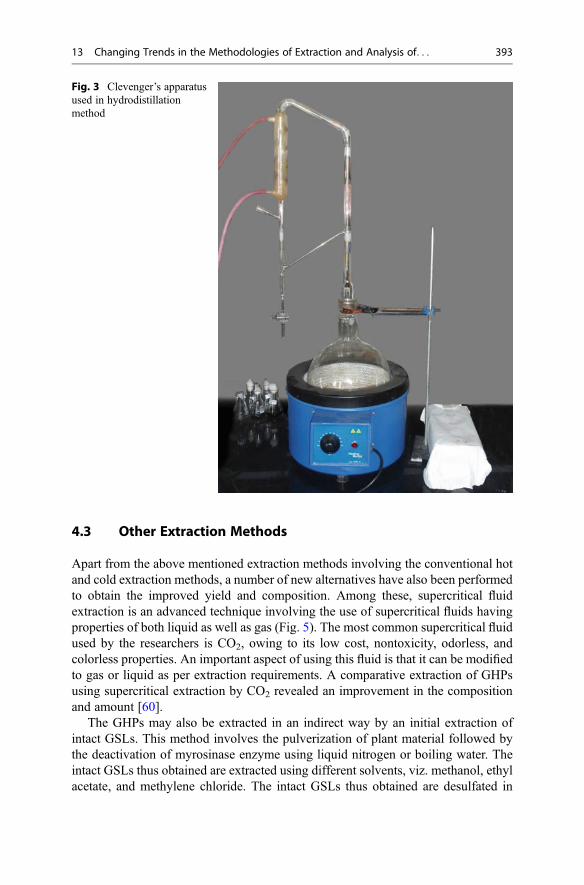
4.3 Other Extraction Methods
Apart from the above mentioned extraction methods involving the conventional hotand cold extraction methods, a number of new alternatives have also been performedto obtain the improved yield and composition. Among these, supercritical fluidextraction is an advanced technique involving the use of supercritical fluids havingproperties of both liquid as well as gas (Fig. 5). The most common supercritical fluidused by the researchers is CO2, owing to its low cost, nontoxicity, odorless, andcolorless properties. An important aspect of using this fluid is that it can be modifiedto gas or liquid as per extraction requirements. A comparative extraction of GHPsusing supercritical extraction by CO2 revealed an improvement in the compositionand amount [60].
The GHPs may also be extracted in an indirect way by an initial extraction ofintact GSLs. This method involves the pulverization of plant material followed bythe deactivation of myrosinase enzyme using liquid nitrogen or boiling water. Theintact GSLs thus obtained are extracted using different solvents, viz. methanol, ethylacetate, and methylene chloride. The intact GSLs thus obtained are desulfated in
Fig. 3 Clevenger’s apparatusused in hydrodistillationmethod
13 Changing Trends in the Methodologies of Extraction and Analysis of. . . 393

Fig. 4 Setup for microwave assisted hydrodistillation
Fig. 5 Equipment forsupercritical fluid extraction
394 R. Arora et al.

DEAE sephadex column using an external supply of either artificial or naturalmyrosinase enzyme (Fig. 6). An important advantage of this method is that thetype of GHPs required may be modified as per our requirement by changing thecolumn conditions (acidic, basic, or neutral pH) and temperature (low, room tem-perature, or high) [61].
5 Glucosinolate Hydrolytic Products Analysis Methods
The GHPs extracted by the above mentioned methods can be utilized for a number ofbiological activities such as antifungal, antibacterial, herbicidal, and anticancerproperties. Before utilizing it for any purpose, the analysis of its composition andquantity of each compound is necessary to analyze the synergistic or antagonisticeffect of each compound. The analytical methods thus employed range from themost common and traditional methods like paper and thin layer chromatography tomuch advanced microchip analysis.
5.1 Chromatography Methods
The chromatographic methods involve the analysis of different components of theextract on the basis of polarity and size.
5.1.1 Paper ChromatographyPaper chromatography (PC) is the oldest method during early 1900s for the analysisof GHPs. The thiourea formed following the reaction amid ammonia andisothiocyanates is detected and separated by paper. While the hydrophobic compo-nents are analyzed using water saturated chloroform, the hydrophilic components onthe other hand involved the mixture of butanol, toluene, and water as mobile phase.The low reproducibility of GHP analysis using this method makes it a difficultchoice for analysis [62].
Fig. 6 Indirect extraction ofglucosinolate hydrolyticproducts using DEAEsephadex column
13 Changing Trends in the Methodologies of Extraction and Analysis of. . . 395

5.1.2 Thin Layer ChromatographyThin layer chromatography (TLC) is much reliable and repeatable method for theanalysis of GHPs. The method uses silica or cellulose gel formed on glass plate as astationary phase, while different combination of solvents as per the polarity are usedas mobile phase. TLC can be used for both qualitative as well as quantitative analysisof GHPs. The repeatability, reproducibility, and the ability to analyze a large numberof samples at the same time make this method highly useful for the initial analysis ofGHPs [63].
5.1.3 High Performance Liquid ChromatographyHigh performance liquid chromatography (HPLC) is the analytical technique mostlypreferred for the analysis of GHPs since late 1900s. The accurate results along withan ease of reproducibility and repeatability makes it an idle analytical method tilldate. The variation in the mobile phase, flow rate, and the column used helps inanalyzing almost all the combination of GHPs present in different extracts (Fig. 7).The different compounds present in the extracts are analyzed using different detec-tors such as photodiode array and the much important mass spectrometer(MS) [64]. The MS attachment makes it easier for the identification of a compoundwithout the presence of any standard by detecting the molecular mass of compound.New and improved versions of HPLC are regularly being formed having all theadvantages of the regular HPLC, while solving its limitations. Some of theseimproved HPLC instruments are ultra high performance liquid chromatography(UHPLC), hydrophilic interaction liquid chromatography (HILIC), and high speedcounter-current chromatography (HSCCC) [65, 66].
Fig. 7 High performance liquid chromatography equipment
396 R. Arora et al.

5.1.4 Gas ChromatographyGas chromatography (GC) is another commonly employed analytic technique usedfor analyzing volatile compounds. The volatile nature of GHPs makes GC a suitabletechnique for analysis. The technique utilizes solid stationary phase and gas mobilephase (Fig. 8). A high initial injection temperature allows volatilization of thesolvent and sample. The type of column is often varied to suit the experiment inhand. Sometimes more than one column with varying polarity is used for elutingcompounds with different polarity. The mass spectrometer (MS) detector is preferredfor the gas chromatography analysis due to its ability to detect the mass spectra ofunknown compounds. Other detectors commonly combined with gas chromatogra-phy are flame ionization detector (FID) and head space (HS). The use of HS alongwith GC provides the ability to detect the compounds present in the vapours ofsample of interest [47, 67, 68].
5.1.5 Strong Ion-Exchange Centrifugal Partition ChromatographyA comparatively new analytical chromatographic method namely strongion-exchange centrifugal partition chromatography (SIXCPC) identifies and sepa-rates purified GHPs using lipophilic entities generated by strong anion exchanger(Fig. 9). A flow rate of 2 ml/min and a productivity of 3.3 g/h/LVC are attained withthis instrument [69, 70]. A modification of this instrument replaces the chromato-graph with an extractor and hence the name strong ion exchange centrifugal partitionextraction (SIXCPE). This modification allows an astonishing increase in produc-tivity up to 8.5-folds, i.e., 28.3 g/h/LVC [70]. The increased productivity and high
Fig. 8 Gas chromatography equipment
13 Changing Trends in the Methodologies of Extraction and Analysis of. . . 397

purity of GHPs obtained using this method allows its use at industrial scale in nearfuture.
5.2 Non-Chromatographic Methods
There are other methods for the analysis of GHPs which do not require anychromatographic technique.
5.2.1 X-Ray SpectroscopyX-ray spectroscopic method of GHP analysis is based on the nondestructive detec-tion of sulfur content in the compound (Fig. 10). This content is then compared withthe sulfur content of the reference material. A number of factors including thereduction of moisture content and controlled heating are required for the properanalysis of GHPs [71, 72].
5.2.2 Nuclear Magnetic Resonance SpectroscopyNuclear magnetic resonance (NMR) spectroscopy utilizes the magnetic properties ofGHPs to analyze the physical and chemical properties of these compounds (Fig. 11).The structural properties of the compound are determined by using a nondestructivemethod. This spectroscopy is also efficient for the analysis of some of the structureswhich cannot be analyzed by any other method. The difficulty in interpretation, highcost, and time consumption has lead to a decreased interest for this method [73, 74].
5.2.3 Near Infrared Reflectance SpectroscopyNear infrared reflectance spectroscopy (NIRS) is a reproducible, rapid, and nonde-structive analytical method for GHPs (Fig. 12). The compound of interest is
Fig. 9 Equipment used for strong ion exchange centrifugal partition chromatography
398 R. Arora et al.
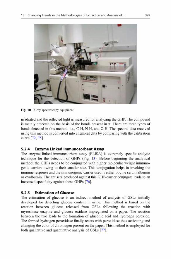
irradiated and the reflected light is measured for analyzing the GHP. The compoundis mainly detected on the basis of the bonds present in it. There are three types ofbonds detected in this method, i.e., C-H, N-H, and O-H. The spectral data receivedusing this method is converted into chemical data by comparing with the calibrationcurve [72, 75].
5.2.4 Enzyme Linked Immunosorbent AssayThe enzyme linked immunosorbent assay (ELISA) is extremely specific analytictechnique for the detection of GHPs (Fig. 13). Before beginning the analyticalmethod, the GHPs needs to be conjugated with higher molecular weight immuno-genic carriers owing to their smaller size. This conjugation helps in invoking theimmune response and the immunogenic carrier used is either bovine serum albuminor ovalbumin. The antisera produced against this GHP-carrier conjugate leads to anincreased specificity against these GHPs [76].
5.2.5 Estimation of GlucoseThe estimation of glucose is an indirect method of analysis of GSLs initiallydeveloped for detecting glucose content in urine. This method is based on thereaction between glucose released from GSLs following the reaction withmyrosinase enzyme and glucose oxidase impregnated on a paper. The reactionbetween the two leads to the formation of gluconic acid and hydrogen peroxide.The formed hydrogen peroxidase finally reacts with peroxidase thus activating andchanging the color of chromagen present on the paper. This method is employed forboth qualitative and quantitative analysis of GSLs [77].
Fig. 10 X-ray spectroscopy equipment
13 Changing Trends in the Methodologies of Extraction and Analysis of. . . 399

Fig. 12 Equipment for near infrared reflectance spectroscopy
Fig. 11 Nuclear magneticresonance spectroscopicequipment
400 R. Arora et al.

5.2.6 Microchip AnalysisThe microchip analysis is a nondestructive and automated method employed forboth quantitative and qualitative purpose with a small quantity of sample. Theanalysis is based on the capillary electrophoresis on a microchip. A charge transfercomplex is formed between the GSLs and xanthenes dyes. It is a rapid method andrequires low quantity of reagents and thus forms a successful alternate to earliertechniques [78].
6 Conclusions
Glucosinolates are important class of secondary metabolites present in a number ofplant families. These metabolites are broken down into their hydrolytic products bythe action of myrosinase, an enzyme present adjacent to it. The GHPs have animmense use in pharmacological industries due to their wide range of biologicalproperties such as antifungal, antibacterial, and anticancer activity. These propertiesof this crucial secondary metabolite make it a significant factor for exploring a rangeof extraction methods, thus improving the yield, composition, time, and cost. Assuch both cold and hot extraction methods are used for its extraction, but due to thevolatility of the compound, the most important method among these is hydrodis-tillation. The extracted compounds are first required to be analyzed for their contentbefore subjecting it for biological analysis and thus a number of analytical methodswere developed to suit the requirement of the researchers. These methods rangedfrom simple and rapid methods like paper and thin layer chromatography to complexand effective microchip analysis. But currently, the most commonly employedmethod is gas chromatography equipped with mass spectrometer. This methodsuits the volatile nature of GHPs and provides accurate results. In nutshell, theselection of extraction and analytical method depends on the problem in hand andthe suitable conditions.
Fig. 13 Analysis of glucosinolate hydrolytic products using enzyme linked immunosorbent assay,where (a) 96 well plate and (b) biotek synergy HT ELISA reader
13 Changing Trends in the Methodologies of Extraction and Analysis of. . . 401

Acknowledgment The study was supported by the funding provided by Department of Scienceand Technology (DST) and University Grants Commission (UGC), New Delhi and Guru NanakDev University, Amritsar.
7 Cross-References
▶Glucosinolates and Plant Defense▶ Investigation of Glucosinolates by Mass Spectrometry
References
1. Ahmed ZF, Rizk AM, Hammouda FM, Seif El-Nasr MM (1972) Naturally occurring glucosiwith special references to those of family Capparidaceae. Planta Med 21:35–60
2. Fenwick GR, Heaney RK, Mullin WJ (1983) Glucosinolates and their breakdown products infood and food plants. Crit Rev Food Sci Nutr 18:123–201
3. Dornemann D, Loffelhardt W, Kindl H (1974) Chain elongation of aromatic acid: the role of2-benzylmalic acid in the biosynthesis of a C6C4 amino acid and a C6C3 mustard oil glucoside.Can J Biochem 52:916–921
4. Cole R (1976) Isothiocyanates, nitriles and thiocyanates as products of autolysis ofglucosinolates in Cruciferae. Phytochemistry 15:759–762
5. Dawson GW, Hick AJ, Bennet RN, Donald AM, Wallsgrove RM (1993) Synthesis ofglucosinolate precursors and investigations into the biosynthesis of phenylalkyl- andmethylthioalkyl glucosinolates. J Biol Chem 268:27154–27159
6. Halkier BA, Du L (1997) The biosynthesis of glucosinolates. Trends Plant Sci 2:425–4317. Haristoy X, Fahey JW, Scholtus I, Lozeniewski A (2005) Evaluation of the antimicrobial effects
of several isothiocyanates on Helicobacter pylori. Planta Med 71:326–3308. Tiedink HGM,Malingre CE, Broekhoven LW, JongenWMF, Lewis J, Fenwick GR (1991) Role
of glucosinolates in the formation of of N-nitroso compounds. J Agric Food Chem 39:922–9269. Mennicke WH, Gorler K, Krumbiegel G, Lorenz D, Rittmann N (1988) Studies on the
metabolism and excretion of benzyl isothiocyanate in man. Xenobiotica 18:441–44710. Smolinska U, Knudsen GR, Morra MJ, Borek V (1997) Inhibition of Aphanomyces euteiches
f. sp. pisi by volatiles produced by hydrolysis of Brassica napus seed meal. Plant Dis81:288–292
11. Fahey JW, Haristoy X, Dolan PM, Fahey JW, Haristoy X, Dolan PM et al (2002) Sulforaphaneinhibits extracellular, intracellular, and antibiotic-resistant strains of Helicobacter pylori andprevents benzo[a]pyrene-induced stomach tumors. Proc Natl Acad Sci U S A 99:7610–7615
12. Lin CM, Kim J, Du WX, Wei CI (2000) Bactericidal activity of isothiocyanate againstpathogens on fresh produce. J Food Prot 63:25–30
13. Greenhalgh JR, Mitchell ND (1976) The involvement of flavour volatiles in the resistance todowny mildew of wild and cultivated forms of Brassica oleracea. New Phytol 77:391–398
14. Nadarajah D, Han JH, Holley RA (2005) Use of mustard flour to inactivate Escherichia coliO157:H7 in ground beef under nitrogen flushed packaging. Int J Food Microbiol 99:257–267
15. Zsolnai T (1966) Antimicrobial effect of thiocyanates and isothiocyanates. Arztl Forsch16:870–876
16. Angus JF (1994) Biofumigation: isothiocyanates released from Brassica roots inhibit thegrowth of the take-all fungus. Plant Soil 162:107–112
402 R. Arora et al.

17. Hooker WJ, Walker JC, Smith FG (1943) Toxicity of beta-phenethyl isothiocyanate to certainfungi. Am J Bot 30:632–637
18. Gamliel A, Stapleton JJ (1993) Characterization of antifungal volatile compounds evolved fromsolarized soil amended with cabbage residues. Phytopathology 83:899–905
19. Manici LM, Lazzeri L, Baruzzi G, Leoni O, Galletti S, Palmieri S (2000) Suppressive activity ofsome glucosinolate enzyme degradation products on Pythium irregulare and Rhizoctonia solaniin sterile soil. Pest Manag Sci 56:921–926
20. Smith BJ, Sarwar M, Wong PTW, Kirkegaard JA (1999) Suppression of cereal pathogens bycanola root tissues in soil. In: Proceedings of the 10th international rapeseed congress, pp 334
21. Dandurand LM, Mosher RD, Knudsen GR (2000) Combined effects of Brassica napus seedmeal and Trichoderma harzianum on two soil borne plant pathogens. Can J Microbiol46:1051–1057
22. Mayton HS, Oliver C, Vaughn SF, Loria R (1996) Correlation of fungicidal activity of Brassicaspecies with allyl isothiocyanate production in macerated leaf tissue. Phytopathology86:267–271
23. Lewis JA, Papavizas GC (1971) Effect of sulphur-containing volatile compounds and vaporsfrom cabbage decomposition on Aphanomyces euteiches. Phytopathology 61:208–214
24. Ekanayake A, Kester JJ, Li JJ, Zehentbauer GN, Bunke PR, Zent JB (2006) Isogard(tm) anatural anti-microbial agent derived from white mustard seed. Acta Hortic 709:101–108
25. Brown PD, Morra MJ (1997) Control of soil-borne plant pests using glucosinolate-containingplants. Adv Agron 61:167–231
26. Seo ST, Tang CS (1982) Hawaiian fruit flies (Diptera: Tephritidae): toxicity of benzyl isothio-cyanate against eggs or 1st instars of three species. J Econ Entomol 75:1132–1135
27. Matthiessen JN, Shackleton MA (2000) Advantageous attributes of larval White fringed weevil,Naupactus leucoloma (Coleoptera: Curculionidae) for bioassaying soil fumigants, andresponses to pure and plant-derived isothiocyanates. Bull Entomol Res 90:349–355
28. Borek V, Elberson LR, McCaffrey JP, Morra MJ (1995) Toxicity of aliphatic and aromaticisothiocyanates to eggs of the black vine weevil (Coleoptera: Curculionidae). J Econ Entomol88:1192–1196
29. Noble RRP, Harvey SG, Sams CE (2002) Toxicity of Indian mustard and allyl isothiocyanate tomasked chafer beetle larvae. Plant Health Prog. doi:10.1094/PHP-2002-0610-01-RS
30. Tsao R, Peterson CJ, Coats JR (2002) Glucosinolate breakdown products as insect fumigantsand their effect on carbon dioxide emission of insects. BMC Ecol 2:5
31. Winkler H, Otto G (1980) Replant losses with strawberries and suggestions for their reduction.Hortic Abstr 50:344
32. Johnson AW, Golden AM, Auld DL, Sumner DR (1992) Effects of rapeseed and vetch as greenmanure crops and fallow on nematodes and soil-borne pathogens. J Nematol 24:117–126
33. Bhardwaj HL, Hamama AA, Porter DM, Reese PF Jr (1996) Rapeseed meal as a naturalpesticide. In: Janick J (ed) Progress in new crops. ASHS Press, Arlington, pp 615–619
34. Serra B, Rosa E, Iori R, Barillari J, Cardoso A, Abreu C (2002) In vitro activity of 2-phenylethylglucosinolate, and its hydrolysis derivatives on the root-knot nematodeGlobodera rostochiensis(Woll.). Sci Hortic 92:75–81
35. Zasada IA, Ferris H (2004) Nematode suppression with brassicaceous amendments: applicationbased upon glucosinolate profiles. Soil Biol Biochem 36:1017–1024
36. Brown PD, Morra MJ (1995) Hydrolysis products of glucosinolates in Brassica napus tissues asinhibitors of seed germination. Plant Soil 181:307–316
37. Sarwar M, Kirkegaard JA (1998) Biofumigation potential of brassicas. Plant Soil 201:91–10138. Krishnan G, Holshauser DL, Nissen SJ (1998) Weed control in soybean (Glycine max) with
green manure crops. Weed Technol 12:97–10239. Norsworthy JK, Brandenberger L, Burgos NR, Riley M (2005) Weed suppression in Vigna
unguiculata with a spring-seeded Brassicaceae green manure. Crop Prot 24:441–447
13 Changing Trends in the Methodologies of Extraction and Analysis of. . . 403

40. Petersen J, Belz R, Walker F, Hurle K (2001) Weed suppression by release of isothiocyanatesfrom turnip–rape mulch. Agron J 93:37–43
41. Sedmikova M, Turek B, Barta I, Strohalm J, Smerak P, Houska M et al (1999) Evaluation of theantimutagenic activity of pressure treated and heat pasteurized cauliflower juice. Czech J FoodSci 17:149–152
42. Mandelova L, Totusek J (2007) Broccoli juice treated by high pressure: chemoprotective effectsof sulforaphane and indole-3-carbinol. High Pressure Res 27:151–156
43. Murugan SS, Balakrishnamurthy P, Mathew YJ (2007) Antimutagenic effect of broccoli flowerhead by the Ames Salmonella reverse mutation assay. Phytother Res 21:545–547
44. Cohen JH, Kristal AR, Stanfordm JL (2000) Fruit and vegetable intakes and prostate cancerrisk. J Natl Cancer Inst 92:61–68
45. Zhang SM, Hunter DJ, Rosner BA, Giovannucci EL, Colditz GA, Speizer FE et al (2000)Intakes of fruits, vegetables, and related nutrients and the risk of non-Hodgkin’s lymphomaamong women. Cancer Epidemiol Biomarkers Prev 9:477–485
46. Verhoeven DT, Goldbohm RA, Van Poppel G, Verhagen H, Van den Brandt PA (1996)Epidemiological studies on Brassica vegetables and cancer risk. Cancer Epidemiol BiomarkersPrev 5:733–748
47. Arora R, Bhushan S, Kumar R, Mannan R, Kaur P, Singh AP, Arora S (2014) Hepaticdysfunction induced by 7, 12-dimethylbenz (α) anthracene and its obviation with erucin usingenzymatic and histological changes as indicators. PLoS One 9(11):e112614. doi:10.1371/journal.pone.0112614
48. Wattenberg LW (1983) Inhibition of neoplasia by minor dietary constituents. Cancer Res43:2448–2453
49. Morse MA, Eklind KI, Hecht SS, Jordan KG, Choi CI, Desai DH (1991) Structure-activityrelationships for inhibition of 4-(methylnitrosamino)- 1-(3-pyridyl)-1-butanone lung tumori-genesis by arylalkyl isothiocyanates in A/J mice. Cancer Res 51:1846–1850
50. Conaway C, Yang YM, Chung FL (2002) Isothiocyanates as cancer chemopreventive agents:their biological activities and metabolism in rodents and humans. Curr Drug Metab 3:233–255
51. Yang YM, Conaway CC, Chiao JW, Wang CX, Amin S, Whysner J (2002) Inhibition of benzo(a)pyrene-induced lung tumorigenesis in A/J mice by dietary N-acetylcysteine conjugates ofbenzyl and phenethyl isothiocyanates during the post initiation phase is associated withactivation of mitogen-activated protein kinases and p53 activity and induction of apoptosis.Cancer Res 62:2–7
52. Bell JM, Youngs CG, Downey RK (1971) A nutritional comparison of various rapeseed andmustard seed solvent-extracted meals of different glucosinolate composition. Can J Anim Sci51:259–269
53. Schreiner RP, Koide RT (1993) Antifungal compounds from the roots of mycotrophic and non‐mycotrophic plant species. New Phytol 123:99–105
54. Kim HK, Verpoorte R (2010) Sample preparation for plant metabolomics. Phytochem Anal21:4–13
55. Arora R, Sharma D, Kumar R, Singh B, Vig AP, Arora S (2014) Evaluating extractionconditions of glucosinolate hydrolytic products from seeds of Eruca sativa (Mill.) Thell.using GC‐MS. J Food Sci 79:C1964–C1969
56. Fahey JW, Zhang Y, Talalay P (1997) Broccoli sprouts: an exceptionally rich source of inducersof enzymes that protect against chemical carcinogens. Proc Natl Acad Sci 94:10367–10372
57. Talreja K, Moon A (2014) Brassica oleracea: phytochemical profiling in Search for anticancercompound. Int J Sci Pharm Res 4. 2250–0480
58. Paunović D, Šolević-Knudsen T, Krivokapić M, Zlatković B, Antić M (2012) Sinalbin degra-dation products in mild yellow mustard paste. Hem Ind 66:29–32
59. Wei L, Zhang Y, Jiang B (2013) Comparison of microwave-assisted hydrodistillation with thetraditional hydrodistillation method in the extraction of essential oils from dwarfedCinnamomum camphora var. Linaolifera Fujita leaves and twigs. Adv J Food Sci Technol5:1436–1442
404 R. Arora et al.

60. Capuzzo A, Maffei ME, Occhipinti A (2013) Supercritical fluid extraction of plant flavours andfragrances. Molecules 18:7194–7238
61. Truscott RJ, Minchinton I, Sang J (1983) The isolation and purification of indole glucosinolatesfrom Brassica species. J Sci Food Agric 34:247–254
62. Kjær A, Rubinstein K (1953) Paper chromatography of thioureas. Nature 171:840–84163. Croft AG (1979) The determination of total glucosinolates in rapeseed meal by titration of
enzyme‐liberated acid and the identification of individual glucosinolates. J Sci Food Agric30:417–423
64. Bennett RN, Mellon FA, Kroon PA (2004) Screening crucifer seeds as sources of specific intactglucosinolates using ion-pair high-performance liquid chromatography negative ionelectrospray mass spectrometry. J Agric Food Chem 52:428–438
65. Troyer JK, Stephenson KK, Fahey JW (2001) Analysis of glucosinolates from broccoli andother cruciferous vegetables by hydrophilic interaction liquid chromatography. J Chromatogr A919:299–304
66. Fahey JW, Wade KL, Stephenson KK, Chou FE (2003) Separation and purification ofglucosinolates from crude plant homogenates by high-speed counter-current chromatography.J Chromatogr A 996:85–93
67. Vaughn SF, Berhow MA (2005) Glucosinolate hydrolysis products from various plant sources:pH effects, isolation, and purification. Ind Crop Prod 21:193–202
68. Taveira M, Fernandes F, de Pinho PG, Andrade PB, Pereira JA, Valentão P (2009) Evolution ofBrassica rapa var. rapa L. volatile composition by HS-SPME and GC/IT-MS. Microchem J93:140–146
69. Toribio A, Boudesocque L, Richard B, Nuzillard JM, Renault JH (2011) Preparative isolation ofglucosinolates from various edible plants by strong ion-exchange centrifugal partition chroma-tography. Sep Purif Technol 83:15–22
70. Hamzaoui M, Hubert J, Reynaud R, Marchal L, Foucault A, Renault JH (2012) Strong ionexchange in centrifugal partition extraction (SIX-CPE): effect of partition cell design anddimensions on purification process efficiency. J Chromatogr A 1247:18–25
71. Schnug E, Haneklaus S (1988) Theoretical principles for the indirect determination of the totalglucosinolate content in rapeseed and meal quantifying the sulphur concentration via X‐rayfluorescence (X‐RF method). J Sci Food Agric 45:243–254
72. Evans EJ, Bilsborrow P, Grant A, Kwiatkowska CA, Spinks E, Heaney RK, Fenwick GR (1989)A comparison of rapid‐(X‐ray fluorescence, near‐infrared reflectance) and glucose‐releasemethods for the determination of the glucosinolate content of oilseed rape (Brassica oleracea).J Sci Food Agric 49:297–305
73. Cox IJ, Hanley AB, Belton PS, Fenwick GR (1984) NMR spectra (1H, 13C) of glucosinolates.Carbohydr Res 132:323–329
74. Prestera T, Fahey JW, Holtzclaw WD, Abeygunawardana C, Kachinski JL, Talalay P (1996)Comprehensive chromatographic and spectroscopic methods for the separation and identifica-tion of intact glucosinolates. Anal Biochem 239:168–179
75. Hom NH, Becker HC, Möllers C (2007) Non-destructive analysis of rapeseed quality by NIRSof small seed samples and single seeds. Euphytica 153:27–34
76. van Doorn HE, van Holst GJ, van der Kruk GC, Raaijmakers-Ruijs NC, Postma E (1998)Quantitative determination of the glucosinolates sinigrin and progoitrin by specific antibodyELISA assays in Brussels sprouts. J Agric Food Chem 46:793–800
77. Arora R, Vig AP, Arora S (2014) Glucosinolates: transposing trends of identification methodsfrom paper chromatography to microchip analysis. Int J Life Sci Biotechnol Pharma Res 3:42–61
78. Fouad M, Jabasini M, Kaji N, Terasaka K, Tokeshi M, Mizukami H, Baba Y (2008) Microchipanalysis of plant glucosinolates. Electrophoresis 29:2280–2287
13 Changing Trends in the Methodologies of Extraction and Analysis of. . . 405

Processing and Preparation of BrassicaVegetables and the Fate of Glucosinolates 14Probo Yulianto Nugrahedi, Matthijs Dekker, and Ruud Verkerk
AbstractThe healthiness of a vegetable cannot solely be inferred from the amount ofhealth-promoting compounds in the raw materials. Brassica vegetables, forexample, are consumed mostly after processing to improve palatability and toextend the shelf life. However, processing also results to various changes in thecontent of glucosinolates which intakes are associated with a reduced risk ofseveral cancers. The large variety in cooking practices and processing methodsaffect the glucosinolate content in the vegetables, particularly due to processesthat allow for enzymatic hydrolysis and thermal degradation of glucosinolates,and leaching of the bioactive components. Knowledge on the effect of preparationand processing of Brassica vegetables is important to evaluate the healthiness ofthe consumed product and to investigate mechanisms to retain high glucosinolatelevels at the stage of consumption and to increase the intake of health-protectivecompounds by the consumer. By using a mechanistic approach, the fate ofglucosinolates during different processing and preparation methods and condi-tions can be explained. Boiling and blanching reduce the glucosinolate contentsignificantly particularly because of the mechanisms of leaching following celllysis and diffusion, and partly due to thermal and enzymatic degradation.Steaming, microwave processing, and stir frying either retain or only slightlyreduce the glucosinolate content due to low degrees of leaching. These methods
P.Y. Nugrahedi (*)Department of Food Technology, SOEGIJAPRANATA Catholic University (Unika) of Semarang,Semarang, Indonesiae-mail: [email protected]
M. Dekker • R. VerkerkFood Quality and Design Group, Department of Agrotechnology and Food Sciences, WageningenUniversity, Wageningen, The Netherlandse-mail: [email protected]; [email protected]
# Springer International Publishing Switzerland 2017J.-M. Mérillon, K.G. Ramawat (eds.), Glucosinolates, Reference Series inPhytochemistry, DOI 10.1007/978-3-319-25462-3_10
407
can enhance the accessibility of glucosinolates from the plant tissue. Fermenta-tion reduces the glucosinolate content considerably, the underlying mechanismsare not yet completely clear, but enzymatic breakdown seems to play an importantrole. Studying the changes of glucosinolates during processing by a mechanisticapproach is shown to be valuable to redesign the processing and to reformulatethe product for improving health benefits of these compounds.
KeywordsGlucosinolate • Preparation • Processing • Mechanistic approach • Brassicavegetable
AbbreviationsESP Epithiospecifier proteinGS GlucosinolateHPP High pressure processingITC IsothiocyanateMW Microwave
Contents1 Introduction . . . . . . . . . . . . . . . . . . . . . . . . . . . . . . . . . . . . . . . . . . . . . . . . . . . . . . . . . . . . . . . . . . . . . . . . . . . . . . . . . . 4082 Brassica Vegetable Intake and Health . . . . . . . . . . . . . . . . . . . . . . . . . . . . . . . . . . . . . . . . . . . . . . . . . . . . . . 4093 Glucosinolates in Brassica vegetables . . . . . . . . . . . . . . . . . . . . . . . . . . . . . . . . . . . . . . . . . . . . . . . . . . . . . . 409
3.1 Occurrence . . . . . . . . . . . . . . . . . . . . . . . . . . . . . . . . . . . . . . . . . . . . . . . . . . . . . . . . . . . . . . . . . . . . . . . . . . . . . 4093.2 Glucosinolate/Myrosinase System . . . . . . . . . . . . . . . . . . . . . . . . . . . . . . . . . . . . . . . . . . . . . . . . . . . . 4103.3 Breakdown Products of Glucosinolates . . . . . . . . . . . . . . . . . . . . . . . . . . . . . . . . . . . . . . . . . . . . . . 412
4 Glucosinolate During Preparation of Brassica Vegetables . . . . . . . . . . . . . . . . . . . . . . . . . . . . . . . . . 4124.1 Mechanisms Underlying the GS Changes . . . . . . . . . . . . . . . . . . . . . . . . . . . . . . . . . . . . . . . . . . . . 4134.2 Thermal Processing and Preparation Methods . . . . . . . . . . . . . . . . . . . . . . . . . . . . . . . . . . . . . . . 4154.3 Other Processing Methods . . . . . . . . . . . . . . . . . . . . . . . . . . . . . . . . . . . . . . . . . . . . . . . . . . . . . . . . . . . . 419
5 Product and Process Design . . . . . . . . . . . . . . . . . . . . . . . . . . . . . . . . . . . . . . . . . . . . . . . . . . . . . . . . . . . . . . . . . 4236 Conclusions . . . . . . . . . . . . . . . . . . . . . . . . . . . . . . . . . . . . . . . . . . . . . . . . . . . . . . . . . . . . . . . . . . . . . . . . . . . . . . . . . . 424References . . . . . . . . . . . . . . . . . . . . . . . . . . . . . . . . . . . . . . . . . . . . . . . . . . . . . . . . . . . . . . . . . . . . . . . . . . . . . . . . . . . . . . . 424
1 Introduction
Many representatives of the Brassicaceae family are of particular importance asvegetables for our human diet, such as Brassica oleracea (e.g., cabbage, cauliflower,broccoli, and Brussels sprouts), Brassica rapa (Chinese cabbage, pak choi, andturnip), Brassica juncea (Indian and Chinese mustard), Brassica napus (rutabagaand swede), and as seasonings and relishes (e.g., mustard and wasabi). Brassicavegetables are unique in that they are rich sources of glucosinolates (GSs), sulfur-containing compounds that deliver a pungent aroma and spicy or bitter taste[1–3]. Moreover, GSs are claimed to be bioactive components responsible formany of the physiological health effects proposed for Brassica vegetables indifferent types of studies, including in vitro, animal, human, and epidemiologicalstudies [4, 5].
408 P.Y. Nugrahedi et al.

Most of the vegetables need some kind of treatment, either household preparationor industrial processing, in order to make them suitable and palatable for consump-tion. The large variety in cooking and processing methods and conditions have incommon that they all affect the profile of GSs in the vegetables, particularly due to(bio)chemical and physical processes such as enzymatic hydrolysis and thermaldegradation of GSs, and leaching of the bioactive components out of the plant tissue[6]. As a consequence, we can say that the healthiness of a vegetable cannot solely beinferred from the amount of the nutritional and health compounds in the rawmaterials. Knowledge on the effect of preparation and processing of Brassicavegetables is important to evaluate the healthiness of the final consumed productand to investigate ways to retain high GS levels at the stage of consumption and toincrease the intake of health-protective compounds by the consumer.
2 Brassica Vegetable Intake and Health
Brassica vegetables, like broccoli, cauliflower, Brussels sprouts, and red cabbage,contain significant levels of health-promoting constituents, including vitamins,minerals, fibers, and numerous types of secondary plant metabolites also calledphytochemicals. Many phytochemicals from vegetables contribute to the reportedantioxidative, anti-inflammatory, anticarcinogenic, and cardiovascular protectiveeffects [7]. One group of phytochemicals occurring almost exclusively in Brassicavegetables is the group of glucosinolates [8]. Epidemiological studies showed thatthe intake of Brassica vegetables is inversely associated with the risk of certain typesof cancer, including colorectal and lung cancers [9–13].
Glucosinolates (GSs) intake is expected to play a significant role in lowering thisrisk of cancer. However, in epidemiologic studies, the intake of the vegetables ismonitored, the real intake of protective compounds, like GSs, is often an unknownvariable. It is demonstrated that many steps in the food production chain, likecultivation, storage, processing, and preparation of vegetables can dramaticallyaffect the content and thus the intake of phytochemicals such as GSs in Brassicavegetables [14].
3 Glucosinolates in Brassica vegetables
3.1 Occurrence
Glucosinolates (GSs), a group of plant secondary metabolites, containβ-thioglucoside N-hydroxysulfates with a sulfur linked β-D-glucopyranose moietyand variable side group (R), which usually classifies the aliphatic, aromatic, andindole GSs. These three GS groups are frequently found in Brassica vegetablespecies (Table 1). Moreover, each species of the family Brassicaceae has a distinctGS profile characterized by major GSs as reviewed by Verkerk et al. [5]. Also,different species of the same genus and different cultivars of the same species can
14 Processing and Preparation of Brassica Vegetables and the Fate of. . . 409

have highly variable GS concentrations. The majority of GSs are found in everyplant organ although the concentration and composition of the GSs can vary greatlyand can also change during plant development [5]. Usually, a single plant speciescontains up to four different GSs in significant amounts, while as many as 15 differ-ent GSs can be found in lower amounts in the same plant. Brassica vegetables occurin different appearances: leafy (e.g., collard green and rocket salad), flowering(cauliflower and broccoli), stems (kohlrabi), roots (radish, rutabaga, and turnips),and buds (Brussels sprouts and cabbage). The content of GSs varies in these differenttissues, for example, GS concentrations are higher in the florets than in the stalks ofbroccoli [5]. The seeds and the sprouting vegetables or cresses, such as garden cressor watercress, usually contain one specific type of GS in substantial amounts. TheGS concentrations in vegetables, although often highly variable, are around 1% dryweight in some Brassica vegetables.
3.2 Glucosinolate/Myrosinase System
The special feature of GS-containing vegetables is the system of compartmentaliza-tion of GSs and the presence of specialized myrosin cells containing the hydrolytic
Table 1 Glucosinolates commonly found in Brassica vegetables [15]
Trivial name Chemical name Main source
Aliphatic
Glucoibervirin 3-Methylthiopropyl-GS Green and white cauliflowers
Glucoerucin 4-Methylthiobutyl-GS Rocket
Glucoiberin 3-Methylsulfinylpropyl-GS Broccoli sprouts, Savoy cabbage
Glucoraphanin 4-Methylsulfinylbutyl-GS Broccoli(cress), Red cabbage
Sinigrin Prop-2-enyl-GS Brussels sprouts, White cauliflower
Gluconapin But-3-enyl-GS Pak choi
Glucobrassicanapin Pent-4-enyl-GS Chinese cabbage, Pak choi
Progoitrin (2R)-2-Hydroxybut-3-enyl Turnip, Chinese broccoli
Indole
Glucobrassicin Indol-3-ylmethyl-GS Broccoli, Cauliflower, and manymore
4-Hydroxy-glucobrassicin
4-Hydroxy-indol-3-ylmethyl-GS
Broccoli, Cauliflower, and manymore
4-Methoxy-glucobrassicin
4-Methoxy-indol-3-ylmethyl-GS
Broccoli, Cauliflower, and manymore
Neo-glucobrassicin N-methoxyindol-3-ylmethyl-GS
Broccoli, Cauliflower, and manymore
Aromatic
Glucotropaeolin Benzyl-GS Garden cress
Gluconasturtiin Phenylethyl-GS Water cress
410 P.Y. Nugrahedi et al.

enzyme myrosinase (thioglucoside glucohydrolase, EC 3.2.1.147). Upon plant’s celldisruption, GSs are highly prone to degradation by myrosinase-catalyzed hydrolysis(see Scheme 1). Subsequently, the GS will degrade into glucose and an unstableaglycon intermediate. The unstable aglycon rearranges into different breakdownproducts, including isothiocyanates (ITCs), thiocyanates, nitriles, and epithionitriles,depending on conditions described in the Sect. 3.3 [4, 8].
Myrosinase is a relative thermo labile enzyme, which can be readily denatured atmoderate to high temperatures. Especially temperatures applied during processing ofBrassica vegetables quickly inactivate myrosinase [16–19]. The optimum tempera-tures for the activity of myrosinase are different between Brassica vegetables, in therange between 30 �C and 60 �C, and the activity is also influenced by pH, thepresence of ascorbic acid, salt, and pressure [20–24]. During the various stages ofstorage, preparation, cooking, and processing of the vegetables, the GS–myrosinasesystem is affected in a complex way. Dekker et al. [25] have estimated that theconcentration of GSs in Brassica vegetables may vary by five to tenfold at eachstage.
The health-promoting effect of GSs is mainly attributed to the ITCs that areformed due to hydrolysis by myrosinase after tissue damage. Since myrosinase ismostly inactivated during processing or preparation, formation of ITCs usually doesnot occur in the product during mastication. However, a myrosinase-like activity isalso provided by the microflora in the human’s large intestine. Intake of Brassicaproducts containing inactive endogenous plant myrosinase still can have benefit bythe formation and absorption of bioactive breakdown products by enzymes from thegut flora. However, their bioavailability is lower than the ones with active plantmyrosinase [10, 26–28].
Scheme 1 Enzymatic breakdown of glucosinolate [adapted from 15]
14 Processing and Preparation of Brassica Vegetables and the Fate of. . . 411

3.3 Breakdown Products of Glucosinolates
Although GSs can be chemically degraded at higher temperatures [29–31], thehydrolysis is mainly enzymatically driven. Previous reports have reviewed themechanisms of GS hydrolysis [e.g., 4, 31–34], which will be briefly describedhere. Several products of hydrolysis of a GS can be produced, such as ITCs,thiocyanates, nitriles, epithionitriles, oxazolidine-2-thiones, or indole compounds,depending on the structure of the GS side chain, the reaction conditions (e.g., pH),presence of additional cofactors (e.g., Fe2+), and proteins (e.g., epithiospecifierprotein (ESP) and thiocyanate-forming protein). Most frequently, the aglyconundergoes a Lossen arrangement to produce an ITC.
At neutral pH, the major hydrolysis products are stable ITCs. For example,hydrolysis of gluconapin and sinigrin produces mainly ITCs, namely, 3-butenyl-ITC and 2-propenyl-ITC, respectively. Sulforaphane, the ITC derived fromglucoraphanin, is the most widely studied as the most bioactive GS hydrolysisproduct. For GSs having a β-hydroxylated side chain or an indole moiety,β-hydroxy-ITCs are unstable and spontaneously cyclize to oxazolidine-2-thiones,while indole ITCs undergo breakdown producing, for example, indole-3-carbinol(I3C).
At low pH, in the presence of an ESP and ferrous ions, gluconapin and sinigrinproduce cyano-epithioalkane, such as 1-cyano-3,4-epithiobutane and 1-cyano-2,3-epithiopropane, respectively, and progoitrin is hydrolyzed into an epithionitriles.Nitriles are the major degradation products under acidic conditions, which can bediminished by heating. It is formed after hydrolysis of a GS with a side chain lackinga double bond, which may involve ESP. Conversion to nitriles is also enhanced in thepresence of ferrous ions. Indole GSs can form indolyl-3-acetonitrile and elementalsulfur. Moreover, ascorbigen and thiocyanate are the major products of indole GSsbetween pH 4 and 7 in the presence of ascorbic acid. For thiocyanates production,the mechanism from GSs is not clear yet.
4 Glucosinolate During Preparation of Brassica Vegetables
Brassica vegetables are mainly consumed after processing, either at a domestic orindustrial level. Broccoli, cauliflower, and cabbage are boiled, steamed, stir-fried, ormicrowave-processed during domestic preparation to produce various dishes.Canned or fermented vegetables are also produced after industrial processing ofthese vegetables. Even when consumed raw, for example, in a salad, these vegetablesare firstly prepared, namely, by washing, cutting, and chopping. Various products ordishes available around the globe are made based on these vegetables. For examples,many kinds of soup, steamed, and stir-fried Brassica produced by domestic prepa-ration and industrial processed products, such as canned and fermented Brassicasuch as sauerkraut, or more local products as sayur asin, and kimchi. In some Asiancountries, the dishes made from these vegetables are usually considered as a sidedish to accompany rice or noodle [35].
412 P.Y. Nugrahedi et al.

Postharvest treatments of Brassica vegetables, such as cutting or chopping andpackaging and storage, can reduce the GS content to a lower degree than the loss dueto preparation itself [5, 36–38]. However, a study on storage of chopped cabbage andbroccoli was reported to increase the indole GSs, which is suspected as physiologicalresponse similar to response due to insect attack [39].
The preparation methods are varied depending on the types of the vegetables, thequality attributes of intended products, and the local customs, particularly forprocessing at domestic level. At industrial processing level, these are more manage-able and standardized. In the Southeast Asian cuisine, for example, cooking vege-tables also commonly involves the addition of spices, garlic, chili, salt, sugar, etc., asingredients for getting the optimum sensorial quality.
Preparation of vegetables is performed to increase the palatability and digestibil-ity, change the sensorial properties (including softening of the texture, improving theappearance and taste), and minimize the risk of microbial contamination. Despitethese advantages, preparation can considerably reduce the content of nutrients andphytochemicals in the vegetables including the GS, polyphenols, and ascorbic acid[40–44].
The changes of GSs due to preparation not always have negative implications tohealth. Although preparation can reduce the GS content, it can at the same timeincrease the GS accessibility of the product [45]. Depending on the decrease of thecontent and the increase of the accessibility, the eventual availability of GS forconversion and absorption during digestion can actually be improved by properpreparation.
4.1 Mechanisms Underlying the GS Changes
Previous studies reported a variety of results on the effects of preparation methods onGS content in Brassica vegetables. These lead to the complexity to interpret datadirectly due to the large variability of processing conditions and analytical methodsthat were used in the various studies. Therefore, a mechanistic approach wasproposed to discuss these data by identifying the relative importance of underlyingmechanisms affecting GS changes of each preparation method [6, 25].
Either sequential or simultaneous mechanisms take place during preparation(Fig. 1), depending on the processing conditions such as the temperature-timeprofile. These can involve physical, (bio)chemical reactions, heat, and mass transfer.These different mechanisms can be described as follows:
1. Lysis of cells and cellular compartmentsCutting or chopping is applied prior to preparation of Brassica vegetable. Con-sequently, vegetable tissue, cells, and cellular compartments are broken. This celllysis continues during preparation, particularly when heat is applied. Duringheating of the vegetable, lysis of the cell will gradually occur. Cell and cellorganelle membranes will collapse and cell walls will soften. The method ofprocessing and the type of the vegetable determine the degree of lysis.
14 Processing and Preparation of Brassica Vegetables and the Fate of. . . 413

2. Diffusion of components through the lysed tissueDue to the disruption, components of the cells and cellular compartments,including GSs and myrosinase, will diffuse giving the opportunity for (bio)chemical reactions between these components.
3. Myrosinase-catalyzed hydrolysis of GSsUpon lysis and diffusion, myrosinase can have a contact to GSs and the hydro-lysis of GSs occurs. The hydrolysis reaction can happen in the lysed tissue and inthe cooking water, when the preparation involves water.
4. Thermal degradation of GSsMost preparation methods on Brassica vegetables apply heat. This is transferredinto the plant tissue, for example, by water, steam, or cooking oil. Consequently,GSs can be chemically degraded due to the elevated temperature.
5. Inactivation of myrosinaseHeat treatment can also cause inactivation of myrosinase, as well as inactivationof the ESP and the thiocyanate-forming protein. Also a loss can occur in theenzymatic cofactors for myrosinase, such as ascorbic acid and Fe2+ affecting theoutcome of the hydrolysis.
6. Leaching of GSs and breakdown products
Fig. 1 Left: Schematic illustration of the main mechanisms responsible for the changes inglucosinolate content during Brassica vegetable preparation. Right: Legend to explain the usedsymbols (Taken from Nugrahedi et al. [6])
414 P.Y. Nugrahedi et al.

When preparation of vegetable involves water, for example, for boiling, GSs aswell as the breakdown products will leach into the water, following lysis anddiffusion.
To identify the underlying mechanisms involved in each preparation method, alldetails of preparation conditions, for example, time, temperature, size or weight ofBrassica vegetables, and water to vegetable ratio, must be taken into account. Hence,the underlying mechanisms of GS changes in each preparation method are specific.For instance, cell lysis, diffusion in tissue, and myrosinase inactivation are identifiedas the main mechanisms in all preparation methods involving heat. Leaching isidentified as the main mechanism affecting GS losses in boiling vegetables, but notfor stir frying, short-term steaming, or microwave processing without additionalwater. Meanwhile, thermal degradation of GS is one of the main mechanismsinvolved in stir-frying, but also for other preparation methods involving heat thiscould play an important role depending on the conditions, such as the method (hotvs. cold start boiling), temperature, time, and the size of the vegetable parts. InSects. 4.2 and 4.3, examples of various conditions between preparation methods willbe described further.
Another benefit of using the mechanistic approach is that the GS content and allfactors/conditions involved in the preparation can be predicted and optimizedquantitatively by applying kinetic modeling. It is a tool to understand what ishappening since the proposed mechanisms need to be confronted with experiments.These mechanisms can be subsequently formulated into mathematical equationsdescribing the rate constant of each mechanism. The reaction rate depends on thetype of GS and the plant matrix. For more detailed information, previous reportshave been studied mathematical modeling of GS changes during preparation ofBrassica vegetables [46–49].
4.2 Thermal Processing and Preparation Methods
Brassica vegetables are mainly cooked by employing high temperature. Heat treat-ment affects the changes of GS content mainly by the mechanisms of cell lysis anddiffusion followed by thermal degradation of GSs and myrosinase inactivation.Depending on the processing conditions, such as temperature and water to vegetableratio, other mechanisms can also play an important role, including enzymatichydrolysis of GSs and leaching.
4.2.1 Boiling, Steaming, Blanching, and CanningBrassica vegetables are commonly prepared either by boiling or steaming. For alonger preservative effect, canning can also be employed. Meanwhile, blanching isconsidered as a pretreatment prior to the core processing. In daily practice, blanchingis sometimes considered as a light boiling without further cooling. Boiling isperformed by immersing the vegetable into cold or already boiling water. Mean-while, steaming is employed by exposing the vegetable to saturated steam. During
14 Processing and Preparation of Brassica Vegetables and the Fate of. . . 415

boiling, heat is transferred mainly by convection of hot water into the vegetabletissue. While for steaming, heat is transferred mainly by condensation of steam at thevegetable surface and by convection.
During boiling or water blanching, the heat of water transferred into the vegetabletissue will lead to cell lysis, which subsequently leads to diffusion of GSs and themyrosinase through the lysed tissue. Part of them will leach into the cooking water.Enzymatic breakdown of GSs can occur in the lysed tissue as well as in the cookingwater as myrosinase can get in contact with GSs. This is usually expected to belimited since the temperature increases quickly and will inactivate myrosinaserapidly, depending to some extent on the stability of the specific myrosinase that ispresent in the vegetable. Simultaneously, inactivation of myrosinase and thermaldegradation of GSs can occur due to the heat.
These preparation methods were reported to reduce considerable amount of GSsin, for example, broccoli (Table 2), white cauliflower [43], Brussels sprouts, and kale[50, 51]. Typically, leaching is the major factor of the loss of GSs during boilingfollowed by GS thermal breakdown. Canning will have a great impact on the loss ofGSs in the products due to the more severe heat treatment. When boiling isperformed at higher pressure than the normal one, a higher degree of GS loss wasreported [41], although one study reported no significant difference in turnipsgreens [52].
Although leaching is the major factor of the loss of GSs during boiling thevegetables, the main part of leached GSs can be recovered in the cooking water. Inpreparation methods that use this water for consumption (e.g., soups), this leachingis not a loss. The rest of the GS loss is likely due to thermal degradation andenzymatic hydrolysis [37, 53]. Contrary to these findings, some other studiessuspected that the mechanism of GS thermal breakdown is more dominant thanleaching in reducing the GS content [54, 55].
A high retention of GS after boiling was reported in broccoli and Brusselssprouts [43, 56], most likely due to the large size of the vegetable parts and theshort boiling time employed. Short heat treatment can result in less-intensive GSloss due to enzymatic hydrolysis, thermal breakdown, and leaching. Most ofthe hydrolytic enzyme myrosinase will be inactivated and there might be anincrease of GS extractability during analysis. D’Antuono et al. [57] reported thatthe total extracted GS content was twofold higher in boiled cauliflower comparedto raw.
Ranges of GS loss after blanching were also reported in broccoli (Table 2) andother Brassica vegetables [50, 53, 58, 59]. Higher loss of GSs than boiling can beexpected [60] especially when the ratio of water to vegetable is higher, which willlead to more extensive leaching. Moreover, the differences in type of Brassicavegetable and blanching technique could influence the behavior of GSs duringblanching. Goodrich et al. [58] have compared total GS contents in broccoli andBrussels sprouts after hot water and steam blanching techniques. The authorsreported no significant losses of total GS contents in Brussels sprouts after hotwater and steam blanching, but these techniques reduced total GS contents inbroccoli significantly.
416 P.Y. Nugrahedi et al.

Thus, preparation time and temperature, the ratio of vegetable to water, thepreparation method, and the type and geometrical shape of the vegetable tissuesare the factors strongly affecting the behavior of GS content during boiling and waterblanching.
For steaming, low magnitude of GS loss is expected, since there is no directcontact between the vegetable tissue and the boiling water. The rate of cell lysis,diffusion, leaching, enzymatic breakdown, and thermal degradation are lower thanthe ones during boiling. Previous studies reported no significant effect of steamingon total GLS content in cabbage and broccoli, cauliflower, and Brussels sprouts [16,26, 38, 41]. Steaming can increase the accessibility of GSs in cauliflower andbroccoli [57, 61, 62]. Nugrahedi et al. [63] reported that duration of steaming affectsthe behavior of GS content. Total GSs in white cabbage was found to increase duringsteaming for 10 min followed by a decline during long-term steaming for 180 min.This can be explained by leaching of the GSs to the condensated water layer on thevegetables that is constantly refreshed by condensation and dripping.
Table 2 Effect of boiling, steaming, and blanching on GS retention in broccoli
GS retention (%)
Temperature(C)
Time(min) Aliphatic Indole Aromatic Total References
Boiling
Boilingwater
30 17.0 n.a. 50.0 19.4 [38]
8 126.9 70.5 n.a. 95.3 [43]
3 126.6 76.1 44.0 90.5 [56]
5 54.2 51.7 50.0 53.5 [61]
2; 5 70.3–85.5 71.3–97.5 n.a. 70.4–88.1 [64]
5 58.8 40.7 n.a. 53.5 [65]
n.a. 10–15 41.5 58.1 Traces 44.2 [50]
5 56.8 18.4 Up 25.5 [41]
Steaming
Boilingwater
15 124.3 147.0 n.a. 137.0 [43]
3.5 111.4 104.5 Up 107.1 [41]
2; 5 93.9–106.9 94.6–119.3 n.a. 96.8–109.6 [64]
5 93.3 63.2 n.a. 84.5 [65]
100 (oven) 13 133.7 143.0 n.a. 138.9 [43]
Oven n.a. 89.5 131.9 n.a. 130.8 [62]
�20–�100 2–30 91.5–136.5 44.8–135.1 n.a. 78.9–131.8 [17]
Blanching
80 3 64.7 97.2 Traces 69.9 [50]
99 4 22.4 11.5 n.a. 17.0 [58]
Steam:99–102
5.5 72.2 47.2 n.a. 60.1
n.a. not available
14 Processing and Preparation of Brassica Vegetables and the Fate of. . . 417
4.2.2 FryingThere are two main methods of frying of food, namely, shallow (stir) and deep-fatfrying. Stir-frying is commonly performed to prepare Brassica vegetable. By using asmall amount of hot oil, the vegetable is stir-fried at high temperature for severalminutes. This is a very common preparation method to prepare vegetables in Asiancountries, usually performed by using a pressure burner to produce a powerful flameused for cooking. Small amounts of water might be added during stir-frying,depending on the local custom, type of Brassica vegetable, and the expectedproduct.
Heat from the hot surface of the frying pan is transferred through a thin layer ofhot oil to the vegetable. The surface temperature of the vegetable rises rapidly and aproportion of water is vaporized [66]. Compared to other thermal preparationmethods, stir-frying applies high temperature of oil and shorter preparation time.Therefore, most of myrosinase is expected to be inactivated. However, the temper-ature of the main part the vegetable tissue will not exceed 100 �C for the short fryingtime usually applied since the tissue will still contain most of its water. Overall, lowdegrees of cell lysis and diffusion, leaching, thermal degradation of GSs, andmyrosinase hydrolysis can be expected during short-time stir-frying. On the con-trary, deep frying to lower water contents will reduce considerable amount of GSsdue to thermal breakdown at the higher product temperatures.
Stir-frying was found to retain the GS content in green cabbage, broccoli,Brussels sprouts, and cauliflower [38, 56] and even increased the extractability ofGS in Chinese cabbage [67]. However, Yuan et al. [65] observed stir-frying ofbroccoli reduced considerable amount of the aliphatic and indole GSs by about55% and 67%, respectively. Possibly this is due to extensive time-temperatureemployed. When an amount of water is added during stir-frying, leaching does notsignificantly contribute to degrade GS content in broccoli as compared to the onewithout additional water. The authors suspected that thermal breakdown of GSs dueto high temperature of stir-frying affects more than leaching [65]. In another study,the effect of various times and temperatures on the GS changes during stir-frying ofChinese cabbage and pak choy was not clearly observed [67].
4.2.3 Microwave ProcessingBrassica vegetables contain dissolved ionic contents and considerable amount ofwater. During the absorption of microwave (MW) energy, the vegetable is heated byrotation of the dipolar water molecules and translation of the ionic components. Heatgenerated within is transferred throughout the tissue by conduction [68]. The mech-anisms affecting the fate of GS content mainly are cell lysis and diffusion, inactiva-tion of myrosinase, and to some extent thermal degradation of GSs.
Magnitude of GS changes is affected by processing time and applied MW power.The longer the processing time, the more the plant cell lysis and thermal degradationwill occur. At moderate temperature, myrosinase activity will increase and inactiva-tion will occur rapidly at higher temperature [45]. When considerable amount ofwater is added to the vegetable, leaching during MW processing can be expected.
418 P.Y. Nugrahedi et al.

Table 3 shows that MW processing affects to various degrees of GS retention inbroccoli, depending on the output power and MW time. Fuller et al. [69] and Songand Thornalley [38] reported that MW processing can retain GS content in cabbage,Brussels sprouts, and cauliflower. Verkerk and Dekker [45] found that the combi-nation of output powers, i.e., 180, 540, and 900W, and various processing times, i.e.,over 24 min, lead to little loss of GSs in red cabbage, while some treatmentsincreased the extractable GS content. Therefore, higher accessibility of GS of theplant tissue can be expected during MW heating.
Loss of GSs was reported when water was added for MW processing [41, 64,70]. Vallejo et al. [41] reported that MW processing for 5 min at 1000 W caused aloss of total GS content of broccoli florets by about 74%, but the recovery of totalGSs was only about 1% in water. Although the amount of evaporated water was notreported, GS thermal degradation may play significant role during MW. When aconsiderable amount of water is lost from the vegetable tissue, the temperature by theMWs can easily increase to values substantially above 100 �C inducing rapidthermal breakdown. A similar microwave processing condition on broccoli, how-ever, reduced only about 18% of total GS content and parts of this loss wererecovered in the cooking water [70]. Although the amount of additional water wasconsiderably small, the great loss of total GSs in broccoli by about 60% aftermicrowave processing for 5 min was also reported when a high power of 1000 Wwas employed [65].
4.3 Other Processing Methods
4.3.1 Chilling and FreezingBrassica vegetables can be minimally prepared by cutting or chopping followed bypackaging and storing at chilling temperature at 0–5 �C. The products are ready tocook or can be prepared as salads. The combination of low temperature and modifiedatmosphere packaging can reduce the rate of biochemical and microbiologicalchanges, hence extending the shelf life.
Changes of GSs can be expected due to mechanisms of cell lysis and diffusion,which will lead to enzymatic hydrolysis reaction between GSs and myrosinase. Theamount of cell damage due to cutting will depend on the size of the vegetable parts,but is expected to be a low fraction of the total amount of cells in the tissue.Refrigerated storage is expected to inhibit the rate of reaction. Storage conditionsat low temperature (<4 �C) and high relative humidity can maintain cellular integrityand preventing the contact of myrosinase and GSs and hence, can retard the loss ofGSs [36].
Meanwhile, freezing applies temperatures below the freezing point of the cellularmoisture. A proportion of water in the vegetable undergoes a change in state to formice crystals that can penetrate the cell membranes and cell walls thereby lysing thecells. During frozen storage, this will not cause big changes, but upon thawing rapidGS hydrolysis by myrosinase can occur if the enzyme was not inactivated byblanching prior to the freezing. The extension of shelf life is acquired by a
14 Processing and Preparation of Brassica Vegetables and the Fate of. . . 419

Table
3Effectof
microwaveprocessing
onGSretentionin
broccoli
GSretention(%
)
Pow
er(W
)Tim
e(m
in)
Brassicaweigh
t(g)
Alip
hatic
Indo
leAromatic
Total
References
300
3010
specim
ens(floret+stem
2.5cm
)97
.7110.4
n.a.
102.2
[43]
500;
700;
1000
2.5;
515
077
.4–110
.557
.6–9
3.8
62.4–8
7.1
64.3–9
7.7
[70]
1100
2;5
150
83.6–1
00.0
86.5–118
.7n.a.
83.9–1
04.1
[64]
1000
515
015
.927
.4Traces
25.5
[41]
1000
520
039
.746
.9n.a.
41.8
[65]
900
0.5–
320–3
0n.a.
n.a.
n.a.
Not
sig.
loss
[38]
420 P.Y. Nugrahedi et al.

combination of low temperatures, reduced water activity, and pretreatment byblanching whenever applied. Compared to chilling, freezing can retain more GScontent of Brassica vegetables.
In general, GS content can be best maintained by freezing, provided thatmyrosinase was inactivated prior to freezing [36]. Freezing the blanched broccoliat �18 �C within 20 min did not substantially change the total GS content butreduced myrosinase activity by 93%. Subsequently, during storage at �20 �C for90 days, the GS content was generally unaltered [56]. Accordingly, Volden et al. [71]found no significant effects of frozen storage of cauliflower at�24 �C for 12 monthson the total GS content. Another study [50] reported that prolonged freezing ofblanched Brassica vegetables at �22 �C for 48 h did not produce any consistentchanges in total GS content. Losses of total GS contents relative to the blanchedvegetables were 50.7% and 4.5% in frozen Brussels sprouts and curly kale, respec-tively. In contrast, total extractable GS contents in frozen green cauliflower andbroccoli increased by 20.9% and 28.5%, respectively [50].
The matrix structure of the vegetable tissue can also affect the magnitude of GSchanges. Loose structure of the broccoli stalk and flower head are very susceptible tothe leaching effects during prior blanching; hence, freezing the blanched broccoli at�20 �C retained the total GS content in the principal inflorescences but significantlydecreased the total GS content in the secondary inflorescences [72].
Freezing can rupture plant cells and soften vegetables because of water crystal-lization in extracellular and intracellular spaces within the vegetable matrix. Freeze-thawing fracture of plant cells can disrupt the vegetable tissue to cause extensive celllysis and diffusion. Subsequently, this will give accessibility of myrosinase tohydrolyze GSs during thawing and eventually cause significant loss of GSs. Songand Thornalley [38] reported that storage of broccoli, Brussels sprouts, cauliflower,and green cabbage at �85 �C for 2 months without prior blanching caused signif-icant loss of GSs upon thawing.
Blanch-freezing as a treatment prior to boiling can enhance the extension of celllysis and diffusion. This will lead to a high degree of GS loss after boiling[43]. However, Rungapamestry et al. [56] reported no significant change of aliphaticand aromatic GS contents after stir-frying of broccoli when prior blanch-freezingwas applied.
4.3.2 DryingAir drying commonly applies circulation of hot dried air on the surface of the foodcausing removal of some amount of moisture. High temperature during drying isexpected to soften the plant tissue and induce cell lysis and diffusion of components.GS loss will be influenced by enzymatic hydrolysis and thermal degradation[73, 74].
Mrkic et al. [75] reported that different combinations of temperatures at 50 �Cthrough 100 �C and velocities of drying air at 1.2 through 2.25 ms�1 affectindividual GS in broccoli differently. Compared to the blanched treated only,the remaining GSs after drying are 32–90% for 4-hydroxy-glucobrassicin, 65–92%for glucobrassicin, 29–90% for 4-methoxy-glucobrassicin, and 36–92% for
14 Processing and Preparation of Brassica Vegetables and the Fate of. . . 421

neoglucobrassicin. Meanwhile, Jin et al. [76] reported that mild air drying ofbroccoli with the constant air temperatures at 40 �C and 50 �C can retain theglucoraphanin content.
4.3.3 FermentationFermentation is depending on the growth and metabolic activity of lactic acidbacteria, either spontaneously or starter-induced. Salt is usually added to inhibitthe growth of undesired microorganisms during the production of fermented prod-ucts [77]. Sauerkraut is a popular fermented product of Brassica vegetable. Someexamples of other locally produced fermented Brassicas especially from Asia areBrassica kimchi from Korea, dakguadong from Thailand, nozawana-zuke fromJapan, suan-tsai from Taiwan, and sayur asin from Indonesia [21, 78–80].
Compared to the preparation methods on Brassica vegetables previouslydescribed, the underlying mechanisms affecting GS changes are different.Depending on the production method of fermentation, cell lysis and diffusion,enzymatic hydrolysis, and leaching can occur, which will lead to GS changes.Moreover, changes of GS content in Brassica during fermentation can be affectedby the type and activity of bacteria, concentration of salt, pH, and fermentationsubstrate and temperature. Tolonen et al. [77] and Suzuki et al. [21] reported thatbacteria and sodium chloride influence on the changes of GS in Brassica vegetablesduring fermentation. Moreover, a concentration of 500 mM NaCl and pH at below5.5 inactivated myrosinase when analyzed in vitro [21].
In general, fermentation reduces the GS content significantly. In sauerkrautproduction and storage, there was no GS detected in the product, irrespective ofcultivation season on the cabbage, type of fermentation, and concentration of salt[81–83]. The breakdown products of GS were detected, such as ITCs, cyanides,indole-3-carbinol, indole-3-acetonitrile, and ascorbigen [77, 81–83]. It is suspectedthat the content of the degradation products is not only influenced by the content ofthe native GS in raw cabbage, but also by to physicochemical properties, such asvolatility, stability, and reactivity in an acidic environment, and microbiologicalstability [81]. Nevertheless, further studies are needed to explain the underlyingmechanisms of GS changes during fermentation.
Sarvan et al. [84] and Nugrahedi et al. [85] showed that inactivation ofmyrosinase prior to the fermentation resulted in an increased retention of GSs inthe final product. Heat treatment (i.e., blanching) was applied to the cabbagefollowed by fermentation (at 25 �C, 4% brine) by Lactobacillus paracasei LMGP22043. This treatment retained 35% (27.2 � 2.3 μmol 100 g�1) of total GSs afterfermentation for 71 h as compared to the one before fermentation. Moreover, duringrefrigerated vacuum storage for 30 days, 23.7 � 1.5 μmol 100 g�1 of GSs stillretained [84]. In another fermentation study, raw Indian mustard was withered bymicrowave processing at 900 W for 2 min to fully inactivate myrosinase. Theconcentration of sinigrin, the most dominant GS in Indian mustard, can be retainedat 30% of the one in the withered leaves, while common fermentation led toconsiderable loss of GSs. After 7 days of fermentation, about 13% of sinigrin stillcan be retained [85]
422 P.Y. Nugrahedi et al.

4.3.4 High Pressure ProcessingHigh pressure processing can prolong the shelf life of food by destroying microor-ganisms. High pressures cause collapse of intracellular vacuoles and damage to cellwalls and cytoplasmic membranes [66]. Besides killing microorganism, combinationof high hydrostatic pressure and mild temperatures can be an alternative to thermalprocessing to retain the health-promoting compounds. Thus, treatment on Brassicavegetables by a high pressure and temperature combination gives an advantage overother conventional preparation methods [86].
Changes of GS during HPP can be expected due to mechanisms of cell lysis anddiffusion, enzymatic breakdown of GS, and possibly also leaching. Nevertheless, theaccessibility of GS from the matrices can be improved by this method.
Van Eylen et al. [86] reported that mild pressure processing of broccoli can inducethe GS hydrolysis. Moreover, loss of 20% of GS was observed after 35 min ofelevated pressure, at 200–300 MPa, and at 20 �C combined treatments. Whentemperature was increased at 40 �C and the range of pressure was 100–500 MPa,the GS degradation was observed after 15 min, and the greatest GS loss at 63% wasobtained at 300 MPa. Thus, the parameters of the process, namely, time, tempera-ture, and pressure, can be varied in order to obtained different amounts of healthbeneficial products.
5 Product and Process Design
To obtain the highest GS content possible in the processed product as well as toincrease the bio-accessibility and bioavailability, the optimization of food prepara-tion and processing methods is inevitably important. At primary production, effortson cultivation and plant breeding have been performed to increase the level of GSs infresh Brassica vegetables, such as “BroccoCress®” and “Beneforte®.” However,since both culinary preparation and industrial processing considerably influence thefate of GSs in the product, there is a need to (re)design and (re)formulate theprocessing and preparation conditions.
To increase the GS intake in the product, as previously described in Sect. 4.3.3,Sarvan et al. [84] have been redesigned the fermentation method of sauerkraut byinactivation of myrosinase prior to fermentation. Similarly, myrosinase wasinactivated prior to fermentation of Indian mustard (Brassica juncea) to producesayur asin, a local fermented Brassica commonly produced in Asia. [85]. Thesestudies indicate that enzymatic hydrolysis plays an important mechanism underlyingthe loss of GSs during fermentation. The reformulation strategy was employedduring pasta-like production by adding broccoli powder. It was reported that thenutritional function, in terms of GS content, in the pasta and noodle can be improvedby enrichment up to 20% (v/v) broccoli powder, and no negative effects on accept-ability were observed [87].
To increase the bioavailability, Oliviero et al. [88] have been designed a mild airdrying technique to obtain powdered broccoli containing high GSs as well asretaining active myrosinase. By optimizing temperature trajectories, broccoli
14 Processing and Preparation of Brassica Vegetables and the Fate of. . . 423

product with fully retained GSs and partially retained myrosinase can be obtained. InSect. 4.3.4, it was shown also that HPP [86] can be considered as a promisingtechnique to increase the bioavailability of GS breakdown products. Moreover, inorder to optimize processing and preparation methods, mathematical modeling of thesimultaneously occurring mechanisms that influence the fate of GSs can be avaluable tool [25].
6 Conclusions
Processing and preparation have a significant impact on the content and accessibil-ity of GSs in Brassica vegetables. Different preparation methods lead to variousdegrees of GS changes. In general, boiling and fermentation considerably reducethe amount of GSs, while short-term steaming, microwave processing, and stir-frying can retain GS content. Moreover, these preparation methods can increase theaccessibility of the compounds from the plant matrix.
A mechanistic approach is valuable to explain and describe the behavior of GSs inBrassica vegetables during preparation. By employing this mechanistic approachunderlying the GS changes during processing alternative procedures or conditionscan be redesigned to improve the retention of GSs. Moreover, reformulation can alsobe performed to modify the product properties in such a way that the intake of health-promoting GSs increases. Redesigning process and reformulating product can con-tribute to the aim of improving the diet, especially by employing mathematicalmodeling techniques.
In addition, by understanding the behavior of GS in Brassica vegetables duringprocessing, a more accurate estimation of the dietary intake of GSs in prepareddishes can be performed. This estimation is important for establishing the relationbetween intake of phytochemicals and health effects like reducing the risk of certaindiseases. However, since GSs do not have health protection effect but the breakdownproducts, it will be useful to further investigate the breakdown products of GSs inBrassica vegetables prior to consumption and the bioavailability of thesecompounds.
References
1. Padilla G, Cartea ME, Velasco P, de Haro A, Ordas A (2007) Variation of glucosinolates invegetable crops of Brassica rapa. Phytochemistry 68:536–545
2. Drewnowski A, Gomez-Carneros C (2000) Bitter taste, phytonutrients, and the consumer: areview. Am J Clin Nutr 72:1424–1435
3. Schonhof I, Krumbein A, Br€uckner B (2004) Genotypic effects on glucosinolates and sensoryproperties of broccoli and cauliflower. Food Nahrung 48:25–33
4. Mithen RF, Dekker M, Verkerk R, Rabot S, Johnson IT (2000) The nutritional significance,biosynthesis and bioavailability of glucosinolates in human foods. J Sci Food Agric 80:967–984
424 P.Y. Nugrahedi et al.

5. Verkerk R, Schreiner M, Krumbein A et al (2009) Glucosinolates in Brassica vegetables: theinfluence of the food supply chain on intake, bioavailability and human health. Mol Nutr FoodRes 53:S219–S265
6. Nugrahedi PY, Verkerk R, Widianarko B, Dekker M (2015) A mechanistic perspective onprocess induced changes in glucosinolate content in Brassica vegetables: a review. Crit RevFood Sci Nutr 55:823–838
7. Jahangir M, Kim HK, Choi YH, Verpoorte R (2009) Health-affecting compounds inBrassicaceae. Comp Rev Food Sci Food Saf 8:31–43
8. Fahey JW, Zalcmann AT, Talalay P (2001) The chemical diversity and distribution ofglucosinolates and isothiocyanates among plants. Phytochemistry 56:5–51
9. Voorrips LE, Goldbohm RA, van Poppel G, Sturmans F, Hermus RJJ, van den Brandt PA(2000) Vegetable and fruit consumption and risks of colon and rectal cancer in a prospectivecohort study; The Netherlands cohort study on diet and cancer. Am J Epidemiol152:1081–1092
10. Higdon JV, Delage B, Williams DE, Dashwood RH (2007) Cruciferous vegetables and humancancer risk: epidemiologic evidence and mechanistic basis. Pharmacol Res 55:224–236
11. Herr I, B€uchler MW (2010) Dietary constituents of broccoli and other cruciferous vegetables:implications for prevention and therapy of cancer. Cancer Treat Rev 36:377–383
12. Brennan P, Hsu CC, Moullan N et al (2005) Effect of cruciferous vegetables on lung cancer inpatients stratified by genetic status: a Mendelian randomisation approach. Lancet366:1558–1560
13. Eid N, Walton G, Costabile A, Kuhnle GG, Spencer JP (2014) Polyphenols, glucosinolates,dietary fibre and colon cancer: understanding the potential of specific types of fruit andvegetables to reduce bowel cancer progression. Nutr Aging 2:45–67
14. Dekker M, Verkerk R (2003) Dealing with variability in food production chains: a tool toenhance the sensitivity of epidemiological studies on phytochemicals. Eur J Nutr 42:67–72
15. Hennig K (2013) Plant science meets food science: genetic effects of glucosinolate degradationduring food processing in Brassica. PhD dissertation, Wageningen University
16. Rungapamestry V, Duncan AJ, Fuller Z, Ratcliffe B (2006) Changes in glucosinolate concen-trations, myrosinase activity, and production of metabolites of glucosinolates in cabbage(Brassica oleracea var. capitata) cooked for different durations. J Agric Food Chem54:7628–7634
17. Verkerk R, Knol JJ, Dekker M (2010) The effect of steaming on the glucosinolate content inbroccoli. Acta Hort 867:37–46
18. Ludikhuyze L, Ooms V, Weemaes C, Hendrickx M (1999) Kinetic study of the irreversiblethermal and pressure inactivation of myrosinase from broccoli (Brassica oleracea L. cv. Italica).J Agric Food Chem 47:1794–1800
19. Hennig K, Verkerk R, Dekker M, Bonnema G (2013) Quantitative trait loci analysis ofnon-enzymatic glucosinolate degradation rates in Brassica oleracea during food processing.Theor Appl Genet 126:2323–2334
20. Ludikhuyze L, Rodrigo L, Hendrickx M (2000) The activity of myrosinase from broccoli(Brassica oleracea L. cv. Italica): influence of intrinsic and extrinsic factors. J Food Protect63:400–403
21. Suzuki C, Ohnishi-Kameyama M, Sasaki K, Murata T, Yoshida M (2006) Behavior ofglucosinolates in pickling cruciferous vegetables. J Agric Food Chem 54:9430–9436
22. Van Eylen D, Oey I, Hendrickx M, Loey AV (2008) Effects of pressure/temperature treatmentson stability and activity of endogenous broccoli (Brassica oleracea L. cv. Italica) myrosinaseand on cell permeability. J Food Eng 89:178–186
23. Springett M, Adams J (1989) Properties of Brussels sprouts thioglucosidase. Food Chem33:173–186
24. Yen G-C, Wei Q-K (1993) Myrosinase activity and total glucosinolate content of cruciferousvegetables, and some properties of cabbage myrosinase in Taiwan. J Sci Food Agr61:471–475
14 Processing and Preparation of Brassica Vegetables and the Fate of. . . 425

25. Dekker M, Verkerk R, Jongen WMF (2000) Predictive modelling of health aspects in the foodproduction chain: a case study on glucosinolates in cabbage. Trends Food Sci Technol11:174–181
26. Conaway CC, Getahun SM, Liebes LL, Pusateri DJ, Topham DKW, Botero-Omary M, ChungF-L (2000) Disposition of glucosinolates and sulforaphane in humans after ingestion of steamedand fresh broccoli. Nutr Cancer 38:168–178
27. Traka M, Mithen R (2009) Glucosinolates, isothiocyanates and human health. Phytochem Rev8:269–282
28. Oliviero T, Verkerk R, Vermeulen M, Dekker M (2014) In vivo formation and bioavailability ofisothiocyanates from glucosinolates in broccoli as affected by processing conditions. Mol NutrFood Res 58:1447–1456
29. MacLeod AJ, Panesar SS, Gil V (1981) Thermal degradation of glucosinolates. Phytochemistry20:977–980
30. Chevolleau S, Gasc N, Rollin P, Tulliez J (1997) Enzymatic, chemical, and thermal breakdownof 3H-labeled glucobrassicin, the parent indole glucosinolate. J Agric Food Chem45:4290–4296
31. Bones AM, Rossiter JT (2006) The enzymic and chemically induced decomposition ofglucosinolates. Phytochemistry 67:1053–1067
32. Hayes JD, Kelleher MO, Eggleston IM (2008) The cancer chemopreventive actions of phyto-chemicals derived from glucosinolates. Eur J Nutr 47:73–88
33. Holst B, Williamson G (2008) Nutrients and phytochemicals: from bioavailability to bioefficacybeyond antioxidants. Curr Opin Biotech 19:73–82
34. Fenwick GR, Heaney RK, Mullin WJ (1983) Glucosinolates and their breakdown products infood and food plants. Crit Rev Food Sci Nutr 18:123–201
35. Van Esterik P (2008) Food culture in Southeast Asia. Greenwood Press, Wesport36. Jones RB, Faragher JD, Winkler S (2006) A review of the influence of postharvest treatments on
quality and glucosinolate content in broccoli (Brassica oleracea var. italica) heads. PostharvestBiol Technol 41:1–8
37. Rungapamestry V, Duncan AJ, Fuller Z, Ratcliffe B (2007) Effect of cooking Brassicavegetables on the subsequent hydrolysis and metabolic fate of glucosinolates. Proc Nutr Soc66:69–81
38. Song L, Thornalley PJ (2007) Effect of storage, processing and cooking on glucosinolatecontent of Brassica vegetables. Food Chem Toxicol 45:216–224
39. Verkerk R, Dekker M, Jongen WMF (2001) Post-harvest increase of indolyl glucosinolates inresponse to chopping and storage of Brassica vegetables. J Sci Food Agric 81:953–958
40. Lee SK, Kader AA (2000) Preharvest and postharvest factors influencing vitamin C content ofhorticultural crops. Postharvest Biol Technol 20:207–220
41. Vallejo F, Tomas-Barberan FA, Garcia-Viguera C (2002) Glucosinolates and vitamin C contentin edible parts of broccoli florets after domestic cooking. Eur Food Res Technol 215:310–316
42. Borowski J, Szajdek A, Borowska EJ, Ciska E, Zielinski H (2008) Content of selected bioactivecomponents and antioxidant properties of broccoli (Brassica oleracea L.). Eur Food ResTechnol 226:459–465
43. Pellegrini N, Chiavaro E, Gardana C et al (2010) Effect of different cooking methods on color,phytochemical concentration, and antioxidant capacity of raw and frozen Brassica vegetables.J Agric Food Chem 58:4310–4321
44. Galgano F, Favati F, Caruso M, Pietrafesa A, Natella S (2007) The influence of processing andpreservation on the retention of health-promoting compounds in broccoli. J Food Sci 72:S130–S135
45. Verkerk R, Dekker M (2004) Glucosinolates and myrosinase activity in red cabbage (Brassicaoleracea L. var. Capitata f. rubra DC.) after various microwave treatments. J Agric Food Chem52:7318–7323
426 P.Y. Nugrahedi et al.

46. Oerlemans K, Barrett DM, Suades CB, Verkerk R, Dekker M (2006) Thermal degradation ofglucosinolates in red cabbage. Food Chem 95:19–29
47. Dekker M, Hennig K, Verkerk R (2009) Differences in thermal stability of glucosinolates in fiveBrassica vegetables. Czech J Food Sci 27:S85–S88
48. Volden J, Wicklund T, Verkerk R, Dekker M (2008) Kinetics of changes in glucosinolateconcentrations during long-term cooking of white cabbage (Brassica oleracea L. ssp. capitataf. alba). J Agric Food Chem 56:2068–2073
49. Sarvan I, Verkerk R, Dekker M (2012) Modelling the fate of glucosinolates during thermalprocessing of Brassica vegetables. Food Sci Technol LEB 49:178–183
50. Cieslik E, Leszczynska T, Filipiak-Florkiewicz A, Sikora E, Pisulewski PM (2007) Effects ofsome technological processes on glucosinolate contents in cruciferous vegetables. Food Chem105:976–981
51. Rosa EAS, Heaney RK (1993) The effect of cooking and processing on the glucosinolatecontent: studies on four varieties of Portuguese cabbage and hybrid white cabbage. J Sci FoodAgric 62:259–265
52. Francisco M, Velasco P, Moreno DA, Garcia-Viguera C, Cartea ME (2010) Cooking methods ofBrassica rapa affect the preservation of glucosinolates, phenolics and vitamin C. Food Res Int43:1455–1463
53. Volden J, Borge GIA, Hansen M, Wicklund T, Bengtsson GB (2009) Processing (blanching,boiling, steaming) effects on the content of glucosinolates and antioxidant-related parameters incauliflower (Brassica oleracea L. ssp. botrytis). Food Sci Technol LEB 42:63–73
54. Ciska E, Kozłowska H (2001) The effect of cooking on the glucosinolates content in whitecabbage. Eur Food Res Technol 212:582–587
55. Kassahun BW, Velisek J, Davidec J (1996) The effect of cooking on cabbage glucosinolates. In:Proceeding Agri-food quality: an interdisciplinary approach. RCS, Cambridge, pp 329–331
56. Rungapamestry V, Duncan A, Fuller Z, Ratcliffe B (2008) Influence of blanching and freezingbroccoli (Brassica oleracea var. italica) prior to storage and cooking on glucosinolate concen-trations and myrosinase activity. Eur Food Res Technol 227:37–44
57. D'Antuono LF, Elementi S, Neri R (2007) Sensory attributes, health promoting aspects and newuses of edible Brassicaceae. In: International information system for the agricultural science andtechnology. vol 741, pp 65–72
58. Goodrich RM, Anderson JL, Stoewsand GS (1989) Glucosinolate changes in blanched broccoliand Brussels sprouts. J Food Process Pres 13:275–280
59. Wathelet JP, Mabon N, Foucart M, Marlier M (1996) Influence du blanchiment sur la qualite duchou de Bruxelles (Brassica oleracea L. cv. gemmifera). Sci Aliments 16:393–402
60. Volden J, Borge GIA, Bengtsson GB, Hansen M, Thygesen IE, Wicklund T (2008) Effect ofthermal treatment on glucosinolates and antioxidant-related parameters in red cabbage (Bras-sica oleracea L. ssp. capitata f. rubra). Food Chem 109:595–605
61. Gliszczyñska-šwiglo A, Ciska E, Pawlak-Lemanska K, Chmielewski J, Borkowski T,Tyrakowska B (2006) Changes in the content of health-promoting compounds and antioxidantactivity of broccoli after domestic processing. Food Addit Contam 23:1088–1098
62. Miglio C, Chiavaro E, Visconti A, Fogliano V, Pellegrini N (2008) Effects of different cookingmethods on nutritional and physicochemical characteristics of selected vegetables. J Agric FoodChem 56:139–147
63. Nugrahedi P, Dekker M, Widianarko B, Verkerk R (2016) Quality of cabbage during longterm steaming; phytochemical, texture and colour evaluation. Food Sci Technol LEB65:421–427
64. Jones RB, Frisina CL, Winkler S, Imsic M, Tomkins RB (2010) Cooking method significantlyeffects glucosinolate content and sulforaphane production in broccoli florets. Food Chem123:237–242
14 Processing and Preparation of Brassica Vegetables and the Fate of. . . 427

65. Yuan G-f, Sun B, Yuan J, Wang Q-m (2009) Effects of different cooking methods on health-promoting compounds of broccoli. J Zhejiang Univ Sci B 10:580–588
66. Fellows P (2000) Food processing technology; principles and practice, 2nd edn. WoodheadPublishing Limited, Cambridge
67. Nugrahedi P, Oliviero T, Heising J, Dekker M, Verkerk R (2015) Stir-frying retainsglucosinolate content in Chinese cabbage (Brassica rapa ssp pekinensis) and pakchoi (Brassicarapa ssp chinensis) (Submitted for Eur Food Res Technol)
68. Ohlsson T, Bengtsson N (2001) Microwave technology and foods. In: Taylor SL (ed) Advancesin food and nutrition research. Academic, San Diego
69. Fuller Z, Louis P, Mihajlovski A, Rungapamestry V, Ratcliffe B, Duncan AJ (2007) Influence ofcabbage processing methods and prebiotic manipulation of colonic microflora on glucosinolatebreakdown in man. Br J Nutr 98:364–372
70. López-Berenguer C, Carvajal M, Moreno DA, GarcÃa-Viguera C (2007) Effects of microwavecooking conditions on bioactive compounds present in broccoli inflorescences. J Agric FoodChem 55:10001–10007
71. Volden J, Bengtsson GB, Wicklund T (2009) Glucosinolates, l-ascorbic acid, total phenols,anthocyanins, antioxidant capacities and colour in cauliflower (Brassica oleracea L. ssp.botrytis); effects of long-term freezer storage. Food Chem 112:967–976
72. Rodrigues AS, Rosa EAS (1999) Effect of post-harvest treatments on the level of glucosinolatesin broccoli. J Sci Food Agric 79:1028–1032
73. Oliviero T, Verkerk R, Dekker M (2012) Effect of water content and temperature onglucosinolate degradation kinetics in broccoli (Brassica oleracea var. italica). Food Chem132:2037–2045
74. Oliviero T, Verkerk R, Van Boekel M, Dekker M (2014) Effect of water content and temperatureon inactivation kinetics of myrosinase in broccoli (Brassica oleracea var. italica). Food Chem163:197–201
75. Mrkic V, Redovnikovic I, Jolic S, Delonga K, Dragovic-Uzelac V (2010) Effect of dryingconditions on indole glucosinolate level in broccoli. Acta Aliment 39:167–174
76. Jin X, Oliviero T, van der Sman RGM, Verkerk R, Dekker M, van Boxtel AJB (2014) Impact ofdifferent drying trajectories on degradation of nutritional compounds in broccoli (Brassicaoleracea var. italica). Food Sci Technol LEB 59:189–195
77. Tolonen M, Taipale M, Viander B, Pihlava J-M, Korhonen H, Ryhanen E-L (2002) Plant-derived biomolecules in fermented cabbage. J Agric Food Chem 50:6798–6803
78. Puspito H, Fleet GH (1985) Microbiology of sayur asin fermentation. Appl MicrobiolBiotechnol 22:442–445
79. Chen YS, Yanagida F, Hsu JS (2006) Isolation and characterization of lactic acid bacteria fromsuan‐tsai (fermented mustard), a traditional fermented food in Taiwan. J Appl Microbiol101:125–130
80. Lee C-H (1997) Lactic acid fermented foods and their benefits in Asia. Food Control 8:259–26981. Ciska E, Pathak DR (2004) Glucosinolate derivatives in stored fermented cabbage. J Agric
Food Chem 52:7938–794382. Daxenbichler ME, VanEtten CH, Williams PH (1980) Glucosinolate products in commercial
sauerkraut. J Agric Food Chem 28:809–81183. Martinez-Villaluenga C, Peñas E, Frias J et al (2009) Influence of fermentation conditions on
glucosinolates, ascorbigen, and ascorbic acid content in white cabbage (Brassica oleracea var.capitata cv. Taler) cultivated in different seasons. J Food Sci 74:C62–C67
84. Sarvan I, Valerio F, Lonigro SL, de Candia S, Verkerk R, Dekker M, Lavermicocca P (2013)Glucosinolate content of blanched cabbage (Brassica oleracea var. capitata) fermented by theprobiotic strain Lactobacillus paracasei LMG-P22043. Food Res Int 54:706–710
85. Nugrahedi PY, Widianarko B, Dekker M, Verkerk R, Oliviero T (2015) Retention ofglucosinolates during fermentation of Brassica juncea: a case study on production of sayurasin. Eur Food Res Technol 240:559–565
428 P.Y. Nugrahedi et al.

86. Van Eylen D, Bellostas N, Strobel BWet al (2009) Influence of pressure/temperature treatmentson glucosinolate conversion in broccoli (Brassica oleraceae L. cv Italica) heads. Food Chem112:646–653
87. Silva E, Gerritsen L, Dekker M, van der Linden E, Scholten E (2013) High amounts of broccoliin pasta-like products: nutritional evaluation and sensory acceptability. Food Funct4:1700–1708
88. Oliviero T (2013) Food processing and health: a case on the glucosinolate-myrosinase system indried broccoli. PhD dissertation, Wageningen University
14 Processing and Preparation of Brassica Vegetables and the Fate of. . . 429
Investigation of Glucosinolates byMass Spectrometry 15Giuliana Bianco, Raffaella Pascale, Filomena Lelario, Sabino A. Bufo,and Tommaso R. I. Cataldi
AbstractThere is an ever-increasing interest in the biological effects of glucosinolates(GSLs), due to their anticarcinogenic properties and potential to contributeto dietary phytonutrient consumption, thereby improving human health andwell-being. Cruciferous vegetables are unique in that they are rich sources ofGSLs. Several epidemiological studies have shown that a high intake of crucif-erous vegetables, for example, cabbage, broccoli, or Brussels sprouts, has bene-ficial influence on human health. A new window of investigations has beenopened by mass spectrometry (MS) and its combination with the various chro-matographic techniques has proved to be highly successful to gain reliable dataabout the presence and abundance of GSLs in vegetables. The purpose of thischapter is focused on the methods that are currently available for their qualitativeand quantitative analysis by MS and tandem MS (MS/MS). Emphasis is placedon the description and value of existing methods as well as on the many MSapplications reported for GSL analysis.
KeywordsGlucosinolates • Isothiocyanate • Mass spectrometry • Tandem mass spectrome-try • High resolution
G. Bianco (*) • F. Lelario (*) • S.A. Bufo (*)Dipartimento di Scienze, Università degli Studi della Basilicata, Potenza, Italye-mail: [email protected]; [email protected]; [email protected]
R. Pascale (*)Scuola di Ingegneria, Dipartimento di Scienze, Università degli Studi della Basilicata, Potenza, Italye-mail: [email protected]
T.R.I. Cataldi (*)Dipartimento di Chimica and Centro Interdipartimentale SMART, Università degli Studi di BariAldo Moro, Campus Universitario, Bari, Italye-mail: [email protected]
# Springer International Publishing Switzerland 2017J.-M. Mérillon, K.G. Ramawat (eds.), Glucosinolates, Reference Series inPhytochemistry, DOI 10.1007/978-3-319-25462-3_12
431

AbbreviationsAPCI Atmospheric pressure chemical ionizationCE Capillary electrophoresisCID Collision-induced dissociationCZE Capillary zone electrophoresisDART Direct analysis in real timeESI Electrospray ionizationFAB Fast atom bombardmentGC Gas chromatographyGLC Gas–liquid chromatographyGSLs GlucosinolatesHPCE High-performance capillary electrophoresisHPLC High-performance liquid chromatographyHRMS High-resolution mass spectrometryIRMPD Infrared multiphoton dissociationITCs IsothiocyanatesITMS Ion trap mass spectrometryLC Liquid chromatographyLC-ESI Liquid chromatography–electrospray ionizationMALDI Matrix-assisted laser desorption/ionizationMS/MS Tandem mass spectrometryMECC Micellar electrokinetic capillary chromatographyMRM Multiple reaction monitoringMS Mass spectrometryMSI Mass spectrometry imagingNMR Nuclear magnetic resonanceRP-HPLC Reversed-phase high-performance liquid chromatographySIM Selected ion monitoringSRM Selected reaction monitoringTIC Total ion currentTOF Time of flightXIC Extracted ion chromatogram
Contents1 Introduction . . . . . . . . . . . . . . . . . . . . . . . . . . . . . . . . . . . . . . . . . . . . . . . . . . . . . . . . . . . . . . . . . . . . . . . . . . . . . . . . . . 4332 Sample Preparation of GSLs . . . . . . . . . . . . . . . . . . . . . . . . . . . . . . . . . . . . . . . . . . . . . . . . . . . . . . . . . . . . . . . . 4353 Analysis of GSLs by Mass Spectrometry . . . . . . . . . . . . . . . . . . . . . . . . . . . . . . . . . . . . . . . . . . . . . . . . . . . 435
3.1 Analysis of Intact GSLs and Their Derivatives by MS . . . . . . . . . . . . . . . . . . . . . . . . . . . . . . 4363.2 Analysis of GSLs by Tandem Mass Spectrometry (MS/MS) . . . . . . . . . . . . . . . . . . . . . . . . 438
4 GSLs by Hyphenated Techniques . . . . . . . . . . . . . . . . . . . . . . . . . . . . . . . . . . . . . . . . . . . . . . . . . . . . . . . . . . . 4454.1 GSL Analysis by Gas Chromatography–Mass Spectrometry (GC/MS) . . . . . . . . . . . . . 4474.2 GSL Analysis by Capillary Electrophoresis–Mass Spectrometry (CE/MS) . . . . . . . . . 4484.3 GSL Analysis by Liquid Chromatography–Mass Spectrometry (LC/MS) . . . . . . . . . . 448
5 GSL Analysis by Off-Line and Shotgun MS . . . . . . . . . . . . . . . . . . . . . . . . . . . . . . . . . . . . . . . . . . . . . . . 4496 Comparison of the Different MS-Based Methods for GSL Analysis . . . . . . . . . . . . . . . . . . . . . . 450
432 G. Bianco et al.

7 GSL Analysis by Mass Spectrometry Imaging (MSI) . . . . . . . . . . . . . . . . . . . . . . . . . . . . . . . . . . . . . . 4548 Conclusions . . . . . . . . . . . . . . . . . . . . . . . . . . . . . . . . . . . . . . . . . . . . . . . . . . . . . . . . . . . . . . . . . . . . . . . . . . . . . . . . . . 456References . . . . . . . . . . . . . . . . . . . . . . . . . . . . . . . . . . . . . . . . . . . . . . . . . . . . . . . . . . . . . . . . . . . . . . . . . . . . . . . . . . . . . . . 456
1 Introduction
Glucosinolates (GSLs) are nitrogen- and sulfur-containing natural compounds thatimpart a pungent aroma and spicy/bitter taste [1]. GSLs are plant secondary metabo-lites, mostly common in the order Capparales, which includes the Brassicaceae familywith agriculturally important crops, Brassica vegetables, and the model plantArabidopsis thaliana. Many commonly consumed cruciferous or Brassica vegetablescome from the Brassica genus, including canola, rapeseed, broccoli, Brussels sprouts,cabbage, cauliflower, collard greens, kale, kohlrabi, mustard, rutabaga, turnips, bokchoy, and Chinese cabbage [2]. Rocket salad, horseradish, radish, wasabi, and water-cress are also cruciferous vegetables, well recognized as rich in GSLs. The accumu-lating evidence of an association between disease prevention and GSLs in the diet hasstimulated a great deal of scientific interest in these plant metabolites [3–6]. Thesevegetables have indeed engendered the interest of researchers as a potential source ofsubstances that may find useful effects in the prevention of oncological diseases.Apparently, the antitumor properties of cruciferous edible plants are linked to theoccurrence of GSLs, which are decomposed into isothiocyanates (ITCs) and indolecompounds in the presence of myrosinase enzyme [7].
All glucosinolates share a common structure consisting of a β-thioglucose moiety,a sulfated oxime group, and a variable aglycone derived from an α-amino acid [8, 9](Fig. 1). Both GSLs and their enzymatic products exhibit biological activities, whichare crucial from the point of view of chemoprevention of neoplasms [10]. It is,therefore, important to identify and quantify these compounds in vegetable tissues.Several methodologies have been developed to quantify total and individual GSLs inplants, such as measurement of the GSL enzymatic breakdown products, named asdesulfated GSLs, and intact GSLs [11]. The most important technique for GSL
Fig. 1 General structure of GSLs as potassium salts. Every glucosinolate contains a central carbonatom, which is bound via a sulfur atom to the thioglucose group and via a nitrogen atom to anO-sulfated anomeric (Z )-thiohydroximate function. In addition, the central carbon is bound to a sidegroup R; different glucosinolates have different side groups, which are responsible for the variationin the biological activities of these secondary metabolites
15 Investigation of Glucosinolates by Mass Spectrometry 433

analysis is based on mass spectrometry (MS) [12] because of its great selectivity,high sensitivity, and the possibility to perform multicomponent and profiling of plantextracts. Detection and, in most of the cases, the identification of unknown anduntargeted compounds is the main mission of MS. More than 120 different GSLs areknown to occur naturally in plants, and for this reason the combination of separationtechniques (e.g., liquid chromatography) with MS has tremendously expanded thechemical examination capability of complex biological samples containing highlypolar compounds.
The basic information available from mass spectra is characterized by its sim-plicity. The spectrum displays monoisotopic masses of the ionized molecule, calcu-lated using the sums of the masses of the component atoms. Each monoisotopic ionthat is used for accurate determinations is always accompanied by additionalisotopologue ions (A + 1, A + 2, A + 3, etc.). The set of these signals is calledisotopic pattern. We must take care, however, to not confuse the monoisotopic massfrom the molecular weight, which is the sum of average atomic weights. An exampleof GSL mass spectrum is shown in Fig. 2 in which the simulated mass spectrum ofglucoraphanin, in its anionic form, is presented.
Fig. 2 Molecular weight (436.486 Da) and monoisotopic mass at m/z 436.041 of glucoraphanin([C12H23NO10S3 � H]�) taken as an example of glucosinolate in a simulated mass spectrum. Them/z values of isotopologue ions A + 1, A + 2, A + 3, and A + 4 at 437.044, 438.039, 439.041, and440.037, respectively, are indicated (Note the difference between the molecular (average) mass andthe monoisotopic exact mass value. The monoisotopic value is an even number due to the presenceof a single nitrogen atom in the structure. Glucoraphanin is also known as4-methylsulfinylbutylglucosinolate)
434 G. Bianco et al.
We herein describe the most common mass spectrometric methods used for GSLanalyses. Our aim is to highlight recent developments with a focus on the advantagesof MS as a stand-alone technique or combined with a separation system. Becausesample preparation has a major impact on the results obtainable, a short discussion ofGSL extraction and purification is also included.
2 Sample Preparation of GSLs
For GSL analysis, it is important to prevent analyte loss from the plant matter and tohave a good extraction procedure able to take out all or the maximum number ofcompounds in their original state and in a quantitative manner [13]. Cutting,chewing, boiling, or fermenting destroys the cell structure of cruciferous vegetables;GSLs are hydrolyzed by myrosinase when heated at 55–65 �C [14–16]. Therefore,when GSL is to be determined, vegetables should be stored at�20 �C until analysis.
Extraction step aims to reduce myrosinase activity, dissolve a maximum numberof metabolites from the plant cells, and ensure a minimal loss of the GSLs to beextracted. The myrosinase is denatured in organic solvent and heating, so mostextractions are performed in ethanol/water (50/50) or methanol/water (70/30) at70 �C [17–22]. Numerous protocols for the extraction and analysis of GSLs havebeen published: various parameters such as solvent composition, temperature, andnumber of required extraction steps were optimized [23].
As far as MS imaging (MSI), no GSLs extraction is needed and the samplepreparation is very different; it consists in sample sectioning (if applicable) andmatrix deposition to the sample surfaces under investigation [24].
3 Analysis of GSLs by Mass Spectrometry
The application of MS to GSL analysis has grown dramatically over the last twodecades, and today MS is the sole most valuable detection mode. Especially theintroduction of ESI and APCI as atmospheric pressure ionization (API) sources inthe late 1980s has revolutionized the bioanalysis of small molecules. There is a greatconsensus that MS plays an important role in the chemical analysis of metabolitesmainly because of its accessibility, versatility, and powerful technology that are best-suited to solve research and analytical problems in most fields including plant metab-olites. Figure 3 illustrates the main different options available to address the GSLanalysis by MS. Moreover, MS combined with a separation technique offers tremen-dous opportunities for analysis of complex samples because it enables the determina-tion and identification of a large number of metabolites in a single run. For GSLanalysis, two issues must be considered in connection with choosing an MS-basedmethod: (i) scope/resolution, because the GSLs comprise a large number of com-pounds with distinct physicochemical properties, and (ii) sensitivity, referring mainlyto very low concentrations. In the last decades, to increase the selectivity of the MSanalysis, mass tandem spectrometry (MS/MS) has been successfully employed.
15 Investigation of Glucosinolates by Mass Spectrometry 435

3.1 Analysis of Intact GSLs and Their Derivatives by MS
In the following, we discuss firstly the MS behavior of GSLs in MS and MS/MSinvestigations. Secondly, we review different hyphenated techniques that combineonline separations with MS (i.e., GC/MS, CE/MS, and LC/MS), thanks to theirbroader applicability in GSL analyses. At the end of this section, we discuss the useof direct infusion into mass spectrometer, covering mainly fingerprinting analysis ofthe samples, and, finally, the application of mass spectrometry imaging for GSLmapping in plant tissues.
Nowadays, among the different analytical approaches available for determiningGSLs in Brassica samples, analysis of intact forms or their desulfo-compounds is themost widely applied [25, 26]. However, the notorious lack of commercially availabledesulfo standards limits its usefulness. For both GSLs intact and desulfo-GSLs, massspectrometry analysis can be performed either in positive or negative ionizationmode. The ESI-MS of desulfo-GSLs is exemplified by a standard solution ofdesulfo-glucoiberin, C11H21NO7S2, 343 MW (Fig. 4) both in positive (panel a) assodium adduct ([C11H21NO7S2 + Na]+, m/z 366) and in negative ionization modes(panel b) as chloride adduct ([C11H21NO7S2 + Cl]�, m/z 378) [27].
Taking into account the possibility to apply both positive and negative MS detec-tions, the desulfo-GSLs are often identified in positive ion mode because theirdeprotonation is relatively difficult without the sulfonate moiety. Figure 5 shows themass spectrum of desulfo-4-methoxyglucobrassicin (C17H22N2O7S, 398 MW); in
Fig. 3 Alternative ways for the most used MS analysis for GSLs. The sample can be directlyanalyzed by MS, or it can be first resolved with different online separation techniques
436 G. Bianco et al.

addition to its protonated molecule [M + H]+ at m/z 399, sodium and potassiumadducts, [M + Na]+ (m/z 421) and [M + K]+ (m/z 437), were observed.
Conversely, thanks to the presence of a sulfonate moiety, the intact GSLs arecategorized as hydrophilic compounds, which occur in the anionic form as [M-H]�
0 2
b
a
4 6
378.1
366.1
382.1
709.1344.1
182.1
380.0
180.0 685.1
13.268
13.264
8 10 12 min
0 2 4 6 8 10 12 min
Fig. 4 LC chromatograms obtained during analyses of standard desulfo-glucoiberin by LC/ESI-MS by (a) MS(+) detection in positive ionization mode, i.e., desulfo-glucoiberin as Na adduct atm/z366, and (b) MS(�) detection in negative ionization mode as Cl adduct at m/z 378 (Reprinted withpermission from Ref. [27])
Fig. 5 Mass spectrum of desulfo-4-methoxyglucobrassicin, m/z 399, [M + H]+ (Reprinted withpermission from Ref. [28])
15 Investigation of Glucosinolates by Mass Spectrometry 437
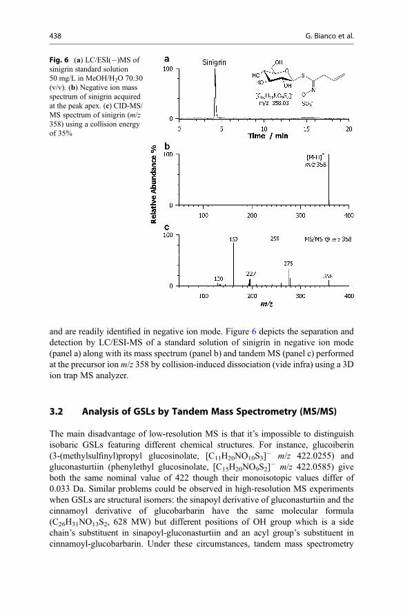
and are readily identified in negative ion mode. Figure 6 depicts the separation anddetection by LC/ESI-MS of a standard solution of sinigrin in negative ion mode(panel a) along with its mass spectrum (panel b) and tandem MS (panel c) performedat the precursor ion m/z 358 by collision-induced dissociation (vide infra) using a 3Dion trap MS analyzer.
3.2 Analysis of GSLs by Tandem Mass Spectrometry (MS/MS)
The main disadvantage of low-resolution MS is that it’s impossible to distinguishisobaric GSLs featuring different chemical structures. For instance, glucoiberin(3-(methylsulfinyl)propyl glucosinolate, [C11H20NO10S3]
� m/z 422.0255) andgluconasturtiin (phenylethyl glucosinolate, [C15H20NO9S2]
� m/z 422.0585) giveboth the same nominal value of 422 though their monoisotopic values differ of0.033 Da. Similar problems could be observed in high-resolution MS experimentswhen GSLs are structural isomers: the sinapoyl derivative of gluconasturtiin and thecinnamoyl derivative of glucobarbarin have the same molecular formula(C26H31NO13S2, 628 MW) but different positions of OH group which is a sidechain’s substituent in sinapoyl-gluconasturtiin and an acyl group’s substituent incinnamoyl-glucobarbarin. Under these circumstances, tandem mass spectrometry
Fig. 6 (a) LC/ESI(�)MS ofsinigrin standard solution50 mg/L in MeOH/H2O 70:30(v/v). (b) Negative ion massspectrum of sinigrin acquiredat the peak apex. (c) CID-MS/MS spectrum of sinigrin (m/z358) using a collision energyof 35%
438 G. Bianco et al.

(MS/MS) is a powerful approach for GSL characterization and identification as thefragmentation pattern helps to distinguish between isobaric and isomeric GSLs.Most theoretical and experimental aspects of collision-induced dissociation (CID)can be found in exhaustive reviews [29, 30].
As discussed above, most published works describe the MS/MS effectiveness innegative ionization mode, thanks to the hydrophilic nature of their sulfonate moiety.The negative ion ESI-MS/MS behavior of GSLs is very useful for structural exam-ination; whereas some product ions are common to all the GSLs, others are highlyspecific due to the variability of the side chain (R1) and/or of the acyl group (R2)occurring on the thioglucose moiety (Fig. 7). The generation of a hydrogen sulfateion, [HSO4]
� (m/z 97), from the precursor ion is observed as the major fragmentationpathway. Also the ions at m/z 139 and 129, corresponding to [C2H3O5S]
� and[HO4S2]
�, respectively, are very common [31]. Several additional product ions areindicative of thioglucose and sulfonated oxime moiety as the N-sulfate ethenimine,[C2H2O4NS]
� at m/z 136 and those at m/z 75 and 154, which correspond most likelyto [C2H3OS]
� and [C2H4O5NS]�, respectively.
Usually, identification of the intact GSLs (i.e., R2 = H in Fig. 7) is based on thedetection of ions with constant neutral losses of the following m/z values[M-196-H]�, [M-178-H]�, and [M-162-H]�, preceded by H-rearrangements, fromthe R1 group by cleavage of the bond on either sides of the sulfur atom and withcharge retention on the sugar aglycone [32, 33]. Possibly, the [M-162-H]� ion canundergo further fragmentation to give the diagnostic fragment M–162–SO3 (�242)[34]. These fragments, depicted in Fig. 7, are very informative for the side chain ofthe GSLs. Frequently, the MS/MS spectrum of GSLs contains also ionscorresponding to the loss of SO3 (80 Da) from the [M � H]� ion [23, 34]. Anotherproduct ion, found at m/z 195, corresponds to the fragment ion of the D-thioglucosegroup [C6H11O5S]
�. The ions at m/z 259 and 275 are well known as glucosinolate-specific fragments; they correspond to the neutral loss of R1-N = C = S andR1-N = C = O from the deprotonated molecule [M-H]�. The ion at m/z 241originates from ion at m/z 259 after loss of a water molecule. Note that all theseions at m/z 275, 259, and 241 are most likely formed through intramolecularrearrangements in which the sulfate group is transferred to the thioglucose moiety.
Acylated derivatives (R2 6¼ H) show mass spectra similar to corresponding intactGSLs (R2 = H), most likely due to the identical chemical core of these compounds[21]. The product ions specific for the variable aglycone side chain exhibit unvariedm/z values; however, acylthioglucose sulfate ions exhibit m/z value increase of theacyl group mass, viz., m/z 176 for isoferuloyl derivative, m/z 130 for cinnamoylGSL, m/z 146 for coumaroyl derivative, m/z 206 for sinapoyl GSL, and m/z 190 fordimethoxy-cinnamoylate derivative. For instance, the ions at m/z 275, 259, 241, and195 correspond to ions at m/z 421 = 275 + 176, 435 = 259 + 176,417 = 241 + 176, and 371 = 195 + 176 in isoferuloyl GSLs. Additional productions, likely related to the acylated nature of GSLs, may also exist. Useful informationcan be obtained on the acyl moiety from the occurrence of ions at m/z 193, 147,163, 223, and 207, corresponding to isoferuloylate ([C10H9O4]
�), cinnamoylate([C9H7O2]
�), coumaroylate ([C9H7O3]�), sinapoylate ([C11H11O5]
�), and
15 Investigation of Glucosinolates by Mass Spectrometry 439

Fig.7
Com
mon
fragmentsof
GSLandtheiracylated
derivativ
esby
ESI-MS/M
S.R
1istheside
chainandR2istheacyl
grou
por
HforintactGSLs
440 G. Bianco et al.

dimethoxy-cinnamoylate ([C11H11O4]�) anions. The most common fragments of
GSLs and their acylated derivatives are depicted in Fig. 7.Although GSL behavior in MS/MS experiments has been investigated mainly in
negative polarity, some researchers use positive ESI mode. In brief, positive productions in ESI-MS/MS are obtained from adducts with alkali metal ions such as Na+ andK+, i.e., [M + 2Na]+ or [M + 2K]+. In Fig. 8 are reported the MS/MS spectra in bothnegative and positive ion modes of progoitrin/epiprogoitrin [C11H18NO10S2]
� at m/z388 and [C11H18NO10S2 + 2Na]+ at m/z 434. The thioglucose product ions at m/z259, 275, and 195 in ESI(�)MS detection corresponds to ions at m/z 305, 321, and241 in ESI (+)MS detection mode. Besides, the aglycone ion product at m/z226 ([M-162]�) corresponds to ion at m/z 272 ([M-162 + 2Na]+) [34].
In the past, the mass spectrometric behavior of GSLs has been investigated bydifferent ionization methods, including fast atom bombardment (FAB) andthermospray (TSP) [12, 32, 33, 35–38]. However, the characterization of intactGSLs was insufficient, as no extensive fragmentation was carried out. Nowadays,the most common fragmentation method is collision-induced dissociation (CID) andinfrared multiphoton dissociation (IRMPD) [18–22, 39]. The application of CID inthe GSL analysis is useful if sufficient collision energy is applied; an example ofmass spectra obtained at relatively low- and high-collision energy of glucoraphaninis shown in Fig. 9a, b, respectively [40].
As already mentioned, ionization techniques that primarily produce positiveadducts or deprotonated molecules (i.e., ESI and APCI) are ideally suitable forcoupling with tandem MS. After selection of precursor ions during preliminaryLC/MS analysis, tandem MS may be used to obtain structurally definitive informa-tion. A specific precursor ion (i.e., deprotonated glucosinolate) is selected andfragmented by a suitable means giving rise to a tandem mass spectrum (MS/MS);whereas the most common fragmentation in low-resolution MS instruments is
Fig. 8 ESI-MS/MS CID spectra in negative (a) and positive (b) ion modes of progoitrin/epiprogoitrin at m/z 388.1 [C11H18NO10S2]
� and m/z 434.1 [C11H18NO10S2 + 2Na]+, respectively(Reprinted with permission from Ref. [34])
15 Investigation of Glucosinolates by Mass Spectrometry 441
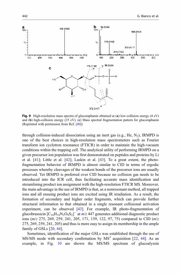
through collision-induced dissociation using an inert gas (e.g., He, N2), IRMPD isone of the best choices in high-resolution mass spectrometers such as Fouriertransform ion cyclotron resonance (FTICR) in order to maintain the high-vacuumconditions within the trapping cell. The analytical utility of performing IRMPD on agiven precursor ion population was first demonstrated on peptides and proteins by Liet al. [41]; Little et al. [42]; Laskin et al. [43]. To a great extent, the photo-fragmentation behavior of IRMPD is almost similar to CID in terms of ergodicprocesses whereby cleavages of the weakest bonds of the precursor ions are usuallyobserved. Yet IRMPD is preferred over CID because no collision gas needs to beintroduced into the ICR cell, thus facilitating accurate mass identification andstreamlining product ion assignment with the high-resolution FTICRMS. Moreover,the main advantage in the use of IRMPD is that, as a nonresonant method, all trappedions and all ensuing product ions are excited using IR irradiation. As a result, theformation of secondary and higher order fragments, which can provide furtherstructural information to that obtained in a single resonant collisional activationexperiment, can be observed [43]. For example, IR photo-fragmentation ofglucobrassicin [C16H19N2O9S2]
� at m/z 447 generates additional diagnostic productions (m/z 275, 269, 259, 241, 205, 171, 139, 122, 97, 75) compared to CID (m/z275, 269, 259, 241, 205) and thus is more easy to assign its membership in the amplefamily of GSLs [20, 44].
Sometimes, identification of the major GSLs was established through the use ofMS/MS mode with secondary confirmation by MS3 acquisition [22, 44]. As anexample, in Fig. 10 are shown the MS/MS spectrum of glucoalyssin
Fig. 9 High-resolution mass spectra of glucoraphanin obtained at (a) low-collision energy (4 eV)and (b) high-collision energy (25 eV). (c) Mass spectral fragmentation pattern for glucoraphanin(Reprinted with permission from Ref. [40])
442 G. Bianco et al.
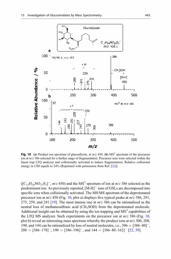
([C13H24NO13S3]�, m/z 450) and the MS3 spectrum of ion at m/z 386 selected as the
predominant ion. As previously reported, [M-H]� ions of GSLs are decomposed intospecific ions when collisionally activated. The MS/MS spectrum of the deprotonatedprecursor ion at m/z 450 (Fig. 10, plot a) displays five typical peaks at m/z 386, 291,275, 259, and 241 [19]. The most intense one at m/z 386 can be rationalized as theneutral loss of methanesulfonic acid (CH3SOH) from the deprotonated molecule.Additional insight can be obtained by using the ion trapping and MS3 capabilities ofthe LTQ MS analyzer. Such experiments on the precursor ion at m/z 386 (Fig. 10,plot b) reveal an interesting mass spectrum whereby the product ions atm/z 306, 208,190, and 144 can be rationalized by loss of neutral molecules, i.e., 306= [386–80]�,208 = [386–178]�, 190 = [386–196]�, and 144 = [386–80–162]� [22, 39].
Fig. 10 (a) Product ion spectrum of glucoallysin, at m/z 450. (b) MS3 spectrum of the precursorion at m/z 386 selected for a further stage of fragmentation. Precursor ions were selected within thelinear trap LTQ analyzer and collisionally activated to induce fragmentation. Relative collisionalenergy in CID equals to 24% (Reprinted with permission from Ref. [22])
15 Investigation of Glucosinolates by Mass Spectrometry 443

Until now, we described only MS/MS behavior of intact GSLs and acylatedderivatives. The MS/MS experiments on their most important closely related deriv-atives, as isothiocyanates (ITCs) [45], desulfo-compounds [31, 46] and azolidineproducts [47], are also of great interest. Concerning desulfo-GSLs, Bialeckiet al. [31] described a comparison of the product ion spectrum of phenyl-d5glucosinolate at m/z 399 (Fig. 11a) with that obtained from its correspondingphenyl-d5desulfoglucosinolate at m/z 319 (Fig. 11b), thus establishing the identity ofproduct ions that still contain the sulfur atom from the sulfate moiety. The productions observed at m/z 75, 119, 157, and 195 were common to both spectra. Becausephenyl-d5desulfoglucosinolate contains no sulfate residue, it is confirmed that theseions do not contain the sulfur atom from the sulfate moiety. In contrast, the productions observed at m/z 80, 96, 97, 206, 259, and 275 all contain S from the sulfatemoiety (Fig. 11a).
The structural characterization of an isothiocyanate has also been pursued byESI-TOF MS/MS [45]. The mass spectrum of hirsutin, i.e., 8-methylsulphinyl octylisothiocyanate (C10H19NOS2, 233 MW), as protonated adduct at m/z 234 (seeFig. 12a), exhibits a prominent product ion at m/z 170 ([M + H-CH4SO]
+), alsoappearing as an intense in-source fragment, which is a group-characteristic ion ofmethylsulphinyl ITCs. Another typical ion is at m/z 161 ([M + H-CH3-NCS]
+),which is a key fragment of mustard oils along with the stable cyclic immoniumion at m/z 114. In Fig. 12b is reported the characteristic MS/MS fragmentationspectrum of thiazolidine-2-thione (C9H9NO2S, 195 MW) as Na+ adduct([C9H9NO2S + Na]+, m/z 218) and proton adduct ([C9H9NO2S + H]+, m/z 196).For azolidine derivatives (see Fig. 12b), the neutral losses of SCO (�60 Da, peak c),H2S (�34 Da, peak d), CH2O (�30 Da, peak e), HOCN (�43 Da, peak f), and H2O(�18 Da, peak g) are common in tandem mass spectrum [47].
Fig. 11 (a) CID-MS/MS spectra of ion atm/z 399 derived from phenyl-d5glucosinolate at collisionenergy setting of 30 eV and (b) ion at m/z 319 derived from phenyl-d5desulfoglucosinolate atcollision energy setting of 15 eV. The inset in b shows the product ion spectrum of in-source-generated m/z 195 ion (cone voltage 25 V) (Reprinted with permission from Ref. [31])
444 G. Bianco et al.

Cataldi and co-workers [19] proposed a general approach for structural elucida-tion of naturally occurring GSLs in crude plant extracts based on the fragmentationof both isotopologue A and A + 2 peaks. All GSLs share a common structure with atleast two sulfur atoms and significant isotopic abundance of 34S. Thus, the dissoci-ation of the +2 Da isotopologue ions containing sulfur gives rise to doublets ofproduct ions featuring a combination of 32S and 34S. Accordingly, their relativeabundances allow one to speed up the structural recognition of GSLs with greatconfidence, as it produces structurally definitive ions than conventional tandem MSperformed on A ions (Fig. 13). Thus, conclusive identification by MS/MS issignificantly easier if the product ion also contains an atom of sulfur that providesa distinctive isotopic pattern.
4 GSLs by Hyphenated Techniques
The isolation and structural elucidation of known and unknown GSLs from plantextracts by tandem MS is a rewarding, but often time-consuming, task, since it is amajor effort to isolate each compound in a pure form, even the known ones as the
Fig. 12 (a) Positive ion ESI-CID-MS/MS of hirsutin atm/z 234 ([C10H19NOS2 + H]+)(Reprinted with permissionfrom Ref. [45]). (b)Characteristic MS2
fragmentation patterns of thethiazolidine-2-thione as Na+
adduct ([C9H9NO2S + Na]+,m/z 218) and proton adduct([C9H9NO2S + H]+, m/z 196).Their product ions arecategorized according to thefollowing letters: c loss ofSCO (�60 Da), (d) loss ofH2S (�34 Da), e loss of CH2O(�30 Da), f loss of HOCN(�43 Da), and g loss of H2O(�18 Da) (Reprinted withpermission from Ref. [47])
15 Investigation of Glucosinolates by Mass Spectrometry 445

simultaneous presence of two or more components in the source region gives rise toan overlapping or mixed spectrum. Furthermore, to obtain the required milligramquantities of all GSLs, especially in the case of minor GSLs, one may need largeamounts of the plant material and of expensive tools and supplies. Another challengeis to identify all GSLs because the variable side chain linked to the thiohydroximate
Fig. 13 Product ion spectra obtained by LC/ESI-LTQ MS/MS in negative ion mode of (A, B)gluconapin and (C, D) sinigrin. The precursor ion base peaks of gluconapin and sinigrin at m/z372 (A) andm/z 358 (c), respectively, and the corresponding A + 2 isotope peaks atm/z 374 (B) andm/z 360 (D) were fragmented at 20% collision energy. The expanded views in plots (B) and (D)show as profile spectra some examples of isotopic peak doublets (Reprinted with permission fromRef. [19])
446 G. Bianco et al.

(see Fig. 1) and the variable acyl group linked to thioglucose afford many differentstructures [8]. The coupling of separation modes with MS has been recognized as avery useful approach for real-world samples analysis, in which complex mixtures arethe norm. In the case of plant extracts, the hyphenation of gas chromatography (GC),high-performance liquid chromatography (HPLC), and capillary electrophoresis(CE) with mass spectrometry has led to several strategies that do not only permitto differentiate between known GSLs but can likewise allow the structural charac-terization of unknown GSLs directly from the crude extract with a minimum amountof sample.
4.1 GSL Analysis by Gas Chromatography–Mass Spectrometry(GC/MS)
Because of their ionic nature, naturally occurring GSLs are not sufficiently volatile tobe separated directly on a GC column, so pre-column conversion to desulfo-GSLderivatives or conversion/hydrolysis to corresponding isothiocyanate (ITC) has to beperformed. Among the myrosinase-catalyzed degradation products of GSLs, ITCs (seetheir formation in Fig. 14) have been generally known as the most biologically activemetabolites, being reported to possess broad-spectrum biological activity againstbacterial and fungal pathogens, nematodes, insects, and weeds [6, 48–53]. Also nitrilesare known to be biologically active, but their effects are more restricted compared toITCs. For these reasons the more recent applications of GC/MS for GSLs analysis aremainly focused on the monitoring of ITC and nitrile metabolites [14, 54–58].
Usually, GC/MS analysis is performed on single-quadrupole mass spectrometers,which provide nominal data ofm/z values. The introduction of GC-TOFMS systemsoffered an attractive supplement to quadrupole instruments and provided greatermass accuracy. TOF instruments also provide high scan speeds that are compatiblewith fast GC separation [59] and the potential to profile complex mixtures in lesstime. Similarly, GC coupled to multistage MS (e.g., MS/MS and MSn) instrumentsenabled the acquisition of several fragmentation data and a higher level of molecularselectivity. Nevertheless, quadrupole mass analyzers still present some extra advan-tages in metabolite analysis such as higher repeatability and large dynamic range forquantitative analysis. According to Nielsen et al. [60], it is possible to improve
Fig. 14 Myrosinase-catalyzed glucosinolate hydrolysis (breakdown) and formation ofisothiocyanates (ITCs). Sulforaphane is an ITC compound formed from glucoraphanin. Whencruciferous vegetables are chopped or chewed, endogenous myrosinase can interact withglucosinolates and release ITCs from their precursors
15 Investigation of Glucosinolates by Mass Spectrometry 447

detection sensitivity from the ng-level to the pg-level through the scanning for aselected number of characteristic ions (selected ion monitoring, SIM), which is veryuseful for quantitative target analysis.
4.2 GSL Analysis by Capillary Electrophoresis–Mass Spectrometry(CE/MS)
Capillary electrophoresis (CE) coupled to mass spectrometry for GSL analysis datesback to 2005 when Bringmann et al. [17] used CZE in combination with electrosprayionization time-of-flight mass spectrometry (ESI-TOF MS). The hyphenated tech-nique combines the advantages of capillary electrophoresis, namely, small samplevolumes needed and nearly no dilution effects, with those of time-of-flight massdetectors, i.e., high sensitivity, accurate mass, and isotopic pattern. Employing anacidic electrolyte and a sheath liquid based on formic acid, the GSLs were separatedand detected as anions, resulting in an excellent selectivity. Thus, the crude plantextracts could be analyzed without any interference of matrix constituents. Thesensitivity together with mass accuracy and true isotopic pattern of the TOF MSallowed the identification of a broad series of glucosinolates in Arabidopsis thalianaseeds.
4.3 GSL Analysis by Liquid Chromatography–Mass Spectrometry(LC/MS)
GSLs and desulfo-GSLs containing sulfinyl or indolic moieties are not amenable toGC/MS analysis. Further, the preanalytical derivatization step may be avoided byemploying LC/MS methods and can be applied to both GSLs and ITCs as validatedby ICH guidelines [61]. Indeed, LC/MS methods have replaced not only the ISO9167-1 method currently used but also all the HPLC–UV methods adopted for thequali-quantitative determination of ITC [8]. Currently, LC/MS is broadly used withESI and APCI sources [18–22, 62–65]. Although ESI-MS is the most used detectiontool for identifying highly polar and heat-labile compounds such as GSLs occurringas ions in solution, the direct comparison with APCI-MS demonstrates that this lastis much more sensitive for aliphatic, aromatic, and indole desulfo-GSLs thanESI-MS. However, no additional information, in terms of structure determination,was obtained by APCI-MS [66].
Several modes of scanning and acquiring MS data during an LC/MS analysis areavailable: (i) full-scan acquisition resulting in the typical total ion current (TIC) plot,(ii) selected ion monitoring (SIM), (iii) selected reaction monitoring (SRM), or(iv) multiple reaction monitoring (MRM). The TIC and SIM modes are mainly usedfor GSLs profiling, whereas SRM scan is used for quantitative analysis. LC coupledwith ESI(�) to a hybrid quadrupole linear ion trap and Fourier transform ion cyclotronresonance (FTICR)mass spectrometer in full-scanmodewas employed for the selectiveand sensitive determination of intact GSLs in several crude sample extracts of broccoli
448 G. Bianco et al.

(Brassica oleraceaL. var. italica), cauliflower (B. oleraceaL. var. botrytis), rocket salad(Eruca sativa L.), and bud flowers of Capparis spinosa (Capparis species, familyCapparaceae). Fragmentation induced by IRMPD was successfully applied to identifytargeted and untargeted GSLs in aqueous/methanol extracts [20–22].
Notably, SIM acquisition mode is more sensitive than the full-scan experimentbecause the mass spectrometer can dwell for a longer time over a smaller mass range;for this reason this scan mode is employed for quantitative analysis if compounds tobe quantified have different m/z values [18, 25, 63]. SRM detection providesimproved sensitivity and selectivity in comparison to both full-scan and selectedion monitoring scanning modes. Tandem MS of a deprotonated GSL produces acharacteristic product ion at m/z 97 ([HSO3]
�) [18–22, 54, 67] with high intensity; itcan be monitored in MS/MS SRM experiments. Tian et al. [68] used SRM of thetransition [M-H]� ! m/z 97 to quantify all GSLs in broccoli, broccoli sprouts,Brussels sprouts, and cauliflower. The same LC/ESI-MS/MS MRMmethod allowedto quantify all the identified glucosinolates in different tissues of Moringa oleiferaand in broccoli, oil seed, Brassica napus seeds, cabbage, Brussels sprouts, cauli-flower, Brassica oleracea seeds, and winter cress Barbarea verna seeds [69–71].
5 GSL Analysis by Off-Line and Shotgun MS
Off-line HPLC followed by matrix-assisted laser desorption/ionization mass spec-trometry (MALDI-MS), or direct analysis of a sample extract by MALDI-TOF MS,through off-line vacuum deposition onto standard stainless-steel MALDI targets, areboth used. The off-line approach allows the decoupling of the separation step fromthe MS analysis, which can be independently optimized in terms of time. Toillustrate the use of MALDI-TOF MS, we discuss the work of Botting et al. [72]who demonstrated the determination of the GSL profile in different crude plantsamples, viz., Lunaria annua seeds, cauliflower florets, rutabaga peel, and turnip.Informative mass spectra were obtained from the plant extracts with predominant ionpeaks corresponding to deprotonated GSLs. In Fig. 15, the mass spectrum obtainedfrom the L. annua extract clearly shows three peaks corresponding to GSLs withisopropyl, 5-methylsulfinyl, and 6-methylsulfinyl side chains. In previous work,these three GSLs were identified by LC/MS upon the analysis of myrosinase-induced breakdown products [73, 74].
Direct infusion MS or shotgun approaches are also been developed for rapidscreening of GSLs in vegetable extracts. Indeed, flow injection ESI-MS has beenused for the profiling of GSLs and phenolic compounds in pulp seed, seed coat,leaves, and roots of M. oleifera 12-day old seedlings [70]. The full-scan MSspectrum of each sample was recorded in triplicate with the aim to rapidly providea visual and statistical evaluation of similarities and differences of secondarymetabolites among tissues. The ESI-MS fingerprints of samples were verydistinguishing, showing distinctive sets of polar markers for each different tissue.Considering the large amount of data set (4 tissues � 3 replicates) obtained bynegative ion ESI-MS fingerprints of extracts under investigation, a chemometric
15 Investigation of Glucosinolates by Mass Spectrometry 449

approach was performed by principal component analysis, in order to characterizethe different plant’s tissue and to evaluate differences in terms of metabolites. Thesimultaneous determination of 11 glucosinolates in different M. oleifera tissueswas described.
The use of high-resolution mass spectrometry (HRMS) in direct analysis greatlyimproves the analytical performance and offers a good combination of selectivity andsensitivity. High mass resolution/accuracy along with measured relative isotopicabundance ratios are typically employed to tentatively identify elemental formulaeof thousands of different compounds. An example of application of HRMS is by directanalysis in real time (DART). In DART, an electrical potential is applied to a heatedgas stream (typically helium or nitrogen) to form a plasma of excited-state atoms andions that desorb lowmolecular weight molecules from the surface of a sample. DART-TOF MS has been fruitfully used to examine yellow mustard seeds [75]; the presenceof 4-hydroxybenzyl GSL, also known as sinalbin, was evidenced through character-istic ions at m/z 180.09 [C6H12O6 + NH4-H2O]
+, 198.10 [C6H12O6 + NH4]+, 214.08
[C6H12O5S + NH4]+, and 296.20 [C4H17O8 + 3H2O + H]+ (Fig. 16a). All these ions
were consistent with the sinalbin standard compound (Fig. 16b). The DART-TOFMSmethod was optimized and validated in comparison to LC/MS.
6 Comparison of the Different MS-Based Methods for GSLAnalysis
There are different issues in the determination of GSLs and their decompositionproducts in cruciferous vegetables. First, there are a lot of compounds with similarphysical and chemical properties which may appear in the investigated samples, even
Fig. 15 MALDI-TOF mass spectra of extracts from L. annua seeds (Reprinted with permissionfrom Ref. [72])
450 G. Bianco et al.

Fig.1
6(con
tinued)
15 Investigation of Glucosinolates by Mass Spectrometry 451

Fig.1
6MSspectrum
of(a)yello
wmustard
seeds(positive
mod
e,ionizatio
ntemperature40
0� C
)and(b)sinalbin1g/l(po
sitiv
emod
e,ionizatio
ntemperature
450
� C).Possiblestructureof
fragmentsanddepend
ence
selected
intensity
ofions
ontemperature
areshow
n(Reprinted
with
perm
ission
from
Ref.[75])
452 G. Bianco et al.

at considerably higher levels than targeted metabolites. Second, reference materials arenot always available, which causes serious problems in carrying out quantitativedeterminations. Table 1 summarizes data from the literature regarding the analyticaltechniques used for samples of cruciferous plants to determine the content of GSLsand their breakdown products: strengths and weaknesses of each method using MS asa selective detector are highlighted. As already mentioned LC/MS is indeed a maturetechnique in GSLs analysis and, as a consequence, has been largely applied asextensively described in the literature. Because of their ionic nature, GSLs cannot beinvestigated directly by GC but must be hydrolyzed to corresponding ITC, nitrile, etc.It has a great potential for quantitative analysis and metabolite profiling, and the
Table 1 Example of application of different MS methods for GSL analysis
Analyticaltechnique Application Advantages Disadvantages References
GC/MS Quantitativeanalysis orqualitative analysisdepends ondetection, the SIM,and SCAN modes,respectively
Highchromatographicresolution, ideal toresolve complexsamples
Sample preparationstep complicatedand time-consuming; unableto analyze thermo-labile GSLs
[57, 58,60]
Nonvolatile GSLsmust be derivatizedbefore analysis
Difficult to identifyunknowncompounds afterderivatization
LC/MS Separation,identification, andquantification of avery broad group ofmetabolites.Limited potential inidentification unlessMS/MS techniquesare used
High sensitivity;average to highchromatographicresolution; noderivatizationrequired; enableanalysis of thermo-labile GSLs
Sample preparationstep labor-intensiveand time-consuming;restriction on LCeluents
[18–21,68, 70]
CE/MS Separation,identification, andquantification ofpolar GSLs, usingsmall samplevolumes
Small volumes;high resolution;flexible technique
Bufferincompatibility;
[17]
Difficulty in MSinterfacing
MS Identification ofGSLs by theircharacteristic m/zvalues. Usuallyapplied in GSLprofiling withoutany quantification
Rapid screening ofmetabolites;versatile andreproducibletechnique
Low sensitivity, noquantificationanalysis
[72, 75]
15 Investigation of Glucosinolates by Mass Spectrometry 453

variation between those two approaches is basically performed through a switching ofthe detection from the SIM to the full-scan mode. However, GC/MS analysis ofbreakdown products possesses specific limitations as some GSL side chains (e.g.,indolyl or hydroxylated side chains) produce poorly volatile or unstable breakdownproducts [72]. An alternative is the conversion of GSLs into their volatile desulfo-GSLderivatives with pre-column derivatization. The mandatory requirement to derivatizenonvolatile metabolites often complicates the identification of unknown compounds,increases the sample preparation variability, adds time to the analysis, causes morecomplex sample handling, and increases variance analysis as well. For these mainreasons, LC/MS and CE/MS are more suitable for GSL analysis. Both intact GSLs anddesulfo-GSLs can be separated and detected by LC/MS. Desulfation results in betterseparation and cleaner samples and also makes the compounds easier to analyze bytraditional mass spectrometric techniques [76, 77].
In evaluating the combination CE/MS, many efforts are needed to establishoptimum composition of the CE background electrolyte, which allows a satisfactoryseparation coupled to an efficient ionization by ESI. Although the low samplevolume required for CE and the high speed would be an advantage over LC/MS,the technique still suffers from low sensitivity. Indeed, compared to LC/MS analysis,one major drawback is the relatively high concentration limit of detection because ofthe very low injection volume used in CE [78].
Both chromatographic and electrophoretic separations reduce ion suppressioneffects originating from the sample matrix. The major drawback of hyphenatedMS-based techniques lies in their limitation to analyze large sets of samples to detectchanges in composition in a fast and simple way. On the contrary there is a strongneed for more rapid, high-throughput MS approaches for nontargeted investigations.Nowadays, direct MS analysis is the method of choice to achieve the maximumhigh-throughput information data from the largest possible number of samples[79]. Direct infusion ESI-MS offers several advantages since it is a very fast,versatile, reproducible, and sensitive technique, which requires little or no samplepreparation [70]. Any separation step prior to MS detection is avoided and thusdirect analysis of samples (previously processed or not) is carried out, making itparticularly attractive when dealing with large sample sets. The analysis ofmulticomponent mixtures by direct infusion MS can, however, be hindered by thecompetitive ionization of the analytes that leads to a low sensitivity. In addition,isobaric interferences in the mass spectrum adversely affect compound identificationas well as other aspects of performance. On the other hand, the high throughput andthe reduction of sample preparation time are the main advantages of direct MS-basedapproaches over conventional hyphenated techniques [79].
7 GSL Analysis by Mass Spectrometry Imaging (MSI)
The surface localization of plant GSLs has been a controversial topic for many years.GSLs are frequently cited as key compounds for plants in deterring generalist insectherbivores and for specialist ones in choosing plants for feeding and oviposition
454 G. Bianco et al.

[7, 80–82]. Since insect behavioral choices are sometimes made after surfacecontact, GSLs have been sought on the surface. With the capabilities of massspectrometry imaging (MSI) to track and map the spatial distribution of smallmolecules in plants, the data generated may essentially lead to improved under-standing of the metabolism of plants in response to diseases and certain stimuli fromthe environment, including pest attack [83].
The MSI method used most frequently is MALDI-MS [84–86]. In plants,MALDI-MSI has been successfully applied to study the spatial distribution ofmolecules such as sugars, lipids, and secondary metabolites like GSLs, flavonoids,and alkanes in leaf tissue and tissue sections of different plants [84, 87–89]. Shroffet al. [84] performed the analysis of GSL distribution within Arabidopsis thalianademonstrating a nonuniform distribution throughout the leaf tissue. The accumula-tion of GSLs close to the leaf margin and the middle vein strongly influences thefeeding preference of Helicoverpa armigera larvae and might play a key role as afirst barrier against chewing herbivores. The identity of these detected compoundswas confirmed by CID mass spectra [84]. Sarsby et al. [86] studied flowerbuds, sepals, and siliques of A. thaliana using MALDI-MSI. More recently Shroffet al. [85] modified their procedures to quantify GSLs occurring on A. thaliana leafsurfaces by matrix deposition through sublimation and confirmed the previousqualitative results concerning nonuniform distribution by monitoringthree different GSL compounds: 4-methylthiobutylglucosinolate (4MTB),4-methylsulfinylbutylglucosinolate (4MSOB), and indol-3-ylmethylglucosinolate(I3M) at m/z 420, 436, and 447, respectively (Fig. 17).
Fig. 17 (A) MALDI-TOF spectra and (B) MALDI-MSI spectra of the Arabidopsis thaliana leafsurface. (c) MALDI-TOF images constructed for abundance of m/z 420 (4MTB,4-methylthiobutylglucosinolate), 436 (4MSOB, 4-methylsulfinylbutylglucosinolate), and447 (I3M, indol-3-ylmethylglucosinolate) (Reprinted with permission from Ref. [85])
15 Investigation of Glucosinolates by Mass Spectrometry 455
8 Conclusions
Over the past two decades, mass spectrometry has proved to be one of the mosteffective techniques in plant metabolite research, including GSLs. Due to their ioniccharacter and low volatility, GSLs are perfectly suitable for determination by liquidchromatography. Thus, the combination of RP-HPLC and ESI-MS/MS has given tous a tool with high sensitivity and selectivity to establish the occurrence of GSLs invegetable samples, also at the low levels of many minor species. We believe that theexisting generation of LC/ESI-MS technology is sufficiently robust for structuralidentification and especially to answering some questions about the role of GSLs inhuman health. Currently, quantitation assays are faced by external standards andconcentration/response curves. As stable isotopically labeled GSL standards becomeavailable, quantitation will be possible by isotope dilution LC/MS, thus improvingprecision and streamline analysis.
Acknowledgments This study was supported by the Università degli Studi di Bari Aldo Moro(Fondi di Ateneo 2012) and Università degli Studi della Basilicata.
References
1. Drewnowski A, Gomez-Carneros C (2000) Bitter taste, phytonutrients, and the consumer: areview. Am J Clin Nutr 72(6):1424–1435
2. Kristal AR, Lampe JW (2002) Brassica vegetables and prostate cancer risk: a review of theepidemiological evidence. Nutr Cancer 42(1):1–9. doi:10.1207/S15327914NC421_1
3. Lund E (2003) Non-nutritive bioactive constituents of plants: dietary sources and health benefitsof glucosinolates. Int J Vitam Nutr Res 73(2003):135–143. doi:10.1024/0300-9831.73.2.135
4. Perocco P, Bronzetti G, Canistro D, Valgimigli L, Sapone A, Affatato A (2006) Glucoraphanin,the bioprecursor of the widely extolled chemopreventive agent sulforaphane found in broccoli,induces phase-I xenobiotic metabolizing enzymes and increases free radical generation in ratliver. Mutat Res 595:125–136. doi:10.1016/j.mrfmmm.2005.11.007
5. Higdon JV, Delage B, Williams DE, Dashwood RH (2007) Cruciferous vegetables and humancancer risk: epidemiologic evidence and mechanistic basis. Pharmacol Res 55:224–236.doi:10.1016/j.apsusc.2006.02.134
6. Oregon State University, Linus Pauling institute’s micronutrient information center. Phytochem-icals. http://lpi.oregonstate.edu/mic/dietary-factors/phytochemicals. Accessed 18 Nov 2015
7. Radojčić RI, Glivetić T, Delonga K, Vorkapić-Furač J (2008) Glucosinolates and their potentialrole in plant. Period Biol 110(4):297–309, ISSN:0031–5362
8. Agerbirk N, Olsen CE (2012) Glucosinolates structures in evolution. Phytochemistry 77:16–45.doi:10.1016/j.phytochem.2012.02.005
9. Fahey JW, Zalcmann AT, Talalay P (2001) The chemical diversity and distribution ofglucosinolates and isothiocyanates among plants. Phytochemistry 56(1):5–51. doi:10.1016/S0031-9422(00)00316-2
10. Mithen RF, Dekker M, Verkerk R, Rabot S, Johnson IT (2000) The nutritional significance,biosynthesis and bioavailability of glucosinolates in human foods. J Sci Food Agric80:967–984, doi:0022-5142/2000
11. Heaney RK, Fenwick RG (1993) Methods for glucosinolate analysis. In: Waterman PG(ed) Methods in plant biochemistry. Academic, London
456 G. Bianco et al.

12. Fenwick GR, Eagles J, Gmelin R, Rakow D (1980) The mass spectra of glucosinolates anddesulfoglucosinolates. Biol Mass Spectrom 7(9):410–412. doi:10.1002/bms.1200070911
13. Maharjan RP, Ferenci T (2003) Global metabolite analysis: the influence of extraction meth-odology on metabolome profiles of Escherichia coli. Anal Biochem 313:145–154. doi:10.1016/S0003-2697(02)00536-5
14. Blažević I, Mastelić J (2009) Glucosinolate degradation products and other bound and freevolatiles in the leaves and roots of radish (Raphanus sativus L.). Food Chem 113(1):96–102.doi:10.1016/j.foodchem.2008.07.029
15. Oerlemans K, Barrett DM, Suades CB, Verkerk R, Dekker M (2006) Thermal degradation ofglucosinolates in red cabbage. Food Chem 95:19–29. doi:10.1016/j.foodchem.2004.12.013
16. Rungapamestry V, Duncan AJ, Fuller Z, Ratcliffe B (2007) Effect of cooking brassica vegeta-bles on the subsequent hydrolysis and metabolic fate of glucosinolates. Proc Nutr Soc 66:69–81.doi:10.1017/S0029665107005319
17. Bringmann G, Kajahn I, Neusuess C, Pelzing M, Laug S, Unger M, Holzgrabe U (2005)Analysis of the glucosinolate pattern of Arabidopsis thaliana seeds by capillary zone electro-phoresis coupled to electrospray ionization-mass spectrometry. Electrophoresis 26:1513–1522.doi:10.1002/elps.200410255
18. Cataldi TR, Rubino A, Lelario F, Bufo SA (2007) Naturally occurring glucosinolates in plantextracts of rocket salad (Eruca sativa L.) identified by liquid chromatography coupled withnegative ion electrospray ionization and quadrupole ion‐trap mass spectrometry. RapidCommun Mass Spectrom 21(14):2374–2388. doi:10.1002/rcm.3101
19. Cataldi TRI, Lelario F, Orlando D, Bufo SA (2010) Collision-induced dissociation of the A+2isotope ion facilitates glucosinolates structure elucidation by electrospray ionization-tandemmass spectrometry with a linear quadrupole ion trap. Anal Chem 82(13):5686–5696.doi:10.1021/ac100703w
20. Bianco G, Lelario F, Battista FG, Bufo SA, Cataldi TRI (2012) Identification of glucosinolatesin capers by LC‐ESI‐hybrid linear ion trap with Fourier-transform ion cyclotron resonance massspectrometry (LC‐ESI‐LTQ‐FTICR MS) and infrared multiphoton dissociation. J MassSpectrom 47(9):1160–1169. doi:10.1002/jms.2996
21. Bianco G, Agerbirk N, Losito I, Cataldi TRI (2014) Acylated glucosinolates with diverse acylgroups investigated by high resolution mass spectrometry and infrared multiphoton dissocia-tion. Phytochemistry 100:92–102. doi:10.1002/jms.2996
22. Lelario F, Bianco G, Bufo SA, Cataldi TR (2012) Establishing the occurrence of major andminor glucosinolates in brassicaceae by LC–ESI-hybrid linear ion-trap and Fourier-transformion cyclotron resonance mass spectrometry. Phytochemistry 73:74–83. doi:10.1016/j.phytochem.2011.09.010
23. Mohn T, Cutting B, Ernst B, Hamburger M (2007) Extraction and analysis of intactglucosinolates-A validated pressurized liquid extraction/liquid chromatography–mass spec-trometry protocol for Isatis tinctoria, and qualitative analysis of other cruciferous plants.J Chromatogr A 1166(1):142–151. doi:10.1016/j.chroma.2007.08.028
24. Schwartz SA, Reyzer ML, Caprioli RM (2003) Direct tissue analysis using matrix-assisted laserdesorption/ionization mass spectrometry: practical aspects of sample preparation. J MassSpectrom 38:699–708. doi:10.1002/jms.505
25. Njumbe Ediage E, Diana Di Mavungu J, Monbaliu S, Van Peteghem C, De Saeger S (2011) Avalidated multianalyte LC–MS/MS method for quantification of 25 mycotoxins in cassavaflour, peanut cake and maize samples. J Agric Food Chem 59(10):5173–5180. doi:10.1021/jf2009364
26. Lee JG, Bonnema G, Zhang N, Kwak JH, de Vos RC, Beekwilder J (2013) Evaluation ofglucosinolate variation in a collection of turnip (Brassica rapa) germplasm by the analysis ofintact and desulfo glucosinolates. J Agric Food Chem 61(16):3984–3993. doi:10.1021/jf400890p
27. Kusznierewicz B, Iori R, Piekarska A, Namieśnik J, Bartoszek A (2013) Convenient identifi-cation of desulfoglucosinolates on the basis of mass spectra obtained during liquid
15 Investigation of Glucosinolates by Mass Spectrometry 457
chromatography–diode array–electrospray ionization mass spectrometry analysis: method ver-ification for sprouts of different Brassicaceae species extracts. J Chromatogr A 1278:108–115.doi:10.1016/j.chroma.2012.12.075
28. Kim SJ, Ishii G (2006) Glucosinolate profiles in the seeds, leaves and roots of rocketsalad (Eruca sativa Mill.) and anti-oxidative activities of intact plant powder andpurified 4-methoxyglucobrassicin. Soil Sci Plant Nutr 52(3):394–400. doi:10.1111/j.1747-0765.2006.00049.x
29. Sleno L, Volmer DA (2004) Ion activation methods for tandem mass spectrometry. J MassSpectrom 39(10):1091–1112. doi:10.1002/jms.703
30. Wells JM, McLuckey SA (2005) Collision‐induced dissociation (CID) of peptides and proteins.Methods Enzymol 402:148–185. doi:10.1016/S0076-6879(05)02005-7
31. Bialecki JB, Ruzicka J, Weisbecker CS, Haribal M, Attygalle AB (2010) Collision‐induceddissociation mass spectra of glucosinolate anions. J Mass Spectrom 45(3):272–283.doi:10.1002/jms.1711
32. Kokkonen PS, Van der Greef J, Niessen WMA, Tjaden UR, Ten Hove GJ, Van de Werken G(1991) Identification of intact glucosinolates using direct coupling of high‐performance liquidchromatography with continuous‐flow frit fast atom bombardment tandem mass spectrometry.Biol Mass Spectrom 20(5):259–267. doi:10.1002/bms.1200200505
33. Kokkonen P, Van der Greef J, Niessen WMA, Tjaden UR, Ten Hove GJ, Van de Werken G(1989) Separation and identification of intact glucosinolates using direct coupling of high‐performance liquid chromatography with frit to fast‐atom bombardment mass spectrometry.Rapid Commun Mass Spectrom 3(4):102–106. doi:10.1002/rcm.1290030404
34. Fabre N, Poinsot V, Debrauwer L, Vigor C, Tulliez J, Fourasté I, Moulis C (2007) Character-isation of glucosinolates using electrospray ion trap and electrospray quadrupole time‐of‐flightmass spectrometry. Phytochem Anal 18(4):306–319. doi:10.1002/pca.983
35. Mellon FA, Chapman JR, Pratt JAE (1987) Thermospray liquid chromatography-mass spec-trometry in food and agricultural research. J Chromatogr A 394(1):209–222. doi:10.1016/S0021-9673(01)94173-1
36. Hogge LR, Reed DW, Underhill EW (1988) The identification of desulfoglucosinolates usingthermospray liquid chromatography/mass spectrometry. J Chromatogr Sci 26(7):348–351.doi:10.1093/chromsci/26.7.348
37. Hogge LR, Reed DW, Underhill EW, Haughn GW (1988) HPLC separation of glucosinolatesfrom leaves and seeds of Arabidopsis thaliana and their identification using thermospray liquidchromatography/mass spectrometry. J Chromatogr Sci 26:551–556
38. Heeremans CEM, Van Der Hoeven RAM, Niessen WMA, Vuik J, De Vos RH, Van Der Greef J(1989) Specific screening methods for glucosinolates in sprout extracts using on-linethermospray liquid chromatography-tandem mass spectrometry. J Chromatogr A472:219–229. doi:10.1016/S0021-9673(00)94108-6
39. Agneta R, Rivelli AR, Ventrella E, Lelario F, Sarli G, Bufo SA (2012) Investigation ofglucosinolates profile and qualitative aspects in sprouts and roots of horseradish (Armoraciarusticana) by using LC-ESI-hybrid linear ion-trap with Fourier-transform ion cyclotron reso-nance mass spectrometry and infrared multiphoton dissociation. J Agric Food Chem 60(30):7474–7482. doi:10.1021/jf301294h
40. Glauser G, Schweizer F, Turlings TC, Reymond P (2012) Rapid profiling of intactglucosinolates in Arabidopsis leaves by UHPLC‐QTOFMS using a charged surface hybridcolumn. Phytochem Anal 23(5):520–528. doi:10.1002/pca.2350
41. Li W, Hendrickson CL, Emmett MR, Marshall AG (1999) Identification of intact proteins inmixtures by alternated capillary liquid chromatography electrospray ionization and LC ESIinfrared multiphoton dissociation Fourier transform ion cyclotron resonance mass spectrometry.Anal Chem 71(19):4397–4402. doi:10.1021/ac990011e
42. Little DP, Speir JP, Senko MW, O’Connor PB, McLafferty FW (1994) Infrared multiphotondissociation of large multiply charged ions for biomolecule sequencing. Anal Chem 66(18):2809–2815. doi:10.1021/ac00090a004
458 G. Bianco et al.

43. Laskin J, Futrell JH (2005) Activation of large lons in FT‐ICR mass spectrometry. MassSpectrom Rev 24(2):135–167. doi:10.1002/mas.20012
44. Millán S, Sampedro MC, Gallejones P, Castellón A, Ibargoitia ML, Goicolea MA, Barrio RJ(2009) Identification and quantification of glucosinolates in rapeseed using liquid chromato-graphy–ion trap mass spectrometry. Anal Bioanal Chem 394(6):1661–1669. doi:10.1007/s00216-009-2823-8
45. von Roepenack-Lahaye E, Degenkolb T, Zerjeski M, Franz M, Roth U, Clemens S (2004)Profiling of Arabidopsis secondary metabolites by capillary liquid chromatography coupledto electrospray ionization quadrupole time-of-flight mass spectrometry. Plant Physiol 134(2):548–559. doi:10.1104/pp.103.032714
46. Agerbirk N, Olsen CE, Heimes C, Christensen S, Bak S, Hauser TP (2015) Multiplehydroxyphenethyl glucosinolate isomers and their tandem mass spectrometric distinction in ageographically structured polymorphism in the crucifer Barbarea vulgaris. Phytochemistry115:130–142. doi:10.1016/j.phytochem.2014.09.003
47. Agerbirk N, Olsen CE (2015) Glucosinolate hydrolysis products in the crucifer Barbareavulgaris include a thiazolidine-2-one from a specific phenolic isomer as well as oxazolidine-2-thiones. Phytochemistry 115:143–151. doi:10.1016/j.phytochem.2014.11.002
48. Brown PD, Morra MJ (1997) Control of soil-borne plant pests using glucosinolate-containingplants. Adv Agron 61:167–231
49. Lin CM, Kim JM, Du WX, Wei CI (2000) Bactericidal activity of isothiocyanate againstpathogens on fresh produce. J Food Prot 63:25–30
50. Manici LM, Lazzeri L, Baruzzi G, Leoni O, Galletti S, Palmieri S (2000) Suppressive activity ofsome glucosinolate enzyme degradation products on Pythium irregulare and Rhizoctonia solaniin sterile soil. Pest Manag Sci 56:921–926. doi:10.1002/1526-4998(200010)
51. Rosa EAS, Rodriges PMF (1999) Towards a more sustainable agriculture system: the effect ofglucosinolates of the control of soilborne diseases. J Hortic Sci Biotechnol 74:667–674,ISSN:1462-0316
52. Wittstock U, Kliebenstein DJ, Lambrix V, Reichelt M, Gershenzon J (2003) Chapter fiveglucosinolate hydrolysis and its impact on generalist and specialist insect herbivores. RecentAdv Phytochem 37:101–125. doi:10.1016/S0079-9920(03)80020-5
53. Zasada IA, Ferris H (2004) Nematode suppression with brassicaceous amendments: applicationbased upon glucosinolate profiles. Soil Biol Biochem 36:1017–1024. doi:10.1016/j.soilbio.2003.12.014
54. Bennett RN, Mellon FA, Botting NP, Eagles J, Rosa EA, Williamson G (2002) Identification ofthe major glucosinolate (4-mercaptobutyl glucosinolate) in leaves of Eruca sativa L. (saladrocket). Phytochemistry 61(1):25–30. doi:10.1016/S0031-9422(02)00203-0
55. Tierens KFJ, Thomma BP, Brouwer M, Schmidt J, Kistner K, Broekaert WF (2001) Study of therole of antimicrobial glucosinolate-derived isothiocyanates in resistance of Arabidopsis tomicrobial pathogens. Plant Physiol 125(4):1688–1699. doi:10.1104/pp.125.4.1688
56. Choi MM, Shuang S, Lai HY, Cheng SC, Cheng RC, Cheung BK, Lee AW (2004) Gaschromatography-mass spectrometric determination of total isothiocyanates in Chinese medic-inal herbs. Anal Chim Acta 516(1):155–163. doi:10.1016/j.aca.2004.04.010
57. Vaughn SF, Berhow MA (2005) Glucosinolate hydrolysis products from various plant sources:pH effects, isolation, and purification. Ind Crop Prod 21(2):193–202. doi:10.1016/j.indcrop.2004.03.004
58. Burow M, Markert J, Gershenzon J, Wittstock U (2006) Comparative biochemical characteriza-tion of nitrile‐forming proteins from plants and insects that alter myrosinase‐catalysed hydrolysisof glucosinolates. FEBS J 273(11):2432–2446. doi:10.1111/j.1742-4658.2006.05252.x
59. Davis SC, Makarov AA, Hughes JD (1999) Ultrafast gas chromatography using time‐of‐flightmass spectrometry. Rapid Commun Mass Spectrom 13(4):237–241. doi:10.1002/(SICI)1097-0231(19990228)
60. Nielsen J (2003) It is all about metabolic fluxes. J Bacteriol 185:7031–7035. doi:10.1128/JB.185.24.7031-7035.2003
15 Investigation of Glucosinolates by Mass Spectrometry 459

61. Guidance for industry: Q2B validation of analytical procedures: methodology, 199662. Hong E, Kim SJ, Kim GH (2011) Identification and quantitative determination of glucosinolates
in seeds and edible parts of Korean Chinese cabbage. Food Chem 128(4):1115–1120.doi:10.1016/j.foodchem.2010.11.102
63. Tolrà RP, Alonso R, Poschenrieder C, Barceló D, Barceló J (2000) Determination ofglucosinolates in rapeseed and Thlaspi caerulescens plants by liquid chromatography–at-mospheric pressure chemical ionization mass spectrometry. J Chromatogr A 889(1):75–81.doi:10.1016/S0021-9673(00)00373-3
64. Kim SJ, Ishida M, Matsuo T, Watanabe M, Watanabe Y (2001) Separation and identification ofglucosinolates of vegetable turnip rape by LC/APCI-MS and comparison of their contents in tencultivars of vegetable turnip rape (Brassica rapa L.). Soil Sci Plant Nutr 47(1):167–177.doi:10.1080/00380768.2001.10408379
65. Griffiths DW, Deighton N, Birch ANE, Patrian B, Baur R, Städler E (2001) Identification ofglucosinolates on the leaf surface of plants from the Cruciferae and other closely related species.Phytochemistry 57(5):693–700. doi:10.1016/S0031-9422(01)00138-8
66. Zimmermann NS, Gerendás J, Krumbein A (2007) Identification of desulfoglucosinolates inBrassicaceae by LC/MS/MS: comparison of ESI and atmospheric pressure chemical ionization‐MS. Mol Nutr Food Res 51(12):1537–1546. doi:10.1002/mnfr.200700103
67. Mellon FA, Bennett RN, Holst B, Williamson G (2002) Intact glucosinolate analysis in plantextracts by programmed cone voltage electrospray LC/MS: performance and comparison withLC/MS/MS methods. Anal Biochem 306(1):83–91. doi:10.1006/abio.2002.5677
68. Tian Q, Rosselot RA, Schwartz SJ (2005) Quantitative determination of intact glucosinolates inbroccoli, broccoli sprouts, Brussels sprouts, and cauliflower by high-performance liquidchromatography–electrospray ionization–tandem mass spectrometry. Anal Biochem 343(1):93–99. doi:10.1016/j.ab.2005.04.045
69. Maldini M, Baima S, Morelli G, Scaccini C, Natella F (2012) A liquid chromatography‐massspectrometry approach to study “glucosinoloma” in broccoli sprouts. J Mass Spectrom 47(9):1198–1206. doi:10.1002/jms.3028
70. Maldini M, Maksoud SA, Natella F, Montoro P, Petretto GL, Foddai M, Pintore G (2014)Moringa oleifera: study of phenolics and glucosinolates by mass spectrometry. J Mass Spectrom49(9):900–910. doi:10.1002/jms.3437
71. Song L, Morrison JJ, Botting NP, Thornalley PJ (2005) Analysis of glucosinolates,isothiocyanates, and amine degradation products in vegetable extracts and blood plasma byLC–MS/MS. Anal Biochem 347(2):234–243. doi:10.1016/j.ab.2005.09.040
72. Botting CH, Davidson NE, Griffiths DW, Bennett RN, Botting NP (2002) Analysis of intactglucosinolates by MALDI-TOF mass spectrometry. J Agric Food Chem 50(5):983–988.doi:10.1021/jf011129a
73. Kjaer A (1960) Naturally derived isothiocyanates (mustard oils) and their parent glucosides.Prog Chem Org Nat Prod 18:122–176. doi:10.1007/978-3-7091-7159-2_4
74. Daxenbichler ME, Spencer GF, Carlson DG, Rose GB, Brinker AM, Powell RG (1991)Glucosinolate composition of seeds from 297 species of wild plants. Phytochemistry30:2623–2638. doi:10.1016/0031-9422(91)85112-D
75. Prchalová J, Kovařík F, Ševčík R, Čížková H, Rajchl A (2014) Characterization of mustardseeds and paste by DART ionization with time‐of‐flight mass spectrometry. J Mass Spectrom49(9):811–818. doi:10.1002/jms.3419
76. Kiddle G, Bennett RN, Botting NP, Davidson NE, Robertson AA, Wallsgrove RM (2001) High‐performance liquid chromatographic separation of natural and synthetic desulfoglucosinolatesand their chemical validation by UV, NMR and chemical ionization‐MS methods. PhytochemAnal 12(4):226–242. doi:10.1002/pca.589
77. Griffiths DW, Bain H, Deighton N, Botting NP, Robertson AA (2000) Evaluation of liquidchromatography–atmospheric pressure chemical ionization–mass spectrometry for the identifi-cation and quantification of desulfoglucosinolates. Phytochem Anal 11(4):216–225.doi:10.1002/1099-1565(200007/08)
460 G. Bianco et al.

78. Karcher A, El Rassi Z (1999) Capillary electrophoresis of glucosinolates and their degradationproducts. Electrophoresis 20:3181–3189. doi:10.1002/(SICI)1522-2683(19991001)
79. Ibáñez C, Simó C, García-Cañas V, Acunha T, Cifuentes A (2015) The role of direct high-resolution mass spectrometry in foodomics. Anal Bioanal Chem 407(21):6275–6287.doi:10.1007/s00216-015-8812-1
80. Agerbirk N, Olsen CE, Nielsen JK (2001) Seasonal variation in leaf glucosinolates and insectresistance in two types of Barbarea vulgaris ssp. arcuata. Phytochemistry 58(1):91–100.doi:10.1016/S0031-9422(01)00151-0
81. Mewis IZ, Ulrich C, Schnitzler WH (2002) The role of glucosinolates and their hydrolysisproducts in oviposition and host-plant finding by cabbage webworm, Hellula undalis. EntomolExp Appl 105:129–139. doi:10.1046/j.1570-7458.2002.01041.x
82. Miles CI, del Campo M, Renwick JAA (2005) Behavioral and chemosensory responses to ahost recognition cue by larvae of Pieris rapae. J Comp Physiol A 191:147–155. doi:10.1007/s00359-004-0580-x
83. Heeren RMA, McDonnell LA, Amstalden E, Luxembourg SL, Altelaar AFM, Piersma SR(2006) Why don’t biologists use SIMS?: a critical evaluation of imaging MS. Appl Surf Sci252(19):6827–6835
84. Shroff R, Vergara F, Muck A et al (2008) Non uniform distribution of glucosinolates inArabidopsis thaliana leaves has important consequences for plant defense. Proc Natl AcadSci U S A 105:6196–6201. doi:10.1073/pnas.0711730105
85. Shroff R, Schramm K, Jeschke V, Nemes P, Vertes A, Gershenzon J, Svatoš A (2015)Quantification of plant surface metabolites by matrix‐assisted laser desorption–ionizationmass spectrometry imaging: glucosinolates on Arabidopsis thaliana leaves. Plant J 81(6):961–972. doi:10.1111/tpj.12760
86. Sarsby J, Towers MW, Stain C, Cramer R, Koroleva OA (2012) Mass spectrometry imaging ofglucosinolates in Arabidopsis flowers and siliques. Phytochemistry 77:110–118. doi:10.1016/j.phytochem.2012.01.026
87. Cha S, Zhang H, Ilarslan HI, Wurtele ES, Brachova L, Nikolau BJ, Yeung ES (2008) Directprofiling and imaging of plant metabolites in intact tissues by using colloidal graphite‐assistedlaser desorption ionization mass spectrometry. Plant J 55(2):348–360. doi:10.1111/j.1365-313X.2008.03507.x
88. Jun JH, Song Z, Liu Z, Nikolau BJ, Yeung ES, Lee YJ (2010) High-spatial and high-massresolution imaging of surface metabolites of Arabidopsis thaliana by laser desorption-ionization mass spectrometry using colloidal silver. Anal Chem 82(8):3255–3265.doi:10.1021/ac902990p
89. Vrkoslav V, Muck A, Cvačka J, Svatoš A (2010) MALDI imaging of neutral cuticular lipids ininsects and plants. J Am Soc Mass Spectrom 21(2):220–231. doi:10.1016/j.jasms.2009.10.003
15 Investigation of Glucosinolates by Mass Spectrometry 461
Index
AAbsorption, 281Accessibility, 417Acetazolamide, 353Acetylcholine (ACh), 287Acetylcholinesterase (AChE), 287Acetyltransferase (ChAT), 287Aci-nitro-compounds, 169Acute promyelocytic leukemia, 356Acylated glucosinolates, 29Adaptor protein NDP52, 285Adenoid cystic carcinoma (ACC), 350Advanced androgen-independent prostate
cancer (AIPC), 363Advanced glycation end products (AGEs), 287Adverse effects, 140Agar, 263Aglycone(s), 281, 433Agronomic crop, 205AITC, 253Akania bidwillii, 32Akaniaceae, 31–32Albugo candida, 243Aldehyde dehydrogenase-1, 359Aldo-keto reductase (AKR), 358Aldoximes, 169Aliphatic, 89, 410
glucosinolates, 5, 172group, 206
Alkenyl glucosinolates, 186, 387Alkyl glucosinolates, 387Allelochemicals, 206Allelopathic effect, 206Allelopathic weed, 206Alliaria petiolata, 40Alliarinoside, 4017-allylamino 17-demethoxygeldanamycin
(17-AAG), 360Allyl cyanide, 255
Allyl ITC, 215, 224, 225, 228Allyl isothiocyanate concentration, 225ALS. See Amyotrophic lateral sclerosis (ALS)Alternaria
A. Alternata, 265A. brassicicola, 266
Alveolar rhadbomyosarcoma (ARMS), 362Alzheimer’s disease (AD), 50, 277American Academy of Otolaryngology, 349American Cancer Society project, 341Amines, 267Amyloid-β (Aβ) peptides, 278Amyloid precursor protein (APP), 278Amyotrophic lateral sclerosis (ALS), 279, 290Angiogenesis, 364Antagonistic effect, 366Antibacterial activity, 253–263Antifungal activity, 263–267, 389Anticancerogenic activity, 49, 91, 390Antifungal compounds, 264Anti-leukemic activity, 306Antimicrobial activity, 49Antimutagenic effect, 390Antioxidant activity, 49Antioxidant response element (ARE), 283Antiproliferative effects, 310Antitumor ability, 390AOP2, 177, 185AOP3, 177, 178Apc mutation, 365Apoptosis, 307Arabidopsis, 37
A. thaliana, 239, 259, 455Arabis spp., 35Arsenic trioxide, 355Arylaliphatic glucosinolates, 5Ascorbic acid, 413Ascorbigen, 119Asexual reproduction, 204
# Springer International Publishing Switzerland 2017J.-M. Mérillon, K.G. Ramawat (eds.), Glucosinolates, Reference Series inPhytochemistry, DOI 10.1007/978-3-319-25462-3
463

Aspergillus parasiticus, 265Astrogliosis, 288AtDof1.1, 172Atherosclerosis, 321, 329
pathophysiology of, 321–322ATR1, 172Autoimmune encephalomyelitis (EAE), 289Autophagosomes, 285Autophagy, 285Auxin, 142Azathioprine, 352Azima tetracantha, 45
BBackground electrolyte, 454Bacteria, 250Bacterial strains, 48Bactericidal activities, 388Barbarea spp., 36Barrett esophageal adenocarcinoma,
352–353Batacae, 47Bax, 308Bcl-2, 308BCR/ABL, 356Beef, 261Bell pepper, 214, 216, 221Benzo(a)pyrene, 3676-Benzylaminopurine, 148Benzyl GL, 19Benzyl glucosinolate 2-phenylethyl, 178Benzyl isothiocyanate (BITC), 253, 367BH4, 288Bioactivation, 306Bioactive compounds, 341Bioavailability, 311–313, 411Biocidal effect, 268Biofumigation, 208, 389
Brassicaceae cover crops, 208–209concentration and residence time of ITC,
214–215GSLs to ITCs conversion efficiency,
212–213intrinsic toxicity, 213–214plasticulture, 215–221potential, 209–212
Biomass, 211Bion®, 241, 243Biosynthetic pathway, 135–137
of glucosinolate, 239Biotic elicitors, 184BITC. See Benzyl isothiocyanate (BITC)
BjuMYB28, 179Blanching, 145, 149, 416Blood brain barrier (BBB), 282Boiling, 118, 415Bortezomib, 354Brain derived neurotropic
factor (BDNF), 287Branched-chain aminotransferases, 166Brassica, 31, 62, 164, 170, 341
B. campestris ssp. pekinensis, 106B. carinata, 107B. juncea, 109, 115, 175B. juncea var. japonica, 110B. napus, 70, 71, 108B. nigra, 78, 115B. oleracea, 29, 67, 68B. oleracea convar. botrytis var.
botrytis, 106B. oleracea convar. capitata, 98B. oleracea ssp. italica, 96B. oleracea var acephala, 108B. oleracea var. alboglabra, 107B. oleracea var gemmifera, 98B. oleracea var. gongylodes, 109B. rapa, 67, 77, 175B. rapa ssp., 179B. rapa ssp. Campestris, 107B. rapa ssp. Chinensis, 110B. rapa ssp. pekinensis, 106B. rapa subsp. nipposinica var.
chinoleifera, 110B. rapa var. rapa, 112B. villosa, 72species, 164vegetables, 304
Brassicaceae, 29, 32–37, 62, 65, 78, 95, 110,112, 164, 203, 206, 209, 213, 215,216, 221
biofumigant, 203cover crops for biofumigation, 208–209
Brassicaceae cover cropsfactors affecting biofumigation, 209–215glucosinolate and isothiocyanate, 206in plasticulture, 215–221weed management, 206
Brassicas, 243Brassinosteroids, 142Breakdown, 139
products, 412, 454Breeding, 140–142
conventionaloilseed crops, 71–72vegetable crops, 72–73
464 Index

Bretschneidera sinensis, 31Broad-spectrum weed, 228Broccoli, 96, 134, 239, 408, 449Bronchial carcinoids, 353–354Brussels sprouts, 98, 408But-3-enyl, 19
CCabbage, 98, 408Camalexin, 181, 187Camelina, 34Cancer, 302, 341
development, 305stem cells, 358therapy, 341
Canning, 416Canola oil, 113Capecitabine, 357Caper, 115CAPOX, 357Capparidaceae, 37–39, 115Capparis, 37
C. ovata, 115C. spinosa, 115
Carcinogen(esis), 303Cardioprotective effects, 321
sulforaphane, 324–332Cardiovascular disease, 91Caricaceae, 39–40Carica papaya, 40, 117Caspase, 308Cauliflower, 106, 408CD44v6, 348Cell cycle, 309Cell disruption, 212Cell lysis, 413Cervical cancer, 351–352Cetuximab, 357Chemical regulation, 142Chemometric approach, 450Chemopreventive, 304, 341
activity, 139Chemoresistance, 348Chemosensitisation, 345Chemosensitivity, 314Chemotherapy, 345Chinese cabbage, 106, 418Chinese kale, 107Chitosan, 262Chloropicrin, 205, 223, 225Cholinesterase, 50Chromagen, 399
Chronic myeloid leukemia, 356Cime di rapa, 107Cinnamic acid, 241Cisplatin, 351, 359c-Jun N-terminal protein kinase (JNK), 287Cleomaceae, 40–41Clevenger’s apparatus, 392Clofarabine, 347Cold extraction, 391Collision induced dissociation, 438, 441Colon cancer, 357–358Colza, 113Commensal bacteria, 48Controlled atmosphere, 146Cooking, 149–150
water, 414Core structure, 66Coronopus, 36Creatine phosphokinase (CPK), 290Critical use exemption, 205Crop injury, 214, 225Cruciferous vegetables, 321, 322, 333, 435C-S lyase, 169Cyclin, 309Cyclin-dependent kinases, 309Cyclodextrin, 262Cyclooxygenase-2 (COX-2), 344CYP family, 69CYP450 family, 306Cyperus esculentus L., 203Cyperus rotundus L., 203Cysteines, 2835-S-cysteinyl-dopamine (CysDA), 288Cytochrome P450 monooxygenase, 146, 169
DDaikon, 111Dakguadong, 422Defattening, 391Degenia velebitica, 34Dehydroerucin, 111Desulfo-glucoiberin, 436Desulfoglucosinolates, 169Desulphoglucosinolate, 387Detoxification enzymes, 91Dexamethasone, 355Diabetes, 91Diamond back moth, 1801,3-Dichloropropene, 205, 223Dietary intake, 424Dietary phytochemicals, 304Differential accumulation, 179, 186
Index 465

Diffusion, 4143, 3’-Diindolylmethane, 3667, 12-Dimethylbenz[a]anthracene, 3453,4-Dimethoxybenzyl GL, 261,4-Dimethoxyglucobrassicin, 36Diplotaxis tenuifolia, 111Direct analysis in real time, 450Direct infusion, 436Disulfanyl-glucocapparin, 38DNA damage, 308DNA double strand breaks (DSB), 351Domestic preparation, 412Domestic processing, 118Dopamine, 278Dormant weed, 223Dose–response relationship, 214Doxorubicin, 355, 359Drug resistance, 313Drumstick tree, 116Drying, 421Drypetes, 48D-thioglucose, 439
EE-cadherin, 348EITC, 263Emblingiaceae, 47Embryonal rhadbomyosarcoma (ERMS), 362Endothelium, 321, 322, 324, 330, 332Enterobacter hormaechei, 259Enterococcus, 258Environment, 138
conditions, 211Enzymatic hydrolysis, 268, 409Enzyme linked immunosorbent assay, 39924-Epibrassinolide, 183Epigallocatechin, 365Epiglucobarbarin, 44Epiglucoisatisin, 36Epithelial-mesenchymal transition (EMT), 364Epithionitriles, 412Epithiospecifier protein, 188ERK1/2, 287Eruca vescicaria ssp sativa, 111Erucic acid, 113Erucin, 281Erythromycin, 260Escherichia coli (E. coli), 256Estrogen receptor (ER)-positive, 346Ethiopian kale, 107Ethylene action, 148Eugenol, 366
Evaporated water, 419Excitotoxicity, 291Exposure period, 225Extraction, 435Ex vivo models, 309
FFamilial adenomatous polyposis, 365Fermentation, 119, 422Fertilization, 144, 211Fibigia triquetra, 34Fingerprints, 449Flame ionization detector, 397Flavin mono oxygenase, 170, 176Flavonoids, 455Flg22, 181Flowering, 2115-Fluorouracil [5FU], 350, 357, 359FOLFOX, 357Food applications, 250Foodborne pathogen, 257FOXO3a, 363Freezing, 118, 145, 148, 212, 419–421Friarielli, 107Fumigant, 203, 205, 224
metam sodium, 216Fumigation, 228
GGaeumannomyces graminis, 266Galectin-3, 348Gas chromatography, 397, 447Gemcitabine, 347, 359Genetic variation, 137–139Genome sequence, 136Genotoxicity, 312Genotype, 138Germination, 203, 208, 213, 214, 221Gleevac®, 356Glioblastoma, 348–349GLSs. See Glucosinolates (GSLs)Glucoalyssin, 34, 110Glucoaubrietin, 43, 46Glucobarbarin, 44Glucoberteroin, 34Glucobrassicanapin, 33, 108, 110, 279Glucobrassicin, 36, 96, 98, 106, 279, 421Glucobretschneiderin, 32Glucocapparin, 37, 95, 116Glucocleomin, 41Glucoconringiin, 8Glucoerucin, 33, 111
466 Index

Glucohesperalin, 18Glucoiberin, 33, 96, 98, 106, 279, 438Glucoibervirin, 33Glucoisatisin, 36Glucolimnanthin, 42Glucomoringin, 95, 117Gluconapin, 33, 108, 109Gluconasturtiin, 35, 110, 279, 410, 438Glucoputranjivin, 35Glucoraphanin, 33, 96, 111, 165, 177, 279, 281,
305, 342, 368, 410, 441Glucosativin, 111Glucose, 183, 399
insensitive mutant, 183Glucosinalbin, 35Glucosinolates (GLSs), 4, 89, 90, 135, 165,
203, 206, 211, 264, 279, 305, 342,385, 433
Akaniaceae, 31–32antibacterial activity, 253–263antifungal activity, 263–267Batacae, 47biological potential, 48–50biosynthesis, 166–170biosynthetic pathways of glucosinolate, 239Brassicaceae, 32–37brassica vegetables, 97Capparidaceae, 37–39chemical structure, 5–30, 238Cleomaceae, 40–41content, 95–96content, sprouts, 95–96Emblingiaceae, 47environmental regulation, 240enzymatic degradation, 252food, 95–96Gyrostemonaceae, 41Koeberliniaceae, 47Limnanthaceae, 42Moringaceae, 42Pentadiplandraceae, 43and phenylalanine ammonia lyase activity,
241–243regulation, 170–189Resedaceae, 44–45role of, 238Salvadoraceae, 45Setchellanthaceae, 47Tovariaceae, 45Tropaeolaceae, 46
Glucosinolate hydrolytic productsbacterial activity, 388cold extraction, 391
enzyme linked immunosorbent assa, 399formation, 386gas chromatography, 397hot extraction, 391to ITC conversion efficiency, 212microchip analysis, 401near infrared reflectance spectroscopy, 398nuclear magnetic resonance, 398paper chromatography, 395strong ion-exchange centrifugal partition
chromatography, 397supercritical fluid extraction, 393thin layer chromatography, 396X-ray spectroscopy, 398
Glucotropaeolin, 46, 117γ-Glutamyl cysteine synthetase (γGCS), 284Glutaredoxin (GLRX), 283Glutathione, 49, 307Glutathione peroxidase (GPX), 283Glutathione reductase (GR), 283, 290Glycinyl-glucocapparin, 38Glycolysis, 213Glyoxalase, 1, 287Goitrogenic, 113GR. See Glutathione reductase (GR)Gram-negative bacteria, 260Gram-positive bacteria, 260Greenhouse, 209, 213, 214, 223, 224Growth conditions, 95GS-ALK, 177GS-AOP, 176GS-ELONG, 176GSL. See Glucosinolates (GSLs)GSL-ALK, 67GSL-ELONG, 66GSL-OHP, 67GS-OH, 176GS-OHP, 176, 177GS-OX, 176GST, 290GTR1, 188GTR2, 188Gyrostemonaceae, 41
HH2O2, 288HAG2/MYB76, 174HAG3/MYB29, 174Head and neck cancer, 349–350Heat shock protein 27 (Hsp27), 285Heat shock protein 90 (Hsp90), 360Helicobacter pylori, 49
Index 467

Helicoverpa armigera, 455Helycobacter pylori, 256Heme oxygenase 1 (HO1), 283, 290, 345Hepatocellular carcinoma, 362Herbicidal properties, 389Herbicide, 205Herbivores, 208Herbivory, 180High aliphatic glucosinolate, 240High performance liquid chromatography,
396, 447High pressure processing, 423High-resolution FTICR MS, 442Histone deacetylase, 344HO1. See Heme oxygenase 1 (HO1)Homogenization, 391Home preparation, 118Honey, 117Hornungia petraea, 257Horseradish, 261Hot extraction, 391hTERT, 366Human epidermal growth factor
receptor-2, 346Human health, 456Hydrodistillation, 392Hydrolysis, 206, 2123-Hydroxybenzyl GL, 254-Hydroxybenzyl GL, 256-Hydroxydopamine (6-OHDA), 284Hydroxyls, 2674-Hydroxygluco-brassicin, 279, 4213-(Hydroxymethyl)pentyl GL, 354-hydroxytamoxifen, 3465-hydroxytryptamine (5-HT), 353Hypoxia, 309
IIAOx, 187Iceberg lettuce, 261Imatinib mesylate, 356Inactivation, 414Indian mustard, 422Indol-3-ylmethyl GL, 17Indole, 89, 410, 448Indole-3-carbinol, 119Indolic glucosinolates, 172Indolyl glucosinolates, 5Indolyl group, 206Inducible nitric oxide synthase (iNOS), 282Inducible resistance, 179Industrial, 118
Industrial processing, 118, 412Infrared multiphoton dissociation, 441Insecticidal properties, 389Intramolecular rearrangements, 439In vivo SFN, 2564-Iodophenyl isothiocyanate (4-IPITC), 2894-IPITC, 290IQD1, 171Isatis tinctoria, 35Isobaric interferences, 454Isoferuloylate, 439Isopropyl malate dehydrogenase, 167Isopropyl malate isomerases, 167Isothiocyanate, 447Isothiocyanates (ITCs), 4, 90, 203, 242, 279,
304, 342, 354, 385, 411.See also Glucosinolates
ITC-cysteine, 282ITC-cysteinylglycine, 282ITC-N-acetylcysteine, 282mulch, 223–224plasticulture, weed in, 224–228weed, 221–223
Isotopologue, 445
JJasmonate-insensitive mutant, 181Jasmonates, 142
KKaempferol, 287Kai-lan, 107Kale, 108Kelch-like-ECH-associated protein
1 (Keap1), 283α-keto acid, 166Kimchi, 107, 422Kinetic modelling, 415Kohlrabi, 109
LLactic acid bacteria, 422Lactobacillus, 261
L. paracasei, 422Lapatanib, 347LC-MS, 342Leaching, 149, 409, 415Lenalidomide, 355Lepidium, 36Leucovorin, 357
468 Index
Leukemia, 303T-cell lines, 308
Lewy bodies, 278Light, 142, 148Light-emitting diode, 148Limnanthaceae, 422-(α-L-Rhamnopyranosyloxy)benzyl GL, 454-(α-l-rhamnosyloxy) benzyl
isothiocyanate, 290Low-density polyethylene (LDPE) mulch, 223Lou Gehrig’s disease, 279
MMacrophage migration inhibitory
factor (MIF), 290MALDI-MS, 449MAM. See Methyl thioalkyl malate
synthase (MAM)Mass spectrometer, 396, 434Mathematical modelling, 424Matrigel invasion assay, 347Matrix, 455
structure, 421MBC, 258MeJA, 172, 174Melphalan, 355Mercapto-glucocapparin, 38Mercapturic acid pathway, 282, 343Metabolic, 435
cross-talk, 187–188Metastasis, 347Metham sodium, 2053-Methoxybenzyl GL, 24, 254-Methoxybenzyl GL, 284-Methoxygluco-brassicin, 279, 421Methyl bromide (MeBr), 203, 205, 220, 221,
223, 2251-Methylcyclopropene, 147–1481-Methylethyl GL, 28Methylglyoxal, 287Methyl ITC, 214, 216, 2261-Methylpropyl GL, 28Methyl-4-phenyl-1,2,3,6-tetrahydropyridine
(MPTP), 284, 288, 2894-(Methylsulfinyl)butylGL, 20Methylsulfinylalkyl glucosinolate, 1766-(Methylsulfinyl)hexyl isothiocyanate, 288Methyl thioalkyl malate synthase
(MAM), 166MAM1, 176MAM2, 176MAM3, 176
MIC. See Minimum inhibitoryconcentration (MIC)
Microbial degradation, 215Microchip analysis, 401Micronuclei, 313MicroRNA-21 (miR-21), 349Microwave, 418–419
cooking, 118MIF tautomerase activity, 290MINELESS, 180Minimum inhibitory concentration (MIC), 256miR826, 185miR-let-7, 368MITC, 263Mitomycin C, 347Mizuna, 110Modified atmosphere packaging, 146–147, 263Molds, 264Monoisotopic ion, 434Montreal Protocol, 205Moringa, 116Moringa oleifera, 42, 116Moringaceae, 42MPTP. See Methyl-4-phenyl-1,2,3,6-
tetrahydropyridine (MPTP)MS. See Multiple sclerosis (MS)Mulch, 202, 203, 205, 206, 210, 215, 216,
223–225Multidrug resistance-associated protein, 4, 352Multidrug resistance protein (MRP), 353Multiple myeloma, 354Multiple sclerosis (MS), 278, 289Multi-target therapy, 313Mustard, 115, 261Mustard greens, 109MYB family, 70
MYB28, 174MYB34, 172MYB51, 173, 182MYB122, 173
MYC2, 175MYC3, 175MYC4, 175Myeloperoxidase, 283Myriosin cells, 188Myrosinase, 48, 89, 139, 206, 281, 305, 342,
385, 411, 449Myzus persicae, 180
NNabicol, 108NADPH oxidase (NOX), 282
Index 469

NADPH quinone oxidoreductase 1 (NQO1),283, 345
Nanog, 364, 368Nasturtium montanum, 35Natural antimicrobial, 251Natural compounds, 268NCT01228084, 369NCT01879878, 370Near infrared reflectance spectroscopy, 398Nematicidal activity, 389Neoangiogenesis, 347Neo-functionalization, 176Neoglucobrassicin, 45, 279, 422Neurodegenerative, 91
diseases, 277disorder, 50
Neuroendocrine tumors (NET), 278, 353Neuroprotective activity, 49Neurotrophic factors, 289Neutral loss, 439NF-kB, 311Nitrative stress, 282Nitriles, 264, 281, 342, 412Noccaea caerulescens, 37Non-melanoma skin cancer, 352Non-small cell lung cancer (NSCLC), 361Normoxic operating conditions, 309Notch signaling, 359Nozawana-zuke, 422NPR, 182NQO1. See NADPH quinone oxidoreductase
1 (NQO1)Nrf2. See Nuclear factor NF-E2-related factor
2 (Nrf2)Nrf2/Keap1/ARE-signaling pathway, 343Nuclear factor (erythroid-derived 2)-related
factor 2, 306Nuclear factor NF-E2-related factor 2 (Nrf2),
49, 283, 324–329Nuclear magnetic resonance, 398Nuts, 265Nutsedge, 204, 206, 209, 221
tubers, 204, 209
OOccurrence, 409–410O-Glycosylated glucosinolates, 76-OHDA, 288O6-methylguanine-DNA
methyltransferase, 348Organic and bio-organic fertilizers, 144Oriental mustard, 263
Osteosarcoma, 361Ovarian cancer, 350Oxaliplatin, 357Oxazolidine-2-thiones, 412Oxidative stress, 268, 282Oxidoreductase activity, 2902-oxoglutarate dependent dioxygenase, 177Ozone, 205
Pp53, 308Packaging, 413Paclitaxel, 350Pak Choi, 110Pak choy, 418Pancreatic cancer, 358–361Pancreatic TICs, 364Papaya, 117Paper chromatography, 395Paraprotein (M protein), 354Pathogens, 208PD, 278PEITC, 256PENETRATION2, 139Pentadiplandraceae, 43Pent-4-enyl GL, 20Permeability, 223Pesticides, 259P. expansum, 265pH, 151Pharmacokinetics, 282, 311Pharmacological profile, 313Phase II enzymes, 306Phenylalanine ammonia lyase (PAL), 239,
241–243Phenyl isothiocyanate, 225Phenyl ITC, 224Phenylpropanoid metabolism, 238Phenylpropanoid pathway, 187Phosphatidylinositol-3-kinase (PI3/Akt)-
mTOR-S6 kinase, 346Phosphonate, 241, 243Phosphorylated ERK1/2, 348Phosphorylated tau, 285Photodynamic treatment
therapy (PDT), 349Photofrin, 349Photosynthetically active radiation, 206Phylogenetic tree, 30Physical suppression, 206Phytochemicals, 279, 409
470 Index

Phytopathogenic bacteria, 259Phytophthora brassicae, 239Phytotoxicity, 214, 224Pieris rapae, 180PI3K/AKT/mTOR, 310Plant, 266
defence, 181, 182, 185, 238–239defense response, 140secondary metabolites, 433spacing, 211
Plant-back interval, 224Plasma concentration, 312Plasmodiophora brassicae, 266Plastic-mulch, 202Plasticulture, 202, 206, 214, 215,
221, 228biofumigation efficacy, 215–221weed management in, 204–205weed problems in, 203–204
Pleiotropic bioactive molecule, 343Pleiotropic therapy, 313p38 MAPK, 287Polyamide layer, 227Polyethylene-mulch, 214, 225Polyethylene-mulched fruit, 203Polyethylene-mulched vegetable, 203Polymer barrier, 223Polyphenols, 413Post-harvest, 118
handling, 144–148POUDER study, 369PR-104A, 358Preparation method, 415Preplant fumigant, 204Preservatives, 260Pringlea antiscorbutica, 33Processing, 95, 112, 118, 119, 148–151
conditions, 415Product, 418
ion, 444and process design, 423–424
Progoitrin, 91, 96, 98, 108, 110, 279Prooxidant activity, 49Prop-2-enyl GL, 19Prostate cancer, 363Proteasome, 285Protein 1 light chain 3 (LC3), 285Proteinopathy, 284Proteostasis, 284Protonated adduct, 444PSMB5, 285Pulsed electric field, 150
Purple nutsedge, 203, 204, 225Putranjivaceae, 30
QQuantitative trait locus (QTL)
GS-AOP, 177GS-ALK, 176glucosinolate synthesis, 74–78
Quercetin, 368
RR2R3-MYB, 172Radiosensitisation, 345Radiotherapy, 345Radish, 110Rape kale, 108Rapeseed, 113Raphanin, 109Raphanus sativus, 110Reactive oxygen species (ROS), 282, 307Re-design, 423Ref2 mutant, 187Reformulation, 423Regulation
aliphatic glucosinolates, 66–68of glucosinolates, 170–189indolic glucosinolates, 69–71
Reseda, 44Resedaceae, 44–45Resveratrol, 366Retention, 420RGS1, 183Rhadbomyosarcoma, 362Riluzole, 291Romanesco, 106(RS)-4-methylsulfinylbutyl glucosinolate, 289
SSalicylic acid, 240Salinity, 143Salmonella, 256Salvadoraceae, 45Sauerkraut, 115, 119, 422Sayur asin, 422S-cells, 165S deficiency, 186Secondary metabolites, 135, 206, 409Secondary transformations, 66Selected ion monitoring, 448Selected reaction monitoring, 448
Index 471
Selenium, 144Selenoamino acids, 29Selenocysteine, 29Selenomethionine, 29Sephadex column, 395Sequence identities, 137Setchellanthaceae, 47SF, 286, 288, 290Signal transducers and activators of
transcription (STAT), 311Signal transducer and activator of transcription
factor-3 (STAT-3), 367Sinapis alba, 115Sinigrin, 33, 98, 106, 109, 255, 279, 342, 422SLIM1, 172, 186Soil amendment, 208, 209, 214, 221Soil-borne pests, 208Soil sorption, 215Sorafenib, 359SOX, 2, 364Soxhlet extraction, 392Specific proteins, 139Spodoptera exigua, 174Sprouts, 112Staphylococcus aureus, 253Steaming, 118, 417Stir-frying, 118, 418Storage, 118, 144, 413Strategies, 141Streptomycin, 260Strong ion-exchange centrifugal partition
chromatography, 397Suan-tsai, 422Sugars, 143Sulforaphane, 33, 119, 256, 281, 304, 321, 342Sulforaphane-cysteine, 343Sulforaphane-N-acetylcysteine, 343
and Barrett esophageal adenocarcinoma,352–353
bioavailability, 323bioavailability and mechanism of action,
342–344and breast cancer, 345–348and bronchial carcinoids, 353–354and cancer stem cells, 363–368cardiovascular beneficial effects of, 323and cervical cancer, 351–352chemistry, 322and chemo-and radiosensitisation, 345and clinical trials, 368–370and colon cancer, 357–358dietary sources, 323and glioblastoma, 348–349
and haematological malignancies, 354–357head and neck cancer, 349–350molecular mechanisms, 324–332and non-melanoma skin cancer, 352and ovarian cancer, 350and pancreatic cancer, 358–361and TRAIL therapy, 361–363
Sulfotransferases, 169Sulfur, 144
deficiency, 171, 172Supercritical fluid extraction, 393Synergistic effect, sulforaphane, 346Synergistic interaction, 366Synthetic ITCs, 289α-Synuclein protein, 278Systemic acquired resistance (SAR), 239, 243
TTamoxifen, 346Tandem mass spectrum, 441Temozolomide, 348Temperature(s), 145–146, 150, 411Temperature-time, 413Thawing, 212, 419Thermal degradation, 150, 409Thermal processing, 415–419Thiazolidine-2-thione, 444Thin layer chromatography, 396Thiocyanate(s), 91, 412Thioglucosidase, 281Thioglucoside, 409β-thioglucoside glucohydrolase, 165β-thioglucoside N-hydroxy-sulfates, 342Thiols, 267Thioredoxin (TX), 283Thioredoxin reductase (TR), 283Threohydroxyaspartate, 291Timing of incorporation, 212TNF-related apoptosis-inducing ligand
(TRAIL), 361Tolerability, 313Tomato, 215, 216, 225Tovaria pendula, 45Tovariaceae, 45Toxicity, 312Transamination, 386Transformed cells, 313Transgenic approaches, 73–74Transplanted crops, 214Transport, 118Trisulfanyl-glucocapparin, 38Tropaeolaceae, 46
472 Index

Tropaeolum, 46Tubers, 204Tumor-initiating cells, 364Tumor microenvironment, 309Tumor necrosis factor (TNF), 361Turnip, 112Turnip rape, 107Twist2, 359
UUbiquitin, 285Ubiquitination, 283Ubiquitin-proteasome system, 285UGT74B1, 169Unfolded protein response (UPR), 355
VVapor phase, 253Vapor pressure, 228Variability, glucosinolates, 175–178Vegetable, 202, 203, 205, 223, 225
crop, 134seeds, 259
Vimentin, 359Virtually impermeable film, 203, 223Volatile(s), 224
compounds, 208
ITC, 215Volatilization, 203, 215, 223
WWater contents, 418Weed, 203
Brassicaceae cover crops, 206emergence, 209, 214management in plasticulture, 204–205problems in plasticulture, 203–204propagule, 225
World Health Organization (WHO), 341Wounding, 184
XXelox, 357X-ray spectroscopy, 398
YYeast, 264Yellow nutsedge, 203–205, 209, 213Yield losses, 204
ZZeb-1, 359
Index 473