Embed Size (px)
DESCRIPTION
dentistry
Citation preview

Periodontal bacterial invasionand infection: contribution toatherosclerotic pathologyReyes L, Herrera D, Kozarov E, Rold�an S, Progulske-Fox A. Periodontal bacterialinvasion and infection: contribution to atherosclerotic pathology. J Clin Periodontol2013; 40 (Suppl. 14): S30–S50. doi: 10.1111/jcpe.12079.
AbstractObjective: The objective of this review was to perform a systematic evaluation ofthe literature reporting current scientific evidence for periodontal bacteria as con-tributors to atherosclerosis.Methods: Literature from epidemiological, clinical and experimental studies con-cerning periodontal bacteria and atherosclerosis were reviewed. Gathered datawere categorized into seven “proofs” of evidence that periodontal bacteria: 1) dis-seminate from the oral cavity and reach systemic vascular tissues; 2) can be foundin the affected tissues; 3) live within the affected site; 4) invade affected cell typesin vitro; 5) induce atherosclerosis in animal models of disease; 6) non-invasivemutants of periodontal bacteria cause significantly reduced pathology in vitroand in vivo; and 7) periodontal isolates from human atheromas can cause diseasein animal models of infection.Results: Substantial evidence for proofs 1 to 6 was found. However, proof 7 hasnot yet been fulfilled.Conclusions: Despite the lack of evidence that periodontal bacteria obtained fromhuman atheromas can cause atherosclerosis in animal models of infection, attain-ment of proofs 1 to 6 provides support that periodontal pathogens can contributeto atherosclerosis.
Leticia Reyes1,†, David Herrera2,†,Emil Kozarov3,4, Silvia Rold�an2 and
Ann Progulske-Fox1
1Department of Oral Biology, College of
Dentistry and Center for Molecular
Microbiology, University of Florida,
Gainesville, FL, USA; 2ETEP (Etiology and
Therapy of Periodontal Disease) Research
Group, University Complutense, Madrid,
Spain; 3Institute for Cardiovascular Science
(CSIC-ICCC), St. Paul Hospital, Barcelona,
Spain; 4Section of Oral and Diagnostic
Sciences, Columbia University Medical
Center, New York, NY, USA
†Contributed equally to this study.
Key words: atherosclerosis; autophagy;
bacteria; endothelial; infection; invasion; oral;
periodontal; review
Accepted for publication 14 November 2012
The proceedings of the workshop were jointly
and simultaneously published in the Journal
of Clinical Periodontology and Journal of
Periodontology.
Introduction
The incidence of atherosclerosis (AS)cannot be fully explained by classicalrisk factors (Katz et al. 2001). Conse-
quently, the significance of infectionsas a potential cause of atherosclerosishas gained favour (Ridker 2002,Epstein et al. 2009), which is sustainedby an abundance of epidemiologicalevidence supporting this notion (Ross1999, Libby et al. 2002). Infectiousagents, including periodontal bacteria,have been implicated in the aetiologyof various vascular conditions via mul-tiple mechanisms, including directmicrobial invasion of endothelial cells.This review will focus on the evidenceand significance of cardiovascular hostcell invasion by periodontal pathogensin the pathogenesis of atherosclerosis,
as well as the different lines of evidencesupporting the role of periodontal bac-teria in cardiovascular diseases.
Overview of cardiovascular disease
pathology
Endothelial cells play a predominantrole in maintaining optimal cardio-vascular function through the pro-duction of paracrine factors thatmodulate vasodilation, inflamma-tion, thrombosis and cellular prolif-eration in the blood vessel (Vita &Loscalzo 2002). Disruption of endo-thelial function is one of the earliestindicators of cardiovascular disease,
Conflict of interest and source offunding statement
The authors declare no conflict ofinterest. The workshop was funded byan unrestricted educational grant fromColgate-Palmolive to the EuropeanFederation of Periodontology and theAmerican Academy of Periodontology.
© 2013 European Federation of Periodontology and American Academy of PeriodontologyS30
J Clin Periodontol 2013; 40 (Suppl. 14): S30–S50 doi: 10.1111/jcpe.12079
which can be initiated by a numberof factors (Vita & Loscalzo 2002,Pober et al. 2009, Kolattukudy &Niu 2012), including infection (Vita& Loscalzo 2002, Pirro et al. 2008).Chronic periodontal infections mayindirectly induce endothelial activa-tion or dysfunction through a stateof systemic inflammation as evi-denced by elevated plasma acute-phase proteins, Interleukin-6 (IL-6)and fibrinogen (Leivadaros et al.2005, Buhlin et al. 2009, Higashiet al. 2009, Fedele et al. 2011). Simi-larly, release of bacterial productsinto the circulation such as outermembrane vesicles (Bartruff et al.2005) or gingipains (Fitzpatricket al. 2009) from Porphyromonasgingivalis, or free soluble bacterialcomponents from Aggregatibacteractinomycetemcomitans (Oscarssonet al. 2008) can induce pro-athero-genic responses in endothelial cells.More significantly, oral bacteria canalso induce endothelial dysfunctionthrough invasion of these cells(Table 1a–c).
Following injury, endothelial cellsinitiate a series of pro-inflammatorysignals such as the release of chemo-kines, increased expression of celladhesion molecules that promoteattachment and transmigration of leu-cocytes into the vascular intima(Braunersreuther et al. 2007, Wool-lard & Geissmann 2010), activation ofsmooth muscle cells and endothelialcell death programs (Pober et al.2009). Damaged endothelia also trig-ger platelet aggregation and initiatethrombus formation at the site ofinjury, which can result in vesselocclusion (Popovic et al. 2012). Acti-vated leucocytes that have migratedinto the subendothelial space continuethe inflammatory cycle throughproduction of additional pro-inflam-matory cytokines, reactive oxygenspecies (ROS) and the release of tissueproteinases that degrade the surround-ing extracellular matrix. Althoughmonocytes represent a predominantleucocyte within vascular plaques, lym-phocytes, neutrophils, mast cells anddendritic cells have also been detectedwithin vascular lesions (Woollard &Geissmann 2010). Smooth muscle cellspresent within the intima and medialayer of the vessel also contribute tovascular pathology by secreting matrixmetalloproteinases (Prochnau et al.2011) and undergoing proliferation
(Popovic et al. 2012). Significantly,periodontal bacteria can affect all ofthese processes either by directly inter-acting with/invading endothelial cells,smooth muscle cells, leucocytes andplatelets, or indirectly by stimulatingthe release of paracrine factors thatmodulate the function of these cells.
Bacterial invasion of host cells
Host cell invasion by pathogenicbacteria represents a sophisticatedstrategy to counter the defences ofthe human body. Tissue or cellularinvasion is a key virulence propertyfor many bacterial species. Itprovides a “privileged niche” withaccess to host protein, iron andother nutritional substrates, a shelterfrom certain immune responses, anda mechanism for persistence in theinfected tissue. An intracellular life-style also provides the bacterium anopportunity to avoid killing by anti-microbial therapy, as observed withChlamydophila pneumoniae infection(Deniset & Pierce 2010). Certainbacteria have evolved to invade non-professional phagocytic cells. Suc-cessful bacterial invasion, defined assurvival within the host cell, can beconsidered to occur in five stages: (1)attachment, (2) entry/internaliza-tion, (3) trafficking, (4) persistenceand (5) exit. Internalization of a bac-terium also results in responses fromthe cell. Thus, during trafficking, asuccessful bacterium locates itself,sometimes by modifying cellularcompartments or vesicles, andusurps host cell functions to coun-teract the cell antimicrobialresponse. Internalized bacteria canremain in a dormant state and/ormultiply, resulting in persistentinfection. Finally, intracellular bac-teria exit either by host cell lysis(Molmeret et al. 2002), or egress viacontrol of a host cell process (Hert-zen et al. 2010), or by both (Hybiske& Stephens 2007).
The concept that oral bacteriacan invade host cells was controver-sial when Meyer et al. (1991)provided evidence that A. actinomy-cetemcomitans invaded KB carci-noma cells (Meyer et al. 1991). Sincethis first report, several studiesreporting various aspects of cellinvasion by periodontal bacteriahave been published (Table 1a–d, 2).
P. gingivalis as a model atherosclerosis
organism
Although P. gingivalis is not theonly periodontopathogenic bacte-rium implicated in cardiovasculardisease, its interactions with cells ofthe cardiovascular system have beenextensively studied and thus providesa model for interactions between aperiodontopathogenic species andcardiovascular cells. Invasion of thehost cell begins with adherence medi-ated by a variety of cell-surfaceadhesins. Since attachment to thetarget cell must precede entry,P. gingivalis adhesion has beenextensively studied (Chavakis et al.2005, Nobbs et al. 2009, Amano2010, Tribble & Lamont 2010). Itsattachment to and invasion of hostcells is mediated by multiple adhe-sins such as the major fimbriae(FimA) for endothelial cells (Desh-pande et al. 1998) and macrophages(Hajishengallis et al. 2006). In mac-rophages, FimA-mediated internali-zation of P. gingivalis involvescrosstalk signalling from Toll-likereceptor 2 (TLR2) to the b2integrinreceptor complex (Harokopakiset al. 2006). Some fimA genotypicclones such as type II are foundmost frequently in vascular tissuesfollowed by types I and IV (Nakanoet al. (2008). In addition to fimbriae,other P. gingivalis adhesins includegingipains (Amano 2010) andhemagglutinins (Song et al. 2005,Belanger et al. 2012). Kozarov et al.(1998) reported an associationbetween an increased number ofrepeat domains in hemagglutinin A(HagA) and the invasive potential ofP. gingivalis. Invasive strains ofP. gingivalis such as 381, 33277 andW83 contain three or more HagArepeats whereas the non-invasivestrain AJW4 has only two repeats.In addition to adhesins, other bacte-ria in the oral microbiome may besignificant since, Fusobacteriumnucleatum facilitates invasion ofendothelial cells by P. gingivalis (Sai-to et al. 2008).
Host cell type, bacterial strainand microbial load directly influenceP. gingivalis trafficking. P. gingivalisinvasion of endothelial cells is anactive process initiated by the bacte-rium that requires actin polymeriza-tion and a metabolically active cell(Deshpande et al. 1998). P. gingivalis
© 2013 European Federation of Periodontology and American Academy of Periodontology
Periodontal bacterial invasion and infection S31

Table 1. Bacterial adhesion and invasion: (a) in vitro P. gingivalis studies, (b) in vitro A. actinomycetemcomitans studies, in vitro studies fordifferent species, (d) in vitro F. nucleatum studies
Reference Country Cells Bacterial strains Research methodology
(a)Deshpande et al. (1998) USA BHEC, BAEC, HUVEC A7436 Antibiotic protection assay,
1-4 h, metronidazole;SEM, TEM
Dorn et al. (1999) USA HCAEC, CASMC, KB E. corrodens,P. gingivalis,P. intermedia
Antibiotic protection assay,1.5 h, gentamicin andmetronidazole; SEM, TEM
Dorn et al. (2000, 2002b) USA HUVEC, KB 26 differentP. gingivalisstrains
Antibiotic protection assay,1.5 h, gentamicin andmetronidazole
Rodrigues et al. (2005) USA HCAEC W83 Invasion 2.5 hTakahashi et al. (2006) USA HAEC 381, major and minor
fimbriaemutants
Antibiotic protection assay,1-6 h, metronidazole
Eick et al. (2006) Germany KB ATCC 33277 andseven clinicalisolates
Antibiotic protection assay,1 h, metronidazole
Hajishengallis et al. (2006) USA Mouse macrophages 381, non-fimbriatedmutant
Cell binding assay,5-60 min., FACS analysis
Wan & Hajishengallis (2008) USA J774A.1 mouse macrophages 381 Cell binding assay, 30 min.,FACS; antibiotic protectionassay, gentamicin andmetronidazole
Hajishengallis et al. (2008) USA Human peripheral bloodmonocytes
33,277 Co-localization, confocalmicroscopy; antibioticprotection assay, gentamicinand metronidazole
Saito et al. (2008) Japan human gingival epithelial(Ca9-22),HAEC
ATCC 33277, W83 antibiotic protection assay,2 h, gentamicin andmetronidazole
Li et al. (2008) USA EC, SMC, KB W83 Antibiotic protection assay,1.5 h, gentamicin andmetronidazole
Zeituni et al. (2009) USA Human DC-SIGN(+/-)Raji cell line,human monocyte-derivedDCs
381, major (DPG-3)-,minor (MFI)-,or double fimbriae(MFB)-deficientmutants
Cell binding assay, 1–18 h,FACS analysis
Wang et al. (2009) USA Mouse macrophages 33,277 (fim-I) andOMZ314 (fim-II)
Cell binding assay, 30 min.,FACS
Suwannakul et al. (2010) UK Oral squamous cellcarcinoma(OSCC) H357;primary gingivalepithelial cells
NCTC 11834,W50and 4 mutantsof W50
Antibiotic protection assay,90 min., metronidazole
Kirschbaum et al. (2010) Germany KB (ATCC CCL 17) ATCC 33277 andM5-1-2
Antibiotic protection assay,1–18 h, penicillin
Liang et al. (2011) USA Mouse macrophages ATCC 33277 andits KDP128mutant
Antibiotic protection assay,30 min., gentamicin andmetronidazole
Aruni et al. (2011) USA HeLa W83 Antibiotic protection assay,45–90 min., metronidazole
Dolgilevich et al. (2011) USA HMEC-1, humanmicrovascularendothelial cell;KB (ATCC CCL 17)
W83, AJW2, AJW4,W83 or mutantsW83DPG0185,W83DPG0186 andW83D0982
Antibiotic protection assay,90 min., gentamicin andmetronidazole
Zhang et al. (2011) USA Primary mouse calvarialosteoblasts
ATCC 33277 orYPF1, fimbriae-deficientmutant
Infection 3 h, confocalmicroscopy
Carrion et al. (2012) USA Human monocyte-derivedDCs
381 FACS analysis
© 2013 European Federation of Periodontology and American Academy of Periodontology
S32 Reyes et al.

Table 1. (continued)
Reference Country Cells Bacterial strains Research methodology
Kinane et al. (2012) USA Human gingival epithelialcells
33277; KDP128,RgpA/RgpB/Kgptriple mutant
Antibiotic protection assay,1–4 h, gentamicin andmetronidazole
Moffatt et al. (2012) USA Human gingivalkeratinocytes
ATCC 33277, isogenic DserB,DserB+pserBand serB::FLAG
Infection 10 min., confocalmicroscopy
Olczak et al. (2012) Germany HeLa cells (CCL-2) A7436, ATCC 33277 Cell invasion assay, 20 h(b)Meyer et al. (1991) USA KB, HEp-2, CHO-Kl,
RPMI-4788Various Antibiotic protection
assay, 2 h, gentamycin;OM, TEM
Sreenivasan et al. (1993) USA KB, KB-R2A SUNY 465 Antibiotic protectionassay, 2 h, gentamycin;OM, TEM
Meyer et al. (1996) USA Mainly KB SUNY 465, Aa652 Antibiotic protectionassay, 2 h, gentamycin;OM, TEM, IF, 24 hpersistence
Lepine et al. (1998) USA, Canada KB 20 RFLP grouped strains Antibiotic protectionassay, 2 h,gentamycin; IF
Brissette & Fives-Taylor(1999)
USA KB 12 strains Antibiotic protectionassay, 2 h,gentamycin; IF
Schenkein et al. (2000) USA HUVEC D045D-40 Antibiotic protectionassay, 4 h,gentamycin; TEM
Asakawa et al. (2003) Japan KB, HGEC Y4 Serotype b, 16 clinicalstrains
Antibiotic protectionassay, 4 h,gentamycin
Li et al. (2004) Canada KB UT32, various strains Antibiotic protectionassay, 2 h,gentamycin; PCR,cloning, qPCR.
Cao et al. (2004) USA KB (HeLa) VT1169, HK1651,mutants
Antibiotic protectionassay, gentamycin;qPCR, IVIAT.
Wu et al. (2006) USA KB Suny 465, derivatives Antibiotic protectionassay, 2 h,gentamycin
Maeda et al. (2010) Japan HeLa R-type, S-type; ATCC 29523 Antibiotic protectionassay, 6 h,gentamycin
Arirachakaran et al. (2012) Thailand PHGF ATCC43718 [Y4] serotype b antibiotic protectionassay, 24 h,gentamycin; SEM,TEM
Komatsuzawa et al. (2002) Japan No Y4 Serotype b Identification of outermembrane proteins
DiRienzo et al. (2002) USA KB, HEp-2 Invasive UP54,non-invasive UP6,UP28, CDT- UP57
Kinetics of theresponse of KB andHEp-2 cells to CDT
(c)Tannerella forsythiaSabet et al. (2003) USA KB 5 strains, including
ATCC 43037Antibiotic protectionassay, 5–6 h,gentamycin &metronidazole
Inagaki et al. (2006) USA KB ATCC 43037, BspA-deficientmutant BFM571
Antibiotic protectionassay, rh, gentamycin& metronidazole
Sakakibara et al. (2007) Japan KB, Ca9-22 ATCC 43037, three deficientmutants
Adherence assay:culture 3 h,immunostaining,CLSM
© 2013 European Federation of Periodontology and American Academy of Periodontology
Periodontal bacterial invasion and infection S33
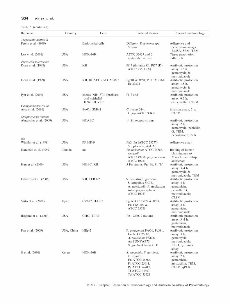
Table 1. (continued)
Reference Country Cells Bacterial strains Research methodology
Treponema denticolaPeters et al. (1999) Endothelial cells Different Treponema spp.
StrainsAdherence andpenetration assays:ELISA, SEM, TEM
Lux et al. (2001) USA HOK-16B ATCC 35405 and 5mutantderivatives
Tissue penetrationafter 8 h
Prevotella intermediaDorn et al. (1998) USA KB Pi17 (fimbriae C), Pi27 (D),
ATCC 25611 (A)Antibiotic protectionassay, 1.5 h,gentamycin &metronidazole
Dorn et al. (1999) USA KB, HCAEC and CASMC Pg381 & W50; Pi 17 & 25611;Ec 23834
Antibiotic protectionassay, 1.5 h,gentamycin &metronidazole
Iyer et al. (2010) USA Mouse NIH 3T3 fibroblast,oral epithelialHN4, HUVEC
Pi17 and Antibiotic protectionassay, 0.5 h,carbenicillin; CLSM
Campylobacter rectusArce et al. (2010) USA BeWo, SM9-1 C. rectus 314,
C. jejuniATCC43457invasion assay, 5 h;CLSM
Streptococcus mutansAbranches et al. (2009) USA HCAEC 14 St. mutans strains Antibiotic protection
assay, 2 h,gentamycin, penicillinG; TEM,persistence 3, 27 h
(d)Winkler et al. (1988) USA PF HR-9 Fn2, Pg (ATCC 33277),
Streptococci, AaGA3Adherence assay
Darenfed et al. (1999) Canada no Fn.nucleatum ATCC 25586,vincentiiATCC 49256, polymorphumATCC 10953
Binding of humanplasminogen toF. nucleatum subsp.nucleatum
Han et al. (2000) USA HGEC, KB 5 Fn strains, Pg, Ec, Pi, Tf Antibiotic protectionassay, 3–4 h,gentamycin &metronidazole; TEM
Edwards et al. (2006) USA KB, TERT-2 S. cristatus,S. gordonii,S. sanguinis SK36,A. naeslundii, F. nucleatumsubsp.polymorphumATCC 10953
Antibiotic protectionassay, 4 h,gentamicin,penicillin G,metronidazole;CLSM
Saito et al. (2008) Japan Ca9-22, HAEC Pg ATCC 33277 & W83,Fn TDC100 &ATCC 25586
Antibiotic protectionassay, 2 h,gentamicin,metronidazole
Ikegami et al. (2009) USA CHO, TERT Fn 12230, 2 mutans Antibiotic protectionassay, 3–4 h,gentamicin,metronidazole
Pan et al. (2009) USA, China HEp-2 P. aeruginosa PAO1, Pg381,Fn ATCC25586,A. naeslundii PK606,Aa SUNYAB75,S. gordoniiChallis CH1
Antibiotic protectionassay, 3 h,gentamycin,metronidazole;FISH, cytokinesassay
Ji et al. (2010) Korea HOK-16B S. sanguinis, S. gordonii,V. atypica,Fn ATCC 25586,Pi ATCC 25611,Pg ATCC 49417,Tf ATCC 43407,Td ATCC 33521
Antibiotic protectionassay, 2 h,gentamicin,amoxicillin; TEM,CLSM, qPCR
© 2013 European Federation of Periodontology and American Academy of Periodontology
S34 Reyes et al.

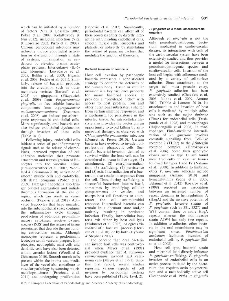
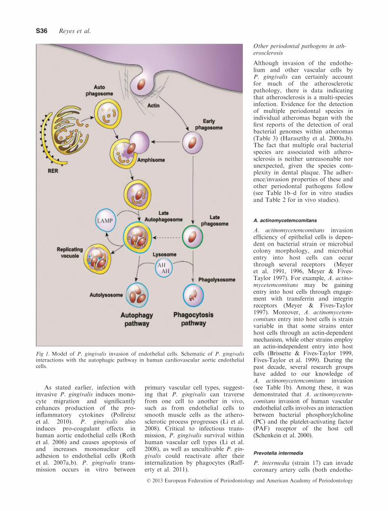
internalizes via lipid rafts in humanaortic endothelial cells (Yamatakeet al. 2007) and traffics via the auto-phagic pathway in cardiovascularendothelial and smooth muscle cells(Dorn et al. 2001); in contrast it uti-lizes the endocytic pathway duringinvasion of oral epithelial cells(Takeuchi et al. 2011) (see Fig. 1).However, only certain strains of P.gingivalis, such as 381 and W83,usurp the autophagic pathway dur-ing invasion of endothelial cells
(Dorn et al. 2001). Invasion via theautophagic pathway is observed at alow multiplicity of infection (100MOI), which is more consistent withbacterial numbers likely to meetendothelial cells because of bactere-mic events. At a higher MOI of1000, the majority of P. gingivalisorganisms travel through endosomesinstead of autophagosomes (Yama-take et al. 2007) which suggests thatbacterial dose plays a role in traf-ficking of P. gingivalis. In epithelial
cells, P. gingivalis exits throughthe endocytic recycling pathway(Takeuchi et al. 2011). It remainsunknown if P. gingivalis exit fromcardiovascular cells (Li et al. 2008) isthrough the same pathway. Regard-less of the pathways employed by P.gingivalis, these strategies are likelyto be critical for microbial dissemi-nation and evasion of host cell clear-ance mechanisms and pathologicalchanges within the cardiovascularsystem.
Table 1. (continued)
Reference Country Cells Bacterial strains Research methodology
Kirschbaum et al. (2010) Germany KB Fn ATCC 25586,Pg ATCC 33277 & M5-1-2,Tf ATCC 43037,Td ATCC 35405.Alone or in combinations.
antibiotic protectionassay, 1, 6, 18 h,penicillin; SEM,cytokine andmRNA assays
SEM, scanning electron microscopy; TEM, scanning electron microscopy; CLSM, confocal laser scanning microscopy, OM, optic micros-copy; IF, immunofluorescence; RFLP, restriction fragment length polymorphism, Aa, A. actinomycetemcomitans; Pg, P. gingivalis; Td, T.denticola; Tf, T. forsythia; Pm, P. micra; Pi, P. intermedia; Fn, F. nucleatum; Ec, E. corrodens; Cr, C. rectus, Pi, P. intermedia; Ec, E. corro-dens. Cell lines: BAEC, bovine aortic endothelial cells; BeWo, human trophoblast cell line derived from a human choriocarcinoma; BHEC,bovine heart endothelial cells; Ca9-22, epithelial cell-like gingival carcinoma cell line; CASMC, coronary artery smooth muscle cell; CHO-Kl, Chinese hamster ovary; EC, endothelial cells; HAEC, human aorta endothelial cells; HCAEC, human coronary artery endothelial cells;HeLa, cervical cancer cells; HEp-2, human larynx epithelial cells, originally derived from an epidermoid carcinoma from the larynx, nowknown to contain contaminant HeLa cells; HGEC, human gingival epithelial cells; HN4, oral epithelial cells; HOK-16B, human gingivalkeratinocyte cell line; HUVEC, monolayers of human vascular endothelial cells; KB, originally thought to be derived from an epidermal car-cinoma of the mouth, have now been shown to be derived from HeLa cell cultures as a contaminant; KB-R2A, a cell line defective for endo-somal acidification; NIH 3T3, mouse fibroblast cells; PF HR-9, induced differentiation of an embryonal carcinoma; PHGF, primary humangingival fibroblasts; RPMI-4788, human intestinal epithelial cells; SMC, smooth muscle cells; SM9-1, mouse trophoblast cell line, derivedfrom a gestational day 9 Swiss–Webster mouse placenta; TERT-2; oral keratinocyte cells.
Table 2. Bacterial adhesion and invasion: in vivo studies
Reference Country Samples Patients Target strains/species Invasion methods
Saglie et al. (1986) USA, Chile Gingival biopsies 6 SevP, 2 LJP, 2 healthy Aa, Pg,Capnocytophaga gingivalis
OM, TEM,immunoperoxidase
Christersson et al.(1987a)
USA 35 (+4 control) biopsies 12 LJP, 2 healthy,1 ChP, 1 monkey
Aa-three serotypes IF (rabbit antisera),TEM
Christersson et al.(1987b)
USA 11 (+3 control,Aa sites) biopsies
6 LPJ Aa Culture after serialwashing
Noiri et al. (1997) Japan 12 teeth 7 SevChP Pg, Cr, A. viscosus Immunohistochemicalstaining
Rudney et al. (2001) USA Cheek samples 24p, 13 m, 11f,1edentulous
Aa, Pg FISH & CLSM;multiplex PCR
Rudney et al. (2005) USA Cheek samples 38p, 20 m, 18f Aa, Pg, Tf FISH & CLSM;qPCR
Leung et al. (2006) USA Supra, sub, cheeksamples
27p, before and 1 mafter ortho
Aa, Tf, Streptococci FISH & CLSM;qPCR
Colombo et al. (2006) Brasil, USA 6 samples (3 pocket,3 sulcus) per patient
49 ChP 33 species Checkerboard
Colombo et al. (2007) Brasil, USA 58 samples (pockets,sulcus, buccal mucosa)
22p, 14 Periodontitis,8 Healthy
Aa, Pg, Tf, Td FISH & CLSM
Johnson et al. (2008) USA buccal mucosa andsubgingival samples
18 AgP (treatment SRP,AmoxiMet, CHX)
Aa, Pg, Tf, Td, Pi FISH & CLSM;qPCR (baseline,3 m, 6 m)
OM, optic microscopy; SEM, scanning electron microscopy; TEM, scanning electron microscopy; IF, immunofluorescence; FISH, fluores-cence in situ hybridization; CLSM, confocal laser scanning microscopy, Sev, severe; Ch, chronic; P, periodontitis; LPJ, localized juvenileperiodontitis; m, male; f, female; SRP, scaling and root planing; AmoxiMet, amoxicillin plus metronidazole; CHX, chlorhexidine, Aa, A. ac-tinomycetemcomitans; Pg, P. gingivalis; Td, T. denticola; Tf, T. forsythia; Pm, P. micra; Pi, P. intermedia; Cr, C. rectus.
© 2013 European Federation of Periodontology and American Academy of Periodontology
Periodontal bacterial invasion and infection S35
As stated earlier, infection withinvasive P. gingivalis induces mono-cyte migration and significantlyenhances production of the pro-inflammatory cytokines (Pollreiszet al. 2010). P. gingivalis alsoinduces pro-coagulant effects inhuman aortic endothelial cells (Rothet al. 2006) and causes apoptosis ofand increases mononuclear celladhesion to endothelial cells (Rothet al. 2007a,b). P. gingivalis trans-mission occurs in vitro between
primary vascular cell types, suggest-ing that P. gingivalis can traversefrom one cell to another in vivo,such as from endothelial cells tosmooth muscle cells as the athero-sclerotic process progresses (Li et al.2008). Critical to infectious trans-mission, P. gingivalis survival withinhuman vascular cell types (Li et al.2008), as well as uncultivable P. gin-givalis could reactivate after theirinternalization by phagocytes (Raff-erty et al. 2011).
Other periodontal pathogens in ath-erosclerosis
Although invasion of the endothe-lium and other vascular cells byP. gingivalis can certainly accountfor much of the atheroscleroticpathology, there is data indicatingthat atherosclerosis is a multi-speciesinfection. Evidence for the detectionof multiple periodontal species inindividual atheromas began with thefirst reports of the detection of oralbacterial genomes within atheromas(Table 3) (Haraszthy et al. 2000a,b).The fact that multiple oral bacterialspecies are associated with athero-sclerosis is neither unreasonable norunexpected, given the species com-plexity in dental plaque. The adher-ence/invasion properties of these andother periodontal pathogens follow(see Table 1b–d for in vitro studiesand Table 2 for in vivo studies).
A. actinomycetemcomitans
A. actinomycetemcomitans invasionefficiency of epithelial cells is depen-dent on bacterial strain or microbialcolony morphology, and microbialentry into host cells can occurthrough several receptors (Meyeret al. 1991, 1996, Meyer & Fives-Taylor 1997). For example, A. actino-mycetemcomitans may be gainingentry into host cells through engage-ment with transferrin and integrinreceptors (Meyer & Fives-Taylor1997). Moreover, A. actinomycetem-comitans entry into host cells is strainvariable in that some strains enterhost cells through an actin-dependentmechanism, while other strains employan actin-independent entry into hostcells (Brissette & Fives-Taylor 1999,Fives-Taylor et al. 1999). During thepast decade, several research groupshave added to our knowledge ofA. actinomycetemcomitans invasion(see Table 1b). Among these, it wasdemonstrated that A. actinomycetem-comitans invasion of human vascularendothelial cells involves an interactionbetween bacterial phosphorylcholine(PC) and the platelet-activating factor(PAF) receptor of the host cell(Schenkein et al. 2000).
Prevotella intermedia
P. intermedia (strain 17) can invadecoronary artery cells (both endothe-
Fig 1. Model of P. gingivalis invasion of endothelial cells. Schematic of P. gingivalisinteractions with the autophagic pathway in human cardiovascular aortic endothelialcells.
© 2013 European Federation of Periodontology and American Academy of Periodontology
S36 Reyes et al.

lial and smooth muscle cells) in vitro(Table 1c). The type C. fimbriae andthe invasin protein, AdpC (belongingto the leucine-rich repeat proteinfamily) appear to mediate the inva-sion process.
Tannerella forsythia
Multiple T. forsythia strains attach toand invade epithelial cells in vitro,mediated by the surface layer com-posed of proteinaceous arrays (Sabetet al. 2003, Sakakibara et al. 2007)(Table 1c). Similar to P. intermedia,epithelial cell attachment and invasionare also dependent on a cell-surfaceassociated leucine-rich repeat protein,BspA. Interestingly, P. gingivalis or itsouter membrane vesicles enhance T.forsythia attachment and invasion (Ina-gaki et al. 2006). At present, there isminimal information about T. forsythiainteractions with cardiovascular cells.
F. nucleatum
Many strains of F. nucleatum areinvasive in vitro (Table 1d). TheF. nucleatum adhesin, FadA, appearsto be required for both attachmentand invasion (Ikegami et al. 2009).F. nucleatum may also transport non-invasive species (e.g. streptococci)into host cells via a combination ofco-aggregation and invasion mecha-nisms and also facilitate P. gingivalisinvasion of human gingival epithelialand endothelial cells (Saito et al.2008), but evidence of F. nucleatumco-aggregation among different com-binations of species including P. gin-givalis, T. denticola or T. forsythia isequivocal (Kirschbaum et al. 2010).
Other oral bacterial species
There is limited in vitro (Table 1a–d)or in vivo (Table 2) evidence that
other bacterial species, such as Trep-onema denticola and Campylobacterrectus, have been detected in ather-oma specimens or demonstrated toinvade host cells.
Emerging mechanisms of atherosclerosis:
autophagy as an endothelial stress
response and its perturbation by P.
gingivalis
Autophagy represents a reparativecellular response that degrades senes-cent cellular organelles, damagedintracellular proteins, and invadingintracellular bacteria (Dorn et al.2002a, Deretic 2011).
There is emerging evidence thatthe autophagic response may influ-ence cardiovascular disease outcomesduring atherosclerosis by affectingplaque stability (Martinet & DeMeyer 2008, Schrijvers et al. 2011).For instance, increased autophagicactivity measured by LC3 activationor expression have been detectedwithin atheromatous plaques fromhumans and murine models of sterileatherosclerosis (Martinet & DeMeyer 2008, Liao et al. 2012). Fur-ther, ApoE-null mice fed high fatdiets exhibit impaired autophagywithin the atheromatous plaque asdisease progresses (Liao et al. 2012),and impaired autophagy has beenlinked to increased inflammation,which has pro-atherogenic effects(Razani et al. 2012).
In sterile atherosclerosis, auto-phagy is considered a protectivemechanism that prevents apoptosisin cells that are undergoing oxidativestress (Scherz-Shouval & Elazar2007, Xie et al. 2011) or endoplasmicreticulum stress (Bernales et al. 2006,Zhang et al. 2010a), which is a com-mon feature of atherosclerotic dis-ease. However, in vitro studies havedemonstrated that over-activation of
autophagy or dysregulation of theautophagic pathway in cardiovascu-lar cells can also be detrimental(Larocca et al. 2012) by promotingcell death referred to as “necropto-sis” (Khan et al. 2012, Kolattukudy& Niu 2012), which is potentiallypro-atherogenic due to its pro-inflammatory effect (Kolattukudy &Niu 2012).
As an innate immune clearancemechanism, autophagy can be acti-vated in response to signalling viatoll-like receptors (TLRs), Nod-likereceptors (NLRs) and/or the seques-tasome (Shin et al. 2010, Anandet al. 2011, Deretic 2011). Someintracellular pathogens including P.gingivalis have developed strategiesto subvert this line of defence (De-retic & Levine 2009). P. gingivalisstrain 381 usurps the autophagicpathway during invasion of humancoronary artery endothelial cells andsmooth muscle cells in vitro, asshown in Fig. 1 (Dorn et al. 2001).Specifically, bacteria delay fusion ofthe autophagosome with lysosomesthus, interfering with autophagicflux. This process may impair theability of the host cell to use theautophagic response as a means of res-cuing the cell from oxidative or endo-plasmic reticulum stress. Rodrigueset al. (2012) have recently demon-strated that not all P. gingivalis strainsmanipulate this pathway during hostcell invasion; W83 perturbs the auto-phagic pathway of endothelial cells ina similar fashion as 381, whereasstrains A7436 and 33,277 do not.
Evaluation of the evidence
In a clinical setting, it is extremelydifficult to establish the causativefactor of atherosclerosis for severalreasons. First, the initiating factor islikely to be missed since the earlyphase of endothelial injury is usuallyasymptomatic (Vita & Loscalzo2002). Second, the atheroscleroticlesion is a common inflammatoryresponse to multiple factors (Keizer2012, Raman et al. 2013), and someor all of those factors may be associ-ated with the lesion at the time ofdiscovery. Third, interventional stud-ies that evaluated the impact of peri-odontal treatment, with or withoutantimicrobial therapy, on systemicinflammation or endothelial dysfunc-tion, have shown mixed results
Table 3. List of “proofs” that has to be fulfilled to demonstrate that periodontal bacteriaare a contributing factor to atherosclerosis
Proof Description
1 Periodontal bacteria can reach systemic vascular tissues.2 Periodontal bacteria can be found in the affected tissues.3 Evidence of live periodontal bacteria at the affected site.4 In vitro evidence of invasion of affected cell types.5 Demonstration that periodontal bacteria can promote atherosclerosis in animal
models of disease.6 In vitro and in vivo evidence that non-invasive mutants cause significantly reduced
pathology (animal model).7 Fulfil modified Koch’s postulate to demonstrate that a human atheroma isolate causes
disease in animal models.
© 2013 European Federation of Periodontology and American Academy of Periodontology
Periodontal bacterial invasion and infection S37

including no change, transient wors-ening of signs immediately aftertreatment, or improvement in signsthat did not necessarily persist overtime (extensively reviewed by Kebsc-hull et al. 2010). Despite these limi-tations, it is possible to, at least,demonstrate biological plausibilitythat invasion of cardiovascular tis-sues by periodontal bacteria havethe potential to promote atheroscle-rosis through fulfilment of severalproofs, discussed elsewhere in thearticle (Table 3).
Proof that periodontal bacteria canreach systemic vascular tissues
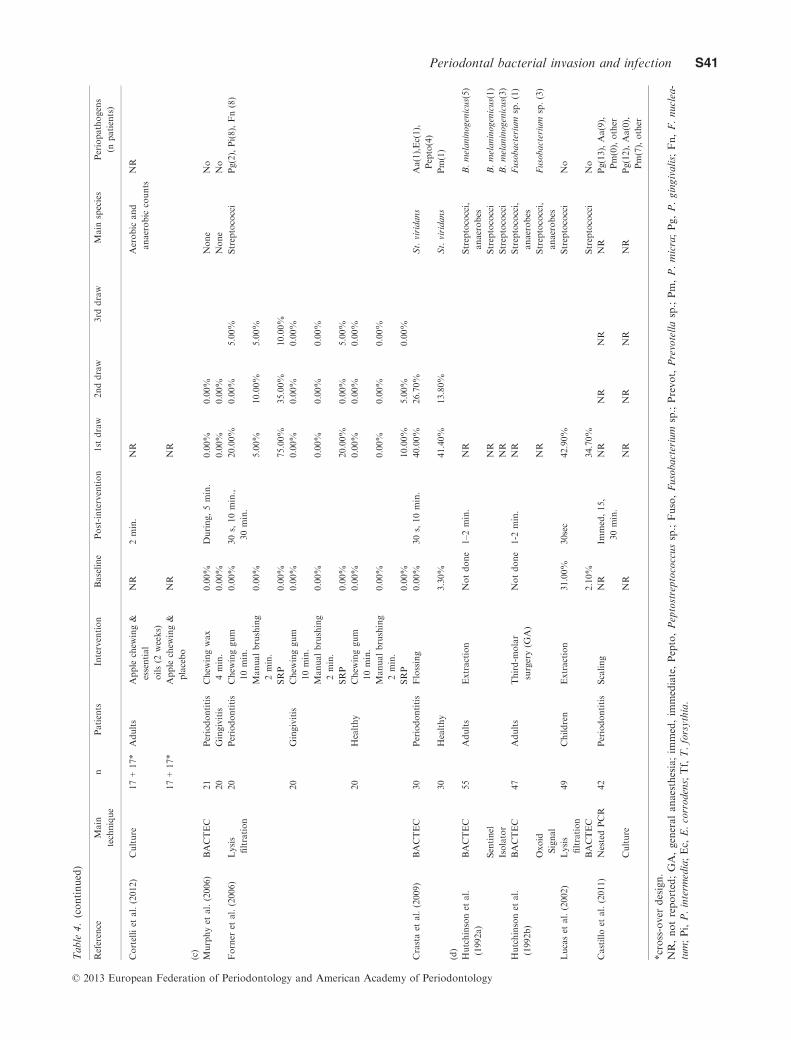
There is no doubt that oral bacterialspecies can enter the circulation andcause bacteremia (Table 4a–d),which has been documented by sev-eral groups. There are potentiallymultiple mechanisms for systemicdissemination of periodontal bacte-ria:
• As the gingival pocket is sepa-rated from gingival micro-capil-laries by a few cells, it is thoughtthat periodontal species thatinvade oral cells can cross thislayer and enter the circulationvia a transcellular mechanism(Takeuchi et al. 2011).
• More often, however, periodontalbacteria likely enter the circula-tion following physical perturba-tions of the gingiva. Amongthese perturbations, some arerelated to dental procedures suchas tooth polishing, scaling, toothextraction, surgical extraction ofthird molar and periodontalprobing (see Table 4a–d). Otherperturbations are daily life activi-ties, including tooth brushing,flossing, chewing or biting anapple, as listed in Table 4a–d. Thefrequency of bacteremia showed awide variability, depending on thestudy design, the microbiologicalmethodology, the stimulus andthe periodontal health status ofthe patients. In some studies, anassociation with plaque and/orgingival indices has been reported(Silver et al. 1977, Forner et al.2006, Lockhart et al. 2009). Arecent systematic review withmeta-analysis on the prevalence ofbacteremia following tooth brush-ing, evaluated the pooled oddsratios of the influence of plaque
and gingival indices at 2.61 and2.77 respectively (Tomas et al.2012). In addition, as shown inTable 4a–d, multiple studieshave detected different bacteremicperiodontal pathogens, especiallyafter subgingival debridementprocedures in periodontitispatients. However, these resultsshould be interpreted with cautiondue to variability among thesestudies, which may be due to limi-tations of the methodology thatwas used that may affect the sensi-tivity and detection thresholds(culture-based methods). In addi-tion, well-validated PCR-basedmethods were not frequently used.
• Another proposed but unprovenmechanism of bacteremia is thatperiodontal bacteria enter the cir-culation and disseminate to dis-tant sites via survival in immunecells (the Trojan horse approach)(Carrion et al. 2012). In suchcases, phagocytosis is beneficialto the bacterial pathogen but det-rimental to the host. In this sce-nario, the internalized pathogenevades microbial killing withinthe leucocyte and is able toescape from the phagocyte afterthe cell reaches another sitewithin the body (Zeituni et al.2009). For example, P. gingivalisgains entry into dendritic cellsand disrupts their phagocyticfunction as evidenced by reten-tion of an immature phenotypecharacterized by a low produc-tion of inflammatory mediators(Zeituni et al. 2009, 2010).Regardless of mechanism, thereis no doubt that oral pathogensfrequently enter the circulation.
Proof that periodontal bacteria canbe found in the affected tissues
Since bacteremias of oral origin arefrequent, the association of peri-odontal pathogens with atheroscle-rotic pathology has been the focusof multiple studies. A variety of oralbacterial species have been identifiedin atheromatous tissues at the DNA,RNA or antigen level. Haraszthyet al. (2000a) were the first to reportthe detection of the genomicDNA of A. actinomycetemcomitans,P. gingivalis and other species usingbacterial 16S rRNA – specific PCRanalysis. These PCR detection stud-
ies were further extended and con-firmed by others (Tables 5, 6).Interestingly, most of these reportsfound evidence of multiple species inindividual atheromas. Thus, there issufficient data to conclude that mul-tiple pathogenic species reach theaffected site. However, the impact ofthese results are somewhat hamperedby several factors. For example, thepresence of bacterial components(DNA, RNA and antigens) does notdistinguish between live and deadbacteria within the tissue. Further,some species of bacteria,, such as Ve-illonella sp, which have been detectedwithin atheromas (Koren et al.2011), are not cultivable (Chalmerset al. 2008). Finally, not all studieshave detected microbial productswithin the atheroma, and this lackof consistency may be due to differ-ences in methodology (Figuero et al.2011) or underlying aetiology of theatheroma.
Evidence of live periodontal bacteriaat the affected site
For several years, multiple groupsattempted to culture periodonto-pathogenic organisms from athero-mas to no avail. Finally, Kozarovet al. (2005) reported evidence oflive P. gingivalis and A. actinomyce-temcomitans in atheromatous tissue.This was accomplished by incubat-ing homogenized atheromatous tis-sue in culture with primary humancoronary artery endothelial cells(HCAECs). After several days ofincubation, the group used fluores-cently labelled antibodies and de-convolution microscopy to visualizeintact bacteria within the endothe-lial cells. Because both species mustbe alive in order to invade, thepresence of the bacteria within thenon-phagocytic cells providedstrong evidence of the existence oflive bacteria in the atheromas. Inaddition, Rafferty et al. (2011) cul-tured atheroma samples with mac-rophages as an intermediate step andwas able to isolate P. gingivalis onculture plates. These studies requirefresh atheromas (angioplasty andstenting have become the standardof care) and the complexity of theprotocol appears to have precludedconfirmatory reports to date. Thus,although periodontal pathogenscannot routinely be cultured fromatheromas directly onto plates, there
© 2013 European Federation of Periodontology and American Academy of Periodontology
S38 Reyes et al.

Table
4.
(a)Clinicalstudiesevaluatingbacterem
iaoforalorigin:frequency
andmain
microbiologicalfindings,
(b)Randomized
clinicaltrials
evaluatingbacterem
iaoforalorigin:frequency
andmain
microbiologicalfindings,
(c)Cohort
studiesevaluatingbacterem
iaoforalorigin:frequency
andmain
microbiologicalfindings,
(d)Validationstudiesevaluatingbacterem
iaoforal
origin:frequency
andmain
microbiologicalfindings
Reference
Main
technique
nPatients
Intervention
Baseline
Post-intervention
1st
draw
2nddraw
3rd
draw
Main
species
Periopathogens
(npatients)
(a)
Silver
etal.(1977)
Culture
96
Gingivitis
Powered
brushing
2.10%
During
42.70%
NR
B.melaninogenicus(7),
Pepto(3)
Silver
etal.(1979)
Culture
36
Healthy
Powered
brushing
0.00%
During
8.30%
NR
No
Heimdahlet
al.
(1990)
Lysis
filtration
20
Adults
Extraction
0.00%
During,10min.
95.00%
40.00%
St.viridans,
anaerobes
No
20
Adults
Third-m
olar
surgery
0.00%
55.00%
40.00%
St.viridans,
anaerobes
Pm(2)
20
Periodontitis
Scaling
0.00%
65.00%
30.00%
St.viridans,
anaerobes
Fn(1)
20
Adults
Endodontic
0.00%
15.00%
5.00%
St.viridans,
anaerobes
No
20
Adults
Tonsillectomy
0.00%
55.00%
0.00%
St.viridans,
anaerobes
Pg(1),Fn(1)
Messiniet
al.(1999)
Culture
18
Disable
Various
procedures
Not
done
Immed,5min.,
30min.
83.30%
Gem
ella,
Streptococci
Pg(4).Pm(1)
Rajasuoet
al.(2004)
Culture
16
Youngadults
Third-m
olar
surgery
NR
10,15,30min.
44.00%
25.00%
13.00%
Streptococci
Prevot.,Eubacterium
sp.,Pepto
Kinaneet
al.(2005)
BACTEC
30
Periodontitis
Probing
6.70%
30s–1min.
20.00%
NR
Pi(1)
Periodontitis
Manual
brushing
NR
<3min.
3.30%
NR
No
Periodontitis
Scaling
No
Immed
13.30%
NR
No
Tomaset
al.(2007a)
BACTEC
53
Disable
Extraction
(under
GA)
NR
30s,15min.,1h
96.20%
64.20%
20.00%
Streptococci
(63.8%
)
Fn(1),Pm(2)
Lafaurieet
al.(2007)
Culture
42
Periodontitis
Scaling
NR
Immed,15,30min.
80.90%
19.00%
NR
Pg(12),Pm(7),Ec(4),
Tf(3),Pi(3),Fuso(5)
Brennanet
al.(2007)
NR
100
Children
Various
procedures
NR
Eightdraws
NR
NR
NR
NR
No
Tomaset
al.(2008)
BACTEC
100
Adults
Third-m
olar
surgery(G
A)
NR
30sec,
15min.
62.00%
67.00%
St.viridans
None
Lucaset
al.(2008)
Lysis
filtration
32
Children&
adolescents
Manual
brushing
NR
30safter
22.00%
19.00%
Variety,no
anaerobes
No
35
Powered
brushing
OralB
NR
26.00%
34.00%
Variety,no
anaerobes
No
33
Powered
brushing
Sonicare
NR
27.00%
33.00%
Variety,no
anaerobes
No
41
Tooth
polishing
NR
15.00%
37.00%
Variety,no
anaerobes
No
Perez-C
haparroet
al.
(2008)
Culture
16
Periodontitis
Scaling
NR
Immed,15,30min.
NR
NR
NR
FocusedonPg
7of16,Pg-positive
© 2013 European Federation of Periodontology and American Academy of Periodontology
Periodontal bacterial invasion and infection S39

Table
4.(continued)
Reference
Main
technique
nPatients
Intervention
Baseline
Post-intervention
1st
draw
2nddraw
3rd
draw
Main
species
Periopathogens
(npatients)
(b)
Diz
Dioset
al.(2006)
BACTEC
53
Disable
Extractions
(GA)&
placebo
NR
30s,15min.,1h
96.00%
64.00%
20.00%
Streptococci
Fn(1),Pm(2)
56
Extractions
(GA)
&amoxi
NR
46.00%
11.00%
4.00%
Streptococci
Prevot(4),
Pepto(4),Ec(2)
54
Extractions
(GA)&clinda
NR
85.00%
70.00%
22.00%
Streptococci
Fuso(2),Pepto(2)
58
Extractions(G
A)
&moxiflo
NR
57.00%
24.00%
7.00%
Streptococci
Fuso(2),Prevot(4),
Pepto(8),Ec(2)
Cherry
etal.(2007)
Lyso-
centrifugation
30
Gingivitis
Scaling&
rinse
povidone
3.30%
30sec,
2min.
3.30%
6.70%
St.viridans
No
30
Scaling&
rinse
saline
10.00%
13.30%
30.00%
St.viridans
Pi(4),Pg(1)
Tomaset
al.(2007b)
BACTEC
53
Disable
Extraction(G
A)
control
9.00%
30s,15min.,1h
96.00%
64.00%
20.00%
Streptococci
(63.8%
)
Fn(1),Pm(2)
53
Extraction(G
A)
preCHX
7.00%
79.00%
30.00%
2.00%
Streptococci
(68%
)
Pm(1)
Bahrani-Mougeotet
al.
(2008)
BACTEC,
16sRNA
sequencing
98
Adults
Manualbrushing
2min.
NR
During,im
med,
20,40,60min.
NR
NR
NR
Streptococci
Pm(3),Pi(1),Dn(1)
96
Extraction
&amoxi
NR
NR
NR
NR
Streptococci
Pm(2),Pi(9),Dn(1)
96
Extraction&
placebo
NR
NR
NR
NR
Streptococci
Pm(16),Pi(3),Dn(6)
Lockhart
etal.(2008)
BACTEC,
16sRNA
sequencing
98
Adults
Manualbrushing
2min.
3cases
During,im
med,
20,40,60min.
Cumulative
32%
60min.,9%
Streptococcus
(49%
),
Prevotella(9%
),
Actinomyces(5%
),
Fusobacterium
(5%
).
Prevotella(9%
),
Fusobacterium
(5%
).96
Extraction
&amoxi
Cumulative
56%
60min.,0%
96
Extraction&
placebo
Cumulative
80%
60min.,2%
Pineiro
etal.(2010)
BACTEC
30
Adults
Implant
placement
3.30%
30s,15min.
6.70%
3.30%
St.viridans
Neisseria
cinerea
No
20
Implant
placement
preCHX
0%
0%
0%
No
No
Asiet
al.(2010)
Culture
30+30*
Periodontitis
Modified
Widmanflap
&amoxi
No
Duringsurgery
13.30%
Staphylococcus
albus,Klebsiella,
Ps.aeruginosa,
St.viridans
No
30+30*
Modified
Widmanflap
No
46.60%
Fineet
al.(2010)
Culture
22+22*
Adults
Apple
chew
ing&
essential
oils(2
weeks)
NR
2min.
NR
Aerobic
and
anaerobic
counts
NR
22+22*
Apple
chew
ing&
placebo
NR
NR
NR
© 2013 European Federation of Periodontology and American Academy of Periodontology
S40 Reyes et al.
Table
4.(continued)
Reference
Main
technique
nPatients
Intervention
Baseline
Post-intervention
1st
draw
2nddraw
3rd
draw
Main
species
Periopathogens
(npatients)
Cortelliet
al.(2012)
Culture
17+17*
Adults
Apple
chew
ing&
essential
oils(2
weeks)
NR
2min.
NR
Aerobic
and
anaerobic
counts
NR
17+17*
Apple
chew
ing&
placebo
NR
NR
(c)
Murphyet
al.(2006)
BACTEC
21
Periodontitis
Chew
ingwax
4min.
0.00%
During,5min.
0.00%
0.00%
None
No
20
Gingivitis
0.00%
0.00%
0.00%
None
No
Forner
etal.(2006)
Lysis
filtration
20
Periodontitis
Chew
inggum
10min.
0.00%
30s,10min.,
30min.
20.00%
0.00%
5.00%
Streptococci
Pg(2),Pi(8),Fn(8)
Manualbrushing
2min.
0.00%
5.00%
10.00%
5.00%
SRP
0.00%
75.00%
35.00%
10.00%
20
Gingivitis
Chew
inggum
10min.
0.00%
0.00%
0.00%
0.00%
Manualbrushing
2min.
0.00%
0.00%
0.00%
0.00%
SRP
0.00%
20.00%
0.00%
5.00%
20
Healthy
Chew
inggum
10min.
0.00%
0.00%
0.00%
0.00%
Manualbrushing
2min.
0.00%
0.00%
0.00%
0.00%
SRP
0.00%
10.00%
5.00%
0.00%
Crastaet
al.(2009)
BACTEC
30
Periodontitis
Flossing
0.00%
30s,10min.
40.00%
26.70%
St.viridans
Aa(1),Ec(1),
Pepto(4)
30
Healthy
3.30%
41.40%
13.80%
St.viridans
Pm(1)
(d)
Hutchinsonet
al.
(1992a)
BACTEC
55
Adults
Extraction
Notdone
1–2
min.
NR
Streptococci,
anaerobes
B.melaninogenicus(5)
Sentinel
NR
Streptococci
B.melaninogenicus(1)
Isolator
NR
Streptococci
B.melaninogenicus(3)
Hutchinsonet
al.
(1992b)
BACTEC
47
Adults
Third-m
olar
surgery(G
A)
Notdone
1-2
min.
NR
Streptococci,
anaerobes
Fusobacterium
sp.(1)
Oxoid
Signal
NR
Streptococci,
anaerobes
Fusobacterium
sp.(3)
Lucaset
al.(2002)
Lysis
filtration
49
Children
Extraction
31.00%
30sec
42.90%
Streptococci
No
BACTEC
2.10%
34.70%
Streptococci
No
Castillo
etal.(2011)
NestedPCR
42
Periodontitis
Scaling
NR
Immed,15,
30min.
NR
NR
NR
NR
Pg(13),Aa(9),
Pm(0),other
Culture
NR
NR
NR
NR
NR
Pg(12),Aa(0),
Pm(7),other
*cross-over
design.
NR,notreported;GA,generalanaesthesia;im
med,im
mediate,Pepto,Peptostreptococcussp.;Fuso,Fusobacterium
sp.;Prevot,Prevotellasp.;Pm,P.micra;Pg,P.gingivalis;
Fn,F.nuclea-
tum;Pi,P.interm
edia;Ec,
E.corrodens;Tf,T.forsythia.
© 2013 European Federation of Periodontology and American Academy of Periodontology
Periodontal bacterial invasion and infection S41

is evidence that live periodontal bac-teria are present in at least some ath-eromas.
In vitro evidence of invasion ofaffected cell types
There are indisputable data that, atleast, some periodontal pathogensinvade human cardiovascular cells invitro (Table 1a–d). Deshpande et al.(1998) and Dorn et al. (1999) werethe first to report invasion of endo-thelial cells by P. gingivalis. Sincethese first reports, there have been anumber of research groups who haveprovided details of the mechanismsof cardiovascular cell invasion by P.gingivalis (Dorn et al. 2000, 2002b,
Rodrigues & Progulske-Fox 2005,Takahashi et al. 2006, Li et al.2008). These have been discussedpreviously.
Demonstration that periodontal bacte-ria can promote atherosclerosis in ani-mal models of disease
Animal models can be used to supportin vitro data-based hypotheses(Graves et al. 2008) and have beenused to demonstrate atheroscleroticpathology caused by periodontalpathogens. For example, P. gingivalishas been reported by multiple groupsto accelerate atherosclerosis in murinemodels (Lalla et al. 2003, Gibsonet al. 2004, Amar et al. 2009). In addi-
tion to murine models, rabbits withexperimentally induced periodontaldisease developed fatty streaks in theaorta faster than in periodontallyhealthy animals (Jain et al. 2003). Innormocholesterolemic pigs, recurrentP. gingivalis bacteremia induced bothaortic and coronary lesions, andP. gingivalis bacteremia also enhancedatherosclerosis in hypercholesterol-emic pigs (Brodala et al. 2005). Thus,in addition to the identification of liveperiodontal organisms in human ath-erosclerotic plaque, in vivo experi-ments in a variety of animal modelshave provided biological plausibilitythat P. gingivalis can enhance athero-genesis.
Table 5. Description of studies evaluating the presence of periodontal pathogens in cardiovascular samples, including atheromatous lesions
Reference Country Patients/samples Surgery Vascular sample
Haraszthy et al. (2000b) USA 50 Carotid endarterectomy Carotid stenosisOkuda et al. (2001) Japan 26 test, 14 control NR 26 atherosclerotic lesions;
14 non-diseased aortaStelzel et al. (2002) Germany 26 Open heart surgery Aortas; vascular regionsMastragelopulos et al.(2002)
Germany 34 Carotid endarterectomyor bypass
NR
Taylor-Robinson et al.(2002)
England 36 samples NR Artherosclerotic major arteries
Marques da Silva et al.(2003)
Norway 49p, 53 samples Aortic aneurysm repair Aneurysms
Cairo et al. (2004) Italy 26 dentate,26 edentulous
Carotid endarterectomy Atheromatous plaques
Ishihara et al. (2004) Japan/USA 51 NR Stenotic coronary artery plaquesKurihara et al. (2004) Japan 32 Aortic aneurysm repair Abdominal aortic aneurysmMarques da Silva et al.(2005)
Norway 51p, 56 lesions Aortic aneurysm repair Aortic aneurysms
Kozarov et al. (2005) USA 1 Endarterectomy CarotidFiehn et al. (2005) Denmark 79 (PCR from 24) NR Atheromatous plaquesPadilla et al. (2006) Chile 12 ChP Endarterectomy Atheromatous plaquesKozarov et al. (2006) USA 29p, 129 samples Aorta, coronary arteries,
carotid, femoralAtheromatous plaques
Ott et al. (2006) Germany 38 samples Catheter-based atherectomy catheter materialZaremba et al. (2007) Poland 20 ChP Bypass Coronary vesselsAimetti et al. (2007) Italy 33 ChP Endarterectomy Carotid atheromatous plaquesRomano et al. (2007) Italy 21 ChP Endarterectomy Carotid atheromatous plaquesPucar et al. (2007) Serbia 15 Coronary artery bypass
grafting surgeryCoronary arteries withatherosclerosis
Zhang et al. (2008) China 51 Coronary artery bypass graft Coronary atheromatous plaquesElkaim et al. (2008) France 22 Cardiac artery bypass Atheromatous plaquesGaetti-Jardim et al. (2009) Brazil 39 ChP, 5 healthy Endarterectomy Atheromatous plaques from
coronary arteriesNakano et al. (2009) Japan 223 Various VariousMahendra et al. (2010) India 51 ChP Coronary artery bypass grafting Atheromatous plaquesMarcelino et al. (2010) Brazil 28 test, 2 control Endarterectomy Atheromatous plaquesFiguero et al. (2011) Spain 42 Endarterectomy of carotid artery Atheromatous plaquesAquino et al. (2011) Brazil 30 Stent angioplasty,
endarterectomy, bypassCarotid, coronary orfemoral arteries
Koren et al. (2011) Sweden 15 atherosclerosis 15 healthy Endarterectomy Carotid arteryOhki et al. (2012) Japan 81 Primary percutaneous
coronary interventionThrombus
NR, not reported; ChP, chronic periodontitis.
© 2013 European Federation of Periodontology and American Academy of Periodontology
S42 Reyes et al.

In vitro and in vivo evidence that non-invasive mutants cause significantlyreduced pathology (animal model)
Mutants with significantly reducedability to invade in vitro have alsobeen evaluated in vivo. In contrastto the invasive wild-type strain of P.gingivalis, the non-invasive fimAdeficient mutant did not accelerateatherosclerosis in ApoE knockoutmice Gibson et al. (2004). Moreover,the fimA deficient mutant was lesspro-atherogenic and elicited a lowerlevel of pro-inflammatory mediatorsthan the invasive parental strain inApoE deficient mice. However, thereis minimal data using other peri-odontal species in animal models.
Fulfill modified Koch’s postulate todemonstrate that a human atheromaisolate causes disease in animal mod-els
The final proof, fulfilling a variationof Koch’s postulates, has not yet beenachieved. It requires the isolation andcharacterization of periodontopatho-gens from human atheromas and thedemonstration that the isolates causeatherosclerotic pathology in animalmodels attributable to the bacterialisolates(s). Given the success by atleast one group in culturing strains ofP. gingivalis from human atheromas(Rafferty et al. 2011), this could beaccomplished in the future. An analo-gous approach has been used to inves-tigate the role of an invasive strain ofStreptococcus mutans in infectiousbrain aneurisms, with demonstrationthat a mutant deleted for the gene thatallows invasion was not able to causedisease (Nakano et al. 2011).
Discussion
In this review, the evidence to assessthe direct role of periodontopatho-gens in atherosclerosis has been eval-uated considering seven proofs:
• Proof 1 – should be consideredvalidated since the literature isconvincing that oral microorgan-isms obtain access to the circula-tion.
• Proof 2 – periodontal bacteriahave been identified in human ath-eromas by several groups usingmultiple detection technologies.Thus, there is little question that
oral bacteria are found in humanatheromas. However, visual proofof bacteria inside cells in the ath-eromas is still lacking. If only aminority of cells contain bacteria,transmission electron microscopy(TEM) may be too inefficient tofind them. Also, there is some var-iation in the organisms identifiedin the tissues. This could be attrib-uted to differences in techniquesused or the inter-individual diver-sity of oral organisms able toinfect atheromas. Eventually, thishypothesis could be tested in ani-mal models but the complexity ofthe microbiome will complicatethe models.
• Proof 3 – data using indirect meansof culture that support proof 3,have been published but additionalcomfirmative studies are needed. Invitro, P. gingivalis becomes dor-mant or viable, but not culturable(Li et al. 2008), and in vivo viablebacteria have been cultured fromatheroma homogenates followingin vitro cultivation in cell lines(Rafferty et al. 2011,). It is possiblethat the same phenomenon occursin vivo and that contact with“fresh” uninfected cells providesthe signals to the bacteria to emergefrom dormancy.
• Proof 4 – in vitro evidence ofinvasion of affected cell types isindisputable. During the past15 years, there have been a seriesof papers reporting the invasionof cardiovascular cell types byperiodontal bacteria.
• Proof 5 – it is also well docu-mented using various animalmodels since the periodontal bac-teria tested in these modelscaused increased frequency andsize of atherosclerotic lesions. Tothe extent that animal models ofatherosclerosis represent humandisease, then this proof is satis-fied. One goal should be theinclusion of more complex mix-tures of bacteria in these models.
• Proof 6 – the invasion and dis-ease phenotype, that is, signifi-cantly reduced disease caused bynon-invasive mutants comparedto the wild-type parental strain isalso documented by multipleresearch groups. Although exper-iments with additional invasivemutants will prove interestingand informative, the present data
confirm this proof for specificstrains.
• The final proof, Proof 7, is yet tobe accomplished. As atheroma-tous specimens become more andmore difficult to obtain, we maybe close to missing the windowof opportunity to test this finalproof.
Proposal of a model
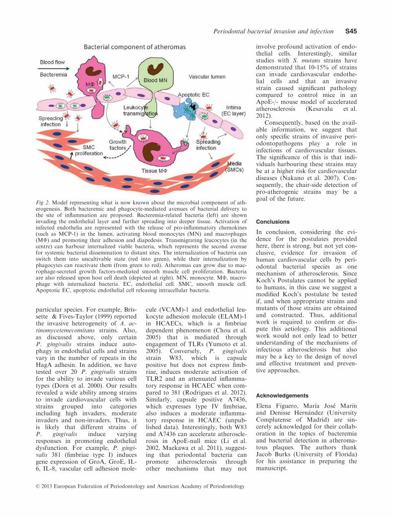
We present a model (Fig. 2) basedon what is known about the oralmicrobial component of atherogenesis.Both bacteremic and phagocyte-mediated avenues of bacterial deliv-ery to the site of inflammation areproposed. Bacteremia-derived bacte-ria invade the endothelial layer andfurther spread into deeper tissue(left). The activation of the infectedendothelia results in the release ofpro-inflammatory chemokines [suchas monocyte chemotactic protein(MCP-1)] in the lumen, resulting inactivation of blood monocytes (MN)and macrophages (MΦ) promotingtheir adhesion and diapedesis. Inaddition, transmigrated leucocytes(in the centre) can harbour internal-ized viable bacteria, which representsthe second avenue for systemic bac-terial dissemination to distant sites.The bacteria adhere to the endothe-lial cells, enter and usurp the endo-thelial cell processes for trafficking,at which point they may become un-cultivable (red into green). Theirinternalization by phagocytes orinteractions with uninfected cells canreactivate them (from green to red).Atheromas can grow due to macro-phage-secreted growth factors result-ing in smooth muscle cellproliferation. Bacteria are alsoreleased upon host cell death(depicted at right) to re-infect addi-tional cells.
Significance of strain
Throughout the microbial world,there are strain differences within aspecies and these differences some-times influence virulence. We pro-pose that such is the case forperiodontopathogenic species in rela-tion to cardiovascular diseases.There are multiple examples cited inthis review that illustrate pheno-typic differences among strains of a
© 2013 European Federation of Periodontology and American Academy of Periodontology
Periodontal bacterial invasion and infection S43

Table 6. Results of studies evaluating the presence of periodontal pathogens in cardiovascular samples, including atheromatous lesions
Reference Periodontal
exam
Subgingival
samples
Universal Aa Pg Td Tf Pi Fn Ec Cr
Haraszthy et al.
(2000b)
No No Specific
PCR
72% 18% 26% 30% 14%
Okuda et al.
(2001)
No No Specific
PCR
NR 0% 0% 23.10% 0% 0%
Stelzel et al.
(2002)
No No Specific
PCR
88.5% 0% 15.4%
Mastra
gelopulos et al.
(2002)
No No Specific
PCR
59% NR NR NR
Taylor-
Robinson et al.
(2002)
NR NR Specific
PCR
31.2% 21.9% 9.4%
Marques
da Silva et al.
(2003)
No No Specific
PCR
7.1% 0% 0%
Cairo et al.
(2004)
Yes Yes Specific
PCR
0% 0% 0% 0% 0%
Ishihara et al.
(2004)
Yes Yes Specific
PCR
23.3% 21.6% 23.5% 5.9% 15.7%
Kurihara et al.
(2004)
Yes Yes Specific
PCR
0% 85% 63% 22% 31% 45%
Marques
da Silva et al.
(2005)
No No Specific
PCR
89.2% 7.1% 0% 0% 0%
Kozarov et al.
(2005)
No No Cell-
culture
100% 100%
Fiehn et al.
(2005)
No No Nested
PCR
100% 0% 4.17% 0% 100% 0%
Padilla et al.
(2006)
Yes Yes Culture 16.67% 0% 0% 0% 0% 0% 0% 0%
Kozarov et al.
(2006)
No No Specific
PCR
40/55.5% 18.3/
88.8%
19.1/
33.3%
15/
22.2%
15/
77.7%
17.5/
22.2%
Zaremba et al.
(2007)
Yes Yes Hybrid-
ization
5.00% 50.0% 30.0% 25.0% 15.0% 20.0% 15.0% 20.0%
Aimetti et al.
(2007)
Yes Yes Nested
PCR
94% 0% 0% 0% 0% 0%
Romano et al.
(2007)
Yes Yes Hybrid-
ization
0% 0% 0% 0% 0%
Pucar et al.
(2007)
No No Specific
PCR
26.7% 53.3% 13.3% 33.3%
Zhang et al.
(2008)
Yes Yes Specific
PCR
0% 33% 31% 18% 12%
Elkaim et al.
(2008)
Yes Yes Hybrid-
ization
54.5% 54.5–72.7% 22.7% 27.3%
Gaetti-
Jardim et al.
(2009)
Yes No Real-time
PCR
46.2% 53.8% 25.6% 59% 0%
Nakano et al.
(2009)
No Yes Specific
PCR
30-35% 15-20% 15-20% 5%
Mahendra et al.
(2010)
Yes Yes Specific
PCR
45.1% 49.01% 21.5%
Figuero et al.
(2011)
Yes No Nested
PCR
66.7% 78.6% 61.9 50% 54.8% 9.5%
Aquino et al.
(2011)
Yes No Specific
PCR
13% 0% 0% 0%
Ohki et al.
(2012)
No No Specific
PCR
21% 3.7% 2.5% 0.0% 0.0%
NR, not reported; ChP, chronic periodontitis, Aa, A. actinomycetemcomitans; Pg, P. gingivalis; Td, T. denticola; Tf, T. forsythia; Pm, P. mi-cra; Pi, P. intermedia; Fn, F. nucleatum; Ec, E. corrodens; Cr, C. rectus.
© 2013 European Federation of Periodontology and American Academy of Periodontology
S44 Reyes et al.
particular species. For example, Bris-sette & Fives-Taylor (1999) reportedthe invasive heterogeneity of A. ac-tinomycetemecomitans strains. Also,as discussed above, only certainP. gingivalis strains induce auto-phagy in endothelial cells and strainsvary in the number of repeats in theHagA adhesin. In addition, we havetested over 20 P. gingivalis strainsfor the ability to invade various celltypes (Dorn et al. 2000). Our resultsrevealed a wide ability among strainsto invade cardiovascular cells withstrains grouped into categoriesincluding high invaders, moderateinvaders and non-invaders. Thus, itis likely that different strains ofP. gingivalis induce varyingresponses in promoting endothelialdysfunction. For example, P. gingi-valis 381 (fimbriae type I) inducesgene expression of GroA, GroE, IL-6, IL-8, vascular cell adhesion mole-
cule (VCAM)-1 and endothelial leu-kocyte adhesion molecule (ELAM)-1in HCAECs, which is a fimbriaedependent phenomenon (Chou et al.2005) that is mediated throughengagement of TLRs (Yumoto et al.2005). Conversely, P. gingivalisstrain W83, which is capsulepositive but does not express fimb-riae, induces moderate activation ofTLR2 and an attenuated inflamma-tory response in HCAEC when com-pared to 381 (Rodrigues et al. 2012).Similarly, capsule positive A7436,which expresses type IV fimbriae,also induces a moderate inflamma-tory response in HCAEC (unpub-lished data). Interestingly, both W83and A7436 can accelerate atheroscle-rosis in ApoE-null mice (Li et al.2002, Maekawa et al. 2011), suggest-ing that periodontal bacteria canpromote atherosclerosis throughother mechanisms that may not
involve profound activation of endo-thelial cells. Interestingly, similarstudies with S. mutans strains havedemonstrated that 10-15% of strainscan invade cardiovascular endothe-lial cells and that an invasivestrain caused significant pathologycompared to control mice in anApoE-/- mouse model of acceleratedatherosclerosis (Kesavalu et al.2012).
Consequently, based on the avail-able information, we suggest thatonly specific strains of invasive peri-odontopathogens play a role ininfections of cardiovascular tissues.The significance of this is that indi-viduals harbouring these strains maybe at a higher risk for cardiovasculardiseases (Nakano et al. 2007). Con-sequently, the chair-side detection ofpro-atherogenic strains may be agoal of the future.
Conclusions
In conclusion, considering the evi-dence for the postulates providedhere, there is strong, but not yet con-clusive, evidence for invasion ofhuman cardiovascular cells by peri-odontal bacterial species as onemechanism of atherosclerosis. SinceKoch’s Postulates cannot be appliedto humans, in this case we suggest amodified Koch’s postulate be testedif, and when appropriate strains andmutants of those strains are obtainedand constructed. Thus, additionalwork is required to confirm or dis-pute this aetiology. This additionalwork would not only lead to betterunderstanding of the mechanisms ofinfectious atherosclerosis but alsomay be a key to the design of noveland effective treatment and preven-tive approaches.
Acknowledgements
Elena Figuero, Mar�ıa Jos�e Mar�ınand Denisse Hern�andez (UniversityComplutense of Madrid) are sin-cerely acknowledged for their collab-oration in the topics of bacteremiaand bacterial detection in atheroma-tous plaques. The authors thankJacob Burks (University of Florida)for his assistance in preparing themanuscript.
Fig 2. Model representing what is now known about the microbial component of ath-erogenesis. Both bacteremic and phagocyte-mediated avenues of bacterial delivery tothe site of inflammation are proposed. Bacteremia-related bacteria (left) are showninvading the endothelial layer and further spreading into deeper tissue. Activation ofinfected endothelia are represented with the release of pro-inflammatory chemokines(such as MCP-1) in the lumen, activating blood monocytes (MN) and macrophages(MΦ) and promoting their adhesion and diapedesis. Transmigrating leucocytes (in thecentre) can harbour internalized viable bacteria, which represents the second avenuefor systemic bacterial dissemination to distant sites. The internalization of bacteria canswitch them into uncultivable state (red into green), while their internalization byphagocytes can reactivate them (from green to red). Atheromas can grow due to mac-rophage-secreted growth factors-mediated smooth muscle cell proliferation. Bacteriaare also released upon host cell death (depicted at right). MN, monocyte. MΦ, macro-phage with internalized bacteria. EC, endothelial cell. SMC, smooth muscle cell.Apoptotic EC, apoptotic endothelial cell releasing intracellular bacteria.
© 2013 European Federation of Periodontology and American Academy of Periodontology
Periodontal bacterial invasion and infection S45

References
Abranches, J., Zeng, L., Belanger, M., Rodrigues,P. H., Simpson-Haidaris, P. J., Akin, D.,Dunn, W. A. Jr, Progulske-Fox, A. & Burne,R. A. (2009) Invasion of human coronaryartery endothelial cells by Streptococcus mutansOMZ175. Oral Microbiology and Immunology24, 141–145.
Aimetti, M., Romano, F. & Nessi, F. (2007) Mi-crobiologic analysis of periodontal pockets andcarotid atheromatous plaques in advancedchronic periodontitis patients. Journal of Peri-odontology 78, 1718–1723.
Amano, A. (2010) Bacterial adhesins to host com-ponents in periodontitis. Periodontology 200052, 12–37.
Amar, S., Wu, S. C. & Madan, M. (2009) IsPorphyromonas gingivalis cell invasion requiredfor atherogenesis? Pharmacotherapeuticimplications. Journal of Immunology 182,1584–1592.
Anand, P. K., Tait, S. W., Lamkanfi, M., Amer,A. O., Nunez, G., Green, D. R. & Kanneganti,T. D. (2011) TLR2 and RIP2 pathways medi-ate autophagy of Listeria monocytogenes viaERK activation. Journal of Biological Chemis-try 286, 42981–42991.
Aquino, A. R., Lima, K. C., Paiva, M. S., Rocas,I. N. & Siqueira, J. F. Jr (2011) Molecular sur-vey of atheromatous plaques for the presenceof DNA from periodontal bacterial pathogens,archaea and fungi. Journal of PeriodontalResearch 46, 303–309.
Arce, R. M., Diaz, P. I., Barros, S. P., Galloway,P., Bobetsis, Y., Threadgill, D. & Offenbacher,S. (2010) Characterization of the invasive andinflammatory traits of oral Campylobacter rec-tus in a murine model of fetoplacental growthrestriction and in trophoblast cultures. Journalof Reproductive Immunology 84, 145–153.
Arirachakaran, P., Apinhasmit, W., Paungmalit, P.,Jeramethakul, P., Rerkyen, P. & Mahanonda, R.(2012) Infection of human gingival fibroblastswith Aggregatibacter actinomycetemcomitans: anin vitro study. Archives of Oral Biology 57,964–972.
Aruni, A. W., Roy, F. & Fletcher, H. M. (2011)Filifactor alocis has virulence attributes thatcan enhance its persistence under oxidativestress conditions and mediate invasion of epi-thelial cells by Porphyromonas gingivalis. Infec-tion and Immunity 79, 3872–3886.
Asakawa, R., Komatsuzawa, H., Kawai, T., Ya-mada, S., Goncalves, R. B., Izumi, S., Fujiw-ara, T., Nakano, Y., Suzuki, N., Uchida, Y.,Ouhara, K., Shiba, H., Taubman, M. A., Kuri-hara, H. & Sugai, M. (2003) Outer membraneprotein 100, a versatile virulence factor of Acti-nobacillus actinomycetemcomitans. MolecularMicrobiology 50, 1125–1139.
Asi, K. S., Gill, A. S. & Mahajan, S. (2010) Post-operative bacteremia in periodontal flap sur-gery, with and without prophylactic antibioticadministration: a comparative study. Journal ofIndian Society Periodontology 14, 18–22.
Bahrani-Mougeot, F. K., Paster, B. J., Coleman,S., Ashar, J., Barbuto, S. & Lockhart, P. B.(2008) Diverse and novel oral bacterial speciesin blood following dental procedures. Journalof Clinical Microbiology 46, 2129–2132.
Bartruff, J. B., Yukna, R. A. & Layman, D. L.(2005) Outer membrane vesicles from Por-phyromonas gingivalis affect the growth andfunction of cultured human gingival fibroblastsand umbilical vein endothelial cells. Journal ofPeriodontology 76, 972–979.
Belanger, M., Kozarov, E., Song, H., Whitlock, J.& Progulske-Fox, A. (2012) Both the uniqueand repeat regions of the Porphyromonas gingi-valis hemagglutin A are involved in adhesionand invasion of host cells. Anaerobe 18, 128–134.
Bernales, S., McDonald, K. L. & Walter, P.(2006) Autophagy counterbalances endoplasmicreticulum expansion during the unfolded pro-tein response. PLoS Biology 4, e423.
Braunersreuther, V., Mach, F. & Steffens, S.(2007) The specific role of chemokines in ath-erosclerosis. Thromombosis and Haemostasis 97,714–721.
Brennan, M. T., Kent, M. L., Fox, P. C., Norton,H. J. & Lockhart, P. B. (2007) The impact oforal disease and nonsurgical treatment on bac-teremia in children. Journal of the AmericanDental Association 138, 80–85.
Brissette, C. A. & Fives-Taylor, P. M. (1999)Actinobacillus actinomycetemcomitans may uti-lize either actin-dependent or actin-independentmechanisms of invasion. Oral Microbiology andImmunology 14, 137–142.
Brodala, N., Merricks, E. P., Bellinger, D. A.,Damrongsri, D., Offenbacher, S., Beck, J.,Madianos, P., Sotres, D., Chang, Y. L., Koch,G. & Nichols, T. C. (2005) Porphyromonasgingivalis bacteremia induces coronary and aor-tic atherosclerosis in normocholesterolemicand hypercholesterolemic pigs. Arteriosclerosis,Thrombosis, and Vascular Biology 25, 1446–1451.
Buhlin, K., Hultin, M., Norderyd, O., Persson,L., Pockley, A. G., Rabe, P., Klinge, B. &Gustafsson, A. (2009) Risk factors for athero-sclerosis in cases with severe periodontitis.Journal of Clinical Periodontology 36, 541–549.
Cairo, F., Gaeta, C., Dorigo, W., Oggioni, M. R.,Pratesi, C., Pini Prato, G. P. & Pozzi, G.(2004) Periodontal pathogens in atheromatousplaques. A controlled clinical and laboratorytrial. Journal of Periodontal Research 39, 442–446.
Cao, S. L., Progulske-Fox, A., Hillman, J. D. &Handfield, M. (2004) In vivo induced antigenicdeterminants of Actinobacillus actinomycetem-comitans. FEMS Microbiology Letters 237,97–103.
Carrion, J., Scisci, E., Miles, B., Sabino, G. J.,Zeituni, A. E., Gu, Y., Bear, A., Genco, C. A.,Brown, D. L. & Cutler, C. W. (2012) Microbialcarriage state of peripheral blood dendritic cells(DCs) in chronic periodontitis influences DCdifferentiation, atherogenic potential. Journalof Immunology 189, 3178–3187.
Castillo, D. M., Sanchez-Beltran, M. C., Castell-anos, J. E., Sanz, I., Mayorga-Fayad, I., Sanz,M. & Lafaurie, G. I. (2011) Detection of spe-cific periodontal microorganisms from bactera-emia samples after periodontal therapy usingmolecular-based diagnostics. Journal of ClinicalPeriodontology 38, 418–427.
Chalmers, N. I., Palmer, R. J.. Jr, Cisar, J. O. &Kolenbrander, P. E. (2008) Characterization ofa Streptococcus sp.-Veillonella sp. communitymicromanipulated from dental plaque. Journalof Bacteriology 190, 8145–8154.
Chavakis, T., Wiechmann, K., Preissner, K. T. &Herrmann, M. (2005) Staphylococcus aureusinteractions with the endothelium: the role ofbacterial “secretable expanded repertoire adhe-sive molecules” (SERAM) in disturbing hostdefense systems. Thrombosis and Haemostasis94, 278–285.
Cherry, M., Daly, C. G., Mitchell, D. & High-field, J. (2007) Effect of rinsing with povidone-
iodine on bacteraemia due to scaling: a ran-domized-controlled trial. Journal of ClinicalPeriodontology 34, 148–155.
Chou, H. H., Yumoto, H., Davey, M., Takahash-i, Y., Miyamoto, T., Gibson, F. C. 3rd & Gen-co, C. A. (2005) Porphyromonas gingivalisfimbria-dependent activation of inflammatorygenes in human aortic endothelial cells. Infec-tion and Immunity 73, 5367–5378.
Christersson, L. A., Albini, B., Zambon, J. J., Wi-kesjo, U. M. & Genco, R. J. (1987a) Tissuelocalization of Actinobacillus actinomycetem-comitans in human periodontitis. I. Light,immunofluorescence and electron microscopicstudies. Journal of Periodontology 58, 529–539.
Christersson, L. A., Wikesjo, U. M., Albini, B.,Zambon, J. J. & Genco, R. J. (1987b) Tissuelocalization of Actinobacillus actinomycetem-comitans in human periodontitis. II. Correla-tion between immunofluorescence and culturetechniques. Journal of Periodontology 58,540–545.
Colombo, A. V., da Silva, C. M., Haffajee, A. &Colombo, A. P. (2007) Identification of intra-cellular oral species within human crevicularepithelial cells from subjects with chronic peri-odontitis by fluorescence in situ hybridization.Journal of Periodontal Research 42, 236–243.
Colombo, A. V., Silva, C. M., Haffajee, A. & Co-lombo, A. P. (2006) Identification of oral bac-teria associated with crevicular epithelial cellsfrom chronic periodontitis lesions. Journal ofMedical Microbiology 55, 609–615.
Cortelli, J. R., Cogo, K., Aquino, D. R., Cortelli,S. C., Ricci-Nittel, D., Zhang, P. & Araujo, M.W. (2012) Validation of the anti-bacteremicefficacy of an essential oil rinse in a Brazilianpopulation: a cross-over study. Brazilian OralResearch 26, 478–484.
Crasta, K., Daly, C. G., Mitchell, D., Curtis, B.,Stewart, D. & Heitz-Mayfield, L. J. (2009) Bac-teraemia due to dental flossing. Journal of Clin-ical Periodontology 36, 323–332.
Darenfed, H., Grenier, D. & Mayrand, D. (1999)Acquisition of plasmin activity by Fusobacteri-um nucleatum subsp. nucleatum and potentialcontribution to tissue destruction during peri-odontitis. Infection and Immunity 67, 6439–6444.
Deniset, J. F. & Pierce, G. N. (2010) Possibilitiesfor therapeutic interventions in disruptingChlamydophila pneumoniae involvement inatherosclerosis. Fundamental & Clinical Phar-macology 24, 607–617.
Deretic, V. (2011) Autophagy in immunity andcell-autonomous defense against intracellularmicrobes. Immunological Reviews 240, 92–104.
Deretic, V. & Levine, B. (2009) Autophagy,immunity, and microbial adaptations. Cell Host& Microbe 5, 527–549.
Deshpande, R. G., Khan, M. B. & Genco, C. A.(1998) Invasion of aortic and heart endothelialcells by Porphyromonas gingivalis. Infection andImmunity 66, 5337–5343.
DiRienzo, J. M., Song, M., Wan, L. S. & Ellen,R. P. (2002) Kinetics of KB and HEp-2 cellresponses to an invasive, cytolethal distendingtoxin-producing strain of Actinobacillus actino-mycetemcomitans. Oral Microbiology andImmunology 17, 245–251.
Diz Dios, P., Tomas Carmona, I., Limeres Posse,J., Medina Henriquez, J., Fernandez Feijoo, J.& Alvarez Fernandez, M. (2006) Comparativeefficacies of amoxicillin, clindamycin, andmoxifloxacin in prevention of bacteremia fol-lowing dental extractions. Antimicrobial Agentsand Chemotherapy 50, 2996–3002.
© 2013 European Federation of Periodontology and American Academy of Periodontology
S46 Reyes et al.

Dolgilevich, S., Rafferty, B., Luchinskaya, D. &Kozarov, E. (2011) Genomic comparison ofinvasive and rare non-invasive strains revealsPorphyromonas gingivalis genetic polymor-phisms. Journal of Oral Microbiology 3, 5764–5774.
Dorn, B. R., Burks, J. N., Seifert, K. N. & Prog-ulske-Fox, A. (2000) Invasion of endothelialand epithelial cells by strains of Porphyromon-as gingivalis. FEMS Microbiology Letters 187,139–144.
Dorn, B. R., Dunn, W. A. Jr & Progulske-Fox,A. (1999) Invasion of human coronary arterycells by periodontal pathogens. Infection andImmunity 67, 5792–5798.
Dorn, B. R., Dunn, W. A. Jr & Progulske-Fox,A. (2001) Porphyromonas gingivalis traffics toautophagosomes in human coronary arteryendothelial cells. Infection and Immunity 69,5698–5708.
Dorn, B. R., Dunn, W. A. Jr & Progulske-Fox,A. (2002a) Bacterial interactions with the auto-phagic pathway. Cellular Microbiology 4, 1–10.
Dorn, B. R., Harris, L. J., Wujick, C. T., Vert-ucci, F. J. & Progulske-Fox, A. (2002b) Inva-sion of vascular cells in vitro byPorphyromonas endodontalis. International En-dododontic Journal 35, 366–371.
Dorn, B. R., Leung, K. & Progulske-Fox, A.(1998) Invasion of human oral epithelial cellsby Prevotella intermedia. Infection and Immu-nity 66, 6054–6057.
Edwards, A. M., Grossman, T. J. & Rudney, J.D. (2006) Fusobacterium nucleatum transportsnoninvasive Streptococcus cristatus into humanepithelial cells. Infection and Immunity 74, 654–662.
Eick, S., Reissmann, A., Rodel, J., Schmidt, K.H. & Pfister, W. (2006) Porphyromonas gingiva-lis survives within KB cells and modulatesinflammatory response. Oral Microbiology andImmunology 21 (4), 231–237.
Elkaim, R., Dahan, M., Kocgozlu, L., Werner,S., Kanter, D., Kretz, J. G. & Tenenbaum, H.(2008) Prevalence of periodontal pathogens insubgingival lesions, atherosclerotic plaques andhealthy blood vessels: a preliminary study.Journal of Periodontal Research 43, 224–231.
Epstein, S. E., Zhu, J., Najafi, A. H. & Burnett,M. S. (2009) Insights into the role of infectionin atherogenesis and in plaque rupture. Circula-tion 119, 3133–3141.
Fedele, S., Sabbah, W., Donos, N., Porter, S. &D’Aiuto, F. (2011) Common oral mucosal dis-eases, systemic inflammation, and cardiovascu-lar diseases in a large cross-sectional USsurvey. American Heart Journal 161, 344–350.
Fiehn, N. E., Larsen, T., Christiansen, N., Holm-strup, P. & Schroeder, T. V. (2005) Identifica-tion of periodontal pathogens in atheroscleroticvessels. Journal of Periodontology 76, 731–736.
Figuero, E., Sanchez-Beltran, M., Cuesta-Frecho-so, S., Tejerina, J. M., del Castro, J. A., Gut-ierrez, J. M., Herrera, D. & Sanz, M. (2011)Detection of periodontal bacteria in atheroma-tous plaque by nested polymerase chain reac-tion. Journal of Periodontology 82, 1469–1477.
Fine, D. H., Furgang, D., McKiernan, M., Teres-ki-Bischio, D., Ricci-Nittel, D., Zhang, P. &Araujo, M. W. (2010) An investigation of theeffect of an essential oil mouthrinse on inducedbacteraemia: a pilot study. Journal of ClinicalPeriodontology 37, 840–847.
Fitzpatrick, R. E., Wijeyewickrema, L. C. & Pike,R. N. (2009) The gingipains: scissors and glueof the periodontal pathogen, Porphyromonasgingivalis. Future Microbiology 4, 471–487.
Fives-Taylor, P. M., Meyer, D. H., Mintz, K. P.& Brissette, C. (1999) Virulence factors of Acti-nobacillus actinomycetemcomitans. Periodontol-ogy 2000 (20), 136–167.
Forner, L., Larsen, T., Kilian, M. & Holmstrup,P. (2006) Incidence of bacteremia after chew-ing, tooth brushing and scaling in individualswith periodontal inflammation. Journal of Clin-ical Periodontology 33, 401–407.
Gaetti-Jardim, E. Jr, Marcelino, S. L., Feitosa, A.C., Romito, G. A. & Avila-Campos, M. J.(2009) Quantitative detection of periodonto-pathic bacteria in atherosclerotic plaques fromcoronary arteries. Journal of Medical Microbi-ology 58, 1568–1575.
Gibson, F. C. 3rd, Hong, C., Chou, H. H., Yum-oto, H., Chen, J., Lien, E., Wong, J. & Genco,C. A. (2004) Innate immune recognition ofinvasive bacteria accelerates atherosclerosis inapolipoprotein E-deficient mice. Circulation109, 2801–2806.
Graves, D. T., Fine, D., Teng, Y. T., Van Dyke,T. E. & Hajishengallis, G. (2008) The use ofrodent models to investigate host-bacteriainteractions related to periodontal diseases.Journal of Clinical Periodontology 35, 89–105.
Hajishengallis, G., Wang, M., Harokopakis, E.,Triantafilou, M. & Triantafilou, K. (2006) Por-phyromonas gingivalis fimbriae proactively mod-ulate beta2 integrin adhesive activity andpromote binding to and internalization by mac-rophages. Infection and Immunity 74, 5658–5666.
Hajishengallis, G., Wang, M., Liang, S., Triantafi-lou, M. & Triantafilou, K. (2008) Pathogeninduction of CXCR4/TLR2 cross-talk impairshost defense function. Proceedings of theNational Academy of Science USA 105, 13532–13537.
Han, Y. W., Shi, W., Huang, G. T., Kinder Ha-ake, S., Park, N. H., Kuramitsu, H. & Genco,R. J. (2000) Interactions between periodontalbacteria and human oral epithelial cells: Fuso-bacterium nucleatum adheres to and invadesepithelial cells. Infection and Immunity 68, 3140–3146.
Haraszthy, V. I., Hariharan, G., Tinoco, E. M.,Cortelli, J. R., Lally, E. T., Davis, E. & Zam-bon, J. J. (2000a) Evidence for the role ofhighly leukotoxic Actinobacillus actinomycetem-comitans in the pathogenesis of localizedjuvenile and other forms of early-onset peri-odontitis. Journal of Periodontology 71,912–922.
Haraszthy, V. I., Zambon, J. J., Trevisan, M.,Zeid, M. & Genco, R. J. (2000b) Identificationof periodontal pathogens in atheromatous pla-ques. Journal of Periodontal Research 71, 1554–1560.
Harokopakis, E., Albzreh, M. H., Martin, M. H.& Hajishengallis, G. (2006) TLR2 transmodu-lates monocyte adhesion and transmigrationvia Rac1- and PI3K-mediated inside-out signal-ing in response to Porphyromonas gingivalis fi-mbriae. Journal of Immunology 176, 7645–7656.
Heimdahl, A., Hall, G., Hedberg, M., Sandberg,H., Soder, P. O., Tuner, K. & Nord, C. E.(1990) Detection and quantitation by lysis-fil-tration of bacteremia after different oral surgi-cal procedures. Journal of Clinical Microbiology28, 2205–2209.
Hertzen, E., Johansson, L., Wallin, R., Schmidt,H., Kroll, M., Rehn, A. P., Kotb, M., Morg-elin, M. & Norrby-Teglund, A. (2010) M1 pro-tein-dependent intracellular traffickingpromotes persistence and replication of Strepto-coccus pyogenes in macrophages. Journal ofInnate Immunity 2, 534–545.
Higashi, Y., Goto, C., Hidaka, T., Soga, J.,Nakamura, S., Fujii, Y., Hata, T., Idei, N.,Fujimura, N., Chayama, K., Kihara, Y. &Taguchi, A. (2009) Oral infection-inflammatorypathway, periodontitis, is a risk factor forendothelial dysfunction in patients with coro-nary artery disease. Atherosclerosis 206, 604–610.
Hutchinson, N. A., Shanson, D. C. & Malins, T.(1992a) Clinical comparison of Sentinel, anovel blood culture system, with radiometricBactec 460 and Isolator 10 in the detection ofstreptococcal and anaerobic bacteraemias.Journal of Clinical Pathology 45, 475–478.
Hutchinson, N. A., Thomas, F. D. & Shanson,D. C. (1992b) The clinical comparison of Ox-oid Signal with Bactec blood culture systemsfor the detection of streptococcal and anaerobicbacteraemias. Journal of Medical Microbiology37, 410–412.
Hybiske, K. & Stephens, R. S. (2007) Mechanismsof host cell exit by the intracellular bacteriumChlamydia. Proceedings of the Natoinal Acad-emy of Science USA 104, 11430–11435.
Ikegami, A., Chung, P. & Han, Y. W. (2009)Complementation of the fadA mutation inFusobacterium nucleatum demonstrates that thesurface-exposed adhesin promotes cellular inva-sion and placental colonization. Infection andImmunity 77, 3075–3079.
Inagaki, S., Onishi, S., Kuramitsu, H. K. & Shar-ma, A. (2006) Porphyromonas gingivalis vesiclesenhance attachment, and the leucine-rich repeatBspA protein is required for invasion of epithe-lial cells by Tannerella forsythia. Infection andImmunity 74, 5023–5028.
Ishihara, K., Nabuchi, A., Ito, R., Miyachi, K.,Kuramitsu, H. K. & Okuda, K. (2004) Correla-tion between detection rates of periodontopathicbacterial DNA in coronary stenotic artery pla-que [corrected] and in dental plaque samples.Journal of Clinical Microbiology 42, 1313–1315.
Iyer, D., Anaya-Bergman, C., Jones, K., Yanam-andra, S., Sengupta, D., Miyazaki, H. & Lewis,J. P. (2010) AdpC is a Prevotella intermedia 17leucine-rich repeat internalin-like protein. Infec-tion and Immunity 78, 2385–2396.
Jain, A., Batista, E. L. Jr, Serhan, C., Stahl, G.L. & Van Dyke, T. E. (2003) Role for peri-odontitis in the progression of lipid depositionin an animal model. Infection and Immunity 71,6012–6018.
Ji, S., Shin, J. E., Kim, Y. C. & Choi, Y. (2010)Intracellular degradation of Fusobacterium nu-cleatum in human gingival epithelial cells. Mol-ecules and Cells 30, 519–526.
Johnson, J. D., Chen, R., Lenton, P. A., Zhang,G., Hinrichs, J. E. & Rudney, J. D. (2008) Per-sistence of extracrevicular bacterial reservoirsafter treatment of aggressive periodontitis.Journal of Periodontology 79, 2305–2312.
Katz, J., Chaushu, G. & Sharabi, Y. (2001) Onthe association between hypercholesterolemia,cardiovascular disease and severe periodontaldisease. Journal of Clinical Periodontology 28,865–868.
Kebschull, M., Demmer, R. T. & Papapanou, P.N. (2010) “Gum bug, leave my heart alone!”–epidemiologic and mechanistic evidence linkingperiodontal infections and atherosclerosis.Journal of Dental Research 89, 879–902.
Keizer, H. G. (2012) The “Mevalonate hypothe-sis”: a cholesterol-independent alternative forthe etiology of atherosclerosis. Lipids in Healthand Disease 11, 149.
Kesavalu, L., Lucas, A. R., Verma, R. K., Liu,L., Dai, E., Sampson, E. & Progulske-Fox, A.
© 2013 European Federation of Periodontology and American Academy of Periodontology
Periodontal bacterial invasion and infection S47
(2012) Increased atherogenesis during Strepto-coccus mutans infection in ApoE-null mice.Journal of Dental Research 91, 255–260.
Khan, M. J., Rizwan Alam, M., Waldeck-We-iermair, M., Karsten, F., Groschner, L., Rie-derer, M., Hallstrom, S., Rockenfeller, P.,Konya, V., Heinemann, A., Madeo, F., Graier,W. F. & Malli, R. (2012) Inhibition of Auto-phagy Rescues Palmitic Acid-induced Necrop-tosis of Endothelial Cells. Journal of BiologicalChemistry 287, 21110–21120.
Kinane, D. F., Riggio, M. P., Walker, K. F.,MacKenzie, D. & Shearer, B. (2005) Bactera-emia following periodontal procedures. Journalof Clinical Periodontology 32, 708–713.
Kinane, J. A., Benakanakere, M. R., Zhao, J.,Hosur, K. B. & Kinane, D. F. (2012) Porphyro-monas gingivalis influences actin degradationwithin epithelial cells during invasion andapoptosis. Cellular Microbiology 14, 1085–1096.
Kirschbaum, M., Schultze-Mosgau, S., Pfister, W.& Eick, S. (2010) Mixture of periodontopatho-genic bacteria influences interaction with KBcells. Anaerobe 16, 461–468.
Kolattukudy, P. E. & Niu, J. (2012) Inflamma-tion, endoplasmic reticulum stress, autophagy,and the monocyte chemoattractant protein-1/CCR2 pathway. Circulation Research 110, 174–189.
Komatsuzawa, H., Asakawa, R., Kawai, T., Ochi-ai, K., Fujiwara, T., Taubman, M. A., Ohara,M., Kurihara, H. & Sugai, M. (2002) Identifi-cation of six major outer membrane proteinsfrom Actinobacillus actinomycetemcomitans.Gene 288, 195–201.
Koren, O., Spor, A., Felin, J., Fak, F., Stomb-augh, J., Tremaroli, V., Behre, C. J., Knight,R., Fagerberg, B., Ley, R. E. & Backhed, F.(2011) Human oral, gut, and plaque microbiotain patients with atherosclerosis. Proceedings ofthe National Academy of Sciences of the UnitedStates of America 108, 4592–4598.
Kozarov, E., Dorn, B., Shelburne, C., Dunn, W.& Progulske-Fox, A. (2005) Human atheroscle-rotic plaque contains viable invasive Porphyro-monas gingivalis and Actinobacillus actinomycetemcomitans. Arteriosclerosis, Thrombosis,and Vascular Biology 25, e17–e18.
Kozarov, E., Sweier, D., Shelburne, C., Prog-ulske-Fox, A. & Lopatin, D. (2006) Detectionof bacterial DNA in atheromatous plaques byquantitative PCR. Microbes and infection/Insti-tut Pasteur 8, 687–693.
Kozarov, E., Whitlock, J., Dong, H., Carrasco,E. & Progulske-Fox, A. (1998) The number ofdirect repeats in hagA is variable among Por-phyromonas gingivalis strains. Infection andImmunity 66, 4721–4725.
Kurihara, N., Inoue, Y., Iwai, T., Umeda, M.,Huang, Y. & Ishikawa, I. (2004) Detection andlocalization of periodontopathic bacteria inabdominal aortic aneurysms. European Journalof Vascular and Endovascular Surgery 28,553–558.
Lafaurie, G. I., Mayorga-Fayad, I., Torres, M.F., Castillo, D. M., Aya, M. R., Baron, A. &Hurtado, P. A. (2007) Periodontopathic micro-organisms in peripheric blood after scaling androot planing. Journal of Clinical Periodontology34, 873–879.
Lalla, E., Lamster, I. B., Hofmann, M. A., Bucc-iarelli, L., Jerud, A. P., Tucker, S., Lu, Y., Pa-papanou, P. N. & Schmidt, A. M. (2003) Oralinfection with a periodontal pathogen acceler-ates early atherosclerosis in apolipoprotein E-null mice. Arteriosclerosis, Thrombosis, andVascular Biology 23, 1405–1411.
Larocca, T. J., Henson, G. D., Thorburn, A., Sin-dler, A. L., Pierce, G. L. & Seals, D. R. (2012)Translational evidence that impaired autophagycontributes to arterial ageing. Journal of Physi-ology 590, 3305–3316.
Leivadaros, E., van der Velden, U., Bizzarro, S.,ten Heggeler, J. M., Gerdes, V. E., Hoek, F. J.,Nagy, T. O., Scholma, J., Bakker, S. J., Gans,R. O., ten Cate, H. & Loos, B. G. (2005) Apilot study into measurements of markers ofatherosclerosis in periodontitis. Journal of Peri-odontology 76, 121–128.
Lepine, G., Caudry, S., DiRienzo, J. M. & Ellen,R. P. (1998) Epithelial cell invasion by Actino-bacillus actinomycetemcomitans strains fromrestriction fragment-length polymorphismgroups associated with juvenile periodontitis orcarrier status. Oral Microbiology and Immunol-ogy 13, 341–347.
Leung, N. M., Chen, R. & Rudney, J. D. (2006)Oral bacteria in plaque and invading buccalcells of young orthodontic patients. AmericanJournal of Orthodontics and Dentofacial Ortho-pedics 130 (698), e611–e698.
Li, L., Matevski, D., Aspiras, M., Ellen, R. P. &Lepine, G. (2004) Two epithelial cell invasion-related loci of the oral pathogen Actinobacillusactinomycetemcomitans. Oral Microbiologyogyand Immunology 19, 16–25.
Li, L., Messas, E., Batista, E. L. Jr, Levine, R. A. &Amar, S. (2002) Porphyromonas gingivalis infec-tion accelerates the progression of atherosclerosisin a heterozygous apolipoprotein E-deficient mur-ine model. Circulation 105, 861–867.
Li, L., Michel, R., Cohen, J., DeCarlo, A. &Kozarov, E. (2008) Intracellular survival andvascular cell-to-cell transmission of Por-phyromonas gingivalis. BMC Microbiology 8,26–36.
Liang, S., Krauss, J. L., Domon, H., McIntosh,M. L., Hosur, K. B., Qu, H., Li, F., Tzekou,A., Lambris, J. D. & Hajishengallis, G.(2011) The C5a receptor impairs IL-12-depen-dent clearance of Porphyromonas gingivalisand is required for induction of periodon-tal bone loss. Journal of Immunology 186, 869–877.
Liao, X., Sluimer, J. C., Wang, Y., Subramanian,M., Brown, K., Pattison, J. S., Robbins, J., Mar-tinez, J. & Tabas, I. (2012) Macrophage auto-phagy plays a protective role in advancedatherosclerosis. Cellular Metabolism 15, 545–553.
Libby, P., Ridker, P. M. & Maseri, A. (2002)Inflammation and atherosclerosis. Circulation105, 1135–1143.
Lockhart, P. B., Brennan, M. T., Sasser, H. C.,Fox, P. C., Paster, B. J. & Bahrani-Mougeot,F. K. (2008) Bacteremia associated with tooth-brushing and dental extraction. Circulation 117,3118–3125.
Lockhart, P. B., Brennan, M. T., Thornhill, M.,Michalowicz, B. S., Noll, J., Bahrani-Mougeot,F. K. & Sasser, H. C. (2009) Poor oral hygieneas a risk factor for infective endocarditis-related bacteremia. Journal of the AmericanDental Association 140, 1238–1244.
Lucas, V. S., Gafan, G., Dewhurst, S. & Roberts,G. J. (2008) Prevalence, intensity and nature ofbacteraemia after toothbrushing. Journal ofDentistry 36, 481–487.
Lucas, V. S., Lytra, V., Hassan, T., Tatham, H.,Wilson, M. & Roberts, G. J. (2002) Compari-son of lysis filtration and an automated bloodculture system (BACTEC) for detection, quan-tification, and identification of odontogenicbacteremia in children. Journal of ClinicalMicrobiology 40, 3416–3420.
Lux, R., Miller, J. N., Park, N. H. & Shi, W.(2001) Motility and chemotaxis in tissue pene-tration of oral epithelial cell layers by Trepo-nema denticola. Infection and Immunity 69,6276–6283.
Maeda, T., Maeda, H., Yamabe, K., Mineshiba,J., Tanimoto, I., Yamamoto, T., Naruishi,K., Kokeguchi, S. & Takashiba, S. (2010)Highly expressed genes in a rough-colony-forming phenotype of Aggregatibacter actino-mycetemcomitans: implication of a mip-likegene for the invasion of host tissue. FEMSImmunology and Medical Microbiology 58,226–236.
Maekawa, T., Takahashi, N., Tabeta, K., Aoki,Y., Miyashita, H., Miyauchi, S., Miyazawa,H., Nakajima, T. & Yamazaki, K. (2011)Chronic oral infection with Porphyromonas gin-givalis accelerates atheroma formation by shift-ing the lipid profile. PLoS ONE 6, e20240.
Mahendra, J., Mahendra, L., Kurian, V. M., Jai-shankar, K. & Mythilli, R. (2010) 16S rRNA-based detection of oral pathogens in coronaryatherosclerotic plaque. Indian Journal of DentalResearch 21, 248–252.
Marcelino, S. L., Gaetti-Jardim, E. Jr, Nakano,V., Canonico, L. A., Nunes, F. D., Lotufo, R.F., Pustiglioni, F. E., Romito, G. A. & Avila-Campos, M. J. (2010) Presence of periodonto-pathic bacteria in coronary arteries frompatients with chronic periodontitis. Anaerobe16, 629–632.
Marques da Silva, R., Caugant, D. A., Lingaas,P. S., Geiran, O., Tronstad, L. & Olsen, I.(2005) Detection of Actinobacillus actinomyce-temcomitans but not bacteria of the red com-plex in aortic aneurysms by multiplexpolymerase chain reaction. Journal of Periodon-tology 76, 590–594.
Marques da Silva, R., Lingaas, P. S., Geiran, O.,Tronstad, L. & Olsen, I. (2003) Multiple bacte-ria in aortic aneurysms. Journal of VascularSurgery 38, 1384–1389.
Martinet, W. & De Meyer, G. R. (2008) Auto-phagy in atherosclerosis. Current Atherosclero-sis Reports 10, 216–223.
Mastragelopulos, N., Haraszthy, V. I., Zambon,J. J. & Zafiropoulos, G. G. (2002) Detection ofperiodontal pathogenic microorganisms in ath-eromatous plaque. Preliminary results. DerChirurg; Zeitschrift fur alle Gebiete der operati-ven Medizen 73, 585–591.
Messini, M., Skourti, I., Markopulos, E., Kou-tsia-Carouzou, C., Kyriakopoulou, E., Kostaki,S., Lambraki, D. & Georgopoulos, A. (1999)Bacteremia after dental treatment in mentallyhandicapped people. Journal of Clinical Peri-odontology 26, 469–473.
Meyer, D. H. & Fives-Taylor, P. M. (1997) Therole of Actinobacillus actinomycetemcomitansin the pathogenesis of periodontal disease.Trends in Microbiology 5, 224–228.
Meyer, D. H., Lippmann, J. E. & Fives-Taylor,P. M. (1996) Invasion of epithelial cells byActinobacillus actinomycetemcomitans: adynamic, multistep process. Infection andImmunity 64, 2988–2997.
Meyer, D. H., Sreenivasan, P. K. & Fives-Taylor,P. M. (1991) Evidence for invasion of a humanoral cell line by Actinobacillus actinomycetem-comitans. Infection and Immunity 59, 2719–2726.
Moffatt, C. E., Inaba, H., Hirano, T. & Lamont,R. J. (2012) Porphyromonas gingivalis SerB-mediated dephosphorylation of host cell cofilinmodulates invasion efficiency. Cellular Microbi-ology 14 (4), 577–588.
© 2013 European Federation of Periodontology and American Academy of Periodontology
S48 Reyes et al.
Molmeret, M., Alli, O. A., Zink, S., Flieger, A., Ci-anciotto, N. P. & Kwaik, Y. A. (2002) icmT isessential for pore formation-mediated egress ofLegionella pneumophila from mammalian andprotozoan cells. Infection and Immunity 70, 69–78.
Murphy, A. M., Daly, C. G., Mitchell, D. H.,Stewart, D. & Curtis, B. H. (2006) Chewingfails to induce oral bacteraemia in patients withperiodontal disease. Journal of Clinical Peri-odontology 33, 730–736.
Nakano, K., Hokamura, K., Taniguchi, N.,Wada, K., Kudo, C., Nomura, R., Kojima, A.,Naka, S., Muranaka, Y., Thura, M., Nakaj-ima, A., Masuda, K., Nakagawa, I., Speziale,P., Shimada, N., Amano, A., Kamisaki, Y.,Tanaka, T., Umemura, K. & Ooshima, T.(2011) The collagen-binding protein of Strepto-coccus mutans is involved in haemorrhagicstroke. Nature Communications 2, 485.
Nakano, K., Inaba, H., Nomura, R., Nemoto,H., Takeuchi, H., Yoshioka, H., Toda, K.,Taniguchi, K., Amano, A. & Ooshima, T.(2008) Distribution of Porphyromonas gingivalisfimA genotypes in cardiovascular specimensfrom Japanese patients. Oral Microbiology andImmunology 23, 170–172.
Nakano, K., Nemoto, H., Nomura, R., Homma,H., Yoshioka, H., Shudo, Y., Hata, H., Toda,K., Taniguchi, K., Amano, A. & Ooshima, T.(2007) Serotype distribution of Streptococcusmutans a pathogen of dental caries in cardio-vascular specimens from Japanese patients.Journal of Medical Microbiology 56, 551–556.
Nakano, K., Nemoto, H., Nomura, R., Inaba,H., Yoshioka, H., Taniguchi, K., Amano, A. &Ooshima, T. (2009) Detection of oral bacteriain cardiovascular specimens. Oral Microbiologyand Immunology 24, 64–68.
Nobbs, A. H., Lamont, R. J. & Jenkinson, H. F.(2009) Streptococcus adherence and coloniza-tion. Microbiology and Molecular BiologyReviews 73, 407–450.
Noiri, Y., Ozaki, K., Nakae, H., Matsuo, T. &Ebisu, S. (1997) An immunohistochemicalstudy on the localization of Porphyromonas gin-givalis, Campylobacter rectus and Actinomycesviscosus in human periodontal pockets. Journalof Periodontal Research 32, 598–607.
Ohki, T., Itabashi, Y., Kohno, T., Yoshizawa, A.,Nishikubo, S., Watanabe, S., Yamane, G. &Ishihara, K. (2012) Detection of periodontalbacteria in thrombi of patients with acute myo-cardial infarction by polymerase chain reaction.American Heart Journal 163, 164–167.
Okuda, K., Ishihara, K., Nakagawa, T., Hiray-ama, A. & Inayama, Y. (2001) Detection ofTreponema denticola in atherosclerotic lesions.Journal of Clinical Microbiology 39, 1114–1117.
Olczak, T., Maszczak-Seneczko, D., Smalley, J.W. & Olczak, M. (2012) Gallium(III), cobalt(III) and copper(II) protoporphyrin IX exhibitantimicrobial activity against Porphyromonasgingivalis by reducing planktonic and biofilmgrowth and invasion of host epithelial cells.Archives of Microbiology 194, 719–724.
Oscarsson, J., Karched, M., Thay, B., Chen, C. &Asikainen, S. (2008) Proinflammatory effect inwhole blood by free soluble bacterial compo-nents released from planktonic and biofilmcells. BMC Microbiology 8, 206.
Ott, S. J., El Mokhtari, N. E., Musfeldt, M., Hell-mig, S., Freitag, S., Rehman, A., Kuhbacher,T., Nikolaus, S., Namsolleck, P., Blaut, M.,Hampe, J., Sahly, H., Reinecke, A., Haake, N.,Gunther, R., Kruger, D., Lins, M., Herrmann,G., Folsch, U. R., Simon, R. & Schreiber, S.(2006) Detection of diverse bacterial signatures
in atherosclerotic lesions of patients with coro-nary heart disease. Circulation 113, 929–937.
Padilla, C., Lobos, O., Hubert, E., Gonzalez, C.,Matus, S., Pereira, M., Hasbun, S. & Descouvi-eres, C. (2006) Periodontal pathogens in ather-omatous plaques isolated from patients withchronic periodontitis. Journal of PeriodontalResearch 41, 350–353.
Pan, N. J., Brady, M. J., Leong, J. M. & Goguen,J. D. (2009) Targeting type III secretion in Yer-sinia pestis. Antimicrobial Agents and Chemo-therapy 53, 385–392.
Perez-Chaparro, P. J., Gracieux, P., Lafaurie, G.I., Donnio, P. Y. & Bonnaure-Mallet, M.(2008) Genotypic characterization of Porphyro-monas gingivalis isolated from subgingival pla-que and blood sample in positive bacteremiasubjects with periodontitis. Journal of ClinicalPeriodontology 35, 748–753.
Peters, S. R., Valdez, M., Riviere, G. & Thomas,D. D. (1999) Adherence to and penetrationthrough endothelial cells by oral treponemes.Oral Microbiologyogy and Immunology 14, 379–383.
Pineiro, A., Tomas, I., Blanco, J., Alvarez, M.,Seoane, J. & Diz, P. (2010) Bacteraemia follow-ing dental implants’ placement. Clinical OralImplants Research 21, 913–918.
Pirro, M., Bagaglia, F., Paoletti, L., Razzi, R. &Mannarino, M. R. (2008) Hypercholesterol-emia-associated endothelial progenitor cell dys-function. Therapeutic Advances inCardiovascular Disease 2, 329–339.
Pober, J. S., Min, W. & Bradley, J. R. (2009)Mechanisms of endothelial dysfunction, injury,and death. Annual Reviews Pathology: Mecha-nisms of Disease 4, 71–95.
Pollreisz, A., Huang, Y., Roth, G. A., Cheng, B.,Kebschull, M., Papapanou, P. N., Schmidt, A.M. & Lalla, E. (2010) Enhanced monocytemigration and pro-inflammatory cytokine pro-duction by Porphyromonas gingivalis infection.Journal of Periodontal Research 45, 239–245.
Popovic, M., Smiljanic, K., Dobutovic, B., Syro-vets, T., Simmet, T. & Isenovic, E. R. (2012)Human cytomegalovirus infection and athero-thrombosis. Journal of Thrombosis and Throm-bolysis 33, 160–172.
Prochnau, D., Lehmann, M., Straube, E., Figulla,H. R. & Rodel, J. (2011) Human cytomegalovi-rus induces MMP-1 and MMP-3 expression inaortic smooth muscle cells. Acta Microbiologicaet Immunologica Hungarica 58, 303–317.
Pucar, A., Milasin, J., Lekovic, V., Vukadinovic,M., Ristic, M., Putnik, S. & Kenney, E. B.(2007) Correlation between atherosclerosis andperiodontal putative pathogenic bacterial infec-tions in coronary and internal mammary arter-ies. Journal of Periodontology 78, 677–682.
Rafferty, B., J€onsson, D., Kalachikov, S., Dem-mer, R. T., Nowygrod, R., Elkind, M. S.,Bush, H. Jr & Kozarov, E. (2011) Impact ofmonocytic cells on recovery of uncultivablebacteria from atherosclerotic lesions. Journal ofInternal Medicine 270, 273–280.
Rajasuo, A., Perkki, K., Nyfors, S., Jousimies-Somer, H. & Meurman, J. H. (2004) Bactere-mia following surgical dental extraction withan emphasis on anaerobic strains. Journal ofDental Research 83, 170–174.
Raman, K., Chong, M., Akhtar-Danesh, G. G.,D’Mello, M., Hasso, R., Ross, S., Xu, F. &Pare, G.(2013) Genetic markers of inflamma-tion and their role in cardiovascular disease.Canadian Journal of Cardiology 29, 67–74.
Razani, B., Feng, C., Coleman, T., Emanuel, R.,Wen, H., Hwang, S., Ting, J. P., Virgin, H.
W., Kastan, M. B. & Semenkovich, C. F.(2012) Autophagy links inflammasomes to ath-erosclerotic progression. Cell Metabolism 15,534–544.
Ridker, P. M. (2002) On evolutionary biology,inflammation, infection, and the causes of ath-erosclerosis. Circulation 105, 2–4.
Rodrigues, P. H. & Progulske-Fox, A. (2005)Gene expression profile analysis of Porphyro-monas gingivalis during invasion of human cor-onary artery endothelial cells. Infection andImmunity 73, 6169–6173.
Rodrigues, P. H., Reyes, L., Chadda, A. S.,B�elanger, M., Wallet, S. M., Akin, D., Dunn,W. Jr & Progulske-Fox, A. (2012) Porphyro-monas gingivalis strain specific interactions withhuman coronary artery endothelial cells: acomparative study. PLoS ONE 7 (12), e52606.
Romano, F., Barbui, A. & Aimetti, M. (2007)Periodontal pathogens in periodontal pocketsand in carotid atheromatous plaques. MinervaStomatologica 56, 169–179.
Roth, G. A., Ankersmit, H. J., Brown, V. B., Pa-papanou, P. N., Schmidt, A. M. & Lalla, E.(2007a) Porphyromonas gingivalis infection andcell death in human aortic endothelial cells.FEMS Microbiology Letters 272, 106–113.
Roth, G. A., Moser, B., Huang, S. J., Brandt, J.S., Huang, Y., Papapanou, P. N., Schmidt, A.M. & Lalla, E. (2006) Infection with a peri-odontal pathogen induces procoagulant effectsin human aortic endothelial cells. Journal ofThrombosis and Haemostasis 4, 2256–2261.
Roth, G. A., Moser, B., Roth-Walter, F., Giac-ona, M. B., Harja, E., Papapanou, P. N.,Schmidt, A. M. & Lalla, E. (2007b) Infectionwith a periodontal pathogen increases mononu-clear cell adhesion to human aortic endothelialcells. Atherosclerosis 190, 271–281.
Ross, R. (1999) Atherosclerosis – an inflamma-tory disease. The New England Journal of Medi-cine 340, 115–126.
Rudney, J. D., Chen, R. & Sedgewick, G. J.(2001) Intracellular Actinobacillus actinomyce-temcomitans and Porphyromonas gingivalis inbuccal epithelial cells collected from humansubjects. Infection and Immunity 69, 2700–2707.
Rudney, J. D., Chen, R. & Sedgewick, G. J.(2005) Actinobacillus actinomycetemcomitans,Porphyromonas gingivalis, and Tannerella for-sythensis are components of a polymicrobialintracellular flora within human buccal cells.Journal of Dental Research 84, 59–63.
Sabet, M., Lee, S. W., Nauman, R. K., Sims, T.& Um, H. S. (2003) The surface (S-) layer is avirulence factor of Bacteroides forsythus.Microbiology 149, 3617–3627.
Saglie, F. R., Smith, C. T., Newman, M. G., Car-ranza, F. A. Jr, Pertuiset, J. H., Cheng, L.,Auil, E. & Nisengard, R. J. (1986) The pres-ence of bacteria in the oral epithelium in peri-odontal disease. II. Immunohistochemicalidentification of bacteria. Journal of Periodon-tology 57, 492–500.
Saito, A., Inagaki, S., Kimizuka, R., Okuda, K.,Hosaka, Y., Nakagawa, T. & Ishihara, K.(2008) Fusobacterium nucleatum enhances inva-sion of human gingival epithelial and aorticendothelial cells by Porphyromonas gingivalis.FEMS Immunology & Medical Microbiology54, 349–355.
Sakakibara, J., Nagano, K., Murakami, Y., Higu-chi, N., Nakamura, H., Shimozato, K. & Yo-shimura, F. (2007) Loss of adherence ability tohuman gingival epithelial cells in S-layer pro-tein-deficient mutants of Tannerella forsythen-sis. Microbiology 153, 866–876.
© 2013 European Federation of Periodontology and American Academy of Periodontology
Periodontal bacterial invasion and infection S49

Schenkein, H. A., Barbour, S. E., Berry, C. R.,Kipps, B. & Tew, J. G. (2000) Invasion ofhuman vascular endothelial cells by Actinobac-illus actinomycetemcomitans via the receptor forplatelet-activating factor. Infection and Immu-nity 68, 5416–5419.
Scherz-Shouval, R. & Elazar, Z. (2007) ROS,mitochondria and the regulation of autophagy.Trends in Cell Biology 17, 422–427.
Schrijvers, D. M., De Meyer, G. R. & Martinet,W. (2011) Autophagy in atherosclerosis: apotential drug target for plaque stabilization.Arteriosclerosis, Thrombosis, and Vascular Biol-ogy 31, 2787–2791.
Shin, D. M., Yuk, J. M., Lee, H. M., Lee, S.H., Son, J. W., Harding, C. V., Kim, J. M.,Modlin, R. L. & Jo, E. K. (2010) Mycobacte-rial lipoprotein activates autophagy viaTLR2/1/CD14 and a functional vitamin Dreceptor signalling. Cell Microbiology 12,1648–1665.
Silver, J. G., Martin, A. W. & McBride, B. C.(1977) Experimental transient bacteraemias inhuman subjects with varying degrees of plaqueaccumulation and gingival inflammation. Jour-nal of Clinical Periodontology 4, 92–99.
Silver, J. G., Martin, A. W. & McBride, B. C.(1979) Experimental transient bacteraemias inhuman subjects with clinically healthy gingivae.Journal of Clinical Periodontology 6, 33–36.
Song, H., Belanger, M., Whitlock, J., Kozarov, E.& Progulske-Fox, A. (2005) Hemagglutinin Bis involved in the adherence of Porphyromonasgingivalis to human coronary artery endothelialcells. Infection and Immunity 73, 7267–7273.
Sreenivasan, P. K., Meyer, D. H. & Fives-Taylor,P. M. (1993) Requirements for invasion of epi-thelial cells by Actinobacillus actinomycetem-comitans. Infection and Immunity 61, 1239–1245.
Stelzel, M., Conrads, G., Pankuweit, S., Maisch,B., Vogt, S., Moosdorf, R. & Flores-de-Jacoby,L. (2002) Detection of Porphyromonas gingiva-lis DNA in aortic tissue by PCR. Journal ofPeriodontology 73, 868–870.
Suwannakul, S., Stafford, G. P., Whawell, S. A.& Douglas, C. W. (2010) Identification of bi-stable populations of Porphyromonas gingivalisthat differ in epithelial cell invasion. Microbiol-ogy 156. Pt 10, 3052–3064.
Takahashi, Y., Davey, M., Yumoto, H., Gibson,F. C. 3rd & Genco, C. A. (2006) Fimbria-dependent activation of pro-inflammatory mol-ecules in Porphyromonas gingivalis infectedhuman aortic endothelial cells. Cellular Micro-biology 8, 738–757.
Takeuchi, H., Furuta, N., Morisaki, I. & Amano,A. (2011) Exit of intracellular Porphyromonasgingivalis from gingival epithelial cells is medi-
ated by endocytic recycling pathway. CellularMicrobiology 13, 677–691.
Taylor-Robinson, D., Aduse-Opoku, J., Sayed, P.,Slaney, J. M., Thomas, B. J. & Curtis, M. A.(2002) Oro-dental bacteria in various athero-sclerotic arteries. European Journal of ClinicalMicrobiology & Infectious Diseases 21, 755–757.
Tomas, I., Alvarez, M., Limeres, J., Potel, C.,Medina, J. & Diz, P. (2007a) Prevalence, dura-tion and aetiology of bacteraemia followingdental extractions. Oral Diseases 13, 56–62.
Tomas, I., Alvarez, M., Limeres, J., Tomas, M.,Medina, J., Otero, J. L. & Diz, P. (2007b)Effect of a chlorhexidine mouthwash on therisk of postextraction bacteremia. InfectionControl and Hospital Epidemiology 28, 577–582.
Tomas, I., Diz, P., Tobias, A., Scully, C. & Do-nos, N. (2012) Periodontal health status andbacteraemia from daily oral activities: system-atic review/meta-analysis. Journal of ClinicalPeriodontology 39, 213–228.
Tomas, I., Pereira, F., Llucian, R., Poveda, R.,Diz, P. & Bagan, J. V. (2008) Prevalence ofbacteraemia following third molar surgery.Oral Diseases 14, 89–94.
Tribble, G. D. & Lamont, R. J. (2010) Bacterialinvasion of epithelial cells and spreading in peri-odontal tissue. Periodontology 2000 52, 68–83.
Vita, J. A. & Loscalzo, J. (2002) Shouldering therisk factor burden: infection, atherosclerosis,and the vascular endothelium. Circulation 106,164–166.
Wan, M. & Hajishengallis, G. (2008) Lipid raft-dependent uptake, signalling and intracellularfate of Porphyromonas gingivalis in mouse mac-rophages. Cellular Microbiology 10, 2029–2042.
Wang, M., Liang, S., Hosur, K. B., Domon, H.,Yoshimura, F., Amano, A. & Hajishengallis,G. (2009) Differential virulence and innateimmune interactions of Type I and II fimbrialgenotypes of Porphyromonas gingivalis. OralMicrobiology and Immunology 24, 478–484.
Winkler, J. R., Matarese, V., Hoover, C. I., Kra-mer, R. H. & Murray, P. A. (1988) An in vitromodel to study bacterial invasion of periodon-tal tissues. Journal of Periodontology 59, 40–45.
Woollard, K. J. & Geissmann, F. (2010) Mono-cytes in atherosclerosis: subsets and functions.Nature Cardiology in Review 7, 77–86.
Wu, H., Lippmann, J. E., Oza, J. P., Zeng, M.,Fives-Taylor, P. & Reich, N. O. (2006) Inacti-vation of DNA adenine methyltransferasealters virulence factors in Actinobacillus actino-mycetemcomitans. Oral Microbiology andImmunology 21, 238–244.
Xie, Y., You, S. J., Zhang, Y. L., Han, Q., Cao,Y. J., Xu, X. S., Yang, Y. P., Li, J. & Liu, C.F. (2011) Protective role of autophagy in AGE-induced early injury of human vascular endo-
thelial cells. Molecular Medicine Reports 4, 459–464.
Yamatake, K., Maeda, M., Kadowaki, T., Takii,R., Tsukuba, T., Ueno, T., Kominami, E., Yo-kota, S. & Yamamoto, K. (2007) Role for gin-gipains in Porphyromonas gingivalis traffic tophagolysosomes and survival in human aorticendothelial cells. Infection and Immunity 75,2090–2100.
Yumoto, H., Chou, H. H., Takahashi, Y., Davey,M. & Gibson, F. C., 3rd, & Genco, C. A.(2005) Sensitization of human aortic endothe-lial cells to lipopolysaccharide via regulation ofToll-like receptor 4 by bacterial fimbria-depen-dent invasion. Infection and Immunity 73, 8050–8059.
Zaremba, M., Gorska, R., Suwalski, P. & Kowal-ski, J. (2007) Evaluation of the incidence ofperiodontitis-associated bacteria in the athero-sclerotic plaque of coronary blood vessels.Journal of Periodontology 78, 322–327.
Zeituni, A. E., Jotwani, R., Carrion, J. & Cutler,C. W. (2009) Targeting of DC-SIGN onhuman dendritic cells by minor fimbriated Por-phyromonas gingivalis strains elicits a distincteffector T cell response. Journal of Immunology183, 5694–5704.
Zeituni, A. E., McCaig, W., Scisci, E., Thanassi,D. G. & Cutler, C. W. (2010) The native 67-kilodalton minor fimbria of Porphyromonasgingivalis is a novel glycoprotein with DC-SIGN-targeting motifs. Journal of Bacteriology192, 4103–4110.
Zhang, W., Ju, J., Rigney, T. & Tribble, G. D.(2011) Fimbriae of Porphyromonas gingivalisare important for initial invasion of osteoblasts,but not for inhibition of their differentiationand mineralization. Journal of PeriodontalResearch 82, 909–916.
Zhang, Y. L., Cao, Y. J., You, S. J., Li, R. X.,Liu, H. H. & Liu, C. F. (2010a) Protectiveeffects of autophagy against oxidized LDL-induced injury in endothelial cells. Zhonghua YiXue Za Zhi 90, 2792–2796.
Zhang, Y. M., Zhong, L. J., Liang, P., Liu, H.,Mu, L. T. & Ai, S. K. (2008) Detection of peri-odontal pathogenic bacteria DNA in coronaryatheromatous plaques from patients underwentcoronary artery bypass graft. Zhonghua XinXue Guan Bing Za Zhi 36, 215–218.
Address:Ann Progulske-Fox, Department of OralBiology, Center for Molecular Microbiology,PO Box 100424 JHMHSC, University ofFlorida, Gainesville, FL 32610-0424, USAE-mail: [email protected]
Clinical Relevance
Scientific rationale for the study: Alink between cardiovascular dis-eases and periodontal diseases hasbeen observed in both epidemiolog-ical studies and in animal modelsof disease. This review was under-taken to evaluate the evidence thatperiodontal pathogen dissemination
contributes to the biological plausi-bility of the link.Principal findings: The evaluation ofthe seven “proofs” of evidence pro-posed in the review support the pos-sible role of periodontal pathogensin the association between periodon-tal and cardiovascular diseases.
Practical implications: These find-ings establish that additional exper-imentation is necessary todetermine if prevention and treat-ment of periodontal diseases woulddisrupt the link with cardiovasculardiseases.
© 2013 European Federation of Periodontology and American Academy of Periodontology
S50 Reyes et al.


