Embed Size (px)
Citation preview
1
Chemistry and Biology of Vision: Introduction
Krzysztof Palczewski
Department of Pharmacology, Case Western Reserve University, Cleveland, OH, 44106-4965.
Running Title: Chemistry of Vision.
Correspondence to: Krzysztof Palczewski, Ph.D., Department of Pharmacology, School of Medicine, Case Western Reserve University, 10900 Euclid Ave., Cleveland, Ohio 44106-4965, USA; Phone: 216-368-4631, Fax: 216-368-1300, E-mail: [email protected]
Keywords: retina, photoreceptors, rhodopsin, rods, cones, 11-cis-retinal, retinoid cycle, visual cycle
Visual perception in humans occurs through absorption of electromagnetic radiation from 400 to 780 nm by photoreceptors in the retina. A photon of visible light carries sufficient amount of energy to cause, when absorbed, a cis-trans geometric isomerization of the 11-cis-retinal chromophore, a vitamin A derivative bound to rhodopsin and cone opsins of retinal photoreceptors. The unique biochemistry of these complexes allows us to reliably and reproducibly collect continuous visual information about our environment. Moreover, other non-conventional retinal opsins such as the circadian rhythm regulator melanopsin also initiate light-activated signaling based on similar photochemistry.
Our visual system operates over an extremely broad dynamic range, detecting variations in light intensity of over eight orders of magnitude, from single photons to more than hundred million photons per second (1). This dynamic range is attributed to adaptation processes in rods and cones, and the remainder arising from pupil contractions, processes within inter-retinal neurons, and the production rate of visual chromophore. The rod cell saturates at several thousand photons/s, whereas cones continue to function at several million-fold higher light intensities (2). The central foundation of our vision is the photochemical isomerization of the vitamin A-derived visual chromophore (11-cis-retinal) from its cis to trans configuration. A single photon of light isomerizes a
single 11-cis-retinal bound to rod or cone opsins. A photon carries ~2.5 eV energy (at 500 nm), but only a fraction (1.5 eV/opsin molecule) is utilized to elicit changes in retinal conformation and subsequently protein conformational changes, whereas the remaining energy is dissipated. The high excess of energy ensures that photoisomerization occurs with high fidelity (3). To regenerate functional receptor after photoactivation, the chromophore must be regenerated metabolically through a series of enzymatic processes including isomerization and oxidation of all-trans-retinyl ester to 11-cis-retinal. The enzymatic re-isomerization of all-trans-retinoid to 11-cis-retinoid requires only 3-4 kcal/mol (or 0.13-0.17 eV/molecule) of energy (4).
The retina is a layered sensory organ containing all necessary functional and structural proteins to support human vision. How this remarkable tissue develops, and operates over such an incredible dynamic range, and how retinoids are recycled, are some of the most stirring questions in biology. Blindness is one of the most feared and debilitating illnesses affecting humans, and without a detailed understanding of the basic events in vision, rational approaches to treating blinding diseases will not be possible. With the current methodology, now is possible to identify all component of the retina and trace mutation to retinopathies.
The complete mouse transcriptome in eye and retina
http://www.jbc.org/cgi/doi/10.1074/jbc.R111.301150The latest version is at JBC Papers in Press. Published on November 10, 2011 as Manuscript R111.301150
Copyright 2011 by The American Society for Biochemistry and Molecular Biology, Inc.
by guest on January 5, 2019http://w
ww
.jbc.org/D
ownloaded from
2
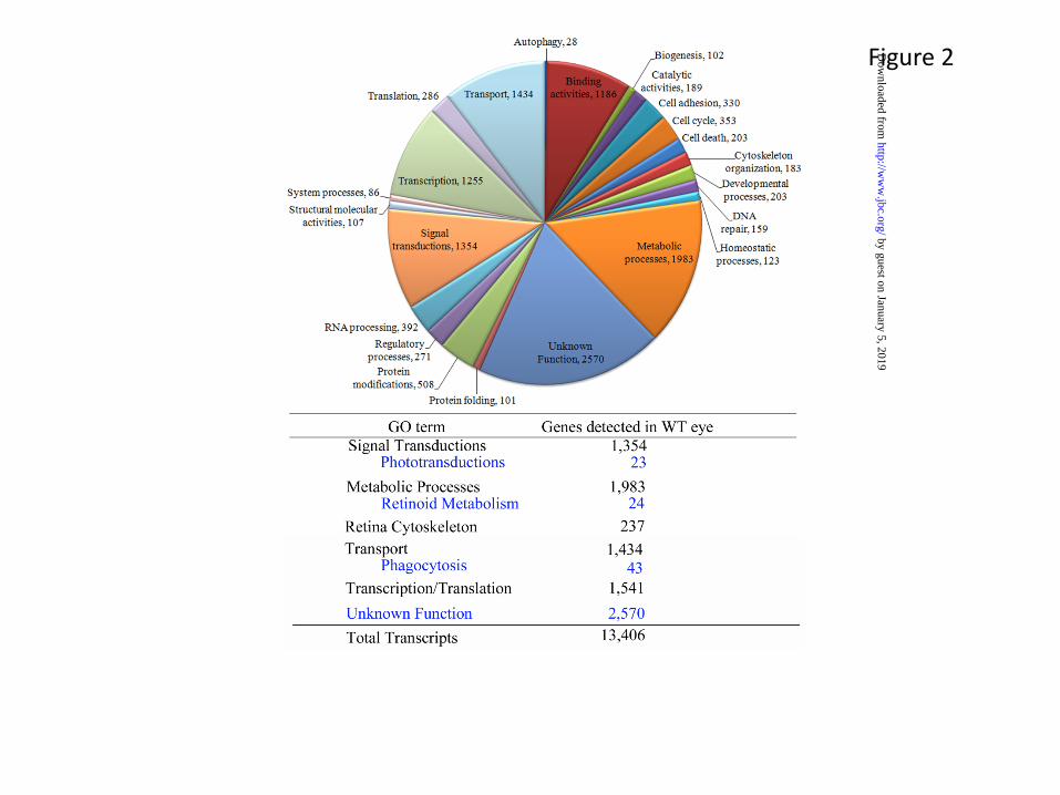
The eye is a complex organ composed of specific tissues that carry out different functions to maintain continuous visual responsiveness. The main players are the cornea and lens in the front, and the retina and retinal pigmented epithelium (RPE) in the back of the eye. The primary light absorption events take place within the retina, a 0.24 mm thick tissue composed of multiple cell layers (Fig. 1). Retinal development and maintenance, as well as light sensitive visual functions are highly regulated. Physical dissection of the different ocular tissues followed by global analysis of their gene expression by massively parallel RNA-sequencing (RNA-seq) have allowed for the assignment of a complete, comprehensive transcriptome of the ocular tissue (http://www.ncbi.nlm.nih.gov/projects/geo/query/acc.cgi?acc=GSE29752) (5). The completeness of such analysis is important prerequisite to understand the structure and physiology of the retinal by identifying all players involved. Using RNA-seq of mature wild-type (WT) mouse ocular tissues (the retina and whole eye), we recently determined the complete composition of these transcriptomes (5). Retinal tissue yielded 13,406 unique transcripts, and as expected, many transcripts from WT retinal tissues had annotated functions that could be linked to specific metabolic processes or structural and regulatory functions (Fig. 2A)(5). In addition, analysis of WT whole eye tissues revealed a large number of genes with unknown function that await further careful analysis (Fig. 2B). These studies complement and greatly expand earlier gene chip based expression analyses in both accuracy and quantification (6,7). This new depth of knowledge of the retinal transcriptome will facilitate large scale analyses of the functional consequences of manipulating photoreceptor gene expression (i.e. using gene transfer by retinal electroporation) (8). In addition to protein-coding mRNAs, a large number of micro-RNA (miR) and other non-coding RNAs are expressed in the eye (9,10). Together with many metabolites and dietary components, these RNAs regulate developmental and circadian control over the translation of proteins in each cell type. At least 78 miRs are preferentially expressed in the mouse retina from 689 miRs that have been identified (miRBASE Release 13.0, March 2009)) (11), suggesting the importance of miRs in modulating gene expression profiles in retinal cells. Moreover, inactivation of Dicer (an essential RNase III endonuclease required for miR maturation) leads to progressive functional and structural degeneration of the mouse retina (12).
Regulation of the miR levels could be an important approaches to treat human retinal disease as demonstrated in a mouse model of light-induced retinal degeneration (13).
Photoreceptor structure
Proteins involved in visual phototransduction are predominantly housed in the photoreceptor outer segments (OS) of rods and cones. Photoreceptor OS of these highly differentiated neurons are actually specialized cilia (Fig. 1A). Structural studies of these cilia were first carried out with guinea pig and frog rod outer segments (ROS) (14,15) and more recently mouse tissue has been favored because of the capability of genetic manipulation (5,16).
The average mouse ROS length and diameter was estimated to be 23.8 ± 1.0 μm, and 1.22 ± 0.12 μm to 1.32 ± 0.12 μm, respectively (17). A mouse ROS contains ~800 membranous disks stacked on top of each other (Fig. 1A). These internal disk membranes increase the total membrane surface area ~1,500-fold compared to the plasma membrane surface alone (18) allowing a high density of the rod visual pigment, rhodopsin. Cryo-electron tomography of vitrified mouse retina was employed to obtain reliable three-dimensional morphological information about this structure (19) (Fig. 1B). Fig. 1C presents a diagram of this ROS structure with distances between different membrane components obtained from cryo-electron tomograms. Based on these and the above-mentioned electron microscopy data, the ROS interior volume, including both the intradiskal and cytoplasmic space, is 32 × 10−12 ml and the cytoplasm occupies 10 × 10−12 ml in the ROS (19). Thus, it is amazing that the cytoplasmic space used for phototransduction represents only ~30% of the space inside a ROS, underscoring the importance of internal membrane structures in phototransduction. This phototransduction cascade occurs as catalytic processes on the interface of disk membranes and the cytoplasm (interfacial catalysis). Cryo-electron tomograms also showed spacers which keep the disks separate from one another and maintain appropriate distances between adjacent disks and the plasma membrane (19). Spacers consist of complexes of proteins with estimated molecular masses of ~500 kDa distributed at a mean density of ~500 molecules/μm2 throughout the disks (19). Intuitively,
by guest on January 5, 2019http://w
ww
.jbc.org/D
ownloaded from
3
the presence of proteins responsible for maintaining this structure could be predicted because structural components are essential for maintaining the architecture of these fluid complex internal membranes. The intervening spacers are likely occupied solely or in part by glutamic acid-rich proteins (GARP) and membrane bound retinal tetraspanin protein called peripherin/rds (20), forming those spacers.
Rhodopsin occupies ~50% of the membrane volume within the disks of ROS (21). This high density of the photoreceptor opsins could be needed to increase the probability of photon absorption. In addition, it appears that rhodopsin could play a critical structural role in establishing ROS morphology, as opsin knockout mice form only small ROS appendices early in life, before the cells degenerate (22). The size of the ROS is dictated by the expression level of rhodopsin (17), as heterozygote knockout mice for the opsin gene possess ~50% smaller ROS (23), and overexpression of this protein leads to rod cell degeneration (24). Rhodopsin is not uniformly distributed throughout disks (25). For example, cryo-electron microscopy images on vitrified unstained native mouse ROS reveals high-density regions on the disk surface. This difference in density could only arise from an uneven distribution of rhodopsin, which is the main protein in these disks, representing more than 90% of all disk proteins. Moreover, patches of disk membrane containing rows of rhodopsin dimers have been observed by atomic force microscopy (AFM), a finding supported by other biochemical methods as summarized previously (3). Para-crystalline patches within carefully isolated fresh disks from photoreceptors of mouse retina, wherein the building blocks consisted of rhodopsin dimers (26), imply functional significance in rhodopsin biosynthesis of function (27) and remains a topic of considerable interest (reviewed in (28)). Interestingly, Corless and colleagues found that crystalline structure is formed from visual pigments in cone cells when frog retinas are exposed to light (29).
Because both the mouse rhodopsin level (~520 pmol/eye) (16,17), and total cell number (6.4 × 10⁶ rods) (30) can be measured precisely, rhodopsin is calculated to have a concentration of 4.62 mM in disk membranes and 8.23 mM with respect to the ROS cytoplasm. The density of rhodopsin in the disk membrane is estimated to be 2.4 × 10⁴ molecules/μm2
on average or up to ∼3.4 × 10⁴ molecules/μm2 in high-density patches. AFM measurements yielded a density of 30,000–55,000 rhodopsin molecules /μm2 and ∼108 rhodopsin molecules per rod, partially organized in para-crystalline arrays (16,26). As the whole retinal transcriptome has now been analyzed and the majority of the ROS proteome identified by mass spectrometry, attention is now focused on the interactions of these proteins and their effects on function and their regulation. Structural studies are required to answer these questions.
Structures of phototransduction and visual cycle components Further molecular understanding of phototransduction inevitably focuses on the structures of phototransduction and retinoid cycle components and their complexes because the spatial organization of photoreceptor proteins underlies their functional ability to harvest light and generate a neuronal signal. Great progress has already been accomplished by defining the structure of a number of full length proteins or fragments, either alone or in complex with effector proteins (see (31)). A few interesting examples are listed below, but it is likely that more will be known in the near future about the structures of different components involved in this G protein-mediated process than about most other signal transduction systems in living nature.
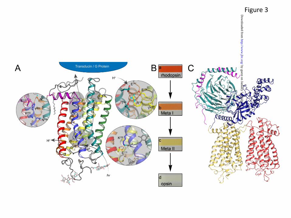
Structures of multiple forms of the G protein-coupled receptor (GPCR), rhodopsin (32-37)(Fig. 3), as well as various forms of G proteins (38-40), receptor capping protein arrestin (41), or likely G protein partners involved in intracellular translocation between photoreceptor compartments (42) have been determined (43). Rhodopsin has been extensively studied as a prototypical GPCR (3), and insights derived from comprehensive biochemical and biophysical studies of rhodopsin and its cognate G protein, transducin, have significantly improved our understanding of GPCR signaling in general (44).
An enhanced insight into the dynamics of rhodopsin activation and interaction with ligand and G protein has been obtained more recently by NMR techniques that show conformational flexibility of this receptor as well as the G protein upon activation (45-48). Several methods demonstrated that membrane proteins (49), including rhodopsin (50), contain
by guest on January 5, 2019http://w
ww
.jbc.org/D
ownloaded from
4
integral, ordered water molecules that play important roles in both structure and function. These water molecules could be key to the initial folding of these proteins as they insert into membranes, facilitating their assembly into functional entities as well as playing roles in the activation process. Using radiolytic footprinting techniques, we found that water molecules are associated with highly conserved and functionally important residues (50). In all sub-3Å resolution GPCR crystal structures determined to date, the observation of “conserved” waters in similar locations supports the notion that these waters are likely to be as important to receptor function as the conserved amino acid residues (32,37,51).
Key myristoylated Ca2+-binding proteins involved in phototransduction, namely guanylate cyclase-activating proteins or GCAPs (52), have been visualized at high-resolution by NMR and crystallographic methods to reveal their internal architecture, but only in their Ca2+-bound forms (53-55). More advanced studies have been performed on another myristoylated photoreceptor protein called recoverin. In recoverin Ca2+ induces the N-terminal extrusion of a myristoyl group that interacts with a lipid membrane bilayer (56,57). This transition, termed a calcium-myristoyl switch, could allow a protein to translocate from the cytoplasm to membranes in a calcium-dependent manner (56). In contrast, GCAP1 has its myristoylated group bound within a cavity formed by the polypeptide chain, but this does not exclude the possibility that this acyl group is mobilized in complexes with targeted guanylate cyclases.
Other important structures of phototransduction proteins include rhodopsin kinase (GRK1) (58), and regulator of G protein signaling 9 (RGS-9), the latter alone or in a complex with the activated α-subunit of the photoreceptor G protein, transducin, and/or an inhibitory subunit of phosphodiesterase 6 (59,60). These studies provide specific information about the termination of signal transduction on photoactivated rhodopsin and the activated G protein, transducin.
In addition to high resolution crystal structures, complementary methods have proven to be informative about complex proteins that are not yet amenable to crystallographic approaches. Among these methods are cryo-electron microscopy and single particle analysis. For example, single particle analysis and modeling provided the first views of phosphodiesterase organization (60,61) and of the
complex of dimeric rhodopsin and heterotrimeric transducin (62) (Fig. 3). However, many additional proteins whose atomic level structural details are critical to understanding the regulation and precise mechanism of phototransduction continue to escape structural interrogation.
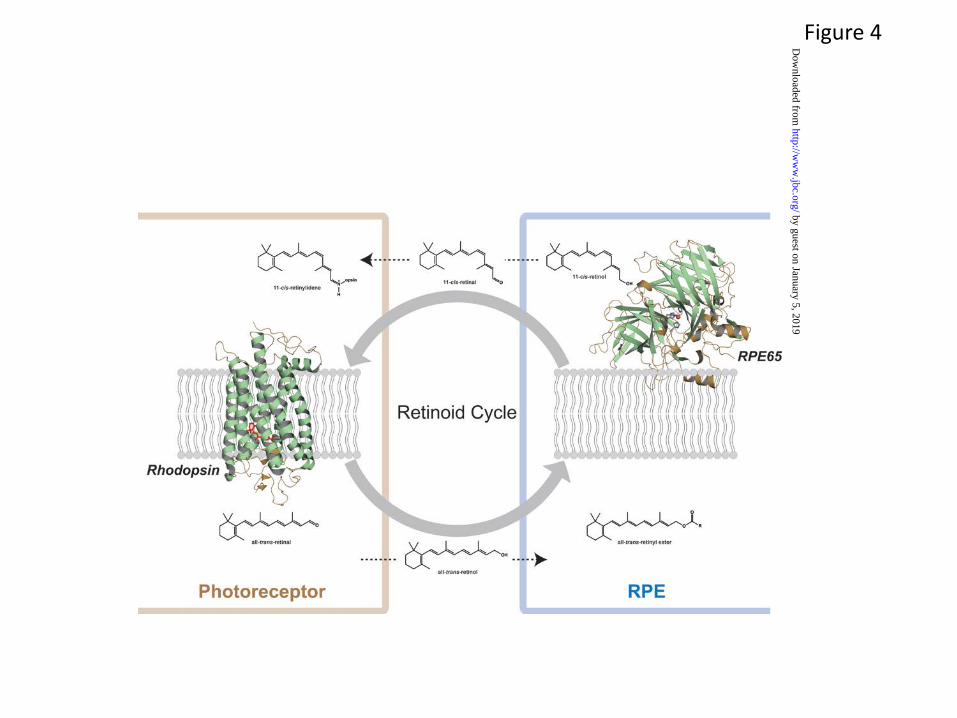
In addition to these functional receptors, enzymes, and structural proteins, the chemical transformation of retinoid metabolites, i.e. the retinoid cycle, is critical for proper visual function. The structure of retinoid isomerase RPE65, the key enzyme of this metabolic pathway, has been determined (63). This crystal structure reveals a seven-bladed β-propeller motif with single-strand extensions on blades VI and VII and a two-strand extension on blade III (Fig. 4). This crystal structure provided a basis for understanding RPE65 membrane binding and enzyme catalyzed retinoid isomerization. The structure of an important 11-cis-retinal binding-protein called cellular retinaldehyde-binding protein (CRALBP) has defined the hydrophobic core that is responsible for sequestering 11-cis-retinal (64). Additionally, the structure of the R234W mutant of CRALBP that is associated with Bothnia dystrophy and compromises visual pigment regeneration, identified the structural basis of that disease (4). Despite these advances, many questions remain with regard to the chemistry of the retinoid cycle.
Regenerating spent chromophore: the retinoid cycle For the retina to remain responsive to light and maintain vision, the 11-cis-retinal which is isomerized to all-trans must be continuously and efficiently regenerated (65). The time constant for rhodopsin regeneration is ~400 s and that for cone pigment regeneration is ~100 s (66). The pioneering work of Böll, Kühne and Wald laid the foundations for our current understanding of the photochemistry of vision (67-69). This process takes place in two cellular systems, retinal photoreceptors and the RPE (Fig. 4). From a chemical perspective, the enzymatic isomerization of the chromophore appears to be a formidable problem in regioselectivity. What regulates the specificity of the conversion of an all-trans-retinol to a specific 11-cis-isomer, when this molecule has only one functional group (-OH) and several possibilities for single or multiple-cis isomerizations?
by guest on January 5, 2019http://w
ww
.jbc.org/D
ownloaded from
5
This reaction also must occur continuously in a membranous/aqueous environment at body temperature. Moreover, the chromophore has other chemical properties that must be cleverly utilized. First, it contains five or six conjugated double bonds that allow light absorption in the visible range of the spectrum when conjugated with protein via a Schiff base. Second, as predicted by Pauling (70) because of the repulsion between two methyl groups, makes 11-cis-retinal an unstable isomer which encourages its isomerization to all-trans-retinal. Third, retinol easily forms one of the most stable carbocations in biology (71), allowing reshuffling of double bonds. And fourth, the isomerization of retinol has a relatively low activation energy (72). Three chemical mechanisms for isomerization of conjugated double bond poly-isoprenoids in biological systems have been identified: (a) a transition state carbocation product is formed from retinyl esters by alkyl cleavage; this carbocation then adjusts to an 11-cis-retinyl-like conformation to fit the active site of the enzyme and double bonds are reestablished when water is added (reviewed in (73)), (b) a specific double bond is saturated; the resulting transition state intermediate rotates and the double bond is reestablished by desaturation (74) as observed in tomato and Arabidopsis carotenoid isomerase (CRTISO) (75); and (c) an oxidative cleavage of carotenoids generates two retinal molecules in cis and trans forms as in the case of NinaB (76). The structural explanation of these disparate dioxygenase and isomerase activities is critical to understanding the molecular mechanisms employed by this class of enzymes.
Remarkable progress has increased our knowledge of the retinoid cycle, expanding the work so brilliantly started over a century ago (Fig. 4). Several extensive reviews provide a current update of this progress (4,65,66,73). While the cycle’s unique photochemistry maintains vision, a high flux of photons by light exposure can lead to elevated levels of toxic retinal metabolites that can accumulate throughout life and induce photoreceptor degeneration (77). Blocking the accumulation and action of these toxic intermediates and preventing such photoreceptor degeneration can alleviate major human visual diseases such as Stargardt disease and age-related macular degeneration (AMD).
As mentioned previously, the broad dynamic range of our vision also raises the intriguing question of how much chromophore is consumed during one’s
lifetime. This estimate requires several assumptions (see for example (78)), but high sensitivity of visual system, large Avogadro number and low molecular mass of the chromophore suggest that the realistic exposure to light would equate to consumption of ~ 1 mmol or only 284 mg of 11-cis-retinal during lifespan!!
While it is unclear why cell types other than photoreceptors are employed for chromophore regeneration per se, the adjacent RPE is vital for maintaining photoreceptor architecture and function. Thus, two cellular compartments are primarily associated with the retinoid cycle, the photoreceptor OS of rods and cones and the closely associated RPE (65) RPE cells are essential for the regeneration of chromophore in both rods and cones (79,80). Cones, in addition, appear to be supplemented with 11-cis-retinol by Müller cells (81,82).
The outflow of retinoids from photoreceptors to the RPE requires RPE-expressed lecithin:retinol acyl transferase (LRAT) which esterifies retinol with fatty acid to form retinyl esters (83) (Fig. 4). Because retinyl esters have propensity to self-aggregate, and they form oil droplets-like structure (84), called retinosomes in the RPE (85,86), the flow of retinol out of rods and cones to the RPE would be expected based on thermodynamic considerations. The flow of 11-cis-retinal back from the RPE to rods and cones is governed by diffusion facilitated by an opsin “sink”, i.e. the virtually irreversible reaction of opsins, especially rod opsin, with chromophore that reestablishes the protonated Schiff base (21). Chromophore undergoes cyclic regeneration for each absorbed photon that causes isomerization of visual pigments. But occasionally retinoids condense with lipids or between themselves to form harmful by-products of the retinoid cycle (87) that require photoreceptor cell regeneration.
Photoreceptor Renewal Rods and cones are extensively exposed to light in the presence of high oxygen levels throughout the life of an animal. This environment would inevitably lead to rapid retinal degeneration if this damaging process was not countered by protective biochemical mechanisms and continuous renewal of these cells. Photoreceptor OS are particularly vulnerable to damage as they contain highly reactive retinoids and high levels of unsaturated phospholipids such as
by guest on January 5, 2019http://w
ww
.jbc.org/D
ownloaded from
6
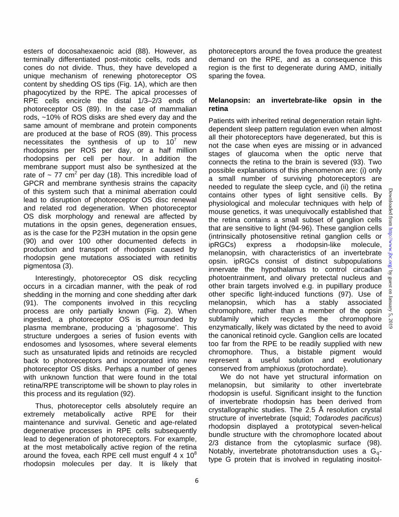
esters of docosahexaenoic acid (88). However, as terminally differentiated post-mitotic cells, rods and cones do not divide. Thus, they have developed a unique mechanism of renewing photoreceptor OS content by shedding OS tips (Fig. 1A), which are then phagocytized by the RPE. The apical processes of RPE cells encircle the distal 1/3–2/3 ends of photoreceptor OS (89). In the case of mammalian rods, ~10% of ROS disks are shed every day and the same amount of membrane and protein components are produced at the base of ROS (89). This process necessitates the synthesis of up to 107 new rhodopsins per ROS per day, or a half million rhodopsins per cell per hour. In addition the membrane support must also be synthesized at the rate of ~ 77 cm2 per day (18). This incredible load of GPCR and membrane synthesis strains the capacity of this system such that a minimal aberration could lead to disruption of photoreceptor OS disc renewal and related rod degeneration. When photoreceptor OS disk morphology and renewal are affected by mutations in the opsin genes, degeneration ensues, as is the case for the P23H mutation in the opsin gene (90) and over 100 other documented defects in production and transport of rhodopsin caused by rhodopsin gene mutations associated with retinitis pigmentosa (3).
Interestingly, photoreceptor OS disk recycling occurs in a circadian manner, with the peak of rod shedding in the morning and cone shedding after dark (91). The components involved in this recycling process are only partially known (Fig. 2). When ingested, a photoreceptor OS is surrounded by plasma membrane, producing a ‘phagosome’. This structure undergoes a series of fusion events with endosomes and lysosomes, where several elements such as unsaturated lipids and retinoids are recycled back to photoreceptors and incorporated into new photoreceptor OS disks. Perhaps a number of genes with unknown function that were found in the total retina/RPE transcriptome will be shown to play roles in this process and its regulation (92).
Thus, photoreceptor cells absolutely require an extremely metabolically active RPE for their maintenance and survival. Genetic and age-related degenerative processes in RPE cells subsequently lead to degeneration of photoreceptors. For example, at the most metabolically active region of the retina around the fovea, each RPE cell must engulf 4 x 10⁸ rhodopsin molecules per day. It is likely that
photoreceptors around the fovea produce the greatest demand on the RPE, and as a consequence this region is the first to degenerate during AMD, initially sparing the fovea.
Melanopsin: an invertebrate-like opsin in the retina Patients with inherited retinal degeneration retain light-dependent sleep pattern regulation even when almost all their photoreceptors have degenerated, but this is not the case when eyes are missing or in advanced stages of glaucoma when the optic nerve that connects the retina to the brain is severed (93). Two possible explanations of this phenomenon are: (i) only a small number of surviving photoreceptors are needed to regulate the sleep cycle, and (ii) the retina contains other types of light sensitive cells. By physiological and molecular techniques with help of mouse genetics, it was unequivocally established that the retina contains a small subset of ganglion cells that are sensitive to light (94-96). These ganglion cells (intrinsically photosensitive retinal ganglion cells or ipRGCs) express a rhodopsin-like molecule, melanopsin, with characteristics of an invertebrate opsin. ipRGCs consist of distinct subpopulations innervate the hypothalamus to control circadian photoentrainment, and olivary pretectal nucleus and other brain targets involved e.g. in pupillary produce other specific light-induced functions (97). Use of melanopsin, which has a stably associated chromophore, rather than a member of the opsin subfamily which recycles the chromophore enzymatically, likely was dictated by the need to avoid the canonical retinoid cycle. Ganglion cells are located too far from the RPE to be readily supplied with new chromophore. Thus, a bistable pigment would represent a useful solution and evolutionary conserved from amphioxus (protochordate). We do not have yet structural information on melanopsin, but similarity to other invertebrate rhodopsin is useful. Significant insight to the function of invertebrate rhodopsin has been derived from crystallographic studies. The 2.5 Å resolution crystal structure of invertebrate (squid; Todarodes pacificus) rhodopsin displayed a prototypical seven-helical bundle structure with the chromophore located about 2/3 distance from the cytoplasmic surface (98). Notably, invertebrate phototransduction uses a Gq-type G protein that is involved in regulating inositol-
by guest on January 5, 2019http://w
ww
.jbc.org/D
ownloaded from
7
1,4,5-trisphosphate production. In contrast to bovine rhodopsin, however, helices V and VI extend into the cytoplasmic medium and comprise part of the G-protein recognition surface. It is suggested that invertebrate rhodopsin can oscillate between cis- and trans-retinal conformations upon photon absorption by one of the forms (99). In physiological native membranes, invertebrate rhodopsin is organized in hexagonally packed microvillar membranes of photoreceptors, and in crystals it is tightly associated in a dimeric form (98).
Focus on vision In this series of reviews, we focus on various molecular aspects of vision. Arshavsky and Burns will provide an update on phototransduction and discuss trafficking of phototransduction proteins in rods and cones; von Lintig University will summarize current molecular insights into the chemistry and enzymology of the retinoid (visual) cycle and carotenoid metabolism; Kefalov will focus on phototransduction and the retinoid cycle, retinal neuron network and the visual cycle studied through pharmacological, genetic and physiological approaches; Van Gelder will
describe the atypical opsin called melanopsin expressed in intrinsically photosensitive retinal ganglion cells; and finally Cepko will concentrate on progress made on therapies that prolong vision in an inherited form of retinal degeneration. Extraordinary progress observed over the last two decades has allowed the development of multiple approaches targeted at blinding diseases. This marriage of basic and translational investigation is the highest standard of current progress in biology.
Acknowledgements
This research was supported by National Institutes of Health Grants EY009339. KP is John H. Hord Professor of Pharmacology. The author would like to thank Dr. Leslie T. Webster Jr., Dr. Andreas Engel, Vadim Arshavsky, Johannes von Lintig, Vladimir Kefalov, Russell Van Gelder, and the Palczewski laboratory for comments on the manuscript, Debarshi Mustafi for Figure 2, Dr. David Lodowski for Figure 3 and Dr. Philip Kiser for preparation of Figure 4.
by guest on January 5, 2019http://w
ww
.jbc.org/D
ownloaded from
8
Figure Legends Fig. 1. Structures of rod and cone outer segments and rod outer segment internal membranes. A. Neuronal organization of a typical mammalian retina. A cross-sectional representation of rod and cone photoreceptors is presented illustrating their connections to retinal pigmented epithelium distally and to relaying cells (bipolar, horizontal, amacrine, and ganglion cells) proximally. The rod structure has a longer outer segment with membrane-enclosed disks tightly packed without connections to the plasma membrane. Cone disks are continuously connected with the plasma membrane. Taken with permission from “Structure of cone photoreceptors” by Mustafi D, Engel AH, Palczewski K. Prog in Retina and Eye Res. 2009; 28(4):289-302 (Elsevier). B. Electron tomogram of vitrified ROS. The electron tomogram is represented in three orthogonal slices through the ROS volume. An x-y slice (right) and a y-z slice (left) display the high order and regular arrangement of stacked disks. Red color represents the high concentrations of rhodopsin found in disk membranes; spacer structures (pillars) are colored green. Bar, 200 nm. C. Blueprint of ROS. A schematic of a plasma membrane and two disks with measured distances between membrane components is shown. Green cylinders represent monomeric rhodopsin, which in native ROS forms a larger cluster. Panels B and C are taken with permission from Three-dimensional architecture of murine rod outer segments determined by cryo-electron tomography” by Nickell S, Park PS, Baumeister W, Palczewski K. J Cell Biol. 2007;177(5):917-25 (Rockefeller Press). Fig. 2. Transcriptome analysis of wild type mouse eye. RNA-Sequencing of WT mouse eye reveals the transcriptional landscape of this tissue and the precise quantification of transcripts present. A breakdown of assigned transcripts is presented along with the number of transcripts in each category. The table below highlights key GO term categories and sub-categories that relate to different aspects of visual processing. Notable are 2,570 transcripts of unknown function out of the total of 13,406 transcripts detected in WT eye, prospects for new avenues of vision research. Data shown were taken with permission from “Defective photoreceptor phagocytosis in a mouse model of enhanced S-cone syndrome causes progressive retinal degeneration” by Mustafi D, Kevany BM, Genoud C, Okano K, Cideciyan AV, Sumaroka A, Roman AJ, Jacobson SG, Engel A, Adams MD, Palczewski K. FASEB J. 2011 25, 3157-76 (American Societies for Experimental Biology).
Fig. 3. Visualization of the photoactivation process and subsequent G protein activation. A. Structural representation of the photoactivation process. Upon adsorption of a photon of light, the bound inverse agonist 11-cis-retinal chromophore isomerizes to the all-trans state. Through a series of small scale changes in protein side chains and their interactions with bound water molecules, this initial signal is transmitted to the cytoplasmic surface 40 Å away where it triggers nucleotide exchange on the heterotrimeric G protein, transducin. Upon nucleotide exchange, transducin dissociates and activates downstream signaling events. B. Photographic documentation of spectral changes in rhodopsin upon activation. Once rhodopsin in its dark- 11-cis-retinal-bound state (a) is exposed to light, it immediately goes through a series of photointermediate states including Meta I rhodopsin (b), and eventually progressing to the Rho* (Meta II) activated state (c). All images shown were taken upon exposure with standard room lighting (10, 40 sec). Upon treatment with hydroxylamine, the chromophore is hydrolyzed resulting in a largely colorless solution (d). C. Model of the G protein, rhodopsin complex based on single particle reconstruction of the negatively stained native entity. A model based on solved X-ray structures was built into constraints imposed by the map provided from single particle analysis (62). This orientation of a G protein and its N and C terminus is recapitulated by the β2AR-GS-nanobody structure (100).
by guest on January 5, 2019http://w
ww
.jbc.org/D
ownloaded from
9
Fig. 4. The retinoid cycle regenerates the visual pigment chromophore, 11-cis-retinal. In ROS, 11-cis-retinal is bound to opsin, forming rhodopsin (structure taken from (32)). Absorption of a photon of light by rhodopsin causes photoisomerization of 11-cis-retinal to all-trans-retinal and productive signaling, eventually leading to release of all-trans-retinal from the chromophore-binding pocket of this opsin. All-trans-retinal is reduced to all-trans-retinol in a reaction catalyzed by NADPH-dependent all-trans-retinol dehydrogenases. Then, all-trans-retinol must diffuse into the adjacent RPE cell layer. This process is enabled by esterification of retinol with fatty acids in a reaction catalyzed by LRAT. In the RPE, these all-trans-retinyl esters tend to form intracellular structures called retinosomes. These esters serve as substrates for RPE65 retinoid isomerase that converts them to 11-cis-retinol (structure taken from (63)), which is further oxidized back to 11-cis-retinal by retinol dehydrogenases. 11-cis-Retinal formed in the RPE diffuses back into the ROS because this reaction is virtually irreversible. This last step also completes the cycle by recombining 11-cis-retinal with opsin to form rhodopsin. The concept embodied in this figure was taken from “Blind dogs that can see: pharmacological treatment of Leber congenital amaurosis caused by a defective visual cycle” by Palczewski K. Arch Ophthalmol. 2010;128(11):1483-5 (American Medical Association).
References
1. Fein, A., and Szuts, E. Z. (1982) Photoreceptors, their role in vision, Cambridge University Press, Cambridge Cambridgeshire ; New York
2. Lobanova, E. S., Finkelstein, S., Song, H., Tsang, S. H., Chen, C. K., Sokolov, M., Skiba, N. P., and Arshavsky, V. Y. (2007) J Neurosci 27(5), 1151-1160
3. Palczewski, K. (2006) Annu Rev Biochem 75, 743-767 4. Travis, G. H., Golczak, M., Moise, A. R., and Palczewski, K. (2007) Annu Rev Pharmacol Toxicol 47,
469-512 5. Mustafi, D., Kevany, B. M., Genoud, C., Okano, K., Cideciyan, A. V., Sumaroka, A., Roman, A. J.,
Jacobson, S. G., Engel, A., Adams, M. D., and Palczewski, K. (2011) FASEB J 25(9), 3157-3176 6. Corbo, J. C., and Cepko, C. L. (2005) PLoS Genet 1(2), e11 7. Bowes Rickman, C., Ebright, J. N., Zavodni, Z. J., Yu, L., Wang, T., Daiger, S. P., Wistow, G., Boon,
K., and Hauser, M. A. (2006) Invest Ophthalmol Vis Sci 47(6), 2305-2316 8. Matsuda, T., and Cepko, C. L. (2004) Proc Natl Acad Sci U S A 101(1), 16-22 9. Arora, A., Guduric-Fuchs, J., Harwood, L., Dellett, M., Cogliati, T., and Simpson, D. A. (2010) BMC
Dev Biol 10, 1 10. Karali, M., Peluso, I., Gennarino, V. A., Bilio, M., Verde, R., Lago, G., Dolle, P., and Banfi, S. (2010)
BMC Genomics 11, 715 11. Xu, S., Witmer, P. D., Lumayag, S., Kovacs, B., and Valle, D. (2007) J Biol Chem 282(34), 25053-
25066 12. Damiani, D., Alexander, J. J., O'Rourke, J. R., McManus, M., Jadhav, A. P., Cepko, C. L., Hauswirth,
W. W., Harfe, B. D., and Strettoi, E. (2008) J Neurosci 28(19), 4878-4887 13. Zhu, Q., Sun, W., Okano, K., Chen, Y., Zhang, N., Maeda, T., and Palczewski, K. (2011) J Biol Chem 14. Daemen, F. J. (1973) Biochim Biophys Acta 300(3), 255-288 15. Blaurock, A. E., and Wilkins, M. H. (1969) Nature 223(5209), 906-909
by guest on January 5, 2019http://w
ww
.jbc.org/D
ownloaded from
10
16. Liang, Y., Fotiadis, D., Filipek, S., Saperstein, D. A., Palczewski, K., and Engel, A. (2003) J Biol Chem 278(24), 21655-21662
17. Liang, Y., Fotiadis, D., Maeda, T., Maeda, A., Modzelewska, A., Filipek, S., Saperstein, D. A., Engel, A., and Palczewski, K. (2004) J Biol Chem 279(46), 48189-48196
18. Mayhew, T. M., and Astle, D. (1997) J Neurocytol 26(1), 53-61 19. Nickell, S., Park, P. S., Baumeister, W., and Palczewski, K. (2007) J Cell Biol 177(5), 917-925 20. Ritter, L. M., Khattree, N., Tam, B., Moritz, O. L., Schmitz, F., and Goldberg, A. F. (2011) J
Neurosci 31(31), 11231-11243 21. Filipek, S., Stenkamp, R. E., Teller, D. C., and Palczewski, K. (2003) Annu Rev Physiol 65, 851-879 22. Humphries, M. M., Rancourt, D., Farrar, G. J., Kenna, P., Hazel, M., Bush, R. A., Sieving, P. A.,
Sheils, D. M., McNally, N., Creighton, P., Erven, A., Boros, A., Gulya, K., Capecchi, M. R., and Humphries, P. (1997) Nat Genet 15(2), 216-219
23. Liang, X., Nazarian, A., Erdjument-Bromage, H., Bornmann, W., Tempst, P., and Resh, M. D. (2001) J Biol Chem 276(33), 30987-30994
24. Olsson, J. E., Gordon, J. W., Pawlyk, B. S., Roof, D., Hayes, A., Molday, R. S., Mukai, S., Cowley, G. S., Berson, E. L., and Dryja, T. P. (1992) Neuron 9(5), 815-830
25. Molday, R. S. (1998) Invest Ophthalmol Vis Sci 39(13), 2491-2513 26. Fotiadis, D., Liang, Y., Filipek, S., Saperstein, D. A., Engel, A., and Palczewski, K. (2003) Nature
421(6919), 127-128 27. Dell'orco, D., and Koch, K. W. (2011) Biochem J, (in press) 28. Fotiadis, D., Jastrzebska, B., Philippsen, A., Muller, D. J., Palczewski, K., and Engel, A. (2006) Curr
Opin Struct Biol 16(2), 252-259 29. Corless, J. M., Worniallo, E., and Schneider, T. G. (1995) Exp Eye Res 61(3), 335-349 30. Jeon, C. J., Strettoi, E., and Masland, R. H. (1998) J Neurosci 18(21), 8936-8946 31. Ridge, K. D., Abdulaev, N. G., Sousa, M., and Palczewski, K. (2003) Trends Biochem Sci 28(9), 479-
487 32. Palczewski, K., Kumasaka, T., Hori, T., Behnke, C. A., Motoshima, H., Fox, B. A., Le Trong, I.,
Teller, D. C., Okada, T., Stenkamp, R. E., Yamamoto, M., and Miyano, M. (2000) Science 289(5480), 739-745
33. Nakamichi, H., and Okada, T. (2006) Proc Natl Acad Sci U S A 103(34), 12729-12734 34. Okada, T., Sugihara, M., Bondar, A. N., Elstner, M., Entel, P., and Buss, V. (2004) J Mol Biol
342(2), 571-583 35. Salom, D., Lodowski, D. T., Stenkamp, R. E., Le Trong, I., Golczak, M., Jastrzebska, B., Harris, T.,
Ballesteros, J. A., and Palczewski, K. (2006) Proc Natl Acad Sci U S A 103(44), 16123-16128 36. Park, J. H., Scheerer, P., Hofmann, K. P., Choe, H. W., and Ernst, O. P. (2008) Nature 454(7201),
183-187 37. Choe, H. W., Kim, Y. J., Park, J. H., Morizumi, T., Pai, E. F., Krauss, N., Hofmann, K. P., Scheerer,
P., and Ernst, O. P. (2011) Nature 471(7340), 651-655 38. Noel, J. P., Hamm, H. E., and Sigler, P. B. (1993) Nature 366(6456), 654-663 39. Lambright, D. G., Noel, J. P., Hamm, H. E., and Sigler, P. B. (1994) Nature 369(6482), 621-628 40. Sondek, J., Lambright, D. G., Noel, J. P., Hamm, H. E., and Sigler, P. B. (1994) Nature 372(6503),
276-279 41. Granzin, J., Wilden, U., Choe, H. W., Labahn, J., Krafft, B., and Buldt, G. (1998) Nature 391(6670),
918-921 42. Arshavsky, V. Y., Lamb, T. D., and Pugh, E. N., Jr. (2002) Annu Rev Physiol 64, 153-187 43. Zhang, H., Constantine, R., Vorobiev, S., Chen, Y., Seetharaman, J., Huang, Y. J., Xiao, R.,
Montelione, G. T., Gerstner, C. D., Davis, M. W., Inana, G., Whitby, F. G., Jorgensen, E. M., Hill, C. P., Tong, L., and Baehr, W. (2011) Nat Neurosci 14(7), 874-880
by guest on January 5, 2019http://w
ww
.jbc.org/D
ownloaded from
11
44. Hofmann, K. P., Scheerer, P., Hildebrand, P. W., Choe, H. W., Park, J. H., Heck, M., and Ernst, O. P. (2009) Trends Biochem Sci 34(11), 540-552
45. Struts, A. V., Salgado, G. F., Martinez-Mayorga, K., and Brown, M. F. (2011) Nat Struct Mol Biol 18(3), 392-394
46. Struts, A. V., Salgado, G. F., and Brown, M. F. (2011) Proc Natl Acad Sci U S A 108(20), 8263-8268 47. Smith, S. O. (2010) Annu Rev Biophys 39, 309-328 48. Van Eps, N., Preininger, A. M., Alexander, N., Kaya, A. I., Meier, S., Meiler, J., Hamm, H. E., and
Hubbell, W. L. (2011) Proc Natl Acad Sci U S A 108(23), 9420-9424 49. Orban, T., Gupta, S., Palczewski, K., and Chance, M. R. (2010) Biochemistry 49(5), 827-834 50. Angel, T. E., Chance, M. R., and Palczewski, K. (2009) Proc Natl Acad Sci U S A 106(21), 8555-
8560 51. Okada, T., Fujiyoshi, Y., Silow, M., Navarro, J., Landau, E. M., and Shichida, Y. (2002) Proc Natl
Acad Sci U S A 99(9), 5982-5987 52. Palczewski, K., Subbaraya, I., Gorczyca, W. A., Helekar, B. S., Ruiz, C. C., Ohguro, H., Huang, J.,
Zhao, X., Crabb, J. W., Johnson, R. S., and et al. (1994) Neuron 13(2), 395-404 53. Ames, J. B., Dizhoor, A. M., Ikura, M., Palczewski, K., and Stryer, L. (1999) J Biol Chem 274(27),
19329-19337 54. Stephen, R., Palczewski, K., and Sousa, M. C. (2006) J Mol Biol 359(2), 266-275 55. Stephen, R., Bereta, G., Golczak, M., Palczewski, K., and Sousa, M. C. (2007) Structure 15(11),
1392-1402 56. Ames, J. B., Ishima, R., Tanaka, T., Gordon, J. I., Stryer, L., and Ikura, M. (1997) Nature 389(6647),
198-202 57. Tanaka, T., Ames, J. B., Harvey, T. S., Stryer, L., and Ikura, M. (1995) Nature 376(6539), 444-447 58. Singh, P., Wang, B., Maeda, T., Palczewski, K., and Tesmer, J. J. (2008) J Biol Chem 283(20),
14053-14062 59. Slep, K. C., Kercher, M. A., He, W., Cowan, C. W., Wensel, T. G., and Sigler, P. B. (2001) Nature
409(6823), 1071-1077 60. Kajimura, N., Yamazaki, M., Morikawa, K., Yamazaki, A., and Mayanagi, K. (2002) J Struct Biol
139(1), 27-38 61. Goc, A., Chami, M., Lodowski, D. T., Bosshart, P., Moiseenkova-Bell, V., Baehr, W., Engel, A., and
Palczewski, K. (2010) J Mol Biol 401(3), 363-373 62. Jastrzebska, B., Ringler, P., Lodowski, D. T., Moiseenkova-Bell, V., Golczak, M., Muller, S. A.,
Palczewski, K., and Engel, A. (2011) J Struct Biol, (in press) 63. Kiser, P. D., Golczak, M., Lodowski, D. T., Chance, M. R., and Palczewski, K. (2009) Proc Natl Acad
Sci U S A 106(41), 17325-17330 64. He, X., Lobsiger, J., and Stocker, A. (2009) Proc Natl Acad Sci U S A 106(44), 18545-18550 65. von Lintig, J., Kiser, P. D., Golczak, M., and Palczewski, K. (2010) Trends Biochem Sci 35(7), 400-
410 66. McBee, J. K., Palczewski, K., Baehr, W., and Pepperberg, D. R. (2001) Prog Retin Eye Res 20(4),
469-529 67. Matthews, R. G., Hubbard, R., Brown, P. K., and Wald, G. (1963) J Gen Physiol 47, 215-240 68. Wald, G. (1968) Science 162(850), 230-239 69. Kuhne, W. (1977) Vision Res 17(11-12), 1269-1316 70. Pauling, L. (1939) The nature of the chemical bond and the structure of molecules and crystals;
an introduction to modern structural chemistry., Cornell University Press; H. Milford, Oxford University Press, Ithaca, N.Y., London.
71. Gurzadyan, G. G., Reynisson, J., and Steenken, S. (2007) Phys Chem Chem Phys 9(2), 288-298
by guest on January 5, 2019http://w
ww
.jbc.org/D
ownloaded from
12
72. McBee, J. K., Kuksa, V., Alvarez, R., de Lera, A. R., Prezhdo, O., Haeseleer, F., Sokal, I., and Palczewski, K. (2000) Biochemistry 39(37), 11370-11380
73. Kiser, P. D., and Palczewski, K. (2010) Prog Retin Eye Res 29(5), 428-442 74. Moise, A. R., von Lintig, J., and Palczewski, K. (2005) Trends Plant Sci 10(4), 178-186 75. Linden, H., Misawa, N., Saito, T., and Sandmann, G. (1994) Plant Mol Biol 24(2), 369-379 76. Oberhauser, V., Voolstra, O., Bangert, A., von Lintig, J., and Vogt, K. (2008) Proc Natl Acad Sci U S
A 105(48), 19000-19005 77. Maeda, A., Maeda, T., Golczak, M., Chou, S., Desai, A., Hoppel, C. L., Matsuyama, S., and
Palczewski, K. (2009) J Biol Chem 284(22), 15173-15183 78. Van Hooser, J. P., Liang, Y., Maeda, T., Kuksa, V., Jang, G. F., He, Y. G., Rieke, F., Fong, H. K.,
Detwiler, P. B., and Palczewski, K. (2002) J Biol Chem 277(21), 19173-19182 79. Redmond, T. M., Yu, S., Lee, E., Bok, D., Hamasaki, D., Chen, N., Goletz, P., Ma, J. X., Crouch, R.
K., and Pfeifer, K. (1998) Nat Genet 20(4), 344-351 80. Batten, M. L., Imanishi, Y., Tu, D. C., Doan, T., Zhu, L., Pang, J., Glushakova, L., Moise, A. R.,
Baehr, W., Van Gelder, R. N., Hauswirth, W. W., Rieke, F., and Palczewski, K. (2005) PLoS Med 2(11), e333
81. Mata, N. L., Radu, R. A., Clemmons, R. C., and Travis, G. H. (2002) Neuron 36(1), 69-80 82. Wang, J. S., and Kefalov, V. J. (2011) Prog Retin Eye Res 30(2), 115-128 83. Batten, M. L., Imanishi, Y., Maeda, T., Tu, D. C., Moise, A. R., Bronson, D., Possin, D., Van Gelder,
R. N., Baehr, W., and Palczewski, K. (2004) J Biol Chem 279(11), 10422-10432 84. Orban, T., Palczewska, G., and Palczewski, K. (2011) J Biol Chem 286(19), 17248-17258 85. Imanishi, Y., Batten, M. L., Piston, D. W., Baehr, W., and Palczewski, K. (2004) J Cell Biol 164(3),
373-383 86. Palczewska, G., Maeda, T., Imanishi, Y., Sun, W., Chen, Y., Williams, D. R., Piston, D. W., Maeda,
A., and Palczewski, K. (2010) Nat Med 16(12), 1444-1449 87. Sparrow, J. R., Wu, Y., Kim, C. Y., and Zhou, J. (2010) J Lipid Res 51(2), 247-261 88. Jastrzebska, B., Debinski, A., Filipek, S., and Palczewski, K. (2011) Prog Lipid Res 50(3), 267-277 89. Young, R. W., and Bok, D. (1969) J Cell Biol 42(2), 392-403 90. Sakami, S., Maeda, T., Bereta, G., Okano, K., Golczak, M., Sumaroka, A., Roman, A. J., Cideciyan,
A. V., Jacobson, S. G., and Palczewski, K. (2011) J Biol Chem 286(12), 10551-10567 91. Bok, D. (1993) J Cell Sci Suppl 17, 189-195 92. Kevany, B. M., and Palczewski, K. (2010) Physiology (Bethesda) 25(1), 8-15 93. Wee, R., and Van Gelder, R. N. (2004) Ophthalmology 111(2), 297-302; discussion 302-293 94. Provencio, I., Rodriguez, I. R., Jiang, G., Hayes, W. P., Moreira, E. F., and Rollag, M. D. (2000) J
Neurosci 20(2), 600-605 95. Provencio, I., Jiang, G., De Grip, W. J., Hayes, W. P., and Rollag, M. D. (1998) Proc Natl Acad Sci U
S A 95(1), 340-345 96. Hattar, S., Liao, H. W., Takao, M., Berson, D. M., and Yau, K. W. (2002) Science 295(5557), 1065-
1070 97. Chen, S. K., Badea, T. C., and Hattar, S. (2011) Nature 476(7358), 92-95 98. Murakami, M., and Kouyama, T. (2008) Nature 453(7193), 363-367 99. Terakita, A., Tsukamoto, H., Koyanagi, M., Sugahara, M., Yamashita, T., and Shichida, Y. (2008) J
Neurochem 105(3), 883-890 100. Rasmussen, S. G., Devree, B. T., Zou, Y., Kruse, A. C., Chung, K. Y., Kobilka, T. S., Thian, F. S., Chae,
P. S., Pardon, E., Calinski, D., Mathiesen, J. M., Shah, S. T., Lyons, J. A., Caffrey, M., Gellman, S. H., Steyaert, J., Skiniotis, G., Weis, W. I., Sunahara, R. K., and Kobilka, B. K. (2011) Nature, (in press)
by guest on January 5, 2019http://w
ww
.jbc.org/D
ownloaded from

Krzysztof PalczewskiChemistry and Biology of Vision
published online November 10, 2011J. Biol. Chem.
10.1074/jbc.R111.301150Access the most updated version of this article at doi:
Alerts:
When a correction for this article is posted•
When this article is cited•
to choose from all of JBC's e-mail alertsClick here
http://www.jbc.org/content/suppl/2012/01/12/R111.301150.DCAuthor_profileRead an Author Profile for this article at
by guest on January 5, 2019http://w
ww
.jbc.org/D
ownloaded from