Embed Size (px)
DESCRIPTION
biomateriale
Citation preview
Synthesis and Characterization of Hydroxyapatite Powder from Natural
Bovine Bone
Muhammad Wasim1, a*, Rameez Saeed Malik2, b,Muhammad UsmanTufail3,c,
Ahsan Ullah Jutt4, d, Rafiq Ahmad5, e, Kashif Mairaj Deen6, f
b ([email protected]),c([email protected]),d([email protected]),
e([email protected]), f(kmdeen.ceet@pu,edu.pk)
*Corresponding Author: Muhammad Wasim, a ([email protected])
1,2,3,4,5,6 Department of Metallurgy & Materials Engineering, College of Engineering & Emerging
Technologies (CEET), University of the Punjab, Lahore-Pakistan
Key Words: Hydroxyapatite; Thermal Decomposition, Calcination
Abstract
Hydroxyapatite was synthesized from bovine cortical bone by thermal decomposition method. The
chemically cleaned bone was heated to 160 0C for 48 hour to remove moisture and any organic
contents followed by decomposition in muffle furnace at 850 oC for 6 hours. The so-obtained white
powder was characterized by Fourier Transform – Infrared (FT-IR) spectroscopy and X-Ray
Diffraction (XRD), SEM and EDX method. The FT-IR results proved the existence of hydroxyl
(OH-) and phosphate (PO4
-3) groups in the powder. XRD analysis was in support to the FT-IR
spectrum, however, an additional phase of tri-calcium phosphate (TCP) was also observed as an
impurity, SEM shows the surface morphology & EDX gives the Calcium (Ca) to Phosphorous (P)
ratio.
1. Introduction
The increasing trend in the use of synthetic materials in body as bioimplant has markedly improved
the physical activity and functionality in human lives. The qualification of a material as biomaterial
has been the key research issue in last two decades. In result many type of materials i.e. metals,
ceramics, polymers and composites are being used as part of a living system in contact with living
tissues and body environment. Structurally different from biological system the biomaterials
characteristics have been modified to produce maximum bio-functionality and compatibility. Due to
high biocompatibility & better osteoconductivity of hydroxyapatite it may be derived from bio
waste bovine bone which would be utilized as efficient source for bone tissues replacement and/or
bone substitution. [1]
As a primary structural part of human body a bone is a composite of collagen fibers reinforced with
Hydroxyapatite (HA)[Ca10 (PO4)6 (OH)2] nano crystals. [7]Due to high bioaffinityand
biocompatibility [8-14], (HA) is being widely used as orthopedic implants coating material as HA
facilitatesthe efficient bonding of bone tissues as a coating interlayer between metal implant and
bone [15-17].
The implant coating material should be chemically stable, corrosion-resistant, non-toxic and non-
carcinogenic with little immunological reaction to the surrounding tissues [12]. The synthesis of HA
is one of the much needed step towards restoration of damaged body-tissue function. The purity
level of HA powder is a function of thermal decomposition process parameters. An increase in the
Journal of Biomimetics, Biomaterials & Tissue Engineering Vol. 19 (2014) pp 35-42Online available since 2014/Mar/24 at www.scientific.net© (2014) Trans Tech Publications, Switzerlanddoi:10.4028/www.scientific.net/JBBTE.19.35
All rights reserved. No part of contents of this paper may be reproduced or transmitted in any form or by any means without the written permission of TTP,www.ttp.net. (ID: 193.226.18.213-12/02/15,07:53:31)
decomposition temperature from critical value can lead to the formation of impurity phases which
deteriorate the bioactivity and biocompatibility of HA.
To synthesize HA various natural sources e.g. fish skeleton, teeth and bovine bones can be used as
raw material. In this study, the focus was to extract HA from bovine cortical bone by thermal
decomposition procedure to evaluate the quality of HA concentrate with simple and economically
feasible method.
The thermal decomposition process is better as compared to initial parameters as described by Eny.
Kusriniet. al [1] for the removal of collagen & other protein fibers. The calcinations process is
beneficial for HA preparation because no further addition of chemical reagent and annealing
treatment for more soluble and easily resorbable by the osteoclasts cells [2].
In this study HA synthesis by thermal decomposition method of cortical bovine bone was carried
out. Further the calcination of decomposed bone product was done at 8000C to eliminate impurities
at conversion into desired powder material (HA).
2. Material& Methods
Bovine bones of male calf were taken as initial raw material. These were chemically cleaned with
acetone to avoid bacterial action. The cleaned bones were then pre-heated to 160 0C for 48 hours in
an oven to further remove moisture and organic materials (fats/lipids etc) ordinarily attached to the
bones surfaces. This heating operation provided bone fragility to crush and grind. The grinding of
bones followed by preheating in an oven at 120 ºC was carried out. The preheated bone particles
were then subjected to calcination in a muffle furnace at 850 0C for 1 hour. The calcined powder
was further sieved to obtain particles of size less than 450 microns. This treatment decomposed the
organic part i.e. collagen fibers which were chemically intermingled with phosphates in the bones.
The schematic approach to synthesize HA powder is shown in figure 1.
The as synthesized HA white powder was then characterized by FT-IR(Shimadzu Model IR-
Prestiage-21), XRD, SEM and EDX analyses. The results were evaluated and compared with the
literature [3].For FTIR analysisthe powder was mixed with KBr in an agate mortar and compressed
in a hydraulic press to make pellets of 1cm2 for FTIR analysis. Transmission IR spectra was
recorded from 4000 to 400 cm-1
with a resolution of 4 cm-1
with averaging over 50 scans.
Phase analysis by XRD (Rigaku RU300) of calcinedpowder was carried out at room temperature
using Cu-Kα as the radiation source at a scan speed of 0.5 o
/min and a step scan of 0.02o. The
crystalline phase compositions were identified and compared with reference to standard JCPDS
cards (15-177 and 18-303). The morphology of HA particles and elemental composition was
studied in SEM (JOEL JSM-6460) coupled with EDX analysis facility.
36 Journal of Biomimetics, Biomaterials & Tissue Engineering Vol. 19
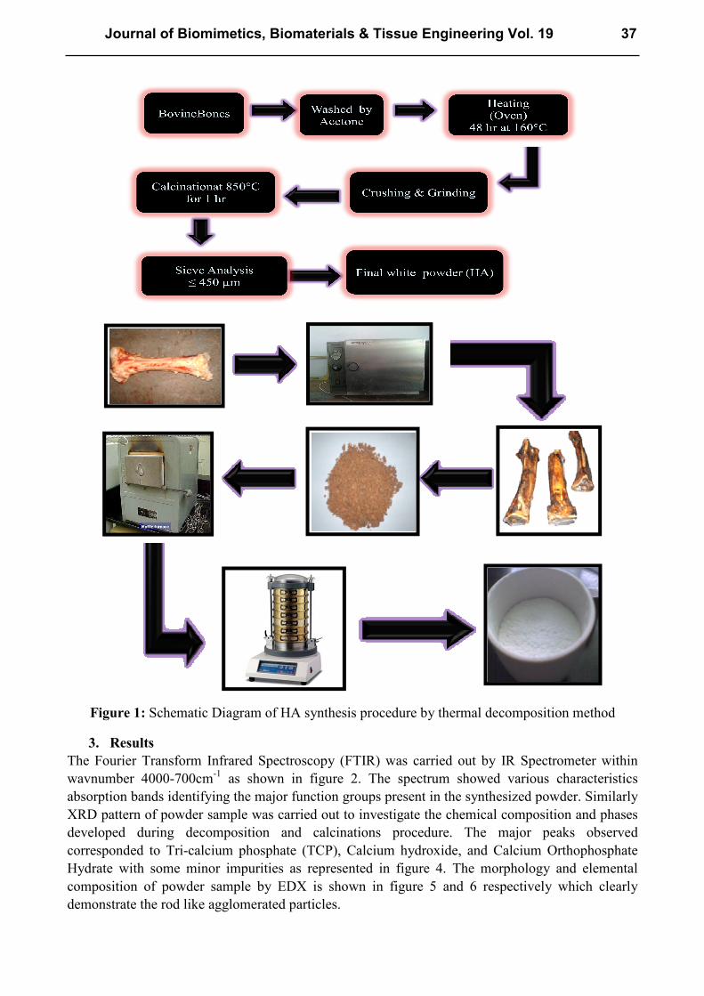
Figure 1: Schematic Diagram of HA synthesis procedure by thermal decomposition method
3. Results
The Fourier Transform Infrared Spectroscopy (FTIR) was carried out by IR Spectrometer within
wavnumber 4000-700cm-1
as shown in figure 2. The spectrum showed various characteristics
absorption bands identifying the major function groups present in the synthesized powder. Similarly
XRD pattern of powder sample was carried out to investigate the chemical composition and phases
developed during decomposition and calcinations procedure. The major peaks observed
corresponded to Tri-calcium phosphate (TCP), Calcium hydroxide, and Calcium Orthophosphate
Hydrate with some minor impurities as represented in figure 4. The morphology and elemental
composition of powder sample by EDX is shown in figure 5 and 6 respectively which clearly
demonstrate the rod like agglomerated particles.
Journal of Biomimetics, Biomaterials & Tissue Engineering Vol. 19 37
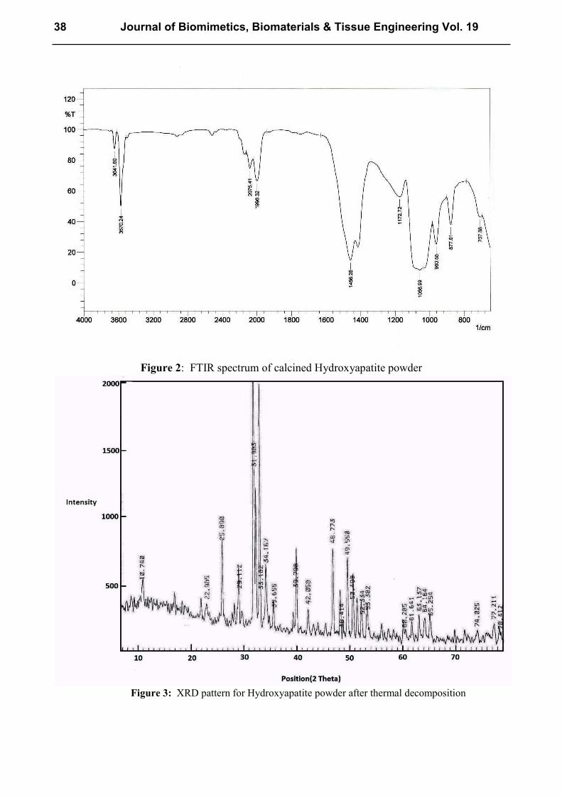
Figure 2: FTIR spectrum of calcined Hydroxyapatite powder
Figure 3: XRD pattern for Hydroxyapatite powder after thermal decomposition
38 Journal of Biomimetics, Biomaterials & Tissue Engineering Vol. 19
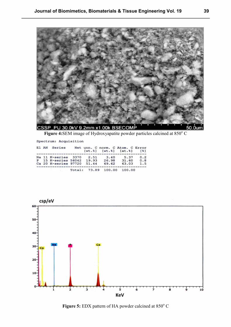
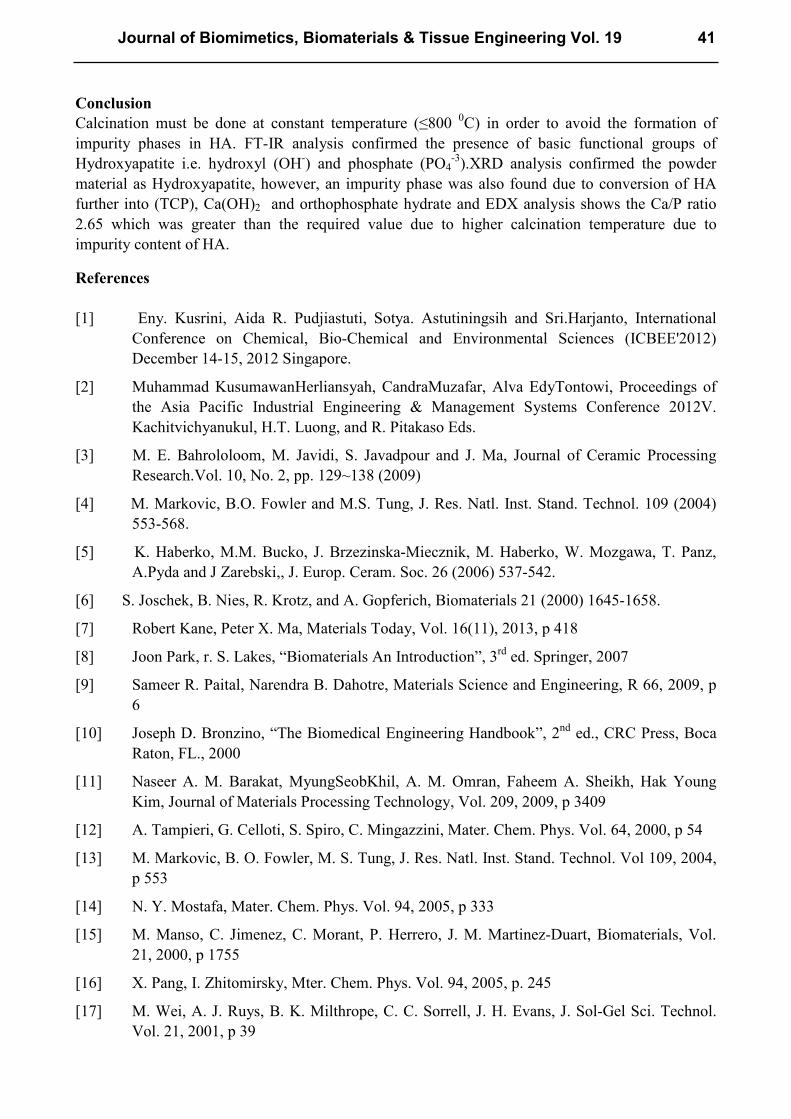
Figure 4:SEM image of Hydroxyapatite powder particles calcined at 850
o C
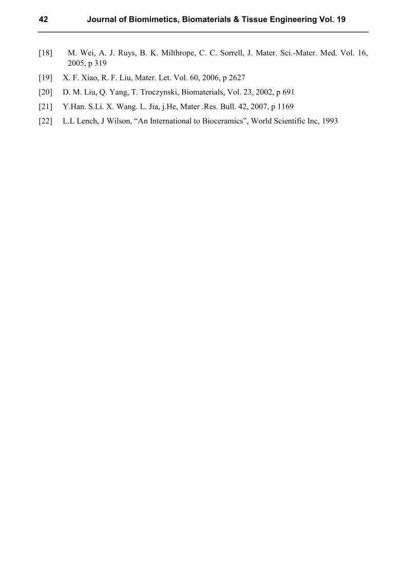
Figure 5: EDX pattern of HA powder calcined at 850
o C
Journal of Biomimetics, Biomaterials & Tissue Engineering Vol. 19 39

4. Discussion
4.1 FTIR Analysis
FTIR analysis was carried out identify the functional groups in the chemical structure of produced
HA as shown in figure 2. It clearly shows the presence of essential functional groups of HA Powder
i.e. hydroxyl (OH-) and phosphate (PO4
-3)at wavenumber 3570.24 cm
-1 and 1056.99 cm
-1
respectively. The stretching vibration band of hydroxyl group at high wavenumber (3570.24 cm-1
)
in combination with a minor signal at 3641.60cm-1
corresponded to strong hydrogen bonding and
presence of water of hydration respectively.The intensity of broad band of (P-O) at 1056.99 cm-1
was stronger than hydroxyl stretching band as required by stoichiometric ratio of calcium and
phosphate. The sharp intensity IR peak of carbonate at 1456.28cm-1
was identified which is an
essential component of natural hydroxyappatite [3-6]. Also, absence of collagen fiber (protein;
amide) was observed in the peak by any of the three main amide regimes(1620, 1636 & 1685 cm- ;
1521, 1533 & 1543 cm-1
; 1232, 1248 & 1281 cm-1
), studied. Thus, the obtained HA powder was
purely phosphates of calcium, and free from organic materials.
4.2 XRD Analysis
Figure 3 shows the XRD pattern of calcined HA powder sample. As per analysis the as calcined
powder sample was composed of four phases in which Hydroxyapatite (HA) was prominent in
abundance with Tri-calcium phosphate (TCP), Calcium hydroxide, and Calcium Orthophosphate
Hydrate as minor impurities. This partial change of phase from HA to TCP was due to variation in
calcination temperature (850 0C). It is found that increase in temperature beyond 875-900ºC may
enhanced the conversion of HA into tricalcium phosphate. However, the stability of HA is affected
even at lower temperatures when sintered for coating purposes onto the metallic substrates due to
the movement of metal ions to Hydroxyapatite coatings [15, 19, 20].
4.3 Scanning Electron Microscopy (SEM)
As shown in the figure 4, the powder particles obtained after thermal decomposition and calcination
were of variable size. The thermally decomposed bone particles were grayish black in color due
burnt organic matrix residue which were oxidized during calcination and turned white. The
particles were agglomerated and grew up at higher temperature due high surface area and were rod
like in shape [21]. The thermal decomposition procedure was repeated for three times for obtaining
the particles of small size but was limited to average size of 130-160 nm. It was deduced that after
thermal decomposition method the particles need further size reduction procedure to get optimum
particle size range for plasma spray on metallic surfaces. It was also evident from particles
morphologies that the powder (HA) wasless dense and mineralized by thermal decomposition of
organic matrix in the bone structure. The calcined powder exhibited a porous structure due to
removal of organic matrix from the bone structure.
4.4 EDX Analysis
A typical EDX spectrum showing the presence of calcium, sodium and phosphorous in calcined
powder sample predominantly is shown in figure 5. In general the analysis indicated that organic
phases of the calcined bovine bone composed mainly of Ca & P as the major constituents with some
minor signals of‘Na’. Generally the Ca/P ratio for all three samples was 2.65 which was higher than
the stiochiometric value of 1.67 required for HA to qualify for coating material for orthopedic
implants. Such deviation in Ca/P ratio from theoretical value the calcined product of thermally
decomposed bovine bone also confirmed the XRD analysis results for the presence of impurity
ingredient in it. The formation of impurity phases in HA was considered due to variation in
calcination temperature and oxidizing condition in the furnace [22].
40 Journal of Biomimetics, Biomaterials & Tissue Engineering Vol. 19
Conclusion
Calcination must be done at constant temperature (≤800 0C) in order to avoid the formation of
impurity phases in HA. FT-IR analysis confirmed the presence of basic functional groups of
Hydroxyapatite i.e. hydroxyl (OH-) and phosphate (PO4
-3).XRD analysis confirmed the powder
material as Hydroxyapatite, however, an impurity phase was also found due to conversion of HA
further into (TCP), Ca(OH)2 and orthophosphate hydrate and EDX analysis shows the Ca/P ratio
2.65 which was greater than the required value due to higher calcination temperature due to
impurity content of HA.
References
[1] Eny. Kusrini, Aida R. Pudjiastuti, Sotya. Astutiningsih and Sri.Harjanto, International
Conference on Chemical, Bio-Chemical and Environmental Sciences (ICBEE'2012)
December 14-15, 2012 Singapore.
[2] Muhammad KusumawanHerliansyah, CandraMuzafar, Alva EdyTontowi, Proceedings of
the Asia Pacific Industrial Engineering & Management Systems Conference 2012V.
Kachitvichyanukul, H.T. Luong, and R. Pitakaso Eds.
[3] M. E. Bahrololoom, M. Javidi, S. Javadpour and J. Ma, Journal of Ceramic Processing
Research.Vol. 10, No. 2, pp. 129~138 (2009)
[4] M. Markovic, B.O. Fowler and M.S. Tung, J. Res. Natl. Inst. Stand. Technol. 109 (2004)
553-568.
[5] K. Haberko, M.M. Bucko, J. Brzezinska-Miecznik, M. Haberko, W. Mozgawa, T. Panz,
A.Pyda and J Zarebski,, J. Europ. Ceram. Soc. 26 (2006) 537-542.
[6] S. Joschek, B. Nies, R. Krotz, and A. Gopferich, Biomaterials 21 (2000) 1645-1658.
[7] Robert Kane, Peter X. Ma, Materials Today, Vol. 16(11), 2013, p 418
[8] Joon Park, r. S. Lakes, “Biomaterials An Introduction”, 3rd
ed. Springer, 2007
[9] Sameer R. Paital, Narendra B. Dahotre, Materials Science and Engineering, R 66, 2009, p
6
[10] Joseph D. Bronzino, “The Biomedical Engineering Handbook”, 2nd
ed., CRC Press, Boca
Raton, FL., 2000
[11] Naseer A. M. Barakat, MyungSeobKhil, A. M. Omran, Faheem A. Sheikh, Hak Young
Kim, Journal of Materials Processing Technology, Vol. 209, 2009, p 3409
[12] A. Tampieri, G. Celloti, S. Spiro, C. Mingazzini, Mater. Chem. Phys. Vol. 64, 2000, p 54
[13] M. Markovic, B. O. Fowler, M. S. Tung, J. Res. Natl. Inst. Stand. Technol. Vol 109, 2004,
p 553
[14] N. Y. Mostafa, Mater. Chem. Phys. Vol. 94, 2005, p 333
[15] M. Manso, C. Jimenez, C. Morant, P. Herrero, J. M. Martinez-Duart, Biomaterials, Vol.
21, 2000, p 1755
[16] X. Pang, I. Zhitomirsky, Mter. Chem. Phys. Vol. 94, 2005, p. 245
[17] M. Wei, A. J. Ruys, B. K. Milthrope, C. C. Sorrell, J. H. Evans, J. Sol-Gel Sci. Technol.
Vol. 21, 2001, p 39
Journal of Biomimetics, Biomaterials & Tissue Engineering Vol. 19 41
[18] M. Wei, A. J. Ruys, B. K. Milthrope, C. C. Sorrell, J. Mater. Sci.-Mater. Med. Vol. 16,
2005, p 319
[19] X. F. Xiao, R. F. Liu, Mater. Let. Vol. 60, 2006, p 2627
[20] D. M. Liu, Q. Yang, T. Troczynski, Biomaterials, Vol. 23, 2002, p 691
[21] Y.Han. S.Li. X. Wang. L. Jia, j.He, Mater .Res. Bull. 42, 2007, p 1169
[22] L.L Lench, J Wilson, “An International to Bioceramics”, World Scientific Inc, 1993
42 Journal of Biomimetics, Biomaterials & Tissue Engineering Vol. 19
Journal of Biomimetics, Biomaterials & Tissue Engineering Vol. 19 10.4028/www.scientific.net/JBBTE.19 Synthesis and Characterization of Hydroxyapatite Powder from Natural Bovine Bone 10.4028/www.scientific.net/JBBTE.19.35
DOI References
[4] M. Markovic, B.O. Fowler and M.S. Tung, J. Res. Natl. Inst. Stand. Technol. 109 (2004) 553-568.
http://dx.doi.org/10.6028/jres.109.042 [5] K. Haberko, M.M. Bucko, J. Brzezinska-Miecznik, M. Haberko, W. Mozgawa, T. Panz, A. Pyda and J
Zarebski, J. Europ. Ceram. Soc. 26 (2006) 537-542.
http://dx.doi.org/10.1016/j.jeurceramsoc.2005.07.033 [6] S. Joschek, B. Nies, R. Krotz, and A. Gopferich, Biomaterials 21 (2000) 1645-1658.
http://dx.doi.org/10.1016/S0142-9612(00)00036-3


![· 2003-06-05 · Let G be a group of type I ... [Se, 19.35] these units give a subgroup of finite index in for arbitrary finite G. We are interested in units of RG, where R is the](https://img.pdfslide.us/doc/110x75/5e82f9742b41820d6218eeb4/2003-06-05-let-g-be-a-group-of-type-i-se-1935-these-units-give-a-subgroup.jpg)