Embed Size (px)
Citation preview
7/25/2019 Javid Et Al-2013-Chemical Biology & Drug Design
http://slidepdf.com/reader/full/javid-et-al-2013-chemical-biology-drug-design 1/11
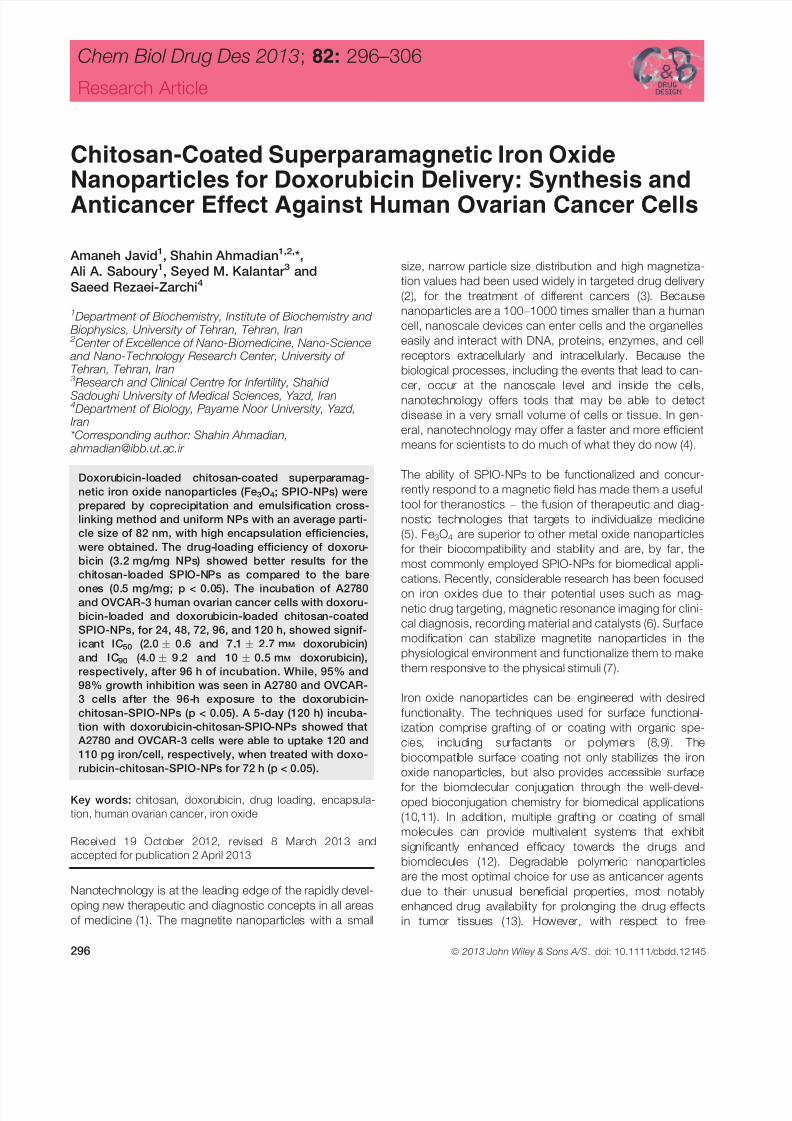
Chitosan-Coated Superparamagnetic Iron OxideNanoparticles for Doxorubicin Delivery: Synthesis andAnticancer Effect Against Human Ovarian Cancer Cells
Amaneh Javid1, Shahin Ahmadian1,2,*,
Ali A. Saboury1, Seyed M. Kalantar 3 and
Saeed Rezaei-Zarchi4
1Department of Biochemistry, Institute of Biochemistry and Biophysics, University of Tehran, Tehran, Iran 2Center of Excellence of Nano-Biomedicine, Nano-Science and Nano-Technology Research Center, University of Tehran, Tehran, Iran3Research and Clinical Centre for Infertility, Shahid
Sadoughi University of Medical Sciences, Yazd, Iran4Department of Biology, Payame Noor University, Yazd,Iran*Corresponding author: Shahin Ahmadian,
Doxorubicin-loaded chitosan-coated superparamag-
netic iron oxide nanoparticles (Fe3O4; SPIO-NPs) were
prepared by coprecipitation and emulsification cross-
linking method and uniform NPs with an average parti-
cle size of 82 nm, with high encapsulation efficiencies,
were obtained. The drug-loading efficiency of doxoru-
bicin (3.2 mg/mg NPs) showed better results for the
chitosan-loaded SPIO-NPs as compared to the bare
ones (0.5 mg/mg; p < 0.05). The incubation of A2780and OVCAR-3 human ovarian cancer cells with doxoru-
bicin-loaded and doxorubicin-loaded chitosan-coated
SPIO-NPs, for 24, 48, 72, 96, and 120 h, showed signif-
icant IC50 (2.0 0.6 and 7.1 2.7 mM doxorubicin)
and IC90 (4.0 9.2 and 10 0.5 mM doxorubicin),
respectively, after 96 h of incubation. While, 95% and
98% growth inhibition was seen in A2780 and OVCAR-
3 cells after the 96-h exposure to the doxorubicin-
chitosan-SPIO-NPs (p < 0.05). A 5-day (120 h) incuba-
tion with doxorubicin-chitosan-SPIO-NPs showed that
A2780 and OVCAR-3 cells were able to uptake 120 and
110 pg iron/cell, respectively, when treated with doxo-
rubicin-chitosan-SPIO-NPs for 72 h (p < 0.05).
Key words: chitosan, doxorubicin, drug loading, encapsula-
tion, human ovarian cancer, iron oxide
Received 19 October 2012, revised 8 March 2013 and
accepted for publication 2 April 2013
Nanotechnology is at the leading edge of the rapidly devel-
oping new therapeutic and diagnostic concepts in all areas
of medicine (1). The magnetite nanoparticles with a small
size, narrow particle size distribution and high magnetiza-
tion values had been used widely in targeted drug delivery
(2), for the treatment of different cancers (3). Because
nanoparticles are a 100 – 1000 times smaller than a human
cell, nanoscale devices can enter cells and the organelles
easily and interact with DNA, proteins, enzymes, and cell
receptors extracellularly and intracellularly. Because the
biological processes, including the events that lead to can-
cer, occur at the nanoscale level and inside the cells,
nanotechnology offers tools that may be able to detect
disease in a very small volume of cells or tissue. In gen-
eral, nanotechnology may offer a faster and more efficient
means for scientists to do much of what they do now (4).
The ability of SPIO-NPs to be functionalized and concur-
rently respond to a magnetic field has made them a useful
tool for theranostics – the fusion of therapeutic and diag-
nostic technologies that targets to individualize medicine
(5). Fe3O4 are superior to other metal oxide nanoparticles
for their biocompatibility and stability and are, by far, the
most commonly employed SPIO-NPs for biomedical appli-
cations. Recently, considerable research has been focused
on iron oxides due to their potential uses such as mag-netic drug targeting, magnetic resonance imaging for clini-
cal diagnosis, recording material and catalysts (6). Surface
modification can stabilize magnetite nanoparticles in the
physiological environment and functionalize them to make
them responsive to the physical stimuli (7).
Iron oxide nanoparticles can be engineered with desired
functionality. The techniques used for surface functional-
ization comprise grafting of or coating with organic spe-
cies, including surfactants or polymers (8,9). The
biocompatible surface coating not only stabilizes the iron
oxide nanoparticles, but also provides accessible surface
for the biomolecular conjugation through the well-devel-oped bioconjugation chemistry for biomedical applications
(10,11). In addition, multiple grafting or coating of small
molecules can provide multivalent systems that exhibit
significantly enhanced efficacy towards the drugs and
biomolecules (12). Degradable polymeric nanoparticles
are the most optimal choice for use as anticancer agents
due to their unusual beneficial properties, most notably
enhanced drug availability for prolonging the drug effects
in tumor tissues (13). However, with respect to free
296 ª 2013 John Wiley & Sons A/S. doi: 10.1111/cbdd.12145
Chem Biol Drug Des 2013; 82: 296–306
Research Article

7/25/2019 Javid Et Al-2013-Chemical Biology & Drug Design
http://slidepdf.com/reader/full/javid-et-al-2013-chemical-biology-drug-design 2/11
drugs, these drug loaded nanoparticles have been shown
to decrease the permeability of the drugs across the cell
membrane (14).
Chitosan, a natural compound and a deacetylated deriva-
tive of chitin, is a positively charged polymer carrier. The
cell adhesion and potential uptake of chitosan particles are
also most favorable due to their attraction to negatively
charged cell membranes, an attractive feature for thetreatment of solid tumors (15,16). Moreover, chitosan has
shown favorable biocompatibility (17) as well as the ability
to increase cell membrane permeability both in vitro (18)
and in vivo (19). Among the various biopolymers, chitosan
along with nanoparticles has been utilized as a stabilizing
agent due to its excellent film-forming ability, mechanical
strength, biocompatibility, non-toxicity, high permeability
towards water, susceptibility to chemical modifications,
cost-effectiveness (20).
Keeping in mind the novel properties of the magnetic
nanoparticles, the Fe3O4 nanoparticles were modified
with chitosan and loaded with the anticancer drug doxo-
rubicin (DOX) for achieving a biocompatible, biodegrad-
able and site-specific carrier of anticancer drugs. The
effect of these drug loaded nanocomposits was also
evaluated on biochemical and morphological parameters
of the human ovarian cancer cell lines of A2780 and OV-
CAR-3.
Experimental Section
Chemicals
Doxorubicin and chitosan (low molecular weight; viscosity,
20 cps) were purchased from Sigma-Aldrich Chemical Co.
(St. Louis, MO, USA). RPMI-1640 medium and all of the
additives were purchased from GIBCO Co. (Grand Island,
NY, USA). The ovarian cancer cell lines, A2780 (NCBI
code, C461) and OVCAR-3 (NCBI code, C430) were pur-
chased from Pasteur Institute, Tehran, Iran. All other
chemicals, used, were of the highest purity and biological
grade available from the commercial sources.
Synthesis of chitosan-coated iron oxide NPs
Chitosan-coated iron oxide (Fe3O4) NPs were prepared by
coprecipitation technique, with some modifications in the
previously reported method (21,22). Firstly, 5.41 g of
FeCl36H2O (99% purity) and 1.99 g FeCl24H2O (99%
purity) were dissolved in 100 mL of distilled water in athree-necked flask. The reaction principle is as follows:
Fe2þ þ 2Fe3þ þ 8OH $ Fe(OH)2 þ 2Fe(OH)3! Fe3O4 þ 4H2O
Chitosan molecules were coated onto the surface of
SPIO-NPs, by adsorbing them onto SPIO-NPs, simulta-
neously during their synthesis. As the chitosan is not
water soluble, a 1% (w/v) aqueous solution was made
by adding 0.2 g of chitosan into a mixture of water
(19 mL) and a 2 N acetic acid (1 mL). The pH of the
mixture (100 mL) of aqueous solutions (0.5 M) of iron
chloride (FeCl2 + FeCl3) and diluted chitosan (0.05% (w/v)
was maintained at pH 6.9 through the slow addition of
25 mL of the aqueous NH4OH (25 – 28%, w/w) while stir-
ring constantly under the protection of dry nitrogen at
80 °C (23). Pressurized air was supplied to the above
solution to oxidize Fe
2+
to Fe
3+
for the formation of mag-netite (Fe3O4) (24 – 26). Change in the color of solution to
dark brown to black, due to the precipitation of Fe3O4,
indicates the formation of bare or chitosan-coated SPIO-
NPs (21,22). The tetramethyl ammonium hydroxide was
used as the surfactant, in preparing the bare SPIO-NPs,
to maintain the aqueous solution of bare SPIO-NPs in
the state of colloidal suspension. The supernatant was
discarded and the resulting precipitate was collected with
strong magnet and rinsed thrice with distilled water to
remove excess NH4OH.
Characterization of synthesized NPs
FT-IR analysis of bare Fe3O4 (SPIO-NPs), chitosan and
SPIO-NPs + chitosan was carried out, via FT-IR Spectro-
photometer (Model 8300; Shimadzu Corporation, Tokyo,
Japan), to investigate the presence of chitosan coating on
the surface of SPIO-NPs. Transmission electron micro-
scope (TEM) and IR results are important because they
evidenced the successful coating of SPIO-NPs by chito-
san. Size and surface morphology of the synthesized NPs
were characterized with the help of TEM (H-7600; Hitachi
High-Technologies Corporation, Tokyo, Japan). A dynamic
light scattering spectrometer (DLS-7000AL; Otsuka Elec-
tronics, Osaka, Japan) was used to determine the average
diameters of the bare and the coated NPs.
Conjugation of DOX to the chitosan-coated SPIO-
NPs
For drug conjugation to modified SPIO-NPs, different con-
centrations (0, 10, 30, 50, 70, 100, 150, and 200 lg) of
chitosan-coated SPIO-NPs were first sonicated with 0.1,
0.2, 0.3, 0.4, 0.5, 1.0, 1.5, 2.0, 2.5, and 3.0 mg/mL con-
centrations of DOX solution for 0.5 h and then stirred over-
night at room temperature in the dark. All the samples
were centrifuged at 18 000 9 g for 1 h. The DOX concen-
tration of all the samples was measured using a standard
DOX concentration curve, generated with a UV-Vis spec-
trophotometer at the wavelength of 233 nm. The drugconjugation to the chitosan modified SPIO-NPs has been
illustrated in Figure 1.
Loading efficiency of incorporated drug
The quantification and release kinetics of the drug-loading
onto the modified SPIO-NPs was carried out through UV/
Visible double-beam spectrophotometer (Hitachi U-2000) at
200 – 800 nm. Drug incorporation efficiency was observed
Chem Biol Drug Des 2013; 82: 296–306 297
Antitumoral Drug-loaded Nanoparticles

7/25/2019 Javid Et Al-2013-Chemical Biology & Drug Design
http://slidepdf.com/reader/full/javid-et-al-2013-chemical-biology-drug-design 3/11
both as drug loading (% w/w) and drug entrapment (%), bythe below-stated eqns (1) and (2), respectively.
Drug loading ð%w/wÞ ¼Mass of drug loaded in NPs 100
Mass of nanoparticles
(1)
Drug entrapment ð%Þ ¼Mass of drug loaded in NPs 100
Mass of drug used in formulation
(2)
Cancer cell culture
The human ovarian cancer cell lines, A2780 and OVCAR-3,
were maintained in RPMI-1640 medium, supplemented with
10% (v/v) FBS, 0.25 IU/mL insulin, 100 lg/mL streptomy-
cin, 100 units/mL penicillin and 0.3 mg/mL glutamine. Cells
were cultured at 37 °C in a humidified atmosphere of 5% (v/
v) CO2 in air. For cytotoxicity data, clonogenic assay was
used. Clonogenic assay is used for studying the effective-
ness of specific agents on the survival and proliferation of
cells. For this assay, 500 cells from each cell line were pla-
ted into 60-mm tissue culture dishes 24 h prior
to nanomedicine treatment. Cells were then treated, in tripli-cate, with 50 lg/mL of the: (i) SPIONs alone, (ii) chitosan-
coated SPIO-NPs, and (iii) DOX-chitosan-SPIONs for 4 h,
after which the experimental media were removed and fresh
medium was added to the cells. Incubation was continued
until the colonies of approximately 50 cells were observed
(7 – 9 days). Growth medium was replaced with fresh med-
ium on day 5. The resulting colonies were stained with 1 mL
of the clonogenic reagent for 45 min, washed with PBS,
blue colonies were counted and the data were compared
with those of the control according to the following relation:
Percent survival ¼ð Average treated count/Average
control countÞ 100
Analysis of iron uptake by cancer cells
To quantitate the iron uptake by cancer cell lines, the
A2780 and OVCAR-3 cells were grown (~ 105 cells/1 mL
medium) with and without 0.5 mg/mL of bare, chitosan-
coated, and chitosan-coated and drug-loaded SPIO-NPs
in 24-well culture plates. Cells were washed with PBS,
resuspended, counted, centrifuged down, and the cell pel-
lets were dissolved in 37% HCl solution at 70 –
80 °C for30 min. The samples were diluted to a final iron concen-
tration of 1.0 – 5.0 lg/mL. Three replicates of each sample
were measured using an inductively coupled plasma emis-
sion spectroscopy (ICP ES) and the results were averaged
with standard deviation.
Cell viability and apoptosis analyses
Viability was determined by staining cancer cells with 1%
methylene blue in 70% ethanol, followed by three washes in
de-ionized water. Data obtained were used to calculate the
IC50 and IC90 values for each cell line, administered with
DOX-chitosan-SPIO-NPs. For all experiments, 3 9 106
cellsfrom each cell line were plated into 150 mm tissue culture
dishes, 24 h prior to the nano-composite treatment. In order
to determine the corresponding IC50 and IC90 values, the
cells were separately treated with the as-prepared bare,
coated and drug-loaded nano-composites for 24, 48, 72,
96, and 120 h at 37 °C. Plates were washed thrice
with PBS and incubated in 10 mL of fresh medium for an
additional 48 h before the final cell collection. All the cells
were collected by sedimenting at 2000 9 g for 5 min.
Figure 1: Illustration of the modification mechanism of SPIO-NPs with chitosan, followed by doxorubicin (DOX) loading.
298 Chem Biol Drug Des 2013; 82: 296–306
Javid et al.

7/25/2019 Javid Et Al-2013-Chemical Biology & Drug Design
http://slidepdf.com/reader/full/javid-et-al-2013-chemical-biology-drug-design 4/11
For the 5-dimethylthiazol-2-yl-2,5-diphenyltetrazolium bro-
mide (MTT) assay, A2780 and OVCAR-3 cells were plated
in 96-well plates and exposed to uncoated SPIO-NPs,
chitosan-SPIO-NPs and DOX-chitosan-SPIO-NPs at a
concentration of 50 lg/mL for 24, 48, 72, 96, and 120 h.
After the culture period, the medium was removed from
each well and replaced with fresh medium. Cellular apop-
tosis was detected by Annexin-V-FLUOS staining and
studied with the help of flow cytometry.
Immuno-blot analysis and investigation of gene
expression
A2780 and OVCAR-3 cells were lysed in a buffer contain-
ing 50 mM Tris/HCl (pH 7.4), 150 mM NaCl, 1 mM EDTA,
0.2% (v/v) Nonidet P40, and protease inhibitor cocktail in
ice for a period of 10 min. Cell debris was pelleted by cen-
trifugation, and the total protein concentration of the solu-
ble extracts was determined by Bradford assay, according
to the manufacturer’s instructions. Total soluble protein
extract (30 lg) for each sample was resolved by SDS
PAGE (12% gels). After electrophoresis, the survivin, Bcl-2,
Bax and NF-K B proteins were transferred to nitrocellulose,
and the blot was blocked for 1 h at room temperature with
a solution of 5% dried milk in PBST (0.1% (v/v) Tween 20
in PBS). The blot was incubated, overnight at 4 °C, with
either of the anti-Bcl-2 monoclonal antibody, anti-Bax,
anti-NF-K B and anti – survivin polyclonal antibodies to evalu-
ate the levels of protein expression. Primary antibodies
were detected using an horseradish peroxidase conju-
gated secondary antibody and enhanced chemilumines-
cence was taken as described by the manufacturer.
Statistical analyses
Results are presented as mean standard deviation. The
two-way ANOVA and the Student’s t -test were used to com-pare data from the different treatment groups. Statistical
significance was accepted at p < 0.05.
Results and Discussion
Synthesis of bare and chitosan-coated SPIO-NPs
The bare and chitosan-coated SPIO-NPs were synthe-
sized by coprecipitation technique. As the chitosan can
precipitate under alkaline conditions, when 2 mL NaOH
(1.0 M) was added to the acidic solution containing the
chitosan, Fe(II) and Fe(III) ions, the SPIO-NPs were formed
during the precipitation of chitosan, leading to the forma-tion of chitosan-coated SPIO-NPs.
Characterization of bare and chitosan coated
SPIO-NPs
FT-IR analysis
The bonding status of chitosan molecules on the surface
of the particle could be checked from the wavelength-
dependent data of transmittance obtained using an FTIR
spectrometer. According to the the SPIO-NPs spectrum of
Figure 2, the 576.25/cm region of the Fe3O4 spectrum
represents the Fe-O group while, the 3401.82/cm peak in
the chitosan spectrum is related to the primary amine
group. The stretching vibrations of O-H and C-H can be
seen through the peaks of 2919 and 2874/cm. While in
the SPIO-NPs + chitosan spectrum, the bio-sorption peak
of primary amine ( –
NH2) is demonstrated by 3392.17 and1613.56/cm. The bio-sorption bands around 1078.01/cm
show the stretch vibration of C – O bond and the 584.32/
cm band represents the Fe-O group of SPIO-NPs. So, the
FT-IR analysis supports the notion that the chitosan coat-
ing is present onto the surface of SPIO-NPs.
Microscopic analysis
The synthesized bare and chitosan coated NPs were char-
acterized by atomic force microscope and the final diame-
ters were obtained to be 10 and 69.0 nm (Figure 3A, B).
The size of prepared nanoparticles was also determined
by TEM analysis. Figure 3C shows the TEM (H-7600; Hit-
achi) micrographs of bare nanoparticles, while Figure 3D
demonstrates the TEM micrograph of chitosan-coated
SPIONs.
Dynamic light scattering analysis
Size distribution of the synthesized SPIO-NPs and chito-
san-coated SPio-NPs was investigated in an aqueous
solution with the help of dynamic light scattering (DLS)
spectrometer. As shown in Figure 4A, B), the mean diam-
eter of the chitosan-SPIO-NPs was recorded to be about
69.0 nm. The size distribution of the bare SPIO-NPs was
also determined and recorded to be about 10 nm.
Doxorubicin loading onto the chitosan-coated
SPIO-NPs
To assess the conjugation capacity of DOX the modified
NPs, the following concentrations of the drug: 0.1, 0.2,
0.3, 0.4, 0.5, 1.0, 1.5, 2.0, 2.5, and 3.0 mg/mL were
added to the chitosan-modified NPs and incubated for
30 min and then stirred overnight at room temperature in
the dark. A DOX concentration curve (233 nm) was con-
structed from standard solutions. As shown in Figure 5A,
DOX loading of modified SPIO-NPs at an initial drug con-
centration of 0.5 mg/mL was nearly saturating.
IC50 and IC90 analyses of the A2780 and OVCAR-3 cell
lines, after 24, 48, 72, 96, and 120 h (5 days) exposure to
DOX-loaded chitosan-coated SPIO-NPs, were recorded at
2.0 0.6 and 7.1 2.7 mM concentration of drug, while,
IC90 of both cell l ines was seen at 4.0 9.2 and
10 0.5 mM DOX concentration after 96 h. This demon-
strates that the OVCAR-3 cells were more resistant to the
lower concentrations of drug as compared to the A2780
cells (Table 1; p < 0.05).
Chem Biol Drug Des 2013; 82: 296–306 299
Antitumoral Drug-loaded Nanoparticles

7/25/2019 Javid Et Al-2013-Chemical Biology & Drug Design
http://slidepdf.com/reader/full/javid-et-al-2013-chemical-biology-drug-design 5/11
DOX release profile
Figure 5B shows the DOX release profiles of the free and
the conjugated drug, at the pH range of 1.5 – 7.0. At pH
7.0, a small amount of the drug release was observed after
the incubation period of 48 h. This is a desirable character-
istic as the pH 7.4 is the undesired pH for the proper
release of the drugs from the nanoconjugated drug carrier.
This will also prevent the premature release of the drugs
before the nanoconjugates reach the cancer cells. Fig-
ure 5B shows that the pH 6.0 provided the desirable condi-
tions for the proper drug release. The first 10 h represent
the period of initial rapid release, followed by a steady state.
This pH-dependent drug release behavior is favorable for
the chemotherapeutic process as it can significantly reduce
the preterm drug release on the body pH level (pH 7.4) and
maximizing the amount of drug reaching the target tumor
cells, once the drug-loaded SPIO-NPs internalize and enter
the tumor by endocytosis (pH 4.5 – 6.5).
Figure 2: FT-IR spectra of bare SPIO-NPs, chitosan, and SPIO-NPs + chitosan.
A B
C D
Figure 3: (A, C) Atomic force and transmission electron microscopic images of the bare SPIO-NPs, B, D) atomic force microscope (AFM)
and transmission electron microscope (TEM) images of chitosan-modified SPIONs, respectively.
300 Chem Biol Drug Des 2013; 82: 296–306
Javid et al.

7/25/2019 Javid Et Al-2013-Chemical Biology & Drug Design
http://slidepdf.com/reader/full/javid-et-al-2013-chemical-biology-drug-design 6/11
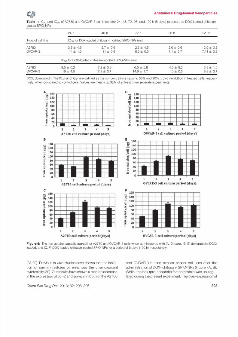
Cell culture and iron uptake by cancer cells
The human ovarian cancer cell lines were cultured in
RPMI-1640 culture medium for a period of 120 h (5 days).
The nanoparticle uptake by the human ovarian cancer cell
lines of A2780 and OVCAR-3 was evaluated after the cells
were grown in medium containing 0.2 mg/mL NPs for up
to 5 days. Figure 6 shows the uptake of bare, DOX-loaded
and DOX-loaded chitosan-coated SPIO-NPs by A2780
and OVCAR-3 ovarian cancer cell lines. The bare SPIO-
NPs were internalized into the cells quickly (14 pg iron/cell)
within the first day, while the uptake amount showed fluc-
tuations during the following 4 days, probably due to the
fast growth of cancer cells. The uptake of 3 pg iron/cellwas recorded after a culture period of 5 days. After the
chitosan coating and drug loading, the nanoparticle uptake
by cancer cells increased significantly with the passage of
time. An iron uptake of 97 and 90 pg/cell was seen in
A2780 and OVCAR-3 cells, respectively, after an incuba-
tion period of 3 days. The average uptake of drug loaded
chitosan-coated NPs by A2780 and OVCAR-3 cells during
the 5 days was 89 and 100 pg/cell. This might be attrib-
uted to the fact that the presence of polymer prevents the
nanoparticle agglomeration in the culture medium and
makes it more soluble and an easy-to-pass entity through
the cell membrane.
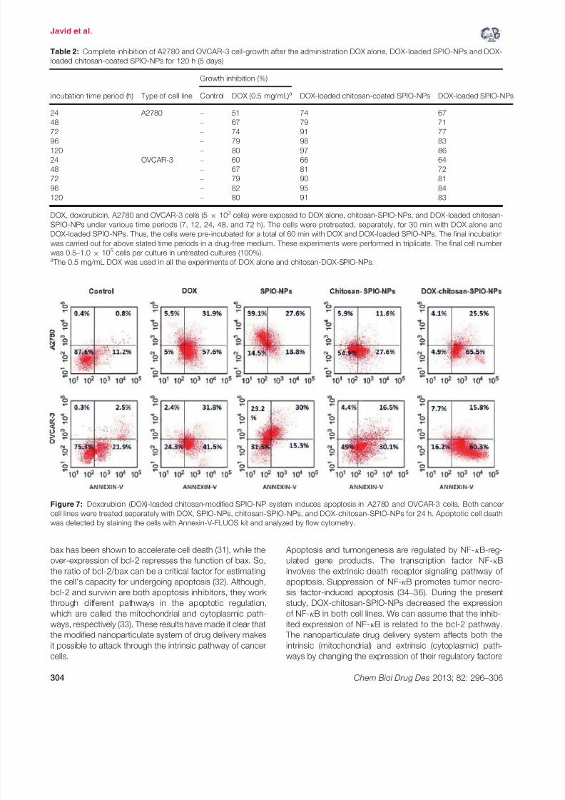
Table 2 shows the results of growth inhibition analysis.
Our results state that the administration of DOX-loaded
(0.5 mg/mL) and chitosan-coated SPIO-NPs caused 98%
and 95% growth inhibition in A2780 and OVCAR-3 cell
lines, during a culture period of 96 h, respectively. Theseresults were compared with those of the effect of 0.5 mg/
mL free DOX (80% and 82% for A2780 and OVCAR-3
cells) and the DOX-loaded SPIO-NPs (83% and 84% for
A2780 and OVCAR-3 cells) under same experimental con-
ditions (p < 0.05).
Cytotoxicity and synergistic effect of DOX-
chitosan-SPIO-NPs on the cancer cells
For the cytotoxicity analysis, A2780 and OVCAR-3 cells
were treated separately with DOX, SPIO-NPs, chitosan-
SPIO-NPs and DOX-chitosan-SPIO-NPs for 24 h. Apopto-
tic cell death was analyzed by staining the cells with
Annexin-V-FLUOS staining kit and analyzed by flow
cytometry. The results revealed a dose-dependent induc-
tion of early or late apoptotic cell death in these two cell
lines (Figure 7). Compared to OVCAR-3 cells, the A2780
cells showed more cell death. A2780 cells showed 91%
apoptosis after the administration of DOX-loaded chitosan-
SPIO-NPs (5 mg/L; p < 0.05). While, 87.1% apoptosis
was seen in the OVCAR-3 cells under same conditions
(p < 0.05), which indicates that chitosan-modified SPIO-
NPs could enhance the DOX-induced apoptosis in both of
the human ovarian cancer cell lines.
Western blot analysis for gene expression
In the Western blot analysis, the survivin and bcl-2 were
used as the representatives of the inhibitor of apoptosis
protein. During the present study, the expression of bax,
bcl-2, NF-jB and survivin proteins was also studied in the
A2780 and OVCAR-3 cells, treated with DOX alone, bare
SPIO-NPs, chitosan-coated SPIO-NPs and DOX- chito-
san-SPIO-NPs, respectively. According to Figure 8, the
results have shown that the level of bcl-2 and survivin pro-
teins had a sharp decrease while NF-jB was also regu-
lated in the cultures, treated with DOX-chitosan-SPIO-NPs
in A2780 and OVCAR-3 cells (Figure 8A, B). This repre-
sents the activation of apoptotic mechanism in cancer
cells after the nanoparticle-based DOX delivery in vitro(p < 0.05).
The SPIO-NPs have a proven candidacy for their biocom-
patibility and wide applications in the medical field (24).
During the present study, the bare and chitosan coated
superparamagnetic iron oxide (SPIO) nanoparticles were
synthesized by the coprecipitation technique. The
synthesized chitosan-coated nanoparticles had the diam-
eter of about 69 nm, which was confirmed by both of
A
B
Figure 4: Demonstration of size distribution of (A) bare SPIO-NPs
and (B) chitosan-SPIO-NPs in an aqueous solution with the help
of dynamic light scattering (DLS) spectrometer. The mean
diameter of the chitosan-SPIO-NPs was recorded to be about
69.0 nm. The size distribution of the bare ferrite particles is also
shown here. The mean diameter of the bare particles was about
10 nm.
Chem Biol Drug Des 2013; 82: 296–306 301
Antitumoral Drug-loaded Nanoparticles

7/25/2019 Javid Et Al-2013-Chemical Biology & Drug Design
http://slidepdf.com/reader/full/javid-et-al-2013-chemical-biology-drug-design 7/11
the TEM and DLS techniques that shows that no aggre-
gation of the nanoparticles occurred in solution. Numer-
ous polymer-based nanoparticles have been developed
for cancer-targeted delivery of therapeutic agents (25).
The polysaccharide coating may provide steric protection
against protein adsorption and macrophage uptake. Addi-
tionally, as polysaccharides offer many available reactivegroups, active targeting could be obtained by grafting
ligands onto the nanoparticle surface (Figure 1). Because
chitosan has high affinity for cell membranes due to its
mucoadhesive properties and positive charge, it was uti-
lized as a coating agent for magnetic nanoparticles, during
the present study. The presence of a chitosan coating
was also studied using zeta potential measurements.
Indeed, chitosan-coated nanoparticles had positive
(+38 mV) zeta potential value (26).
Drug loading is affected by particle size. Preparations of
smaller particles have larger total surface area. Therefore,
most of the drug associated would be at or near the
particle surface, leading to fast drug release. Whereas,
larger particles have large cores, which allow more drug
to be encapsulated and slowly diffuse out (27). Our
results have shown that the DOX-loaded and chitosan-
coated SPIO-NPs showed better drug delivery andrelease performance as compared to the bare nanoparti-
cles.
Apoptosis can be prompted in the cancer cells either by
activating the proteins upstream of apoptotic signaling path-
way or by inhibiting the antiapoptotic factors. An over-
expression of survivin is seen in all the human cancers. The
presence or the overly expressed survivin means resistance
to apoptosis, which is associated with increased malignancy
A
B
Figure 5: (A) Doxorubicin loading onto the chitosan-coated SPIO-NPs. Doxorubucin concentrations of 0.1, 0.2, 0.3, 0.4, 0.5, 1.0, 1.5,
2.0, 2.5, and 3.0 mg/mL were added to the chitosan-modified SPIO-NPs and incubated for 30 min and then stirred overnight at room
temperature in the dark and the measurements were carried out with the help of UV-Visible spectrophotometer at the wavelength of 233.
(B) In vitro release profile of doxorubicin (DOX) from chitosan-coated SPIO-NPs at different pH values at 37 °C, after the incubation period
of 48 h ( n = 3).
302 Chem Biol Drug Des 2013; 82: 296–306
Javid et al.
7/25/2019 Javid Et Al-2013-Chemical Biology & Drug Design
http://slidepdf.com/reader/full/javid-et-al-2013-chemical-biology-drug-design 8/11
(28,29). Previous in vitro studies have shown that the inhibi-
tion of survivin restores or enhances the chemoreagent
cytotoxicity (30). Our results have shown a marked decrease
in the expression of bcl-2 and survivin in both of the A2780
and OVCAR-3 human ovarian cancer cell lines after the
administration of DOX- chitosan- SPIO-NPs (Figure 7A, B).
While, the bax (pro-apoptotic factor) protein was up-regu-
lated during the present experiment. The over-expression of
Table 1: IC50 and IC90 of A2780 and OVCAR-3 cell lines after 24, 48, 72, 96, and 120 h (5 days) exposure to DOX-loaded chitosan-
coated SPIO-NPs
Type of cell line
24 h 48 h 72 h 96 h 120 h
IC50 for DOX-loaded chitosan-modified SPIO-NPs (mM)
A2780 3.6 4.5 2.7 0.9 2.3 4.5 2.0 0.6 2.0 0.6
OVCAR-3 14 1.5 11 0.9 8.6 0.5 7.1 2.7 7.11 0.8
IC90 for DOX-loaded chitosan-modified SPIO-NPs (mM)
A2780 8.5 0.2 7.2 0.9 6.5 0.8 4.0 9.2 3.8 1.0
OVCAR-3 19 4.5 17.2 3.7 14.9 1.7 10 0.5 8.8 3.7
DOX, doxorubicin. The IC50 and IC90 are defined as the concentrations causing 50% and 90% growth inhibition in treated cells, respec-
tively, when compared to control cells. Values are means SEM of at least three separate experiments.
A
B
C
D
E
F
Figure 6: The iron uptake capacity (pg/cell) of A2780 and OVCAR-3 cells when administered with (A, D) bare, (B, E) doxorubicin (DOX)
loaded, and (C, F) DOX-loaded chitosan-coated SPIO-NPs for a period of 5 days (120 h), respectively.
Chem Biol Drug Des 2013; 82: 296–306 303
Antitumoral Drug-loaded Nanoparticles
7/25/2019 Javid Et Al-2013-Chemical Biology & Drug Design
http://slidepdf.com/reader/full/javid-et-al-2013-chemical-biology-drug-design 9/11
bax has been shown to accelerate cell death (31), while the
over-expression of bcl-2 represses the function of bax. So,the ratio of bcl-2/bax can be a critical factor for estimating
the cell’s capacity for undergoing apoptosis (32). Although,
bcl-2 and survivin are both apoptosis inhibitors, they work
through different pathways in the apoptotic regulation,
which are called the mitochondrial and cytoplasmic path-
ways, respectively (33). These results have made it clear that
the modified nanoparticulate system of drug delivery makes
it possible to attack through the intrinsic pathway of cancer
cells.
Apoptosis and tumorigenesis are regulated by NF-jB-reg-
ulated gene products. The transcription factor NF-jBinvolves the extrinsic death receptor signaling pathway of
apoptosis. Suppression of NF-jB promotes tumor necro-
sis factor-induced apoptosis (34 – 36). During the present
study, DOX-chitosan-SPIO-NPs decreased the expression
of NF-jB in both cell lines. We can assume that the inhib-
ited expression of NF-jB is related to the bcl-2 pathway.
The nanoparticulate drug delivery system affects both the
intrinsic (mitochondrial) and extrinsic (cytoplasmic) path-
ways by changing the expression of their regulatory factors
Table 2: Complete inhibition of A2780 and OVCAR-3 cell-growth after the administration DOX alone, DOX-loaded SPIO-NPs and DOX-
loaded chitosan-coated SPIO-NPs for 120 h (5 days)
Incubation time period (h) Type of cell line
Growth inhibition (%)
DOX-loaded chitosan-coated SPIO-NPs DOX-loaded SPIO-NPsControl DOX (0.5 mg/mL)a
24 A2780 – 51 74 67
48 – 67 79 71
72 – 74 91 77
96 –
79 98 83120 – 80 97 86
24 OVCAR-3 – 60 66 64
48 – 67 81 72
72 – 79 90 81
96 – 82 95 84
120 – 80 91 83
DOX, doxorubicin. A2780 and OVCAR-3 cells (5 9 103 cells) were exposed to DOX alone, chitosan-SPIO-NPs, and DOX-loaded chitosan-
SPIO-NPs under various time periods (7, 12, 24, 48, and 72 h). The cells were pretreated, separately, for 30 min with DOX alone and
DOX-loaded SPIO-NPs. Thus, the cells were pre-incubated for a total of 60 min with DOX and DOX-loaded SPIO-NPs. The final incubation
was carried out for above stated time periods in a drug-free medium. These experiments were performed in triplicate. The final cell number
was 0.5 – 1.0 9 105 cells per culture in untreated cultures (100%).a The 0.5 mg/mL DOX was used in all the experiments of DOX alone and chitosan-DOX-SPIO-NPs.
Figure 7: Doxorubicin (DOX)-loaded chitosan-modified SPIO-NP system induces apoptosis in A2780 and OVCAR-3 cells. Both cancer
cell lines were treated separately with DOX, SPIO-NPs, chitosan-SPIO-NPs, and DOX-chitosan-SPIO-NPs for 24 h. Apoptotic cell death
was detected by staining the cells with Annexin-V-FLUOS kit and analyzed by flow cytometry.
304 Chem Biol Drug Des 2013; 82: 296–306
Javid et al.

7/25/2019 Javid Et Al-2013-Chemical Biology & Drug Design
http://slidepdf.com/reader/full/javid-et-al-2013-chemical-biology-drug-design 10/11
such as bax and bcl-2, and the transcription factor NF-jB
(36).
So, we have demonstrated that biodegradable drug deliv-
ery through the nanoparticulate systems can promote
apoptosis and lessen the negative effects of chemothera-
peutic agents during the in vitro conditions. To optimize
this drug delivery system, greater understanding of the dif-
ferent mechanisms of biological interactions, and particle
engineering, is still required. Further advances are needed
in order to turn the concept of nanoparticle technology
into a realistic practical application as the next generation
of drug delivery system.
References
1. Ferrari M. (2005) Cancer nanotechnology: opportunities
and challenges. Nat Rev Cancer;5:161 – 171.
2. Chertok B., Moffat B.A., David A.E., Yu F., Bergemann
C., Ross B.D. (2008) Iron oxide nanoparticles as a drug
delivery vehicle for MRI monitored magnetic targeting
of brain tumors. Biomaterials;29:487 – 496.
3. Kikumori T., Kobayashi T., Sawaki M., Imai T. (2009)
Anti-cancer effect of hyperthermia on breast cancer by
magnetite nanoparticle-loaded anti-HER2 immunolipo-somes. Breast Cancer Res Treat;113:435 – 441.
4. Marchal F., Pic E., Pons T., Dubertret B., Bolotine L.,
Guillemin F. (2008) Quantum dots in oncological sur-
gery: the future for surgical margin status? Bull Can-
cer;95:1149 – 1153.
5. Kim E.H., Lee H.S., Kwak B.K., Kim B.K. (2005) Syn-
thesis of ferrofluid with magnetic nanoparticles by so-
nochemical method for MRI contrast agent. J Magn
Magn Mater;289:328 – 330.
6. Hong S.W., Chang Y., Hwang M.J., Rhee I., Kang D.S.
(2004) Ni-Fe2O4 nanoparticles as contrast agents for
magnetic resonance imaging. J Korean Soc Magn
Reson Med;4:27 – 32.
7. Ren J., Jia M., Ren T., Yuan W., Tan Q. (2008) Prepa-
ration and characterization of PNIPAAm- b-PLA/Fe3O4
thermo-responsive and magnetic composite micelles.
Mater Lett;62:4425 – 4427.
8. Chandra S., Mehta S., Nigam S., Bahadur D. (2010)
Dendritic magnetite nanocarriers for drug delivery appli-
cations. New J Chem;34:648 – 655.
9. Khosroshahi M.E., Ghazanfari L. (2010) Preparation
and characterization of silica-coated iron-oxide bio-nanoparticles under N2 gas. Physica E;42:1824 – 1829.
10. Sahoo Y., Pizem H., Fried T., Golodnitsky D., Bur-
stein L., Sukenik S.N., Markovich G. (2001) Alkyl
phosphonate/phosphate coating on magnetite nano-
particles: a comparison with fatty acids. Lang-
muir;17:7907 – 7911.
11. Nayak S., Lee S., Chmielewski J., Lyon L.A. (2004)
Folate-mediated cell targeting and cytotoxicity using
thermoresponsive microgels. J Am Chem
Soc;126:10258 – 10259.
12. Mammen M., Choi S.K., Whitesides G.M. (1998) Poly-
valent interactions in biological systems: implications
for design and use of multivalent ligands and inhibitors. Angew Chem Int Ed;37:2754 – 2794.
13. Fonseca C., Sim~oes S., Gaspar R. (2002) Paclitaxel-
loaded PLGA nanoparticles: preparation, physico-
chemical characterization and in vitro anti-tumoral
activity. J Control Release;83:273 – 286.
14. M€uller B., Kreuter J. (1999) Enhanced transport of
nanoparticle associated drugs through natural and arti-
ficial membranes – a general phenomenon? Int J
Pharm;178:23 – 32.
A B
Figure 8: Expression of survivin, bax, bcl-2, and NF-jB proteins in (A) A2780 cells and (B) OVCAR-3 cells by Western blot after 48 h’
treatment with bare SPIO-NPs (Column 2), doxorubicin (DOX; Column 3), chitosan-coated SPIO-NPs (Column 4), and DOX-loaded
chitosan-coated SPIO-NPs (Column 5). The column 1 shows control experiment.
Chem Biol Drug Des 2013; 82: 296–306 305
Antitumoral Drug-loaded Nanoparticles
7/25/2019 Javid Et Al-2013-Chemical Biology & Drug Design
http://slidepdf.com/reader/full/javid-et-al-2013-chemical-biology-drug-design 11/11
15. Xu Y., Du Y. (2003) Effect of molecular structure of
chitosan on protein delivery properties of chitosan
nanoparticles. Int J Pharm;250:215 – 226.
16. Qi L.F., Xu Z.R., Jiang X. (2004) Preparation and anti-
bacterial activity of chitosan nanoparticles. Carbohydr
Res;339:2693 – 2700.
17. Artursson P., Lindmark T., Davis S.S., Illum L. (2004)
Effect of chitosan on the permeability of monolayers of
intestinal epithelial cells (Caco-2). Pharm Res;11:1358 –
1361.
18. Fernandez-Urrusuno R., Calvo P., Remu~nan-Lopez C.,
Vila-Jato J.L., Alonso M.J. (1999) Enhancement of
nasal absorption of insulin using chitosan nanoparti-
cles. Pharm Res;16:1576 – 1581.
19. V arum K.M., Myhr M.M., Hjerde R.J., Smidsrød O.
(1997) In vitro degradation rates of partially N -acety-
lated chitosans in human serum. Carbohydr
Res;299:99 – 101.
20. Miao Y., Tan S.N. (2000) Amperometric hydrogen per-
oxide biosensor based on immobilization of peroxidase
in chitosan matrix crosslinked with glutaraldehyde.
Analyst;125:1591 – 1594.
21. Hong S., Chang Y., Rhee I. (2010) Chitosan-coated
ferrite (Fe3O4) nanoparticles as a T 2 contrast agent for
magnetic resonance imaging. J Korean Phys
Soc;56:868 – 873.
22. Mohammadi-Samani S., Miri R., Salmanpour M., Kha-
lighian N., Sotoudeh S., Erfani S.N. (2013) Preparation
and assessment of chitosan-coated superparamagnet-
ic Fe3O4 nanoparticles for controlled delivery of metho-
trexate. Res Pharm Sci;8:25 – 33.
23. Jain T.K., Morales M.A., Sahoo S.K., Leslie-Pelecky
D.L., Labhasetwar V. (2005) Iron oxide nanoparticles
for sustained delivery of anticancer agents. Mol
Pharm;2:194 – 205.
24. Gupta A.K., Gupta M. (2005) Synthesis and surface
engineering of iron oxide nanoparticles for biomedical
applications. Biomaterials;26:3995 – 4021.
25. Saifuddin N., Dinara S. (2012) Immobilization of Sac-
charomyces cerevisiae onto cross-linked chitosan
coated with magnetic nanoparticles for adsorp-
tion of Uranium (VI) ions. Adv Nat Appl Sci;6:249 –
267.
26. Schwertmann U., Cornell R.M. (2007) Iron Oxides in
the Laboratory: Preparation and Characterization, 2nd
edn. Germany: Wiley-VCH Verlag GmbH; p. 5 – 18.
27. Hong S., Lim W.T., Rhee I. (2006) Relaxation rate of
hydrogen protons of water molecules in an aqueous
solution of dextran coated ferrite nanoparticles. J Kor-
ean Phys Soc;49:676 – 681.
28. Chen Y., Mohanraj V.J., Parkin J.E. (2003) Chitosan-
dextran sulfate nanoparticles for delivery of an antian-giogenesis peptide. Lett Pept Sci;10:621 – 627.
29. Vila A., Sanchez A., Tobio M., Calvo P., Alonso M.J.
(2002) Design of biodegradable particles for protein
delivery. J Control Release;78:15 – 24.
30. Redhead H.M., Davis S.S., Illum L. (2001) Drug deliv-
ery in poly(lactide-co-glycolide) nanoparticles surface
modified with poloxamer 407 and poloxamine 908: in
vitro characterization and in vivo evaluation. J Control
Release;70:353 – 363.
31. Wang T.T., Wei J., Qian X.P., Ding Y.T., Yu L.X., Liu
B.R. (2008) Gambogic acid, a potent inhibitor of survi-
vin, reverses docetaxel resistance in gastric cancer
cells. Cancer Lett;262:214 – 222.
32. Wang L., Zhang G.M., Feng Z.H. (2003) Down-regula-
tion of survivin expression reversed multidrug resis-
tance in adriamycin- resistant HL-60/ADR cell line.
Acta Pharmacol Sin;24:1235 – 1240.
33. Ling X., Bernacki R.J., Brattain M.G., Li F.Z. (2004)
Induction of survivin expression by taxol (paclitaxel) is
an early event, which is independent of taxol mediated
G2/M arrest. J Biol Chem;279:15196 – 15203.
34. Adams J.M., Cory S. (1998) The Bcl-2 protein family:
arbiters of cell survival. Science;281:1322 – 1326.
35. Kirkin V., Joos S., Zornig M. (2004) The role of Bcl-2
family members in tumorigenesis. Biochim Biophys
Acta;1644:229 – 249.
36. Pandey M.K., Sung B., Ahn K.S. (2007) Gambogic acid,
a novel ligand for transferrin receptor, potentiates TNF-
induced apoptosis through modulation of the nuclear
factor-jB signaling pathway. Blood;110:3517 – 3525.
306 Chem Biol Drug Des 2013; 82: 296–306
Javid et al.