Embed Size (px)
Citation preview
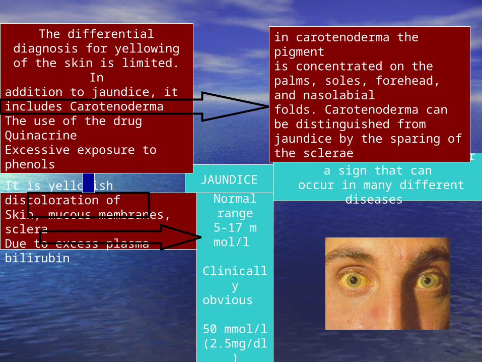
JAUNDICE
It is yellowish discoloration of Skin, mucous membranes, scleraDue to excess plasma bilirubin
Is not a disease but rather a sign that can
occur in many different diseases
Normal range
5-17 m mol/l
Clinically obvious 50 mmol/l (2.5mg/dl)
The differential diagnosis for yellowing of the skin is
limited. Inaddition to jaundice, it includes CarotenodermaThe use of the drug Quinacrine Excessive exposure to phenols
in carotenoderma the pigmentis concentrated on the palms, soles, forehead, and nasolabialfolds. Carotenoderma can be distinguished from jaundice by the sparing of the sclerae
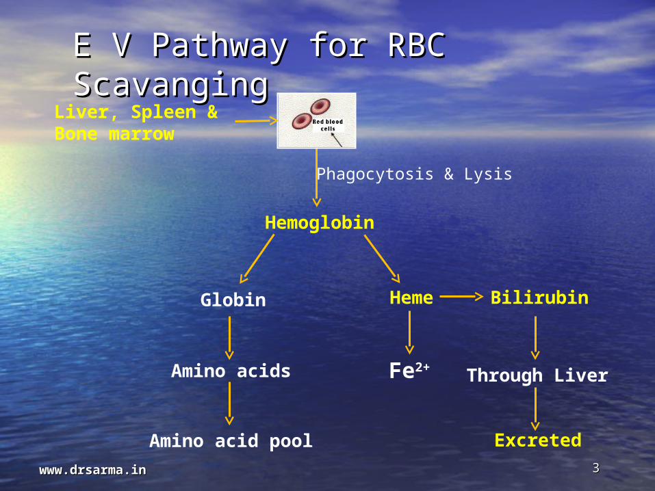
E V Pathway for RBC E V Pathway for RBC ScavangingScavanging
Liver, Spleen & Bone marrow
Hemoglobin
Globin
Amino acids
Amino acid pool
Heme Bilirubin
Fe2+
Excreted
Phagocytosis & Lysis
Through Liver
33www.drsarma.inwww.drsarma.in

Bilirubin Production & Bilirubin Production & Metabolism:Metabolism:Fo
rmatio
n o
f Biliru
bin
Main
ly in
RES (S
ple
en
)
Conju
gatio
n o
f biliru
bin
in H
epato
cyte
About 70 to 80% of the 250 to 300
mg of bilirubinproduced each day is derived
from the breakdown ofhemoglobin in senescent red
blood cellsThe remainder
comes fromprematurely destroyed
erythroid cells in bone marrow and
from theturnover of
hemoproteins such as
myoglobin and cytochromes
foundin tissues
throughout the body.
Excretion

Etiology Of Jaundice:Etiology Of Jaundice:
Incre
ase
of
pro
ductio
n
Imp
aire
d o
f C
leara
nce


Direct Hyperbilirubiemia
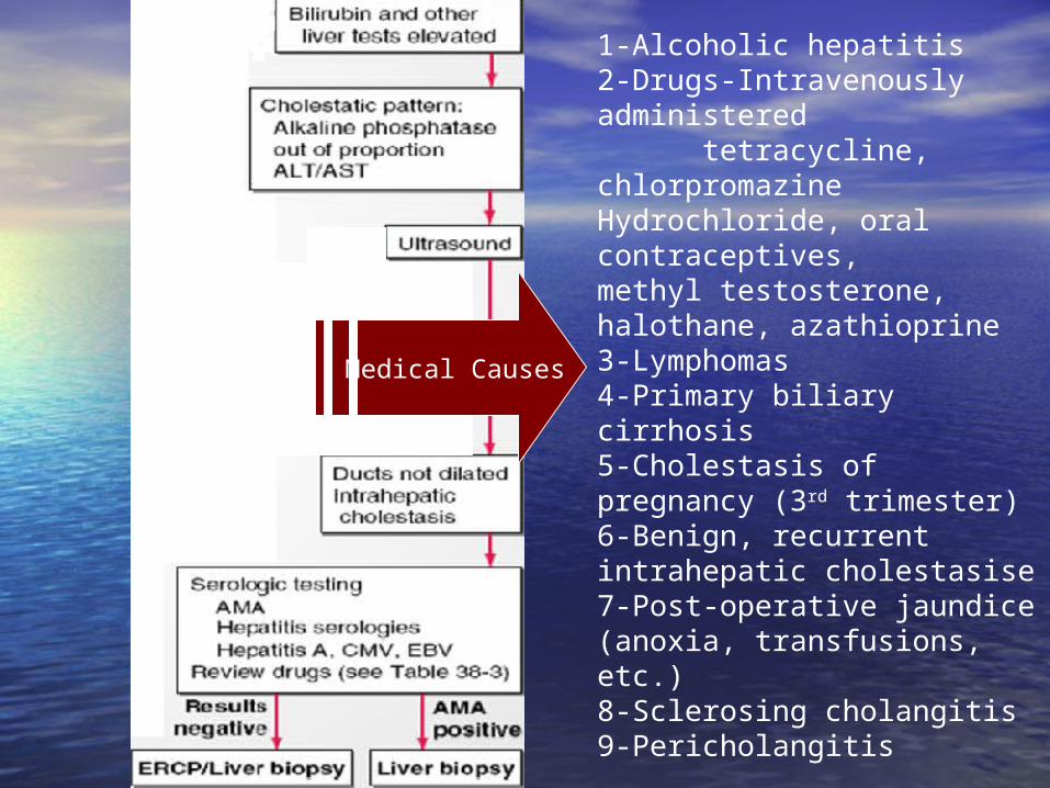
Medical Causes
1-Alcoholic hepatitis2-Drugs-Intravenously administered tetracycline, chlorpromazineHydrochloride, oral contraceptives,methyl testosterone, halothane, azathioprine3-Lymphomas4-Primary biliary cirrhosis5-Cholestasis of pregnancy (3rd trimester)6-Benign, recurrent intrahepatic cholestasise7-Post-operative jaundice (anoxia, transfusions, etc.)8-Sclerosing cholangitis9-Pericholangitis

Surgical Causes
Medical Causes
Very common (25 to 35 percent)Choledocholithiasis
Carcinoma of head of pancreasCommon (5 to 10 percent)
Carcinoma of common ductStricture of common duct
Ampullary carcinomaUncommon (I to 5 percent)
Chronic pancreatitisSclerosing cholangitis
LymphomaMetastatic carcinoma
Primary liver cell carcinomaRare (less than I percent)
Post-bulbar ulcerHepatic artery aneurysm
Choledochal cystBiliary atresia
Duodenal diverticulumhemobilia

Anatomy of biliary systemAnatomy of biliary system

Gallstones are also associated with certain medical conditions including:
1-Diabetes 2-Liver disease 3-Crohn's disease 4-Blood disorders like sickle-cell anaemia 5-Stomach surgery - gallstones are more common if you have had surgery to remove part of your stomach
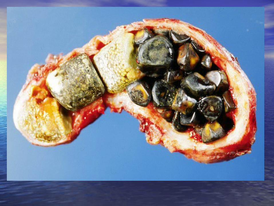
Gall bladder Stone
Risk Fa
ctors

Gall bladder StoneThe majority of cases
(approximately 80%)are asymptomatic (silent) gall
stones , discovered accidentally by abdominal
sonar .
Oth
er sy
mpto
ms a
re re
late
d to
site
of m
ovem
en
t of sto
ne
A gall stone may impact in the neck of gall
bladder or in the
cystic duct giving biliary pain or
cholecystitis
Biliary pain usually occurs in the
epigastrium and right
hypochondrium
Obstruction of common bile duct leading to pain &
jaundicePancreatitis.
Gall stones increase risk of carcinoma of the gall bladder
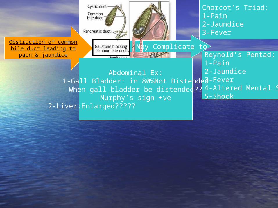
Obstruction of common bile duct leading to pain
& jaundiceMay Complicate to
Charcot’s Triad:1-Pain2-Jaundice3-Fever
Reynold’s Pentad:1-Pain 2-Jaundice3-Fever4-Altered Mental State5-Shock
Abdominal Ex:1-Gall Bladder: in 80%Not Distended
When gall bladder be distended??Murphy’s sign +ve
2-Liver:Enlarged?????

Treatment of Treatment of Choledocholithiasis:Choledocholithiasis:Preoperative Preparation:
Correct Clotting DysfunctionGuard vs LCFGuard vs RF
Definitive Treatment:Remove Source of Obstruction (stone) Remove Source of Stone (Gall bladder)
Reynold’s PentadObstructive Jaundice Charcot’s TriadChronic
cholecystitis
Treatment
ttt Of
Shock
3 rd
Genera
tion
Cephalo
sporin
ERCP
Chole
cyste
ctom
y

Carcinoma of head of pancreas
Symptoms Signs
CachecxiaCriteria of obstructive jaundicePain which is common, characterized by starting as vague
( Lower abdomen or back)Usually worsen in supine position & relived by lining forward It may be caused by:
A) Tumor invasion of splanchnic plexuses & retroperitoneum
B) Obstruction of pancreatic ductDigestive symptoms
JaundicePalpable liverPalpable gall bladderTendernessAscitesAbdominal mass In advanced cases:Nodular liverEnlarged supraclavicular
lymph node Periumblical adenopathy

Courvoisier’s sign = painless, palpable/distended gallbladder on
exam (think of CA)
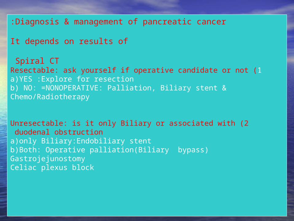
Diagnosis & management of pancreatic cancer:
It depends on results of
Spiral CT 1 )Resectable: ask yourself if operative candidate or not
a)YES :Explore for resectionb) NO: =NONOPERATIVE: Palliation, Biliary stent & Chemo/Radiotherapy
2 )Unresectable: is it only Biliary or associated with duodenal obstruction a)only Biliary:Endobiliary stentb)Both: Operative palliation(Biliary bypass)GastrojejunostomyCeliac plexus block

Whipple operation:Whipple operation:

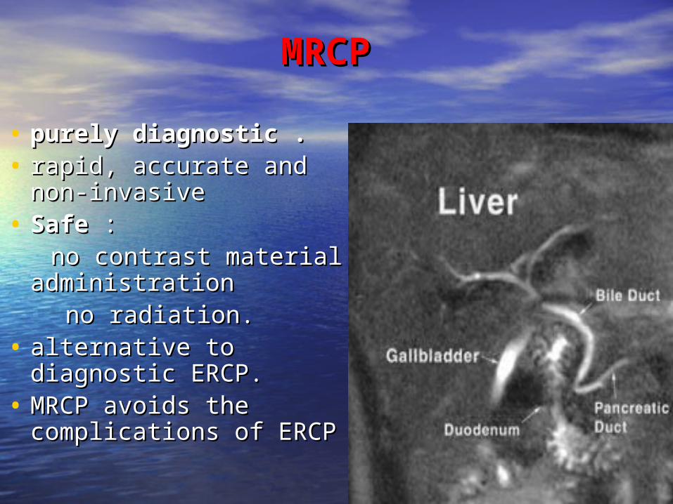
Diagnostic: MRCP and ERCPMagnetic resonance cholangiopancreatography (MRCP)– Advantage
• Detects choledocholithiasis, neoplasms, strictures, biliary dilations
• Sensitivity of 81-100%, specificity of 92-100% of choledocholithiasis
• Minimally invasive- avoid invasive procedure in 50% of patients
– Disadvantage: • cannot sample bile, test cytology, remove stone• Contraindications: pacemaker, implants, prosthetic valves
– Indications• If cholangitis not severe, and risk of ERCP high, MRCP
useful• If Charcot’s triad present, therapeutic ERCP with drainage
should not be delayed.
Endoscopic retrograde cholangiopancreatography (ERCP)-Gold standard for diagnosis of CBD stones, pancreatitis, tumors, sphincter of Oddi dysfunction-Advantage
•Therapeutic option when CBD stone identified•Stone retrieval and sphincterotomy
-Disadvantage•Complications: pancreatitis, cholangitis, perforation of duodenum or bile duct, bleeding•Diagnostic ERCP complication rate 1.38% , mortality rate 0.21%
MRCPMRCP
• purely diagnostic . purely diagnostic . • rapid, accurate and rapid, accurate and
non-invasive non-invasive • SafeSafe : : no contrast material no contrast material
administrationadministration no radiation. no radiation. • alternative to alternative to
diagnostic ERCP. diagnostic ERCP. • MRCP avoids the MRCP avoids the
complications of ERCP complications of ERCP

• Case 1: Normal MRCP. Note good Case 1: Normal MRCP. Note good delineation of normal caliber pancreatic delineation of normal caliber pancreatic and bile ducts. Fluid in stomach and and bile ducts. Fluid in stomach and duodenum also demonstrated.duodenum also demonstrated.

• Case 2: MRCP. Large common hepatic Case 2: MRCP. Large common hepatic duct stone (asterisk) within dilated duct stone (asterisk) within dilated bile ducts. Note multiple gallstonesbile ducts. Note multiple gallstones

Surgical treatment
• Endoscopic biliary drainage– Endoscopic sphincterotomy with stone extraction and
stent insertion
• CBD stones removed in 90-95% of cases
• Therapeutic mortality 4.7% and morbidity 10%, lower than surgical decompression
• Surgery– Emergency surgery replaced by non-operative biliary drainage– Once acute cholangitis controlled, surgical exploration of CBD for difficult stone removal– Elective surgery: low M & M compared with emergency survey– If emergent surgery, choledochotomy carries lower M&M compared with cholecystectomy with CBD exploration

ERCPERCP
•
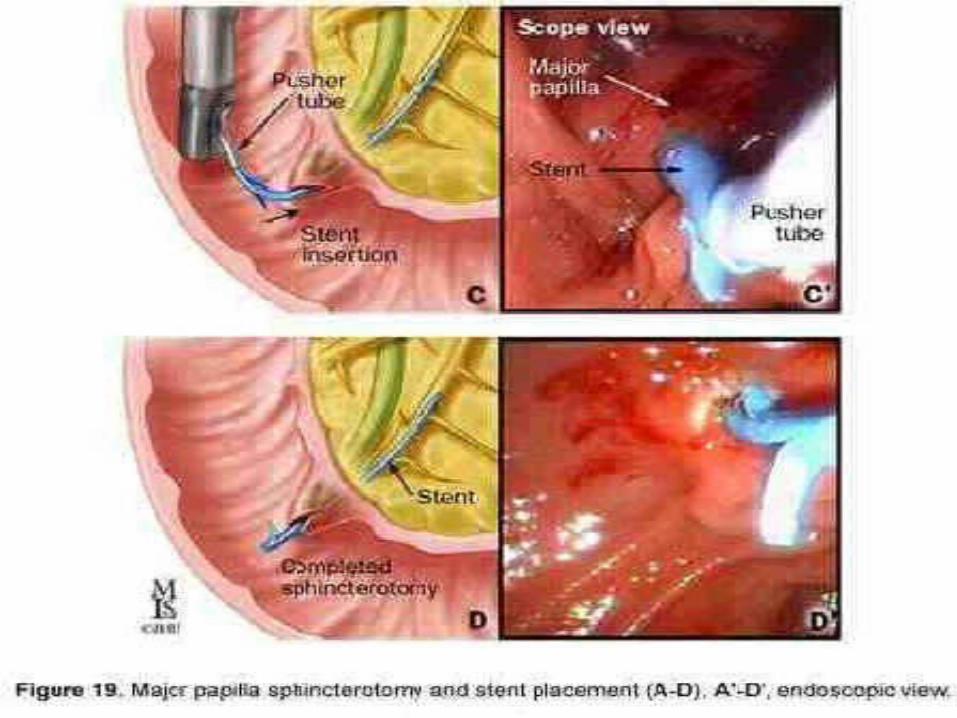

ERCP(theraputic)ERCP(theraputic)
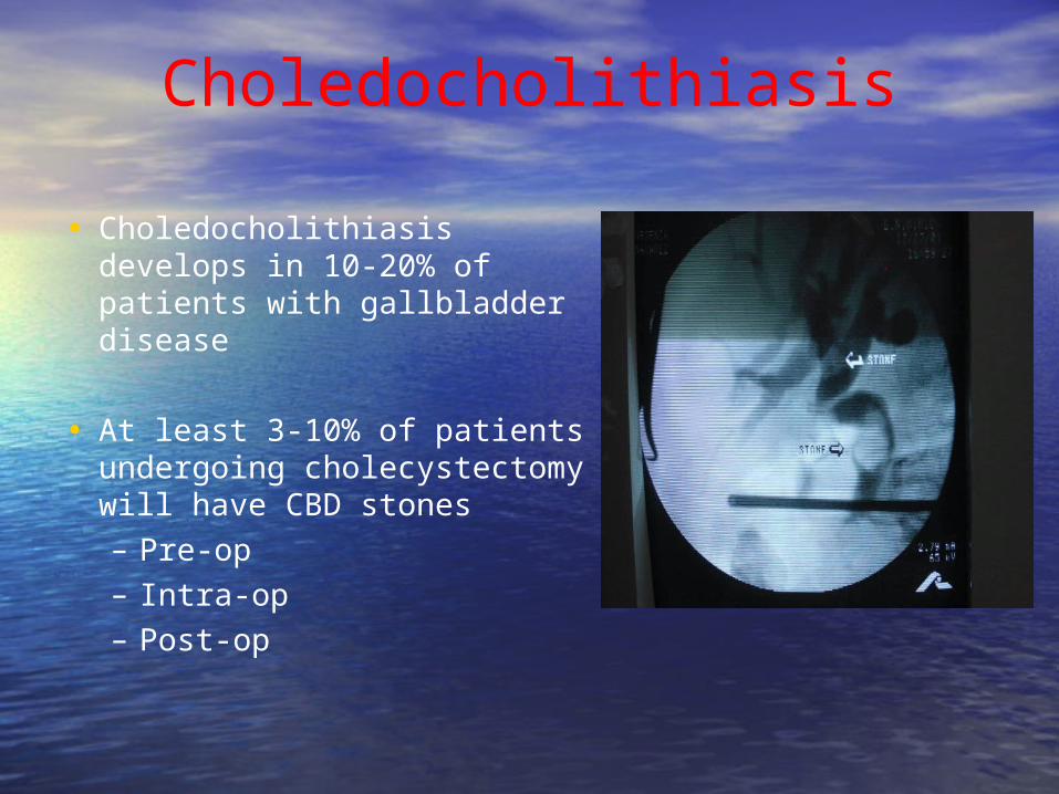
Choledocholithiasis
• Choledocholithiasis develops in 10-20% of patients with gallbladder disease
• At least 3-10% of patients undergoing cholecystectomy will have CBD stones– Pre-op– Intra-op– Post-op

Pre-op diagnosis & management
– Diagnosis: Clinical history and exam, LFTs, Abdominal U/S, CT, MRCP
• High risk (>50%) of choledocholithiasis: – clinical jaundice, cholangitis,– CBD dilation or choledocholithiasis on ultrasound– Tbili > 3 mg/dL correlates to 50-70% of CBD stone
• Moderate risk (10-50%): – h/o pancreatitis, jaundice correlates to CBD stone in
15%– elevated preop bili and AP, – multiple small gallstones on U/S
• Low risk (<5%): – large gallstones on U/S– no h/o jaundice or pancreatitis, – normal LFTs-Treatment:
•ERCP•Surgery
Intra-op diagnosis and management
• Diagnosis: intraoperative cholangiography (IOC)– Cannulation of cystic duct, filling of L and R hepatic ducts, CBD and
common hepatic duct diameter, presence or absence of filling defects.
– Detect CBD stones– Potentially identify bile duct abnormalities, including iatrogenic
injuries– Sensitivity 98%, specificity 94%– Morbidity and mortality low
• Treatment -Open CBD exploration
Most surgeons prefer less invasive techniques -Laparoscopic CBD exploration
•via choledochotomy: CBD dilatation > 6mm•via cystic duct (66-82.5%)•CBD clearance rate 97%•Morbidity rate 9.5%•Stones impacted at Sphincter of Oddi most difficult to extract
-Intraoperative ERCP

Early years: Open CBD exploration & Introduction of endoscopic
sphincterotomy• 1889, 1st CBD exploration by Ludwig
Courvoisier, a Swiss surgeon – Kocherization of duodenum and short
longitudinal choledochotomy– Stones removed with palpation, irrigation
with flexible catheters, forceps, – Completion with T-tube drainage– For many years, this was the standard
treatment for cholecystocholedocholithiasis
• 1970s, endoscopic sphincterotomy (ES)
-Gained wide acceptance as good, less invasive, effective alternative -In patients with CBD stones who have previously undergone cholecystectomy, ES is the method of choice
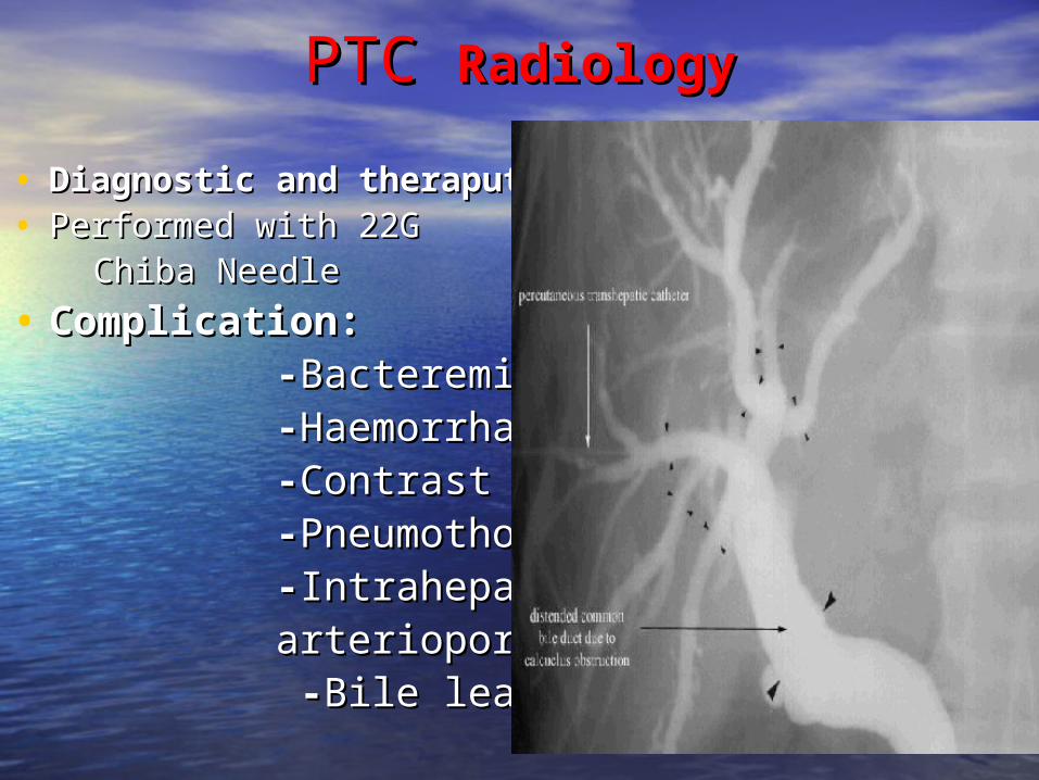
PTC PTC RadiologyRadiology
• Diagnostic Diagnostic and theraputicand theraputic• Performed with 22G Performed with 22G Chiba Needle Chiba Needle
• Complication:Complication: --Bacteremia Bacteremia --HaemorrhageHaemorrhage --Contrast reactionContrast reaction --PneumothoraxPneumothorax --IntrahepaticIntrahepatic arterioportal fistulaarterioportal fistula --Bile leakageBile leakage
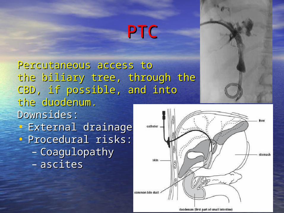
PTCPTC
Percutaneous access to Percutaneous access to the biliary tree, through the the biliary tree, through the CBD, if possible, and into CBD, if possible, and into the duodenum. the duodenum. Downsides:Downsides:• External drainageExternal drainage• Procedural risks:Procedural risks:
– CoagulopathyCoagulopathy– ascitesascites