Embed Size (px)
Citation preview

Plant Molecular Biology 51: 895–911, 2003.© 2003 Kluwer Academic Publishers. Printed in the Netherlands.
895
Jasmonate biosynthesis and the allene oxide cyclase family of Arabidopsisthaliana
Irene Stenzel1, Bettina Hause2, Otto Miersch1, Tobias Kurz1, Helmut Maucher3, HeikoWeichert3, Jörg Ziegler1, Ivo Feussner3 and Claus Wasternack1,∗1Institute of Plant Biochemistry, Department of Natural Product Biotechnology, Weinberg 3, 06120Halle/Saale,Germany (∗author for correspondence; e-mail [email protected]); 2Institute of Plant Biochem-istry, Department of Secondary Metabolism, Weinberg 3, 06120 Halle/Saale, Germany; 3Institute for Plant Scienceand Crop Research (IPK), Department of Molecular Cell Biology, Corrensstrasse 3, 06466 Gatersleben, Germany
Received 21 April 2002; accepted 19 September 2002
Key words: allene oxide cyclase family, Arabidopsis thaliana, jasmonate biosynthesis, opr3 mutant, oxylipins
Abstract
In biosynthesis of octadecanoids and jasmonate (JA), the naturally occurring enantiomer is established in a step catalysedby the gene cloned recently from tomato as a single-copy gene (Ziegler et al., 2000). Based on sequence homology, fourfull-length cDNAs were isolated from Arabidopsis thaliana ecotype Columbia coding for proteins with AOC activity. Theexpression of AOC genes was transiently and differentially up-regulated upon wounding both locally and systemically andwas induced by JA treatment. In contrast, AOC protein appeared at constitutively high basal levels and was slightly increasedby the treatments. Immunohistochemical analyses revealed abundant occurrence of AOC protein as well as of the precedingenzymes in octadecanoid biosynthesis, lipoxygenase (LOX) and allene oxide synthase (AOS), in fully developed tissues, butmuch less so in 7-day old leaf tissues. Metabolic profiling data of free and esterified polyunsaturated fatty acids and lipidperoxidation products including JA and octadecanoids in wild-type leaves and the jasmonate-deficient mutant OPDA reductase3 (opr3) revealed preferential activity of the AOS branch within the LOX pathway. 13-LOX products occurred predominantlyas esterified derivatives, and all 13-hydroperoxy derivatives were below the detection limits. There was a constitutive high levelof free 12-oxo-phytodienoic acid (OPDA) in untreated wild-type and opr3 leaves, but an undetectable expression of AOC. Uponwounding opr3 leaves exhibited only low expression of AOC, wounded wild-type leaves, however, accumulated JA and AOCmRNA. These and further data suggest regulation of JA biosynthesis by OPDA compartmentalization and a positive feedbackby JA during leaf development.
Abbreviations: α-LeA, α-linolenic acid; AOC, allene oxide cyclase; cet, mutant with constitutive expression ofthionin; dad1, mutant with delayed anther dehiscence1; dn-OPDA, dinor-12-oxo-phytodienoic acid; 13-HPOT,13S-hydroperoxy-(9Z,11E,15Z)-octadecatrienoic acid; JA, jasmonic acid; JAME, JA methyl ester; L.A, linoleicacid; LOX, lipoxygenase; OPDA, 12-oxo-phytodienoic acid; opr3, mutant defective in OPR3; OPR3, OPDAreductase3; PLA1, phospholipase of the A1 type; PUFA, polyunsaturated fatty acids; SA, salicylate
Introduction
Generation of lipid-derived signal molecules is acommon phenomenon in higher organisms. In plantsjasmonates and octadecanoids are of particular im-
The nucleotide sequence data reported will appear in the EMBLGenBank and DDBJ Nucleotide Sequence Databases under the ac-cession numbers AJ308483 (AOC1); AJ308484 (AOC2); AJ308485(AOC3) and AJ308486 (AOC4).
portance (Bergey et al., 1996; Ryan, 2000). Theyoriginate from polyunsaturated fatty acids (PUFA) andare formed by one of the seven different branchesof the LOX pathway, the AOS branch (Feussner andWasternack, 2002). The other branches lead to leafaldehydes and leaf alcohols as well as various deriv-atives of PUFAs such as epoxy-, hydroxy-, keto- orether PUFA and epoxy hydroxy PUFA (Feussner andWasternack, 2002).

896
Figure 1. Scheme of JA biosynthesis and further13-LOX products derived from α-LeA, 13-HPOT and(9Z,11E,15Z)-13-keto-(9,11,15)-octadecatrienoic acid (13-KOT).Identical reactions occur with LA as substrate, whereas thecorresponding 9-derivatives are formed via 9-LOX catalysis.
The biosynthesis of jasmonic acid (JA) and itsmethyl ester (JAME) was elucidated by Vick and Zim-merman (1983) and Hamberg and Hughes (1988).With α-linolenic acid (α-LeA) as substrate, molecu-lar oxygen is inserted by a 13-LOX at carbon atom13 leading to the formation of a fatty acid hydroper-oxide 13S-hydroperoxy-(9Z,11E,15)-octadecatrienoicacid, 13-HPOT (Figure 1). This compound is de-hydrated by the allene oxide synthase (AOS) to anunstable allene oxide which can be either hydrol-ysed non-enzymatically to α- and γ -ketols or cyclizedto racemic 12-oxo-phytodienoic acid (OPDA). In thepresence of an allene oxide cyclase (AOC), preferen-tial formation of the (9S,13S) enantiomer of OPDAoccurs. The AOC-catalysed step is regarded as the cru-cial step in octadecanoid and jasmonate biosynthesisbecause only this enantiomeric form is the substrate
for the naturally occurring (+)-7-iso-JA, which isformed after reduction of OPDA by a specific OPDAreductase (OPR3) and three cycles of β-oxidation(Schaller et al., 2000; Ziegler et al., 2000).
Several cDNAs coding for LOXs, AOSs and OPRshave been cloned from different plant species as re-viewed by Schaller (2001) and Feussner and Waster-nack (2002). The first AOC was recently cloned fromtomato and found to be encoded by a single-copygene (Ziegler et al., 2000). Treatment of plants withproducts of the AOS branch, the octadecanoids andjasmonates, led to accumulation of mRNAs codingfor LOX, AOS, AOC and OPR3 suggesting a feed-forward regulation in JA biosynthesis (Sasaki et al.,2001). Also, biotic or abiotic stresses, which resultin endogenous increases of octadecanoids and jas-monates, are usually accompanied by a transcriptionalup-regulation of AOS, AOC and OPR3 (Howe et al.,2000; Maucher et al., 2000; Ziegler et al., 2000).However, the function of this mRNA accumulation isnot understood, because it was not accompanied byendogenous formation of jasmonates as shown by iso-topic dilution analysis using barley and tomato leavesduring the first 24 h of mRNA accumulation (Kramellet al., 2000; Miersch and Wasternack, 2000). Fur-thermore, the rise in JA upon wounding appearedbefore the onset of AOS or AOC mRNA accumulation(Ziegler et al., 2001; Stenzel, 2002).
A chloroplast location of the first half of biosyn-thetic steps to JA is generally assumed. Since chloro-plast LOXs are predominantly localized within thestroma and the inner envelope membranes of chloro-plasts (Feussner et al., 1995; Blée and Joyard, 1996),α-LeA of chloroplast envelope membranes is sug-gested as the substrate for jasmonate biosynthesis. In-deed, the recently identified jasmonate-deficient mu-tant dad1 (delayed anther dehiscence1) is defectivein a chloroplast-located phospholipase of the A1 type(PLA1) (Ishiguro et al., 2001). Furthermore, it seemsubiquitous that the location of 13-LOXs, AOSs andAOC is the chloroplast, since the proteins were de-tected in the chloroplast by import studies (Bell et al.,1995; Froehlich et al., 2001), by chloroplast iso-lation (Harms et al., 1995; Feussner et al., 1995)or by immunocytochemical analysis (Feussner et al.,1995; Ziegler et al., 2000). Even the barley AOSlacking a transit sequence for chloroplast import wasimmunocytochemically shown to be located withinthe chloroplast (Maucher et al., 2000). By contrast,OPR3 carries a peroxisomal target sequence (Stintziand Browse, 2000), and might thus be co-localized
897
with the subsequent β-oxidation steps in the peroxi-somes (Kindl, 1987). It is still unclear, however, howmetabolites in JA biosynthesis are channelled betweenthe two compartments involved (Laudert and Weiler,1998; Froehlich et al., 2001).
In Arabidopsis, AOS (Kubigsteltig et al., 1999) andOPR3 (Sanders et al., 2000; Stintzi and Browse, 2000)were shown to be expressed at distinct stages of flowerdevelopment. Two mutants defective in OPR3 (dde1,Sanders et al., 2000; opr3, Stintzi and Browse, 2000)and the dad1 mutant are male-sterile. In leaves ofthe opr3 mutant, OPDA accumulated upon wounding,and plants were resistant to fungal and insect attack(Stintzi et al., 2001). Thus, this mutant is a powerfultool to dissect distinct signalling properties of OPDAand JA which have been suggested by several groups(Blechert et al., 1999; Kramell et al., 2000; Vollenwei-der et al., 2000). Furthermore, opr3 plants seem to bea useful tool to analyse regulation of JA biosynthesis.
In Arabidopsis cDNAs for all enzymes of JAbiosynthesis have been cloned, except for AOC andthose of β-oxidation. To date, two different LOX cD-NAs have been described (AtLOX2, Bell and Mullet,1993; AtLOX1, Melan et al., 1993). Recently, fourother LOX cDNAs were listed in the databases (At-LOX3, AAF79461; AtLOX4, AAF21176; AtLOX5,CAC19365; AtLOX6, AAG52309). Out of these sixLOXs, four might be involved in JA biosynthesis(AtLOX2, 3, 4, 6) (Bell et al., 1995; I. Feussner, un-published). For AOS, a single-copy gene was found(Laudert et al., 1996). Of the three different OPRs,only the recently cloned OPR3 exhibited the exclusiveformation of the naturally occurring cyclopentanonecompound (Müssig et al., 2000; Sanders et al., 2000;Schaller et al., 2000).
The unique role of AOC in generating the correctenantiomeric form in JA biosynthesis, and its specificoccurrence in all vascular bundles and in flower tissuesof tomato (Hause et al., 2000a) prompted us to analyseAOC(s) of Arabidopsis.
Here, we describe cloning and characterization offour cDNAs coding for proteins with AOC activity.Northern blot analysis revealed transient and differen-tial expression upon wounding. However, AOC, LOXas well as AOS protein appeared constitutively in fullydeveloped leaf tissues with only minor additional ac-cumulation upon wounding. Untreated wild-type andopr3 leaves exhibited a constitutive high level of freeOPDA, but undetectable expression of AOC. Uponwounding opr3 leaves exhibited only low expressionof AOC; wounded wild-type leaves, however, accu-
mulated JA and AOC mRNA. These and further dataon profiles of free and esterified PUFAs and oxylip-ins suggest regulation of JA biosynthesis by OPDAcompartmentalization and positive feedback duringgrowth.
Material and methods
Materials
Opr3 mutant seeds (in the Wassilewskija background)were kindly provided by Prof. J. Browse and DrA. Stintzi. All non-isotopic jasmonates and octade-canoids were prepared or purchased, checked on pu-rity and used as described (Kramell et al., 2000; Hauseet al., 2000a). (13S)-HPOT was prepared from α-LeA by incubation with soybean LOX (Sigma, St.Louis, MO). Other hydroperoxy and hydroxy PUFAsas well as other oxylipins were prepared or purchasedas described (Kohlmann et al., 1999; Weichert et al.,1999).
The [2H6]-JA was synthesized as described (Mier-sch, 1991). [2H5]-OPDA was prepared from [17-2H2,18-2H3]-linolenic acid (Zimmerman and Feng,1978), and both compounds were used as internalstandards in GC-MS analysis. Rabbit polyclonal an-tibodies against the lipid body LOX of cucumber(Hause et al., 2000b), the recombinant AOS of A.thaliana (Laudert and Weiler, 1998) and the recom-binant AOC2 of A. thaliana were used.
Plant growth and treatments
A. thaliana ecotype Columbia and ecotype Was-silewskija were cultivated in controlled chambers (Per-cival, CLF) at 70% relative humidity under short-dayconditions of 8 h light,
210 µE m−2 s−1 for 6 weeks. For treatments thecomplete rosette of a plant was cut above the roots andfloated on distilled water, 50 µM JAME, 50 µM 13-HPOT, 1 M sorbitol, 0.5 M glucose, 50 µM salicylate(SA) or 1 M NaCl for indicated times. Wounding wasperformed by crushing the leaves with a forceps acrossthe mid-vein.
cDNA isolation, RT-PCR, cloning and sequencing
The cDNA library prepared from 4-week old rosetteleaves of ecotype Columbia treated for 24 h with100 µM JAME and maintained in γ ZAP expressStratagene was kindly provided by Dr Stefan Bau

898
(Cologne). The cDNA library was hybridized withthe full-length AOC cDNA from tomato (Ziegleret al., 2000) (55 ◦C, 5× SSC, 5× Denhardt’s reagent,100 µg/ml shared salmon sperm DNA, 0.1% w/v SDS,1 × 106 dpm ml−1 of labelled probe, 18 h). Positiveclones were isolated and converted into phagemids byin vivo excision, and were sequenced on both strandswith IRO 700 labelled primers with the ThermoSequenase DYEnamic Direct Cycle Sequencing Kit(Amersham Pharmacia Biotech). Only one AOC ho-mologue of 991 bp was isolated and designated asAOC1.
Based on three other database sequences with par-tial homology to the AOC sequence, we used RT-PCRfor isolation of full-length cDNAs. mRNA isolatedfrom rosette leaves floated on 100 µM JAME for2 h was used with the following primer combinations:AOC2, 5′ primer CCAATCAAGTAGAGTTTCTTC,3′ primer ACACAGCGATACGAGAAAC; AOC3, 5′primer CCATTAAAACCAAAGATCAATC, 3′ primerCTAACCTCACACAACTCATC; AOC4, 5′ primerATTAAACTAACTCAGGCAAG, 3′ primer CCAT-GACAAAGACACCCAC. Generated RT-PCR prod-ucts were cloned into the vector pCR II TOPO (In-vitrogen) and sequenced.
RNA and immunoblot analysis
Total RNA was extracted from frozen tissues withbuffer (4 M guanidinium isothiocyanate, 50 mMsodium citrate pH 4.0, 0.5% sarcosyl, 0.1% 2-mercaptoethanol) and purified by treatments withbuffered phenol/chloroform/isoamyl alcohol 25:24:1(v/v/v). After ethanol precipitations 20 µg per lanewas subjected to RNA gel blot analysis according toSambrook et al. (1989). Gel loading was checkedby comparing ethidium bromide-stained rRNAs. Hy-bridization was performed at 60 ◦C for 16 h with32P-labelled full-length cDNAs of AOC1 (Figure 6) orof AOC3 (Figures 5 and 9), and AOS of A. thaliana.For the creation of gene-specific probes used in Fig-ure 5, we amplified 150 bp of the 3′-untranslatedregion (3′-UTR) starting after the stop codon of eachAOC. These PCR products were cloned into the pCRII-TOPO vector and sequenced. The specificity ofeach probe was tested with PCR-amplified genomicfragments which contained the complete sequenceas well as 150 bp of the 5′-UTR and 3′-UTR ofeach AOC. Gel loading was checked by comparingethidium bromide-stained rRNAs. Proteins were iso-lated from the phenolic phase and were used for
separation and immunoblot analysis as described byHause et al. (1996, 2000a). Immunoblot analysis wasperformed by using an antibody raised against re-combinant AOC2 (A. thaliana) (1:5000 dilution) oragainst recombinant AOS (A. thaliana) (1:2500 di-lution) or against cucumber lipid body LOX (Hauseet al., 2000b) (1:1000 dilution).
AOC expression in E. coli and AOC activity assay
Expression of AOC1, AOC2, AOC3 and AOC4was performed after cloning of amplified PCRfragments for each AOC as follows. For over-expression of AOC1, a fragment covering aminoacids 31–253 was generated with the 5′ primer CGCGGATCCC159TTGGTTTCTCTAAATCCTT (BamHIrestriction site underlined) and the 3′ primer ACCGGTCGACC835ACTAATTTGTAAAGTTGCTT AC (SalIrestriction site underlined). After cloning into the pCRII-TOPO vector and sequencing, the 680 bp fragmentwas subcloned into pQE30 with the BamHI/SalI re-striction sites. For over-expression of AOC2, AOC3and AOC4, the same approach was used withCGCGGATCCC139TTGGTTCCTCTAAATCCTT as5′ primer and ACCGGTCGACA812ATTAGTTGGTATAgTTACTTAT as 3′ primer for AOC2, CGCGGATCC169TTGGTTTCTCAAGATCTTTC as 5′ primerand ACCGGTCGACA851CTTAATTAGTAAAGTTACTTAT as 3′ primer for AOC3, and, finally,CGCGGATCCC114 TCGGTTTCTCTAGATCCTT as5′ primer and ACCGGTCGACT790TTCAATTAGTAAAGTTAGCGAT as 3′ primer for AOC4. pQE30,without or with these inserts, were transformedinto the host strain E. coli M15. Total protein ofisopropyl-β-thiogalactopyranoside (IPTG)-induced ornon-induced cultures were isolated and purified asdescribed (Maucher et al., 2000). The resulting super-natant was used for AOC activity assay as described(Ziegler et al., 1997, 1999, 2000).
Quantitative measurement of α-linolenic acid,jasmonates, octadecanoids and other LOX-derivedcompounds
For quantitative analysis of OPDA, dinor-12-oxo-phytodienoic acid (dn-OPDA) and JA, 1 g (freshweight) of plant tissue was frozen with liquid nitrogen,homogenized in a mortar and extracted with 10 mlof methanol, and 100 ng of [2H6]-JA and 100 ng of[2H5]-OPDA were added as internal standards. Thefiltrate was loaded onto 3 ml of DEAE-SephadexA25 columns (Ac− form, methanol), and the columns

899
were washed with 3 ml of methanol. After subse-quent washing with 3 ml of 0.1 M acetic acid inmethanol, eluents obtained with 3 ml of 1 M aceticacid in methanol and with 3 ml of 1.5 M acetic acidin methanol were collected, evaporated and separatedon preparative HPLC (column: Eurospher 100-C18,5 µm, 250 mm × 4 mm, flow rate 1 ml/min). Sepa-ration was performed with solvent A (methanol) andsolvent B (0.2% acetic acid in H2O) with a gradientof 40% solvent A to 100% within 25 min. Fractionseluting between 12 and 13.3 min, between 18.30 and20 min and between 20.30 and 22 min were collectedand evaporated. For subsequent derivatization sampleswere dissolved in a mixture of 200 µl CHCl3 andN,N-diisopropylethylamine (1:1, v/v) and derivatizedwith 10 µl of pentafluorobenzyl bromide at 20 ◦Covernight. The samples were evaporated, dissolved in5 ml of n-hexane and passed through a SiOH column(500 mg; Machery-Nagel). The pentafluorobenzyl es-ters were eluted with a mixture of 7 ml of n-hexaneand diethyl ether (2:1, v/v). After evaporation probeswere dissolved in 100 µl of acetonitrile and subjectedto GC-MS analysis with a GCQ Finnigan instrumentin the following conditions: 70 eV, NCI, ionizationgas NH3, source temperature 140 ◦C, column Rtx-5w/Integra Guard (Restek, Germany), 5 m inert pre-column; 30 m × 0.25 mm, 0.25 µm film thickness,injection temperature 250 ◦C, interface temperature275 ◦C; helium 40 cm/s; splitless injection; columntemperature program: 1 min at 100 ◦C, 25 ◦C/min to200 ◦C, 5 ◦C/min to 300 ◦C, 20 min at 300 ◦C. Reten-tion times were for [2H6]-JA-pentafluorobenzyl ester11.92 min, for JA-pentafluorobenzyl ester 11.98 min,for dn-OPDA-pentafluorobenzyl ester 18.59 min, for[2H5]-OPDA-pentafluorobenzyl ester 21.31 min, andfor OPDA-pentafluorobenzyl ester 21.39 min. Frag-ments m/z 209 (JA), 215 (JA-standard), 291 (OPDA),296 (OPDA-standard) and 263 (dn-OPDA) were usedfor quantification.
PUFA and oxylipins were extracted, derivatizedand separated into the corresponding positional andstereoisomeric forms by the following methods. Ox-idized fatty acids were extracted according to themethod of Weichert et al. (1999); for esterified deriv-atives 0.5 g fresh weight (f.w.) and for free fatty acidderivatives 1.0 g f.w. of frozen leaf tissue was addedto 10 ml of extraction solvent (isohexane/isopropanol,3:2 v/v with 0.0025% w/v BHT) and immediately ho-mogenized with an Ultra Turrax under a stream ofargon on ice for 30 s. The extract was centrifuged at4500 × g at 4 ◦C for 10 min. The clear upper phase
was collected and the pellet extracted three times with3 ml each of extraction solvent. To the combinedorganic phases a 6.7% w/v solution of potassium sul-fate was added to a volume of 47 ml. After vigorousshaking the upper hexane-rich layer was removed.The upper organic phase containing the oxylipin andfatty acid derivatives was dried under nitrogen and re-dissolved in 1.5 ml of isohexane/2-propanol (100:5v/v), and stored under argon at −80 ◦C until use.
For the analysis of esterified fatty acids, the sol-vent was removed, and 333 µl of a mixture of tolueneand methanol (1:1 v/v) and 167 µl of 0.5 mM sodiummethoxide was added. As internal standards trihep-tadecanoate and triricinoleate were added (100 µg perprobe). After incubation of the samples for 20 min,0.5 ml of 1 M sodium chloride and 50 µl of HCl(37% v/v) were added, and fatty acid methyl esterswere extracted twice each with 0.75 ml of hexane.The combined organic phases were evaporated to dry-ness under a nitrogen stream and the correspondingfatty acid methyl esters were reconstituted in 50 µl ofmethanol/water/acetic acid (85:15:0.1, v/v).
For the analysis of non-esterified oxylipinsin the isohexane/isopropanol extract, the solventwas removed and the sample was solved in400 µl of methanol. As internal standards heptade-canoic acid and (15S,11Z,13E)-15-hydroxy-11,13-eicosadienoic acid were added. Then 10 µl of anEDAC (1-ethyl-3-(3-dimethylaminopropyl)-carbodii-mide) solution (1 mg EDAC in 10 µl methanol) wasadded and incubated for 2 h. After adding 200 µl of0.1 M Tris-HCl, pH 7.5, the fatty acid methyl es-ters were extracted twice each with 1 ml of hexane.The combined organic phases were evaporated to dry-ness under a nitrogen stream and the correspondingfatty acid methyl esters were reconstituted in 50 µl ofmethanol/water/acetic acid (85:15:0.1, v/v).
Oxylipins were analysed by HPLC on an Agilent1100 HPLC system coupled to a diode array detector(Weichert et al., 1999). At first oxylipins were puri-fied on reversed-phase HPLC. This was carried out ona ET250/2 Nucleosil 120-5 C18 column (Macherey-Nagel, 2.1 mm × 250 mm, 5 µm particle size) withmethanol/water/acetic acid (85:15:0.1, v/v) as solventsystem, at a flow rate of 0.18 ml/min. Straight-phaseHPLC of the hydro(pero)xy fatty acids was carriedout on a Zorbax Rx-SIL column (Agilent, 2.1 mm× 150 mm, 5 µm particle size) with n-hexane/2-propanol/acetic acid (100:1:0.1, v/v) as solvent sys-tem, at a flow rate of 0.1 ml/min. Chiral-phase HPLCof the hydro(pero)xy fatty acids was carried out on a

900
Chiralcel OD-H column (Daicel, 2.1 mm × 150 mm,5 µm particle size) with n-hexane/2-propanol/aceticacid (100:5:0.1, v/v) as solvent system, at a flow rate of0.1 ml/min. Absorbance at 234 nm was monitored. Alloxylipins were identified by comparison to the elutiontimes of authentic standards.
Analysis of fatty acid derivatives was carried outby gas chromatography (GC). GC analysis was per-formed with an Agilent GC 6890 system coupledwith a FID detector and equipped with a capillaryHP INNOWAX column (0.32 mm × 30 m; 0.5 µmcoating thickness; Agilent, Germany). Helium wasused as carrier gas (30 cm/s). The samples were mea-sured with a split of 60:1 at an injector temperatureof 220 ◦C. The temperature gradient was 150 ◦C for1 min, 150–200 ◦C at 15 ◦C/min, 200–250 ◦C at2 ◦C/min, and 250 ◦C for 10 min. All fatty acids wereidentified by comparison with the retention times ofauthentic standards.
Immunocytochemistry
Immunocytochemical analysis was performed as de-scribed recently (Hause et al., 2000a). Cross-sectionsof leaves (2 µm thickness) were immuno-labelled withanti-AOC2 antibody (A. thaliana) (diluted 1:2000),anti-LOX antibody (cucumber, diluted 1:500) or anti-AOS antibody (A. thaliana, diluted 1:2000). As asecondary antibody a goat anti-rabbit IgG conjugatedwith Alexa 488 (Molecular Probes, Eugene, OR) wasused in the dilution 1:500. Each immunocytochemicalstaining was analysed under an epifluorescence micro-scope (Axioskop, Carl Zeiss, Jena, Germany) with theproper filter combination.
Results
Cloning of four cDNAs coding for allene oxidecyclase: primary sequence and gene structure
To identify the A. thaliana homologue(s) of the re-cently cloned tomato AOC (Ziegler et al., 2000),sequence database information revealed existence offour homologous genes. Full-length cDNAs were iso-lated by screening a λZAP expression library madefrom mRNA of rosette leaves treated with 100 µMJAME for 24 h, or were generated via RT-PCR frommRNA of rosette leaves floated on 100 µM JAMEfor 2 h. For this, specific primers for the putativeAOC homologues found in the sequence databasewere used. The four different full-length cDNAs were
Table 1. Molecular characteristics of AOC1, AOC2, AOC3 andAOC4 of A. thaliana ecotype Columbia.
AOC1 AOC2 AOC3 AOC4
ORF (bp) 762 759 774 759
Number of amino acids 254 253 258 253
Molecular mass (kDa) 27.8 27.6 28.4 27.7
pI (full-length) 9.11 6.91 9.19 9.27
pI (without putative chloroplast
target sequence) 5.94 5.4 8.7 8.37
Location on chromosome 3 3 3 1
designated as AOC1, AOC2, AOC3 and AOC4. Theopen reading frames ranged from 759 to 774 bp (Ta-ble 1), corresponding to proteins containing 253–258amino acid residues. The calculated molecular massesfor all AOCs were about 28 kDa. Using full-lengthsequences, a neutral pI was calculated for AOC2,whereas for AOC1, AOC3 and AOC4 an alkaline pIwas found. In contrast, calculations with sequenceslacking the putative chloroplast signal peptide re-vealed an acidic pI for AOC1 and AOC2, whereasfor AOC3 and AOC4 an alkaline pI was calculated(Table 1).
For activity assays, 5′-truncated cDNAs werecloned into the pQE30 vector and expressed in E. coli.Induction of expression in bacteria led to the appear-ance of an additional band at 26 kDa, which wasabsent in control bacteria transformed with the emptyvector only (data not shown). Examination of bacterialextracts on AOC activity was performed as described(Ziegler et al., 1997, 1999). The assay, which includedrecombinant AOS protein of barley as a helper enzyme(Maucher et al., 2000) revealed cis-(+)-OPDA forma-tion with AOC1, AOC2, AOC3 and AOC4, with thehighest activity for AOC2 (data not shown). Giventhe specificity of the assay conditions, the cis-(+)-OPDA formation is indicative of AOC activity. In eachAOC sequence of Arabidopsis, N-glycosylation sites,phosphorylation sites for protein kinase C, tyrosine ki-nase and casein kinase II, as well as N-myristolationsites were identified by computional analysis, buttheir functionality has not been tested yet. A com-mon feature of AOC1, AOC2, AOC3 and AOC4 isthe occurrence of a putative chloroplast transit pep-tide. All N-terminal regions are rich in Ser residues(26–30% in the first 50 amino acids of AOC1–AOC4),the start Met is followed by an Ala residue, andamong the first 10 amino acids there is no charged

901
Figure 2. Amino acid sequence comparison of the four isolated AOCs from A. thaliana ecotype Columbia with that of tomato Lycopersiconesculentum cv. Lukullus published recently (Ziegler et al., 2000). Putative chloroplast transit peptides are boxed in light grey. Conservedsequences are boxed in dark-grey and marked with asterisks.
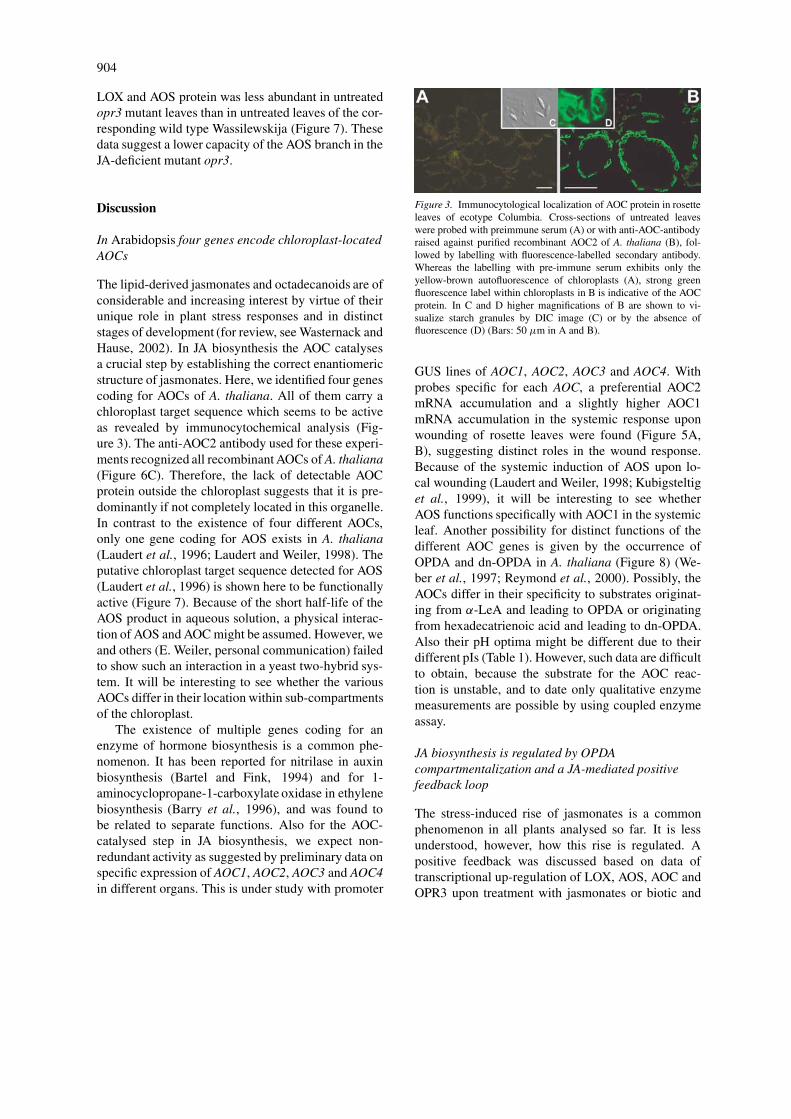
amino acid. Computer analysis of the first 100 aminoacids was performed with the ChloroP V 1.1 program(http://www.dtu.dk/services/ChloroP) (Emanuelssonet al., 1999) and the TargetP program ver. 1.0(http://www.cbs.dtu.dk/services/TargetP) (Emanuels-son et al., 2000). In both analyses a chloroplast local-ization was predicted. The putative cleavage site liesbetween the amino acids 78 and 54 for AOC1, AOC2,AOC3 and AOC4 (Figure 2, shaded in light grey).Using an anti-AOC2 antibody (see below), we con-firmed the localization of the AOC protein in chloro-plasts by immunocytological analysis. Cross-sectionsof untreated rosette leaves incubated with anti-AOC2antibody exhibited a strong green fluorescence la-bel within chloroplasts (Figure 3B, D) indicating theoccurrence of AOC protein. After incubation with pre-immune serum only yellow-brown autofluorescencewas observed (Figure 3A).
Comparison of the cDNA sequences of AOC1,AOC2, AOC3 and AOC4 with sequence databaseinformation (http://www.ucbi.nih.gov/) revealed map-ping and genomic structure as follows (Figure 4A):
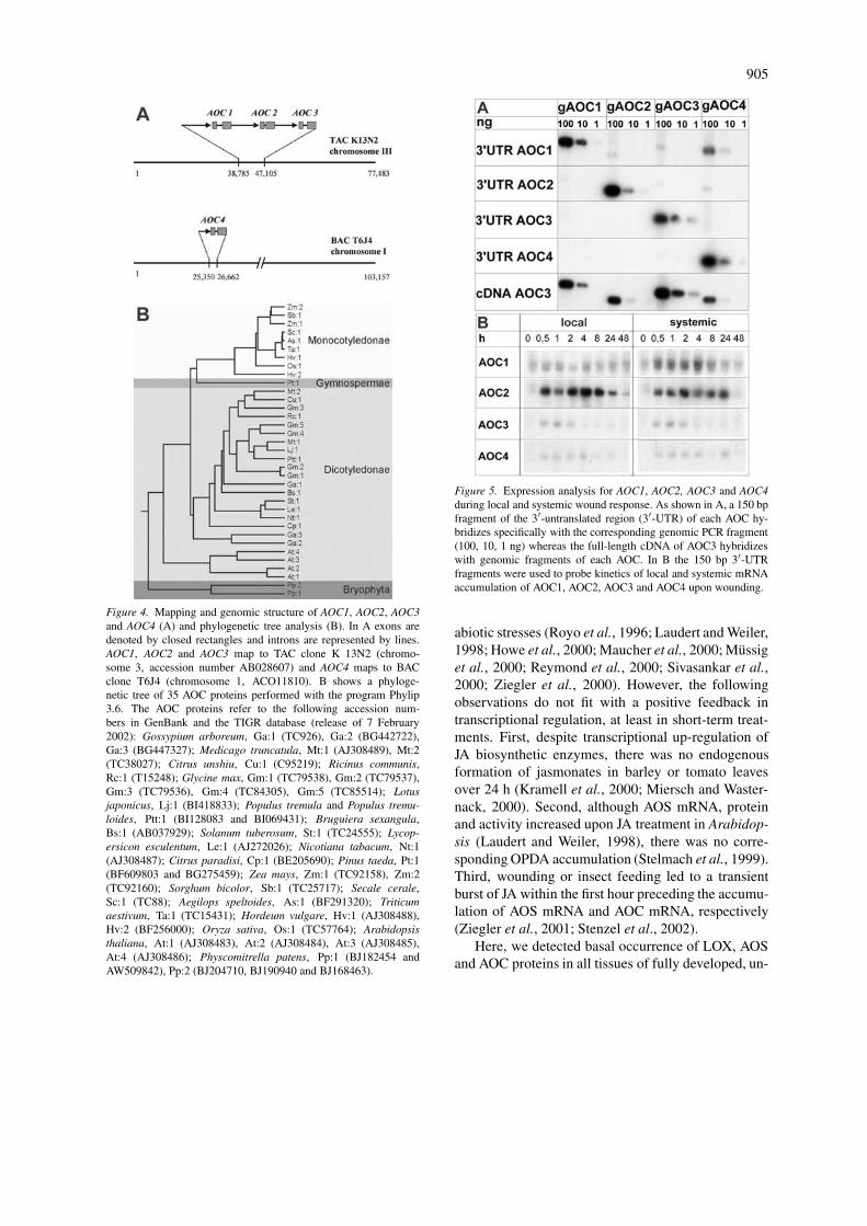
AOC1, AOC2 and AOC3 are located on chromo-some 3, whereas AOC4 is located on chromosome1. All four coding regions contain one intron of369 bp, 103 bp, 271 bp and 164 bp, respec-tively. A search with AOC1 protein sequence withthe tblastn program in the EMBL EST database(http://www.ncbi.nlm.nih.gov/blast/) (Altschul et al.,1997) and in the TIGR database (http://www.tigr.org/tab/e2K1/agi/ath1) (Altschul et al., 1990) 35 pu-tative AOC sequences could be found and recon-structed, respectively. Sequence comparison wasperformed which did not include putative chloro-plast signal sequences in the conserved region start-ing at KVYEL up to the C-terminus. Among thefour AOCs an identity of 74–94% was found be-tween AOC2, AOC3 and AOC4, whereas AOC1exhibited 60–74% identity to all other AOCs. Forphylogenetic tree analysis the program Phylip 3.6(http://www.evolution.genetics.washington.edu/phylip.html) was used (Figure 4B). The AOCs of A.thaliana fall into their own subgroup like solanaceousAOCs. The monocotyledonous AOCs are grouped in
902
a clearly different branch. Gene duplications mighthave occurred in the case of A. thaliana, Zea mays,Physcomitrella patens and Gossypium arboretum.
In order to record differential expression for AOC1,AOC2, AOC3 and AOC4, we used specific primersamplified 150 bp after the stop codon of the cor-responding 3′-UTR of each AOC. Specificity of theprimers were found, with 1, 10 and 100 ng genomicPCR products of each AOC which contained the cor-responding gene and 150 bp of each 5′- and 3′-UTR(Figure 5A). In the local and systemic wound responseof rosette leaves AOC2 mRNA accumulated signifi-cantly more than that of AOC1, AOC3 and AOC4.Furthermore, the systemic response in terms of in-crease in mRNA accumulation was stronger for AOC1than for the AOC3 and AOC4. Beside these pref-erential mRNA accumulations, each AOC exhibitedtransiently a local and systemic mRNA accumulation.
Abundant occurrence of AOC protein in leaves isindependent of stress-induced AOC mRNAaccumulation
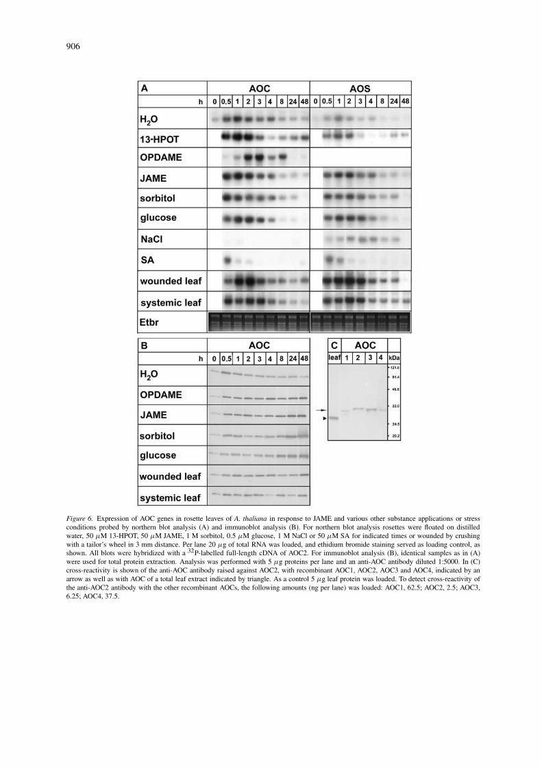
Accumulation of jasmonates and octadecanoids, oc-curring upon sorbitol treatment (Bohlmann et al.,1998) or wounding (Laudert and Weiler, 1998; Rey-mond et al., 2000) of A. thaliana leaves, correlates intiming with the mRNA accumulation of 13-LOX (Bellet al., 1995) and of AOS (Laudert and Weiler, 1998).We complemented these data by recording accumula-tion of AOC mRNA. The full-length cDNA (AOC1)which hybridized also with all other mRNAs cod-ing for AOCs was used. Total RNA of rosette leavestreated with JAME, its precursor 13-HPOT or variousstresses was probed (Figure 6A). Rapid and transientAOC mRNA accumulation occurred already in de-tached water-treated rosette leaves, reflecting a woundresponse by detachment. In contrast, untreated leavesof intact plants exhibited a negligible AOC mRNAaccumulation (Figure 8C). Treatment with 50 µMJAME or 13-HPOT (Figure 6A) led to additional AOCmRNA accumulation, which was similar upon treat-ment with OPDA or JA-amino acid conjugates (datanot shown). In case of 13-HPOT treatment, 3-foldhigher levels of OPDA and JA could be detectedwithin 30 min (data not shown), indicating endoge-nous formation of both compounds. Accumulationof AOC mRNA occurred also in a similar amountand time range, when leaves were floated on 1 Msorbitol or 0.5 M glucose. In contrast, with 50 µMSA and, in a more pronounced fashion, with 1 M
NaCl an inhibitory effect on AOC mRNA accumula-tion compared to water-treated leaves was found. Forcomparison, AOS mRNA accumulation was analysed.Except for NaCl treatment, similar if not identicalresults were found.
The AOC mRNA levels were not reflected by theAOC protein levels. In most treatments, the AOC pro-tein exhibited constitutively high level with slight risein the first hour and a significant rise after 8 h (Figure6B). The anti-AOC2 antibody was able to recognizeall four recombinant AOCs (Figure 6C). However,the AOC2 protein was preferentially recognized (cf.legend to Figure 6C). Therefore, AOC2 might be pref-erentially detected in the immunoblot analysis andthe immunocytochemical analysis. Immunocytolog-ical inspection revealed an abundant appearance ofAOC protein in all tissues of fully developed rosetteleaves of the ecotype Columbia, whereas developingrosette leaves showed less AOC protein (Figure 7).A similar distribution and abundance were detectedfor LOX and AOS at the protein level. This consti-tutive occurrence of LOX, AOS and AOC protein inuntreated fully developed leaf tissues is in apparentcontrast to the stress-induced transient accumulationof their mRNAs (Figure 6A) (Bell et al., 1995; Laudertand Weiler, 1998).
The oxylipin profiles of untreated rosette leaves revealconstitutive LOX pathway reactions with preferentialactivity of the AOS branch
The abundant occurrence of LOX, AOS and AOCproteins in untreated leaf tissues prompted us todetermine profiles of fatty acids and various LOX-derived metabolites including the initial JA-precursor13-HPOT. Each of them was recorded quantitativelyfor the ecotype Columbia in the free and the esteri-fied form (Table 2). The Arabidopsis genome containsno gene coding for divinyl ether synthase. Therefore,formation of divinyl ethers is not to be expected. Theamounts of C6 volatiles as well as α- and γ -ketolswere below the detection limit which is in the range of10 pmol per gram f.w. for all compounds tested. In thecase of esterified fatty acids a strong preponderance ofpolyenoic fatty acids was observed with preferentialoccurrence of α-LeA up to the micromolar range (Ta-ble 2). Among esterified compounds originating fromLOX activity, substantial amounts (20 nmol/g f.w.) of13-HOT were detected, whereas other LOX productswere found esterified to less than 10-fold lower levels.

903
Table 2. Amounts of free and esterified fatty acids andthe corresponding hydroxy derivatives as well as of freedn-OPDA, OPDA, and JA in untreated 6-week old rosetteleaves of ecotype Columbia. Leaf tissue was colleted from 3to 4 different plants and subjected to extraction and separationas described in Materials and methods.
Fatty acid Esterified, Free,
pmol per gram f.w. pmol per gram f.w.
16:0 620 000 222 000
16:1 60 000 n.d.a
16:3 990 000 n.d.
18:0 50 000 280 000
18:1 180 000 4 000
18:2 900 000 7 000
18:3 2 820 000 10 000
9-HOD 100 000 30
13-HODd 400 000 32
13-/9-KOD n.d. 10
9-KOD n.d. 12
9-HOT 300 28
13-HOT 20 179
13-/9-KOT 2 200 n.d.
12-HOT n.d. n.d.
16-HOT n.d. n.d.
OPDA –c 951
dn-OPDA –c 75
JA – 43
aNot detectable; b13-KOD ((9Z,11E)-13-keto-(9,11)-octadecadienoic acid); cNot determined;d(13S,9Z,11E,15Z)-hydroxy-(9,11,15)-octadecadienoicacid.
Among the free fatty acids 16:0 and 18:0 domi-nated, whereas linoleic acid (LA) and α-LeA werefound at 7 and 10 nmol per gram f.w., respectively.The substrate for generation of dn-OPDA, the 16:3fatty acid, could not be detected in its free form.Also products of the LOX reaction itself, HPOD origi-nating from LA and HPOT originating from α-LeA,could not be detected, although these hydroperoxycompounds are stable at least in part in our work-up procedure (Feussner et al., 1997). However, HODand HOT derivatives, both indicative of the reduc-tase branch of the LOX pathway, were detected withpreference to 13-HOT. LA derivatives accumulatedto more than 6-fold lower level without preferenceof a positional isomer. Among all free LOX-derivedproducts, compounds of the AOS branch were domi-nant. Nearly 1 nmol per gram f.w. OPDA was found,whereas for dn-OPDA and JA 13-fold and 22-foldlower levels were detected, respectively.
Oxylipin profiles and AOC expression in opr3 mutantleaves suggest a positive feedback loop in JAbiosynthesis
The abundant occurrence of OPDA and the 10-foldhigher level of the LOX substrate, α-LeA, as well asconstitutive high levels of LOX, AOS and AOC proteinin untreated rosette leaves raise the question of howregulation of JA biosynthesis occurs. To dissect theeffects of JA and OPDA, we performed metabolic pro-filing and expression analyses of JA biosynthetic geneswith untreated and wounded leaves of the JA-deficientmutant opr3 as well as the corresponding wild-typeWassilewskija. In the case of free and esterified fattyacids, as well as hydroxy and hydroperoxy fatty acidderivatives, most levels were similar if not identicalin the opr3 mutant compared to the wild type. Only13-HOT levels were 5-fold lower in the mutant, sug-gesting decreased capacity of the LOX-catalysed step(data not shown).
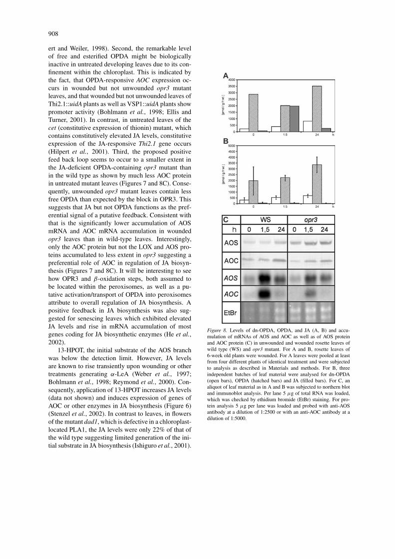
In untreated leaves of the ecotype Wassilewskijaabout 2.9 nmol per gram f.w. OPDA was found, whichis somewhat above the range of previously detectedlevels (Stelmach et al., 1999; Reymond et al., 2000;Stintzi and Browse, 2000). This amount exceeds thatof JA and dn-OPDA 13- to 35-fold, respectively (Fig-ure 8A). OPDA is also the dominant compound inuntreated opr3 mutant leaves. Despite these remark-able OPDA levels in untreated leaves of both the wildtype and the mutant, and the fact that AOC expressionis OPDA responsive, AOC mRNA accumulation wasbelow the detection limit (Figure 8C). Upon wound-ing of wild-type leaves, no dramatic changes occurredin OPDA and dn-OPDA levels compared to a 24-foldincrease in JA levels within the first 1.5 h. Even in theopr3 mutant, OPDA levels did not increase dramati-cally. The JA level decreased up to 24 h upon wound-ing of wild-type leaves, whereas OPDA and dn-OPDAlevels increased in this time. AOC mRNA and AOSmRNA accumulated abundantly within 1.5 h uponwounding of wild-type leaves (Figure 6A, Figure 8C),possibly caused by the preferential accumulation ofJA. Indeed, in wounded leaves of the opr3 mutant ex-hibiting the expected JA deficiency (Figure 8B), muchless AOC mRNA and AOS mRNA accumulated thanin the wild type during 1.5 h of wounding (Figure 8C).Most interestingly, the level of AOC protein, but notof AOS protein, was much less in the untreated opr3mutant leaves and increased slightly upon wounding(Figure 8C). This is also indicated by the immuno-cytochemical inspection, where AOC protein but not
904
LOX and AOS protein was less abundant in untreatedopr3 mutant leaves than in untreated leaves of the cor-responding wild type Wassilewskija (Figure 7). Thesedata suggest a lower capacity of the AOS branch in theJA-deficient mutant opr3.
Discussion
In Arabidopsis four genes encode chloroplast-locatedAOCs
The lipid-derived jasmonates and octadecanoids are ofconsiderable and increasing interest by virtue of theirunique role in plant stress responses and in distinctstages of development (for review, see Wasternack andHause, 2002). In JA biosynthesis the AOC catalysesa crucial step by establishing the correct enantiomericstructure of jasmonates. Here, we identified four genescoding for AOCs of A. thaliana. All of them carry achloroplast target sequence which seems to be activeas revealed by immunocytochemical analysis (Fig-ure 3). The anti-AOC2 antibody used for these experi-ments recognized all recombinant AOCs of A. thaliana(Figure 6C). Therefore, the lack of detectable AOCprotein outside the chloroplast suggests that it is pre-dominantly if not completely located in this organelle.In contrast to the existence of four different AOCs,only one gene coding for AOS exists in A. thaliana(Laudert et al., 1996; Laudert and Weiler, 1998). Theputative chloroplast target sequence detected for AOS(Laudert et al., 1996) is shown here to be functionallyactive (Figure 7). Because of the short half-life of theAOS product in aqueous solution, a physical interac-tion of AOS and AOC might be assumed. However, weand others (E. Weiler, personal communication) failedto show such an interaction in a yeast two-hybrid sys-tem. It will be interesting to see whether the variousAOCs differ in their location within sub-compartmentsof the chloroplast.
The existence of multiple genes coding for anenzyme of hormone biosynthesis is a common phe-nomenon. It has been reported for nitrilase in auxinbiosynthesis (Bartel and Fink, 1994) and for 1-aminocyclopropane-1-carboxylate oxidase in ethylenebiosynthesis (Barry et al., 1996), and was found tobe related to separate functions. Also for the AOC-catalysed step in JA biosynthesis, we expect non-redundant activity as suggested by preliminary data onspecific expression of AOC1, AOC2, AOC3 and AOC4in different organs. This is under study with promoter
Figure 3. Immunocytological localization of AOC protein in rosetteleaves of ecotype Columbia. Cross-sections of untreated leaveswere probed with preimmune serum (A) or with anti-AOC-antibodyraised against purified recombinant AOC2 of A. thaliana (B), fol-lowed by labelling with fluorescence-labelled secondary antibody.Whereas the labelling with pre-immune serum exhibits only theyellow-brown autofluorescence of chloroplasts (A), strong greenfluorescence label within chloroplasts in B is indicative of the AOCprotein. In C and D higher magnifications of B are shown to vi-sualize starch granules by DIC image (C) or by the absence offluorescence (D) (Bars: 50 µm in A and B).
GUS lines of AOC1, AOC2, AOC3 and AOC4. Withprobes specific for each AOC, a preferential AOC2mRNA accumulation and a slightly higher AOC1mRNA accumulation in the systemic response uponwounding of rosette leaves were found (Figure 5A,B), suggesting distinct roles in the wound response.Because of the systemic induction of AOS upon lo-cal wounding (Laudert and Weiler, 1998; Kubigsteltiget al., 1999), it will be interesting to see whetherAOS functions specifically with AOC1 in the systemicleaf. Another possibility for distinct functions of thedifferent AOC genes is given by the occurrence ofOPDA and dn-OPDA in A. thaliana (Figure 8) (We-ber et al., 1997; Reymond et al., 2000). Possibly, theAOCs differ in their specificity to substrates originat-ing from α-LeA and leading to OPDA or originatingfrom hexadecatrienoic acid and leading to dn-OPDA.Also their pH optima might be different due to theirdifferent pIs (Table 1). However, such data are difficultto obtain, because the substrate for the AOC reac-tion is unstable, and to date only qualitative enzymemeasurements are possible by using coupled enzymeassay.
JA biosynthesis is regulated by OPDAcompartmentalization and a JA-mediated positivefeedback loop
The stress-induced rise of jasmonates is a commonphenomenon in all plants analysed so far. It is lessunderstood, however, how this rise is regulated. Apositive feedback was discussed based on data oftranscriptional up-regulation of LOX, AOS, AOC andOPR3 upon treatment with jasmonates or biotic and
905
Figure 4. Mapping and genomic structure of AOC1, AOC2, AOC3and AOC4 (A) and phylogenetic tree analysis (B). In A exons aredenoted by closed rectangles and introns are represented by lines.AOC1, AOC2 and AOC3 map to TAC clone K 13N2 (chromo-some 3, accession number AB028607) and AOC4 maps to BACclone T6J4 (chromosome 1, ACO11810). B shows a phyloge-netic tree of 35 AOC proteins performed with the program Phylip3.6. The AOC proteins refer to the following accession num-bers in GenBank and the TIGR database (release of 7 February2002): Gossypium arboreum, Ga:1 (TC926), Ga:2 (BG442722),Ga:3 (BG447327); Medicago truncatula, Mt:1 (AJ308489), Mt:2(TC38027); Citrus unshiu, Cu:1 (C95219); Ricinus communis,Rc:1 (T15248); Glycine max, Gm:1 (TC79538), Gm:2 (TC79537),Gm:3 (TC79536), Gm:4 (TC84305), Gm:5 (TC85514); Lotusjaponicus, Lj:1 (BI418833); Populus tremula and Populus tremu-loides, Ptt:1 (BI128083 and BI069431); Bruguiera sexangula,Bs:1 (AB037929); Solanum tuberosum, St:1 (TC24555); Lycop-ersicon esculentum, Le:1 (AJ272026); Nicotiana tabacum, Nt:1(AJ308487); Citrus paradisi, Cp:1 (BE205690); Pinus taeda, Pt:1(BF609803 and BG275459); Zea mays, Zm:1 (TC92158), Zm:2(TC92160); Sorghum bicolor, Sb:1 (TC25717); Secale cerale,Sc:1 (TC88); Aegilops speltoides, As:1 (BF291320); Triticumaestivum, Ta:1 (TC15431); Hordeum vulgare, Hv:1 (AJ308488),Hv:2 (BF256000); Oryza sativa, Os:1 (TC57764); Arabidopsisthaliana, At:1 (AJ308483), At:2 (AJ308484), At:3 (AJ308485),At:4 (AJ308486); Physcomitrella patens, Pp:1 (BJ182454 andAW509842), Pp:2 (BJ204710, BJ190940 and BJ168463).
Figure 5. Expression analysis for AOC1, AOC2, AOC3 and AOC4during local and systemic wound response. As shown in A, a 150 bpfragment of the 3′-untranslated region (3′-UTR) of each AOC hy-bridizes specifically with the corresponding genomic PCR fragment(100, 10, 1 ng) whereas the full-length cDNA of AOC3 hybridizeswith genomic fragments of each AOC. In B the 150 bp 3′-UTRfragments were used to probe kinetics of local and systemic mRNAaccumulation of AOC1, AOC2, AOC3 and AOC4 upon wounding.
abiotic stresses (Royo et al., 1996; Laudert and Weiler,1998; Howe et al., 2000; Maucher et al., 2000; Müssiget al., 2000; Reymond et al., 2000; Sivasankar et al.,2000; Ziegler et al., 2000). However, the followingobservations do not fit with a positive feedback intranscriptional regulation, at least in short-term treat-ments. First, despite transcriptional up-regulation ofJA biosynthetic enzymes, there was no endogenousformation of jasmonates in barley or tomato leavesover 24 h (Kramell et al., 2000; Miersch and Waster-nack, 2000). Second, although AOS mRNA, proteinand activity increased upon JA treatment in Arabidop-sis (Laudert and Weiler, 1998), there was no corre-sponding OPDA accumulation (Stelmach et al., 1999).Third, wounding or insect feeding led to a transientburst of JA within the first hour preceding the accumu-lation of AOS mRNA and AOC mRNA, respectively(Ziegler et al., 2001; Stenzel et al., 2002).
Here, we detected basal occurrence of LOX, AOSand AOC proteins in all tissues of fully developed, un-
906
Figure 6. Expression of AOC genes in rosette leaves of A. thaliana in response to JAME and various other substance applications or stressconditions probed by northern blot analysis (A) and immunoblot analysis (B). For northern blot analysis rosettes were floated on distilledwater, 50 µM 13-HPOT, 50 µM JAME, 1 M sorbitol, 0.5 µM glucose, 1 M NaCl or 50 µM SA for indicated times or wounded by crushingwith a tailor’s wheel in 3 mm distance. Per lane 20 µg of total RNA was loaded, and ethidium bromide staining served as loading control, asshown. All blots were hybridized with a 32P-labelled full-length cDNA of AOC2. For immunoblot analysis (B), identical samples as in (A)were used for total protein extraction. Analysis was performed with 5 µg proteins per lane and an anti-AOC antibody diluted 1:5000. In (C)cross-reactivity is shown of the anti-AOC antibody raised against AOC2, with recombinant AOC1, AOC2, AOC3 and AOC4, indicated by anarrow as well as with AOC of a total leaf extract indicated by triangle. As a control 5 µg leaf protein was loaded. To detect cross-reactivity ofthe anti-AOC2 antibody with the other recombinant AOCs, the following amounts (ng per lane) was loaded: AOC1, 62.5; AOC2, 2.5; AOC3,6.25; AOC4, 37.5.

907
Figure 7. Immunohistochemical analysis on the occurrence of LOX, AOS, and AOC in untreated rosette leaves of the ecotypes Columbia(Col-O) and Wassilewskija (WS) and of the opr3 mutant. Cross-sections were probed with an anti-LOX-antibody (dilution 1:500), an anti-AOSantibody (dilution 1:2000) or an anti-AOC antibody, used for Columbia in a dilution of 1:2000 and for Wassilewskija and opr3 in a dilution1:1000. The green fluorescence indicates occurrence of LOX, AOS and AOC protein, respectively. The fluorescence intensity is clearly morepronounced in fully developed leaves than in developing leaves of the ecotype Columbia. Whereas in the ecotype Wassilewskija and the opr3mutant similar label intensity is indicated for LOX and AOS proteins, the AOC protein level is significantly less in the mutant leaves than inthe wild type. Yellow-brown autofluorescence appeared only upon treatment with the corresponding preimmune sera (data not shown, see alsoFigure 3A). Bars represent 100 µm in all figures.
treated rosette leaves (Figure 7). In contrast, tomatoleaves exhibit specific expression of AOC in vascu-lar bundles (Hause et al., 2000a), accompanied bypreferential accumulation of jasmonates in main veinscompared to intercostal regions (Stenzel et al., 2002).The constitutive high level of LOX, AOS and AOC inall leaf tissues of A. thaliana suggests that OPDA canbe formed constitutively. Indeed, levels of free OPDAwere detected of about 1 nmol per gram f.w. in theecotype Columbia (Table 2) (Reymond et al., 2000)and about 2.9 nmol/g in the ecotype Wassilewskija(Figure 8A). Furthermore, besides this remarkableamount of free OPDA, the main portion is esterifiedin chloroplast lipids (Stelmach et al., 2001). Althoughits formation is unclear to date, one possibility is theaction of LOX, AOS and AOC with esterified sub-strates, generating esterified OPDA. Upon woundingesterified OPDA is rapidly released (Stelmach et al.,2001).
The remarkable level of free OPDA in untreatedleaves (Figure 8) and the significant increase in LOX,AOS and AOC protein between young leaves to fullydeveloped leaves (Figure 7) suggest a positive feed
back loop during leaf development. Arabidopsis leavestreated with OPDA accumulate mRNA of LOX andAOS (Laudert and Weiler, 1998; Stintzi et al., 2001)and of AOC (this paper). However, despite the re-markable levels of free OPDA (Figure 8) and esterifiedOPDA (Stelmach et al., 2001) in untreated wild-typeleaves, AOS RNA accumulated only very weakly andexpression of AOC genes appeared only upon wound-ing (Figure 8C). Also in untreated opr3 mutant leaves,AOC mRNA was not detectable despite high levels ofOPDA. We suggest that over extended periods suchas development of a wild-type leaf, release of OPDAfrom the chloroplast and generation of JA can occurat low levels. This amount of JA may induce ex-pression of undetectable levels of mRNAs of LOX,AOS and AOC while leading to accumulation of thecorresponding proteins detected by immunoblot andimmunocytochemical analyses (Figures 7 and 8C).This scenario is supported by several findings. First,upon external application of JA or stress, both ofwhich lead to an endogenous rise of jasmonates, mR-NAs of LOX, AOS and AOC accumulate rapidly buttransiently (Figure 6) (Bell and Mullet, 1993; Laud-
908
ert and Weiler, 1998). Second, the remarkable levelof free and esterified OPDA might be biologicallyinactive in untreated developing leaves due to its con-finement within the chloroplast. This is indicated bythe fact, that OPDA-responsive AOC expression oc-curs in wounded but not unwounded opr3 mutantleaves, and that wounded but not unwounded leaves ofThi2.1::uidA plants as well as VSP1::uidA plants showpromoter activity (Bohlmann et al., 1998; Ellis andTurner, 2001). In contrast, in untreated leaves of thecet (constitutive expression of thionin) mutant, whichcontains constitutively elevated JA levels, constitutiveexpression of the JA-responsive Thi2.1 gene occurs(Hilpert et al., 2001). Third, the proposed positivefeed back loop seems to occur to a smaller extent inthe JA-deficient OPDA-containing opr3 mutant thanin the wild type as shown by much less AOC proteinin untreated mutant leaves (Figures 7 and 8C). Conse-quently, unwounded opr3 mutant leaves contain lessfree OPDA than expected by the block in OPR3. Thissuggests that JA but not OPDA functions as the pref-erential signal of a putative feedback. Consistent withthat is the significantly lower accumulation of AOSmRNA and AOC mRNA accumulation in woundedopr3 leaves than in wild-type leaves. Interestingly,only the AOC protein but not the LOX and AOS pro-teins accumulated to less extent in opr3 suggesting apreferential role of AOC in regulation of JA biosyn-thesis (Figures 7 and 8C). It will be interesting to seehow OPR3 and β-oxidation steps, both assumed tobe located within the peroxisomes, as well as a pu-tative activation/transport of OPDA into peroxisomesattribute to overall regulation of JA biosynthesis. Apositive feedback in JA biosynthesis was also sug-gested for senescing leaves which exhibited elevatedJA levels and rise in mRNA accumulation of mostgenes coding for JA biosynthetic enzymes (He et al.,2002).
13-HPOT, the initial substrate of the AOS branchwas below the detection limit. However, JA levelsare known to rise transiently upon wounding or othertreatments generating α-LeA (Weber et al., 1997;Bohlmann et al., 1998; Reymond et al., 2000). Con-sequently, application of 13-HPOT increases JA levels(data not shown) and induces expression of genes ofAOC or other enzymes in JA biosynthesis (Figure 6)(Stenzel et al., 2002). In contrast to leaves, in flowersof the mutant dad1, which is defective in a chloroplast-located PLA1, the JA levels were only 22% of that ofthe wild type suggesting limited generation of the ini-tial substrate in JA biosynthesis (Ishiguro et al., 2001).
Figure 8. Levels of dn-OPDA, OPDA, and JA (A, B) and accu-mulation of mRNAs of AOS and AOC as well as of AOS proteinand AOC protein (C) in unwounded and wounded rosette leaves ofwild type (WS) and opr3 mutant. For A and B, rosette leaves of6-week old plants were wounded. For A leaves were pooled at leastfrom four different plants of identical treatment and were subjectedto analysis as described in Materials and methods. For B, threeindependent batches of leaf material were analysed for dn-OPDA(open bars), OPDA (hatched bars) and JA (filled bars). For C, analiquot of leaf material as in A and B was subjected to northern blotand immunoblot analysis. Per lane 5 µg of total RNA was loaded,which was checked by ethidium bromide (EtBr) staining. For pro-tein analysis 5 µg per lane was loaded and probed with anti-AOSantibody at a dilution of 1:2500 or with an anti-AOC antibody at adilution of 1:5000.

909
To date the function of homologous proteins of DAD1in rosette leaves is unknown. The undetectable amountof 13-HPOT and the high level of free and esterifiedα-LeA suggest the LOX reaction as another or addi-tional control point in the substrate generation for JAbiosynthesis. To date, however, there is no proof asto which of the six LOX forms found in the genomeof A. thaliana functions in JA biosynthesis (Feussnerand Wasternack, 2002). Four of them (LOX2, 3, 4,6) are 13-LOXs and exhibit putative chloroplast targetsequences. They might be detected in the immunocy-tochemical analysis shown in Figure 7, and they arecandidates to function in JA biosynthesis possibly indifferent tissues of the plant. Based on previous data,it is very likely that in the case of leaves LOX2 is themajor LOX involved in JA biosynthesis (Bell et al.,1995).
The abundant occurrence of enzymes of JA biosyn-thesis within the chloroplast and rise in JA levelsupon external stimuli such as wounding shown here,together with the recently found covalently linkedOPDA derivatives, point to sequestration of enzymesand substrates as an important type of control in JAbiosynthesis. LOX, AOS and AOC protein compart-mentalized within the chloroplast (Figures 3 and 7)(Bell et al., 1995; Laudert and Weiler, 1998) mightfunction by substrate channelling during basal JA for-mation, and wounding may allow substrate accessibil-ity by the relevant enzymes, thereby increasing JA for-mation. This model was discussed recently (Froehlichet al., 2001) and is in accordance to previous data on35S::LOX2antisense plants (Bell et al., 1995), whichexhibited a decrease in wound-induced JA formationonly, but were still able to form residual levels of JA.
Another possibility in regulation of JA biosynthe-sis would be an activity control by post-translationalmodifications of the pre-existing enzymes such asAOC. Such a control would allow the plant to respondrapidly upon wounding or pathogen/insect attack byformation of JA which functions subsequently as asignal in defence gene expression. Such a rapid burstof JA and/or OPDA was found in tobacco and otherplants upon insect attack (Heil et al., 2001; Ziegleret al., 2001) or wounding (Laudert et al., 2000; Stenzelet al., 2002, submitted). The functional role of subse-quent transcriptional up-regulation of JA-biosyntheticenzymes observed here and by many others remains tobe elucidated.
Acknowledgements
We thank Prof. J. Browse and Dr A. Stintzi for supplyof the opr3 mutant seeds, Prof. E. Weiler for supplyof the anti-AOS antibody, Dr S. Rosahl for criticalreading and C. Dietel for typing the manuscript.
References
Altschul, S.F., Gish, W., Miller, W., Myers, E.W. and Lipman, D.J.1990. Basic local alignment search tool. J. Mol. Biol. 215: 403–410.
Altschul, S.F., Madden, T.L. and Schäffer, A.A. 1997. GappedBLAST and PSI-BLAST: a new generation of protein databasesearch programs. Nucl. Acid Res. 25: 3389–3402.
Barry, C.S., Blume, B., Bouzayen, M., Cooper, W., Hamil-ton, A.J. and Grierson, D. 1996. Differential expression ofthe 1-aminocyclopropane-1-carboxylate oxidase gene family oftomato. Plant J. 9: 525–535.
Bartel, B. and Fink, G.R. 1994. Differential regulation of an auxin-producing nitrilase gene family in Arabidopsis thaliana. Proc.Natl. Acad. Sci. USA 91: 6649–6653.
Bell, E. and Mullet, J.E. 1993. Characterization of an Arabidopsislipoxygenase gene responsive to methyl jasmonate and wound-ing. Plant Physiol. 103: 1133–1137.
Bell, E., Creelman, R.A. and Mullet, J.E. 1995. A chloroplastlipoxygenase is required for wound-induced jasmonic acid ac-cumulation in Arabidopsis. Proc. Natl. Acad. Sci. USA 92:8675–8679.
Bergey, D.R., Howe, G.A. and Ryan, C.A. 1996. Polypeptidesignaling for plant defensive genes exhibits analogies to de-fense signaling in animals. Proc. Natl. Acad. Sci. USA 93:12053–12058.
Blechert, S., Bockelmann, C., Füsslein, M., von Schrader, T., Stel-mach, B., Niesel, U. and Weiler, E.W. 1999. Structure activityanalyses reveal the existence of two separate groups of activeoctadecanoids in elicitation of the tendril-coiling response ofBryonia dioica Jacq. Planta 207: 470–479.
Blée, E. and Joyard, J. 1996. Envelope membranes from spinachchloroplasts are a site of the metabolism of fatty acid hydroper-oxides. Plant Physiol. 110: 445–454.
Bohlmann, H., Vignutelli, A., Hilpert, B., Miersch, O., Wasternack,C. and Apel, K. 1998. Wounding and chemicals induce expres-sion of the Arabidopsis thaliana gene Thi2.1, encoding a fungaldefense thionin, via the octadecanoid pathway. FEBS Lett. 437:281–286.
Ellis, C. and Turner, J.G. 2001. The Arabidopsis mutant cev1 hasconstitutively active jasmonate and ethylene signal pathways andenhanced resistance to pathogens. Plant Cell 13: 1025–1033.
Emanuelsson, O., Nielsen, H., Bonnak, S. and von Heijne, G.2000. Predicting subcellular localization of proteins based onN-terminal amino acid sequence. J. Mol. Biol. 300: 1005–1016.
Emanuelsson, O., Nielsen, H. and von Heijne, G. 1999. ChloroP,a neural network-based method for predicting chloroplast transitpeptides and their cleavage sites. Protein Sci. 8: 978–984.
Feussner, I. and Wasternack, C. 2002. The lipoxygenase pathway.Annu. Rev. Plant Biol. 53: 275–297.
Feussner, I., Hause, B., Vörös, K., Parthier, B. and Wasternack,C. 1995. Jasmonate- and stress-induced lipoxygenase forms arelocalized in chloroplast of barley leaves (Hordeum vulgare cv.Salome). Plant J. 7: 949–957.
910
Feussner, I., Balkenhohl, T.J., Porzel, A., Kühn, H. and Wasternack,C. 1997. Structural elucidation of oxygenated storage lipids incucumber cotyledons. Implication of lipid body lipoxygenasein lipid mobilization during germination. J. Biol. Chem. 272:21635–21641.
Froehlich, J.E., Itoh, A. and Howe, G.A. 2001. Tomato allene oxidesynthase and fatty acid hydroperoxide lyase, two cytochromeP450s involved in oxylipin metabolism, are targeted to dif-ferent membranes of chloroplast envelope. Plant Physiol. 125:306–317.
Hamberg, M. and Hughes, M. 1988. Fatty acid allene oxides.III. Albumin-induced cyclization of 12,13(S)-epoxy-9(Z),11-octadecadienoic acid. Lipids 23: 469–475.
Harms, K., Atzorn, R., Brash, A.R., Kühn, H., Wasternack, C.,Willmitzer, L. and Peña-Cortés, H. 1995. Expression of a flaxallene oxide synthase cDNA leads to increased endogenous jas-monic acid (JA) levels in transgenic potato plants but not to acorresponding activation of JA-responding genes. Plant Cell 7:1645–1654.
Hause, B., Demus, U., Teichmann, C., Parthier, B. and Wasternack,C. 1996. Developmental and tissue-specific expression of JIP-23, a jasmonate-inducible protein of barley. Plant Cell Physiol.37: 641–649.
Hause, B., Stenzel, I., Miersch, O., Maucher, H., Kramell, R.,Ziegler, J. and Wasternack, C. 2000a. Tissue-specific oxylipinsignature of tomato flowers: allene oxide cyclase is highly ex-pressed in distinct flower organs and vascular bundles. Plant J.24: 113–126.
Hause, B., Weichert, H., Höhne, M., Kindl, H. and Feussner,I. 2000b. Expression of cucumber lipid-body lipoxygenase intransgenic tobacco: lipid-body lipoxygenase is correctly targetedto seed lipid bodies. Planta 210: 708–714.
He, Y., Fukushige, H., Hildebrand, D.F. and Gan, S. 2002. Ev-idence supporting a role of jasmonic acid in Arabidopsis leafsenescence. Plant Physiol. 128: 876–884.
Heil, M., Koch, T., Hilpert, A., Fiala, B., Boland, W. andLinsenmair, K.E. 2001. Extrafloral nectar production of the ant-associated plant, Macaranga tanarius, is an induced, indirect,defensive response elicited by jasmonic acid. Proc. Natl. Acad.Sci. USA 98: 1083–1088.
Hilpert, B., Bohlmann, H., op den Camp, R., Przybyla, D., Miersch,O., Buchala, A. and Apel, K. 2001. Isolation and characterizationof signal transduction mutants of Arabidopsis thaliana that con-stitutively activate the octadecanoid pathway and form necroticmicrolesions. Plant J. 26: 435–446.
Howe, G.A., Lee, G.I., Itoh, A., Li, L. and DeRocher, A.E.2000. Cytochrome P450- dependent metabolism of oxylipins intomato. Cloning and expression of allene oxide synthase andfatty acid hydroperoxide lyase. Plant Physiol. 123: 711–724.
Ishiguro, S., Kawai-Oda, A., Ueda, J., Nishida, I. and Okada,K. 2001. The defective in anther dehiscence 1 gene encodes anovel phospholipase A1 catalyzing the initial step of jasmonicacid biosynthesis, which synchronyzes pollen maturiaton, antherdehiscence, and flower opening in Arabidopsis. Plant Cell 13:(AUTHOR: PLEASE PROVIDE PAGE DATA)
Kindl, H. 1987. β-Oxidation of fatty acids by specific organelles. In:P.K. Stumpf and E.E. Conn (Eds.) The Biochemistry of Plants,Vol 9. Academic Press, London, pp. 31–52.
Kohlmann, M., Bachmann, A., Weichert, H., Kolbe, A., Balken-hohl, T., Wasternack, C. and Feussner, I. 1999. Formation oflipoxygenase-pathway-derived aldehydes in barley leaves uponmethyl jasmonate treatment. Eur. J. Biochem. 260: 885–895.
Kramell, R., Miersch, O., Atzorn, R., Parthier, B. and Wasternack,C. 2000. Octadecanoid-derived alteration of gene expression and
the ‘oxylipin signature’ in stressed barley leaves: implicationsfor different signalling pathways. Plant Physiol. 123: 177–186.
Kubigsteltig, I., Laudert, D. and Weiler, E.W. 1999. Structure andregulation of the Arabidopsis thaliana allene oxide synthasegene. Planta 208: 463–471.
Laudert, D., Pfannschmidt, U., Lottspeich, F., Holländer-Czytko,H. and Weiler, E.W. 1996. Cloning, molecular and functionalcharacterization of Arabidopsis thaliana allene oxide synthase(CYP74), the first enzyme of the octadecanoid pathway tojasmonates. Plant Mol. Biol. 31: 323–335.
Laudert, D., Schaller, A. and Weiler, E.W. 2000. Transgenic Nico-tiana tabacum and Arabidopsis thaliana plants overexpressingallene oxide synthase. Planta 211: 163–165.
Laudert, D. and Weiler, E.W. 1998. Allene oxide synthase: a majorcontrol point in Arabidopsis thaliana octadecanoid signalling.Plant J. 15: 675–684.
Maucher, H., Hause, B., Feussner, I., Ziegler, J. and Wasternack, C.2000. Allene oxide synthases of barley (Hordeum vulgare cv. Sa-lome): tissue specific regulation in seedling development. PlantJ. 21: 199–213.
Melan, M.A., Dong, X., Kendara, M.E., Davis, K.R., Ausubel, F.M.and Peterman, T.K. 1993. An Arabidopsis thaliana lipoxygenasegene can be induced by pathogens, abscisic acid, and methyljasmonate. Plant Physiol. 101: 441–450.
Miersch, O. 1991. Synthesis of (±)-(10-2H,11-2H3,12-2H3)-jasmonic acid. Z. Naturforsch. 46b: 1724–1729.
Miersch, O. and Wasternack, C. 2000. Octadecanoid and jasmonatesignalling in tomato leaves (Lycopersicon esculentum Mill.):endogenous jasmonates do not induce jasmonate biosynthesis.Biol. Chem. 381: 715–722.
Müssig, C., Biesgen, C., Lisso, J., Uwer, U., Weiler, E.W. andAltmann, T. 2000. A novel stress-inducible 12-oxophytodienoatereductase from Arabidopsis thaliana provides a potential linkbetween brassinosteroid action and jasmonic acid synthesis. J.Plant Physiol. 157: 155–165.
Reymond, P., Weber, H., Diamond, M. and Farmer, E.E. 2000. Dif-ferential gene expression in response to mechanical woundingand insect feeding in Arabidopsis. Plant Cell 12: 707–719.
Royo, J., Vancanneyt, G., Perez, A.G., Sanz, C., Störmann, K.,Rosahl, S. and Sanchez-Serrano, J.J. 1996. Characterizationof three potato lipoxygenases with distinct enzymatic activi-ties and different organ-specific and wound-regulated expressionpatterns. J. Biol. Chem. 271: 21012–21019.
Ryan, C.A. 2000. The systemin signaling pathway: differential ac-tivation of plant defensive genes. Biochim. Biophys. Acta 1477:112–121.
Sambrook, J., Fritsch, E.F. and Maniatis, T. 1989. MolecularCloning: A Laboratory Manual, 2nd ed. Cold Spring HarborLaboratory Press, Plainview, NY.
Sanders, P.M., Lee, P.Y., Biesgen, C., Boone, J.D., Beals,T.P., Weiler, E.W. and Goldberg, R.B. 2000. The Arabidop-sis DELAYED DEHISCENCE1 gene encodes an enzyme in thejasmonic acid synthesis pathway. Plant Cell 12: 1041–1061.
Sasaki, Y., Asamizu, E., Shibata, D., Nakamura, Y., Kaneko, T.,Awai, K., Amagai, M., Kuwata, C., Tsugane, T., Masuda, T.,Shimada, H., Takamiya, K., Ohta, H. and Tabata, S. 2001. Mon-itoring of methyl jasmonate-responsive gene in Arabidopsis bycDNA macroarray: self-activation of jasmonic acid biosynthe-sis and crosstalk with other phytohormone signaling pathways.DNA Res. 8: 153–161.
Schaller, F. 2001. Enzymes of the biosynthesis of octadecanoid-derived signalling molecules. J. Exp. Bot. 52: 11–23.

911
Schaller, F., Biesgen, C., Müssig, C., Altmann, T. and Weiler, E.W.2000. 12-Oxophytodienoate reductase 3 (OPR3) is the isoen-zyme involved in jasmonate biosynthesis. Planta 210: 979–984.
Sivasankar, S., Sheldrick, B. and Rothstein, S.J. 2000. Expressionof allene oxide synthase determines defense gene activation intomato. Plant Physiol. 122: 1335–1342.
Stelmach, B.A., Müller, A. and Weiler, E.W. 1999. 12-oxo-phytodienoic acid and indole-3-acetic acid in jasmonic acid-treated tendrils of Bryonia dioica. Phytochemistry 51: 187–192.
Stelmach, B.A., Müller, A., Hennig, P., Gebhardt, S., Schubert-Zsilavecz, M. and Weiler, E.W. 2001. A novel class of oxylipins,sn1-O-(12-oxophytodienoyl)-sn2-O-(hexadecatrienoyl)-monogalactosyl diglyceride, from Arabidopsis thaliana. J.Biol. Chem. 276: 1282–1283.
Stenzel, I., Hause, B., Maucher, H., Pitzschke, A., Miersch, O.,Ziegler, J., Ryan C.A., Wasternack, C. (2002) Allene oxide cy-clase dependence of the wound response and vascular bundlespecific generation of jasmonates in tomato – Amplification inwound-signalling. Plant J. (in press).
Stintzi, A. and Browse, J. 2000. The Arabidopsis male-sterilemutant, opr3, lacks the 12-oxophytodienoic acid reductase re-quired for jasmonate synthesis. Proc. Natl. Acad. Sci. USA 97:10625–10630.
Stintzi, A., Weber, J., Reymond, P., Browse, J.A. and Farmer, E.E.2001. Plant defense in the absence of jasmonic acid: the role ofcyclopentenones. Proc. Natl. Acad. Sci. USA 98: 12837–12842.
Vick, B.A. and Zimmerman, D.C. 1983. The biosynthesis ofjasmonic acid: a physiological role for plant lipoxygenase.Biochem. Biophys. Res. Comm. 111: 470–477.
Vollenweider, S., Weber, H., Stolz, S., Chételet, A. and Farmer, E.E.2000. Fatty acid ketodienes and fatty acid ketotrienes: Michael
addition acceptors that accumulate in wounded and diseasedArabidopsis leaves. Plant J. 24: 467–476.
Wasternack, C. and Hause, B. 2002. Jasmonates and octadecanoids:signals in plant stress responses and plant development. Prog.Nucl. Acid Res. 72: 165–221.
Weber, H., Vick, B.A. and Farmer, E.E. 1997. Dinor-oxo-phytodienoic acid: a new hexadecanoid signal in the jasmonatefamily. Proc. Natl. Acad. Sci. USA 94: 10473–10478.
Weichert, H., Stenzel, I., Berndt, E., Wasternack, C. and Feussner, I.1999. Metabolic profiling of oxylipins upon salicylate treatmentsin barley leaves – preferential induction of the reductase pathwayby salicylate. FEBS Lett. 464: 133–137.
Ziegler, J., Hamberg, M., Miersch, O. and Parthier, B. 1997. Purifi-cation and characterization of allene oxide cyclase from dry cornseeds. Plant Physiol. 114: 565–573.
Ziegler, J., Wasternack, C. and Hamberg, M. 1999. On the speci-ficity of allene oxide cyclase. Lipids 34: 1005–1015.
Ziegler, J., Stenzel, I., Hause, B., Maucher, H., Miersch, O., Ham-berg, M., Grimm, M., Ganal, M. and Wasternack, C. 2000.Molecular cloning of allene oxide cyclase: the enzyme establish-ing the stereochemistry of octadecanoids and jasmonates. J. Biol.Chem. 275: 19132–19138.
Ziegler, J., Keinänen, M. and Baldwin, I.T. 2001. Herbivore-induced of allene oxide synthase transcripts and jasmonic acidin Nicotiana attenuata. Phytochemistry 58: 729–738.
Zimmerman, D.C. and Feng, P. 1978. Characterization of aprostaglandin-like metabolite of linolenic acid produced by a flaxseed extract. Lipids 13: 313–316.


![A computational study of anionic alkoxide-allene and amide ... · Anionic alkoxide-allene cyclizations have now been investigated,[3] for which two different pathways have previously](https://img.pdfslide.us/doc/110x75/60c15ce0279b8a303b7250e2/a-computational-study-of-anionic-alkoxide-allene-and-amide-anionic-alkoxide-allene.jpg)