Embed Size (px)
Citation preview
Jarvis, Chapter 9
Vital Signs
Vital Signs
• Classic Vital Signs – TPR/BP– Temperature– Pulse– Respirations– Blood Pressure
• Additional Vital Signs– Height– Weight– BMI (Kg/m2) or (702Xlbs/in2)– Supine, orthostatic BP
Temperature
• Measurement of metabolic activity– Core vs Surface– Exercise– Size – Mass to Body surface area– Fat deposits – insulation– Environment
• Location, Location, Location– Oral (PO)– Ear (Tym)– Arm pit (ax)– Anal (PR)
• Normal values?
Pulse
• Measurement of Heart Activity– One aspect of Cardiac Output
• Stroke Volume * Heart Rate
• Rate, regularity, quality• Locations
– Commonly palpated: Carotid, Radial, Femoral, Posterior Tibial, Dorsal Pedalis
– Less Common: Ulnar, Antecubital, Popliteal
• Normal Values?
Respiration
• More properly ventilation
• Measure of how ease of ventilation and respiratory demand
• Rate and quality
• Normal values?
Blood Pressure
• Surrogate measure for blood flow
• Pressure drives blood flow– Necessary to push blood against gravity and
keep it moving– Only affects arterial pressure– Venous blood returns due to muscle
contraction and valves
• Generated by heart and arteries
Blood Pressure
• Measurement is only as good as your instrument
• Interpretation is only as good as your theory• Sphygmomanometers - 64% unreliable
– 21% inaccurate
• Aneroids – 70% unreliable– 44% in the hospital setting – 61% in private medical
Mion & Pierin, (1998), J Hum Hypertens, 12(4)
Cuff-Based Measurement
• Riva Rocci, 1896• Korotkoff, 1905• Theory
– Blood flow is normally laminar– Cuff occludes blood flow– Moment blood flow returns, cuff pressure
equals arterial pressure
• Usual site of measurement is brachial artery
Direct Measurement
• Intensive Care or Surgery– Arterial line direct blood pressure– Usually radial pressure
• Cath Lab– Ventricular pressure– Aortic pressure
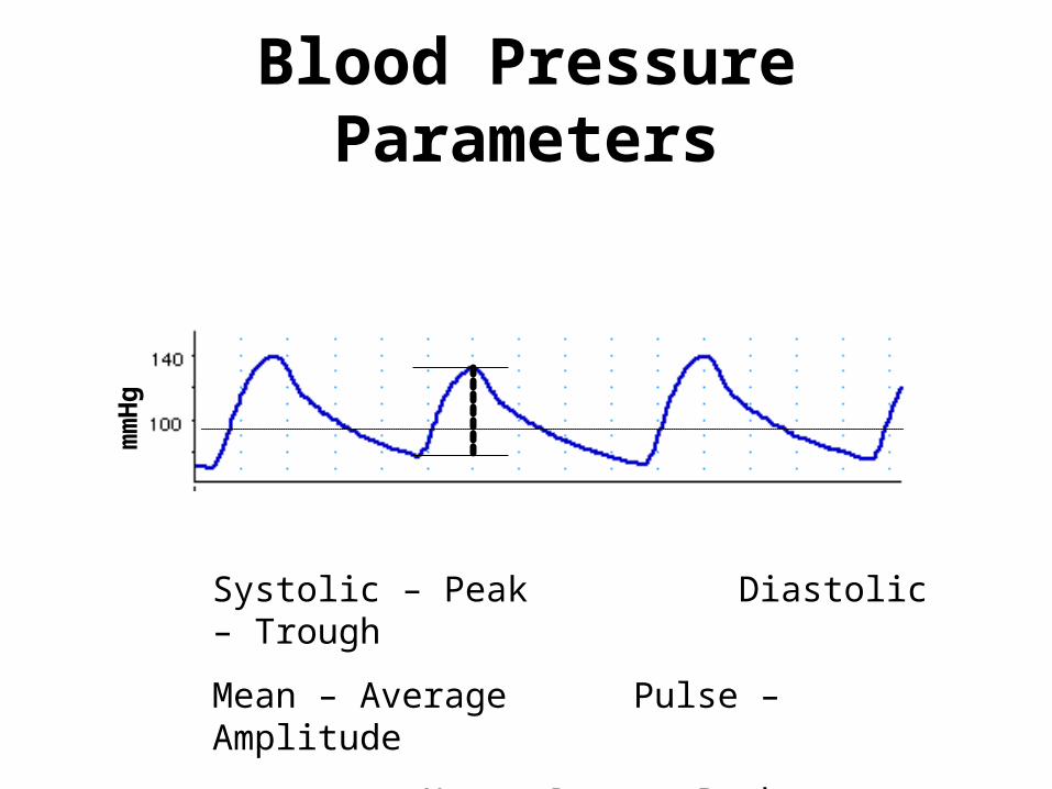
Blood Pressure Parametersm
mH
g
Systolic – Peak Diastolic – Trough
Mean – Average Pulse – Amplitude
Heart Rate – Peak to Peak
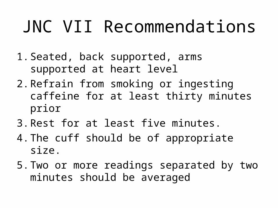
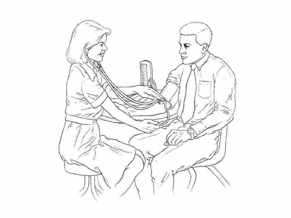
JNC VII Recommendations
1. Seated, back supported, arms supported at heart level
2. Refrain from smoking or ingesting caffeine for at least thirty minutes prior
3. Rest for at least five minutes.
4. The cuff should be of appropriate size.
5. Two or more readings separated by two minutes should be averaged
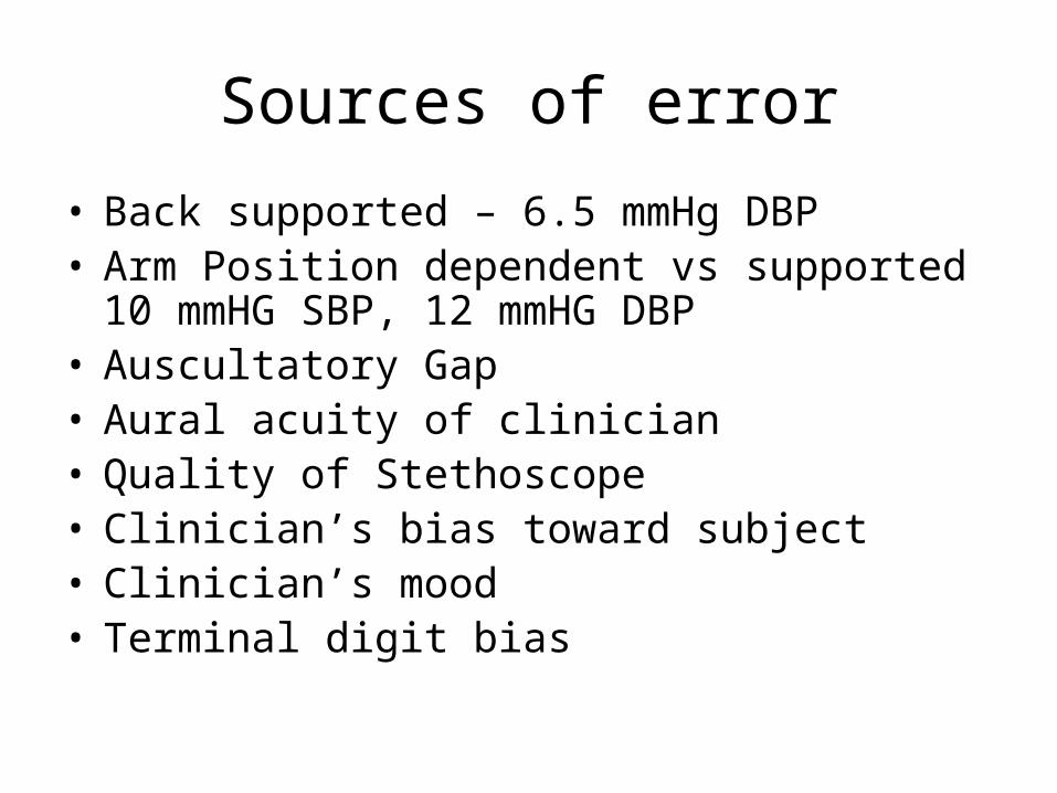
Sources of error
• Back supported – 6.5 mmHg DBP• Arm Position dependent vs supported
10 mmHG SBP, 12 mmHG DBP• Auscultatory Gap• Aural acuity of clinician• Quality of Stethoscope• Clinician’s bias toward subject• Clinician’s mood• Terminal digit bias
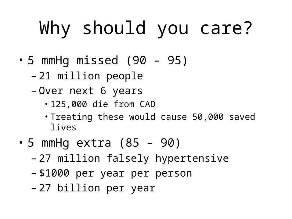
Why should you care?
• 5 mmHg missed (90 – 95)– 21 million people– Over next 6 years
• 125,000 die from CAD• Treating these would cause 50,000 saved lives
• 5 mmHg extra (85 – 90)– 27 million falsely hypertensive– $1000 per year per person– 27 billion per year
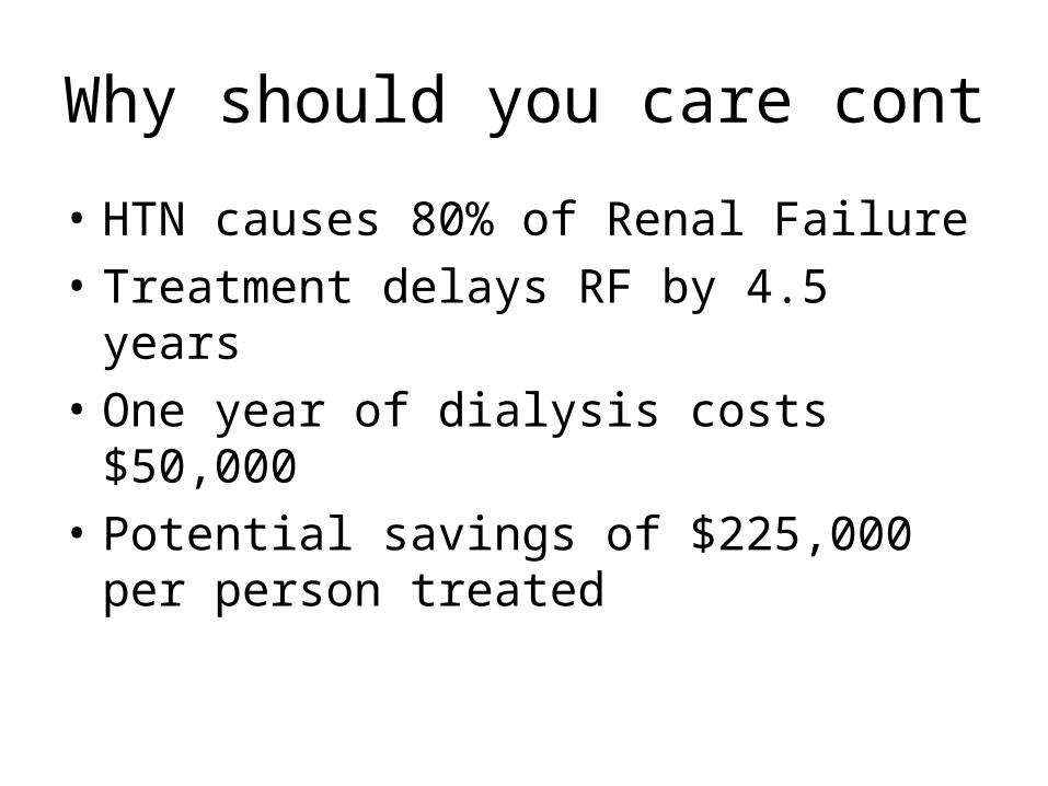
Why should you care cont
• HTN causes 80% of Renal Failure
• Treatment delays RF by 4.5 years
• One year of dialysis costs $50,000
• Potential savings of $225,000 per person treated

Why should you care cont
• Seven national surveys had “serious BP measurement errors”– Finland– Norway– United States– Australia– England
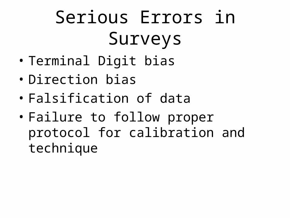
Serious Errors in Surveys
• Terminal Digit bias
• Direction bias
• Falsification of data
• Failure to follow proper protocol for calibration and technique
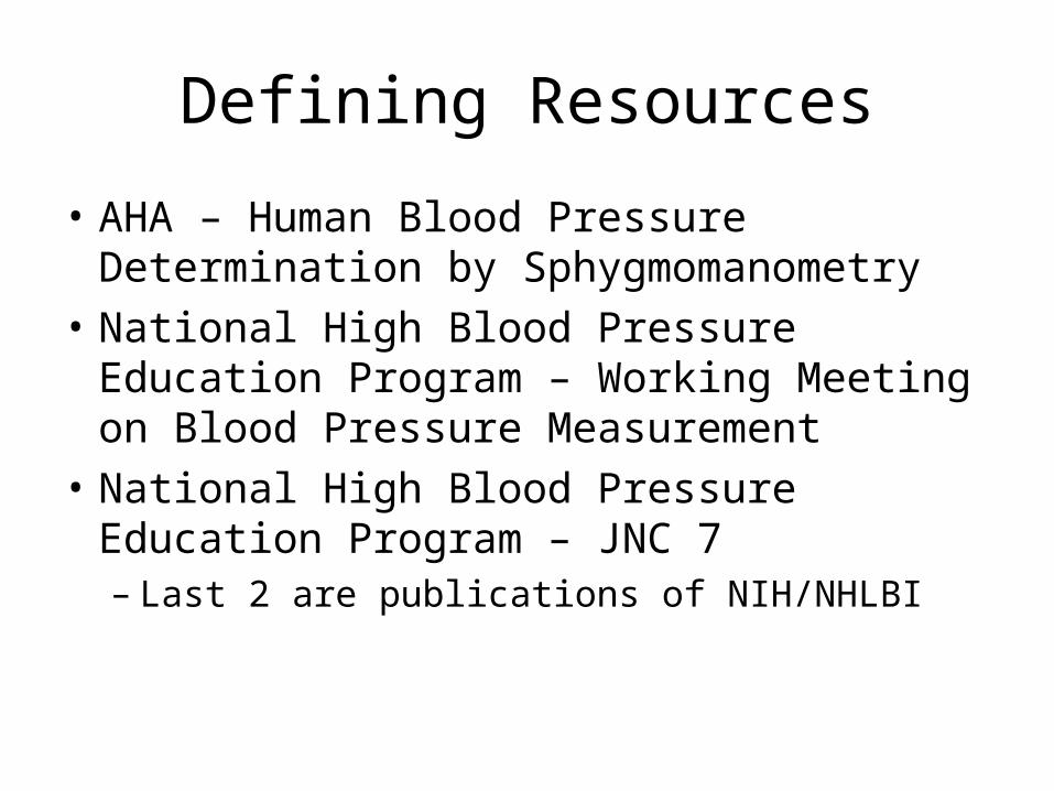
Defining Resources
• AHA – Human Blood Pressure Determination by Sphygmomanometry
• National High Blood Pressure Education Program – Working Meeting on Blood Pressure Measurement
• National High Blood Pressure Education Program – JNC 7– Last 2 are publications of NIH/NHLBI
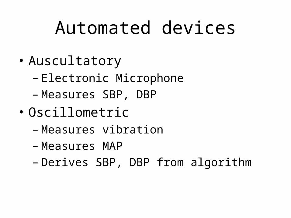
Automated devices
• Auscultatory– Electronic Microphone– Measures SBP, DBP
• Oscillometric– Measures vibration– Measures MAP– Derives SBP, DBP from algorithm
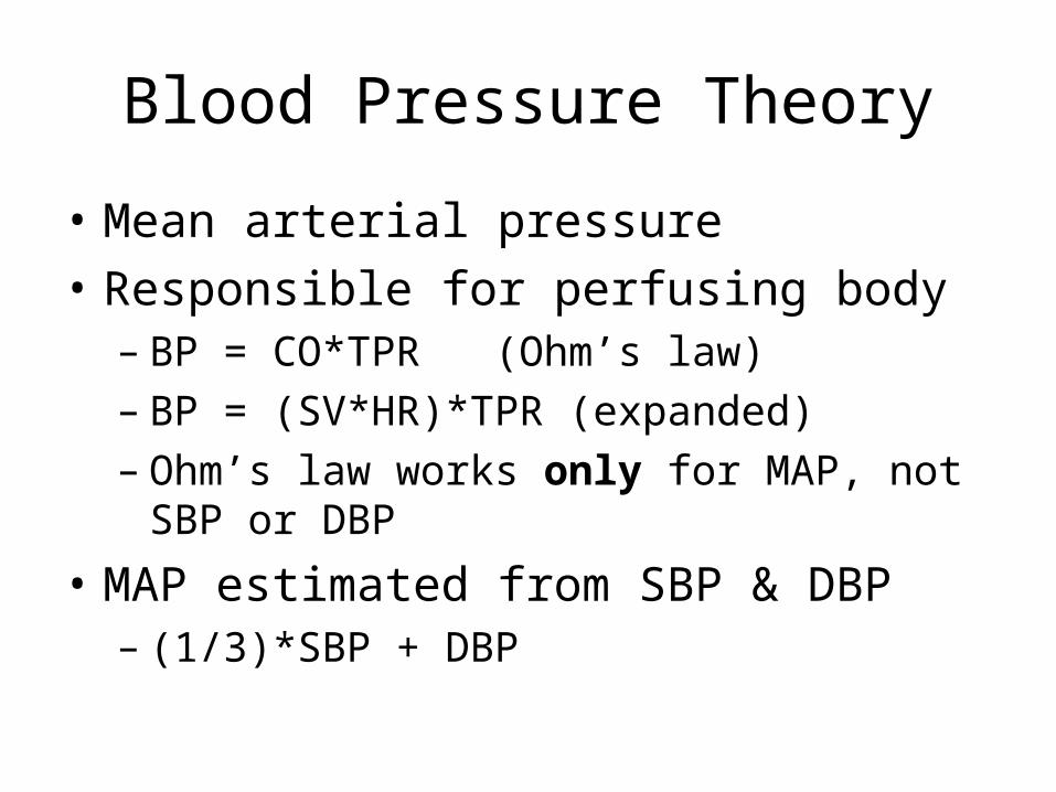
Blood Pressure Theory
• Mean arterial pressure
• Responsible for perfusing body– BP = CO*TPR (Ohm’s law)– BP = (SV*HR)*TPR (expanded)– Ohm’s law works only for MAP, not SBP or
DBP
• MAP estimated from SBP & DBP– (1/3)*SBP + DBP
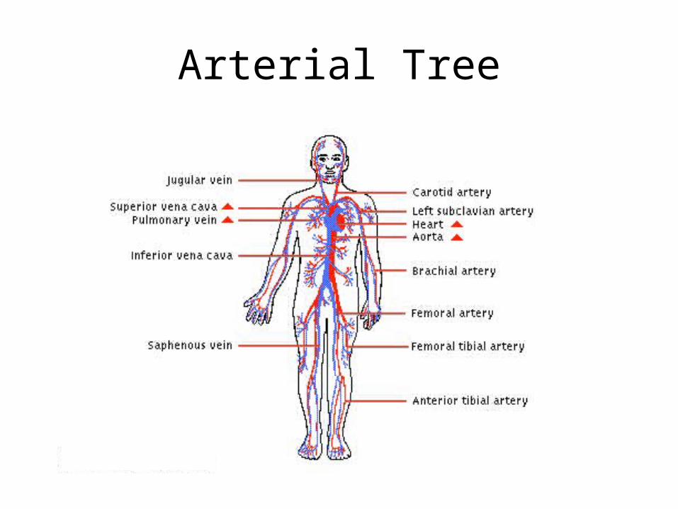
Arterial Tree
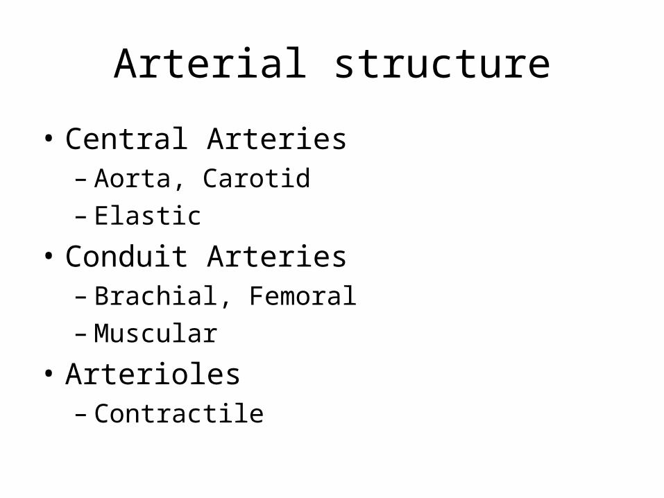
Arterial structure
• Central Arteries– Aorta, Carotid– Elastic
• Conduit Arteries– Brachial, Femoral– Muscular
• Arterioles– Contractile
Application
• Shock– Difficult to diagnose early stages– Arterioles compensate for decreased cardiac
output– Sudden decompensation with little warning
• Central Pressure drops before brachial
Ventricular-Vascular Coupling
• Heart does not pump against all of the body’s blood, only against what is in the aorta– Aorta stretches to absorb stroke volume– When valves close, heart relaxes, then the
aorta contracts and blood flows downward• Effect
– Decrease systolic pressure– Increase diastolic pressure– Hardening of arteries causes opposite effects
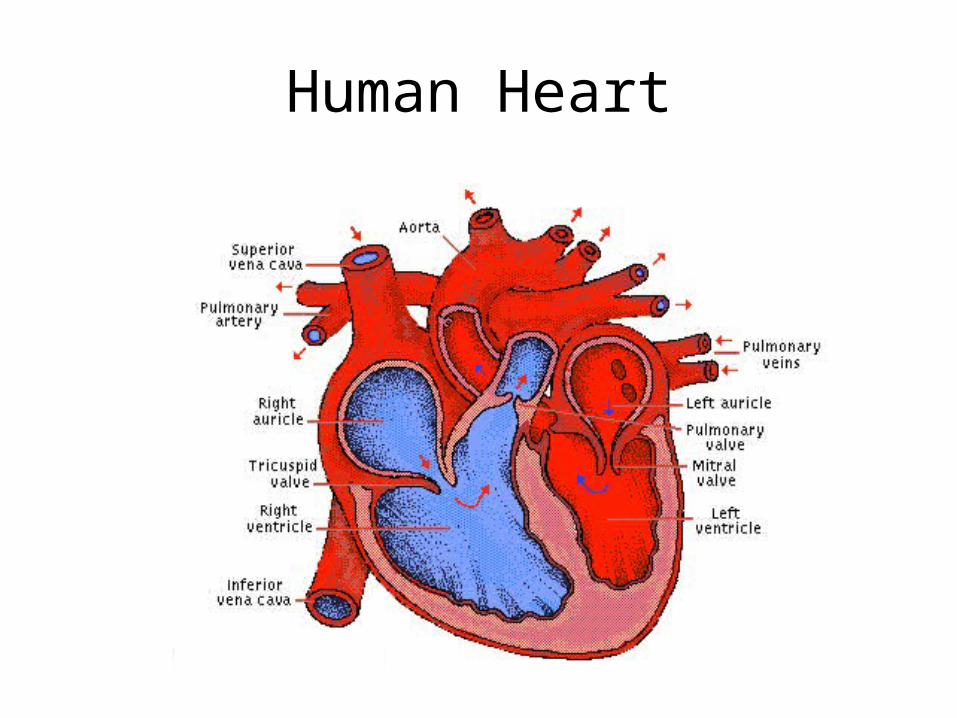
Human Heart
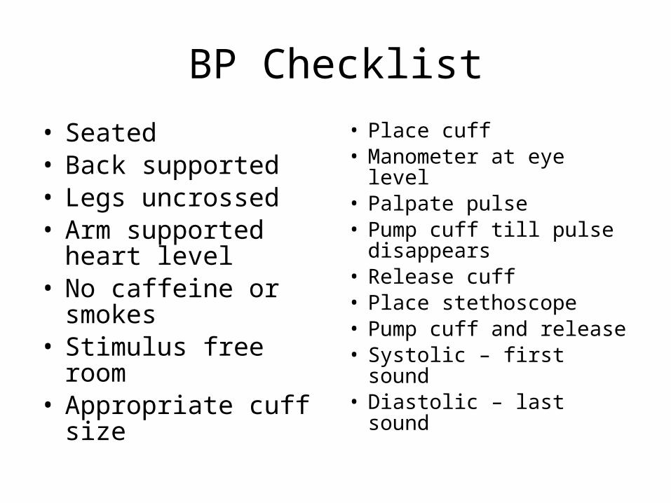
BP Checklist
• Seated• Back supported• Legs uncrossed• Arm supported heart
level• No caffeine or
smokes• Stimulus free room• Appropriate cuff size
• Place cuff• Manometer at eye
level• Palpate pulse• Pump cuff till pulse
disappears• Release cuff• Place stethoscope• Pump cuff and release• Systolic – first sound• Diastolic – last sound
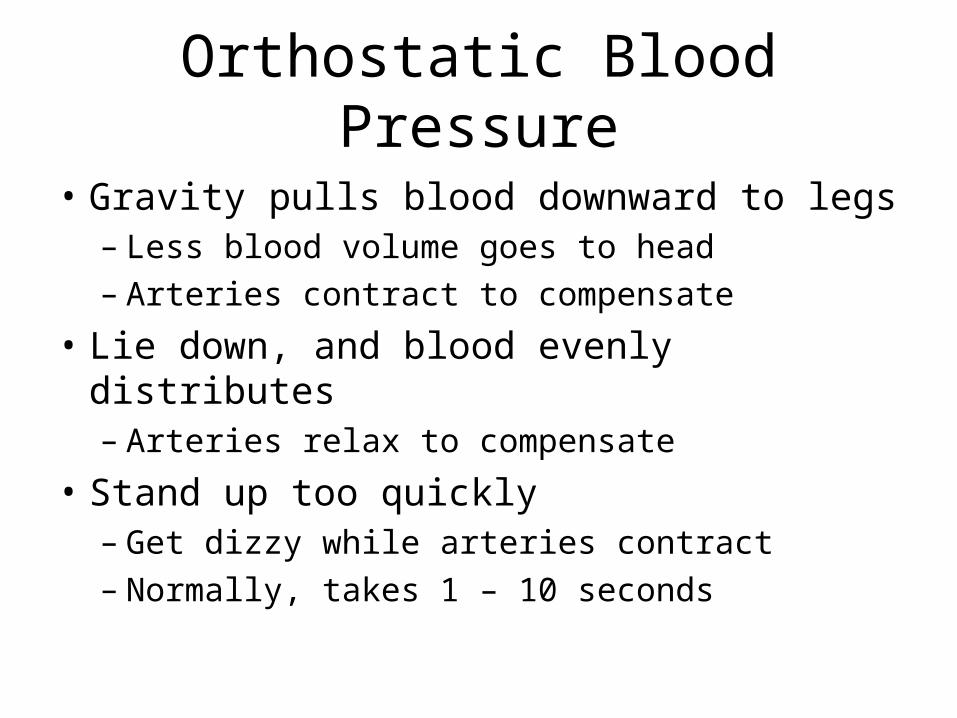
Orthostatic Blood Pressure
• Gravity pulls blood downward to legs– Less blood volume goes to head– Arteries contract to compensate
• Lie down, and blood evenly distributes– Arteries relax to compensate
• Stand up too quickly– Get dizzy while arteries contract– Normally, takes 1 – 10 seconds
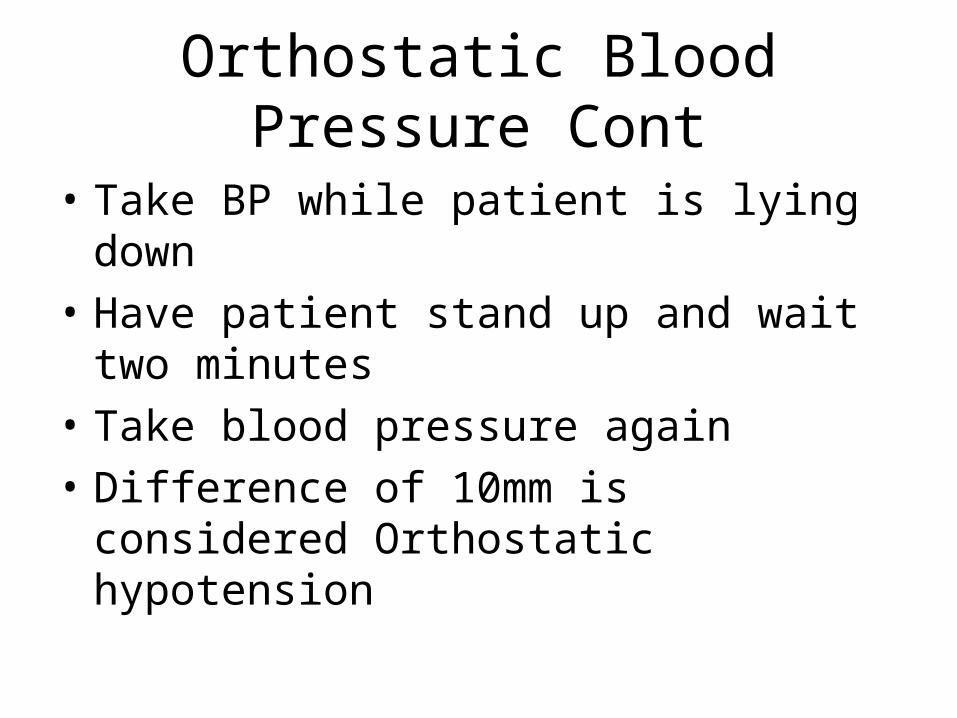
Orthostatic Blood Pressure Cont
• Take BP while patient is lying down
• Have patient stand up and wait two minutes
• Take blood pressure again
• Difference of 10mm is considered Orthostatic hypotension
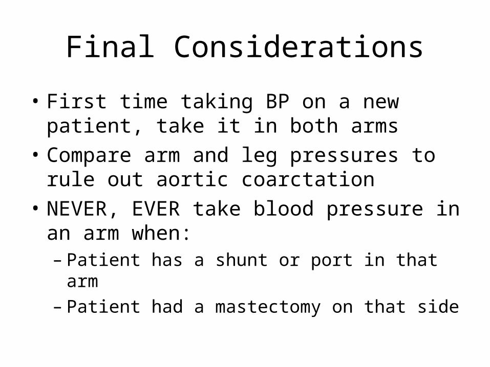
Final Considerations
• First time taking BP on a new patient, take it in both arms
• Compare arm and leg pressures to rule out aortic coarctation
• NEVER, EVER take blood pressure in an arm when:– Patient has a shunt or port in that arm– Patient had a mastectomy on that side