Embed Size (px)
Citation preview
JAK-INHIBITORER – MODE OF ACTION DANBIO KURSUS 19. JANUAR 2018
Uffe Møller Døhn
Center for Rheumatology and Spine Diseases & COPECARE
Rigshospitalet, Glostrup
Denmark
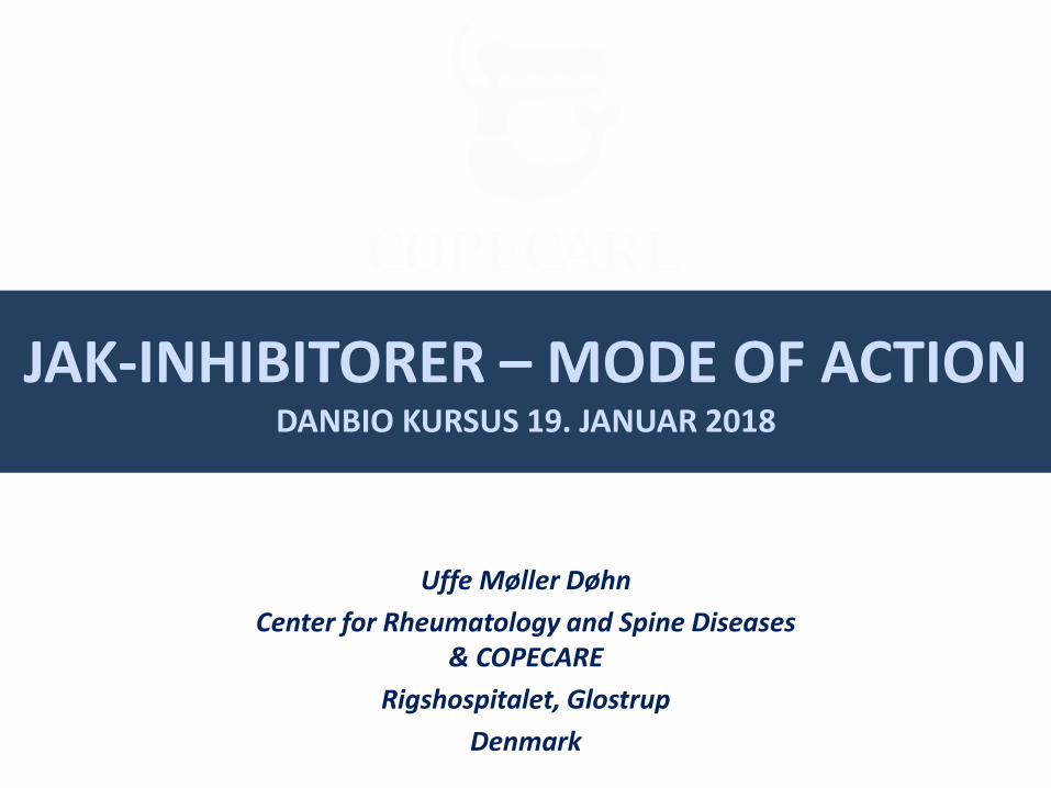
RA PATHOGENESIS
Figure adapted from http://www.medical-artist.com/scientific-illustrations.html.
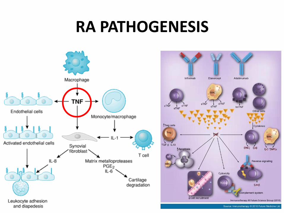
B cell
Dendritic cell
Cytokines IL-1, IL-6
TNF
Co-stimulation
Rheumatoid factor and other antibodies
T cell
T cell T cell
T cell
Macrophage
Rheumatoid arthritis
Biologics target cytokines and
extracellular signalling
Small molecules target intracellular signalling
pathways
The inflammatory cascade continues downstream into the cell
TARGETED THERAPIES Intracellular pathways can be targeted by small molecules
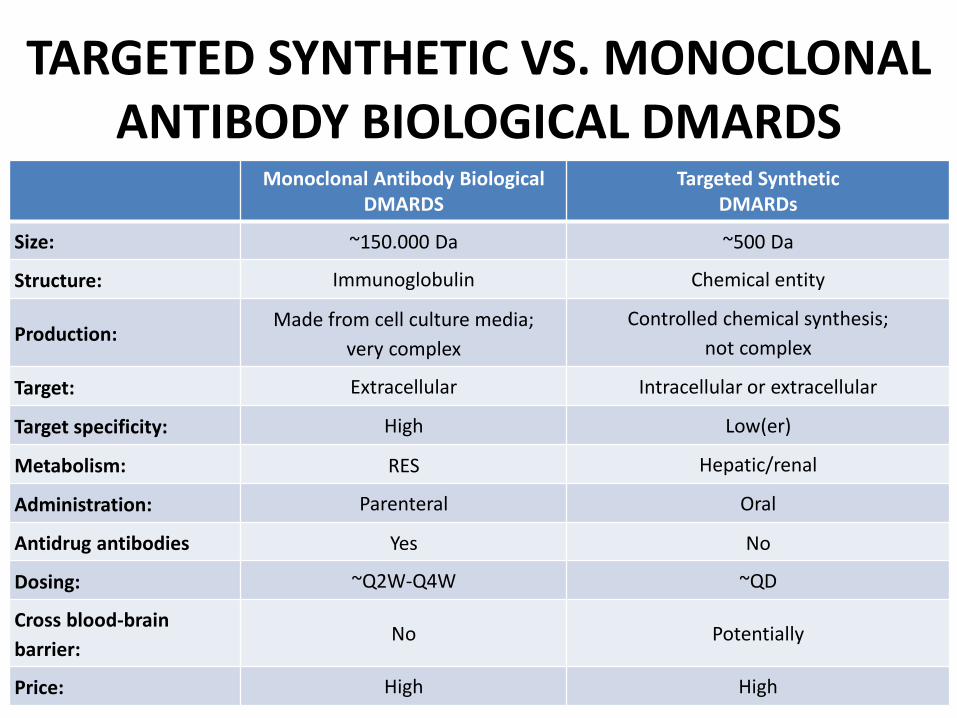
Monoclonal Antibody Biological DMARDS
Targeted Synthetic DMARDs
Size: ~150.000 Da ~500 Da
Structure: Immunoglobulin Chemical entity
Production: Made from cell culture media;
very complex
Controlled chemical synthesis;
not complex
Target: Extracellular Intracellular or extracellular
Target specificity: High Low(er)
Metabolism: RES Hepatic/renal
Administration: Parenteral Oral
Antidrug antibodies Yes No
Dosing: ~Q2W-Q4W ~QD
Cross blood-brain
barrier: No Potentially
Price: High High
TARGETED SYNTHETIC VS. MONOCLONAL ANTIBODY BIOLOGICAL DMARDS
Smolen J et al.: Ann Rheum Dis 2016
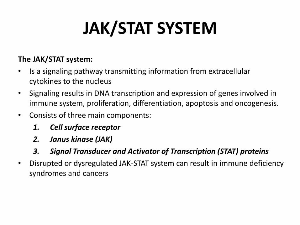
JAK/STAT SYSTEM
The JAK/STAT system:
• Is a signaling pathway transmitting information from extracellular cytokines to the nucleus
• Signaling results in DNA transcription and expression of genes involved in immune system, proliferation, differentiation, apoptosis and oncogenesis.
• Consists of three main components:
1. Cell surface receptor
2. Janus kinase (JAK)
3. Signal Transducer and Activator of Transcription (STAT) proteins
• Disrupted or dysregulated JAK-STAT system can result in immune deficiency syndromes and cancers
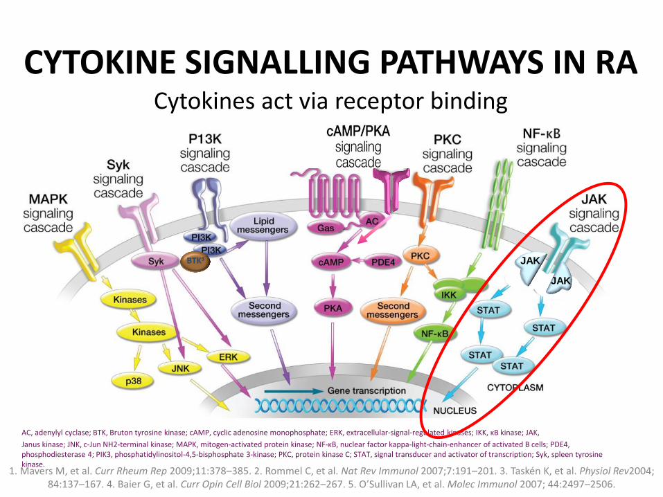
CYTOKINE SIGNALLING PATHWAYS IN RA
1. Mavers M, et al. Curr Rheum Rep 2009;11:378–385. 2. Rommel C, et al. Nat Rev Immunol 2007;7:191–201. 3. Taskén K, et al. Physiol Rev2004; 84:137–167. 4. Baier G, et al. Curr Opin Cell Biol 2009;21:262–267. 5. O’Sullivan LA, et al. Molec Immunol 2007; 44:2497–2506.
BTK3 JAK
JAK
AC, adenylyl cyclase; BTK, Bruton tyrosine kinase; cAMP, cyclic adenosine monophosphate; ERK, extracellular-signal-regulated kinases; IKK, κB kinase; JAK,
Janus kinase; JNK, c-Jun NH2-terminal kinase; MAPK, mitogen-activated protein kinase; NF-κB, nuclear factor kappa-light-chain-enhancer of activated B cells; PDE4, phosphodiesterase 4; PIK3, phosphatidylinositol-4,5-bisphosphate 3-kinase; PKC, protein kinase C; STAT, signal transducer and activator of transcription; Syk, spleen tyrosine kinase.
Cytokines act via receptor binding
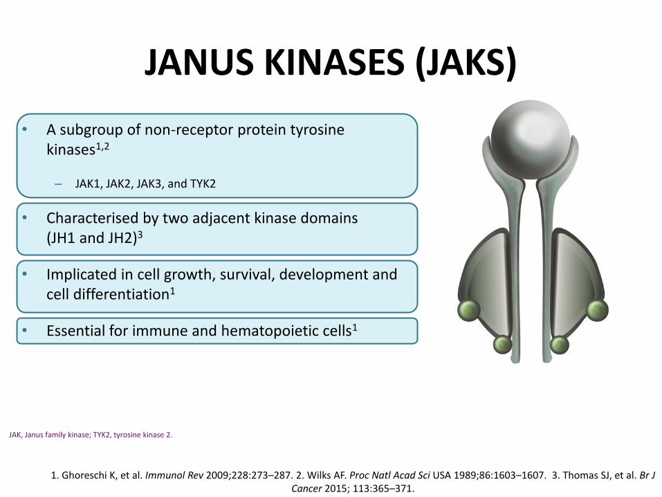
JANUS KINASES (JAKS)
• A subgroup of non-receptor protein tyrosine kinases1,2
– JAK1, JAK2, JAK3, and TYK2
• Characterised by two adjacent kinase domains (JH1 and JH2)3
• Implicated in cell growth, survival, development and cell differentiation1
• Essential for immune and hematopoietic cells1
1. Ghoreschi K, et al. Immunol Rev 2009;228:273–287. 2. Wilks AF. Proc Natl Acad Sci USA 1989;86:1603–1607. 3. Thomas SJ, et al. Br J Cancer 2015; 113:365–371.
JAK, Janus family kinase; TYK2, tyrosine kinase 2.

BINDING OF CYTOKINE RECEPTORS ACTIVATES JAK SIGNALLING PATHWAYS
• Rapid membrane to nucleus signalling: – Cytokines bind trans-membrane
receptors that are associated with JAKs
JAK, Janus kinase; P, phosphate; STAT, signal transducer and activator of transcription. 1. Shuai K, et al. Nat Rev Immunol 2003;3:900–911.
Gene transcription
Binding activates JAKs
STATs bind to DNA and activate gene transcription to produce proteins mediating immune response/inflammation
JAKs phosphorylate receptors
STATs bind to receptors
JAKs phosphorylate STATs
STATs translocate to the nucleus
JAKs activate STATs, which then act as transcription factors
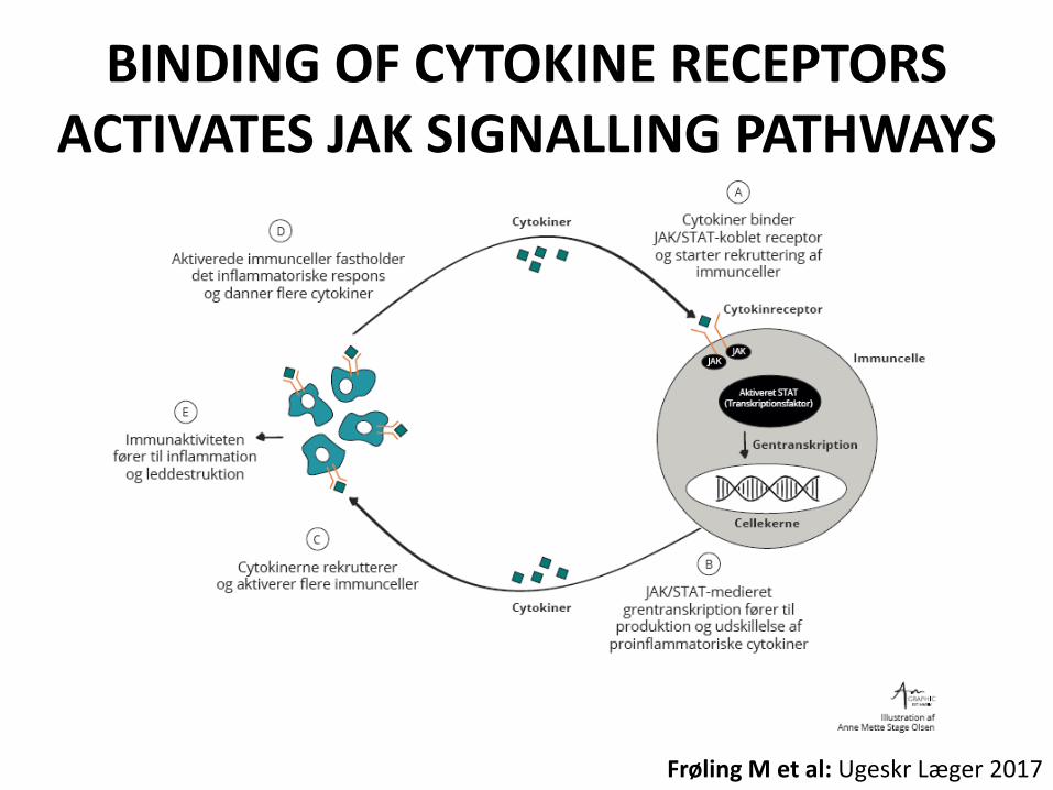
Frøling M et al: Ugeskr Læger 2017
BINDING OF CYTOKINE RECEPTORS ACTIVATES JAK SIGNALLING PATHWAYS

Winthrop KL. Nat Rev Rheumatol. 2017 Mar 2. doi: 10.103.
BINDING OF CYTOKINE RECEPTORS ACTIVATES JAK SIGNALLING PATHWAYS
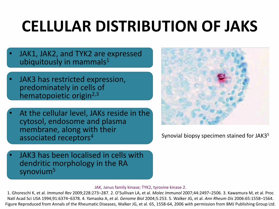
CELLULAR DISTRIBUTION OF JAKS
• JAK1, JAK2, and TYK2 are expressed ubiquitously in mammals1
• JAK3 has restricted expression, predominately in cells of hematopoietic origin2,3
• At the cellular level, JAKs reside in the cytosol, endosome and plasma membrane, along with their associated receptors4
• JAK3 has been localised in cells with dendritic morphology in the RA synovium5
JAK, Janus family kinase; TYK2, tyrosine kinase 2.
1. Ghoreschi K, et al. Immunol Rev 2009;228:273–287. 2. O’Sullivan LA, et al. Molec Immunol 2007;44:2497–2506. 3. Kawamura M, et al. Proc Natl Acad Sci USA 1994;91:6374–6378. 4. Yamaoka A, et al. Genome Biol 2004;5:253. 5. Walker JG, et al. Ann Rheum Dis 2006:65:1558–1564.
Figure Reproduced from Annals of the Rheumatic Diseases, Walker JG, et al. 65, 1558-64, 2006 with permission from BMJ Publishing Group Ltd.
Synovial biopsy specimen stained for JAK35
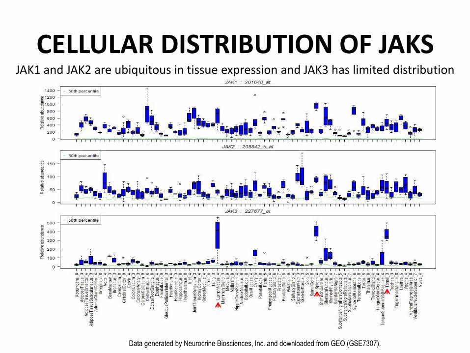
Tao Wei TS&T June 2012 Data generated by Neurocrine Biosciences, Inc. and downloaded from GEO (GSE7307).
JAK1 and JAK2 are ubiquitous in tissue expression and JAK3 has limited distribution
CELLULAR DISTRIBUTION OF JAKS
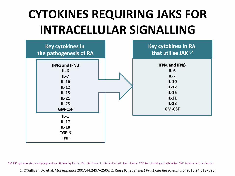
IFNα and IFNβ
IL-6 IL-7
IL-10 IL-12 IL-15 IL-21 IL-23
GM-CSF
IL-1 IL-17 IL-18 TGF-β TNF
CYTOKINES REQUIRING JAKS FOR INTRACELLULAR SIGNALLING
1. O’Sullivan LA, et al. Mol Immunol 2007;44:2497–2506. 2. Riese RJ, et al. Best Pract Clin Res Rheumatol 2010;24:513–526.
GM-CSF, granulocyte-macrophage colony-stimulating factor, IFN, interferon; IL, interleukin; JAK, Janus kinase; TGF, transforming growth factor; TNF, tumour necrosis factor.
Key cytokines in the pathogenesis of RA
IFNα and IFNβ IL-6 IL-7
IL-10 IL-12 IL-15 IL-21 IL-23
GM-CSF
Key cytokines in RA that utilise JAK1,2
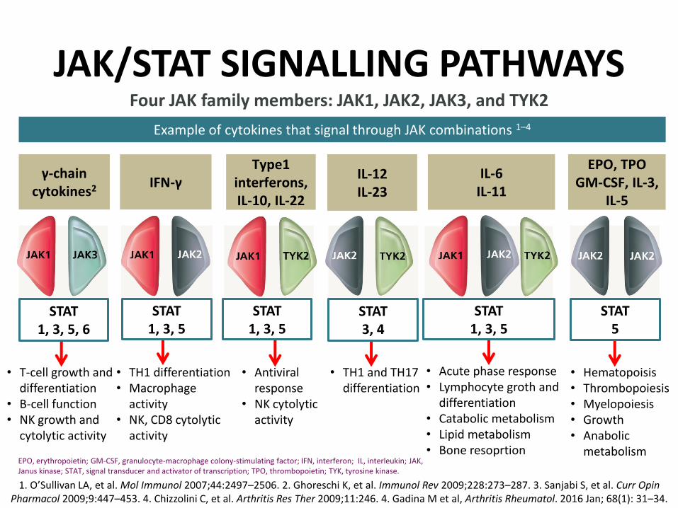
γ-chain cytokines2
Type1 interferons, IL-10, IL-22
IL-6
IL-11 IFN-γ
IL-12 IL-23
EPO, TPO GM-CSF, IL-3,
IL-5
Example of cytokines that signal through JAK combinations 1–4
JAK/STAT SIGNALLING PATHWAYS
1. O’Sullivan LA, et al. Mol Immunol 2007;44:2497–2506. 2. Ghoreschi K, et al. Immunol Rev 2009;228:273–287. 3. Sanjabi S, et al. Curr Opin Pharmacol 2009;9:447–453. 4. Chizzolini C, et al. Arthritis Res Ther 2009;11:246. 4. Gadina M et al, Arthritis Rheumatol. 2016 Jan; 68(1): 31–34.
EPO, erythropoietin; GM-CSF, granulocyte-macrophage colony-stimulating factor; IFN, interferon; IL, interleukin; JAK, Janus kinase; STAT, signal transducer and activator of transcription; TPO, thrombopoietin; TYK, tyrosine kinase.
Four JAK family members: JAK1, JAK2, JAK3, and TYK2
• T-cell growth and differentiation
• B-cell function • NK growth and
cytolytic activity
• TH1 differentiation • Macrophage
activity • NK, CD8 cytolytic
activity
• TH1 and TH17 differentiation
• Hematopoisis • Thrombopoiesis • Myelopoiesis • Growth • Anabolic
metabolism
• Acute phase response • Lymphocyte groth and
differentiation • Catabolic metabolism • Lipid metabolism • Bone resoprtion
• Antiviral response
• NK cytolytic activity
STAT 1, 3, 5, 6
STAT 1, 3, 5
STAT 1, 3, 5
STAT 1, 3, 5
STAT 3, 4
STAT 5
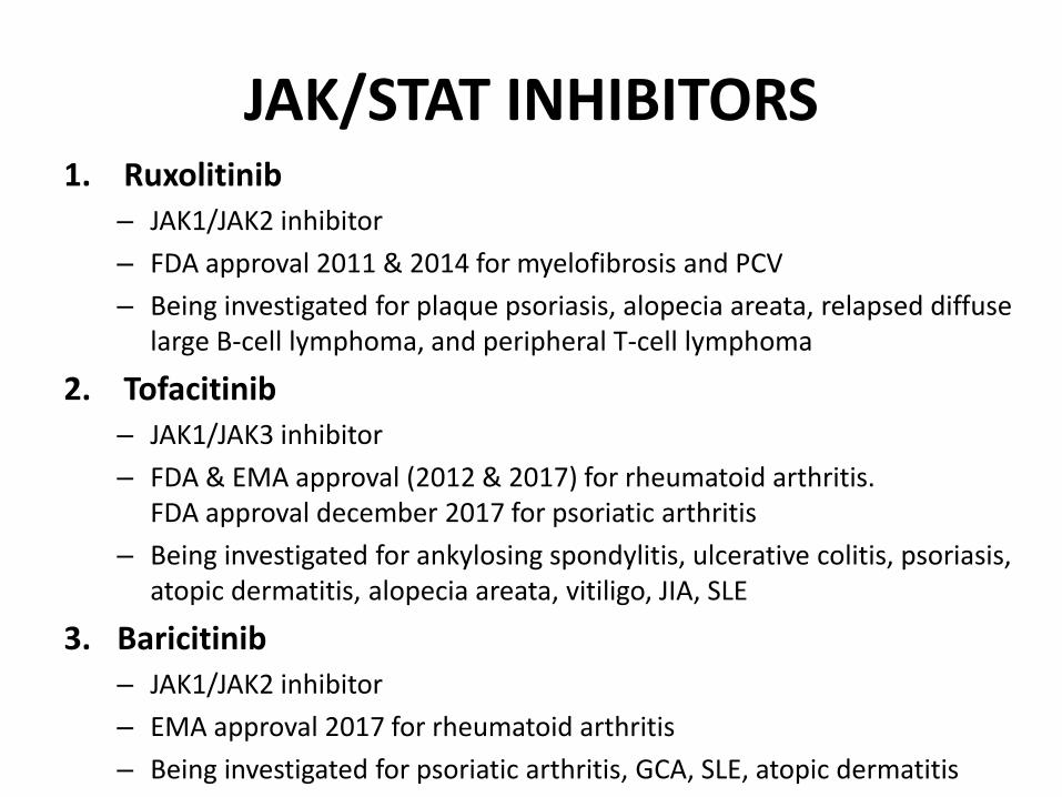
JAK/STAT INHIBITORS 1. Ruxolitinib
– JAK1/JAK2 inhibitor
– FDA approval 2011 & 2014 for myelofibrosis and PCV
– Being investigated for plaque psoriasis, alopecia areata, relapsed diffuse large B-cell lymphoma, and peripheral T-cell lymphoma
2. Tofacitinib – JAK1/JAK3 inhibitor
– FDA & EMA approval (2012 & 2017) for rheumatoid arthritis. FDA approval december 2017 for psoriatic arthritis
– Being investigated for ankylosing spondylitis, ulcerative colitis, psoriasis, atopic dermatitis, alopecia areata, vitiligo, JIA, SLE
3. Baricitinib – JAK1/JAK2 inhibitor
– EMA approval 2017 for rheumatoid arthritis
– Being investigated for psoriatic arthritis, GCA, SLE, atopic dermatitis

THE JAK/STAT SIGNALLING PATHWAYS
Furumoto et al. BioDrugs . 2013; 27: 431–438.
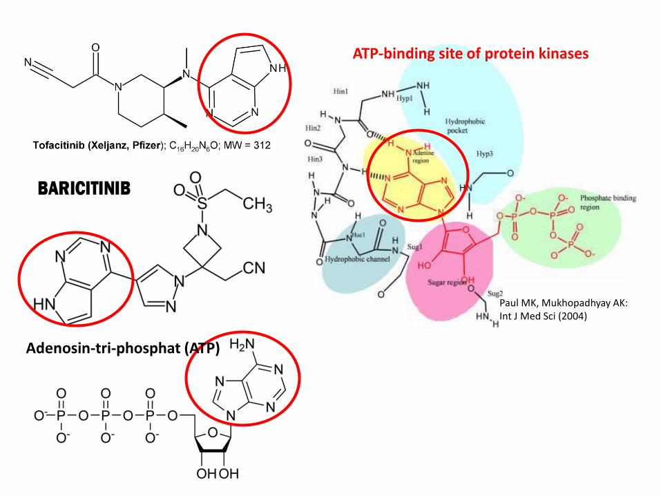
ATP-binding site of protein kinases
Paul MK, Mukhopadhyay AK: Int J Med Sci (2004)
Adenosin-tri-phosphat (ATP)

Sulfasalazin
Methotrexate
Adenosin-tri-phosphat (ATP)

• Methotrexate were independently identified as strong inhibitors of the Drosophila JAK/STAT pathway, an effect conserved to human cells.
• Methotrexate did not affect protein phosphorylation in other intracellular signalling pathways.
• Methotrexate caused significant suppression of JAK/STAT activation at a concentration equivalent to that seen in patients taking low-dose oral methotrexate (p≤0.001).
JAK/STAT INHIBITORS
Thomas S.: The Lancet, Volume 385, Supp. 1, 2015, Page S98
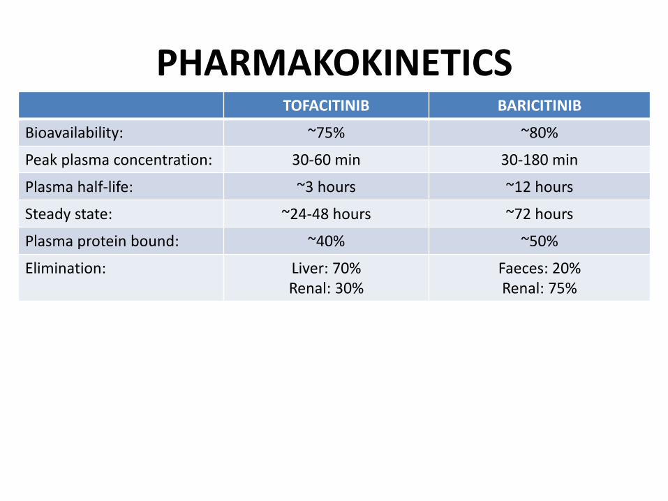
PHARMAKOKINETICS
TOFACITINIB BARICITINIB
Bioavailability: ~75% ~80%
Peak plasma concentration: 30-60 min 30-180 min
Plasma half-life: ~3 hours ~12 hours
Steady state: ~24-48 hours ~72 hours
Plasma protein bound: ~40% ~50%
Elimination: Liver: 70% Renal: 30%
Faeces: 20% Renal: 75%

TOFACITINIB BARICITINIB
Mode of action: JAK1, JAK3, (JAK2) JAK1, JAK2
Indication: Mono therapy or in combination with MTX or other csDMARDs
Mono therapy or in combination with MTX
Dosing 5 mg BID 4 mg QD
Elimination: Hepatic Renal
Reduced dose: Moderate liver impairment Moderate kidney impairment Age >75 yrs
Drug interactions: CYP3A4, CYP2C (Clarithromycin, Ketoconazole …)
OAT3 (Probenecid)
Recommended lab tests:
Neutrophiles, leukocytes, Hgb, transaminases, lipids
Neutrophiles, leukocytes, Hgb, transaminases, lipids, creatinine
PHARMAKOKINETICS

Hodge et al.: Clin Exp Rheumatol 2016; 34: 318-328
MODE-OF-ACTION
JAK inhibitors has high in-vitro passive permeability properties
and entries intracellularly by transcellular diffusion
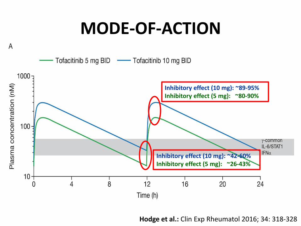
Hodge et al.: Clin Exp Rheumatol 2016; 34: 318-328
Inhibitory effect (10 mg): ~89-95% Inhibitory effect (5 mg): ~80-90%
Inhibitory effect (10 mg): ~42-60% Inhibitory effect (5 mg): ~26-43%
MODE-OF-ACTION
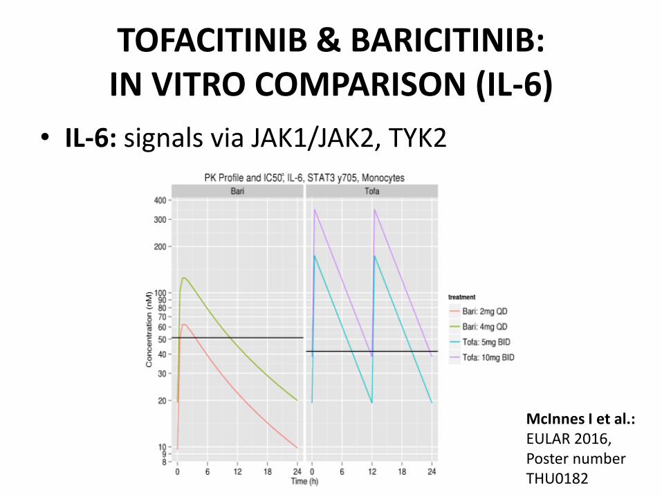
TOFACITINIB & BARICITINIB: IN VITRO COMPARISON (IL-6)
• IL-6: signals via JAK1/JAK2, TYK2 a
McInnes I et al.: EULAR 2016, Poster number THU0182
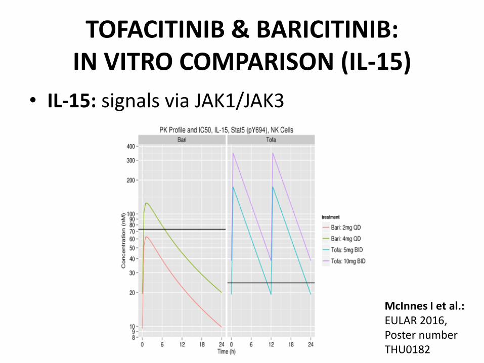
• IL-15: signals via JAK1/JAK3
26
TOFACITINIB & BARICITINIB: IN VITRO COMPARISON (IL-15)
McInnes I et al.: EULAR 2016, Poster number THU0182

• IL-21: signals via JAK1/JAK3
TOFACITINIB & BARICITINIB: IN VITRO COMPARISON (IL-21)
McInnes I et al.: EULAR 2016, Poster number THU0182
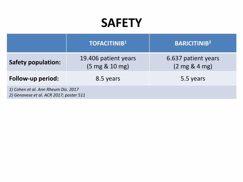
TOFACITINIB1 BARICITINIB2
Safety population: 19.406 patient years
(5 mg & 10 mg) 6.637 patient years
(2 mg & 4 mg)
Follow-up period: 8.5 years 5.5 years
1) Cohen et al. Ann Rheum Dis. 2017 2) Genovese et al. ACR 2017; poster 511
SAFETY
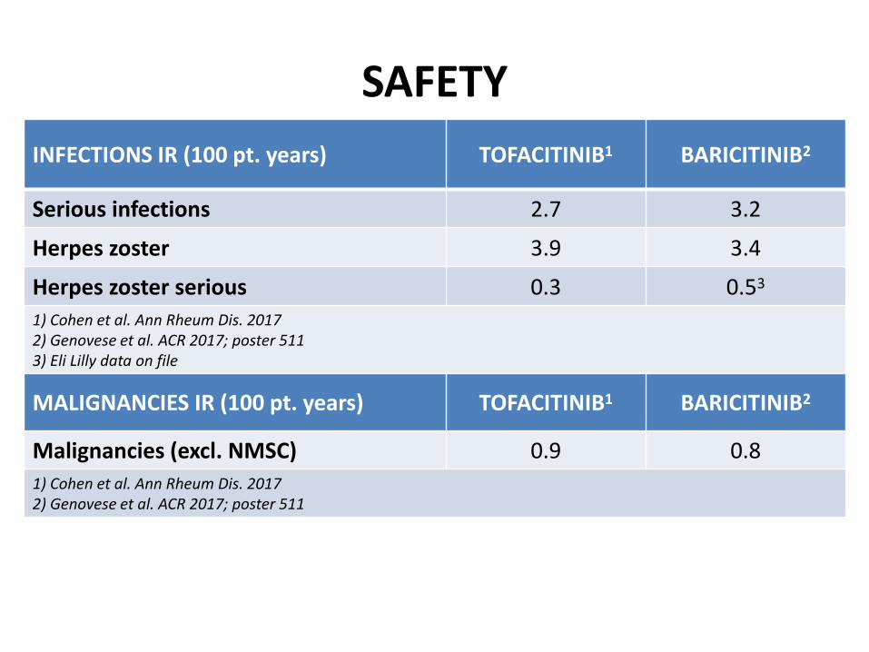
INFECTIONS IR (100 pt. years) TOFACITINIB1 BARICITINIB2
Serious infections 2.7 3.2
Herpes zoster 3.9 3.4
Herpes zoster serious 0.3 0.53
1) Cohen et al. Ann Rheum Dis. 2017 2) Genovese et al. ACR 2017; poster 511 3) Eli Lilly data on file
MALIGNANCIES IR (100 pt. years) TOFACITINIB1 BARICITINIB2
Malignancies (excl. NMSC) 0.9 0.8
1) Cohen et al. Ann Rheum Dis. 2017 2) Genovese et al. ACR 2017; poster 511
SAFETY
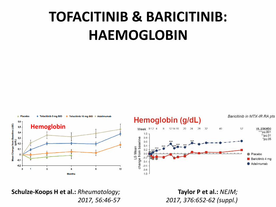
TOFACITINIB & BARICITINIB: HAEMOGLOBIN
Hemoglobin
Schulze-Koops H et al.: Rheumatology; 2017, 56:46-57
Taylor P et al.: NEJM; 2017, 376:652-62 (suppl.)

Neutrophiles
Lymphocytes
Schulze-Koops H et al.: Rheumatology; 2017, 56:46-57
TOFACITINIB & BARICITINIB: WHITE BLOOD CELLS
Taylor P et al.: NEJM; 2017, 376:652-62 (suppl.)
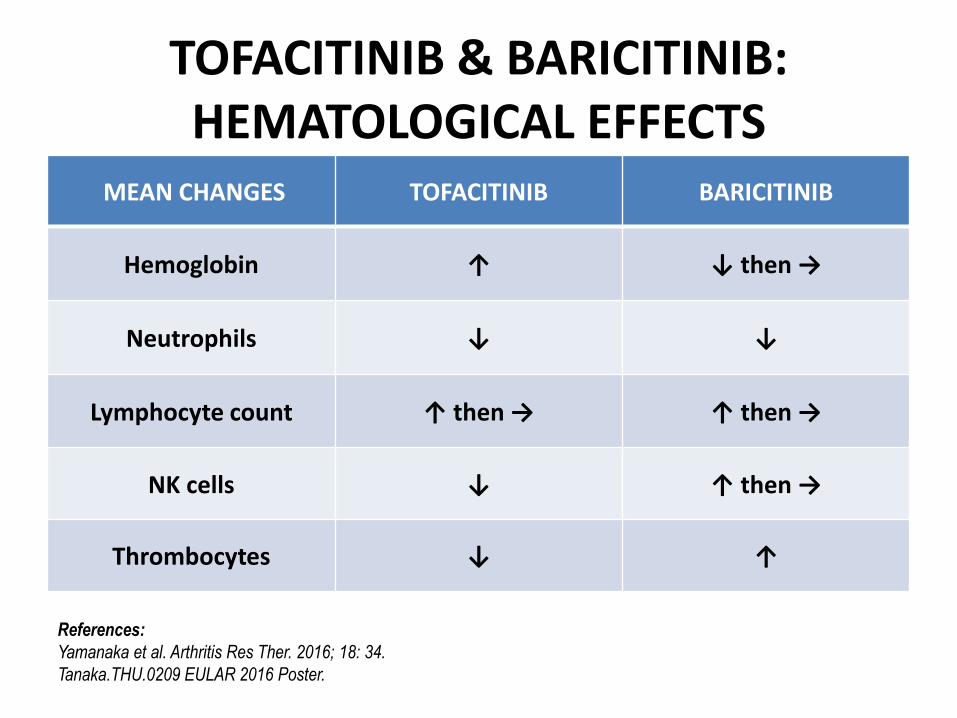
MEAN CHANGES TOFACITINIB BARICITINIB
Hemoglobin ↑ ↓ then →
Neutrophils ↓ ↓
Lymphocyte count ↑ then → ↑ then →
NK cells ↓ ↑ then →
Thrombocytes ↓ ↑
References:
Yamanaka et al. Arthritis Res Ther. 2016; 18: 34.
Tanaka.THU.0209 EULAR 2016 Poster.
TOFACITINIB & BARICITINIB: HEMATOLOGICAL EFFECTS
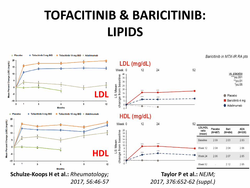
LDL
HDL
Schulze-Koops H et al.: Rheumatology; 2017, 56:46-57
TOFACITINIB & BARICITINIB: LIPIDS
Taylor P et al.: NEJM; 2017, 376:652-62 (suppl.)
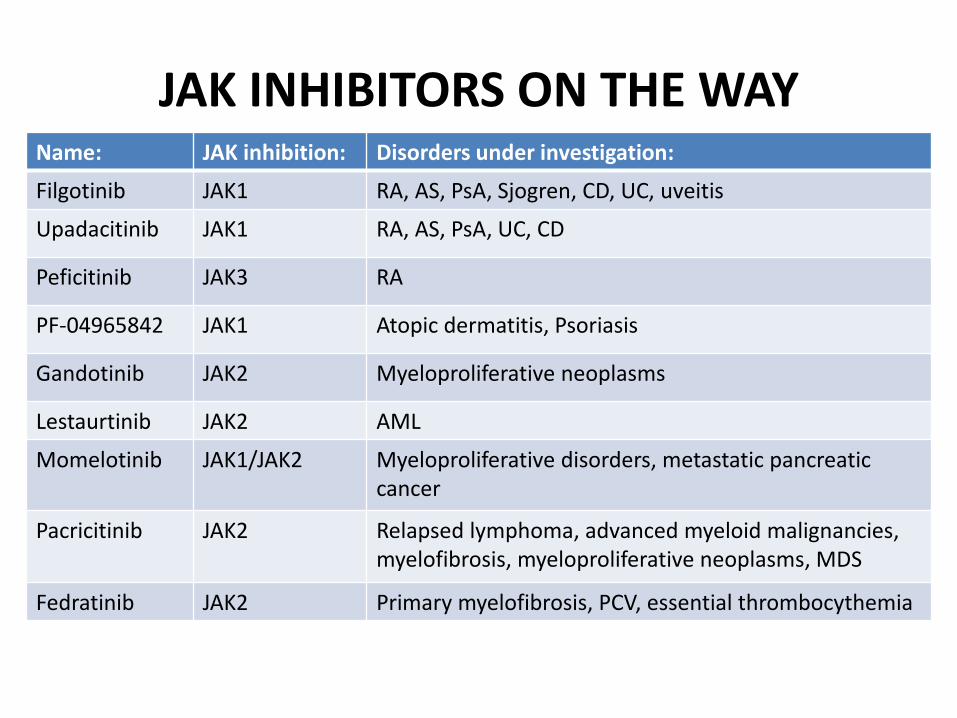
JAK INHIBITORS ON THE WAY Name: JAK inhibition: Disorders under investigation:
Filgotinib JAK1 RA, AS, PsA, Sjogren, CD, UC, uveitis
Upadacitinib JAK1 RA, AS, PsA, UC, CD
Peficitinib JAK3 RA
PF-04965842 JAK1 Atopic dermatitis, Psoriasis
Gandotinib JAK2 Myeloproliferative neoplasms
Lestaurtinib JAK2 AML
Momelotinib JAK1/JAK2 Myeloproliferative disorders, metastatic pancreatic cancer
Pacricitinib JAK2 Relapsed lymphoma, advanced myeloid malignancies,
myelofibrosis, myeloproliferative neoplasms, MDS
Fedratinib JAK2 Primary myelofibrosis, PCV, essential thrombocythemia

Coffeecitinib Chococitinib