Embed Size (px)
Citation preview
Jack of All Trades, Master of All:
The New D8 ADVANCE Plus with EIGER2 R 500K
Benjamin Krueger, Ph.D. XRD Applications Scientist [email protected]
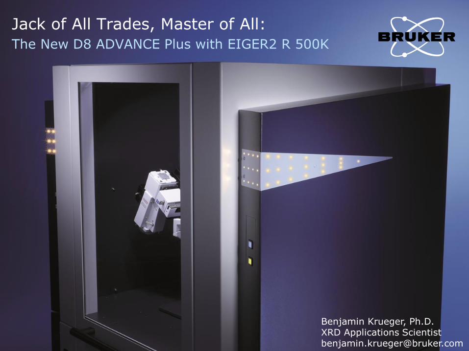
D8 ADVANCE Plus The Perfect Balance of Simplicity and Sophistication
2
• Guided Method Development with WIZARD
• Full Control with COMMANDER
• Unrestricted Access to Diagnostics with TOOLS
Intuitive Instrument Control
• Rapid Results with Industry Standard Methods
• Dig Deeper with the Latest Analytical Techniques
Advanced Analysis Techniques
• No Touch Optics
• Bayonet-Mounted Stages
• Multi-Mode Detector Technology
Effortless Equipment Setup
5/8/2019 D8 ADVANCE Plus
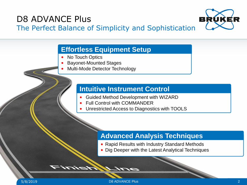
D8 ADVANCE Plus Fundamentally Flexible
3
Primary Optic
Bench
Sample Stage
Bayonet
Goniometer Accessory
Mount*
Secondary Optic
Bench*
Universal Detector
Mount
*Optional 5/8/2019 D8 ADVANCE Plus
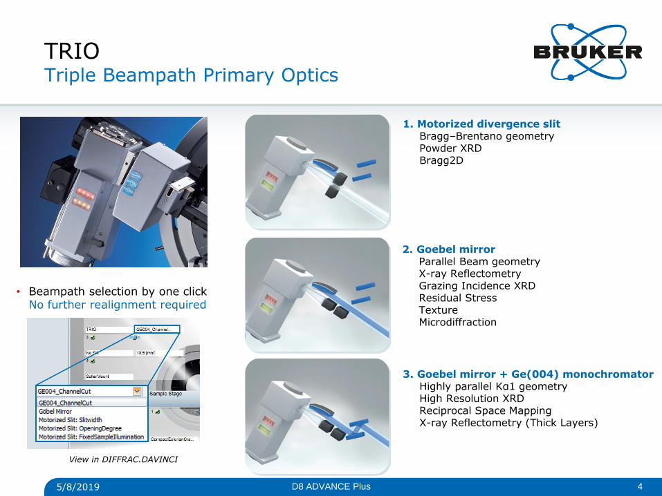
TRIO Triple Beampath Primary Optics
1. Motorized divergence slit Bragg–Brentano geometry Powder XRD Bragg2D
2. Goebel mirror Parallel Beam geometry X-ray Reflectometry Grazing Incidence XRD Residual Stress Texture Microdiffraction
3. Goebel mirror + Ge(004) monochromator Highly parallel Kα1 geometry High Resolution XRD Reciprocal Space Mapping X-ray Reflectometry (Thick Layers)
• Beampath selection by one click No further realignment required
View in DIFFRAC.DAVINCI
5/8/2019 4 D8 ADVANCE Plus
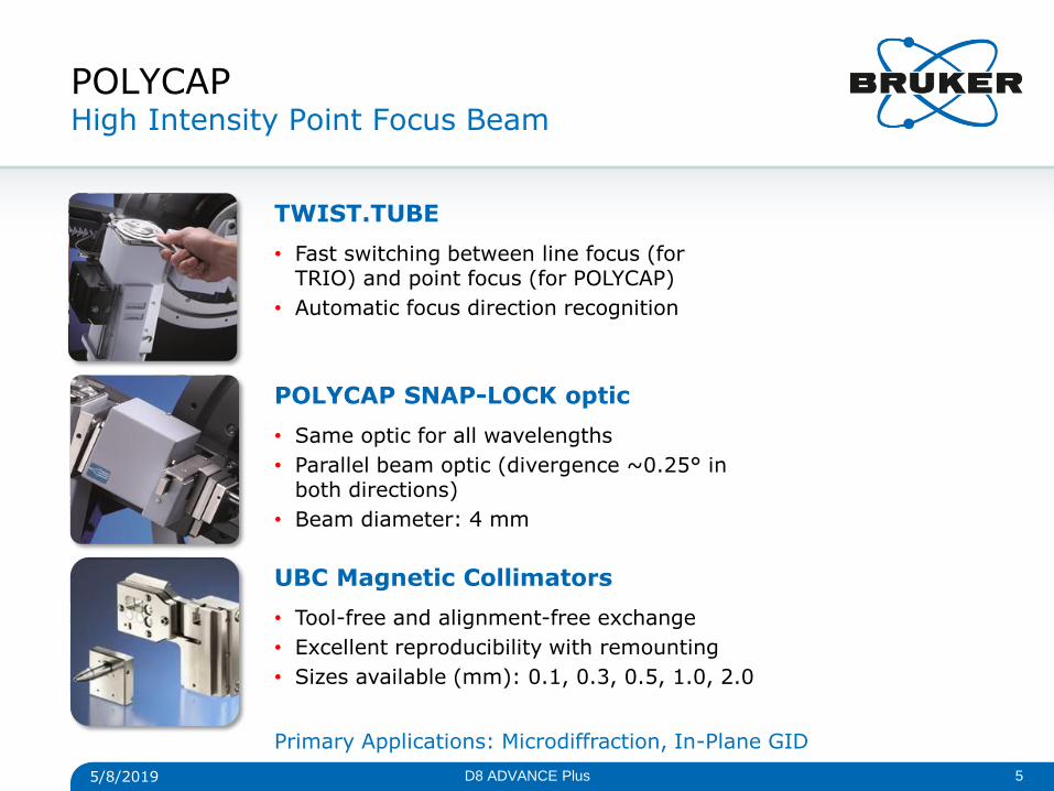
POLYCAP High Intensity Point Focus Beam
TWIST.TUBE
• Fast switching between line focus (for TRIO) and point focus (for POLYCAP)
• Automatic focus direction recognition
POLYCAP SNAP-LOCK optic
• Same optic for all wavelengths
• Parallel beam optic (divergence ~0.25° in both directions)
• Beam diameter: 4 mm
UBC Magnetic Collimators
• Tool-free and alignment-free exchange
• Excellent reproducibility with remounting
• Sizes available (mm): 0.1, 0.3, 0.5, 1.0, 2.0
Primary Applications: Microdiffraction, In-Plane GID
5/8/2019 5 D8 ADVANCE Plus
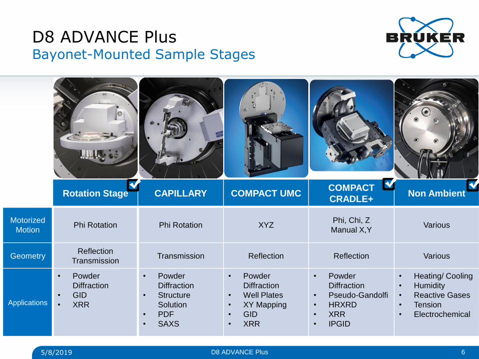
D8 ADVANCE Plus Bayonet-Mounted Sample Stages
Rotation Stage CAPILLARY COMPACT UMC COMPACT
CRADLE+ Non Ambient
Motorized
Motion Phi Rotation Phi Rotation XYZ
Phi, Chi, Z
Manual X,Y Various
Geometry Reflection
Transmission Transmission Reflection Reflection Various
Applications
• Powder
Diffraction
• GID
• XRR
• Powder
Diffraction
• Structure
Solution
• SAXS
• Powder
Diffraction
• Well Plates
• XY Mapping
• GID
• XRR
• Powder
Diffraction
• Pseudo-Gandolfi
• HRXRD
• XRR
• IPGID
• Heating/ Cooling
• Humidity
• Reactive Gases
• Tension
• Electrochemical
5/8/2019 6 D8 ADVANCE Plus
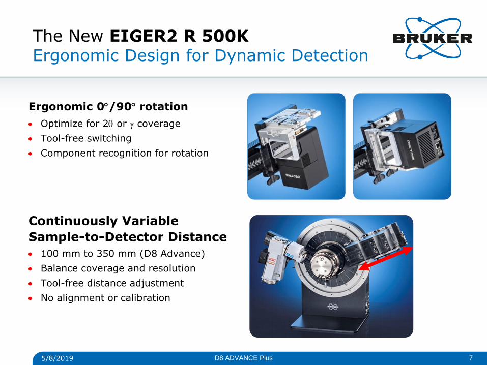
The New EIGER2 R 500K Ergonomic Design for Dynamic Detection
5/8/2019 7
Ergonomic 0/90 rotation
Optimize for 2 or coverage
Tool-free switching
Component recognition for rotation
Continuously Variable
Sample-to-Detector Distance
100 mm to 350 mm (D8 Advance)
Balance coverage and resolution
Tool-free distance adjustment
No alignment or calibration
D8 ADVANCE Plus
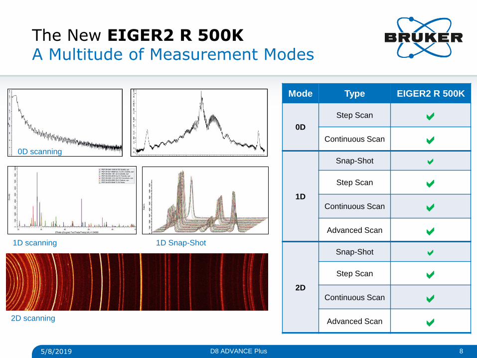
The New EIGER2 R 500K A Multitude of Measurement Modes
8
0D scanning
1D scanning 1D Snap-Shot
2D scanning
5/8/2019
Mode Type EIGER2 R 500K
0D
Step Scan
Continuous Scan
1D
Snap-Shot
Step Scan
Continuous Scan
Advanced Scan
2D
Snap-Shot
Step Scan
Continuous Scan
Advanced Scan
D8 ADVANCE Plus
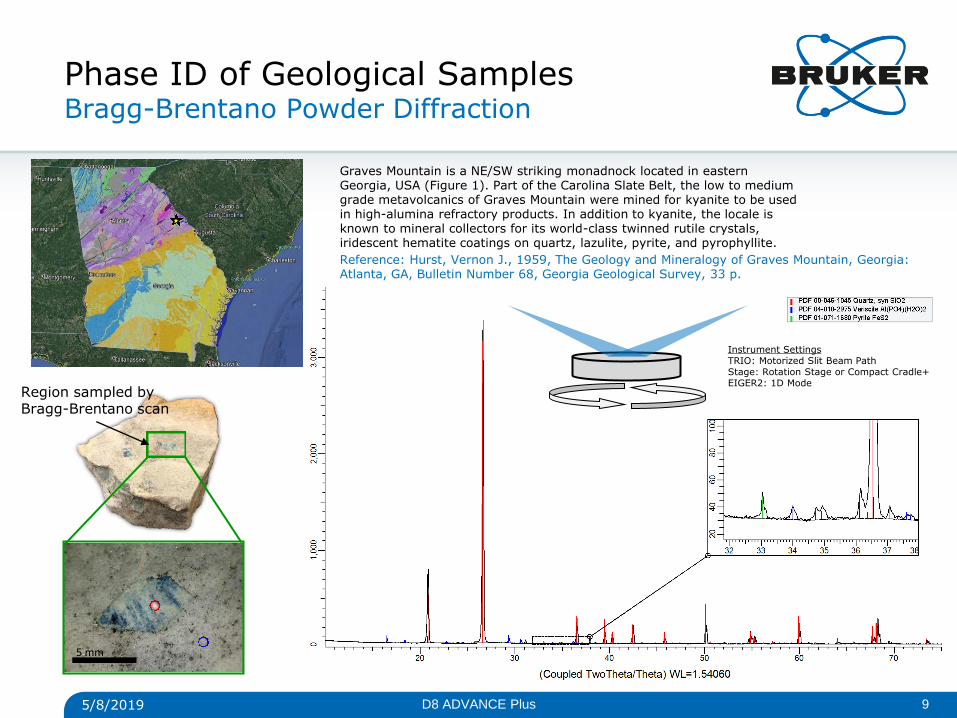
Phase ID of Geological Samples Bragg-Brentano Powder Diffraction
5 mm
Graves Mountain is a NE/SW striking monadnock located in eastern Georgia, USA (Figure 1). Part of the Carolina Slate Belt, the low to medium grade metavolcanics of Graves Mountain were mined for kyanite to be used in high-alumina refractory products. In addition to kyanite, the locale is known to mineral collectors for its world-class twinned rutile crystals, iridescent hematite coatings on quartz, lazulite, pyrite, and pyrophyllite.
Reference: Hurst, Vernon J., 1959, The Geology and Mineralogy of Graves Mountain, Georgia: Atlanta, GA, Bulletin Number 68, Georgia Geological Survey, 33 p.
5/8/2019 9 D8 ADVANCE Plus
Region sampled by Bragg-Brentano scan
Instrument Settings TRIO: Motorized Slit Beam Path Stage: Rotation Stage or Compact Cradle+ EIGER2: 1D Mode
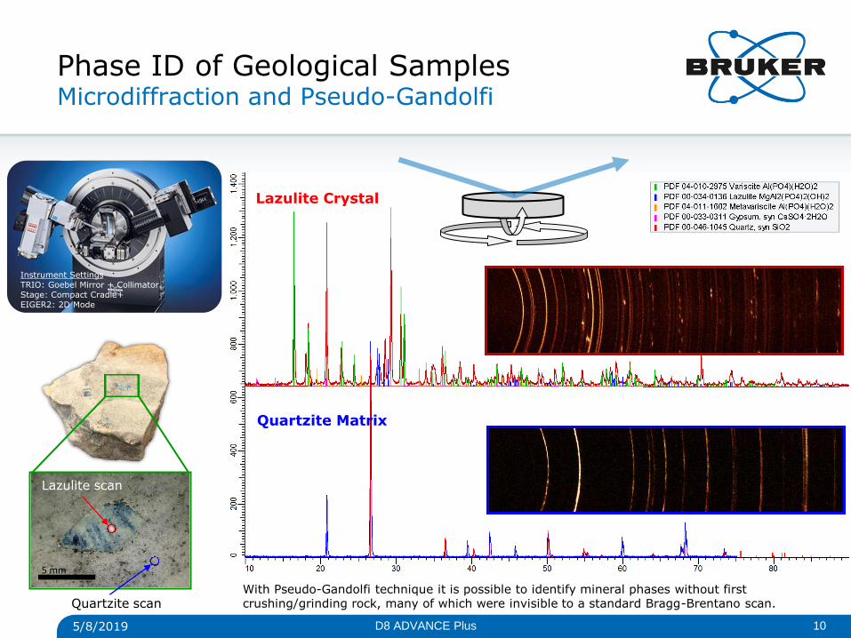
Phase ID of Geological Samples Microdiffraction and Pseudo-Gandolfi
5 mm
Quartzite Matrix
Lazulite Crystal
5/8/2019 10 D8 ADVANCE Plus
Lazulite scan
Quartzite scan With Pseudo-Gandolfi technique it is possible to identify mineral phases without first crushing/grinding rock, many of which were invisible to a standard Bragg-Brentano scan.
Instrument Settings TRIO: Goebel Mirror + Collimator Stage: Compact Cradle+ EIGER2: 2D Mode
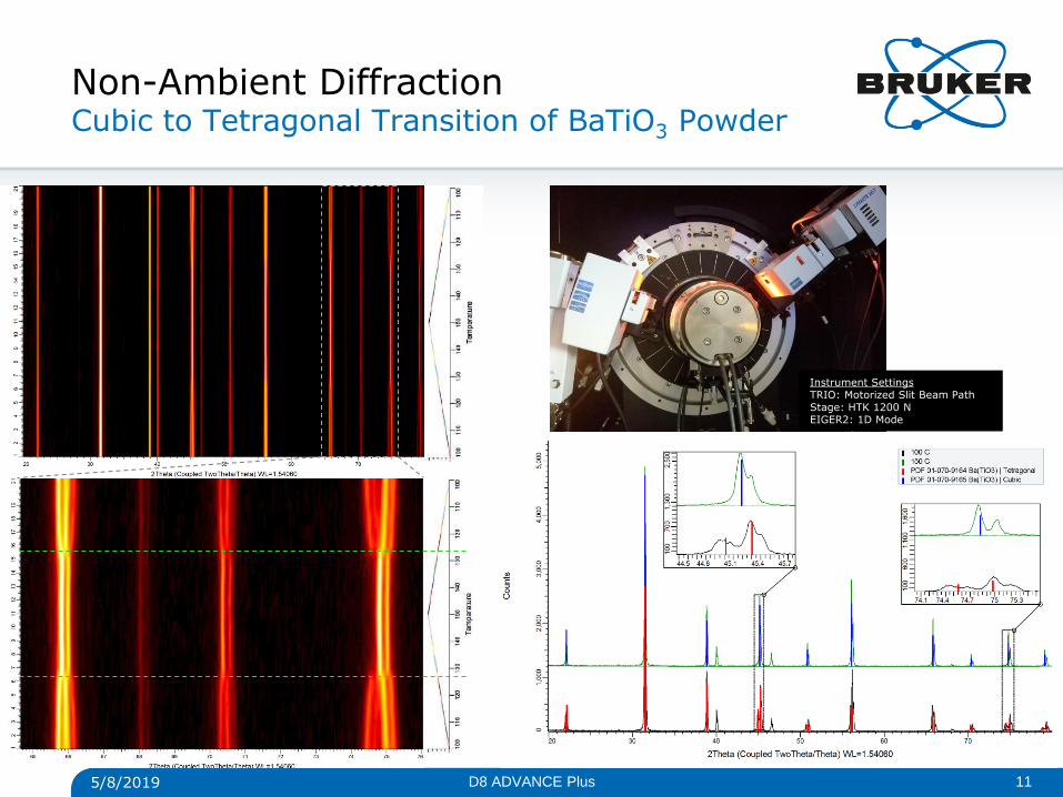
Non-Ambient Diffraction Cubic to Tetragonal Transition of BaTiO3 Powder
5/8/2019 11 D8 ADVANCE Plus
Instrument Settings TRIO: Motorized Slit Beam Path Stage: HTK 1200 N EIGER2: 1D Mode
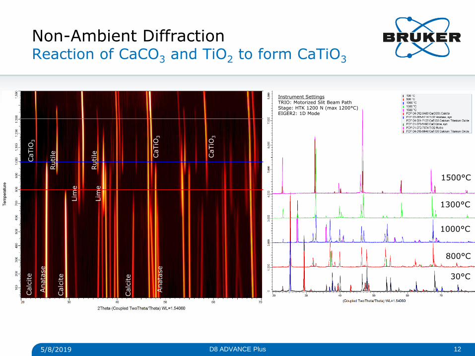
Non-Ambient Diffraction Reaction of CaCO3 and TiO2 to form CaTiO3
30°C
800°C
1300°C
1000°C
1500°C
Calc
ite
Calc
ite
Calc
ite
Anata
se
Anata
se
Rutile
Rutile
Lim
e
Lim
e
CaTiO
3
CaTiO
3
CaTiO
3
5/8/2019 12 D8 ADVANCE Plus
Instrument Settings TRIO: Motorized Slit Beam Path Stage: HTK 1200 N (max 1200°C) EIGER2: 1D Mode
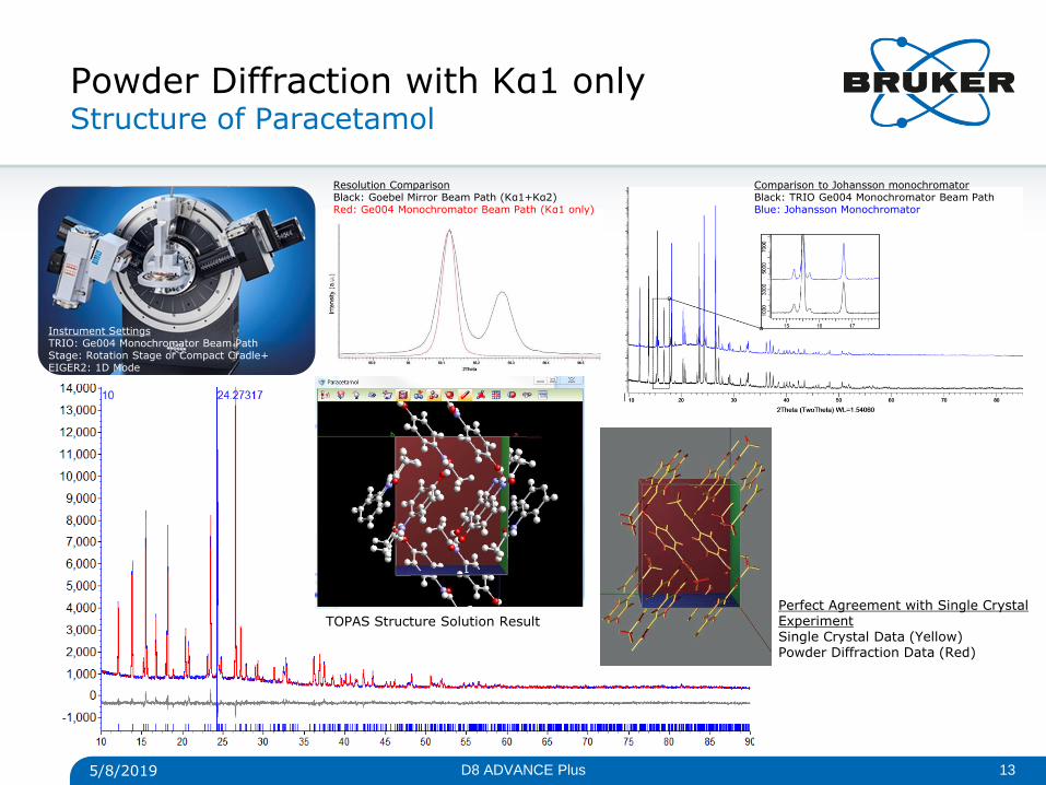
Powder Diffraction with Kα1 only Structure of Paracetamol
Perfect Agreement with Single Crystal Experiment Single Crystal Data (Yellow) Powder Diffraction Data (Red)
5/8/2019 13 D8 ADVANCE Plus
Instrument Settings TRIO: Ge004 Monochromator Beam Path Stage: Rotation Stage or Compact Cradle+ EIGER2: 1D Mode
Resolution Comparison Black: Goebel Mirror Beam Path (Kα1+Kα2) Red: Ge004 Monochromator Beam Path (Kα1 only)
TOPAS Structure Solution Result
Comparison to Johansson monochromator Black: TRIO Ge004 Monochromator Beam Path Blue: Johansson Monochromator
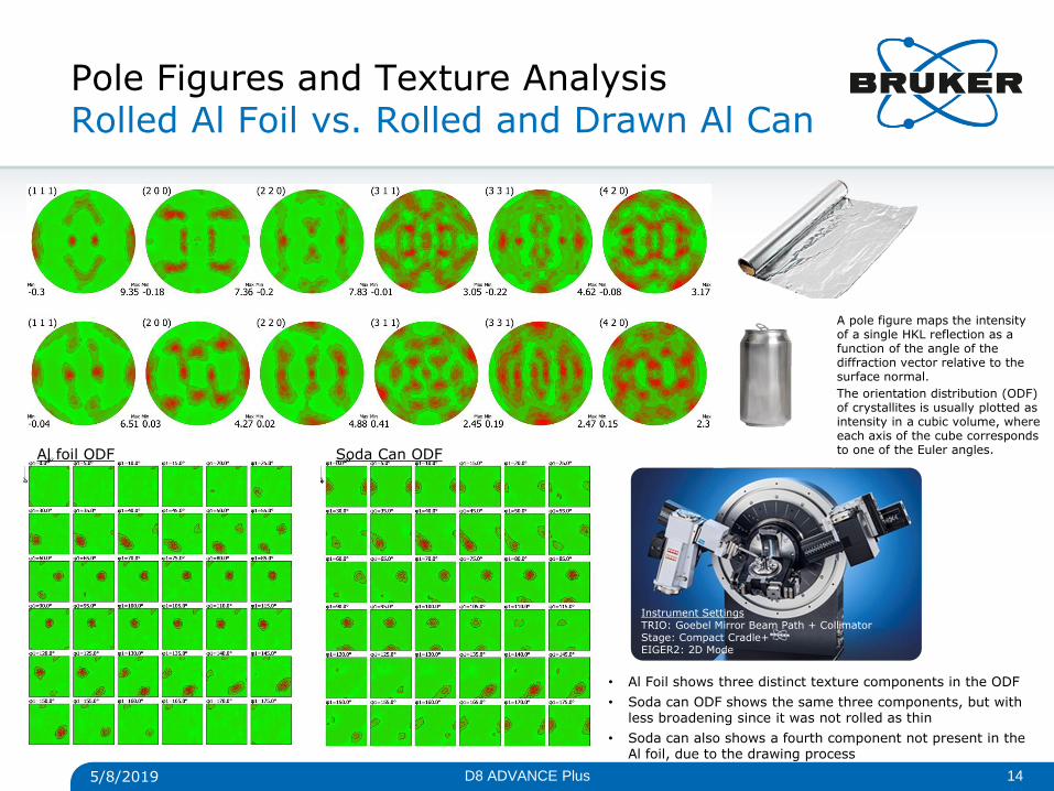
Pole Figures and Texture Analysis Rolled Al Foil vs. Rolled and Drawn Al Can
5/8/2019 14 D8 ADVANCE Plus
Instrument Settings TRIO: Goebel Mirror Beam Path + Collimator Stage: Compact Cradle+ EIGER2: 2D Mode
• Al Foil shows three distinct texture components in the ODF
• Soda can ODF shows the same three components, but with less broadening since it was not rolled as thin
• Soda can also shows a fourth component not present in the Al foil, due to the drawing process
Al foil ODF Soda Can ODF
A pole figure maps the intensity of a single HKL reflection as a function of the angle of the diffraction vector relative to the surface normal.
The orientation distribution (ODF) of crystallites is usually plotted as intensity in a cubic volume, where each axis of the cube corresponds to one of the Euler angles.
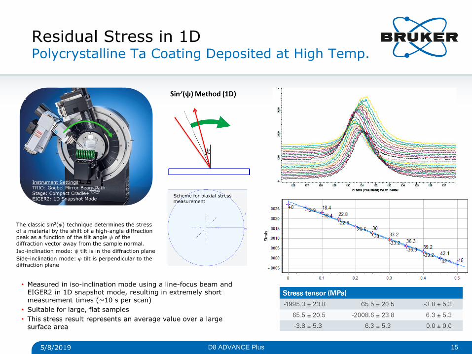
Residual Stress in 1D Polycrystalline Ta Coating Deposited at High Temp.
• Measured in iso-inclination mode using a line-focus beam and EIGER2 in 1D snapshot mode, resulting in extremely short measurement times (~10 s per scan)
• Suitable for large, flat samples
• This stress result represents an average value over a large surface area
5/8/2019 15 D8 ADVANCE Plus
Instrument Settings TRIO: Goebel Mirror Beam Path Stage: Compact Cradle+ EIGER2: 1D Snapshot Mode
𝜓
Scheme for biaxial stress measurement
The classic sin2(𝜓) technique determines the stress of a material by the shift of a high-angle diffraction peak as a function of the tilt angle 𝜓 of the diffraction vector away from the sample normal.
Iso-inclination mode: 𝜓 tilt is in the diffraction plane
Side-inclination mode: 𝜓 tilt is perpendicular to the diffraction plane
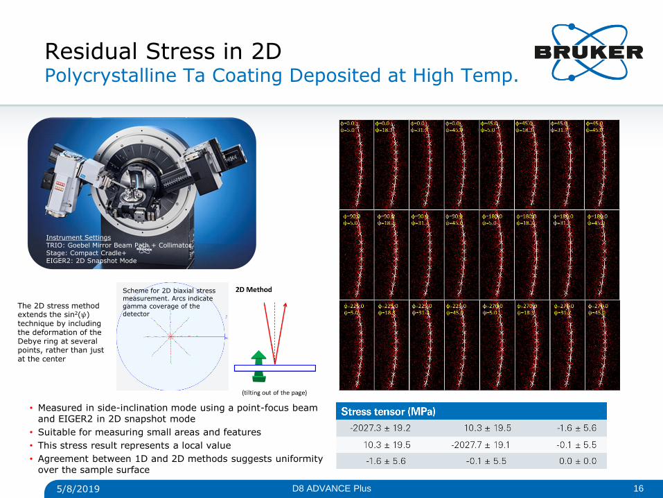
Residual Stress in 2D Polycrystalline Ta Coating Deposited at High Temp.
• Measured in side-inclination mode using a point-focus beam and EIGER2 in 2D snapshot mode
• Suitable for measuring small areas and features
• This stress result represents a local value
• Agreement between 1D and 2D methods suggests uniformity over the sample surface
5/8/2019 16 D8 ADVANCE Plus
Scheme for 2D biaxial stress measurement. Arcs indicate gamma coverage of the detector
Instrument Settings TRIO: Goebel Mirror Beam Path + Collimator Stage: Compact Cradle+ EIGER2: 2D Snapshot Mode
The 2D stress method extends the sin2(𝜓) technique by including the deformation of the Debye ring at several points, rather than just at the center
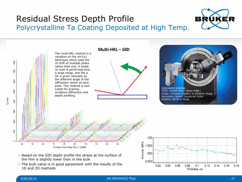
Residual Stress Depth Profile Polycrystalline Ta Coating Deposited at High Temp.
• Based on the GID depth profile the stress at the surface of the film is slightly lower than in the bulk
• The bulk value is in good agreement with the results of the 1D and 2D methods
5/8/2019 17 D8 ADVANCE Plus
The multi-HKL method is a variation on the sin2(𝜓) technique which uses the 2𝜃 shift of multiple peaks rather than one. A single 2𝜃 scan is performed over a large range, and the 𝜓 tilt is given naturally by the different angle of the diffraction vector at each peak. This method is well suited for grazing-incidence diffraction and depth profiling.
Instrument Settings TRIO: Goebel Mirror Beam Path Stage: Compact Cradle+ or Rotation Stage Secondary Optics: Equatorial Soller EIGER2: 0D Scan Mode
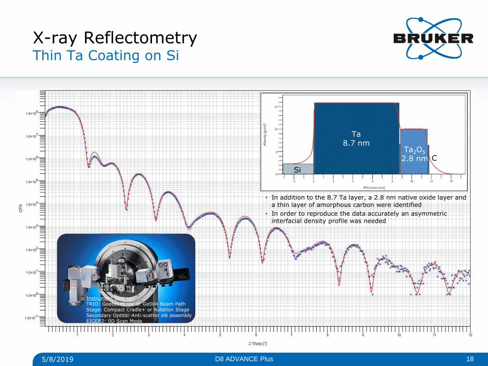
X-ray Reflectometry Thin Ta Coating on Si
Si
Ta 8.7 nm
Ta2O5 2.8 nm C
• In addition to the 8.7 Ta layer, a 2.8 nm native oxide layer and a thin layer of amorphous carbon were identified
• In order to reproduce the data accurately an asymmetric interfacial density profile was needed
5/8/2019 18 D8 ADVANCE Plus
Instrument Settings TRIO: Goebel Mirror or Ge004 Beam Path Stage: Compact Cradle+ or Rotation Stage Secondary Optics: Anti-scatter slit assembly EIGER2: 0D Scan Mode
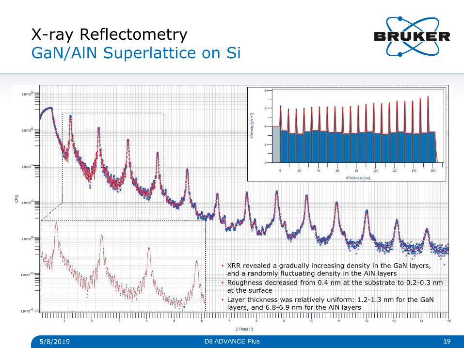
X-ray Reflectometry GaN/AlN Superlattice on Si
• XRR revealed a gradually increasing density in the GaN layers, and a randomly fluctuating density in the AlN layers
• Roughness decreased from 0.4 nm at the substrate to 0.2-0.3 nm at the surface
• Layer thickness was relatively uniform: 1.2-1.3 nm for the GaN layers, and 6.8-6.9 nm for the AlN layers
5/8/2019 19 D8 ADVANCE Plus
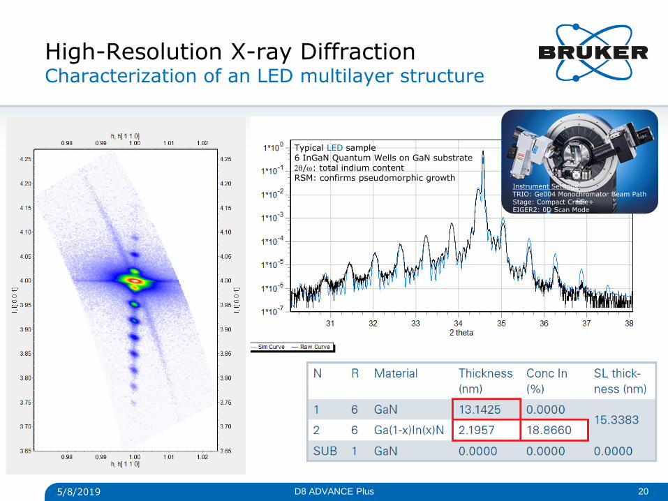
High-Resolution X-ray Diffraction Characterization of an LED multilayer structure
Typical LED sample 6 InGaN Quantum Wells on GaN substrate /: total indium content RSM: confirms pseudomorphic growth
5/8/2019 20 D8 ADVANCE Plus
Instrument Settings TRIO: Ge004 Monochromator Beam Path Stage: Compact Cradle+ EIGER2: 0D Scan Mode
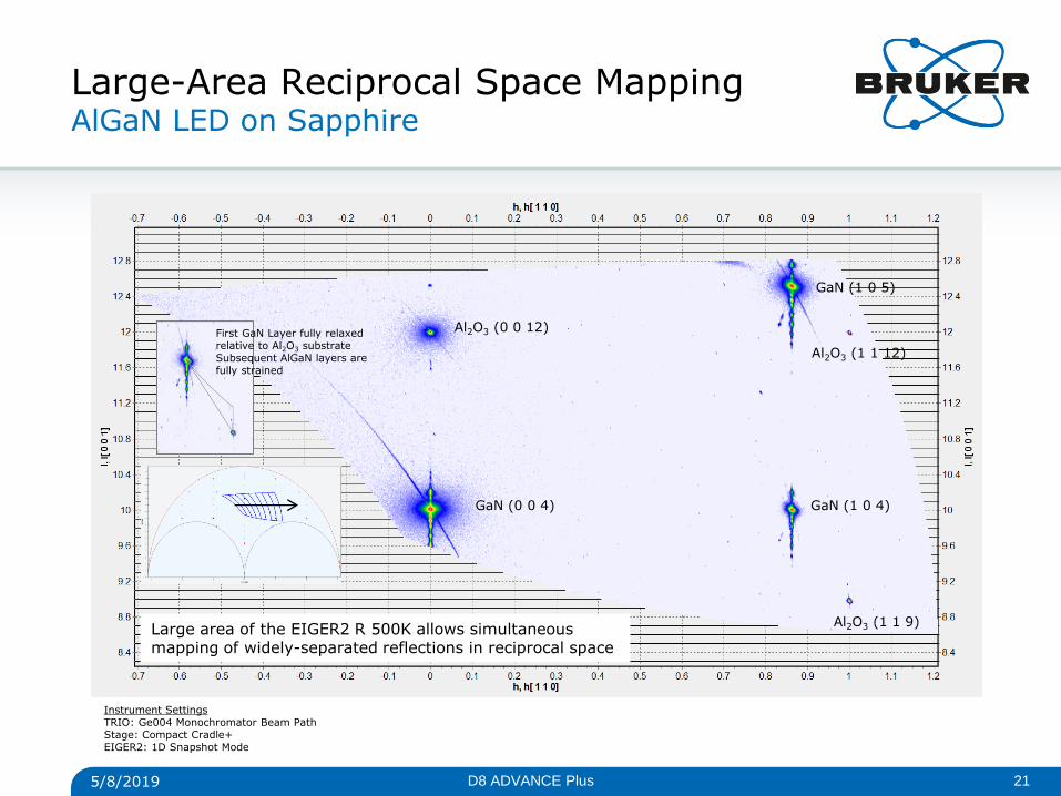
Large-Area Reciprocal Space Mapping AlGaN LED on Sapphire
Al2O3 (0 0 12)
Al2O3 (1 1 12)
GaN (0 0 4)
GaN (1 0 5)
GaN (1 0 4)
Al2O3 (1 1 9) Large area of the EIGER2 R 500K allows simultaneous mapping of widely-separated reflections in reciprocal space
5/8/2019 21 D8 ADVANCE Plus
Instrument Settings TRIO: Ge004 Monochromator Beam Path Stage: Compact Cradle+ EIGER2: 1D Snapshot Mode
First GaN Layer fully relaxed relative to Al2O3 substrate Subsequent AlGaN layers are fully strained
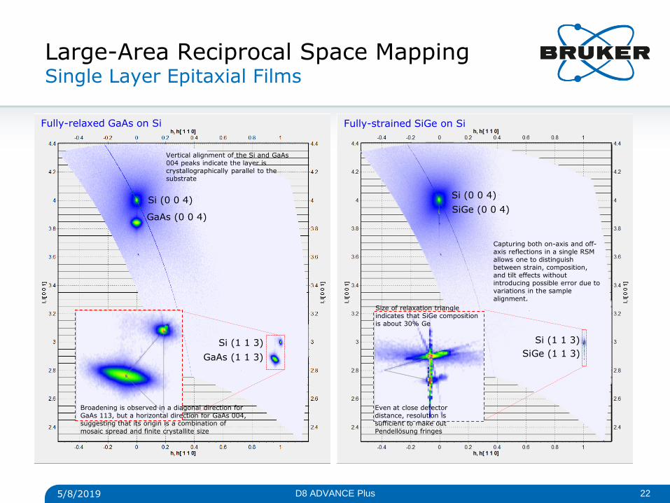
Large-Area Reciprocal Space Mapping Single Layer Epitaxial Films
Si (0 0 4) Si (0 0 4)
Si (1 1 3)
GaAs (0 0 4) SiGe (0 0 4)
GaAs (1 1 3) SiGe (1 1 3)
Si (1 1 3)
5/8/2019 22 D8 ADVANCE Plus
Vertical alignment of the Si and GaAs 004 peaks indicate the layer is crystallographically parallel to the substrate
Broadening is observed in a diagonal direction for GaAs 113, but a horizontal direction for GaAs 004, suggesting that its origin is a combination of mosaic spread and finite crystallite size
Even at close detector distance, resolution is sufficient to make out Pendellösung fringes
Fully-relaxed GaAs on Si Fully-strained SiGe on Si
Size of relaxation triangle indicates that SiGe composition is about 30% Ge
Capturing both on-axis and off-axis reflections in a single RSM allows one to distinguish between strain, composition, and tilt effects without introducing possible error due to variations in the sample alignment.
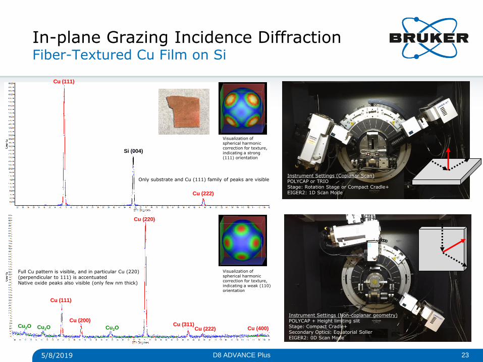
In-plane Grazing Incidence Diffraction Fiber-Textured Cu Film on Si
Cu (111)
Cu (200)
Cu (220)
Cu (311) Cu (222) Cu (400) Cu2O Cu2O Cu2O
Cu (111)
Cu (222)
Si (004)
5/8/2019 23 D8 ADVANCE Plus
Instrument Settings (Coplanar Scan) POLYCAP or TRIO Stage: Rotation Stage or Compact Cradle+ EIGER2: 1D Scan Mode
Instrument Settings (Non-coplanar geometry) POLYCAP + Height limiting slit Stage: Compact Cradle+ Secondary Optics: Equatorial Soller EIGER2: 0D Scan Mode
Only substrate and Cu (111) family of peaks are visible
Full Cu pattern is visible, and in particular Cu (220) (perpendicular to 111) is accentuated Native oxide peaks also visible (only few nm thick)
Visualization of spherical harmonic correction for texture, indicating a strong (111) orientation
Visualization of spherical harmonic correction for texture, indicating a weak (110) orientation
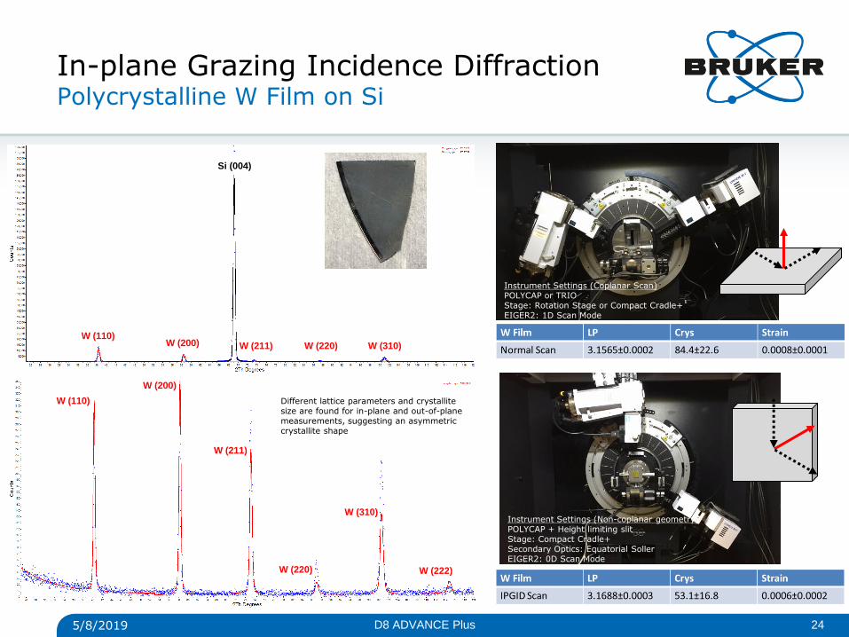
In-plane Grazing Incidence Diffraction Polycrystalline W Film on Si
W (110)
W (200)
W (211)
W (220)
W (310)
W (222)
W (110) W (200) W (211) W (220) W (310)
Si (004)
5/8/2019 24 D8 ADVANCE Plus
Different lattice parameters and crystallite size are found for in-plane and out-of-plane measurements, suggesting an asymmetric crystallite shape
Instrument Settings (Coplanar Scan) POLYCAP or TRIO Stage: Rotation Stage or Compact Cradle+ EIGER2: 1D Scan Mode
Instrument Settings (Non-coplanar geometry) POLYCAP + Height limiting slit Stage: Compact Cradle+ Secondary Optics: Equatorial Soller EIGER2: 0D Scan Mode
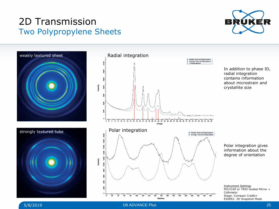
2D Transmission Two Polypropylene Sheets
Radial integration
Polar integration
5/8/2019 25 D8 ADVANCE Plus
In addition to phase ID, radial integration contains information about microstrain and crystallite size
Polar integration gives information about the degree of orientation
weakly textured sheet
strongly textured tube
Instrument Settings POLYCAP or TRIO Goebel Mirror + Collimator Stage: Compact Cradle+ EIGER2: 2D Snapshot Mode
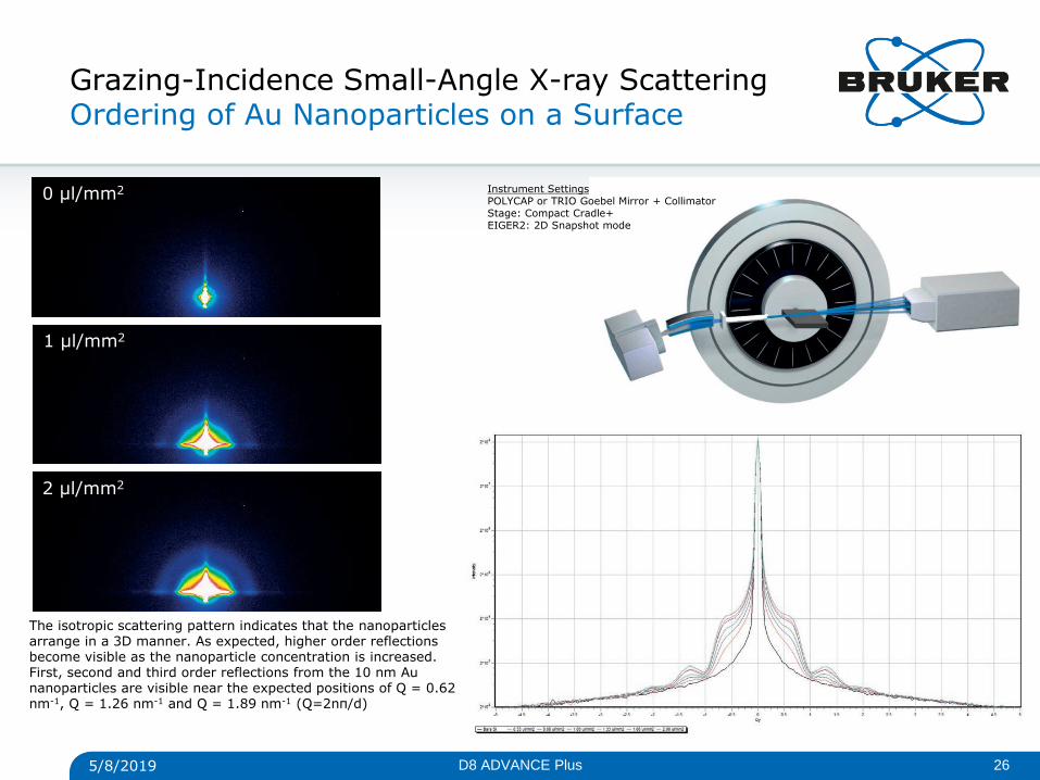
Grazing-Incidence Small-Angle X-ray Scattering Ordering of Au Nanoparticles on a Surface
The isotropic scattering pattern indicates that the nanoparticles arrange in a 3D manner. As expected, higher order reflections become visible as the nanoparticle concentration is increased. First, second and third order reflections from the 10 nm Au nanoparticles are visible near the expected positions of Q = 0.62 nm-1, Q = 1.26 nm-1 and Q = 1.89 nm-1 (Q=2nπ/d)
0 μl/mm2
1 μl/mm2
2 μl/mm2
5/8/2019 26 D8 ADVANCE Plus
Instrument Settings POLYCAP or TRIO Goebel Mirror + Collimator Stage: Compact Cradle+ EIGER2: 2D Snapshot mode
• Powder Diffraction in Bragg-Brentano or Parallel Beam
• Phase ID
• Quantification
• Indexing
• Rietveld Refinement
• Structure Solution
• Bragg 2D
• Non-Ambient Diffraction
• Microdiffraction
• Transmission
D8 ADVANCE with TRIO and EIGER2 R 500K Applications Summary
• Residual Stress Analysis
• 2D Method
• Sin2(Psi) Method
• Multi-HKL method
• Texture Analysis
• Pole Figures
• Orientation Distribution Function
• Component Fitting
• Harmonic Fitting
• Inverse Pole Figures
• High-resolution XRD
• Rocking Curves
• On-Axis Coupled Scans
• Reciprocal Space Mapping
• Grazing Incidence Diffraction
• Phase ID
• Depth Profiling
• GI-SAXS
• In-Plane Grazing Incidence Diffraction
• 2D Materials and ultra-thin films
• Surface oxides
• In-plane lattice parameters and crystallite size
• Epitaxial films relationship to substrate
• X-ray Reflectometry
• Thickness, density, roughness of single and multi-layered films
• Crystalline, amorphous, or liquid layers
5/8/2019 27 D8 ADVANCE Plus
Innovation with Integrity
© Copyright Bruker Corporation. All rights reserved.