Embed Size (px)
DESCRIPTION
Self-assembly of viral capsid protein
Citation preview
7/17/2019 J. Virol.-2012-Cadena-Nava-3318-26
http://slidepdf.com/reader/full/j-virol-2012-cadena-nava-3318-26 1/9
Self-Assembly of Viral Capsid Protein and RNA Molecules of Different Sizes: Requirement for a Specific High Protein/RNA MassRatio
Ruben D. Cadena-Nava, Mauricio Comas-Garcia, Rees F. Garmann, A. L. N. Rao,* Charles M. Knobler, and William M. Gelbart
Department of Chemistry and Biochemistry University of California, Los Angeles, California, USA
Virus-like particles can be formed by self-assembly of capsid protein (CP) with RNA molecules of increasing length. If the pro-
tein “insisted” on a single radius of curvature, the capsids would be identical in size, independent of RNA length. However, there
would be a limit to length of the RNA, and one would not expect RNA much shorter than native viral RNA to be packaged unlessmultiple copies were packaged. On the other hand, if the protein did not favor predetermined capsid size, one would expect the
capsid diameter to increase with increase in RNA length. Here we examine the self-assembly of CP from cowpea chlorotic mottle
virus with RNA molecules ranging in length from 140 to 12,000 nucleotides (nt). Each of these RNAs is completely packaged if and only if the protein/RNA mass ratio is sufficiently high; this critical value is the same for all of the RNAs and corresponds to
equal RNA and N-terminal-protein charges in the assembly mix. For RNAs much shorter in length than the 3,000 nt of the viral
RNA, two or more molecules are assembled into 24- and 26-nm-diameter capsids, whereas for much longer RNAs (>4,500 nt), a
single RNA molecule is shared/packaged by two or more capsids with diameters as large as 30 nm. For intermediate lengths, asingle RNA is assembled into 26-nm-diameter capsids, the size associated with T3 wild-type virus. The significance of these
assembly results is discussed in relation to likely factors that maintain T3 symmetry in vivo.
Many mature infectious viruses with positive-sense RNA ge-nomes are known to consist of a single copy of the genome
inside a one-molecule-thick shell of protein (the capsid protein[CP]). In many instances, this nucleocapsid structure is believedto arise spontaneously from self-assembly of the protein aroundits associated nucleic acid, a process that has been mimicked invitro by directmixing of thetwo purified components (i.e.,CP andRNA) in an optimized physiological buffer. A particularly well-studied example is that of cowpea chloroticmottle virus (CCMV),first synthesized in the laboratory in 1967 (8). The native virus hasa multipartite genome encoding four genes contained in threesingle-stranded RNA (ssRNA) molecules: RNA1, 3,171 nucleo-tides (nt); RNA2, 2,774 nt; and RNA3, 2,173 nt (2, 18). The firsttwo genomic RNAs are packaged alone, and the third genomicRNA is copackaged with a subgenomic RNA4 of 824 nt, so thateach CCMV virion contains about 3,000 nt. Just 2 years later, itwas demonstrated that CCMV CP was also capable of packag-ing heterologous ssRNA, as well as synthetic anionic polymers(9). A more extensive study of CCMV CP assembly aroundfourheterologous viral RNAs ranging in length from 4,000 to 6,400nt and around short homopolynucleotides [poly(A), poly(C),
and poly(U))] was carried out by Adolph and Butler (1).Similarly, the CP of a closely related virus, brome mosaic virus
(BMV), has been shown to package gold nanoparticles of varioussizes that have been functionalized with anionic moieties (43) anda negatively charged near-infrared chromophore (26). Also, wehave investigated the packaging by CCMV CP of poly(styrene sul-fonate) (PSS) polymers with molecular masses ranging from 38kDa to 3.4 MDa (12, 23). We chose to examine PSS because itsphysical properties are well established: it has a persistence lengthand charge density comparable to that of ssRNA, it has solutionproperties (e.g., radius of gyration) that are well understood, andit is readily availablein a large range of molecularmasses. Carryingout assemblies of virus-like particles (VLPs) for mixes of CCMV
CP with PSS of various molecular masses, we determined the ef-fects of polymer length on the formation of VLPs of different sizes(23) and explained the observed behavior in terms of a competi-tion between the polymer-polymer and polymer-capsid interac-tions and the preferred curvature of the CP, as discussed by Zandiand van der Schoot (49). In particular, we found that a large rangeof PSSlengths (and hence charges) could be accommodatedby thesame size protein capsid, up to a certain threshold length at whichlarger capsids begin to form as the dominant assembly product(12, 23).
In the findings reported here we extend this study of VLP self-assembly to include a broader range of polyanion lengths andcapsid sizes. Also, while we continue to use the same (CCMV) CP,we now feature—instead of PSS—ssRNA molecules with molec-ular masses varying over a larger range, from about 45 kDa (140nt) to 4 MDa (12,000 nt).
There are several reasons for working with ssRNA instead of PSS. First, ssRNA is the genome of CCMV and of a majority of viruses pathogenic to plants, animals, and humans. Second, froma physical point of view, RNA is not a linear polymer like PSS.Self-complementary base-pairing interactions in ssRNA lead to
a branched structure containing double-stranded and single-stranded regions that are organized with respect to each other intocomplex tertiary structures. As a consequence, the radius of gyra-
Received 20 October 2011 Accepted 22 December 2011
Published ahead of print 28 December 2011
Address correspondence to William M. Gelbart, [email protected].
* Present address: Department of Plant Pathology and Microbiology, University of
California, Riverside, California, USA.
Copyright © 2012, American Society for Microbiology. All Rights Reserved.
doi:10.1128/JVI.06566-11
3318 jvi.asm.org 0022-538X/12/$12.00 Journal of Virology p. 3318–3326
on J un e 3 0 ,2 01 5 b
y UNIVER SIDAD
A UT ON OMA
DE SLP
h t t p: / / jv
i. a sm. or g /
D ownl o a d e dfr om
7/17/2019 J. Virol.-2012-Cadena-Nava-3318-26
http://slidepdf.com/reader/full/j-virol-2012-cadena-nava-3318-26 2/9
tion and shape of the RNA increase and change with molecularmass in ways that are difficult to predict (48), and it must beexpected to interact very differently with CP (10) than does alinear homopolymer such as PSS.
MATERIALS AND METHODS
Materials. Restriction enzymes wereobtained from New England Biolabs.SP6 RNA polymerase was obtained from Ambion and T7 RNA polymer-ase was a gift from Feng Guo (Department of Biological Chemistry, Uni-versity of California, Los Angeles). Enzymes were used as recommendedby themanufacturer.The plasmids were grown, purified, andanalyzedby standard methods (39). Plasmids pT7B1and pT7B3Sn contain full-lengthcDNA clones corresponding to BMV RNA 1 (B1) (3,234 nt) and BMVRNA3 (B3) (2,117nt), respectively (17), andplasmids p30B, pEYFP-Rep,and pTE12 encodethe fulllength of TMV RNA (6,395nt) (15), a Sindbis-derived replicon RNA containing enhanced yellow fluorescent protein(8,600 nt) and full-length Sindbis virus RNA (11,700 nt), respectively (30). The plasmids were grown, purified, and analyzed by standard meth-ods. All of the other chemicals used were DNase-, RNase-, and protease-free.
Combining B1 and B3 RNAs by directional subcloning. Directional
subcloning of pT7B1 requires the plasmid to have overhangs; these werecreated by linearization with BamHI and EcoRI. The insert that encodesB3 was engineered from pT7B3Sn by PCR using the forward primer d(TTGCGCGGATCCAACTAATTCTCGTTCG) (the BamHI site is under-lined) and the reverse primer d(TGGCCGGAATTCAACACTGTACGGTAC) (the EcoRI site is underlined). These primers give 8 and 28 fewernucleotides at the5= and 3= ends, respectively, than genomic BMVRNA 3.The resulting PCR product was double digested with EcoRI and BamHIandsubclonedinto pT7B1 previously digested withBamHI/EcoRI to givethe new cloning cDNA, pRDCT7B1B3. The authenticity of ligated prod-uct, RNA B1B3, was confirmed by restriction analysis followed by se-quencing.
PCR amplification of DNA templates to transcribe short RNAs.DNA templates with lengths shorter than BMV RNA 1 were prepared by PCR of pT7B1 plasmid; all truncations were carried out from the 3 = end.
We used the universal 5= primer d(TAATACGACTCACTATAGGTAGACCACGGAACGAGGTTC) (the T7 promoter is underlined) and, as re-verse primers, d(GCAATCAACTTCAGCAAATCG), d(CACATCCTCTCCTCATGTC), d(GTCTTCAAACCATACACAGTG), d(CTTGCTCAAATTCTTCAACG), and d(GGATACAACCAGTTACCGTTG) to obtainthe corresponding DNA templates for transcribing RNAs with lengths of 141, 499, 999, 1,498 and 1,960 nt, respectively.
RNA transcription. Plasmids pT7B3Sn and pT7B1 were linearizedwith BamHI, plasmids p30B and pTE12 were linearized with XhoI, andplasmid pEYFP-Rep was linearized with SacI. pRDCT7B1B3 was linear-ized with BglII and EcoRI to produce RNA transcripts of 4,452 and 5,319nt, respectively. All of the templates were purified by standard procedures(39). All DNA templates and plasmids encoding for RNAs between 140and 6,395 nt were transcribed with T7 RNA polymerase; plasmids pEYFP
and pTE12 were transcribed with SP6 RNA polymerase.CCMV CP purification. CCMV was purified frominfected Californiacowpeaplant (Vigna ungiculata cv. Black Eye) (7), and capsid protein wasisolated largely as described previously (3). Nucleocapsids were disruptedby 24-h dialysis againstdisassembly buffer (0.5 M CaCl
2, 50mM Tris[pH
7.5], 1 mM EDTA, 1 mM dithiothreitol [DTT], 0.5 mM phenylmethyl-sulfonyl fluoride [PMSF]) at 4°C and then by ultracentrifugation at100,000 rpmfor 100min (4 105 g ) at4°Cin a BeckmanTLA110 rotor.The RNA was pelleted, and the CP was extracted from the supernatant indifferent fractions. Each fraction was immediately dialyzed against pro-tein buffer (1 M NaCl, 20 mM Tris [pH 7.2], 1 mM EDTA, 1 mM DTT, 1mM PMSF). The protein concentration and its purity, with respect toRNA contamination, were measured by UV-Vis spectrophotometry; only protein solutions with 280/260 ratios greater than 1.5 were used for as-sembly. SDS-PAGE and matrix-assisted laser desorption ionization–time
of flight mass spectrometrywere used to ascertainthatthe purified proteinwas not cleaved.
In vitro assembly: preliminary titrations and gel retardation assays.Assembly reactions of CCMV CP with RNAs of different lengths wereperformed in RNA assembly buffer (RAB; 50 mM NaCl, 10 mM KCl, 5mM MgCl
2, 1 mM DTT, 50 mM Tris-HCl [pH 7.2]). CCMV CP was
titrated at different mass ratios into a constant RNA concentration (30ng/l). After overnight assembly at 4°C, a 10-l aliquot of each ratio wasmixed with 2 l of 100% glycerol (RNase-, DNase-, and protease-free)and loaded into a 1% agarose gel (EMD) in either virus buffer (0.1 Msodium acetate, 1 mM EDTA [pH 4.8]) or electrophoresis buffer (0.1 Msodium acetate, 1 mM EDTA, pH 6). The samples were electrophoresedfor 1.25 h at 70 V (electrophoresis buffer) or 4 h at 50 V (virus buffer) in ahorizontal gelapparatus (Fisher) at 4°C. Thesamples were then stained ina solution of 5 g of ethidium bromide/ml and visualized with anAlphaimager system.
In vitro assembly of VLPs. Dissociated CCMV CP subunits and de-sired RNA (30 ng/l) transcripts were mixed in a ratio of 6:1 (wt/wt) andthen dialyzed for 24 h at 4°Cagainst RAB. Thesamples were then dialyzedagainst virus suspension buffer (50 mM sodium acetate, 8 mM magne-sium acetate [pH 4.5]) for at least 4 h. For assemblies with RNAs longerthan BMV RNA1, 100 to 300 l of the assembly reactions were subjected
to sucrose gradient (10 to 40% [wt/wt]) centrifugation in virus bufferusing an ultracentrifuge SW 50.1 rotor at 33,000 rpm (1.3 105 g ) for2 h. The fraction corresponding to the VLPs was recovered and dialyzedovernight against virus buffer or virus suspension buffer to remove thesucrose. VLPs from allRNA assemblies were purified andconcentrated by washing with virus suspension buffer using a 100-kDa Amicon centrifugefilter (0.5 ml)at 3,000 g for 5 min; this last stepwas repeatedfourtimes.All of the procedures were carried out at 4°C. Finally, the samples werecharacterized by UV-Vis spectroscopy with a NanoDrop spectrophotom-eter (Fisher).
TEM analysis of VLPs. For negative staining, purified VLPs were ap-plied to glow-discharged copper grids (400-mesh) that previously hadbeen coatedwith Parlodionand carbon. A 6-l aliquot of VLPs wasspreadonto the grid for 1 min, blotted with Whatman filter paper, and thenstained with6 l of 1%uranyl acetate for 1 min. Excess stain was removed
by blotting with filter paper. The samples were stored overnight in a des-iccator and analyzed with a JEM 1200-EX transmission electron micro-scope equipped with a wide-angle (top mount) BioScan 600-W 11Kpixel digital camera operated at 80 keV. The reported average diameter of the VLPs is that of the geometric mean of two orthogonal measurementsof the capsids obtained with ImageJ (U.S. National Institutes of Health)software from recorded images.
RESULTS AND DISCUSSION
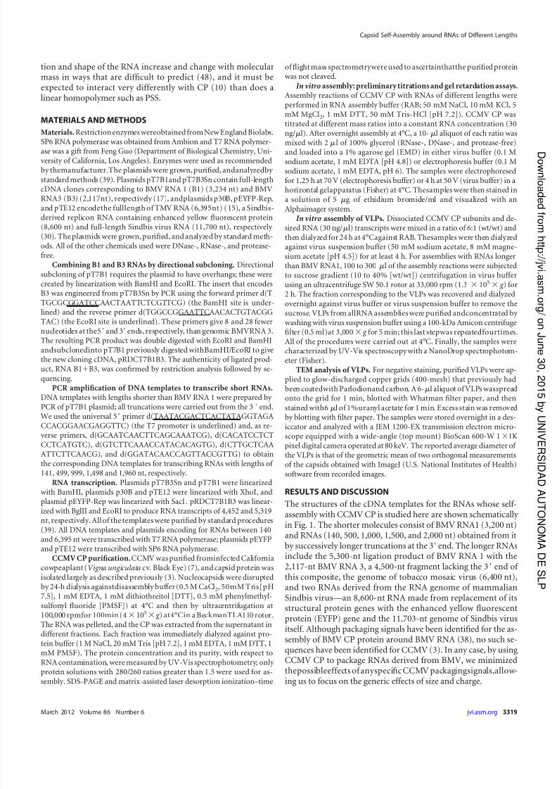
The structures of the cDNA templates for the RNAs whose self-assembly with CCMV CP is studied here are shown schematically in Fig. 1. The shorter molecules consist of BMV RNA1 (3,200 nt)and RNAs (140, 500, 1,000, 1,500, and 2,000 nt) obtained from itby successively longer truncations at the 3= end. The longer RNAs
include the 5,300-nt ligation product of BMV RNA 1 with the2,117-nt BMV RNA 3, a 4,500-nt fragment lacking the 3= end of this composite, the genome of tobacco mosaic virus (6,400 nt),and two RNAs derived from the RNA genome of mammalianSindbis virus—an 8,600-nt RNA made from replacement of itsstructural protein genes with the enhanced yellow fluorescentprotein (EYFP) gene and the 11,703-nt genome of Sindbis virusitself. Although packaging signals have been identified for the as-sembly of BMV CP protein around BMV RNA (38), no such se-quences have been identified for CCMV (3). In any case, by usingCCMV CP to package RNAs derived from BMV, we minimizedthepossibleeffects of anyspecific CCMV packagingsignals,allow-ing us to focus on the generic effects of size and charge.
Capsid Self-Assembly around RNAs of Different Lengths
March 2012 Volume 86 Number 6 jvi.asm.org 3319
on J un e 3 0 ,2 01 5 b
y UNIVER SIDAD
A UT ON OMA
DE SLP
h t t p: / / jv
i. a sm. or g /
D ownl o a d e dfr om
7/17/2019 J. Virol.-2012-Cadena-Nava-3318-26
http://slidepdf.com/reader/full/j-virol-2012-cadena-nava-3318-26 3/9
The packaging experiments were preceded by studies to iden-tify the optimal molar ratio of CP to RNA for packaging. This isthe ratio at which there is just sufficient protein to package all of the RNA into RNase-resistant VLPs. It was identified by carryingout, in parallel, assembly reactions in RAB using a variety of pro-
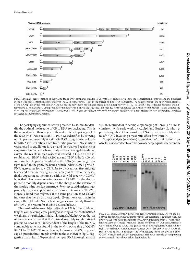
tein/RNA (wt/wt) ratios. Each fixed-ratio protein/RNA solutionwas allowed to equilibrate for 24 h and then dialyzed against virussuspensionbuffer before beinganalyzed by agarose gel retardationassays. The results in each case, as illustrated in Fig. 2 by the as-semblies with BMV RNA1 (3,200 nt) and TMV RNA (6,400 nt),were similar. As protein is added to the RNA (i.e., moving fromright to left in the gels), the bands, which indicate small protein-RNA aggregates for low CP/RNA (wt/wt) ratios, first migratefaster and then increasingly more slowly as the ratio increases,finally appearing at the same position as wild-type (wt) CCMV.Note that it has been shown in the case of CCMV that the electro-phoretic mobility depends only on the charge on the exterior of thecapsid andnot on itscontents, with empty capsids migratingatprecisely the same position as virions containing RNA (25).
Hence, a band that migrates at the same position as wt CCMVindicates that there is an intact capsid. (Note, however, that in thecase of the 6,400-nt RNA the band migrates more slowly than thatof CCMV; the reason for this is discussed below.)
Theresults of theassemblystudies show RNAs of very differentlengths can be completely packaged as long as the protein/RNAweight ratio is sufficiently high. It is remarkable, however, that weobserve in every case that the optimal assembly weight ratio of protein to RNA is 6:1, independent of the length of the RNA. Acomparable ratio was found in the in vitro packaging of CCMVRNA1 by CCMV CP; in particular, Johnson et al. (24) reportedcapsid-protein titration gels similar to those shown in Fig. 2, sug-gesting that at least 130 protein dimers per RNA (a weight ratio of
5:1) are required for the complete packaging of RNA1. This is alsoconsistent with early work by Adolph and Butler (1), who re-ported a significant fraction of free RNA in their reassembly stud-ies of CCMV involving a mass ratio of 5:1 for CP/RNA.
Simple analysis (see below) shows that the “magic ratio” value
of 6:1is associated with a condition of charge equality between the
FIG1 Schematic representation of the plasmids and DNA templates used for RNA syntheses. The arrows denote the transcription promoter, and the cloverleaf at the 3= end represents the highly conserved tRNA-like structure (3=TLS) in the corresponding RNA transcripts. The boxes represent the open reading framesof the RNAs; 1a is a viral replicase; MP and CP are the movement protein and capsid protein, respectively; E1, E2, E3, and 6K are structural proteins; and NSrepresents all nonstructural viral proteins for Sindbis virus. EYFP is the sequence that encodes for the enhanced yellow fluorescent protein, RDRP denotes theRNA-dependent RNApolymerasegenes, andU5CPis theCP gene of strainU5 of tobacco mild green mosaicvirus. Therepresentations of theplasmid templatesare scaled to their relative lengths.
FIG 2 CP-RNA assembly titrations: gel retardation assays. Shown are 1%agarose gels stained with ethidiumbromide.At theleft is a titrationof 3,217-ntBMV RNA1 with various amounts of CCMV CP ranging from 0 (right-mostlane,RNA) to the“magic”ratio,6:1 (lane secondfromleft). CP/RNAratiosarewt/wt ratios of CP to RNA. This gel was run in electrophoresis buffer. On theright is a similar gelretardationassaycarried outwith 6,395-nt TMV RNAandrun in virus buffer. In both gels, the leftmost lane shows the position of wtCCMV.Note,in each gel, theappearanceof a smearof intensityaccompanyingevery assembly carried out below the magic ratio.
Cadena-Nava et al.
3320 jvi.asm.org Journal of Virology
on J un e 3 0 ,2 01 5 b
y UNIVER SIDAD
A UT ON OMA
DE SLP
h t t p: / / jv
i. a sm. or g /
D ownl o a d e dfr om
7/17/2019 J. Virol.-2012-Cadena-Nava-3318-26
http://slidepdf.com/reader/full/j-virol-2012-cadena-nava-3318-26 4/9
RNAand the N-terminal arginine-rich motif of the CP.Analyticalultracentrifugation results (unpublished data) confirm the geltitration results, establishing the magic ratio as the CP/RNAthreshold for assuring complete packaging of the RNA, indepen-dent of RNA length. We emphasize that this 6:1 mass ratio doesnot depend on any assumptions or models; we are simply observ-ing that this threshold composition in the assembly mix is re-
quired for there to be no free RNA remaining.Again without any assumptions or models, we can say more,on a microscopic level, about the meaning of this special massratio. We can write the total mass of protein in the solution as
M CP nCP MM CP, where nCP is the total number of moles of CPand MM CP is the CP molecular mass (20,000 Da). Similarly,for the total mass of RNA, we write M RNA nRNALRNA MM nt,where nRNA is the total number of moles of RNA, LRNA is theRNA length in nucleotides, and MM nt is the average molecularmass of a nucleotide (330 Da). It follows that the special ratioof the number of CP subunits (nCP) to the number of nucleo-tides (nnt nRNALRNA) is given by nCP/nnt ( MM nt/ MM CP)( M CP/ M RNA) (330/20,000) (6/1) 1/10. This implies that weneed one CP subunit for every 10 RNA nucleotides in order for
there to be no free RNA present in the assembly mix. It does notimply that at this point we have no free protein, nor anythingabout the amount of cationic charge from protein that is inter-acting with RNA or otherwise involved in the assembly process.
Nevertheless, from the fact that at the magic ratio one CP sub-unit is present for each 10 nt of RNA we infer (since each CP Nterminus has 10 cationic residues) that 10 N-terminal cationicresidues are present for each 10 nt. Although there are other cat-ionic residues in the CPs that could be involved in RNA binding,several different kinds of experiment (45, 14) imply strong inter-action of the 10 N-terminal basic residues of the CPs with RNA,with binding energies on the order of 10 k BT (with k B the Boltz-mann constant). This is also consistent with charge matching of
oppositely charged colloidal particles, e.g., the nonspecific bind-ing of proteins to DNA (16), being the main driving force for theirassociation/binding in aqueous solution. More explicitly, due tothe entropy gain associated with mobile counter-ion release (31),the free energy of polycation/nucleic acid association is roughly k BT per charge. For the 10 CP N termini, this suggests bindingenergies on the order of 10 k BT, implying a saturation of binding,
from which we conclude that super-stoichiometric numbers of proteins are bound, specifically, 1 per 10 nt, e.g., 300 per wt lengthof RNA rather than the 180 that eventually form the ordered shellof a T3 capsid.
The need for an amount of protein in excess of the stoichio-metric quantity has also been demonstrated in several self-assembly studies involving CCMV CP and heterologous RNAs.For example, Adolph and Butler (1) found unencapsidated RNAfollowing reaction of CCMV and RNAs from turnip crinkle virus(4,051 nt), bushy stunt virus (4,776nt), turnipyellow mosaicvirus(6,318 nt), and tobacco mosaic virus (6,400 nt) at CP/RNA weightratios of 4:1, exceeding in all cases the stoichiometric ratios ex-pected for T3 capsids. Even if the longer RNAs assembled intoT4 capsids, the stoichiometric ratios would still be well below
the 4:1 used.The 6:1 weight ratio for complete packaging of RNA was also
found by Kobayashi and Ehara (27) in their in vitro encapsidationstudies of the closely related plant virus, cucumber mosaic virus(CMV), whose CP N termini also carry a charge of 10. Porter-field et al. (35) have used gel assays to examine the in vitro assem-bly of hepatitis B virus (HBV) core protein (molecular weight,21,000) around CCMV RNA 1 and HBV pregenomic RNA, both3,200 nt long, and around Xenopus elongation factor RNA, 1,900nt in length. The HBV protein has 17 arginines at its N terminus.Taking this into account, we expect the optimal weight ratio inthese experiments to be 3.6. The observed ratios are indeed 4.
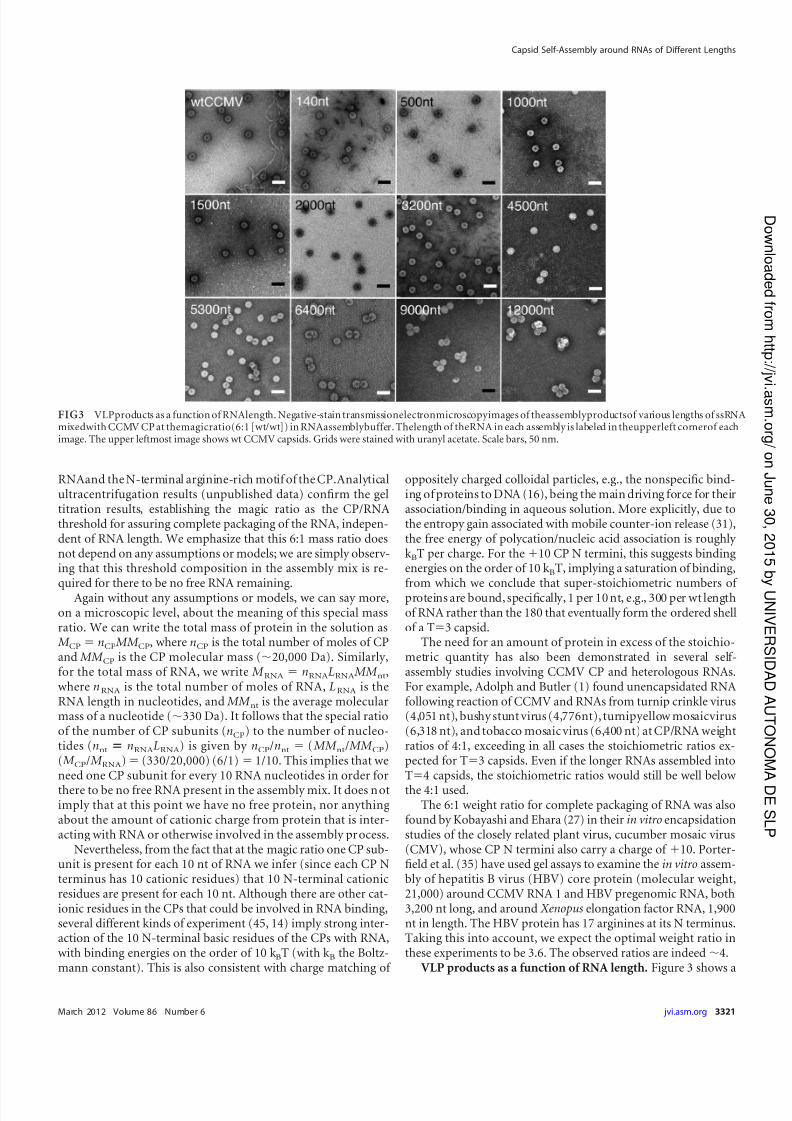
VLP products as a function of RNA length. Figure 3 shows a
FIG3 VLPproducts as a function of RNAlength. Negative-stain transmissionelectronmicroscopyimages of theassemblyproductsof various lengths of ssRNAmixedwith CCMV CP at themagicratio(6:1 [wt/wt]) in RNAassemblybuffer. Thelength of theRNA in each assembly is labeled in theupperleft cornerof eachimage. The upper leftmost image shows wt CCMV capsids. Grids were stained with uranyl acetate. Scale bars, 50 nm.
Capsid Self-Assembly around RNAs of Different Lengths
March 2012 Volume 86 Number 6 jvi.asm.org 3321
on J un e 3 0 ,2 01 5 b
y UNIVER SIDAD
A UT ON OMA
DE SLP
h t t p: / / jv
i. a sm. or g /
D ownl o a d e dfr om
7/17/2019 J. Virol.-2012-Cadena-Nava-3318-26
http://slidepdf.com/reader/full/j-virol-2012-cadena-nava-3318-26 5/9
panel of typical negative-stain electron micrographs of the assem-bly mixes corresponding to 6:1 CP/RNA mass ratio, for all 11 of the increasing lengths of RNA studied. The upper left-hand pic-ture is wt CCMV. The numbers of capsids sharing a single RNAmolecule (in the case of the large nucleotide lengths), the sizes of VLPs formedin each instance (Fig.4A), and the numbers of RNAspackaged per VLP (in the case of the shorter RNA lengths) arediscussed below.
When Verduin and Bancroft (47) examined the assembly of CCMV CP around TMV RNA, they found some difficulty in as-signingaTnumbertotheVLPsthatformed.TheTEMdataagreed
with T4, but sedimentation analyses were more consistent withT7. We have already noted that the gels for the assembly of CCMV CP around TMV RNA seem to show a product at themagic ratio that migrates more slowly than wt CCMV, suggestingtoo that the capsidmay belarger than T3. A careful examinationof the electron micrographs for the TMV VLPs makes clear thereason forthis ambiguity. Themajorityof thecapsids are paired—see, for example, the 6,400-nt frame in Fig. 3 and the upper right-hand image (“image B”) in Fig. 4B. Such doublets, which are mostevident when the density of capsids in the image is not so high thatthe particles are all closely packed, could also be seen clearly in theearly studies of TMV RNA packaging by CCMV CP (22) but werenot mentioned. The doublets also account for the appearance of
two sedimentation bands in Adolph and Butler’s packaging stud-ies of TMV RNA by CCMV CP (1).
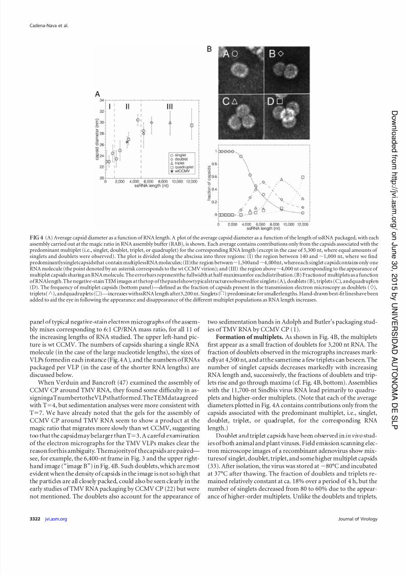
Formation of multiplets. As shown in Fig. 4B, the multipletsfirst appear as a small fraction of doublets for 3,200 nt RNA. Thefraction of doublets observed in the micrographs increases mark-edlyat 4,500 nt, and atthe sametime a few triplets can beseen. Thenumber of singlet capsids decreases markedly with increasingRNA length and, successively, the fractions of doublets and trip-lets rise and go through maxima (cf. Fig. 4B, bottom). Assemblieswith the 11,700-nt Sindbis virus RNA lead primarily to quadru-plets and higher-order multiplets. (Note that each of the average
diameters plotted in Fig. 4A contains contributions only from thecapsids associated with the predominant multiplet, i.e., singlet,doublet, triplet, or quadruplet, for the corresponding RNAlength.)
Doublet and triplet capsids have been observed in in vivo stud-ies of both animal and plant viruses. Field emission scanning elec-tron microscope images of a recombinant adenovirus show mix-turesof singlet, doublet, triplet, and some higher multiplet capsids(33). After isolation, the virus was stored at80°C and incubatedat 37°C after thawing. The fraction of doublets and triplets re-mained relatively constant at ca. 18% over a period of 4 h, but thenumber of singlets decreased from 80 to 60% due to the appear-ance of higher-order multiplets. Unlike the doublets and triplets,
FIG 4 (A) Average capsid diameter as a function of RNA length. A plot of the average capsid diameter as a function of the length of ssRNA packaged, with eachassembly carried out at the magic ratio in RNA assembly buffer (RAB), is shown. Each average contains contributions only from the capsids associated with thepredominant multiplet (i.e., singlet, doublet, triplet, or quadruplet) for the corresponding RNA length (except in the case of 5,300 nt, where equal amounts of singlets and doublets were observed). The plot is divided along the abscissa into three regions: (I) the region between 140 and 1,000 nt, where we findpredominantlysingletcapsidsthat contain multiplessRNA molecules; (II)the region between1,500and4,000nt, whereeach singlet capsidcontains only oneRNA molecule (the point denoted by an asterisk corresponds to the wt CCMV virion); and (III) the region above4,000 nt corresponding to the appearance of multiplet capsids sharing an RNA molecule. The errorbars representthe fullwidth at half-maximumfor eachdistribution.(B) Fractionof multiplets as a functionof RNAlength.The negative-stain TEM images at thetop of thepanelshowtypicalstructuresobservedfor singlets (A), doublets (B), triplets (C), andquadruplets(D). The frequency of multiplet capsids (bottom panel)—defined as the fraction of capsids present in the transmission electron microscopy as doublets (),triplets(o), andquadruplets ()—increases withssRNA length after3,200 nt. Singlets (Œ) predominate for smallerlengths. Hand-drawn best-fit lineshave been
added to aid the eye in following the appearance and disappearance of the different multiplet populations as RNA length increases.
Cadena-Nava et al.
3322 jvi.asm.org Journal of Virology
on J un e 3 0 ,2 01 5 b
y UNIVER SIDAD
A UT ON OMA
DE SLP
h t t p: / / jv
i. a sm. or g /
D ownl o a d e dfr om
7/17/2019 J. Virol.-2012-Cadena-Nava-3318-26
http://slidepdf.com/reader/full/j-virol-2012-cadena-nava-3318-26 6/9
the higher-order multiplets in the present study appeared to beaggregates of virusand proteinaceous material. The doublet struc-tures have the superficial appearance of geminiviruses (50), whichoften form triplets and quadruplets (11). In the case of the Africancassava mosaic geminiviruses containing defective interfering
ssDNA,the number of multiplets increases with DNAlength (21).The multiplet VLPs that we observed in CCMV VLPs are un-
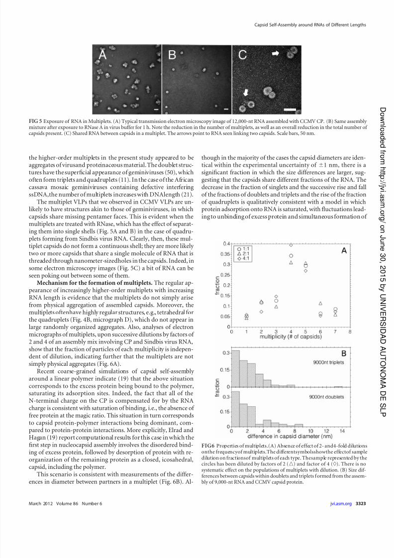
likely to have structures akin to those of geminiviruses, in whichcapsids share missing pentamer faces. This is evident when themultiplets are treated with RNase, which has the effect of separat-ing them into single shells (Fig. 5A and B) in the case of quadru-plets forming from Sindbis virus RNA. Clearly, then, these mul-tiplet capsids do not form a continuous shell; they are more likely two or more capsids that share a single molecule of RNA that isthreaded through nanometer-sizedholes in the capsids. Indeed, insome electron microscopy images (Fig. 5C) a bit of RNA can beseen poking out between some of them.
Mechanism for the formation of multiplets. The regular ap-
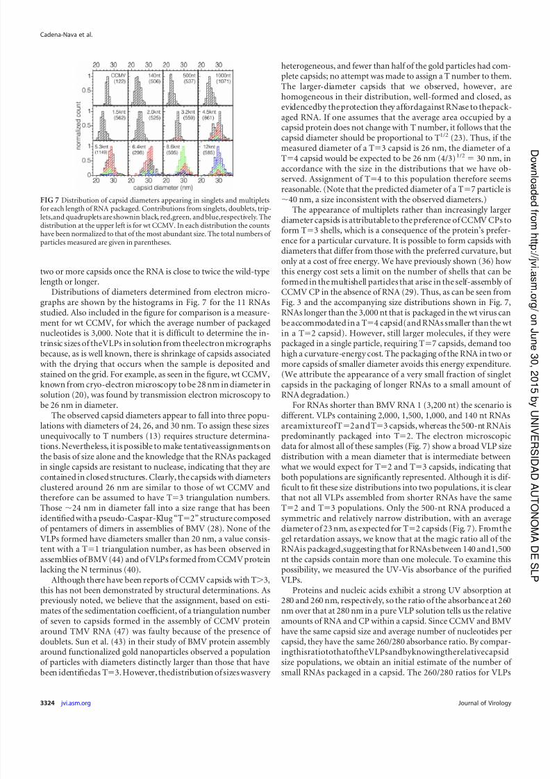
pearance of increasingly higher-order multiplets with increasingRNA length is evidence that the multiplets do not simply arisefrom physical aggregation of assembled capsids. Moreover, themultiplets oftenhave highly regular structures, e.g., tetrahedral forthe quadruplets (Fig. 4B, micrograph D), which do not appear inlarge randomly organized aggregates. Also, analyses of electronmicrographs of multiplets, upon successive dilutions by factors of 2 and 4 of an assembly mix involving CP and Sindbis virus RNA,show that the fraction of particles of each multiplicity is indepen-dent of dilution, indicating further that the multiplets are notsimply physical aggregates (Fig. 6A).
Recent coarse-grained simulations of capsid self-assembly around a linear polymer indicate (19) that the above situationcorresponds to the excess protein being bound to the polymer,
saturating its adsorption sites. Indeed, the fact that all of theN-terminal charge on the CP is compensated for by the RNAcharge is consistent with saturation of binding, i.e., the absence of free protein at the magic ratio. This situation in turn correspondsto capsid protein-polymer interactions being dominant, com-pared to protein-protein interactions. More explicitly, Elrad andHagan (19) report computational results for this case in which thefirst step in nucleocapsid assembly involves the disordered bind-ing of excess protein, followed by desorption of protein with re-organization of the remaining protein as a closed, icosahedral,capsid, including the polymer.
This scenario is consistent with measurements of the differ-ences in diameter between partners in a multiplet (Fig. 6B). Al-
though in the majority of the cases the capsid diameters are iden-tical within the experimental uncertainty of 1 nm, there is asignificant fraction in which the size differences are larger, sug-gesting that the capsids share different fractions of the RNA. Thedecrease in the fraction of singlets and the successive rise and fall
of the fractions of doublets and triplets and the rise of the fractionof quadruplets is qualitatively consistent with a model in whichprotein adsorption onto RNA is saturated, with fluctuations lead-ing to unbinding of excess protein and simultaneous formation of
FIG 5 Exposure of RNA in Multiplets. (A) Typical transmission electron microscopy image of 12,000-nt RNA assembled with CCMV CP. (B) Same assembly mixture after exposure to RNase A in virus buffer for 1 h. Note the reduction in the number of multiplets, as well as an overall reduction in the total number of capsids present. (C) Shared RNA between capsids in a multiplet. The arrows point to RNA seen linking two capsids. Scale bars, 50 nm.
FIG6 Properties of multiplets.(A) Absence of effect of 2- and4-fold dilutionsonthe frequencyof multiplets.The differentsymbolsshowthe effectof sampledilution on fractionsof multiplets of each type. Thesample represented by thecircles has been diluted by factors of 2 (o) and factor of 4 (). There is nosystematic effect on the populations of multiplets with dilution. (B) Size dif-ferences between capsids within doublets and triplets formed from the assem-bly of 9,000-nt RNA and CCMV capsid protein.
Capsid Self-Assembly around RNAs of Different Lengths
March 2012 Volume 86 Number 6 jvi.asm.org 3323
on J un e 3 0 ,2 01 5 b
y UNIVER SIDAD
A UT ON OMA
DE SLP
h t t p: / / jv
i. a sm. or g /
D ownl o a d e dfr om
7/17/2019 J. Virol.-2012-Cadena-Nava-3318-26
http://slidepdf.com/reader/full/j-virol-2012-cadena-nava-3318-26 7/9
two or more capsids once the RNA is close to twice the wild-typelength or longer.
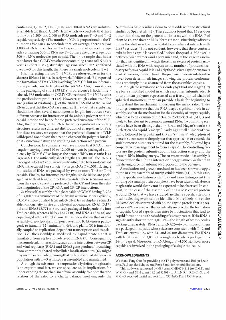
Distributions of diameters determined from electron micro-graphs are shown by the histograms in Fig. 7 for the 11 RNAsstudied. Also included in the figure for comparison is a measure-ment for wt CCMV, for which the average number of packagednucleotides is 3,000. Note that it is difficult to determine the in-trinsic sizes of theVLPs in solution from theelectron micrographsbecause, as is well known, there is shrinkage of capsids associatedwith the drying that occurs when the sample is deposited andstained on the grid. For example, as seen in the figure, wt CCMV,known from cryo-electron microscopy to be 28 nm in diameter insolution (20), was found by transmission electron microscopy to
be 26 nm in diameter.The observed capsid diameters appear to fall into three popu-lations with diameters of 24, 26, and 30 nm. To assign these sizesunequivocally to T numbers (13) requires structure determina-tions. Nevertheless, it is possible to make tentativeassignments onthe basis of size alone and the knowledge that the RNAs packagedin single capsids are resistant to nuclease, indicating that they arecontained in closed structures. Clearly, the capsids with diametersclustered around 26 nm are similar to those of wt CCMV andtherefore can be assumed to have T3 triangulation numbers.Those 24 nm in diameter fall into a size range that has beenidentified with a pseudo-Caspar-Klug “T2” structure composedof pentamers of dimers in assemblies of BMV (28). None of theVLPs formed have diameters smaller than 20 nm, a value consis-
tent with a T1 triangulation number, as has been observed inassemblies of BMV (44) and of VLPs formed from CCMV proteinlacking the N terminus (40).
Although there have been reports of CCMV capsids with T3,this has not been demonstrated by structural determinations. Aspreviously noted, we believe that the assignment, based on esti-mates of the sedimentation coefficient, of a triangulation numberof seven to capsids formed in the assembly of CCMV proteinaround TMV RNA (47) was faulty because of the presence of doublets. Sun et al. (43) in their study of BMV protein assembly around functionalized gold nanoparticles observed a populationof particles with diameters distinctly larger than those that havebeen identifiedas T3. However, thedistribution of sizes wasvery
heterogeneous, and fewer than half of the gold particles had com-plete capsids; no attempt was made to assign a T number to them.The larger-diameter capsids that we observed, however, arehomogeneous in their distribution, well-formed and closed, asevidencedby the protection they affordagainst RNase to thepack-aged RNA. If one assumes that the average area occupied by acapsid protein does not change with T number, it follows that the
capsid diameter should be proportional to T1/2 (23). Thus, if themeasured diameter of a T3 capsid is 26 nm, the diameter of aT4 capsid would be expected to be 26 nm (4/3)1/2 30 nm, inaccordance with the size in the distributions that we have ob-served. Assignment of T4 to this population therefore seemsreasonable. (Note that the predicted diameter of a T7 particle is40 nm, a size inconsistent with the observed diameters.)
The appearance of multiplets rather than increasingly largerdiameter capsids is attributable to the preference of CCMV CPs toform T3 shells, which is a consequence of the protein’s prefer-ence for a particular curvature. It is possible to form capsids withdiameters that differ from those with the preferred curvature, butonly at a cost of free energy. We have previously shown (36) how
this energy cost sets a limit on the number of shells that can beformed in the multishell particles that arise in the self-assembly of CCMV CP in the absence of RNA (29). Thus, as can be seen fromFig. 3 and the accompanying size distributions shown in Fig. 7,RNAs longer than the 3,000 nt that is packaged in the wt virus canbe accommodated in a T4 capsid(and RNAs smaller than the wtin a T2 capsid). However, still larger molecules, if they werepackaged in a single particle, requiring T7 capsids, demand toohigh a curvature-energy cost. The packaging of the RNA in two ormore capsids of smaller diameter avoids this energy expenditure.(We attribute the appearance of a very small fraction of singletcapsids in the packaging of longer RNAs to a small amount of RNA degradation.)
For RNAs shorter than BMV RNA 1 (3,200 nt) the scenario isdifferent. VLPs containing 2,000, 1,500, 1,000, and 140 nt RNAsareamixtureofT2andT3 capsids, whereas the 500-nt RNAispredominantly packaged into T2. The electron microscopicdata for almost all of these samples (Fig. 7) show a broad VLP sizedistribution with a mean diameter that is intermediate betweenwhat we would expect for T2 and T3 capsids, indicating thatboth populations are significantly represented. Although it is dif-ficult to fit these size distributions into two populations, it is clearthat not all VLPs assembled from shorter RNAs have the sameT2 and T3 populations. Only the 500-nt RNA produced asymmetric and relatively narrow distribution, with an averagediameter of 23 nm, as expected for T2 capsids (Fig. 7). Fromthegel retardation assays, we know that at the magic ratio all of the
RNAis packaged,suggesting that for RNAs between 140 and1,500nt the capsids contain more than one molecule. To examine thispossibility, we measured the UV-Vis absorbance of the purifiedVLPs.
Proteins and nucleic acids exhibit a strong UV absorption at280 and 260 nm, respectively, so the ratio of the absorbance at 260nm over that at 280 nm in a pure VLP solution tells us the relativeamounts of RNA and CP within a capsid. Since CCMV and BMVhave the same capsid size and average number of nucleotides percapsid, they have the same 260/280 absorbance ratio. By compar-ingthisratiotothatoftheVLPsandbyknowingtherelativecapsidsize populations, we obtain an initial estimate of the number of small RNAs packaged in a capsid. The 260/280 ratios for VLPs
FIG 7 Distribution of capsid diameters appearing in singlets and multipletsfor each length of RNA packaged. Contributions from singlets, doublets, trip-lets,and quadruplets are shownin black, red,green, and blue,respectively. Thedistribution at the upper left is for wt CCMV. In each distribution the countshave been normalized to that of the most abundant size. The total numbers of particles measured are given in parentheses.
Cadena-Nava et al.
3324 jvi.asm.org Journal of Virology
on J un e 3 0 ,2 01 5 b
y UNIVER SIDAD
A UT ON OMA
DE SLP
h t t p: / / jv
i. a sm. or g /
D ownl o a d e dfr om
7/17/2019 J. Virol.-2012-Cadena-Nava-3318-26
http://slidepdf.com/reader/full/j-virol-2012-cadena-nava-3318-26 8/9
containing 3,200-, 2,000-, 1,000-, and 500-nt RNAs are indistin-guishable from that of CCMV, from which we conclude that thereis only one 3,200- and 2,000-nt RNA molecule per T3 and T2capsid, respectively. (The number of CPs is proportional to the Tnumber.) We can also conclude that, on average, there are two1,000-nt RNA molecules per T2 capsid. Similarly, since the cap-sids containing 500-nt RNA are T2, there are on average four
500-nt RNA molecules per capsid. The only sample that had aratio lower than CCMV wasthe one containing 1,500-ntRNA (1.5versus 1.7 for CCMV), strongly suggesting, since T2 is preferredover T3 for this length, that there is a single molecule in T2.
It is interesting that no T1 VLPs are observed, even for theshortest RNAs (140 nt). In early work, Pfeiffer et al. (34) reportedthe formation of T1 VLPs involving BMV CP, but no informa-tion is provided on the lengths of the ssRNAs. Also, in our studiesof the packaging of short (38 kDa), fluorescence (rhodamine)-labeled, PSS molecules by CCMV CP, we found T1 VLPs to bethe predominant product (12). However, rough estimates of thesize (radius of gyration[R g ]) of the 38-kDa PSS and of the 140-ntRNAsuggest that the RNAs are smaller. It may be that a rigid-ring,
rhodamine label, several nanometers in length, introduces a very different scenario for interaction of the anionic polymer with thecapsid interior and hence for the preferred curvature of the VLP.Also, the branching of the RNA due to its significant secondary structure results in a different distribution of charge than for PSS.For these reasons, we expect that the preferred diameter of VLPwilldepend not onlyon the sizeand chargeof the polymer but alsoon its structural nature and resulting interaction with protein.
Conclusions. In summary, we have shown that RNA of any length—varying from 140 to 12,000 nt—can be packaged com-pletely by CCMV CP as long as the protein/RNA mass ratio is aslarge as 6:1. For sufficiently short lengths (2,000 nt), the RNA ispackaged into T2andT3 capsids with oneto four moleculesof
RNA in the capsid. For sufficiently large RNAs (
4,500 nt), singlemolecules of RNA are packaged by two or more T3 or T4capsids. Finally, for intermediate lengths, single RNAs are pack-aged, as with wt length, into T3 capsids. These scenarios arisefrom the capsid curvature preferred by the CP and from the rela-tive magnitudes of the CP-RNA and CP-CP interactions.
In vitro self-assembly of single capsids of CCMV having RNAsof 3,000 nt is reminiscent of the in vivo scenario. More explicitly,CCMV virions purified from infected leaf tissue display a remark-able homogeneity in size and physical appearance: RNA1 (3,171nt) and RNA2 (2,774 nt) are each packaged independently intoT3 capsids, whereas RNA3 (2,173 nt) and RNA 4 (824 nt) arecopackaged into a third virion. It has been shown that in vivoassembly of nucleocapsids in positive-strand RNA viruses patho-
genic to humans (32), animals (4, 46), and plants (3) is function-ally coupled to replication-dependent transcription and transla-tion, i.e., the assembly is mediated by capsid protein that istranslated from replication-derived mRNA (5). Consequently,macromolecular interactions, such as the interaction between CPand viral replicase (RNA1 and RNA2 gene products), resultingfrom commonly shared subcellular localization sites (6), mightplay an importantrole,ensuringthat only onekind of stablevirionpopulation with T3 symmetry is assembled and maintained.
Although theexistence of theoperationally definedmagicratiois an experimental fact, we can speculate on its implications forunderstanding the mechanism of viral assembly. We note that therelation of the ratio to a charge balance involving only the
N-terminus basic residues seems to be at odds with the structuralstudies by Speir et al. (42). These authors found that 13 residuesother than those on the protein tail interact with the RNA, 7 of them basic, and that the RNA “forms three distinct bulges directly under the shell near the quasi-3-fold axes, where it interacts withLys87 residues.” It is not evident, however, that these contactsexist before a capsid is assembled. Indeed, the quasi-3-fold axes lie
between two hexamers and a pentamer and, at the stage in assem-bly that we identified in which there is an excess of protein asso-ciated with the RNA with respect to the number of proteins nec-essary to form a capsid, it is unlikely that distinct capsidpentamersexist.Moreover, thestructure of theprotein dimersin solution hasnever been determined: images showing the protein conforma-tions are simply those abstracted from the assembled capsids.
Although the simulations of assembly by Elrad and Hagan (19)are for a simplified model in which capsomer subunits adsorbonto a linear polymer represented as a freely jointed chain of spherical monomers, they can provide a basis for beginning tounderstand the mechanism underlying the magic ratio. Thesefindings demonstrate that the RNA plays a significant role in as-
sembly, so that the mechanism for the assembly of empty capsids,which has been examined in detail by Zlotnick et al. (51), is notlikely to be relevant to assembly around RNA. Two limiting sce-narios have been distinguished in Elrad and Hagan’s work: (i)nucleation of a capsid “embryo” involving a small number of pro-teins, followed by growth and (ii) an “en masse” adsorption of protein subunits onto the polymer that approaches or exceeds thestoichiometric numbers required for the assembly, followed by acooperative rearrangement to form a capsid. The controlling fac-tors are the protein subunit-subunit interaction energy and theprotein-RNA binding energy. The en masse mode of assembly isfavored when the subunit interaction energy is much weaker thanthe driving force for subunit adsorption onto the polymer.
A nucleation and growth mechanism has been shown to apply to the in vitro assembly of turnip crinkle virus (41). In this case,both a specific nucleation center (37) and a nucleating event (thebinding of a small protein complex) have been identified. Here, amagic ratio would clearly not be expected to be observed. In con-trast, in the case of the assembly of the CCMV capsid proteinaround RNAs that we have studied, neither a specific site nor alocal nucleating event can be identified. More likely, the entireRNAmoleculeis saturated with bound capsid protein that is pres-ent in a 70% excess over that eventually involved in the formationof capsids. Closed capsids then arise by fluctuations that lead tocapsid formation and the shedding of excess protein. If the RNAissignificantly shorter than 3,000 nt—the length of wt molecules
packaged separately (RNA1 and RNA2)—two or more of themare packaged in capsids whose sizes are consistent with T2 andT3 structures, i.e., with 24- and 26-nm diameters. For RNAswith lengths around 3,000 nt, a single molecule is packaged in a26-nm capsid. Moreover, for RNA lengths4,500 nt, two or morecapsids are involved in the packaging of a single molecule.
ACKNOWLEDGMENTS
We thank Feng Guo for providing the T7 polymerase and Robijn Bruin-
sma, Paul van der Schoot, and Roya Zandi for helpful discussions.This study was supported by NSF grant CHE 0714411 (to C.M.K. and
W.M.G.) and NIH grant 1R21AI82301 (to A.L.N.R.). R.D.C.-N. and
M.C.-G. received partial support from CONACyT and UC-Mexus.
Capsid Self-Assembly around RNAs of Different Lengths
March 2012 Volume 86 Number 6 jvi.asm.org 3325
on J un e 3 0 ,2 01 5 b
y UNIVER SIDAD
A UT ON OMA
DE SLP
h t t p: / / jv
i. a sm. or g /
D ownl o a d e dfr om
7/17/2019 J. Virol.-2012-Cadena-Nava-3318-26
http://slidepdf.com/reader/full/j-virol-2012-cadena-nava-3318-26 9/9
ADDENDUM IN PROOF
The existence of multiplets has also recently been reported in thecase of self-assembly of CCMV CP around a conjugated polyelec-trolyte (M. Brasch and J. J. M. Cornelisse, Chem. Commun. 48:1446–1448, 2012).
REFERENCES
1. Adolph KW, Butler PJG. 1977. Studies on the assembly of a sphericalplant virus. III. Reassembly of infectious virions under mild conditions. J.Mol. Biol. 109:345–357.
2. Allison RF, Janda M, Ahlquist P. 1988. Infectious in vitro transcripts fromcowpea chlorotic mottle virus cDNAclones and exchange of individual RNAcomponents with brome mosaic virus.1988 J. Virol. 62:3581–3588.
3. Annamalai P, Rao ALN. 2005. Dispensability of 3= tRNA-like sequencefor packaging cowpea chlorotic mottle virus genomic RNAs. Virology 332:650–658.
4. Annamalai P, Rao ALN. 2006. Packaging of brome mosaic virus sub-genomic RNA is functionally coupled to replication-dependent transcrip-tion and translation of coat protein. J. Virol. 80:10096–10108.
5. Annamalai P, Rofail F, DeMason DA, Rao ALN. 2008. Replication-coupled packaging mechanism in positive-strand RNA viruses: synchro-nized coexpression of functional multigenome RNA components of ananimal and a plant virus in Nicotiana benthamiana cells by agroinfiltra-
tion. J. Virol. 82:1484–1490.6. Bamunusinghe D, Seo J-K, Rao ALN. 2011. Subcellular localization and
rearrangement of endoplasmic reticulum by brome mosaic virus capsidprotein. J. Virol. 85:2953–2963.
7. Bancroft JB. 1970. The self-assembly of spherical plant viruses. Adv.VirusRes. 16:99–134.
8. Bancroft JB, Hiebert E. 1967. Formation of an infectious nucleoproteinfrom protein and nucleic acid isolated from a small spherical virus. Virol-ogy 32:354–356.
9. Bancroft JB, Hiebert E, Bracker CE. 1969. The effects of various poly-anions on shellformation of somespherical viruses. Virology 39:924–930.
10. Basnak G, et al. 2010. Viral genomic single-stranded RNA directs thepathway towards a T3 capsid. J. Mol. Biol. 395:924–936.
11. Briddon RW, Markham PG. 1995. Family Geminiviridae, p 158–165. InMurphy FA, et al. (ed), Virus taxonomy: archives in virology. Springer-Verlag, New York, NY.
12. Cadena-Nava RD, et al. 2011. Exploiting fluorescent polymers to probethe self-assembly of virus-like particles. J. Phys. Chem. B 115:2386–2391.13. Caspar DLD, Klug A. 1962. Physical principles in the construction of
regular viruses. Cold Spring Harbor Symp. Quant. Biol. 27:1–24.14. Choi YG, Rao ALN. 2000. Molecular studies on bromovirus capsid pro-
tein. VII. Selective packaging of BMV RNA4 by specific N-terminal argi-nine residues. Virology 275:207–217.
15. ChoiYG, Rao ALN. 2000. Packaging of tobacco mosaic virus subgenomicRNAs by brome mosaic virus coat protein exhibits RNA controlled poly-morphism. Virology 275:249–257.
16. deHaseth PL, Lohman TM, Record MT, Jr. 1977. Nonspecific interac-tions of lac repressor with DNA: an association reaction driven by counter-ion release. Biochemistry 16:4783–4790.
17. Dreher TW, Rao ALN, Hall TC. 1989. Replication in vivo of mutant bromemosaic virus RNAs defective in aminoacylation. J. Mol. Biol. 206:425–438.
18. Dzianott A, Bujarski J. 1991. The nucleotide sequence and genome or-
ganizationof the RNA-1segmentin two bromoviruses:broad beanmottlevirus and cowpea chlorotic mottle virus. Virology 185:553–562.19. Elrad OM, Hagan MF. 2010. Encapsulation of a polymer by an icosahe-
dral virus. Phys. Biol. 7:045003.20. Fox JM, et al. 1998. Comparison of the native CCMV virion with in vitro
assembled virions by cryoelectron microscopy and image reconstruction.Virology 244:212–218.
21. Frischmuth T, Ringel M, Kocher C. 2001. The size of encapsidatedsingle-stranded DNA determines the multiplicity of African cassava mo-saic virus particles. J. Gen. Virol. 82:673–676.
22. Hiebert E, Bancroft JB, Bracker CF. 1968. The assembly in vitro of somesmall spherical viruses, and other nucleoproteins. Virology 34:492–508.
23. Hu Y, Zandi R, Anavitarte A, Knobler CM, Gelbart WM. 2008. Pack-aging of a polymer by a viral capsid: the interplay between polymer lengthand capsid size. Biophys. J. 94:1428–1436.
24. Johnson JM, Willits DA, Young MJ, Zlotnick A. 2004. Interaction with
capsid protein alters RNA structure and the pathway for in vitro assembly of cowpea chlorotic mottle virus. J. Mol. Biol. 335:455–464.
25. Johnson MW, Wagner GW, Bancroft JB. 1973. A titrimetric and elec-trophoretic study of cowpea chlorotic mottle virus and its protein. J. Gen.Virol. 19:263–273.
26. Jung B, Rao ALN, Anvari B. 2011. Optical nano-constructs composed of genome-depleted Brome mosaic virus doped with a near infrared chro-mophore for potential biomedical applications. ACS Nano. 5:1243–1252.
27. Kobayashi A, Ehara Y. 1995. In vitro encapsidation of cucumber mosaicvirus RNA species. Ann. Phytopathol. Soc. Jpn. 61:99–102.28. Kroll MA, et al. 1999. RNA-controlled polymorphism in the in vivo
assembly of a 180-subunit and 120-subunit virions from a single capsidprotein. Proc. Natl. Acad. Sci. U. S. A. 96:13650–13655.
29. Lavelle L, et al. 2009. Phase diagram of self-assembled viral capsid proteinpolymorphs. J. Phys. Chem. B 113:3813–3820.
30. Lustig S, et al. 1988. Molecular basis of Sindbis virus neurovirulence inmice. J. Virol. 62:2329–2336.
31. Mascotti DP, Lohman TM. 1990. Thermodynamic extent of counter-ionrelease upon binding oligo-lysines to single-stranded nucleic acids. Proc.Natl. Acad. Sci. U. S. A. 87:3142–3146.
32. Nugent CI, Johnson KL, Sarnow P, Kirkegaard K. 1997. Functionalcoupling between replication and packaging of poliovirus replicon RNA.J. Virol. 73:427–435.
33. Obenauer-Kutner LJ, et al. 2002. The use of field emission scanning
electron microscopy to assess recombinant adenovirus stability. Hum.Gene Ther. 13:1687–1696.34. Pfeifer P, Herzog M, Hirth L. 1976. Stabilization of brome mosaic virus.
Philos. Trans. R. Soc. Lond. B Biol. Sci. 276:99–107.35. Porterfield JZ, et al. 2010. Full-length hepatitis B virus core protein
packages viral and heterologous RNA with similarly high levels of coop-erativity. J. Virol. 84:7174–7184.
36. Prinsen P, van der Schoot P, Gelbart WM, Knobler CM. 2010. Shellstructures of virus capsid proteins. J. Phys. Chem. B 114:5522–5533.
37. Qu F, Morris TJ. 1997. Encapsidation of turnip crinkle virus is defined by a specific packaging signal size RNA. J. Virol. 71:1428–1435.
38. Rao ALN. 2006. Genome packaging by spherical plant RNA viruses.Annu. Rev. Phytopathol. 44:61–87.
39. Sambrook J, Fritsch EF, Maniatis T. 1989. Molecular cloning: a labora-tory manual. Cold Spring Harbor Laboratory Press, Cold Spring Har-bor, NY.
40. Sikkema FD, et al. 2007. Monodisperse polymer-virus hybrid nanopar-ticles. Org. Biomol. Chem. 5:54–57.
41. Sorger PK, Stockley PG, Harrison SC. 1986. Structure and assembly of turnip crinkle virus. II. Mechanism of reassembly in vitro. J. Mol. Biol.191:639–658.
42. Speir JA, Munshi S, Wang G, Baker TS, Johnson JE. 1995. Structuresof the native and swollen forms of cowpea chlorotic mottle virus de-termined by X-ray crystallography and cryo-electron microscopy.Structure 3:63–78.
43. Sun J, et al. 2007. Core-controlled polymorphism in virus-like particles.Proc. Natl. Acad. Sci. U. S. A. 104:1354–1359.
44. Tang J,et al. 2006. Therole of subunit hingesand molecular “switches” inthe control of viral capsid polymorphism. J. Struct. Biol. 154:59–67.
45. van der Graaf M, Scheek RM, van der Linden CC, Hemminga MA.1992. Conformation of a pentacosapeptide representing the RNA-bindingN terminus of cowpea chlorotic mottle virus coat protein in the presenceof oligophosphates: a two-dimensional proton nuclear magnetic reso-nance and distance geometry study. Biochem. 3:9177–9182.
46. Venter PA, Krishna NK, Schneemann A. 2005. Capsid protein synthesisfrom replicating RNA directs specific packaging of the genome of a mul-tipartite, positive-strand RNA virus. J. Virol. 79:6239–6248.
47. Verduin BJM, Bancroft JB. 1969. The infectivity of tobacco mosaic virusRNA in coat proteins from spherical viruses. Virology 37:501–506.
48. Yoffe AM, et al. 2008. Predicting the sizes of large RNA molecules. Proc.Natl. Acad. Sci. U. S. A. 105:16153–16158.
49. Zandi R, van der Schoot P. 2009. Size regulation of ssRNA viruses.Biophys. J. 96:9–20.
50. Zhang W, et al. 2001. Structure of the maize streak virus geminate parti-cle. Virology 279:471–477.
51. Zlotnick A, Aldrich R, Johnson JM, Ceres P, Young MJ. 2000. Mecha-nism of capsidassemblyfor an icosahedral plant virus.Virology 277:450–456.
Cadena-Nava et al.
3326 jvi.asm.org Journal of Virology
on J un e 3 0 ,2 01 5 b
y UNIVER SIDAD
A UT ON OMA
DE SLP
h t t p: / / jv
i. a sm. or g /
D ownl o a d e dfr om