Embed Size (px)
Citation preview
8/11/2019 J. Lipid Res. 1994 de Craemer 1241 50
http://slidepdf.com/reader/full/j-lipid-res-1994-de-craemer-1241-50 1/10
Peroxisomes in liver, heart, and kidney of mice fed
a commercial fish oil preparation: original data and
review on peroxisomal changes induced by high-fat
diets
Dirk De Craemer,',* Joseph Vamecq,t Frank Roels,l* Louis VallCe, t Marina Pauwels? and
Christ iane Van den Branden*
Menselijke Anatomie & Embryologie,' Vrije Universiteit Brussel, Belgium; North France Center for the
Study of Childhood Epilepsy.1 Lille, France; and Menselijke Ontleedkunde & Embryologic,* Universiteit
Gent, Belgium
Abstract
Male NMRI mice were fed a diet with 10% w/w
Beromegan for up to three weeks. Beromegan is a commercial
fish (salmon) oil preparation rich in eicosapentaenoic acid and
docosahexaenoic acid. Peroxisomal P-oxidation capacity, cata-
lase activity, and ultrastructural morphometry of the hepatic
peroxisomes were investigated. In myocardium and kidney, cata-
lase activity, peroxisomal s taining after catalase cytochemistry,
peroxisomal morphology, and morphometry (in myocardium)
were evaluated. In liver, we found a significant increase in perox-
isomal fl-oxidation, catalase activity, and peroxisomal number
already after 3 days of dietary treatment. These changes were
more pronounced after 3 weeks. Peroxisomal size was not
changed. Positive correlations were found between peroxisomal
enzyme activities and the number but not the size
of
the peroxi-
somes, and between catalase activity and P-oxidation capacity.
The mean peroxisomal diameter per animal was inversely
proportional to catalase activity measured in homogenate. In
myocardium, catalase activity was increased with duration of
fish oil feeding. Peroxisomal staining, number, and size were
also increased when compared to controls. In kidney, no altera-
tions were observed. e ur results indicate a beneficial effect
of a diet supplemented with fish oil on the peroxisomal metabo-
lism in liver and myocardium; it differs from the changes
in-
duced by xenobiotic peroxisome proliferation.- De Craemer,
D.,
J.
Vamecq,
E
Roels,
L.
Vallte,
M .
Pauwels, and C.
Van den Branden.
Peroxisomes in liver, heart, and kidney of
mice fed a commercial fish oil preparation: original data and
review on peroxisomal changes induced by high-fat diets.
J Lipid
Res.
1994. 35: 1241-1250.
Supplementary
key
W O ~ S atalase activity catalase cytochemistry
morphometry docosahexaenoic acid polyunsaturated fatty acids
peroxisomal @-oxidation
Peroxisomes are cell organelles characterized by the
presence
of
hydrogen peroxide-producing oxidases and
high amounts of catalase which breaks down hydrogen
peroxide to water an d oxygen (1). Based on its abund anc e
in peroxisomes, catalase is used as a marker enzyme in
fractionation studies and in morphologic studies using
(immuno-) cytochemical techniques. Peroxisomes also
contain enzymes that intervene in several metabolic pro-
cesses suc h as the /?-oxidation of very long chai n fatty
acids and the synthesis of plasmalogens (ether-phospho-
lipids) and bile acids
(2).
Peroxisomes had long been considered as relatively un-
important organelles until it was reported that peroxi-
somes were absent in the livers
of
patients with the
cerebro-hepato-renal syndrome of Zellweger
(3).
Today it
is known that at least 13 metabolic diseases are related to
a deficiency in o ne o r more peroxisomal enzymes
4, ).
Besides severe clinical symptoms,
an
accumulation of the
very long chain fatty acids is always observed in patients
with an inborn error
of
peroxisomal &oxidation (6).
A
dietary therapy for patients suffering from one of these
disorders (X-linked adrenoleukodystrophy) consists of a
diet supplemented with
a
mixture of glyceryltrioleate and
glyceryltrierucate (Lorenzo's oil) an d a dietary restriction
of the very long chain fatty acids (7). This results in a
reductio n of the level
of
very long chain fatty acids in the
majority of treated patients. Clinical symptoms however,
do not improve. Only some patients with
a
mild form of
the disease or at the onset of the clinical sympto ms showed
a
stabilization of the neurological impairment (7-10).
Recently, Martinez
(11, 12)
found that patients with
generalized peroxisomal diseases revealed a constant bio-
chemical abnormality: the content of docosahexaenoic
acid, a polyunsaturated fatty acid (C22:6) of the n-3
series, was decreased in brain, liver and kidney. In a
'To
whom correspondence should be addressed at: Vrije Univeniteit
Brussel, Human Anatomy & Embryology, Laarbeeklaan 103, B-1090
Brussels, Belgium.
Journal of Lipid Research Volume
35,
1994
1241
8/11/2019 J. Lipid Res. 1994 de Craemer 1241 50
http://slidepdf.com/reader/full/j-lipid-res-1994-de-craemer-1241-50 2/10
single published case, supplementation with the pure
ethyl ester of docosahexaenoic acid resulted in a clearcut
improvement (13). Following these data, investigation of
polyunsaturated fatty acids in all peroxisomal deficient
patients was requested (14). In a large scale trial in the
United States, n-3 fatty acids in the form
of
fish oil are
now being added to the diet of patients with X-linked
adrenoleukodystrophy in order
to
avoid essential
fa t ty
acid deficiency in patients on Lorenzo's oil therapy (10).
Our aim was to investigate the effects of a diet supple-
mented with fish oil, which contains a high concentration
of
docosahexaenoic acid, on the peroxisomal metabolism
of laboratory animals for up to 3 weeks. We used the com-
mercial fish oil preparation (Beromegan@) in a concentra-
tion that made
i t
possible to compare our results with
those of Yamazaki, Shen, and Schade (15): the two main
constituents in fish oil (eicosapentaenoic acid C20:5, n-3
and docosahexaenoic acid C22:6, n-3) reached
a
final
concentration in the chow administered to the laboratory
animals of approximately 3 and
2 ,
respectively.
The results showed that fish oil activates peroxisomal
functions in liver and myocardium but not in kidney. Pre-
liminary findings in the liver of mice on dietary treatment
for
3
days were presented at the Joint Meeting of the
Dutch and Belgian Societies for Electron Microscopy and
the Belgian Society for Cell Biology
(16).
MATERIAL AND METHODS
Adult male NMRI mice were randomly assigned into
six groups. Five groups were fed commercial powdered
chow supplemented with 10% Beromegan@ a gift from
FHER, division of Boehringer Ingelheim) for
1, 2, 3 , 14,
or 21 days. Beromegan@ s a fish (salmon) oil extract con-
taining mainly polyunsaturated fatty acids (57 ) (Table 1).
TABLE
1 .
Fatty acid composition of Beromegan
Fatty Acid
Percent nf Total
14:O
16:O
16:1
16:2
7 : O
18:0
18: 1
18:2
18:4
20:o
2O:4
20:5
22:4
2215
6
13
8
1
1
3
10
1 .5
The control group received standard powdered chow
(A04 meal-UAR, Epinay, France). All animals had free
access to food and water and were fed the standard
powdered chow at least 5 days before the start of the ex-
periment.
All mice were killed by cervical dislocation after ether
anesthesia and the liver, heart, and right kidney were re-
moved. For histological analysis, thin slices (1 mm thick)
were cut from the middle lobe of the liver, from the left
ventricle wall, and from the kidney cortex. The liver and
kidney slices were fixed in cacodylate-buffered 4% for-
maldehyde for 24 h at room temperature (17). Slices of
heart were fixed in the same solution for 18 h at O°C. This
modification permits an enhancement
of
the peroxisomal
staining in tissues with relatively small amounts of cata-
lase (I. Kerckaert and F. Roels, unpublished observation).
Cryosections (50 pm thick) of the fixed material were in-
cubated with diaminobenzidine at pH 10.5 (catalase
staining of peroxisomes)
(18)
and evaluated under the
light microscope, or were postosmicated in the presence of
K,Fe(CN),, and embedded in epoxy resin. On 2-pm
epoxy resin sections, periportal areas in the liver and
longitudinal sectioned myocardial fibers were selected for
ultrastructural investigation. Morphometry of the hepato-
cellular and myocardial peroxisomes was performed on
random electron micrographs
(17).
Th e remaining parts of the liver were frozen to -8OOC
before enzyme assays were performed. Peroxisomal fatty
acyl-CoA oxidase specific activity (first step of the perox-
isomal P-oxidation) was studied in homogenate using the
method of Vamecq (19) and is expressed in nmol hydrogen
peroxide generated per minute and mg protein. Cyanide-
insensitive fatty acyl-CoA oxidation was measured
as
de-
scribed by Lazarow and de Duve (20) and is expressed
as
nmol NAD+ reduced per minute and mg protein. Cata-
lase activity was assayed at 0°C in homogenate of liver,
heart, and kidney of each mouse using the titanium oxy-
sulfate method (21, 22). One UB is the amount of cata-
lase that breaks down
90% of
the substrate (1.5 mM
H202/1) n a volume of 50 ml at O°C in
1
min; maximal
reaction time
is
10 min.
As statistical methods (23), we used linear regression
analysis and non-parametrical Kruskall-Wallis test.
Sig-
nificance was obtained when
P <
0.05.
RESULTS
I
1 Relative
organ
weights
0 . 5
30
1
The diet rich in fish oil was well accepted by the mice.
At the end of the experiment, body weight of Beromegan@-
fed animals was similar to the weight of control animals
(not shown). Hepatomegaly was observed in Beromegan@-
fed animals: liver weight expressed
as
percentage Of
body
weight was increased 25% after 3 , 14, and
21
days of feed-
22:6 20
Besides fatty acids, Beromegan also contains
0 . 025 a-tocopherol
and
1
cholesterol.
1242 Journal
of
Lipid Research Volume 3 5 ,
1994
8/11/2019 J. Lipid Res. 1994 de Craemer 1241 50
http://slidepdf.com/reader/full/j-lipid-res-1994-de-craemer-1241-50 3/10
ing, when compared to animals fed the control diet.
N o
differences in heart or kidney weights were observed.
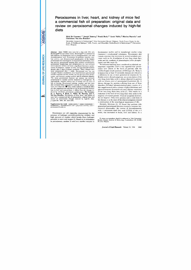
Enzyme assays
Already after 2 and 3 days, hepatic catalase activity was
significantly increased in mice fed the chow supplemented
with 10% fish oil (110 f 17 and 136 10 uB/g her,
respectively) when compared to the animals fed the stan-
dard chow (86 16 uB/g liver). In mice fed the fish oil
diet for up to 3 days, a linear increase in hepatic catalase
activity as a function of duration of feeding was observed
(Fig.
1). A continuing significant but less pronounced in-
crease in hepatic catalase activity was found between the
third and fourteenth day of feeding (157 f
9
uB/g liver).
Feeding the mice 1 week longer did not provoke an addi-
tional increase in catalase activity (161 14 uB/g liver)
(Fig. 1).
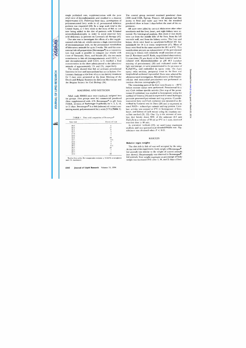
Peroxisomal 0-oxidation of fatty acyl-CoAs in the liver
gradually increased with longer periods of fish oil intake.
The rate-limiting step in peroxisomal @-oxidation, mea-
sured as lauroyl-CoA and palmitoyl-CoA oxidase activi-
ties, was already significantly increased after 3 days of fish
oil feeding (Fig.
2).
After 3 weeks of Beromegan@-
LIVER
C
-7
iii
KIDNEY
[
i
EART
f
I I
0 3
I
I
14 21
Duration of feeding (days)
Fig. 1. Effect of duration of feeding on the catalase activity in liver,
kidney, and myocardium of mice fed a diet supplemented with 10%
(w/w) fish oil (Beromegan ). Points without a common superscript are
significantly different at P < 0.05.
1st step
3rd step
C
i m
i
I I
0 3 14 21
Duration
of
feeding (days)
Fig. 2.
Effect of duration of feeding on the first and third step
of
the
peroxisomal 0-oxidation capacity in the liver of mice fed a diet supple-
mented with
10%
(w/w) fish oil (Beromegan ) using lauroyl-CoA ( 0 )
and palmitoyl-CoA 0) s substrates. Points without a common super-
script are significantly different at P < 0.05.
supplemented diet, the activities of lauroyl-CoA oxidase
and palmitoyl-CoA oxidase were increased, respectively,
7.7- and 6.6-fold when compared to the activities in con-
trol mouse livers (Fig. 2).
Hepatic peroxisomal 0-oxidation capacity was also
measured as the cyanide-insensitive fatty acyl-CoA-
dependent NAD+ reduction using lauroyl-CoA and
palmitoyl-CoA as substrates (third step in peroxisomal
0-
oxidation). The amount of NAD+ reduced was sig-
nificantly increased as
a
function of duration of feeding
the fish oil-supplemented diet (Fig. 2). After 3 weeks,
cyanide-insensitive lauroyl-CoA and palmitoyl-CoA oxi-
dation were increased, respectively, 8.3- and 7.2-fold.
Hepatic catalase activity was positively correlated
(Y
>
0.90; P
<
0.001) with peroxisomal @-oxidation (results
not shown).
Renal catalase activity in mice fed the fish oil diet for
periods up to 3 weeks was never different from that in con-
trol mice (Fig. 1). Myocardial catalase activity was
significantly increased after 3 days (1.82
f
0.30 uB/g)
when compared to the activity in control myocardium
(1.26
+
0.16 uB/g) and reached
2.99
0.37 UB/g in mice
fed the fish oil-supplemented diet for
3
weeks (Fig. 1).
De Cmemer et al. Peroxisomes and fish
oil
diet 1243
8/11/2019 J. Lipid Res. 1994 de Craemer 1241 50
http://slidepdf.com/reader/full/j-lipid-res-1994-de-craemer-1241-50 4/10
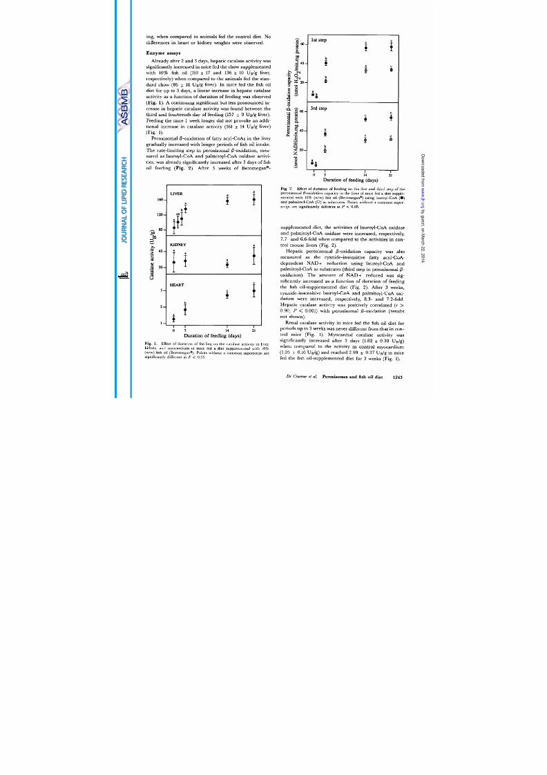
Morphology of peroxisomes
Light microscopic evaluation revealed peroxisomal pro-
liferation and an increase in catalase staining of the
peroxisomes in all livers of mice fed the fish oil-
supplemented diet (Fig. 3). Peroxisomal size was un-
changed. No accumulation of lipid droplets in the hepato-
cytes was observed. Ultrastructural observation of the
hepatic peroxisomes revealed an increase in number in
fish oil-fed animals. In all livers, peroxisomal matrix was
homogeneously filled with electron-dense catalase reac-
tion product. In all control as well as treated livers, perox-
isomal shape varied widely: oval, elongated, triangular,
angular, and reniform organelles were observed. Some-
times short DAB-positive tails were present. In this
respect, mouse liver differs from rat and human liver.
In the myocardium of mice fed the diet with Rero-
megan@, staining intensity of the peroxisomes was in-
creased (Fig. 4). By light microscopic evaluation, peroxi-
somes were larger and more numerous than in the
myocardial cells of control mice. Ultrastructural analysis
of the myocardium revealed the presence of lipid droplets
in two mice fed the fish oil-supplemented diet and in one
control mouse. After catalase cytochemistry, peroxisomes
were recognized by a reticular matrix in the myocardium
of both control and fish oil-fed mice. They were found in
the near vicinity of mitochondria and elements of sarco-
plasmic reticulum. By visual evaluation, peroxisomal
number and size were increased after fish oil feeding
confirming light microscopic observations.
By light microscopy, catalase staining of the renal
peroxisomes did not reveal differences in staining inten-
sity, number, or distribution of the organelles between
animals fed the fish oil-supplemented versus the control
diet (not shown).
Morphometry
of
peroxisomes
Ultrastructural morphometry of the hepatic peroxi-
somes confirmed the light microscopic impression: the
number of peroxisomes was doubled in fish oil-fed mice
for
3
days (+98%) and for 21 days (+124%). Although
mean peroxisomal size was unchanged, this resulted in a
significant doubling of the volume and surface density of
the peroxisomal compartment. Between the third and the
twenty first day of fish oil feeding, differences in perox-
isomal diameter, number, volume, and surface density
were no longer observed (Table 2).
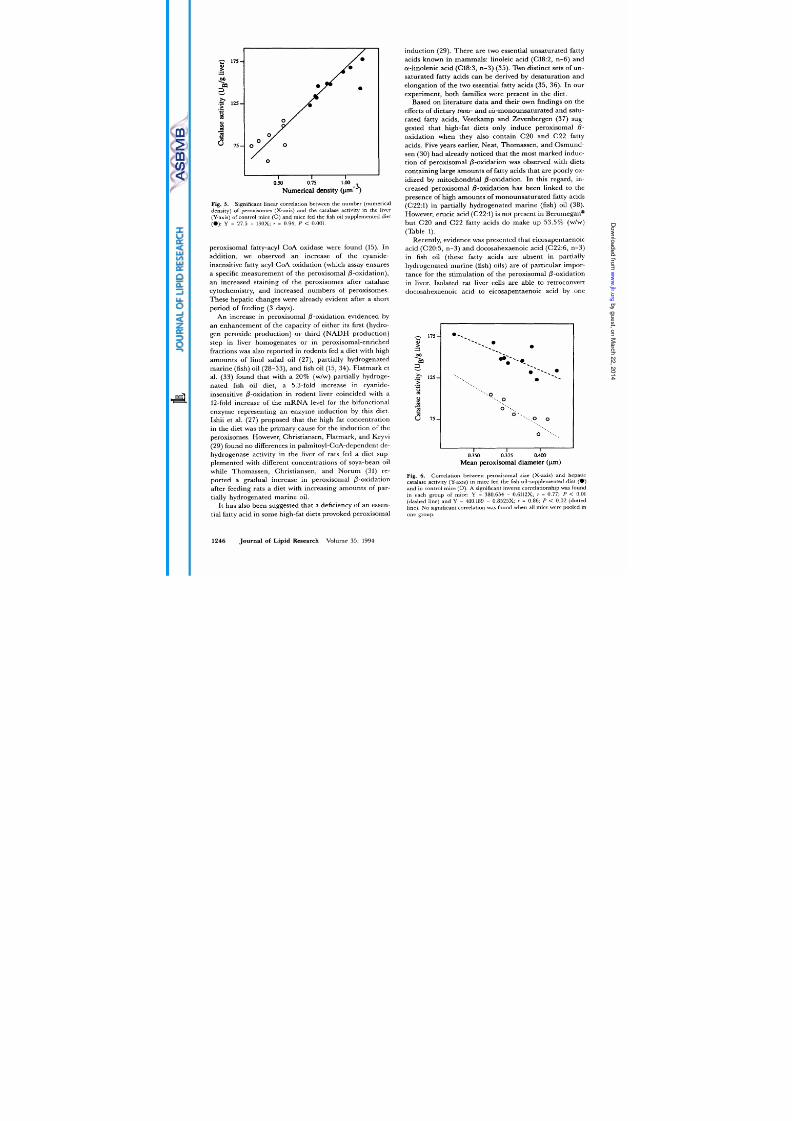
A significant linear positive relationship was found be-
tween the number of peroxisomes (numerical density) on
the one hand, and the catalase activity (Fig. 5 , the
palmitoyl-CoA oxidase activity, and the cyanide-insensitive
palmitoyl-CoA oxidation on the other hand. Similar rela-
tionships were found with volume and surface density of
Fig.
3.
Light micrographs of the liver of a mouse fed the control diet a) and of a mouse fed the fish oil-supplemented diet for 3 days
(b).
Catalase
stain; peroxisomes are recognized as black dots in the cytoplasm. An increase in peroxisomal number is observed in the liver of.mice fed the fish
oil-supplemented diet. Final magnification: x
500.
1244
Journal of Lipid Research Volume 35, 1994

8/11/2019 J. Lipid Res. 1994 de Craemer 1241 50
http://slidepdf.com/reader/full/j-lipid-res-1994-de-craemer-1241-50 5/10
1
4a
.
t
.
. I
C ;
..
I
1
b
. *
Fig. 4. Light micrographs of the myocardium of a m ouse fed the control diet (a) and of a m ouse fed the fish oil-supplemented diet for
14
days b).
Catalase stain; peroxisomes (black dots) are larger and more num erous in myocardium of m ice fed the fish oil-supplemented diet. Final magnification:
x 1250.
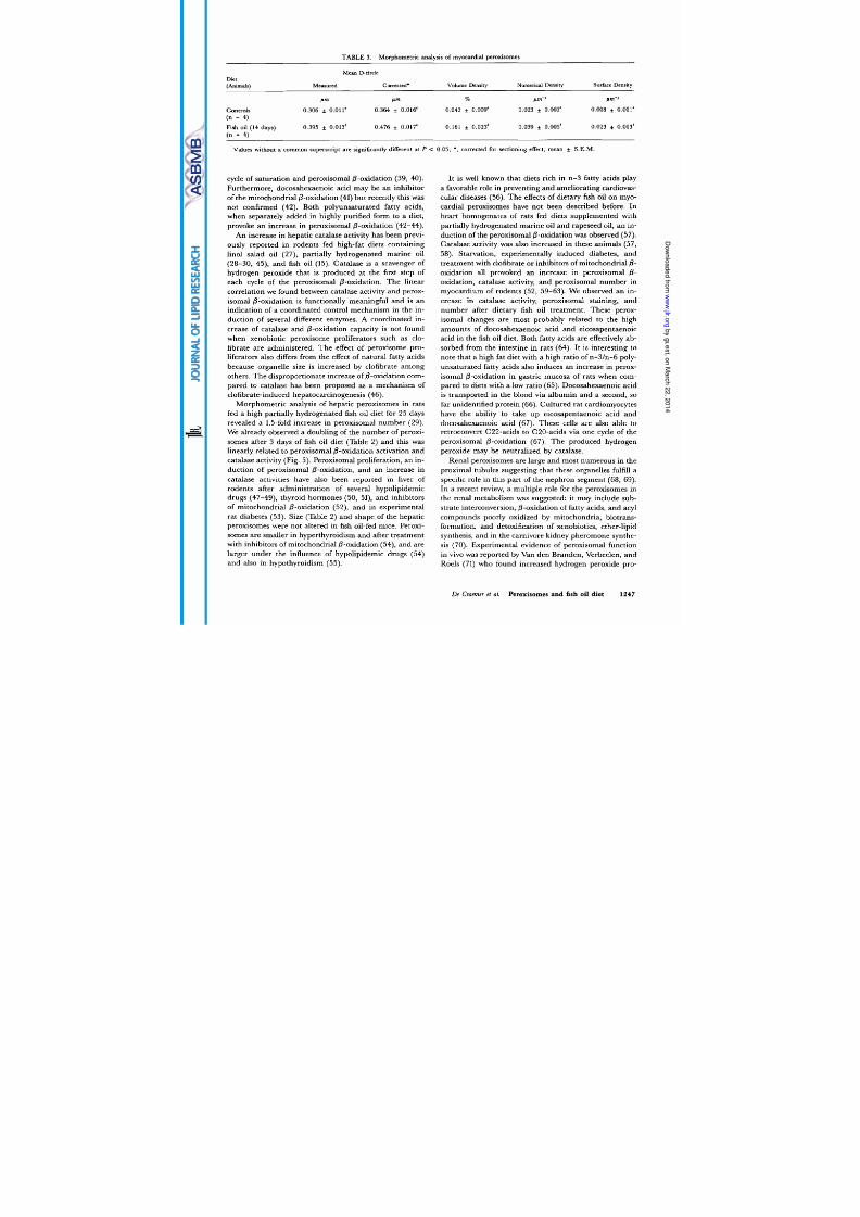
the hepatic peroxisomes. In contrast, mean peroxisomal
diameter was not related to catalase activity
Fig. 6)
and
peroxisomal @-oxidation when control and treated
animals were pooled into one group. However, we did find
an inverse relationship between mean peroxisomal diam-
eter and catalase activity per animal in control mice as
well as in fish oil-fed mice (Fig. 6). This means that
smaller hepatic peroxisomes have a higher enzyme ac-
tivity than larger peroxisomes. This agrees with the
finding of Roels and Comelis (24) that smaller peroxi-
somes have higher catalase concentrations than larger
ones. It was proposed that the larger surface area to
volume ratio of smaller peroxisomes is favorable for trans-
location of the enzyme molecules (which are synthesized
in the cytoplasm), as well as for exchange of metabolites
between peroxisomes and cytosol 25, 26).
With respect to peroxisomal shape, no changes in mean
or median axial ratio were found between livers of
animals fed the fish oil-supplemented diet and control
mice, not even when peroxisomes were divided in groups
according to size.
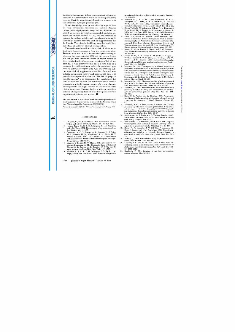
Ultrastructural morphometry of the peroxisomes in the
myocardium of mice fed the Beromegan@-supplemented
diet for 14 days was in agreement with the light and elec-
tron microscopic impression.
A
1.7-fold increase in num-
ber (numerical density) and a 1.3-fold increase in size
were found when compared to control mice. This resulted
in a 3.8-fold and 2.9-fold increase in volume density and
surface density, respectively
Table
3).
DISCUSSION
Our study on mouse liver confirms previous observa-
tions in rats after a fish oil-supplemented diet for 14 days;
hepatomegaly and increased activities
of
catalase and
TABLE 2. Morphometric analysis of hepatic peroxisomes
Diet
(Animals)
Mean D-circle
Measured Corrected' Volume Density Numerical Density Surface Density
Irm
Pm p m -3 pm-'
0.470 f
0.007
1.641 f 0.146' 0.433 * 0.037" 0.240
f 0.020"
ontrols 0.382
f 0.006'
Fish oil (3 days) 0.390 + 0.007 0.483 f 0.010 3.374 f 0.133' 0.857 f 0.067' 0.482 f 0.022*
Fish oil (21 days) 0.370
f 0.010
0.457 f 0.014' 3.299 + 0.235' 0.970
f
0.058' 0.491
f
0.023'
(n - 8)
(n - 5)
(n - 5)
Values without a common superscript are significantly different at
P
< 0.01; *, corrected for sectioning effect; mean S.E.M.
De
C c r et
al. Peroxisomes
and fish
oil diet
1245
8/11/2019 J. Lipid Res. 1994 de Craemer 1241 50
http://slidepdf.com/reader/full/j-lipid-res-1994-de-craemer-1241-50 6/10
I
I I
0.50 0.75 1
oo
Numerical density
p~n-~)
-
175-
I
P
>
.
m
2
0
e 125
>
.
v 75-
Fig. 5 . Significant linear correlation between the number (numerical
density) of peroxisomes (X-axis) and the catalase activity in the liver
(Y-axis) of control mice 0) nd mice fed the fish oil-supplemented diet
(0 ) :
=
27.5
+
130X;
r =
0.94; P
<
0.001.
peroxisomal fatty-acyl CoA oxidase were found (15). In
addition, we observed an increase of the cyanide-
insensitive fatty acyl-CoA oxidation (which assay ensures
a
specific measurement of the peroxisomal @-oxidation),
an increased staining of the peroxisomes after catalase
cytochemistry, and increased numbers of peroxisomes.
These hepatic changes were already evident after
a
short
period of feeding (3 days).
An increase in peroxisomal 6-oxidation evidenced by
an enhancement of the capacity of either its first (hydro-
gen peroxide production) or third (NADH production)
step in liver homogenates or in peroxisomal-enriched
fractions was also reported in rodents fed a diet with high
amounts of linol salad oil (27), partially hydrogenated
marine (fish) oil (28-33), and fish oil (15, 34). Flatmark et
al. (33) found that with a 20% (w/w) partially hydroge-
nated fish oil diet, a 5.3-fold increase in cyanide-
insensitive @-oxidation in rodent liver coincided with a
12-fold increase of the mRNA level for the bifunctional
enzyme representing an enzyme induction by this diet.
Ishii et al. (27) proposed that the high fat concentration
in the diet was the primary cause for the induction of the
peroxisomes. However, Christiansen, Flatmark, and Kryvi
(29) found no differences in palmitoyl-CoA-dependent de-
hydrogenase activity in the liver of rats fed a diet sup-
plemented with different concentrations of soya-bean oil
while Thomassen, Christiansen, and Norum (31) re-
ported a gradual increase in peroxisomal 6-oxidation
after feeding rats a diet with increasing amounts of par-
tially hydrogenated marine oil.
It has also been suggested that a deficiency of an essen-
tial fatty acid in some high-fat diets provoked peroxisomal
induction (29). There are two essential unsaturated fatty
acids known in mammals: linoleic acid (C18:2, n-6) and
a-linolenic acid (C18:3, n-3) (35). Two distinct sets of un-
saturated fatty acids can be derived by desaturation and
elongation of the two essential fatty acids (35, 36). In our
experiment, both families were present in the diet.
Based on literature data and their own findings on the
effects of dietary
tram-
and cis-monounsaturated and satu-
rated fatty acids, Veerkamp and Zevenbergen (37) sug-
gested that high-fat diets only induce peroxisomal @
oxidation when they also contain C20 and C22 fatty
acids. Five years earlier, Neat, Thomassen, and Osmund-
sen (30) had already noticed that the most marked induc-
tion of peroxisomal @-oxidationwas observed with diets
containing large amounts of fatty acids that are poorly ox-
idized by mitochondrial @-oxidation. In this regard, in-
creased peroxisomal @-oxidation has been linked to the
presence of high amounts
of
monounsaturated fatty acids
(C22:l) in partially hydrogenated marine (fish) oil (38).
However, erucic acid (C22:l) is not present in Beromegan@
but C20 and C22 fatty acids do make up 53.5% (w/w)
(Table 1).
Recently, evidence was presented that eicosapentaenoic
acid (C20:5, n-3) and docosahexaenoic acid (C22:6, n-3)
in fish oil (these fatty acids are absent in partially
hydrogenated marine (fish) oils) are of particular impor-
tance for the stimulation of the peroxisomal 0-oxidation
in liver. Isolated rat liver cells are able to retroconvert
docosahexaenoic acid to eicosapentaenoic acid by one
..Q... 0
O ..
.,
0 .
..
...,
0 0
0 '.. .
I I
0.350 0.375 0.400
Mean
peroxisomal
diameter (pm)
Fig. 6
Correl ation between peroxisomal size (X-axis) and hepatic
catalase activity (Y-axis) in mice fed the fish oil-supp lemented diet ( 0 )
and in control mice 0). significant inverse correlationship was found
in each group of mice: Y = 380.654 0.6112X; r = 0.77; P < 0.01
(dashed line) and Y = 410.189 ~ 0.8525X; r = 0.86;
P <
0.02 (dotted
line).
No
significant correlation was found when all mice were pooled in
one group.
1246 Journal of
Lipid
Research
Volume
35 ,
1994
8/11/2019 J. Lipid Res. 1994 de Craemer 1241 50
http://slidepdf.com/reader/full/j-lipid-res-1994-de-craemer-1241-50 7/10
TABLE
3.
Morphometric analysis of myocardial peroxisomes
Diet
(Animals)
Mean D-circle
Surface
Density
easured Corrected' Volume
Density
Numerical Density
pm f im
p m W 3
pm-1
Controls
0.306 0.011 0.364 f
0.016
0.042 f 0.009 0.023 0.002
0.008 0.001
(n
=
4)
Fish oil (14 days) 0.39 5 0.013' 0.476 f 0.017' 0.161 0.023' 0.039 f 0.005' 0.023 f 0.003'
(n =
4)
Values without a commo n superscript are significantly different at P <
0.05;
, corrected for sectioning effect; mean
f
S . E . M .
cycle of saturation and peroxisomal @oxidation (39, 40).
Furthermore, docosahexaenoic acid may be an inhibitor
of the mitochondrial 0-oxidation (41) but recently this was
not confirmed (42). Both polyunsaturated fatty acids,
when separately added in highly purified form to a diet,
provoke
an
increase in peroxisomal 0-oxidation (42-44).
An increase in hepatic catalase activity has been previ-
ously reported in rodents fed high-fat diets containing
linol salad oil (27), partially hydrogenated marine oil
(28-30, 45), and fish oil (15). Catalase is a scavenger of
hydrogen peroxide that is produced at the first step of
each cycle of the peroxisomal 0-oxidation. The linear
correlation we found between catalase activity and perox-
isomal @-oxidation is functionally meaningful and is an
indication of a coordinated control mechanism in the in-
duction of several different enzymes. A coordinated in-
crease of catalase and 0-oxidation capacity is not found
when xenobiotic peroxisome proliferators such as clo-
fibrate are administered. Th e effect of peroxisome pro-
liferators also differs from the effect of natural fatty acids
because organelle size is increased by clofibrate among
others. The disproportionate increase of @-oxidationcom-
pared to catalase has been proposed as a mechanism of
clofibrate-induced hepatocarcinogenesis (46).
Morphometric analysis of hepatic peroxisomes in rats
fed a high partially hydrogenated fish oil diet for 25 days
revealed a 1.5-fold increase in peroxisomal number (29).
We already observed a doubling of the number of peroxi-
somes after 3 days of fish oil diet (Table 2) and this was
linearly related to peroxisomal 0-oxidation activation and
catalase activity (Fig. 5). Peroxisomal proliferation, an in-
duction of peroxisomal 0-oxidation, and an increase in
catalase activities have also been reported in liver of
rodents after administration of several hypolipidemic
drugs (47-49), thyroid hormones (50, 51), and inhibitors
of mitochondrial 0-oxidation (52), and in experimental
rat diabetes (53). Size (Table 2) and shape of the hepatic
peroxisomes were not altered in fish oil-fed mice. Peroxi-
somes are smaller in hyperthyroidism and after treatment
with inhibitors of mitochondrial @-oxidation
54),
and are
larger under the influence of hypolipidemic drugs (54)
and also in hypothyroidism (55).
It is well known that diets rich in n-3 fatty acids play
a favorable role in preventing and ameliorating cardiovas-
cular diseases (56). The effects of dietary fish oil on myo-
cardial peroxisomes have not been described before. In
heart homogenates of rats fed diets supplemented with
partially hydrogenated marine oil and rapeseed oil, an in-
duction of the peroxisomal @-oxidationwas observed (57).
Catalase activity was also increased in these animals (57,
58). Starvation, experimentally induced diabetes, and
treatment with clofibrate or inhibitors of mitochondrial
0
oxidation all provoked an increase in peroxisomal 0
oxidation, catalase activity, and peroxisomal number in
myocardium of rodents (52, 59-63). We observed an in-
crease in catalase activity, peroxisomal staining, and
number after dietary fish oil treatment. These perox-
isomal changes are most probably related to the high
amounts of docosahexaenoic acid and eicosapentaenoic
acid in the fish oil diet. Both fatty acids are effectively ab-
sorbed from the intestine in rats (64). It is interesting to
note that a high fat diet with a high ratio of n-3ln-6 poly-
unsaturated fatty acids also induces an increase in perox-
isomal 0-oxidation in gastric mucosa of rats when com-
pared to diets with a low ratio (65). Docosahexaenoic acid
is transported in the blood via albumin and a second, so
far unidentified protein (66). Cultured rat cardiomyocytes
have the ability to take up eicosapentaenoic acid and
docosahexaenoic acid (67). These cells are also able
to
retroconvert C22-acids to C20-acids via one cycle of the
peroxisomal 0-oxidation (67). The produced hydrogen
peroxide may be neutralized by catalase.
Renal peroxisomes are large and most numerous in the
proximal tubules suggesting that these organelles fulfill
a
specific role in this part of the nephron segment (68, 69).
In a recent review, a multiple role for the peroxisomes in
the renal metabolism was suggested: it may include sub-
strate interconversion, 0-oxidation of fatty acids, and acyl
compounds poorly oxidized by mitochondria, biotrans-
formation, and detoxification of xenobiotics, ether-lipid
synthesis, and in the carnivore kidney pheromone synthe-
sis (70). Experimental evidence of peroxisomal function
in vivo was reported by Van den Branden, Verbeelen, and
Roels (71) who found increased hydrogen peroxide pro-
De Craemer et al. Peroxisomes and fish oil diet 1247
8/11/2019 J. Lipid Res. 1994 de Craemer 1241 50
http://slidepdf.com/reader/full/j-lipid-res-1994-de-craemer-1241-50 8/10
ductio n in the remn ant kidney concommitant with the in-
crease in Na’ reab sorption, which is a n energy-req uiring
process. Possibly, peroxisomal 0-oxidation accounts for
the additional hydrogen peroxide (71).
To our knowledge, data on the effects of high fat diets
on renal peroxisomal functions are lacking. Rodents
treated with hypolipidemic dru gs or fed thyroxine re-
vealed
an
increase in renal peroxisomal @-oxidation en-
zyme a nd catalase activity (47,
72,
73). We observed no
changes in catalase activity and peroxisomal staining in
the kidneys of intact mice fed a fish oil-supplemente d diet
for
3
weeks. Therefore, in th e kidney a s well as in the liver,
the effects of clofibrate and fat feeding differ.
T he mechanism
by
which a dietary fish oil elicits an in -
ductio n of the peroxisomes in liver and heart is
not
clear.
Recently,
a
nuclear receptor inducible by peroxisome pro-
liferators has been reported (74) but the natural ligand
has not yet been identified. Based on recent results on
diets enriched w ith different concentrations of fish oil and
cor n oil, it was speculated that a n n-3 fatty acid or a
molecule derived from it m ay induce the peroxisome pro-
liferator activated receptor (75). O u r observations indi-
cate that a fish oil su ppleme nt in the diet
of
normal mice
induces peroxisomes in liver
and
heart as did diets with
partially hydrogenated marine oils. T he fish oil prepara-
tion Beromegan* is an inexpensive diet supplement that
may increase the extreme low concentrations of docosa-
hexaenoic acid in the nervous system of a gr oup of perox-
isomal patients; this might result in an amelioration of the
clinical symptoms. However, furthe r studies on the effects
of lower (therapeutic) doses of fish
oils
on peroxisomes of
experimental animals are needed.
The authors wish to thank RenC Stien for his indispensable tech-
nical assistance. Supported by a grant of the National Fonds
voor Wetenschappelijk Onderzoek (2923220790).
M a n w e r i p received 27 September 1993 and in revisedform 24 January 199 4.
REFERENCES
1.
2.
3.
4.
5.
De Duve, C., and
P.
Baudhuin. 1966. Peroxisomes (micro-
bodies and related particles). Physiol. Rev 46: 323-357.
van den Bosch, H., R. B. H. Schutgens, R.
J.
A. Wanders,
and
J.
M. Tager. 1992. Biochemistry of peroxisomes.
Annu.
Rev. Biochem.
61:
157-197.
Goldfisher, S.,
C
L. Moore, A. B. Johnson, A. J. Spiro,
M.
P.
Valsamis, H. M . Wisniewski, R. H. Ritch, W. T.
Norton, I. Rapin, and L. M. Gartner. 1973. Peroxisomal
and mitochondrial defects in the cerebro-hepato-renal syn-
drome.
Science. 182:
62-64.
Lazarow, P. B., and H. W. Moser. 1989. Disorders of per-
oxisome biogenesis. In The Metabolic Basis of Inherited
Disease. C. R. Scriver, A. L. Beaudet, W. S.
Sly,
and D.
Valle, editors. McGraw-Hill, New York. 1479-1509.
Wanders, R. J. A,, R. B. H. Schutgens, P.
G.
Barth, J. M.
Tager, and H. van den Bosch. 1993. Postnatal diagnosis of
peroxisomal disorders: a biochemical approach.
Biochimie.
6. Wanders, R. J. A., C.
W.
T. van Roermund,
R .
B. H.
Schutgens, P. G. Barth, H. S. A. Heymans, H. van den
Bosch, and J. M. Tager. 1990. The inborn errors of perox-
isomal 0-oxidation: a review. J
Inhe,: Metab. Dis. 13:
3-36.
7. Rizzo, W. B., R. T. Leshner,
A.
Odone, A. L. Dammann,
D. A . Craft, M. E. Jensen, S. S. Jennings, S. Davis, R.
Jaitly, and J. A. Sgro. 1989. Dietary erucic acid therapy for
X-linked adrenoleukodystrophy. Neurology, 39: 1415-1422.
8. Uziel, G., E. Bertini, M. Rimoldi, and M. Gambetti. 1990.
Italian multicentric dietary therapeutical trial in adreno-
leukodystrophy. In Adrenoleukodystrophy and Other Per-
oxisomal Disorders. Clinical, Biochemical, Genetic and
Therapeutic Aspects. G. Uziel, R. J. A. Wanders, and M.
Cappa, editors. Excerpta Medica, Amsterdam. 163-180.
9. Moser, H. W., A. Bergin, S. Naidu, and P. W. Ladenson.
1991. Adrenoleukodystrophy.Endocnnol. Metab. Ciin. N Am.
Moser, H. W. A. B. Moser,
K .
D. Smith, A. Bergin,
J.
Borel, J. Shankroff,
0
C. Stine,
C.
Merette,
J. Ott,
W.
Krivit, and
E.
Shapiro. 1992. Adrenoleukodystrophy:
phenotypic variability and implications for therapy.J Inher.
Metab. Dis. 15: 645-664.
Martinez, M. 1991. Developmental profiles of polyunsatu-
rated fatty acids in the brain of normal infants and patients
with peroxisomal diseases: severe deficiency of docosahex-
aenoic acid in Zellweger’s and Pseudo-Zellweger’s Syn-
dromes. In World Review of Nutrition and Dietetics. A. P.
Simopoulos, R. E. Kifer, R. E. Martin, and
S.
M. Barlow,
editors. Karger, Basel. 87-102.
12. Martinez, M . 1992. Abnormal profiles of polyunsaturated
fatty acids in the brain, liver, kidney and retina of patients
with peroxisomal disorders.
Brain Res. 583:
171-182.
13. Martinez, M. 1992. Treatment with docosahexaenoic acid
favorably modifies the fatty acid composition of erythro-
cytes in peroxisomal patients. Prog. Clin. Biol. Res. 375:
389-397.
14. Roels, F., S. Fischer, and
W.
Kissling. 1993. Polyunsatu-
rated fatty acids in peroxisomal disorders: a hypothesis and
a proposal for treatment.
J
Neurol. Neumsurg. Psychiat. 56:
937.
15. Yamazaki, R. K., T. Shen, and G. B. Schade. 1987. A diet
rich in n-3) fatty acids increases peroxisomal @-oxidation
activity and lowers plasma triacylglycerols without inhibit-
ing glutathione-dependent detoxication activities in the rat
liver. Biochim. Biophys. Acta. 920 62-67.
16. De Craemer,
D.
E Roels, and C. Van den Branden. 1993.
Rapid effects of dietary fish oil on peroxisomes in mouse
liver. Eut: J Morphol. 30: 331-335.
De Craemer, D., I. Kerckaert, and F. Roels. 1991. Hepato-
cellular peroxisomes in human alcoholic and drug-induced
hepatitis: a quantitative study. Hepatology. 14: 811-817.
18.
Roels, F., A. Cornelis, B. T. Poll-The, P. Aubourg, H.
Ogier,
J.
Scotto, and
J.
M. Saudubray. 1986. Hepatic per-
oxisomes are deficient in infantile Refsum disease: a
cytochemical study of 4 cases. Am. J .
Med. Genet. 25:
257-
271.
19. Vamecq, J. 1990. Fluorometric assay of peroxisomal oxi-
dases. Anal. Biochem. 186: 340-349.
20. Lazarow, P. B., and C. de Duve. 1976. A fatty acyl-CoA
oxidizing system in rat liver peroxisomes: enhancement by
clofibrate, a hypolipidemic drug. Proc. Nail. Acad. Scz. USA.
21. Baudhuin, P. 1974. Isolation of rat liver peroxisomes.
75:
269-279.
20:
297-318.
10.
11.
17.
73:
2043-2046.
Methods Enzymoi.
31 358-
3
68.
1248 Journal
of Lipid
Research Volume 35, 1994
8/11/2019 J. Lipid Res. 1994 de Craemer 1241 50
http://slidepdf.com/reader/full/j-lipid-res-1994-de-craemer-1241-50 9/10
22.
Van den Branden, C., I. Kerckaert, and F. Roels. 1984.
Peroxisomal @-oxidation from endogenous substrates.
Demonstration through HzOzproduction in the unanaes-
thetized mouse.
Biochem.
J 218:
697-702.
23. Snedecor, G.W., and W. G. Cochran. 1980. Statistical
Methods. Iowa State University Press, Ames, IA.
24. Roels, F., and A. Cornelis. 1989.Heterogenity of catalase
staining in human hepatocellular peroxisomes.
J Histochem.
25.
Roels, E,
M.
speel, and D. De Craemer.
1991.
Liver
pathology and immunocytochemistry in congenital perox-
isomal diseases.
J
Znher Metab. Dis. 1 4 857-875.
26. Roels, E, M. Espeel, F. Poggi, H. Mandel, L. Van Malder-
gem, and
J.
M. Saudubray. 1993.Human liver pathology
in peroxisomal diseases: a review including novel data.
Biochimie. 75: 281-292.
27.
Ishii, H., N. Fukumori, S. Horie, and T. Suga.
1980.
Effects of fat content in the diet on hepatic peroxisomes of
the rat.
Biochim. Biofihys. Acta.
617: 1 11.
28. Neat, C. E., M. S. Thomassen, and H. Osmundsen. 1980.
Induction of peroxisomal @-oxidation n rat liver by high-
fat diets.
Biochem.
J . 186: 369-371.
29.
Christiansen, E. N.,
T.
Flatmark, and H. Kryvi.
1981.
Effects of marine oil diet on peroxisomes and mitochondria
of rat liver. A combined biochemical and morphometric
study. Eur. J
Cell Biol.
26:
11-20.
30.
Neat, C. E., M. S. Thomassen, and H. Osmundsen.
1981.
Effects of high-fat diets on hepatic fatty acid oxidation in
the rat.
Biochem.J
196: 149-159.
Thomassen, M.
S.,
E. N. Christiansen, and K. R. Norum.
1982.
Characterization of the stimulatory effect of high-fat
diets on peroxisomal 6-oxidation in rat liver.
Biochem.
J
32.
Thomassen,
M. ., .
Norseth, and E. N. Christiansen.
1985.
Long-term effects of high-fat diets on peroxisomal
6
oxidation in male and female rats.
Lipids.
20: 668-674.
33.
Flatmark, T. . Nilsson,
J.
Kvannes, T.
S.
Eikhom, M. H.
Fukami, H. Kryvi, and E. N. Christiansen.
1988.
On the
mechanism of induction of the enzyme systems for perox-
isomal &oxidation of fatty acids in rat liver by diets rich in
partially hydrogenated fish oil.
Biochim. Biophys. Acta.
962:
34.
Rustan, A. C., E. N. Christiansen, and C. A. Drevon.
1992.
Serum lipids, hepatic glycerolipid metabolism and perox-
isomal fatty acid oxidation in rats fed 0-3 and w-6 fatty
acids.
Biochem.
J. 283: 333-339.
35.
Sardesai, V.
M.
992.Nutritional role of polyunsaturated
fatty acids.
J Nutr. Biochem.
3:
154-166.
36. Drevon, C. A. 1992.Marine oils and their effects.
Nutr.
Rev.
37.
Veerkamp,
J.
H., and
J. L.
Zevenbergen.
1986.
Effect
of
dietary fat on total and peroxisomal fatty acid oxidation in
rat tissues.
Biochim. Biophys. Acta.
878:
102-109.
38.
Bremer, J., and K. R . Norum.
1982.
Metabolism of very
long-chain monounsaturated fatty acids
22:l)
and the
adaptation to their presence in the diet. J.
Lipid
Res 23:
39.
Hagve,
T.
A., and
B.
0 Christophersen.
1986.
Evidence for
peroxisomal retroconversion of adrenic acid 22:4 n-6))
and docosahexaenoic acids 22:6 n-3)) in isolated liver
cells. Biochim. Biobhys. Acta. 875: 165-173.
40.
Gronn, M., E. Christensen,
T.
A. Hagve, and B.
0
Christophersen. 1991.Peroxisomal retroconversion of doco-
sahexaenoic acid 22:6 n-3)) to eicosapentaenoic acid
20:5 n-3))
studied in isolated rat liver cells.
Biochim.
Biophys. Acta.
1081:
85-91.
Cytochem. 37:
331-337.
31.
206:
195-202.
122-130.
5 :
38-45.
243-256.
41.
Osmundsen, H., and K. Bjornstad.
1985.
nhibitory effects
of some long-chain unsaturated fatty acids on mitochon-
drial 6-oxidation.
Biochem.
J 230: 329-337.
42.
Willumsen, N., S.Hexeberg,
J.
Skorve,
M.
Lundquist, and
R. K. Berge.
1993.
Docosahexaenoic acid shows no tri-
glyceride-lowering effects but increases the peroxisomal
fatty acid oxidation in liver of rats.
J. Lipid Res
3 4
13-22.
43.
Aarsland, A., M. Lundquist, B. Borretsen, and R. K.
Berge.
1990.
On the effect of peroxisomal P-oxidation and
carnitine palmitoyl-transferase activity by eicosapentaenoic
acid in liver and heart from rats.
Lipids.
25:
546-548.
44. Demoz, A., N. Willumsen, and R . K. Berge. 1992.Eicosa-
pentaenoic acid at hypotriglyceridemic dose enhances the
hepatic antioxidant defense in mice.
Lipids.
27:
968-971.
45.
Holmer,
G.
. E. Hoy, and D. Kintein.
1982.
Influence of
partially hydrogenated vegetable and marine oils on lipid
metabolism in rat liver and heart. Lipids. 17: 585-593.
46.
Conway,
J.
G., R. C. Cattley,
J.
A. Popp, and B. E. Butter-
worth. 1989.Possible mechanisms in hepatocarcino-genesis
by the peroxisome proliferator di 2-ethylhexy1)phthalate.
rug
Metab.
Rev. 21: 65-102.
47.
Reddy,
J.
K., J. R. Warren, M. K. Reddy, and N. D.
Lalwani.
1982.
Hepatic and renal effects of peroxisome
proliferators: biological implications.
Ann. N Y Acad. Sci.
48.
Kawashima,
Y.,
A. Hirose,
Y.
Hirose, T. Adachi, and H.
Kozuka.
1986.
nducing effects of clofibric acid on l-acyl-
glycerophosphorylcholine
acyltransferase in kidney and in-
testinal mucosa of rats.
Biochim. Biophys Acta.
875:
549-553.
49.
Katoh, H.,
Y
Kawashima, H. Watanuki, H. Kozuka, and
H. Isono.
1987.
Effects of clofibric acid and tiadenol on
cytosolic long-chain acyl-CoA hydrolase and peroxisomal
beta-oxidation in liver and extrahepatic tissues of rats.
Biochim. Biophys. Acta.
920:
171-179.
50.
Fringes,
B.,
nd A. Reith.
1982.
Time course of peroxisome
biogenesis during adaptation to mild hyperthyroidism in
rat liver.
Lab. Invest.
47:
19-26.
Kerckaert, I ., A. Claeys, W. Just, A. Cornelis, and F. Roels.
1989.
Automated image analysis of rat liver peroxisomes
after treatment with thyroid hormones: changes in number,
size and catalase reaction.
Micmn Microsc. Acta.
20:
9-18.
52. Vamecq, J., F. Roels, C. Van den Branden, and
J.
P.
Draye.
1987.
Peroxisomal proliferation in heart and
liver of mice receiving chlorpromazine, ethyl
2 5 4-chloro-
phenyl)pentyl)oxiran-2-carboxylicacid or high fat diet: a
biochemical and morphometrical comparative study.
Pediatr.
Res 22: 748-754.
53. Horie, S.,H. Ishii, and T. Suga.
1981.
Changes in perox-
isomal fatty acid oxidation in the diabetic rat liver.
J Biochem.
90:
1691-1696.
54.
Roels, F.
1991.
Peroxisomes, a Personal Account. VUB-
Press, Brussels.
55. Riede, U. N., P. R. Riede, R. Horn, R. Batthiany,
G.
Kiefer, and W. Sandritter.
1978.
Mechanisms of adaptation
of hepatocytes to a chronical hypothyroidism (a cyto-
photometrical and morphometrical study).
Pathol. Res Pract.
56.
Leaf,
A.,
nd P. C. Weber.
1988.
Cardiovascular effects of
n-3
fatty acids.
N
Engl.
J Med.
318:
549-557.
57.
Norseth, J., and M.
S.
Thomassen.
1983.
Stimulation of
microperoxisomal 6-oxidation
in
rat heart by high-fat diets.
Biochim. Biophys. Acta.
751:
312-320.
58.
Norseth,
J. 1979.
The effect of feeding rats with partially
hydrogenated marine oil
or
rapeseed oil on the chain
shortening
of
erucic acid in perfused heart.
Biochim. BiOphys.
386: 81-110.
51.
162:
398-419.
De
C e r
et al.
Peroxisomes and fish oil diet 1249
8/11/2019 J. Lipid Res. 1994 de Craemer 1241 50
http://slidepdf.com/reader/full/j-lipid-res-1994-de-craemer-1241-50 10/10
Acta.
575:
1-9.
59. Norseth,
J.
1980. Increased 0-oxidation of erucic acid in
perfused hearts from rats fed clofibrate. Biochim. Biophys.
Acta. 617: 183-191.
60. Lammi-Keefe, C.
J.,
P. V.
J.
Hegarty, and P. B. Swan. 1981.
Effect of starvation and refeeding on catalase and super-
oxide dismutase activities in skeletal and cardiac muscles
from 12-month-old rats. Experimtiu. 37: 25-27.
61. Crescimanno, M. ,
M.
G. Armata,
L.
Rausa, M. C. Gueli,
C. Nicotra, and N. DAlessandro. 1989. Card iac perox-
isomal enzymes and starvation.
Free
Rad
Res
Commun. 7:
62. Yokota,
S.,
an d K. Asayama. 1990. Peroxisomes of the rat
cardiac and soleus muscles increase after starvation. A bio-
chemical and immunocytochemical study.
Histochemisty
63. Yokota,
S.,
and K. Asayama. 1992. Proliferation of myo-
cardial peroxisomes in experimental rat diabetes: a bio-
chemical and immunocytochemical study.
Virchows Arch. [B/
64. Chernenko, G. A., J. A. Barrowman,
K.
T. Kean,
G.
R.
Herzberg, and K. M. W. Keough. 1989. Intestinal absorp-
tion and lymphatic transport of fish oil (MaxEPA) in the
rat. Biochim. Biophys. Acta.
1004:
95-102.
65. Grataroli,
R.,
J.
Vamecq,
J.
H.
Poupaert,
J.
Ltonardi, E.
Termine,
H.
Lafont, and G. Nalbone. 1992. Effects of
dietary n-6/n-3 ratios on lipid and prostaglandin E2
metabolism in rat gastric mucosa. J Lipid Mediators.
5 :
227-236.
66. Li, J., M. G. Wetzel, and P.
J.
OBri en. 1992. Transport on
n-3 fatty acids from the intestine to the retina in rats.
67-72.
93: 287-293.
63: 43-49.
J Lipid Res
33:
539-548.
67. Mohammed , B. S., T. A. Hagve, and
H.
Sprecher. 1990.
Th e metabolism of 20- and 22-carbon unsaturated acids in
rat heart and myocytes as mediated by feeding fish oil.
L i p i h .
25:
854-858.
Beard, M. E., and A. B. Novikoff. 1969. Dis tribution of
peroxisomes (microbodies) in the nephron of the rat:
a
cytochemical study.
J Cell Biol.
42: 501-518.
69. Roels,
E ,
and
S.
Goldfischer. 1979. Cytochemistry
of
human catalase. The demonstration of hepatic and renal
peroxisomes by a high temperature procedure.
J Histachem.
Cytochem. 27: 1471-1477.
70. Zaar, K . 1992. Stru cture and function of peroxisomes in the
mammalian kidney.
Eur. J Cell. Biol.
59: 233-254.
71. Van den Branden, C., D. Verbeelen, and E Roels. 1990.
Peroxisomal @-oxidation n the rat kidney during increased
work load.
Eur. J Cell. Biol. 53:
53.
72. Ohno,
S.
1985. Peroxisomes
of
the kidney. Znt Reu. Cytol.
73. Kramar, R., K. Kremser, M. Hohenegger, and M. Mayer.
1986. Fatty acyl-CoA oxidase in rat kidney and liver after
application of thyroxine. Enyme. 35: 27-33.
74. Issemann, I.,
R.
Prince,
J.
Tugwood, and
S.
Green. 1992.
A role for fatty acids and liver fatty acid binding protein in
peroxisome proliferation?
Biochem.
SOC.
ans. 20:
824-827.
75. Vamecq, J.,
L.
Vallte, P. Lechene de la Porte, M. Fontaine,
D. De Craemer, C., Van den Branden, H., Lafont, R.
Grataroli, and G. Nalbone. 1993. Effect of various n-3/n-6
fatty acid ratio contents of high fat diets on rat liver and
heart peroxisomal and mitochondrial @-oxidation.Biochim.
Biophys. Acta.
1170:
151-157.
68.
95: 131-162.
1250
Journal
of
Lipid
Research Volume 35, 1994


![SAGEM [email protected] 1201/1241 - Support Sagemcom](https://img.pdfslide.us/doc/110x75/61fb45e62e268c58cd5c372a/sagem-emailprotected-12011241-support-sagemcom.jpg)