Embed Size (px)
Citation preview
Brit. J. Ophthal. (I 972) 56, 338
Deposition of melanin and iron inocular structures in haemochromatosisGEOFFREY DAVIES,t IAIN DYMOCKJ JOHN HARRY,*AND ROGER WILLIAMS4
tDepartment of Ophthalmology and the tM.R.C. Group on Metabolism and Haemodynamics of LiverDisease, King's College Hospital, London, and the *Department of Pathology, Institute of Ophthal-mology, University of London
Cutaneous pigmentation is a well-recognized feature of idiopathic haemochromatosis and,in various series, has been recorded in up to 8o per cent. of patients (Finch and Finch,I955). In the world literature there are occasional reports of pigmentation in the gums(Saundby, I890), buccal mucous membranes (Richardiere, I895), lips (Parker, I903), andtongue (Hess and Zurhelle, I905), and Maddox (I933) recorded the presence of dis-colouration around the disc margin of the retina in four patients with haemochromatosis.The past finding was also observed by Hudson (1953) in one of the five patients heexamined. Apart from these reports, however, abnormal pigmentation in the eye hasreceived little attention. In this paper we describe findings related to the occurrence anddistribution of melanin and iron in the extraocular structures and within the eye, inhaemochromatosis, based on a study of 44 patients.
Patients
Of the 44 patients examined, 42 had primary idiopathic haemochromatosis, defined according tothe criteria of Williams (I968), and the other two had alcoholic cirrhosis with secondary iron over-load. All 44 patients were of the Caucasian race.
Methods
Pigmentation of the conjunctiva and lid margins was looked for using direct slit-lamp examination,and a careful search was made for pigmentation in the cornea, uvea, lens, and retina. Ocularphotographs were taken with a coupled Zeiss-Ikon camera using high-speed Ektachrome film.
Results
CONJUNCTIVAL PIGMENTATION
Brown pigmentation of the conjunctiva was detected in eight patients. The pigment waspresent in the bulbar conjunctiva and was confined to the area adjacent to the limbus,encroaching on to the cornea. It was most marked along the inferior border of thelimbus extending to the medial aspect of the globe opposite the interpalpebral fissure.Some pigmentation was also detected along the superior border of the limbus. Thedeposits of pigment in the conjunctiva tended to be given a radial striation by the inter-vening lymphatic channels, while in one patient horizontal striae were seen which wereprobably due to the mechanical effects of rucking of the conjunctiva with movements ofthe lower lid (Fig. I, opposite).
Received for publication July 7, 1971Address for reprints: King's College Hospital, Denmark Hill, London SE5 gRS, or Institute of Ophthalmology, Judd Street,London, WCiH 9QS
on 26 May 2018 by guest. P
rotected by copyright.http://bjo.bm
j.com/
Br J O
phthalmol: first published as 10.1136/bjo.56.4.338 on 1 A
pril 1972. Dow
nloaded from
DEPOSITION OF MELANIN AND IRON IN OCULAR STRUCTURES
F I G. I Conjunctival pigmentation (by Courtesy of Mr. Peter Wright)
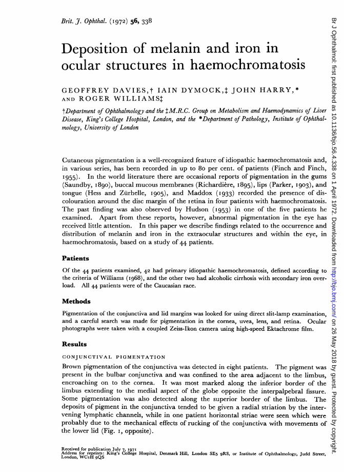
FIG. 3 Pigmentation of lid margin
To face page 338
:i;
'I
on 26 May 2018 by guest. P
rotected by copyright.http://bjo.bm
j.com/
Br J O
phthalmol: first published as 10.1136/bjo.56.4.338 on 1 A
pril 1972. Dow
nloaded from
Melanin and iron deposition in haemochromatosis
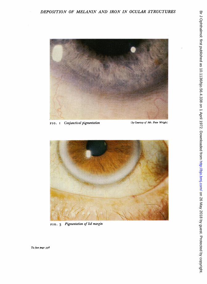
A biopsy of the affected conjunctiva was performed in one patient (Case 2). Microscopyshowed the presence of melanin pigment, particularly in the basal layers of the epithelium(Fig. 2), while a Perls-stained section demonstrated the presence of a minute amount offree ferric iron, mainly within the epithelium.
F I G. 2 Section of conjunctivato show melanin pigment inepithelium. Fontana x 570
A. .4. .
.. Ava.
LID PIGMENTATION
A similar brown pigmentation of the lid margin was present in nine patients. It was seenthroughout the length of the margin, being more prominent around the lash follicles. Thepigmentation was confined to the cutaneous side of the muco-cutaneous junction and waspresent in both the upper and lower lids, being more apparent in the latter (Fig. 3, seecol. pl.). Four of these patients also showed pigmentation of the conjunctiva.One patient (Case I 2), an alcoholic with cirrhosis and secondary siderosis, in whom lid
pigmentation was present, subsequently died from rupture of oesophageal varices. Awedge-shaped portion of the lower lid was removed at autopsy. Histological examinationof this tissue revealed increased melanin pigmentation, particularly within the basal layersof the epithelium of the skin (Fig. 4), but no free iron could be detected.
.:...
FIG. 4 Section of lid to showmelanin pigment in epithelium
I rof skin Fontana x 570
RELATION OF PIGMENTATION TO CLINICAL STATUS (Table, overleaf)
Five of the patients with idiopathic haemochromatosis and the two patients with alcoholiccirrhosis and secondary iron overload were either untreated or had been only partially
339
on 26 May 2018 by guest. P
rotected by copyright.http://bjo.bm
j.com/
Br J O
phthalmol: first published as 10.1136/bjo.56.4.338 on 1 A
pril 1972. Dow
nloaded from

Geoffrey Davies, Iain Dymock, john Harry, and Roger Williams
Table Clinical and biochemical details of thirteen patients with pigmentation of extraocularstructures
Cases I-I had idiopathic haemochromatosisCases 12-I3 had alcoholic cirrhosis with secondary siderosis
6o53455946506o65635'394764
Sex Venesection SerumSextherapY ([tg/ironthray (jig./oI 0MI.)
M
M
M
M
M
M
FM
M
M
M
M
M
NoneIn progress
In progress
CompletedCompletedCompletedCompletedCompletedCompletedCompletedJust completedNoneNone
14026533570135260205
245I40
270
50I50
220
Saturationof serumTIBC*(per cent.)
829899269696939874100
139494
Fv( g./kg.).t
24I9I 726713822711
484319
90
56066i79
4X6
* TIBC-Total iron binding capacityt Fv -A measure of the total chelatable body iron estimated by the differential
Normal range 0-300 I±g./kg. (Smith and others, i969).
Pigmentation
Conjunctiva Lids Skin
+ + ++ + ++ 0 +++± + +++ ± ++o ± +o + +±
o + ±+ 0 +
± 0 +o + ++o ± ±+-t
ferrioxamine test.
treated by venesection therapy. Of these, one had conjunctival pigmentation and twolid pigmentation, the other two having pigmentation at both sites. The remaining 37patients had been treated by venesection therapy and eight of them had pathologicalpigmentation distributed as follows: conjunctivae alone-three; lid alone-three; bothsites-two. With the exception of Case 8, who had bled recently from a duodenal ulcer,each of these patients had re-accumulated iron to some extent, as shown by an increase intotal chelatable body iron when measured by the differential ferrioxamine test (Smith,Lestas, Miller, Dymock, Pitcher, and Williams, I969). This re-accumulation hadoccurred during the few years which had elapsed since the completion of venesectiontherapy. Other patients, however, who had re-accumulated iron to a similar extent didnot show pigmentation.Although skin pigmentation is difficult to assess, in general the patients with either
conjunctival or lid pigmentation also showed prominent skin pigmentation. One of theuntreated patients had prominent skin pigmentation and, in spite of a careful search, no
lid or conjunctival pigmentation could be detected.
DEPOSITION OF IRON WITHIN THE EYE
During the course of this study two patients died, one (Case 12) from rupture of oesophagealvarices, and the other, a man of 70 with primary haemochromatosis, from liver failure.An eye removed post mortem from Case 12 was examined histopathologically and showed,on a Perls-stained section, the presence of minute traces of free ferric iron in the cornealepithelium and in the non-pigmented epithelium of the ciliary body. Similar amounts of
340
Case Ageno. (yrs)
2
3456
789I0
I I
12
' 3
on 26 May 2018 by guest. P
rotected by copyright.http://bjo.bm
j.com/
Br J O
phthalmol: first published as 10.1136/bjo.56.4.338 on 1 A
pril 1972. Dow
nloaded from
Melanin and iron deposition in haemochromatosis
iron with the same distribution were found in both eyes of the other patient who died, theiron in the corneal epithelium being present mainly in the limbal region and extendinginto the conjunctiva; bleached sections of these eyes also showed the presence of a minutetrace of free iron within the epithelium of the iris.
Discussion
Although conjunctival pigmentation was reported in two patients with idiopathichaemochromatosis by Ridder (i9io), pigmentation of the lid has not previously beendescribed. The slate-coloured pigmentation around the disc margin, observed byMaddox (I933) in four patients and in one of five patients by Hudson (I953), was not seenin the present series of cases of idiopathic haemochromatosis, although one of the twowith secondary haemochromatosis (Case 12) had a faint halo-like pigmentation around thedisc margin. Hudson also described a brownish-green discolouration of the iris in hispatients, which we have not seen, and it is of interest that neither he nor Maddox describedthe pigmentation of the lids and conjunctivae which was such a striking feature of ourpatients.The cutaneous pigmentation of patients with idiopathic haemochromatosis usually
diminishes with venesection therapy (Williams, Smith, Spicer, Barry, and Sherlock, I969).The most marked ocular pigmentation in our patients was in those who were either un-treated or had been only partially treated, and it was less frequently found in patients whohad been subjected to previous venesections. This would suggest that the pigmentationin the two sites has a similar pathogenesis. The mechanism of cutaneous pigmentation inhaemochromatosis has, however, not been determined. It has been suggested that theiron present in the cutaneous tissues may favour the deposition of melanin by increasingthe progressive oxidation of the amino acid tyrosine (Robert and Zurcher, I 960), a featureof normal melanogenesis (Brunet, I960; Fitzpatrick, Seiji, and McGugan, I96I). Dis-orders of the endocrine system are known to affect skin pigmentation, for example inAddison's disease, and there is some evidence from animal studies that oestrogens increaseskin pigmentation (Bischitz and Snell, I960), although androgens have no such effect(Bischitz and Snell, 1959). The response of the adrenal cortex to ACTH is normal inhaemochromatosis, but these patients usually have hypogonadism. Whether the skinpigmentation in haemochromatosis is due to a disordered hypothalamic-pituitary axis, tohyperoestrogenism consequent upon impaired hepatic inactivation, or to other factorsremains to be determined (Harris, I969).Although the mechanism of the ocular pigmentation is not clear, it seems likely that it
represents an extension of the pigmentary phenomena seen in other sites, and it wastherefore ofsome intex est to find lid and conjunctival pigmentation similar to that observedin haemochromatosis in a patient with Addison's disease who had no demonstrabledisturbance of iron metabolism. We have also seen similar pigmentation in the lids aftersunburn and in the conjunctivae of coloured races.While the deposition of iron in various organs of the body is the outstanding character-
istic of haemochromatosis, the presence of iron within the eye has not, as far as we areaware, been previously reported. It is of interest that this iron, which for the eye can beconsidered to be of endogenous origin, was found in the corneal epithelium and ciliarybody epithelium, which are two of the characteristic sites of iron deposition in siderosisbulbi, a condition in which iron infiltrates the tissues either from a retained intraocularforeign body or from an intraocular haemorrhage.
34I1
on 26 May 2018 by guest. P
rotected by copyright.http://bjo.bm
j.com/
Br J O
phthalmol: first published as 10.1136/bjo.56.4.338 on 1 A
pril 1972. Dow
nloaded from
342 Geoffrey Davies, Iain Dymock, John Harry, and Roger Williams
Summary
A systematic ophthalmic examination of 44 patients with haemochromatosis revealedpigmentation of the conjunctiva or lid margin in thirteen (29 per cent.). This pigmenta-tion was present in three of five untreated patients with idiopathic haemochromatosis andin both patients with secondary haemochromatosis who were also untreated, whereas onlyeight of the 37 patients who had previously completed venesection therapy showed pig-mentation. Histopathological examination of three eyes removed at autopsy showed thepresence of iron within the corneal epithelium and in the ciliary body, and this is the firsttime that this has been recorded.
We are indebted to Mr. V. J. Elwood of the Department of Pathology, Institute of Ophthalmology, fortechnical assistance, and to Mrs. E. P. Burr for secretarial help.
References
BISCHITZ, P. G., and SNELL, R. S. (1959) J. invest. Derm., 33, 299(I960) J. Endocr., 20, 3I2
BRUNET, P. c. j. (I960) "Melanogenesis", in "Progress in the Biological Sciences in Relation toDermatology", ed. A. Rook, p. I5. Cambridge University Press, Cambridge
FINCH, S. C.,and FINCH, C. A. (I955) Medicine (Baltimore), 34, 38IFITZPATRICK, T. B., SEIJI, M., and McGUGAN, A. D. (I96I) New Engl. J. Med., 265, 328HARRIS, P. W. R. (I969) Guy's Hosp. Rep., II8, 387HESS, 0., and ZURHELLE, E. (I905) Z. klin. Med., 57, 344HUDSON, J. R. (I953) Brit. J. Ophthal., 37, 242MADDOX, K. (I933) Ibid., 179 393
PARKER, G. (1903) Brit. med. J., 2, I052RICHARDIFRE, H. (I895) Un. mid (Paris), 4e ser., I, 577RIDDER, M. (I91O) Dtsch. med. Wschr., 36, i647ROBERT, P., and ZURCHER, H. (1950) Dermatologica (Basel), 100, 217SAUNDBY, R. (I890) Brit. med J., 2, 1457SMITH, P. M., LESTAS, A. M., MILLER, J. P. G., DYMOCK, I. W., PITCHER, C. S., and WILLIAMS, R. (I969)
Lancet, 2, 402WILLIAMS, R. (I968) In "Recent Advances in Medicine", I5th ed., ed. D. N. Baron, N. Compston,and A. M. Dawson, p. I 70. Churchill, London
SMITH, P. M., SPICER, E. J. F., BARRY, M., and SHERLOCK, s. (I969) Quart. J. Med., 38, I on 26 May 2018 by guest. P
rotected by copyright.http://bjo.bm
j.com/
Br J O
phthalmol: first published as 10.1136/bjo.56.4.338 on 1 A
pril 1972. Dow
nloaded from


![38689320 Melanin Physics by NEB HERU[1]](https://img.pdfslide.us/doc/110x75/577d1eb21a28ab4e1e8f093e/38689320-melanin-physics-by-neb-heru1.jpg)


![Melanin Translation[1]](https://img.pdfslide.us/doc/110x75/577d22411a28ab4e1e96f1ae/melanin-translation1.jpg)