Embed Size (px)
Citation preview
Expression of Epidermal Keratins and Filaggrin during Human Fetal Skin Development
BEVERLY A. DALE, **~ KAREN A. HOLBROOK, ~tl JANET R. KIMBALL,* MARY HUFF, and TUNG-TIEN SUN ~ Departments of *Periodontics, *Orat Biology, tMedicine/Dermatology, and it Biological Structure, University of Washington, Seattle, Washington 98195; and ~Departments of Dermatology and Pharmacology, New York University School of Medicine, New York, New York 10016
ABSTRACT The major structural proteins of epithelia, the keratins, and the keratin filament- associated protein, fitaggrin, were analyzed in more than 50 samples of human embryonic and fetal skin by one-dimensional SDS PAGE and immunoblotting with monoclonal and polyclonal antibodies. Companion samples were examined by immunohistochemistry and electron microscopy. Based on structural characteristics of the epidermis, four periods of human epidermal development were identified. The first is the embryonic period (before 9 wk estimated gestational age), and the others are within the fetal period: stratification (9-14 wk), follicular keratinization (14-24 wk), and interfollicular keratinization (beginning at ~24 wk). Keratin proteins of both the acidic (AEl-reactive, type I) and the basic (AE3-reactive, type II) subfamilies were present throughout development. Keratin intermediate filaments were recognized in the tissue by electron microscopy and immunohistochemical staining. Keratins of 50 and 58 kD were present in the epidermis at all ages studied (8 wk to birth), and those of 56.5 and 67 kD were expressed at the time of stratification and increased in abundance as development proceeded. 40- and 52-kD keratins were present early in development but disappeared with keratinization. Immunohistochemical staining suggested the presence of keratins of 50 and 58 kD in basal cells, 56.5 and 67 kD in intermediate cells, and 40 and 52 kD in the periderm as well as in the basal cells between the time of stratification and birth. Filaggrin was first detected biochemically at ~15 wk and was localized immunohistochemically in the keratinizing cells that surround hair follicles. It was identified 8-10 wk later in the granular and cornified cell layers of keratinized interfollicular epidermis. These results dem- onstrate the following. (a) An intimate relationship exists between expression of structural proteins and morphologic changes during development of the epidermis. (b) The order of expression of individual keratins is consistent with the known expression of keratins in simple vs. stratified vs. keratinized epithelia. (c) Expression of keratins typical of stratified epithelia (50 and 58 kD) precedes stratification, and expression of keratins typical of keratinization (56.5 and 67 kD) precedes keratinization, which suggests that their expression marks the tissue commitment to those processes. (d) Because only keratins that have been demonstrated in various adult tissues are expressed during fetal development, we conclude that there are no "fetal" keratins per se. (e) Filaggrin expression occurs at the time of keratinization first in the hair follicle and later in the interfollicular epidermis. All of these epithelial structural proteins are expressed by I4-16 wk estimated gestational age, the time that amniocentesis is per- formed, which suggests that biochemical and immunohistochemical analysis of amniotic fluid cells may have potential use in the diagnosis of genetic disorders of keratinization expressed in utero.
THE JOURNAL OF CELL BIOLOGY • VOLUME 101 OCTOBER 1985 1257-1269 1257 © The Rockefeller University Press - 0021-9525/85/10/1257/13 $1.00
on August 28, 2012
jcb.rupress.orgD
ownloaded from
Published October 1, 1985
The major structural proteins of the epidermis are the kera- tins, a family of water-insoluble polypeptides in the range of 40-70 kD, that form the major intermediate filament network of all epithelial cells (l 4, 15, 38, 52-54). The members of the keratin family that are expressed within a given cell vary with the tissue, the stage of embryonic development, and the state of differentiation (3, 4, 10, 12, 15, 17, 33, 38-40, 46, 47, 50, 52-54, 56, 57, 59). The histidine-rich protein, filaggrin, is another epidermal structural protein and is expressed in the upper layers of the epidermis (2, 5-7, 9, 19, 22, 35, 37, 43, 49). It aggregates with keratin filaments in vitro and may function as the keratin matrix protein in cornified cells (8, 51).
During embryonic and fetal development, the epidermis changes from a simple epithelium covered by the periderm to a stratified epithelium and finally to the keratinized epidermis. Some morphologic and ultrastructural aspects of this process have been documented in humans (23-26); however, only recently have investigators attempted to relate biochemical changes in the structural proteins of the epidermis to mor- phologic changes during development. Schweizer and Winter (47) and Dale et al. (10) analyzed keratin polypeptides in developing rodent epidermis from the time the tissue stratified until keratinization was complete. Banks-Schlegel (3) ana- lyzed keratins in developing rabbit epidermis beginning at the stage when the tissue was a simple epithelium. Moll et at. (38) reported on changes in the keratin polypeptides during human fetal skin development. In each case, the general pattern was that the higher molecular weight keratins appeared and in- creased in quantity as differentiation proceeded and that the changes during development occurred in the same order as cellular differentiation within the adult epidermis (17, 57).
Monoclonal antibodies to keratins can be useful for study- ing the keratin changes associated with development and differentiation. AE1 and AE3 monoclonal antibodies react with almost all epithelial cells, whereas AE2 reacts with only suprabasal cells of keratinized epithelia (57). On immunoblots of SDS polyacrylamide gels, AE1 and AE3 react with two distinct, nonoverlapping subsets of keratins. The acidic subfamily (related to type I wool keratin) is recognized by AEI and the basic subfamily (type II) reacts with AE3. The AE2 antibody reacts with the highest molecular weight mem- ber of each subfamily, i.e., the 56.5-kD acidic and 65-67-kD basic keratins, which are thought to be specific markers for keratinization (12, 40, 53, 54, 56). The monoclonal antibodies 34¢~E12 and 35BH 11 react with keratins of the basic subfamily and can be used to distinguish simple (35/3H11 positive) from stratified (34BE 12 positive) epithelia (20, 21).
Filaggrin is frequently cited as a marker of epidermal dif- ferentiation (2, 6, 22, 49). Its precursor is associated with keratohyalin granules (6, 7, 9, 16). Antibody to this protein reacts with keratohyalin granules and cornified cells in tissue sections of epidermis (7, 19, 37) but has only a slight reaction with epidermal cells before the formation of keratohyalin granules during development (19).
We report here the sequence of appearance of the keratins and filaggrin, and their immunolocalization in the epidermis, during the development of human skin. The expression of these proteins is correlated with the uttrastructural changes of keratinocytes during development. Polyclonal antibody to human filaggrin, five monoclonal antibodies to human kera- tins, and one monospecific anti-40-kD keratin antiserum (58, 59) were used for these studies. More than 50 fetal samples
1258 THE JOURNAL OF CELL BIOLOGY • VOLUME 101, 1985
were analyzed by SDS PAGE, immunoblotting, and light microscopy. Companion samples of tissue were examined by immunohistochemistry and electron microscopy.
MATERIALS AND METHODS
Tissues: Human fetal skin was obtained from abortuses of 50-160 d estimated gestational age (EGA) ~ and stillborn term infants through the courtesy of the Central Laboratory for Human Embryology, University of Washington. Fetal age was estimated by crown-rump length, foot length, and menstrual age. Skin specimens were taken from limbs (either upper arm or thigh) and processed for biochemistry and morphology. For biochemistry, one sample was placed in saline, then transferred to 5 mM EDTA in phosphate-buffered saline (PBS) and incubated for 3 min at 50"C. The tissue was plunged into ice-cold PBS and the epidermis was peeled away from the dermis. More successful separation of tissue from specimens of 50-60 d EGA was achieved alter incubation at 37"C for 15 min. The epidermis was stored at -20"C until extracted. A second portion of each specimen was fixed in Carnoy's fixative for immunohislo- chemistry and a third portion was fixed in half-strength Karnovsky's fixative for routine histology and electron microscopy.
Antibodies: Monoclonal antibodies AEI, AE2, AE3, 348EI2, and 358H11 to human keratins were prepared as described elsewhere (20, 56, 57). Monoclonal antibodies 34~E 12 and 35BH 11 were generously donated by Drs. Allen Gown and Art Vogel, Department of Pathology, University of Washing- ton. A monospecific polyclonal antibody against the 40-kD keratin (58, 59) was a generous gift from Dr. James Rheinwald, Dana Farber Cancer Research Institute, Boston. Polyclonal antibody to the 37-kD human filaggrin was elicited in rabbits by injection of the purified protein (37) mixed with complete Freund's adjuvant. Antibody to filaggrin was adsorbed twice with adult human dermis and ultracentrifuged for 1 h at 35,000 g before use.
Immunohistology: Specimens fixed in Carnoy's fixative were rinsed in absolute ethanol, rehydrated to 70% ethanol, and processed for paraffin embedding. 5-t~m sections were stained by the avidin-biotin-peroxidase com- plex method (28). Antibody dilutions were 1/1,000 to 1/5,000 for the mono- clonal antibodies and control ascites fluid, and 1/1,000 for antifilaggrin and anti--40-kD keratin. Sections were incubated in normal horse (goat [changes for polyclonal antibodies are given in parentheses]) serum, then in monoclonal antibody (polyclonal antifilaggrin) for 30 min, biotin-conjugated horse anti- mouse IgG and IgM (goat anti-rabbit IgG) for 30 min, and then in avidin mixed with biotin-conjugated peroxidase for 45 min (Vector Laboratories, Inc., Burlingame, CA). Sections were rinsed with PBS extensively between incuba- tions. Color was developed by incubation in freshly prepared substrate solution containing 50 mM Tris-HCl, pH 7.6, 3,3'-diaminobenzidine-HCl (0.05 mg/ ml), and 0.01% hydrogen peroxide at room temperature for 10 min.
Protein Extractiorl: Epidermal proteins were extracted by homogeni- zation at 4"C in 8 M urea/0.05 M Tris-HCl, pH 7.5, 1 mM dithiothreitol, 0. I M 2-mercaptoethanol, and the protease inhibitors phenylmethylsulfonyl fluo- ride (100 ~g/ml) and aprotinin (100 ug/ml). Extracts were stored at -20"C.
SDS Gel Electrophoresis and Immunoblotting: Thediscontin- uous buffer system of Laemmli (32) was used in polyacrylamide gradient gels (7.5-15 %). The samples contained 10-14 ug protein per lane and were boiled for 3-5 rain with 2% SDS and 3% 2-mercaptoethanol before loading for electrophoresis. Proteins were stained with Coomassie Brilliant Blue. Proteins from duplicate unstained gels were transferred electrophoretically to nitrocel- lulose membrane using a Bio-Rad Transblot apparatus (Bio-Rad Laboratories, Palo Alto, CA; overnight at 4°C 50 V in Tris-giycine buffer, pH 7.5, with 20% methanol). The blots were incubated for 1 h in 3% bovine serum albumin to block additional protein binding sites, then incubated sequentially in monoclo- nal antibody at 1/200 to 1/500 dilution for 2 h, goat anti-mouse IgG and IgM (Tago, Inc.) for 1 h, then in mouse peroxidase anti-peroxidase (1/100 dilution; Sternberger-Mayer Co., Jarretsville, MD) for 1 h (55). Other blots were incu- bated in rabbit antisera, then with goat-anti rabbit lgG (Cappel Laboratories, Cochranville, PA) then with rabbit peroxidase anti-peroxidase (Miles Labora- tories, Inc., Elkhart, IN; 1/80 dilution). In both cases, the incubations were done at room temperature on a rocker platform, with buffer washes between each step. Color was developed by incubation in substrate as above for from 10 s to 2 rain. Trial experiments showed that optimal resolution ofimmunoblots using antibodies AEI and AE3 was obtained by reducing the protein loaded to -5 #g per lane. Control immunoblots with an unrelated ascites fluid and normal rabbit serum both gave negative results.
Electron Microscopy: Tissues were fixed for 2-4 h in the cold, then washed in buffer and postfixed in 1% OsO4 in distilled water for an additional hour. They were washed, then flooded with 1% aqueous uranyl acetate for 1 h
~Abbreviation used in this paper: EGA, estimated gestational age.
on August 28, 2012
jcb.rupress.orgD
ownloaded from
Published October 1, 1985
to stain en bloc. Dehydration was carried out in a graded series of alcohols, transferred into propylene oxide, and embedded in Epon (36). 1-#m sections were cut for light microscopy and stained by the method of Richardson et al. (45). Thin sections were cut at ~80 nm in thickness, stained with uranyl acetate and lead citrate (44), and viewed in a Philips 420 scanning/transmission electron microscope.
RESULTS
Overview From the biochemical and ultrastructural data, four stages
of human epidermal development were identified. The first is the embryonic period (5-8 wk, Fig. 1) in which the epidermis consists of basal and periderm cell layers. Both are formed from the embryonic ectoderm, but only the basal layer ap- pears to give rise to the epidermis and its appendages. The other three stages are within the fetal period. Epidermal stratification begins at ~9-10 wk (Fig. 2), with the formation of a third layer of cells between the basal and periderm layers. The number of "intermediate" cell layers gradually increases as the period of stratification proceeds. At ~ 12 wk, append- ages begin to form from cells of the basal layer. Groups of cells invaginate and develop into hair follicles and associated glandular structures. Follicular keratinization begins at ~14 wk (Fig. 3), and represents the earliest formation of terminally differentiated cornified cells which occur in the infundibulum of the follicle and in hair canals surrounding the developing hair. The period ofinterfollicular keratinization (Fig. 4) begins at -24 wk EGA. At this time the four cell layers typical of adult epidermis are present and the periderm cells have re- gressed and are shed into the amniotic fluid.
For each period of development, a description of the mor- phology is followed by results ofimmunohistochemistry (Figs. 1-4). Results of SDS polyacrylamide gels and immunoblots summarizing all periods of development are shown in Fig. 5, but to integrate the findings results for each age are described along with other data for that period. The specificity of the antibodies used and a summary of the immunohistochemical staining results are shown in Table I.
Embryonic Period The embryonic epidermis consists of basal and periderm
cell layers (Figs. 1, a and j). The basal layer gives rise to the epidermis proper and the periderm is a single-layered "epithe- lium" which covers the developing epidermis until it is fully keratinized.
Sections of embryonic epidermis stained positively with the AE1 and AE3 monoclonal antibodies in both periderm and basal layers (Figs. 1, b and d). Periderm cells stained more intensely and evenly with both the AE 1 and AE3 antibodies than basal cells, which exhibited a patchy distribution of reaction product along basal and upper borders. The periderm was intensely stained by the antibody that recognizes the 40- kD keratin (Fig. 1 e) and antibody 35/~H11 (Fig. l f ) , which recognizes the 52-kD keratin. These antibodies did not stain basal cells. Antibody 34~E12 showed weak and irregular staining at this developmental stage (Fig. 1 g). The AE2 anti- body, which typically recognizes the higher molecular weight differentiation-specific keratins, did not stain either basal or periderm cells (Fig. 1 c). Staining with the antifilaggrin anti- body was also negative at this stage (Fig. 1 h).
The immunohistochemical staining patterns reflected the organization of intermediate filaments in both cell layers as seen by electron microscopy (Fig. 1, i-k). In basal cells, the
filaments were organized in a network in the plane of the basal plasma membrane (Fig. 1 i) and in small bundles asso- ciated with desmosomes (Fig. 1 k). Filaments were distributed in periderm cells in small bundles throughout the cytoplasm (Fig. 1 l).
SDS polyacrylamide gels of extracts prepared from embry- onic epidermis and immunoblots of the gels stained with the AE 1 and AE3 antikeratin antibodies demonstrated the pres- ence of S0 (No. 14)- and 58 (No. 5)-kD keratins, characteristic of adult basal cells (Fig. 5). (Numbers in parentheses are catalogue numbers according to Moll et al. [38]). Other prom- inent keratins that were identified in this period include the 52 (No. 8)-kD keratin, which stains with AE3 and 35/3H1 l, and the 40 (No. 19)-kD keratin, which stains with AE1 and the anti-40kD antibody. These bands are absent from normal adult epidermis, although both may occasionally be present in the newborn and in mucosal region of the foreskin, which are highly stratified but not keratinized. A diffuse band of 60- 68 kD was stained on the gel blots by the AE2 and AE3 antibodies in all embryonic and fetal skin samples. This band did not correspond readily to any band on the stained gels; it probably represents stratum corneum protein contamination of reagents (4 l). This broad band is exaggerated in extracts of the younger samples which usually have a lower protein concentration so that a greater volume must be loaded on gels. A 43-kD protein present in the stained gel for all speci- mens is assumed to be actin. The actin band may mask the presence of a 45 (No. 18)-kD keratin, which was previously described in human embryonic epidermis by Moll et al. (38), but it did not stain with any of the antibodies used in the present study. Similarly, keratins of 46 (No. 17) and 51 (No. 13) kD, also present in embryonic epidermis (38), show little or no reaction with our antibodies. A keratin of 59 (No. 4) kD, previously identified at this stage of development by two- dimensional SDS gels (38), cannot be distinguished from the major 58 (No. 5) kD keratin by one-dimensional SDS gels or immunoblots with AE3 shown here.
Fetal Period: Epidermal Stratification Between 9 and l0 wk a third layer of cells forms between
the basal and periderm layers (Figs. 2,a and i). Cells of the intermediate layer appear randomly at first, but by 10-11 wk they form a complete third cell layer. By 12 wk, clusters of basal cells are recognized as hair germs (Fig. 2 e).
All of the keratin monoclonal antibodies stained tissue sections of the three-layered fetal epidermis (Figs. 2, b-h). AE3 stained all of the epidermal layers (Fig. 2 d). AE 1 stained the basal and periderm layers in the same manner as embryonic epidermis; it stained the intermediate cell layer less intensely (Fig. 2 b). The anti-40-kD keratin antibody (Fig. 2 e) and the 35~H11 (52-kD keratin) (Fig. 2 f ) recognized keratins in both periderm and basal cells but not in intermediate cells. The intensity of the reaction in the periderm with all of these antibodies was striking. In contrast, antibody 34/~E 12 staining (Fig. 2g) was negative in the periderm but slightly positive in basal and certain intermediate cell layers. Cells of the inter- mediate layer were weakly stained by AE2 at the cell periphery (Fig. 2c). The reaction was faint at first and became more intense around 11 wk EGA. The antifilaggrin antibody still did not stain cells of this stratified epidermis (Fig. 2 h). The keratins of 40, 50, 52, and 58 kD were readily recognized on gels and immunoblots by antibodies AE I, AE3, anti-40 kD,
D A t e e t AL. Keratin and Filaggrin in Human Fetal Epidermis 1259
on August 28, 2012
jcb.rupress.orgD
ownloaded from
Published October 1, 1985
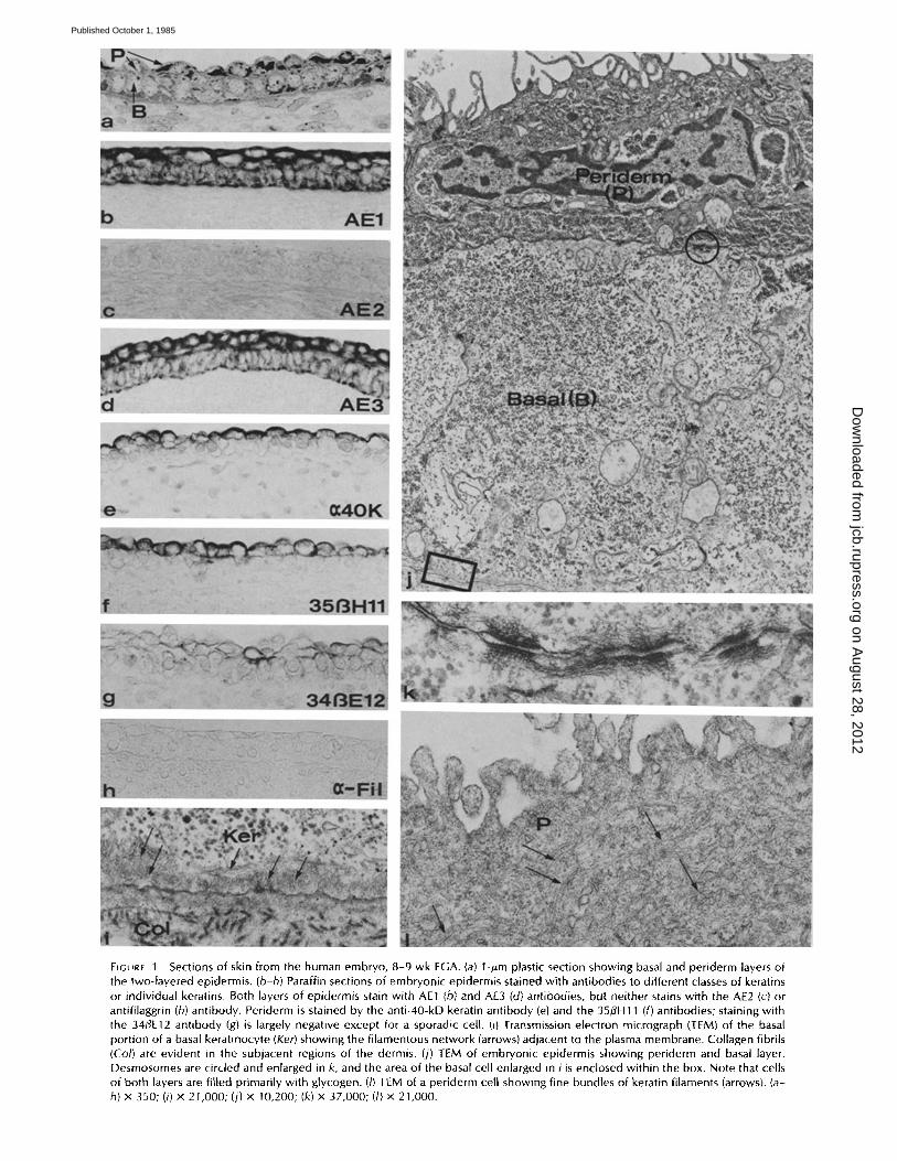
FIGURE 1 Sections of skin from the human embryo, 8-9 wk EGA. (a) l-#m plastic section showing basal and periderm layers of the two-layered epidermis. (b-h) Paraffin sections of embryonic epidermis stained with antibodies to different classes of keratins or individual keratins. Both layers of epidermis stain with AE1 (b) and AE3 (d) antibodies, but neither stains with the AE2 (c) or antifilaggrin (h) antibody. Periderm is stained by the anti-40-kD keratin antibody (e) and the 35flH11 (f) antibodies; staining with the 34/~E12 antibody (g) is largely negative except for a sporadic cell. (i) Transmission electron micrograph (TEM) of the basal portion of a basal keratinocyte (Ker) showing the filamentous network (arrows) adjacent to the plasma membrane. Collagen fibrils (Co/) are evident in the subjacent regions of the dermis. (j) TEM of embryonic epidermis showing periderm and basal layer. Desmosomes are circled and enlarged in k, and the area of the basal cell enlarged in i is enclosed within the box. Note that cells of both layers are filled primarily with glycogen. (I) TEM of a periderm cell showing fine bundles of keratin filaments (arrows). (a- h) x 350; (i) x 21,000; (j) x 10,200; (k) x 37,000; (/) x 21,000.
on August 28, 2012
jcb.rupress.orgD
ownloaded from
Published October 1, 1985
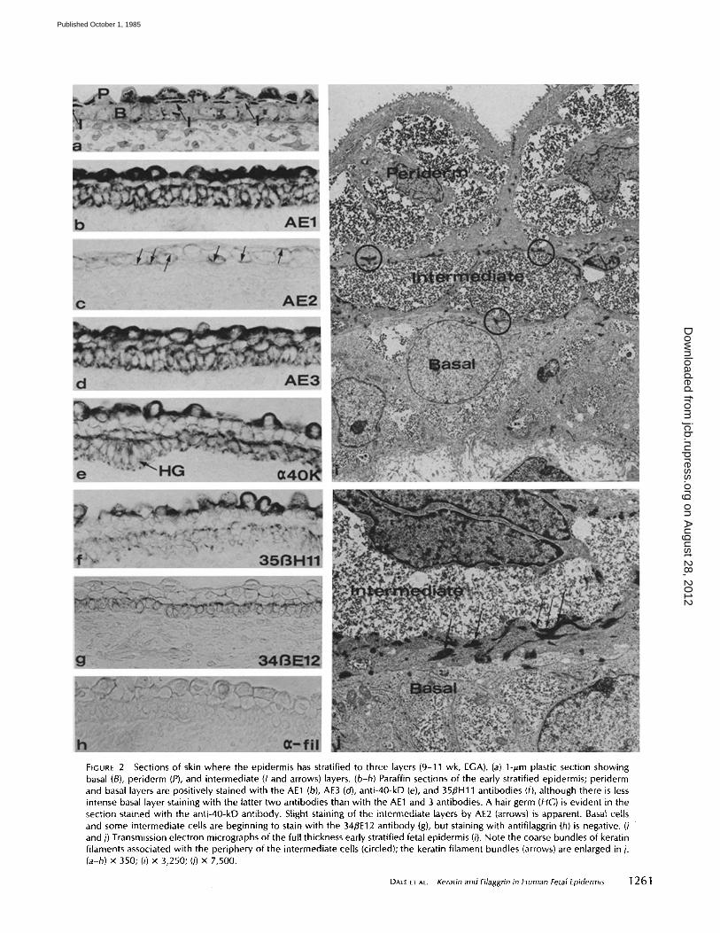
FIGURE 2 Sections of skin where the epidermis has stratified to three layers (9-11 wk, EGA). (a) 1-pm plastic section showing basal (B), periderm (P), and intermediate (I and arrows) layers. (b-h) Paraffin sections of the early stratified epidermis; periderm and basal layers are positively stained with the AE1 (b), AE3 (cO, anti-40-kD (e), and 35/3H11 antibodies (f), although there is less intense basal layer staining with the latter two antibodies than with the AE1 and 3 antibodies. A hair germ (HG) is evident in the section stained with the anti-40-kD antibody. Slight staining of the intermediate layers by AE2 (arrows) is apparent. Basal cells and some intermediate cells are beginning to stain with the 34BE12 antibody (g), but staining with antifilaggrin (h) is negative. (i and j) Transmission electron micrographs of the full thickness early stratified fetal epidermis (i). Note the coarse bundles of keratin filaments associated with the periphery of the intermediate cells (circled); the keratin filament bundles (arrows) are enlarged in j. (a-h) x 350; (i) x 3,250; (j) x 7,500.
DALE ET AL. Keratin and Filaggrin in Human Fetal Epidermis 1261
on August 28, 2012
jcb.rupress.orgD
ownloaded from
Published October 1, 1985
1262 THE JOURNAL OF CELL BIOLOGY • VOLUME 101, 1985
on August 28, 2012
jcb.rupress.orgD
ownloaded from
Published October 1, 1985
and 3513H 11, depending on specificity (Fig. 5). Weak staining of the 56.5- and 67-kD keratins by AE1 and 2 or AE3 and 2, respectively, suggests that these antigens may be present in small quantities.
The epidermis was clearly not yet keratinized at this stage as judged by electron microscopy (Fig. 2 i). The filaments in basal cells were still organized along the basal border, although the bundles associated with desmosomes were becoming larger and more prominent and extending further into the cytoplasm as the number of desmosomes increased. The filaments of the periderm layer formed bundles throughout the cytoplasm and were organized in a dense network beneath the apical margin of the cell. The bundles of keratin filaments in intermediate cells were organized in larger and more elec- tron-dense bundles than those in either periderm or basal cells (Fig. 2j). This corresponded with the greater number of desmosomes among cells of the intermediate layer and be- tween intermediate cells and adjacent layers than between cells within the basal layers or within the periderm layer (Fig. 2h).
Fetal Period: Follicular Keratinization
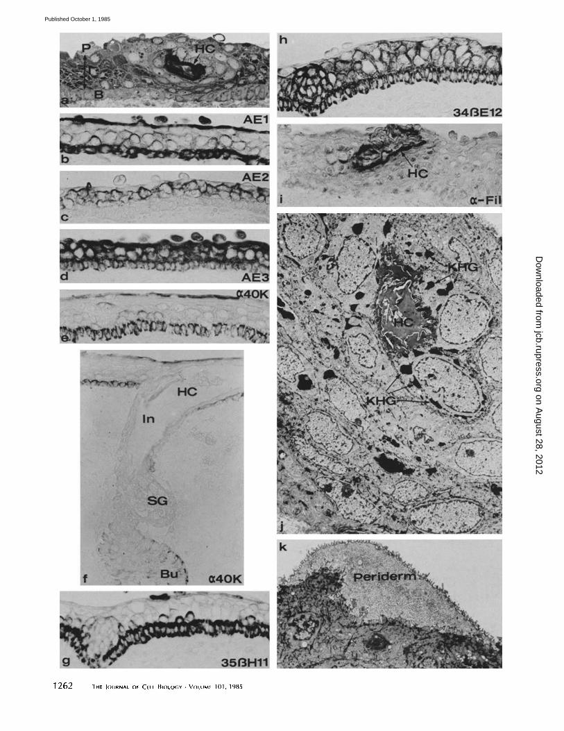
Shortly after the epidermis stratifies into three layers, at 12 wk, hair follicles begin to form, at first as hair germs (Fig.
2 e), then as elongated cords of cells that grow downward into the dermis (hair pegs and bulbous hair pegs). The follicle has concentric layers of cells, which, in terms of the differentiation of keratin filaments, correspond to basal and intermediate layers of the epidermis. At ~ 14 wk, the follicles on the trunk of the fetus (earlier on the head) begin to keratinize. Keratin- ized cells are recognized in developing hair, in cells (granular and cornified) that line the infundibulum of the follicle and its contiguous intraepidermal channel, the hair canal (Fig. 3, a, f h, and l). The interfoUicular epidermis is still covered by a complete layer of periderm (Fig. 3, a-d, k) and shows no indication of keratinization even though the epidermis has added an additional layer of intermediate cells and is at least four layers thick (Fig. 3, a-e).
The immunohistochemical staining (Fig. 3,b-i) showed that, as in earlier stages, the periderm stained with AE 1 (Fig. 3, b), AE3 (Fig. 3 d), anti-40-kD (Fig. 3, e and f ) keratin, and 35/3Hll (Fig. 3g) but was negative with AE2 (Fig. 3c) and 3413E 12 (Fig. 3 h). All of the keratinocytes of the interfollicular and follicular epidermis stained with AE3 and 34¢/E 12. Stain- ing by the AE2 antibody was restricted to the intermediate layers of the epidermis (Fig. 3 c) and to the suprabasal layers of the infundibulum and internal root sheath of the follicle. AE 1 stained a continuous basal layer of both the interfoUicular
epidermis and the external root sheath; a few cells of the intermediate layer also stained with AE1 (Fig. 3 b). The anti- 40-kD keratin stained periderm and basal cells, but interme- diate cells were negative. Beginning at this period, regional variation in the immunohistochemical staining of interfollic- ular areas and developing follicles could be seen with the anti- 40 kD, 35~H 11, AE2, and antifilaggrin antibodies. The outer cells of the hair germs were positive for the 40-kD keratin (Fig. 2e), but as development proceeded to hair peg and bulbous hair peg stages, the staining became fainter and was restricted to the bulge and to the cells of the outer root sheath distal to the bulge (Fig. 3f). Antibody 3513H11 staining was, in general, weaker in developing follicles than in the interfol- licular areas. Similarly, AE2 staining in the follicles was weaker than in the adjacent areas (data not shown). The patent portions of the hair canal were surrounded by keratin- ized cells, which stained with the antifilaggrin antibody (Fig. 3i).
Keratin filaments were more dense in the intermediate cells where they were associated with the desmosomes and also organized in large, electron-dense bundles in the cytoplasm. Basal cells contained fine bundles of keratin filaments in the cytoplasm which were evident primarily at the ultrastructural level. The filaments of the periderm cell were organized as in the earlier stages. The staining of filaggrin in the hair follicle corresponded to the presence of granular cells limited to that region and to the development of keratohyalin granules within those cells (Fig. 3j).
This period is distinguished by a marked increase and coordinated expression of the 67- and 56.5-kD keratins (Fig. 5). These keratins have been described by Sun et al. (53, 54) as markers of keratinizing epithelia. The 56.5-kD keratin is recognized by the AE 1 and AE2 antibodies, whereas the 67- kD keratin is stained by the AE2 and AE3 antibodies. For the first time in this developmental series, the antifilaggrin anti- body clearly identified a protein (often a doublet) on the immunoblot preparations at the 37-kD position characteristic for human filaggrin (Fig. 5). Filaggrin was detected at 15-16 wk EGA, after the dramatic increase in the 56.5- and 67-kD keratins. An apparent dimer of ~75 kD was also present, but other high molecular weight, immunoreactive proteins were not seen on the gels or blots until birth. Such high molecular weight proteins identified with the anti-filaggrin antibody are typically present on gels of rapidly extracted adult epidermal proteins and have been identified as the high molecular weight, precursor form of filaggrin (profilaggrin) (2, 22, 35, 43). Profilaggrin is extremely sensitive to proteolytic degra- dation, and therefore it may not be recovered in the fetal
FIGURE 3 Segments of skin from fetuses at ages where hair follicles are beginning to keratinize but the interfollicular epidermis is nonkeratinized, b-d are taken from specimens early in this period (~14 wk EGA), and other photos are from slightly older specimens (15-16 wk EGA) with more highly developed follicles. (a) 1-#m plastic section through a portion of the epidermis traversed by a hair canal (HC). Approximately three layers of intermediate cells (I) are shown between the continuous periderm (P) layer and the basal (B) layer. (b-i) Paraffin sections stained with antibodies to keratin filaments and filaggrin. Periderm and basal cells only were stained with AE1 (b), anti-40-kD (e and f), and 35~H11 (g) antibodies. The AE3 antibody (d) stained all layers of the epidermis. The anti-40-kD antibody also stained cells of the bulge (Bu) but not cells of the infundibulum (In), hair canal (HC), or sebaceous gland (SG). AE2 (c) stained the intermediate cells but spared periderm and basal cells. 34~E12 (h) stained all layers except the periderm. Cells surrounding the hair canal (HC) were positively stained with the antifilaggrin antibody (i). (j and k) Transmission electron micrographs through a hair canal (HC) as shown in a illustrating the presence of large keratohyalain granules (KHG). The periderm is complete over the surface of the epidermis (k), and abundant keratin filament bundles (arrows) are seen in the cytoplasm of subjacent epidermal cells. (a-i) x 350; (j) x 2,650; (k) x 8,000.
DALe et AL. Keratin and Filaggrin in Human Fetal Epidermis 1263
on August 28, 2012
jcb.rupress.orgD
ownloaded from
Published October 1, 1985
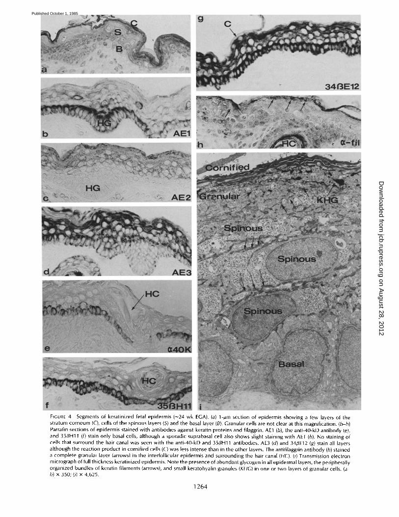
FIGURE 4 Segments of keratinized fetal epidermis (~24 wk EGA). (a) 1-~m section of epidermis showing a few layers of the stratum corneum (C), cells of the spinous layers (S) and the basal layer (B). Granular cells are not clear at this magnification. (b-h) Parrafin sections of epidermis stained with antibodies against keratin proteins and filaggrin. AE1 (b), the anti-40-kD antibody (e),. and 35flH11 (f) stain only basal cells, although a sporadic suprabasal cell also shows slight staining with AE1 (b). No staining of cells that surround the hair canal was seen with the anti-40-kD and 35flH11 antibodies. AE3 (d) and 34~E12 (g) stain all layers although the reaction product in cornified cells (C) was less intense than in the other layers. The antifilaggrin antibody (h) stained a complete granular layer (arrows) in the interfollicular epidermis and surrounding the hair canal (HC). (i) Transmission electron micrograph of full thickness keratinized epidermis. Note the presence of abundant glycogen in all epidermal layers, the peripherally organized bundles of keratin filaments (arrows), and small keratohyalin granules (KHG) in one or two layers of granular cells. (a- h) x 350; (i) x 4,625.
1264
on August 28, 2012
jcb.rupress.orgD
ownloaded from
Published October 1, 1985
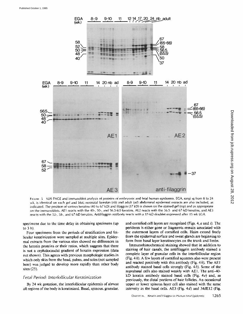
FIGURE 5 SDS PAGE and immunoblot analysis of proteins of embryonic and fetal human epidermis. EGA, rangi ig from 8 to 24 wk, is identical on each gel and blot; neonatal foreskin (nb) and adult (ad) abdominal epidermal extracts are also included, as indicated. The position of various keratins (40 to 67 kD) and filaggrin (37 kD) is shown on the stained gel (top) and as appropriate on the immunoblots. AE1 reacts with the 40-, 50-, and 56.5-kD keratins, AE2 reacts with the 56.5- and 67-kD keratins, and AE3 reacts with the 52-, 58-, and 67-kD keratins. Antifilaggrin antibody reacts with a 37-kD doublet expressed after 15 wk EGA.
specimens due to the time delay in obtaining specimens (up to 3 h).
Four specimens from the periods of stratification and fol- licular keratinization were sampled at multiple sites. Epider- mal extracts from the various sites showed no differences in the keratin proteins or their ratios, which suggests that there is not a cephalocaudal gradient of keratin expression (data not shown). This agrees with previous morphologic studies in which only skin from the head, palms, and soles (not sampled here) was judged to develop more rapidly than other body sites (25).
Fetal Period: Interfollicular Keratinization
By 24 wk gestation, the interfollicular epidermis of almost all regions of the body is keratinized. Basal, spinous, granular,
and cornified cell layers are recognized (Figs. 4, a and i). The periderm is either gone or fragments remain associated with the outermost layers of cornified cells. Hairs extend freely from the epidermal surface and sweat glands are beginning to form from basal layer keratinocytes on the trunk and limbs.
Immunohistochemical staining showed that in addition to staining of hair canals, the antifilaggrin antibody stained a complete layer of granular cells in the interfollicular region (Fig. 4 h). A few layers of cornified squames also were present and reacted positively with this antibody (Fig. 4h). The AEI antibody stained basal cells strongly (Fig. 4b). Some of the suprabasal cells also stained weakly with AEI. The anti-40- kD keratin antibody stained basal cells (Fig. 4e) and, as previously, the distal portions of hair follicles. An occasional upper or lower spinous layer cell also stained with the same intensity as the basal cells. AE3 (Fig. 4d) and 34~E12 (Fig.
DALE ET AL. Keratin and Filaggrin in Human Fetal Epidermis 1265
on August 28, 2012
jcb.rupress.orgD
ownloaded from
Published October 1, 1985
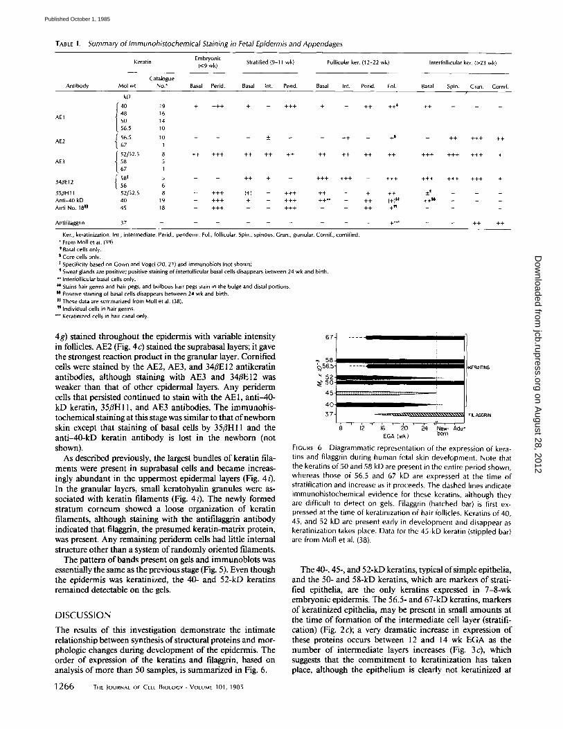
TABLE I. Summary of Immunohistochemical Staining in Fetal Epidermis and Appendages
Embryon ic Kerat in (<9 wk ) Strati f ied (9-11 wk ) Fol l icu lar ker. (12 -22 wk ) In ter fo l l icu lar ker. (>23 w k l
Catalogue Antibody Mol wt No.* Basal Perid. Basal Int. Perid. Basal Int. Perid. FoL Basal Spin. Gran. Corni f .
kD /
40 19 + + + + + - + + + + - ++ ++* ++ - -
AE1 48 16 50 14
56.5 10
AE2 ~" 56.5 10 - - - _ - - + + - +~ - + + + + + + +
t 67 1
t 52[52.5 8 + + + + + + + + + + + + + + + + + + + + + + + + + + + + + AE3 58 5
67 1
581 5 - - + + + - + + + + + + - +4 -+ ++4- +4-4- 4 -++ + 34/~E12 56 6
3 5 # H t 1 52152.5 8 _ + + + (+) _ + + + + + _ + + + +4 _ _ _
A n t i - 4 0 kD 40 19 _ + + + + _ + + + + + ~ _ + + (+)*t, + + ~ _ _ _ Anti-No. 18 It 4 5 1 B I + + + - - I + + + I I + + + [ I - - - -
Antifilaggrin 37 . . . . . . . . . +*~" - - ++ ++
Ker., keratinization. Int., intermediate, Perid., periderm. Fol., follicular. Spin., spinous. Gran., granular. Cornif., cornified. * From Moll et al. (39). * Basal cells only.
Core cells only. I Specificity based on Gown and Vogel (20, 21) and immunoblots (not shown}.
Sweat glands are positive; positive staining of interfollicutar basal cells disappears between 24 wk and birth. ** Intertollicular basal cells only. ~* Stains hair germs and hair pegs, and bulbous hair. pegs stain in the bulge and distal portions. II Positive staining of basal cells disappears between 24 wk and birth, II These data are summarized from Moll et al. (38). t'l Individual cells in hair germs.
Keratinized cells in hair canal only.
4g) stained throughout the epidermis with variable intensity in follicles. AE2 (Fig. 4 c) stained the suprabasal layers; it gave the strongest reaction product in the granular layer. Cornified cells were stained by the AE2, AE3, and 34/~E 12 antikeratin antibodies, although staining with AE3 and 34#E12 was weaker than that of other epidermal layers. Any periderm cells that persisted continued to stain with the AE1, anti-40- kD keratin, 35#H1 l, and AE3 antibodies. The immunohis- tochemical staining at this stage was similar to that of newborn skin except that staining of basal cells by 35#H11 and the anti-40-kD keratin antibody is lost in the newborn (not shown).
As described previously, the largest bundles of keratin fila- ments were present in suprabasal cells and became increas- ingly abundant in the uppermost epidermal layers (Fig. 4 i). In the granular layers, small keratohyalin granules were as- sociated with keratin filaments (Fig. 4i). The newly formed stratum corneum showed a loose organization of keratin filaments, although staining with the antifilaggrin antibody indicated that filaggrin, the presumed keratin-matrix protein, was present. Any remaining periderm cells had little internal structure other than a system of randomly oriented filaments.
The pattern of bands present on gels and immunoblots was essentially the same as the previous stage (Fig. 5). Even though the epidermis was keratinized, the 40- and 52-kD keratins remained detectable on the gels.
D I S C U S S I O N
The results of this investigation demonstrate the intimate relationship between synthesis of structural proteins and mor- phologic changes during development of the epidermis. The order of expression of the keratins and filaggrin, based on analysis of more than 50 samples, is summarized in Fig. 6.
67-
58 ~565-
~52
45-
40
57-
F IGURE 6
. . . . . I I IIII III II I l l l I ~R~T~S
I I -
r , , , , , , , , f f , 8 12 16 2 0 24 New- ,hdult
EGA ( w k ) born
D i a g r a m m a t i c r e p r e s e n t a t i o n o f t h e e x p r e s s i o n o f k e r a -
tins and filaggrin during human fetal skin development. Note that the keratins of 50 and 58 kD are present in the entire period shown, whereas those of 56.5 and 67 kD are expressed at the time of stratification and increase as it proceeds. The dashed lines indicate immunohistochemical evidence for these keratins, although they are difficult to detect on gels. Filaggrin (hatched bar) is first ex- pressed at the time of keratinization of hair follicles. Keratins of 40, 45, and 52 kD are present early in development and disappear as keratinization takes place. Data for the 45-kD keratin (stippled bar) are from Moll et al. (38).
The 40-, 45-, and 52-kD keratins, typical of simple epithelia, and the 50- and 58-kD keratins, which are markers of strati- fied epithelia, are the only keratins expressed in 7-8-wk embryonic epidermis. The 56.5- and 67-kD keratins, markers of keratinized epithelia, may be present in small amounts at the time of formation of the intermediate cell layer (stratifi- cation) (Fig. 2c); a very dramatic increase in expression of these proteins occurs between 12 and 14 wk EGA as the number of intermediate layers increases (Fig. 3c), which suggests that the commitment to keratinization has taken place, although the epithelium is clearly not keratinized at
1266 THE }OURNAL OF CELL BIOLOGY , VOLUME 101 , 1 9 8 5
on August 28, 2012
jcb.rupress.orgD
ownloaded from
Published October 1, 1985
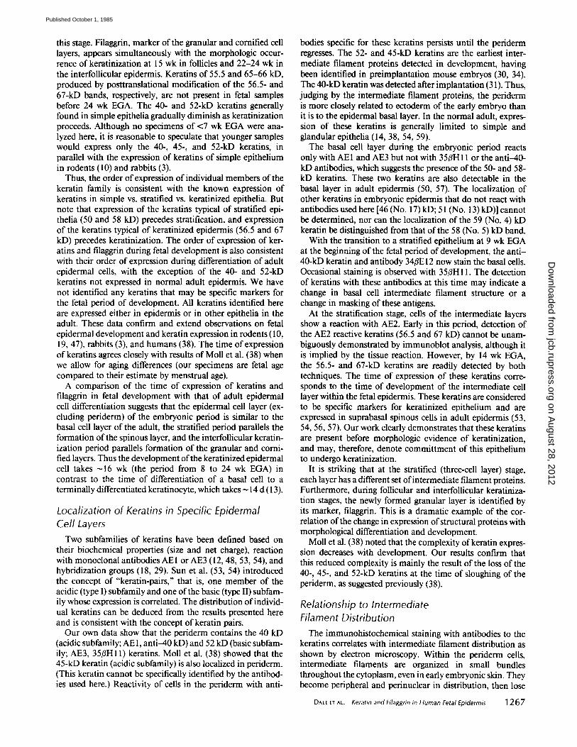
this stage. Filaggrin, marker of the granular and cornified cell layers, appears simultaneously with the morphologic occur- rence of keratinization at 15 wk in follicles and 22-24 wk in the interfollicular epidermis. Keratins of 55.5 and 65-66 kD, produced by posttranslational modification of the 56.5- and 67-kD bands, respectively, are not present in fetal samples before 24 wk EGA. The 40- and 52-kD keratins generally found in simple epithelia gradually diminish as keratinization proceeds. Although no specimens of <7 wk EGA were ana- lyzed here, it is reasonable to speculate that younger samples would express only the 40-, 45-, and 52-kD keratins, in parallel with the expression of keratins of simple epithelium in rodents (10) and rabbits (3).
Thus, the order of expression of individual members of the keratin family is consistent with the known expression of keratins in simple vs. stratified vs. keratinized epithelia. But note that expression of the keratins typical of stratified epi- thelia (50 and 58 kD) precedes stratification, and expression of the keratins typical of keratinized epidermis (56.5 and 67 kD) precedes keratinization. The order of expression of ker- atins and filaggrin during fetal development is also consistent with their order of expression during differentiation of adult epidermal cells, with the exception of the 40- and 52-kD keratins not expressed in normal adult epidermis. We have not identified any keratins that may be specific markers for the fetal period of development. All keratins identified here are expressed either in epidermis or in other epithelia in the adult. These data confirm and extend observations on fetal epidermal development and keratin expression in rodents (10, 19, 47), rabbits (3), and humans (38). The time of expression of keratins agrees closely with results of Moll et al. (38) when we allow for aging differences (our specimens are fetal age compared to their estimate by menstrual age).
A comparison of the time of expression of keratins and filaggrin in fetal development with that of adult epidermal cell differentiation suggests that the epidermal cell layer (ex- cluding periderm) of the embryonic period is similar to the basal cell layer of the adult, the stratified period parallels the formation of the spinous layer, and the interfollicular keratin- ization period parallels formation of the granular and corni- fled layers. Thus the development of the keratinized epidermal cell takes ~16 wk (the period from 8 to 24 wk EGA) in contrast to the time of differentiation of a basal cell to a terminally differentiated keratinocyte, which takes ~ 14 d (13).
Localization of Keratins in Specific Epidermal Cell Layers
Two subfamilies of keratins have been defined based on their biochemical properties (size and net charge), reaction with monoclonal antibodies AEI or AE3 (12, 48, 53, 54), and hybridization groups (18, 29). Sun et al. (53, 54) introduced the concept of "keratin-pairs," that is, one member of the acidic (type I) subfamily and one of the basic (type II) subfam- ily whose expression is correlated. The distribution of individ- ual keratins can be deduced from the results presented here and is consistent with the concept of keratin pairs.
Our own data show that the periderm contains the 40 kD (acidic subfamily; AE 1, anti-40 kD) and 52 kD (basic subfam- ily; AE3, 35~H11) keratins. Moll et al. (38) showed that the 45-kD keratin (acidic subfamily) is also localized in periderm. (This keratin cannot be specifically identified by the antibod- ies used here.) Reactivity of cells in the periderm with anti-
bodies specific for these keratins persists until the periderm regresses. The 52- and 45-kD keratins are the earliest inter- mediate filament proteins detected in development, having been identified in preimplantation mouse embryos (30, 34). The 40-kD keratin was detected after implantation (31). Thus, judging by the intermediate filament proteins, the periderm is more closely related to ectoderm of the early embryo than it is to the epidermal basal layer. In the normal adult, expres- sion of these keratins is generally limited to simple and glandular epithelia (14, 38, 54, 59).
The basal cell layer during the embryonic period reacts only with AEI and AE3 but not with 35/3H11 or the anti-40- kD antibodies, which suggests the presence of the 50- and 58- kD keratins. These two keratins are also detectable in the basal layer in adult epidermis (50, 57). The localization of other keratins in embryonic epidermis that do not react with antibodies used here [46 (No. 17) kD; 51 (No. 13) kD)] cannot be determined, nor can the localization of the 59 (No. 4) kD keratin be distinguished from that of the 58 (No. 5) kD band.
With the transition to a stratified epithelium at 9 wk EGA at the beginning of the fetal period of development, the anti- 40-kD keratin and antibody 34/3E12 now stain the basal cells. Occasional staining is observed with 35/3H11. The detection of keratins with these antibodies at this time may indicate a change in basal cell intermediate filament structure or a change in masking of these antigens.
At the stratification stage, cells of the intermediate layers show a reaction with AE2. Early in this period, detection of the AE2 reactive keratins (56.5 and 67 kD) cannot be unam- biguously demonstrated by immunoblot analysis, although it is implied by the tissue reaction. However, by 14 wk EGA, the 56.5- and 67-kD keratins are readily detected by both techniques. The time of expression of these keratins corre- sponds to the time of development of the intermediate cell layer within the fetal epidermis. These keratins are considered to be specific markers for keratinized epithelium and are expressed in suprabasal spinous cells in adult epidermis (53, 54, 56, 57). Our work clearly demonstrates that these keratins are present before morphologic evidence of keratinization, and may, therefore, denote committment of this epithelium to undergo keratinization.
It is striking that at the stratified (three-cell layer) stage, each layer has a different set of intermediate filament proteins. Furthermore, during follicular and interfollicular keratiniza- tion stages, the newly formed granular layer is identified by its marker, filaggrin. This is a dramatic example of the cor- relation of the change in expression of structural proteins with morphological differentiation and development.
Moll et al. (38) noted that the complexity of keratin expres- sion decreases with development. Our results confirm that this reduced complexity is mainly the result of the loss of the 40-, 45-, and 52-kD keratins at the time of sloughing of the periderm, as suggested previously (38).
Relationship to Intermediate Filament Distribution
The immunohistochemical staining with antibodies to the keratins correlates with intermediate filament distribution as shown by electron microscopy. Within the periderm cells, intermediate filaments are organized in small bundles throughout the cytoplasm, even in early embryonic skin. They become peripheral and perinuclear in distribution, then lose
DALE e t A t . Keratin and Filaggrin in Human Fetal Epidermis 1267
on August 28, 2012
jcb.rupress.orgD
ownloaded from
Published October 1, 1985
their bundled organization and become randomly distributed as the periderm cell develops a cornified cell envelope and regresses. Staining with antibodies AE 1, AE3, 35~H 11, and anti-40-kD keratin mimics this distribution. In contrast, in- termediate filaments in the basal cells are fine, organized in small bundles, less numerous than in the other cell layers, and stain less intensely With AEI and AE3 than do those in the periderm. There are fewer changes in the amount, struc- ture, staining pattern, and organization of keratin filaments in the basal cells than in any other cell layer and less change in the quantity of the 50- and 58-kD keratins than in the other keratin polypeptides.
Before 12 wk EGA, the immunostaining reaction within the intermediate cells is weak and primarily located at the cell periphery, consistent with the peripheral distribution of inter- mediate filaments as seen by electron microscopy. After ~ 12 wk EGA, the quantity of intermediate filament bundles in the upper cell layers increases. These filaments are organized in larger bundles than in the basal cells, undergo the most change during development, and stain with the greatest inten- sity relative to other cell layers.
Potential Applications This work establishes the normal time of expression of
specific proteins associated with epidermal keratinization in normal human fetal development. All of the keratins and filaggrin are present by 14-16 wk EGA. This is when amni- ocentesis is performed for diagnosis of genetic disorders. It is known that many of the cells present in amniotic fluid express keratins (42). The results of this study will serve as a base line against which to evaluate protein abnormalities in fetuses at risk for genetic diseases of the epidermis that are expressed before birth. These disorders may be amenable to early diag- nosis via epidermal cells present in amniotic fluid. At the present, some severe genetic disorders that affect the epidermis are diagnosed by morphologic criteria using biopsies obtained by the more difficult and invasive method of fetoscopy, performed at 19-21 wk EGA (1). The potential for diagnosis of epidermolytic hyperkeratosis via morphologic characteris- tics of amniotic fluid cells has already been suggested (1 I, 27). Biochemical analysis and immunohistochemical staining of amniotic fluid cells may be a valuable adjunct to diagnosis of genetic disorders of keratinization in which the expression of keratins and filaggrin is altered.
W e g r a t e f u l l y a c k n o w l e d g e J u l i e S c o f i e l d , A l e x i s L y n l e y , a n d M a r i e
D o m a n f o r e x c e l l e n t t e c h n i c a l a s s i s t a n c e w i t h v a r i o u s a s p e c t s o f t h i s
w o r k .
T h i s w o r k w a s s u p p o r t e d b y U . S. P u b l i c H e a l t h S e r v i c e g r a n t s
H D 1 7 6 6 4 a n d A M 2 1 5 5 7 f r o m t h e N a t i o n a l I n s t i t u t e s o f H e a l t h .
R e c e i v e d f o r p u b l i c a t i o n 4 M a r c h 1985 , a n d in r e v i s e d f o r m 2 5 A p r i l
1985 .
REFERENCES
1. Anton-Lamprecht, 1. 1982. Prenatal diagnosis of genetic disorders of the skin by means of electron microscopy. Hum. Genet. 59:392--405.
2. Ball, R. C., G. K. Walker, and I. A. Bernstein. 1978. Histidine-rich proteins as molecular markers of epidermal differentiation. J. BioL Chem. 253:5861-5868.
3. Banks-schlegel, S. P. 1982. Keratin alterations during embryonic epidermal differentia- tion: a presage of adult epidermal maturation. J. CellBiol. 93:551-559.
4. Bowden, P. E., and W. J. Cunliffe. 1981. Modifications of human prekeratin during epidermal differentiation. Biochem. J. 199:145-154.
5. Dale, B. A. 1977. Purification and characterization of a basic protein from the stratum corneum of mammalian epidermis. Biochim. Biophys. Acta. 491 : 193-204.
1268 THE JOURNAL Of CELL BIOLOGY • VOLUME I01, 1985
6. Dale, B. A., and S. Y. Ling. I979. Evidence of a precursor form of stratum corneum basic protein in rat epidermis. Biochemistry. 18:3539-3546.
7. Dale, B. A., and S. Y. Ling. 1979. Immunologic cross-reaction of stratum corneum basic protein and a keratohyalin granule protein. J. Invest. DermatoL 72:257-261.
8. Dale, B. A., K. A. Holbrook, and P. M. Steinert. 1978. Assembly of stratum corneum basic protein and keratin filaments in macrofibrils. Nature (Lond.). 276:729-73 I.
9. Dale, B. A., K. A. Resing, and J. D. Lonsdale-Eccles. 1985. Filaggrin, a keratin filament assooated protein. Ann. NYAcad Sci. In press.
10. Dale, B. A., I. B. Stern, M. Rabin, and L.-Y. Huang. 1976. The identification of fibrnus proteins in fetal rat epidermis by electrophoretic and immunologic techniques..L Invest. Dermatol. 66:230-235.
l 1. Eady, R. A. J., D. B. Gunner, and I. M. Leigh. 1984. Detection of tonofilament clumping in epidermal and amniotic fluid cells enables prenatal diagnosis of hullous ichthyosiform erythroderma (BIE). J. Cutaneous Pathology. 11:212.
12. Eichner, R., Bonitz, P., and Sun, T.-T. 1984. Classification of epidermal keratins according to their immunoreactivity, isoelectric point, and mode of exp~ssion. J. Cell BioL 98:1388-1396.
13. Epstein, W., and A. Maibach. 1965. Cell renewal in human epidermis. Arch. Dermatol 92:462--468.
14. Franke, W. W., K. Weber, M. Osborn, E. Schmid, and C. Freudenstein. 1978. Antibody to prekeratin: decoration of tonofilament-like arrays in various cells of epithelial char- acter. Exp. Cell Res. 116:429-445.
15. Fmnke, W. W., D. L. SchiLler, R. Moll, S. Winter, E. Schmid, I. Engelbrecbt, H. Denk, R. Krepler, and E. Plalzer. 1981. Diversity of cytokeratins: differentiation-specific expression of cytokeratin polypeptides in epithelial cells and tissues. J. Mol BJol 153:933-959.
16. Freinkel, R. K., and K. A. Wier. 1975. Changing patterns of incorporation of t4C histidine and 3H leucine into epidermal proteins during differentiation of fetal rat skin. J. Invest. DermatoL 65:482-487.
17. Fuchs, E., and H. Green. 1980. Changes in keratin gene expression during terminal differentiation of the keratinocyte. Cell. 19:1033-1042.
18. Fuchs, E., S. M. Coppock, H. Green, and D. W. Cleveland. 1981. Two distinct classes of keratin genes and their evolutionary significance. Cell. 27:75-84.
19. Fukuyama, K., L Marshburn, and W. L. Epstein. 1981. Histidine-rich protein in developing rat epidermis. Dev. BioL 81:201-207.
20. Gown, A. M., and A. M. Vogel. 1982. Monoclonal antibodies to intermediate filament proteins of human cells: unique and cross-reacting antibodies. J. Cell Biol. 95:414-424.
21. Gown, A. M., and A. M Vogel. 1984. Monoclonal antibodies to human intermediate filament proteins II. Distribution of filament proteins in normal human tissues. Am. J Pathol. 114:309-321.
22. Harding, C. R., and 1. R. Scott. 1983. Histidine-rich proteins (filaggnns): structural and functional heterogeneity during epidermal differentiation..L MoL Biol. 170:651-673.
23. Holbrook, K.A.,andG. F. Odland. 1975. Finestrnctureofdevetopinghumanepidermis: light, scanning and transmission electron microscopy of the periderm. ,L Invest. Der- matol. 65:16-38.
24. Holbrook, K. A., and G. F. Odland. 1978. Structure of the human fetal hair canal and initial hair eruption, d. Invest Dermatol. 71:385-390.
25. Holbrook, K. A., and G. F. Odland. 1980. Regional development of the human epidermis in the first trimester embryo and the second trimester fetus (ages related to the timing of amniocentesis and fetal biopsy). Z Invest. Dermatol. 80: l 61-168.
26. Holbrook, K. A., and L. T. Smith. 198t. Ultrastructural aspects of human skin dunng the embryonic, fetal, premature, neonatal, and adult periods of life. Birth Defects: Orig. Attic. Ser. XVII (2):9-38.
27. Holbrook, K. A., B. A. Dale, V. P. Sybert, and R. W. Sagebiel. 1983. Epidermolytic hyperkeratosis: ultrastrncture and biochemistry of skin and amniotic fluid cells from two affected fetuses and a newborn infant..L Invest. Dermatol. 80:222-227.
28. Hsu, S. M., L. Raine, and H. Fanger. 1981. Use of avidin-biotin-peroxidase complex (ABC) in immunoperoxidase techniques: a comparison between ABC and unlabeled antibody (PAP) procedures. J. Histochem. Cytochem. 29:577-580.
29. Kim, K. H., J. Rheinwald, and E. V. Fuchs. 1983. Tissue specificity of epithelial keratins: differential expression of mRNAs from two multiple families. MoL Cell. Biol 3:495- 502.
30. Jackson, B. W., C. Grund, S. Winter, W. W. Franke, and K. lllmensee. 1981. Formation of cytoskeletal elements during mouse embryogenesis. II. Epithelial differentiation and intermediate-sized filaments in early postimplantation embryos. Differentiation 20:203- 216.
31. Jackson, B. W., C. Grand, E. Schmid; K. Bark, W. W. Franke, and K. Illmensee. 1980. Formation of cytoskeletal elements during mouse embryogenesis: intermediate filaments of the cytokeratin type and desmosomes in preimplantation embryos. Differentiation. 17:161-179.
32. Laemmli, U. K. 1970. Cleavage of structural proteins during the assembly of the head of the bacteriophage T4. Nature (Lond.) 226:680-685.
33. Lazarides, E. 1982. Intermediate filaments: a chemically heterogeneous, developmentally regulated class of proteins. Annu. Rew. Biochem. 51:219-250.
34. Lehtonen, E., V. P. Lehto, T. Vartio, R. A. Badley, and 1. Virtanen. 1983. Expression of cytokeratin polypeptides in mouse oocytes and preimplantation embryos. Dew. BioL 100:158-165.
35. Londale-Eeeles, J. D., K. A. Resing, R. L. Meek, and B. A. Dale. 1984. High-molecular weight precursor of epidermal filaggnn and hypothesis for its tandem repeating structure. Biochemistry. 23:1239-1245.
36. Lult, J. H. 1961. Improvements in epoxy resin embedding methods. J Biophys. Biochem. CytoL 9:409-414.
37. Lynley, A. M., and B. A. Dale. 1983. Characterization of human epidermal filaggnn, a histidine-rich, keratin filament-aggregating protein. Biochim. Biophys. Acta. 744:28-35.
38. Moll, R., I. Moll, and W. Wiest. 1983. Changes in the pattern ofcytokemtin polypeptides in epidermis and hair follicles during skin development in human fetuses. Differentiation 23:170-178.
39. Moll, R., W. W. Franke, D. L Schiller, B. Gieger, and R. Krepler. 1982. The catalog of human cytokeratins: patterns of expression in normal epithelia, tumors and cultured cells. Cell. 31:11-24.
40. Nelson, W. G., and T.-T. Sun. 1983. The 50- and 58-kdalton keratin classes as molecular markers for stratified squamous epithelia: cell culture studies..1. ('ell Biol. 97:224-251.
41. Ochs, D. 1983. Protein contaminants of SDS-polyacrylamide gels. Anal. Biochem 135:470474.
42. Ochs, B. A., W. W. Franke, R. Moll, C. Grund, M. Cremer, and T. Cremer. 1983. Epithelial character and morphologic diversity of cell cultures from human amniotic fluids examined by immunofluorescence microscopy and gel electrophoresis of cyto- skeletal proteins. Differentiation. 24:153-173.
on August 28, 2012
jcb.rupress.orgD
ownloaded from
Published October 1, 1985
43. Ramsden, M., D. Loehren, and A. Balmain. 1983. Identification of a rapidly labeled 350k histidine-rich protein in neonatal mouse epidermis. Differentiation. 23:243-249.
44. Reynolds, E. S. 1963. The use of lead citrate at high pH as an electron opaque slain in electron microscopy. ,L Cell BioL 17:208-212.
45. Richardson, K. C., L. Jarrett, and E. H. Finke. 1960. Embedding in epoxy resins for ultrathin sectioning in electron microscopy. Stain Technol. 35:313-323.
46. Roop, D. R., P. Hawley-Nelson, C. K. Cheng, and S. H. Yuspa. 1983. Keratin gene expression in mouse epidermis and cultured epidermal cells. Proc. NatL Acad Sci. USA. 80:716-720.
47. Schweizer, J. andH. Winter. 1982. Keratinpolypeptideanalysisin felalandin terminally differentiating newborn mouse epidermis. Differentiation. 22:19-24.
48. Schiller, D. L., W. W. Franke, and G. Geiger. 1982. A subfamily of rclatively large and basic cytokeratin polypeptides as defined by peptide mapping is represented by one or several polypeptides in epithelial cells. EMBO (Eur. Mol. Biol. Organ.) J. 1:761-769.
49. Sibrack, L. A., R. H. Gray, and I. A. Bernstein. 1974. Localization of the histidine-rich protein in keratohyalin: a morphologic and macromolecular marker in epidermal differentiation. J. Invest. Dermatol. 62:394-405.
50. Skerrow, D., and C. J. Skerrow. 1983. Tonofilament differentiation in human epidermis: isolation and polypeptide chain composition of keratinocyte subpopulation. Exp. Cell Res. 143:27-35.
5 I. Steinert, P. M., J. S. Cantieri, D. C. Teller, J. D. Lonsdale-Eccles, and B. A. Dale. 1981~ Characterization of a class of cationic proteins that specifically interact with intermediate filaments. Prtx'. Natl. Acad Sci. USA. 78:40974101.
52. Sun, T.-T. and H. Green. 1978. lmmunofiuorescent staining of keratin fibers in cultured cells. Cell. 14:469-476.
53. Sun, T.-T., R. Eichner, A. Schermer, D. Cooper, W. G. Nelson, and R. A. Weiss. 1984. Classification, expression, and possible mechanisms of evolution of mammalian epithe- lial keratins: a unifying model. In Cancer Cell. Vol. 1. The Transformed Phenotype. A. Levine, W. Topp, G. van de Woude, and J. D. Watson, editors. Cold Spring Harbor Laboratory, New York. 169-176.
54. Sun, T.-T., R. Eichner, W. G. Nelson. S. C. G. Tseng, R. A. Weiss, M. Jarvinen, and J. Woodcock-Mitchell. 1983. Keratin classes: molecular markers for different types of epithelial differentiation. J. Invest. DermatoL 81:109s--I 15s.
55. Towbin, H., T. Staehelin, and J. Gordon. 1979. Electrophorctic transfer of proteins from polyacrylamide gels to nitrocellulose sheets: procedure and some applications. Proc. Natl. Acad Sci. USA. 76:4350--4354.
56. Tseng, S. C. G., M. Jarvinen, W. G. Nelson, H. W. Huang, J. Woodcock-Mitchell, and T.-T. Sun. 1982. Correlation of specific keratins with different types of epithelial differentiation: monoclonal antibody studies. Cell. 30:361-372.
57. Woodcock-Mitchell, J., R. Eichner, W. G. Nelson, and T.-T. Sun. 1982. lmmunolocal- ization of keratin polypeptides in human epidermis using monoclonal antibodies. J. Cell BioL 95:580-588.
58. Wu, Y. and J. G. Rheinwald. 1981. A new small (40 Kd) keratin filament protein made in some cultured human squamous cell carcinomas. Cell. 25:627-635.
59. Wu, Y., L. Parker, N. Binder, M. Beckett, J. Sinard, C. Griffiths, and J. Rheinwald. 1982. The mesothelial keratins: a new family of cytoskeletal proteins identified in cultured mesothelial cells and nonkeratinizing epithelia. Cell. 31:693-703.
DALE ET AL. Keratin and Filaggrin in Human Fetal Epidermis 1269
on August 28, 2012
jcb.rupress.orgD
ownloaded from
Published October 1, 1985