Embed Size (px)
Citation preview
Ivančić-Baće, Ivana and Cass, Simon and Wearne, Stephen J. and Bolt, Edward L. (2015) Different genome stability proteins underpin primed and naïve adaptation in E. coli CRISPR-Cas immunity. Nucleic Acids Research . pp. 1-10. ISSN 1362-4962
Access from the University of Nottingham repository: http://eprints.nottingham.ac.uk/31310/1/Ivancic-Bace%20et%20al%20CRISPR%202015%20NAR.pdf
Copyright and reuse:
The Nottingham ePrints service makes this work by researchers of the University of Nottingham available open access under the following conditions.
· Copyright and all moral rights to the version of the paper presented here belong to
the individual author(s) and/or other copyright owners.
· To the extent reasonable and practicable the material made available in Nottingham
ePrints has been checked for eligibility before being made available.
· Copies of full items can be used for personal research or study, educational, or not-
for-profit purposes without prior permission or charge provided that the authors, title and full bibliographic details are credited, a hyperlink and/or URL is given for the original metadata page and the content is not changed in any way.
· Quotations or similar reproductions must be sufficiently acknowledged.
Please see our full end user licence at: http://eprints.nottingham.ac.uk/end_user_agreement.pdf
A note on versions:
The version presented here may differ from the published version or from the version of record. If you wish to cite this item you are advised to consult the publisher’s version. Please see the repository url above for details on accessing the published version and note that access may require a subscription.
For more information, please contact [email protected]
Nucleic Acids Research, 2015 1
doi: 10.1093/nar/gkv1213
Different genome stability proteins underpin primedand naıve adaptation in E. coli CRISPR-Cas immunity
Ivana Ivancic-Bace1, Simon D Cass2, Stephen J Wearne2 and Edward L Bolt2,*
1Faculty of Science, Department of Molecular Biology, University of Zagreb, Horvatovac 102a, Zagreb, Croatia and2School of Life Sciences, Queen’s Medical Centre, University of Nottingham, NG72UH, UK
Received July 19, 2015; Revised October 26, 2015; Accepted October 28, 2015
ABSTRACT
CRISPR-Cas is a prokaryotic immune system builtfrom capture and integration of invader DNA intoCRISPR (Clustered Regularly Interspaced ShortPalindromic Repeats) loci, termed ‘Adaptation’,which is dependent on Cas1 and Cas2 proteins. In Es-cherichia coli, Cascade-Cas3 degrades invader DNAto effect immunity, termed ‘Interference’. Adaptationcan interact with interference (‘primed’), or is inde-pendent of it (‘naıve’). We demonstrate that primedadaptation requires the RecG helicase and PriA pro-tein to be present. Genetic analysis of mutant phe-notypes suggests that RecG is needed to dissipateR-loops at blocked replication forks. Additionally,we identify that DNA polymerase I is important forboth primed and naive adaptation, and that RecBis needed for naıve adaptation. Purified Cas1-Cas2protein shows specificity for binding to and nickingforked DNA within single strand gaps, and collapsingforks into DNA duplexes. The data suggest that differ-ent genome stability systems interact with primed ornaıve adaptation when responding to blocked or col-lapsed invader DNA replication. In this model, RecGand Cas3 proteins respond to invader DNA replica-tion forks that are blocked by Cascade interference,enabling DNA capture. RecBCD targets DNA ends atcollapsed forks, enabling DNA capture without inter-ference. DNA polymerase I is proposed to fill DNAgaps during spacer integration.
INTRODUCTION
CRISPR-Cas are adaptive immune systems in prokaryotesthat act against invasive genetic elements (e.g. phages andplasmids) (1). Immunity is based on a CRISPR (ClusteredRegularly Interspaced Short Palindromic Repeats (2,3)) lo-cus that comprises numerous repeat DNA sequences alter-nating with ‘spacer’ DNA sequences derived from an in-vader. Cas (CRISPR-associated) proteins catalytically pro-
cess CRISPR DNA and RNA to bring about immunitythrough targeting and destruction of invader nucleic acids.Building of the CRISPR immune system requires Cas1 andCas2 proteins, and is likely to occur in two major events;capture of DNA fragments (‘protospacers’) from an in-vader, and integration of protospacers into a CRISPR lo-cus as a new spacer. Spacer integration is accompanied bysynthesis of a new repeat by an unknown factor. These pro-cesses are calledCRISPR ‘Adaptation’ or ‘Acquisition’ (1,4–9).
Transcription of a CRISPR locus yields ‘pre-crRNA’ thatis cleaved to ‘crRNA’ within repeat sequences and assem-bled into ribonucleoprotein complexes (Cascades (10), Cas9(11), CMR (12) and CSM (13)). Each crRNA comprisesa spacer sequence that is targeted by these complexes tohomologous invader nucleic acids, triggering their degra-dation. These processes are termed ‘interference’. Interfer-ence complexes show mechanistic and/or structural differ-ences, relected by classiication of CRISPR-Cas systemsinto two major Classes that comprise ive Types (I–V), withfurther division into 16 sub-Types (14). In Type I CRISPR-Cas systems, which include Escherichia coli, interferencewith invader DNA is catalyzed by the ‘Cascade’ ribonucleo-protein protein complex and the Cas3 translocase-nuclease(10). Cascade catalyses base pairing of crRNA to the doublestrandedDNAprotospacer, producing a structure called anR-loop (15,16). Structural analyses of Cascade complexeshave revealed details of crRNA nucleoprotein ilament for-mation and their targeting to DNA (15,17–21). Cascadeinitiates interference by binding to negatively supercoiledDNA at sequences called ‘Protospacer Adjacent Motifs’(PAMs) located in invader DNA (22–24,25,26). CRISPRloci lack PAMs, providing a mechanism to prevent self-destruction by interference. In E. coli Cascade, sub-unitCse1 binds to a PAM (8,19,27,28), beginning R-loop for-mation between the crRNA and protospacer in an eight-nucleotide seed that is extended over 30–33 nucleotides toconformationally lock Cascade (28–30). ‘Escape’ mutationsor polymorphisms in PAM or protospacer DNA cause mis-matches in crRNA–DNA that alter the disposition of Cas-cade reducing the effectiveness of interference, resulting inincomplete immunity (30–32).
*To whom correspondence should be addressed. Tel: +44 115 8230194; Fax: +44 115 823 0142; Email: [email protected]
C⃝ The Author(s) 2015. Published by Oxford University Press on behalf of Nucleic Acids Research.
This is an Open Access article distributed under the terms of the Creative Commons Attribution License (http://creativecommons.org/licenses/by/4.0/), which
permits unrestricted reuse, distribution, and reproduction in any medium, provided the original work is properly cited.
Nucleic Acids Research Advance Access published November 17, 2015
2 Nucleic Acids Research, 2015
Intriguing interplay between adaptation and interferencehas been observed, when escape mutations that inluenceCascade binding to invader DNA also stimulate Cas1-Cas2catalyzed adaptation (4,6,32–34). This is called ‘primed’adaptation and relies on Cascade binding to a non-optimalPAM or with bound crRNA from a pre-existing spacer thatimperfectlymatches a protospacer. Therefore, primed adap-tation can re-establish immunity against an invader thatwould otherwise have acquired resistance. The genetic re-quirements for primed adaptation are deined as cas1, cas2,cas3, cascade and a sub-optimal PAM or a spacer that im-perfectly matches a protospacer target (32). Adaptation inthe absence of interference, termed ‘naıve adaptation’ (35),generates immunity against an invader that has not beenpreviously encountered. The ability of Cas1 and Cas2 tocatalyze naıve adaptation independently of Cascade hasbeen demonstrated in vivo (5,36) and using puriied Cas1and Cas2 proteins (37).InE. coli, adaptation requires catalytic activity fromCas1
in complex with Cas2 forming an oligomer of two or fourCas1 monomers (36). The integration stage of adaptation,generating a new spacer-repeat pair within a CRISPR lo-cus, proceeds by Cas1 nicking the irst repeat giving 5′ DNAends that are joined to 3′ protospacer ends via transesteri-ication reactions (36–38). Integration targets CRISPR re-peat DNA that may form structures inluenced by DNA su-percoiling or other factors (37). Less is known about howprotospacer DNA capture occurs prior to integration, al-though replication forks at ter sites in E. coli provide a ma-jor source of new spacer DNA in naıve adaptation (39).The same analysis also highlighted a fascinating role for theRecBCD complex in naıve adaptation, providing a mech-anism for DNA capture that could specify invader DNArather than host DNA.We investigated requirements for E. coli host genomic
stability proteins during adaptation, comparing naıve andprimed adaptation because of their potential differencesowing to the absence or presence of Cascade interference.Genetic analysis demonstrated that DNA polymerase I,RecG and PriA facilitate primed adaptation. DNA poly-merase I and RecB were needed for naıve adaptation, butRecG was not needed. Genetic analysis of recG and priAin primed adaptation gave phenotypes corresponding toknown roles of RecG at blocked replication forks, and in-dicated that RecG is required to remove R-loop complexes.We analyzed activities of puriied Cas1 and Cas2 proteins atlow concentrations (0–25 nM), and observed strong prefer-ence for binding and catalysis targeted to single strandedDNA gaps in fork substrates. A model is presented suggest-ing new roles for genome stability enzymes that underpinCRISPR immunity.
MATERIALS AND METHODS
Strains, plasmids and reagents
Gene deletion strains are listed in Supplementary Table S1,and plasmids for genetic analysis and protein puriicationare listed in Supplementary Table S2. Some strains were ob-tained from the Coli Genetic Stock Center (CGSC) (http://cgsc.biology.yale.edu/DatabaseInfo.php) and further ma-nipulated using P1 transductions and strain veriication as
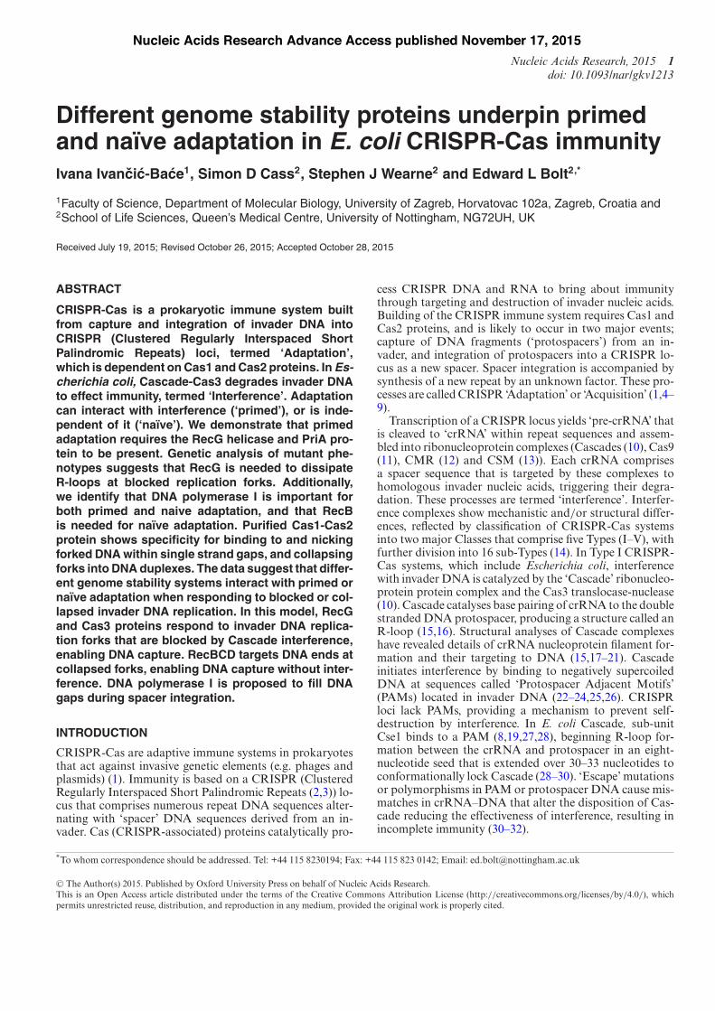
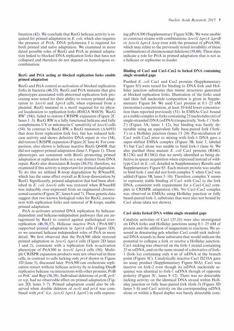
Figure 1. RecG, PriA and DNA polymerase I (PolA) are neededfor primed adaptation. (A) Summary of the modiied Escherichia coliCRISPR-Cas system used to analyze primed adaptation. Genes encodingthe interference complex (cas3, and casA-E ‘cascade’), the adaptation com-plex (cas1, cas2) and the crRNA spacer T3 (spT3), which are all requiredfor priming, were induced by arabinose (pARA) and IPTG (pIPTG). As-terisks illustrate annealing positions of primers used in PCR reactions todetect CRISPR expansion. In CRISPR, each repeat is denoted as a illedrectangle and each spacer as a diamond. (B) DNAgels fromPCR reactionsassaying for CRISPR expansion in the ‘wild type’ (wt) strain for primedadaptation (lanes 1 and 2), compared to isogenenic strains with gene dele-tions in recG or priA as indicated. E indicates CRISPR expansion from a723 bp parental length, P, to 784 bp, when CRISPR-Cas was induced byaddition of arabinose and IPTG (lane 1), compared to without inducers(lane 2). Plasmid expression of missing proteins, or their active site mu-tants, was used as indicated to determine if primed adaptation could be re-stored in each case. Agarose gels were stained using ethidium bromide andare displayed in reverse contrast. (C) Agarose gel as in (B), showing loss ofprimed adaptation from deleting polA (lane 2), and its complementationby plasmid polA (pPolA+, lane 4), compared to empty plasmid vector (lane3) and to recB and ruvC gene deletions (lanes 5 and 6).
described in Supplementary Material. The !recG !priAdoublemutant required for data shown in Figure 2 was con-structed by P1 transduction of !recG into !priA cells con-taining pPriA300 to maintain viability. The !polA strainused is described in (40)(JJ1038) and lacks polymerase func-tion but has improved viability because it retains the exonu-clease domain.
Phage infectivity assays for spacer acquisition
The strain used for primed adaptation (Figure 1A) con-tained the CRISPR-Cas genetic elements deined as nec-essary for priming (32). The engineered spacer (spT3) wasto target an essential gene of a virulent lambda phage (!vir) (41). Primed adaptation was assayed in E. coli strainIIB969 (Figure 1A) and its derivatives. Overnight culturesof appropriate strains were inoculated into LB containinginducers IPTG (1.0 mM) and arabinose (0.2% w/v) as indi-cated. At optical density (OD600) of 0.3, ! virwas added to amultiplicity of infection (MOI) of 1.0 followed by phage ad-sorption for 20 min. Cells were then diluted 1:10 into freshLB containing inducers as required and growth was contin-ued for 12–16 h. These infectivity assays were repeated atleast three times, to monitor spacer acquisition by PCR, us-ing primers annealing to CRISPR positions annotated byasterisks in Figure 1A, detailed in Supplementary Meth-ods. Template DNA was derived from either bacterial cul-
Nucleic Acids Research, 2015 3
ture lysed by boiling in water, or from puriied genomicDNA extracted from 1 ml of bacterial culture using a kit(GeneJET, Thermo Scientiic). For each different culturethe OD600 was measured and cultures were diluted to be atequal turbidity prior to isolating DNA. Typical OD600 val-ues observed for deletion strain cultures during these assaysare given in Supplementary Table S5B. Individual survivorcolonies obtained from plated cell cultures were picked forDNA sequencing corresponding to newly acquired spacer,as shown in Supplementary Table S3. PCR products wereanalyzed on 2.0% agarose Tris-acetate-EDTA (TAE) gels. Ifno spacer acquisition was detected in CRISPR-1, the samePCR method was used to monitor CRISPR-2 for expan-sion, using primer pairs listed in Supplementary Materials.If there was still no detectable spacer acquisition, infectiv-ity assays were repeated with further rounds of infectivityusing the same method as described above.Naıve adaptation assays (Figure 2A) followed a proce-
dure similar to that in (5). Cells lacking Cas3, CasC and/orCas1 and a priming spacer (spT3) were transformed bypEB628 expressing Cas1 and Cas2 or the empty plasmidas a control. Expression of Cas1-Cas2 was induced by ad-dition of 0.2% (w/v) arabinose, and cells were sub-culturedthree or four times by 1:300 dilution of the previous culture.Antibiotics were not included in these rounds of growth inLB to allow plasmid curing from spacer acquisition.
Puriication of E. coli proteins
Coomassie stained gels of puriied proteins are shown inSupplementary Figure S3. Cas1 and Cas2 proteins wereover-produced with N-terminal (His)6 tags in strain BL21AI. Cells were grown at 37◦C to optical density of 0.5–0.6 in LB ampicillin (50 "g/ml) and induced using arabi-nose (0.2% w/v), with growth continued for 3 h after induc-tion. Cas1 or Cas2 expressing cells were harvested for re-suspension in buffer H (20 mM Tris-HCl pH7.5, 500 mMNaCl, 5 mM imidazole, 10% glycerol) for storage at −80◦Cprior to protein puriication. The irst puriication step wasidentical for both Cas1 and Cas2: sonicated and clariiedsoluble cell extract was passed into a 5 ml Hi-Trap Nickelchelating column, Cas1 or Cas2 eluting within a gradientof increasing imidazole. Salt was reduced by dialysis intobuffer H2 (20 mM Tris-HCl pH7.5, 100 mM NaCl, 10%glycerol, 1 mMDTT). For Cas1, fractions were loaded intoa 5 ml Hi-Trap heparin column and eluted in a gradientof NaCl at 200–300 mM. Cas1 fractions were pooled forstorage at −80◦C in buffer H2 containing 40% glycerol.Cas1 mutant proteins D218A, R84G, R95G, R123G andR138G were puriied in the same way as wild-type Cas1.Further rationale and details about these Cas1 arginine mu-tants are given in Supplementary Text and Figure S5. Af-ter the Nickel chelation step, Cas2 was loaded onto a S300size exclusion column in buffer H2. Cas2 fractions wereloaded onto a 5ml heparin column and collected in the non-binding low through or wash. Cas2 fractions were storedas for Cas1. RecG and PriA proteins were a gift from Prof.Bob Lloyd FRS (University of Nottingham) and RusA waspuriied (42).
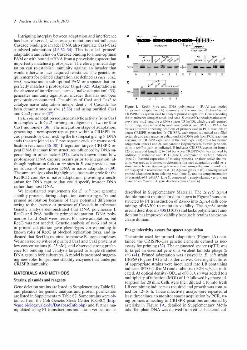
Figure 2. (A) A summary of the modiied E. coli CRISPR-Cas systemused to analyze naıve adaptation. The strain has deletions in cas3, casC andcas1, each indicated as!, resulting in loss of interference (‘! interference’)and loss of chromosomally encoded adaptation (‘! adaptation’). Adap-tation was restored by expression of Cas1-Cas2 expression from pEB628(lane 1, pCas1-Cas2), shown in an agarose gel of PCR reactions as ex-pansion of CRISPR from 662 bp parental (P) to 723 bp expanded (E).Spacer acquisition was much reduced when D218A Cas1 replaced wildtype enzyme (lane 2, pCas1ND-Cas2 for ‘nuclease defective’) and emptyplasmid vector (pEmpty) showed no spacer acquisition (lane 3). Asterisksillustrate annealing positions of primers used in PCR reactions to detectCRISPR expansion (B) Summary agarose gel of CRISPRDNA fromPCRreactions against genomic DNA from naıve cells that can acquire spacerwhen deleted for recG, recA or ruv (lanes 3 and 4 and 7–10), or cannot ac-quire spacer when deleted for polA and recB (lanes 5–6 and 11–12). (C)Agarose gels summarizing effects on primed adaptation in the recG dele-tion strain when expressing mutant RecG proteins from a plasmid (lanes1–5), or RNaseHI inducibly expressed from the chromosome (Lanes 6 and7). (D) Effects on primed adaptation of expressing helicase defective PriA(‘PriA300’) in !recG!priA cells (lanes 1 and 2), or of deleting primosomeassembly proteins PriB, PriC or Rep (lanes 3–7).
Assays on DNA
Base sequences of DNA strands used to construct sub-strates are given in the Supplementary Figure S5. DNAstrands were custom synthesized and HPLC puriied bySigma-Aldrich. DNA strands (300 ng) were 32P labeled attheir 5′ ends by incubation with T4 polynucleotide kinase(PNK) and # 32P-ATP (1 h, 37◦C) followed by heat inactiva-tion of PNK.UnincorporatedATPwas removed from thesereactions using Bio-Spin 6 columns (Bio-Rad). Resulting
4 Nucleic Acids Research, 2015
end-labeled DNA was annealed to unlabeled DNA strands(900 ng) in buffer SSC (150 mM sodium chloride, and 15mMsodium citrate, pH7.0) by heating to 95◦C for 2min fol-lowed by gradual cooling to room temperature. DNA sub-strates were then puriied, to remove un-annealed oligonu-cleotide or incomplete DNA structures, by electrophore-sis through a 10% acrylamide Tris-Borate-EDTA (TBE) gelfollowed by autoradiography, excision of gel slice and elu-tion by diffusion at 4◦C into 250 "l of 10 mM Tris-HCl,50 mM NaCl pH 7.5. Cas1/Cas2 binding to substrateswas analyzed in electrophoretic mobility shift assays (EM-SAs) through 5%acrylamideTBEgels at room temperature.Prior to electrophoresis, protein and DNA substrate weremixed for 10 min at ambient temperature in buffer SBHB (7mMTris-HCl pH 8.5, 9% glycerol, 50mMNaCl, 100"g/mlBSA) supplemented with 5 mM EDTA, in 20 "l reactionvolumes. Gels were dried and exposed by phosphorimagingto detect 32P labeled DNA. Nuclease and end joining assayswere in buffer SBHB supplementedwith 10mMmagnesiumchloride at 37◦C for 10 min. Reactions were stopped by ad-dition of 1 mg/ml proteinase K, 2.5% w/v SDS, formamidegel loading dye and heating to 75◦C prior to electrophore-sis through 15 or 20% acrylamide gels containing 5 M ureain 1xTBE buffer. Holliday junction and fork substrates Chiand ChiSma were generated according to the method (43).
RESULTS
Differential requirements for RecG, PriA, RecB and DNApolymerase I in primed and naıve adaptation
We investigated adaptation in E. coli strains deleted forgenes encoding proteins that help to maintain genome sta-bility by DNA repair and homologous recombination. Toassay primed adaptation we generated an E. coli strain de-rived from strains in references (32,41), described in Supple-mentary Table S1. This contained chromosomally induciblegenes encodingCas1, Cas2, Cas3 andCascade proteins, andan inducible CRISPR spacer (spT3) (Figure 1A). SpacerspT3 encodes crRNA that has a perfect sequence matchwith the essential gene R in virulent lambda phage (!vir)(41), but with a non-consensus PAM (5′-CCA), giving onlypartial protection against phage (44). Primed adaptationwas detected as expansion of CRISPR after PCR amplii-cation of genomic DNA extracted from cells surviving afterinfection with !vir. Induction of chromosomal CRISPR-Cas resulted in expanded CRISPR consistent with additionof a single spacer-repeat unit (723 bp increased to 784 bp,Figure 1B lane 1). PCR and DNA sequencing of individualcolony survivors with expanded CRISPR conirmed thatnew spacer sequences were acquired from !vir (Supplemen-tary Table S3).Most gene deletions tested in primed adaptation assays
had no observable effect on CRISPR expansion listed inSupplementary Table S4. Elimination of genes encodingRecG or PriA helicases (!recG or !priA) corresponded toloss of detectable CRISPR expansion (Figure 1B). DNAbands present in this, and subsequent, agarose gels were theonly bands visible; untrimmed gels are shown in Supple-mentary Figure S1. We did not detect CRISPR expansionfromDNA extracted from these deletion strains after infec-tion with !vir. Overall, !vir infectivity was not signiicantly
reduced by !recG, !priA or !polA (Supplementary TableS5A), consistent with the gene products acting on host celladaptation rather than other events during phage infection.Loss of CRISPR expansion from !recG cells was restoredby plasmid expression of RecG, but helicase inactive RecGQ640R (45) did not restore it (Figure 1B lanes 3–6). RecGhelicase promotes genome stability in most species of bac-teria (46,47), by rescuing stalled replication forks (43,48)and dissociating R-loops (49,50). Loss of primed adapta-tion from !priA cells was reversed when PriA or helicaseinactive PriA (K230R, also called ‘priA300’) was expressedfrom a plasmid (Figure 1B lanes 10–12), giving at leasttwo CRISPR expansion products, observations returned tolater.The DNA gap-illing enzyme DNA polymerase I, en-
coded by polA, was also essential for primed adaptation(Figure 1C). CRISPR expansion could not be detectedwhen DNA synthesis activity of DNA polymerase I waslacking (!polA, Figure 1C lane 2). Expression of DNApolymerase I from a plasmid (pPolA+) restored spacer ac-quisition but empty plasmid vector did not (Figure 1C,lanes 3 and 4). Therefore, gene deletions in recG, priA orpolA corresponded to a loss of primed adaptation, in con-trast to deletions in other DNA recombination-repair genesthat were proicient at primed adaptation (exempliied by!recB and !ruvC, Figure 1C lanes 5 and 6).To test naıve adaptation interference was eliminated by
deleting genes encoding Cas1, Cas3 and the major Cas-cade component CasC, and spacer spT3 was absent (Figure2A and Supplementary Table S1A). Expansion of CRISPRby incorporation of a new spacer-repeat unit in naıve cellswas detectable by inducible expression of Cas1-Cas2 froma plasmid (662 bp increased to 723 bp, Figure 2A lane 1),but no expansion was present after expressing catalyticallydefective Cas1 D218A (51), or empty plasmid (Figure 2Alanes 2 and 3). In contrast to primed adaptation,!recG cellsdid give detectable naıve adaptation, showing that RecGis dispensable for adaptation when interference is absent(summarized in Figure 2B with additional gels in Supple-mentary Figure S2). However, !polA naıve cells showedno detectable CRISPR expansion, indicating that it was re-quired in both types of adaptation (Figure 2B and Supple-mentary Figure S2).We have been unable to test naıve adap-tation in !priA cells because PriA is required for propa-gation of the Cas1-Cas2 plasmid. A recent report demon-strated crucial roles for RecBCD in supporting naıve adap-tationwhen replication forks are collapsed (39). In our naıveadaptation assays, cells lacking RecB (!recB) also lackeddetectable CRISPR expansion (Figure 2B lanes 5 and 6).RecBCD can initiate homologous recombination at DNAends by providing a substrate for RecA to generate D-loops(52), which can be converted into Holliday junctions byRuvABC.However, recA and ruvC deletions did not abolishnaıve or primed adaptation (Figure 2B and SupplementaryTable S4). Therefore, these genetic data on recB, recA andruvC indicate that naıve adaptation occurs independentlyof DNA double strand break repair, but is in agreementwith RecBCD being required for DNA capture at collapsedforks when Cas3 nuclease is absent. This is also consistentwith RecG being required for primed, but not naıve, adap-tation because RecG acts independently of RecBCD recom-
Nucleic Acids Research, 2015 5
bination (42). We conclude that RecG helicase activity is re-quired for primed adaptation in E. coli, which also requiresthe presence of PriA. DNA polymerase I is required forboth primed and naıve adaptation. We examined in moredetail possible roles of RecG and PriA in primed adapta-tion linked to blocked DNA replication forks that have notcollapsed and therefore do not depend on homologous re-combination.
RecG and PriA acting at blocked replication forks enableprimed adaptation
RecG and PriA control re-activation of blocked replicationforks in bacteria (46,53). RecG and PriA mutants that givephenotypes associated with abnormal replication fork pro-cessing were tested for their ability to restore primed adap-tation to !recG and !priA cells, when expressed from aplasmid. RecG mutated in a motif required for its physi-cal localization to replication forks (R682A W683S; ‘RecGRW’ (54)), failed to restore CRISPR expansion (Figure 2Clanes 1–3). RecG RW is a fully functional helicase and fullycomplements UV or mitomycin C sensitivity of !recG cells(54). In contrast to RecG RW, a RecG mutation (!A693)that does form replication fork foci, but has reduced heli-case activity and shows defective DNA repair in vivo (54),did restore CRISPR expansion (Figure 2C lane 4). For com-parison, also shown is helicase inactive RecG Q640R thatdid not support primed adaptation (lane 5, Figure 1). Thesephenotypes are consistent with RecG promoting primedadaptation at replication forks in a way distinct from DNArepair. RecG also dissociates R-loops (50,55), therefore, weexamined if this activity is important for primed adaptation.To do this we utilized R-loop degradation by RNaseHI,which has the same effect overall as R-loop dissociation byRecG. Signiicantly, primed adaptation that had been abol-ished in E. coli !recG cells was restored when RNaseHIwas inducibly over-expressed from an engineered chromo-somal cassette (Figure 2C, lanes 6 and 7). These genetic datasuggest that two known biological roles for RecG, associa-tion with replication forks and removal of R-loops, enableprimed adaptation.PriA re-activates arrested DNA replication by helicase-
dependent and helicase-independent pathways that are an-tagonized by RecG to control against pathological over-replication (46,56,57). Helicase inactive PriA (‘PriA300’)supported primed adaptation in !priA cells (Figure 1D),so we assessed helicase independent roles of PriA in moredetail. We irst observed that the PriA300 allele restoredprimed adaptation in !recG !priA cells (Figure 2D lanes1 and 2), consistent with a replication fork re-activationphenotype of PriA300 in !recG !priA cells (56). Multi-ple CRISPR expansion products were not observed in thesecells, in contrast to cells lacking only priA shown in Figure1D (lane 5), discussed later. PriA300 can orchestrate repli-cation restart without helicase activity by re-loading DnaBreplicative helicase via interactions with other proteins, PriBor PriC and Rep (56,58). Individual deletions of priB, priCor rep, had no observable effect on primed adaptation (Fig-ure 2D, lanes 3–7). Primed adaptation could also be ob-served when double deletion of recG and priA was com-bined with priC (i.e. !recG !priA !priC) in cells express-
ing pPriA300 (Supplementary Figure S2B).We were unableto construct strains with combinations!recG!priA!priBor !recG !priA !rep even with the presence of pPriA300,which may relate to the previously noted inviability of thesecombinations of chromosomal deletions (59,60). These dataindicate a role for PriA in primed adaptation that is not asa helicase or replisome re-loader.
Binding of Cas1 and Cas1-Cas2 to forked DNA containingsingle stranded gaps
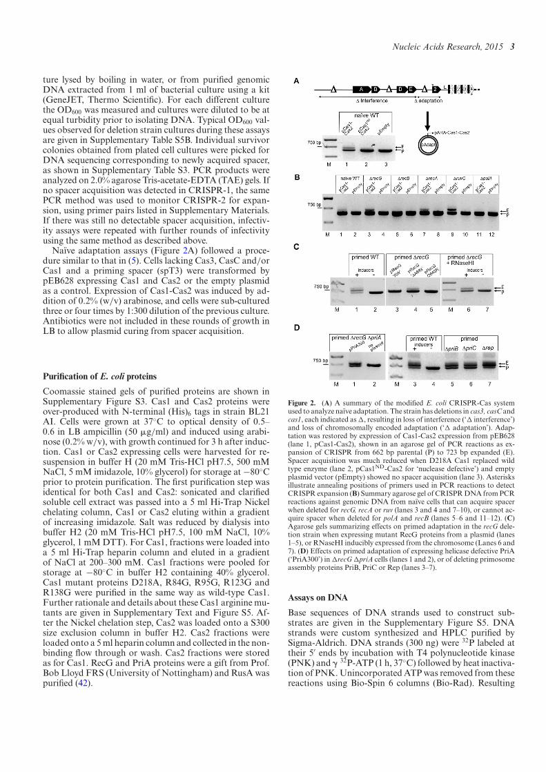
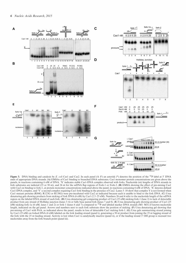
Puriied E. coli Cas1 and Cas2 proteins (SupplementaryFigure S3) were tested for binding to DNA fork and Hol-liday junction substrates that mimic structures generatedat blocked replication forks. Illustration of the forks usedand their full nucleotide composition is given in Supple-mentary Figure S4. We used Cas1 protein at 0.1–25 nM(monomer) concentration, at least 10-fold lower concentra-tion than reported previously (51). In EMSAs Cas1 boundas a stable complex to forks containing 25 nucleotides (nt) ofsingle-strandedDNA (ssDNA) (respectively, ‘fork-1’/’fork-2′) (Figure 3A, lanes 1–12), but binding was barely de-tectable using an equivalent fully base-paired fork (‘fork-3’) or a Holliday junction (lanes 13–24). Pre-incubation ofCas1 with Cas2 prior to mixing with fork-1 DNA gave asuper-shifted EMSA complex (Figure 3B, lane 3, labeledY) but Cas2 alone was unable to bind fork-1 (lane 4). Wealso puriied three mutant E. coli Cas1 proteins (R84G,R123G and R138G) that we newly identiied as being de-fective in spacer acquisition when expressed instead of wild-type Cas1 in E. coli, detailed in Supplementary Results andSupplementary Figure S5. Each mutant protein was unableto bind fork-1 and did not form complex Y when Cas2 wasadded (Figure 3B, lanes 5–10). Therefore, complex Y seemsto represent stable binding of Cas1 with Cas2 to the forkDNA, consistent with requirement for a Cas1-Cas2 com-plex in CRISPR adaptation (36). No Cas1-Cas2 complexwas observed in EMSAs using Holliday junction or fullybased-paired fork-3, substrates that were also not bound byCas1 alone (data not shown).
Cas1 nicks forked DNA within single stranded gaps
Catalytic activities of Cas1 (37,51) were also investigatedon DNA forks and Holliday junctions using 0.1–25 nM ofprotein and the addition of magnesium to reactions. We as-sessed in denaturing gels whether Cas1 could nick individ-ual DNA strands in these substrates that would indicate thepotential to collapse a fork or resolve a Holliday junction.Cas1 nicking was observed on the fork-1 strand containing25 nt ssDNA, and on the same strand of a derivative of fork-1 (fork-1a) containing only 4 nt of ssDNA at the branchpoint (Figure 3C). Catalytically inactive Cas1 D218A gaveno assay product (Supplementary Figure S6A). Cas1 wasinactive on fork-2 even though its ssDNA nucleotide se-quence was identical to fork-1 ssDNA though of oppositepolarity (Figure 3C, lanes 9–12). There was no detectablenicking activity on the identical DNA strand within Holl-iday junction or fully base-paired fork (fork-3) (Figure 3Dlanes 3–8) and Cas1 activity on the corresponding ssDNAalone or within a layed duplex was barely detectable com-
6 Nucleic Acids Research, 2015
Figure 3. DNA binding and catalysis by E. coli Cas1 and Cas2. In each panel (A–F) an asterisk (*) denotes the position of the 32P label at 5′ DNAends of appropriate DNA strands. (A) EMSAs of Cas1 binding to branched DNA substrates. Cas1 monomer protein concentrations are given above thepanels, in reactions containing 6 nM of DNA. ‘X’ indicates stable Cas1-DNA complex observed with forks. Nucleotide (nt) lengths of DNA strands infork substrates are indicted (25 or 50 nt), and 26 nt for the ssDNA lap regions of Fork-1 or Fork-2. (B) EMSA showing the effect of pre-mixing Cas1with Cas2 on binding to fork-1, at protein monomer concentrations indicated above the panel, in reactions containing 6 nM of DNA. ‘X’ denotes deinedCas1-DNA complex, and ‘Y’ a second complex requiring Cas1 fork binding in the presence of Cas2. Lanes 5–10 show that complex Y is not formed whenCas1 mutant proteins (R84G, R123G or R138G) were pre-incubated with Cas2 as indicated because each is unable to bind to the fork DNA. (C) Ureadenaturing gels showing products from nicking of fork DNA (6 nM) by Cas1 (2.5–25 nM). Numbers 26 and 4 refer to the nucleotide length of the ssDNAregion on the labeled DNA strand of each fork. (D) Urea denaturing gel comparing product of Cas1 (25 nM) nicking fork-1 (lane 2) to lack of detectableproduct from any strand of Holliday junction (lanes 3–6) or fully base paired fork (lanes 7 and 8). (E) Urea denaturing gels showing product of Cas1 (25nM) nicking fork-1a (6 nM, lanes 1 and 2) or fork-1 (lanes 6 and 7) compared to 32P end labeled marker DNA strands (M1–M3) of known nucleotidelength, indicated on the gel panel. Arrows and numbers next to each fork substrate show the position of nicking. (F) Urea denaturing gel showing thatpre-mixing of Cas1 with PriA, as indicated above the panel, results in loss of detectable Cas1 nicking fork-1. (G) Urea gels summarizing strand joiningby Cas1 (25 nM) on forked DNA (6 nM) labeled on the fork leading strand (panel i), generating a 50 nt product from joining the 25 nt lagging strand ofthe fork with the 25 nt leading strand. Activity is lost when Cas1 is catalytically inactive (panel ii), or if the leading strand 3′ OH group is recessed twonucleotides away from the fork branch point (panel iii).
Nucleic Acids Research, 2015 7
pared to Fork-1 (Supplementary Figure S6B). The inabil-ity of Cas1 to cut fork-3 and Holliday junction in these as-sayswas conirmed in alternate assays usingChi andChiSma,large substrates sensitive for detecting structure-speciic res-olution of Holliday junctions and forks by nucleases (43),when compared to a bona ide resolving enzyme RusA (61)(Supplementary Figure S6C).Cas1 nicked fork-1 and fork-1a within single stranded
DNA (ssDNA) at 18 and 26 nucleotides from the 32P-labeled 5′-DNA end (Figure 3E). Nicking of fork-1a waswithin the ssDNA gap at the branch point, showing thatit does not require availability of a ssDNA end for nickingactivity. Binding of Cas1 to fork-1a in EMSAs was simi-lar to binding of fork-1 (Supplementary Figure S6D). Sincepre-incubation ofE. coliCas1 with Cas2 formed complex Y(Figure 3B) we also tested if Cas2 stimulated the Cas1 nick-ing activity on fork-1, which was maximally 20% productfrom 25 nM Cas1. However, Cas2 had no effect on nick-ing by Cas1 on any substrate, and Cas2 alone had no de-tectable nuclease activity (Supplementary Figure S5E). In-terestingly, pre-mixing of Cas1 with PriA abolished Cas1nicking of fork-1, but Cas1 was still active if RecG wasadded instead of PriA (Figure 3F). We conclude that Cas1assayed at low concentrations can bind and nick fork DNAin single strand DNA gaps (e.g. fork-1a), with high speci-icity compared to other branched DNA. This activity ofCas1 could collapse a fork, generating DNA ends for pro-cessing and capture during CRISPR adaptation.End labeling of fork-1a on alternative strands revealed
Cas1 catalyzed strand joining of the leading strand 3′ OHDNA to the 5′ end generated from Cas1 nicking in ssDNAof the same fork (Figure 3G). Strand joining in the forkwas lost if Cas1 D218A was used, or if the leading strand3′ OH group was located two nucleotides (or more) awayfrom the fork branch point (fork-1b, Figure 3H panels (ii)and (iii)). Cas1 catalyzed transesteriication reactions onfork substrates were recently detailed in (68), and are re-quired for spacer integration into CRISPR, exempliied byintegration of a radiolabeled duplex DNA fragment intoa supercoiled plasmid (37), a reaction also supported byCas1 in this study (Supplementary Figure S7). Cas1 strandjoining reactions are unlikely to occur at blocked replica-tion forks because the necessary 3′ OH would be located>2 nucleotides away from the cut branch point. Instead,fork nicking by Cas1 may enable DNA capture at replica-tion forks, for strand joining during spacer integration atCRISPR loci, each event aided by the identiied host fac-tors as discussed below and in Figure 4.
DISCUSSION
We present new insights into how genome stability systemsunderpin the building of CRISPR-Cas immunity by eitherprimed or naıve adaptation in E. coli. In primed adapta-tion, Cascade-Cas3 interference complexes that are not pro-icient in degrading invader DNA stimulate adaptation asa means to update immunity by acquisition of new spac-ers (32). In naıve adaptation immunity can be establishedby Cas1-Cas2 without interference reactions (5). A strikingoutcome of our genetic analysis was that primed adapta-tion required RecG as an active helicase that can localize to
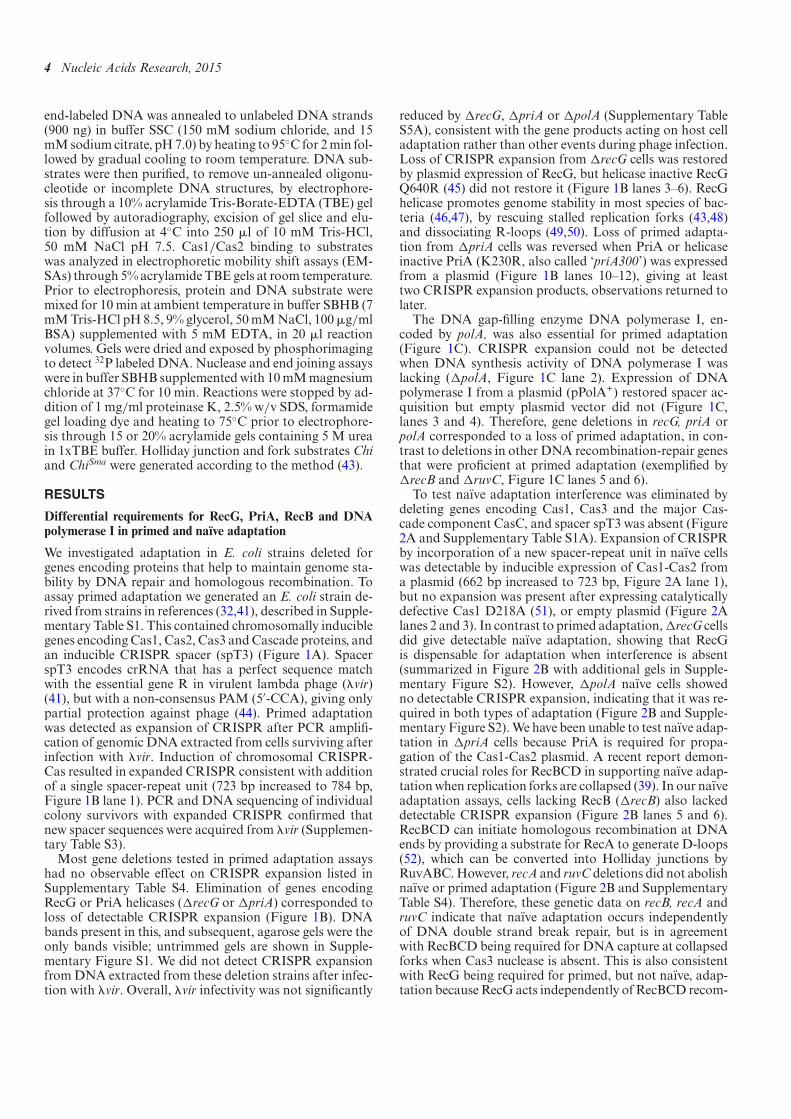
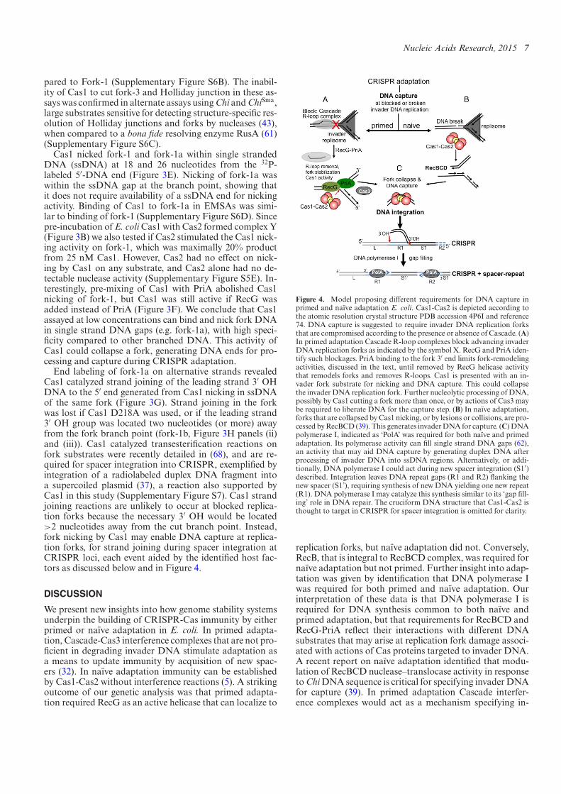
Figure 4. Model proposing different requirements for DNA capture inprimed and naıve adaptation E. coli. Cas1-Cas2 is depicted according tothe atomic resolution crystal structure PDB accession 4P6I and reference74. DNA capture is suggested to require invader DNA replication forksthat are compromised according to the presence or absence of Cascade. (A)In primed adaptation Cascade R-loop complexes block advancing invaderDNA replication forks as indicated by the symbol X. RecG and PriA iden-tify such blockages. PriA binding to the fork 3′ end limits fork-remodelingactivities, discussed in the text, until removed by RecG helicase activitythat remodels forks and removes R-loops. Cas1 is presented with an in-vader fork substrate for nicking and DNA capture. This could collapsethe invader DNA replication fork. Further nucleolytic processing of DNA,possibly by Cas1 cutting a fork more than once, or by actions of Cas3 maybe required to liberate DNA for the capture step. (B) In naıve adaptation,forks that are collapsed byCas1 nicking, or by lesions or collisions, are pro-cessed byRecBCD (39). This generates invaderDNA for capture. (C)DNApolymerase I, indicated as ‘PolA’ was required for both naıve and primedadaptation. Its polymerase activity can ill single strand DNA gaps (62),an activity that may aid DNA capture by generating duplex DNA afterprocessing of invader DNA into ssDNA regions. Alternatively, or addi-tionally, DNA polymerase I could act during new spacer integration (S1’)described. Integration leaves DNA repeat gaps (R1 and R2) lanking thenew spacer (S1’), requiring synthesis of new DNA yielding one new repeat(R1). DNA polymerase I may catalyze this synthesis similar to its ‘gap ill-ing’ role in DNA repair. The cruciform DNA structure that Cas1-Cas2 isthought to target in CRISPR for spacer integration is omitted for clarity.
replication forks, but naıve adaptation did not. Conversely,RecB, that is integral to RecBCD complex, was required fornaıve adaptation but not primed. Further insight into adap-tation was given by identiication that DNA polymerase Iwas required for both primed and naıve adaptation. Ourinterpretation of these data is that DNA polymerase I isrequired for DNA synthesis common to both naıve andprimed adaptation, but that requirements for RecBCD andRecG-PriA relect their interactions with different DNAsubstrates that may arise at replication fork damage associ-ated with actions of Cas proteins targeted to invader DNA.A recent report on naıve adaptation identiied that modu-lation of RecBCD nuclease–translocase activity in responsetoChiDNA sequence is critical for specifying invaderDNAfor capture (39). In primed adaptation Cascade interfer-ence complexes would act as a mechanism specifying in-
8 Nucleic Acids Research, 2015
vader DNA, acting as ‘programmed’ roadblocks to invaderreplication, triggering RecG helicase activity to remove theblockages, exposing DNA for capture.A model for involvement of genome stability proteins in
underpinning adaptation is presented in Figure 4, summa-rized into three parts. (i) In primed adaptation, CascadeR-loop complexes block invader DNA replication. RecGand PriA respond, with RecG helicase activity dissociat-ing the R-loop by unwinding RNA–DNA hybrids and re-moving bound proteins, including possibly Cascade, PriAand SSB. This remodels the blocked replication fork intoexposed double and ssDNA regions for DNA capture bythe catalytic activity of Cas1. Cas3, which is also essentialfor primed adaptation (32), may also contribute to gener-ating DNA fragments at this stage. (ii) In naıve adaptationRecBCD nuclease–helicase activity resects DNA ends gen-erated by collapsed invader replication forks, independentlyof Cascade. This generates DNA substrate for capture. Forknicking by Cas1 (Figure 3) could be responsible for collapseof forks. (iii) DNA polymerase I catalyzes ‘gap illing’ DNAsynthesis (62) of a new CRISPR repeat during spacer inte-gration.Cascade R-loops have potential to act as replication
roadblocks similarly to stalled RNA polymerase and otherprotein–DNA complexes (63–65). It was signiicant thatprimed adaptation required RecG helicase activity anda functioning RecG fork localization motif. Additionally,primed adaptation defects caused by elimination of RecGcould be corrected by ectopic over-expression of RNaseHI(Figures 1 and 2). Therefore, removal of replication fork-blocking R-loop nucleoprotein complexes by RecG heli-case is implicated as being crucial for the mechanism ofprimed adaptation, in line with other reported roles forRecG (50,54,55). Future experiments to ascertain directly ifCascade can block DNA replication will require in vitro re-constitution of the replisome based on previous studies inE.coli (63,66). Another activity of RecG, conversion of forkedDNA into a Holliday junction structure (43,67), was ini-tially appealing to us for facilitating primed adaptation, byCas1 cleaving aHolliday junction to generate ends forDNAcapture. However, Cas1 in our assays (0–25 nMCas1: 6 nMof DNA) showed strong preference for binding to and nick-ing fork substrates with ssDNA gaps. Cas1 was inactive onlargeChi structures that representHolliday junction or fullybase-paired fork DNA, giving further evidence that Cas1prefers branched DNA substrates that are at least partiallyssDNA. Cas1 assayed at much greater monomer concen-trations (250 nM to low "M) was observed to cleave sim-ilar structures most eficiently in a previous analysis (51),although we did not observe any nicking of Holliday junc-tion or fully base-paired fork DNA. The nicking activityof Cas1 that we observed on ssDNA forks is in line with avery recent analysis of a Cas1-Cas2-forked DNA complexpresented at atomic resolution (74).Binding of Cas1, and Cas1-Cas2, to forks containing ss-
DNA gave distinct stable in-gel complexes. Binding was lostwhen using any of the acquisition defective Cas1 mutantsR84G, R123G or R138G (Figure 3B and SupplementaryS5). Cas1 collapsed the same forks by nicking DNA withinthe single strand gap. Cas1 catalyzes transesteriication, ordis-integration, reactions that join together DNA strands
(37). Strand joining by Cas1 was also eficient within forkedDNA, shown here and in (68). DNA strand joining byCas1 is crucial for incorporation of new spacer DNA into aCRISPR locus, and utilizes DNA sequence speciicity thatmatches integration sites for new spacers at the leader endof an E. coli CRISPR (68). Blocked replication forks areunlikely substrates for Cas1 strand joining reactions be-cause they lack DNA 3′ OH located exactly proximal to thefork branch point. Therefore, in that context strand joiningwould not be possible in the way it can take place duringspacer integration into CRISPR. Therefore, Cas1 may beversatile during adaptation, by nicking and collapsing forksfor DNA capture, and joining DNA strands when suit-able ends are available for transesteriication in CRISPR.In conclusion we suggest that requirement for RecG heli-case in primed adaptation centers on aiding Cas1 in captureof protospacer DNA, rather than DNA integration intoa CRISPR locus, because if RecG DNA helicase were re-quired for integration this would be expected to correlatewith impaired naıve adaptation in !recG cells, which alsoneed integration to occur, but this was not the case.PriA fully inhibited Cas1 activity, a surprising observa-
tion given that PriA was required for primed adaptation.PriA binds to fork branch points in a position to accom-modate the leading strand 3′ end into a binding pocket(69,70), which would most likely block access to the forkbranch-point by Cas1. However, PriA binding may alsolimit fork conversion into substrates for homologous re-combination (57,71), an effect that could be advantageousfor primed adaptation if Cas1 nicks an intact fork. We alsoobserved that plasmid expression of PriA or PriA300 inRecG+ primed adaptation cells gave additional CRISPR ex-pansion products (Figure 1D), but did not when RecG wasabsent (Figure 2D). We speculate that this may relect in-creased mobilization of RecG helicase to blocked forks inresponse to artiicially increased PriA levels, with the corre-sponding enhancement of adaptation.AntagonismbetweenRecG and PriA is important for maintaining genome stabil-ity in bacteria when replication forks stall or require termi-nation at ter sites (46,72,73). They may be a factor in theobserved bias toward spacer acquisition from ter sites ob-served in a recent study, although that was not tested (39).RecG and PriA are present in most species of bacteria, in-cluding those utilizingCas9 for interference, raising the pos-sibility that primed adaptation enabled by these helicasesresponding to blocked replication may be widely relevant.
SUPPLEMENTARY DATA
Supplementary Data are available at NAR Online.
ACKNOWLEDGEMENTS
We thank Bob Lloyd for E. coli strains and proteins, Ekata-rina Semenova for E. coli strains, and Christian Rudolphand Ronald Chalmers for comments on the manuscript.
FUNDING
UK BBSRC PhD studentships; UK BBSRC[BB/M020541/1]; The University of Nottingham
Nucleic Acids Research, 2015 9
SoLs pump-priming funds; The University of Zagreb[202761]. Funding for open access charge: BBSRC[BB/M020541/1]/University of Nottingham Open AccessFunds.Conlict of interest statement.None declared.
REFERENCES
1. Barrangou,R., Fremaux,C., Deveau,H., Richards,M., Boyaval,P.,Moineau,S., Romero,D.A. and Horvath,P. (2007) CRISPR providesacquired resistance against viruses in prokaryotes. Science, 315,1709–1712.
2. Mojica,F.J., Diez-Villasenor,C., Soria,E. and Juez,G. (2000)Biological signiicance of a family of regularly spaced repeats in thegenomes of Archaea, Bacteria and mitochondria.Mol. Microbiol.,36, 244–246.
3. Jansen,R., Embden,J.D., Gaastra,W. and Schouls,L.M. (2002)Identiication of genes that are associated with DNA repeats inprokaryotes.Mol. Microbiol., 43, 1565–1575.
4. Richter,C., Dy,R.L., McKenzie,R.E., Watson,B.N., Taylor,C.,Chang,J.T., McNeil,M.B., Staals,R.H. and Fineran,P.C. (2014)Priming in the Type I-F CRISPR-Cas system triggersstrand-independent spacer acquisition, bi-directionally from theprimed protospacer. Nucleic Acids Res., 42, 8516–8526.
5. Yosef,I., Goren,M.G. and Qimron,U. (2012) Proteins and DNAelements essential for the CRISPR adaptation process in Escherichiacoli. Nucleic Acids Res., 40, 5569–5576.
6. Li,M., Wang,R. and Xiang,H. (2014) Haloarcula hispanica CRISPRauthenticates PAM of a target sequence to prime discriminativeadaptation. Nucleic Acids Res., 42, 7226–7235.
7. Cady,K.C., Bondy-Denomy,J., Heussler,G.E., Davidson,A.R. andO’Toole,G.A. (2012) The CRISPR/Cas adaptive immune system ofPseudomonas aeruginosa mediates resistance to Naturally occurringand engineered phages. J. Bacteriol., 194, 5728–5738.
8. Swarts,D.C., Mosterd,C., van Passel,M.W. and Brouns,S.J. (2012)CRISPR interference directs strand speciic spacer acquisition. PLoSOne, 7, e35888.
9. Tyson,G.W. and Banield,J.F. (2008) Rapidly evolving CRISPRsimplicated in acquired resistance of microorganisms to viruses.Environ. Microbiol., 10, 200–207.
10. Brouns,S.J., Jore,M.M., Lundgren,M., Westra,E.R., Slijkhuis,R.J.,Snijders,A.P., Dickman,M.J., Makarova,K.S., Koonin,E.V. and vander Oost,J. (2008) Small CRISPR RNAs guide antiviral defense inprokaryotes. Science, 321, 960–964.
11. Deltcheva,E., Chylinski,K., Sharma,C.M., Gonzales,K., Chao,Y.,Pirzada,Z.A., Eckert,M.R., Vogel,J. and Charpentier,E. (2011)CRISPR RNA maturation by trans-encoded small RNA and hostfactor RNase III. Nature, 471, 602–607.
12. Hale,C.R., Zhao,P., Olson,S., Duff,M.O., Graveley,B.R., Wells,L.,Terns,R.M. and Terns,M.P. (2009) RNA-guided RNA cleavage by aCRISPR RNA-Cas protein complex. Cell, 139, 945–956.
13. Marrafini,L.A. and Sontheimer,E.J. (2008) CRISPR interferencelimits horizontal gene transfer in Staphylococci by targeting DNA.Science, 322, 1843–1845.
14. Makarova,K.S., Wolf,Y.I., Alkhnbashi,O.S., Costa,F., Shah,S.A.,Saunders,S.J., Barrangou,R., Brouns,S.J., Charpentier,E., Haft,D.H.et al. (2015) An updated evolutionary classiication of CRISPR-Cassystems. Nat. Rev. Microbiol., 13, 722–736.
15. Jore,M.M., Lundgren,M., van Duijn,E., Bultema,J.B., Westra,E.R.,Waghmare,S.P., Wiedenheft,B., Pul,U., Wurm,R., Wagner,R. et al.(2012) Structural basis for CRISPR RNA-guided DNA recognitionby Cascade. Nat. Struct. Mol. Biol., 18, 529–536.
16. Ivancic-Bace,I., Al Howard,J. and Bolt,E.L. (2012) Tuning in tointerference: R-Loops and cascade complexes in CRISPR immunity.J. Mol. Biol., 422, 607–616.
17. Wiedenheft,B., Lander,G.C., Zhou,K., Jore,M.M., Brouns,S.J., vander Oost,J., Doudna,J.A. and Nogales,E. (2011) Structures of theRNA-guided surveillance complex from a bacterial immune system.Nature, 477, 486–489.
18. Lintner,N.G., Kerou,M., Brumield,S.K., Graham,S., Liu,H.,Naismith,J.H., Sdano,M., Peng,N., She,Q., Copie,V. et al. (2011)Structural and functional characterization of an archaeal clusteredregularly interspaced short palindromic repeat (CRISPR)-associated
complex for antiviral defense (CASCADE). J. Biol. Chem., 286,21643–21656.
19. Mulepati,S., Heroux,A. and Bailey,S. (2014) Crystal structure of aCRISPR RNA-guided surveillance complex bound to a ssDNAtarget. Science, 345, 1479–1484.
20. Jackson,R.N., Golden,S.M., van Erp,P.B., Carter,J., Westra,E.R.,Brouns,S.J., van der Oost,J., Terwilliger,T.C., Read,R.J. andWiedenheft,B. (2014) Crystal structure of the CRISPR RNA-guidedsurveillance complex from Escherichia coli. Science, 345, 1473–1479.
21. Zhao,H., Sheng,G., Wang,J., Wang,M., Bunkoczi,G., Gong,W.,Wei,Z. and Wang,Y. (2014) Crystal structure of the RNA-guidedimmune surveillance Cascade complex in Escherichia coli. Nature,515, 147–150.
22. Westra,E.R., van Erp,P.B., Kunne,T., Wong,S.P., Staals,R.H.,Seegers,C.L., Bollen,S., Jore,M.M., Semenova,E., Severinov,K. et al.(2012) CRISPR Immunity Relies on the Consecutive Binding andDegradation of Negatively Supercoiled Invader DNA by Cascadeand Cas3.Mol. Cell, 46, 595–605.
23. Bolotin,A., Quinquis,B., Sorokin,A. and Ehrlich,S.D. (2005)Clustered regularly interspaced short palindrome repeats (CRISPRs)have spacers of extrachromosomal origin.Microbiology, 151,2551–2561.
24. Deveau,H., Barrangou,R., Garneau,J.E., Labonte,J., Fremaux,C.,Boyaval,P., Romero,D.A., Horvath,P. and Moineau,S. (2008) Phageresponse to CRISPR-encoded resistance in Streptococcusthermophilus. J. Bacteriol., 190, 1390–1400.
25. Shah,S.A., Erdmann,S., Mojica,F.J. and Garrett,R.A. (2013)Protospacer recognition motifs: mixed identities and functionaldiversity. RNA Biol., 10, 891–899.
26. Heler,R., Marrafini,L.A. and Bikard,D. (2014) Adapting to newthreats: the generation of memory by CRISPR-Cas immune systems.Mol. Microbiol., 93, 1–9.
27. Sashital,D.G., Wiedenheft,B. and Doudna,J.A. (2012) Mechanism offoreign DNA selection in a bacterial adaptive immune system.Mol.Cell, 46, 606–615.
28. Szczelkun,M.D., Tikhomirova,M.S., Sinkunas,T., Gasiunas,G.,Karvelis,T., Pschera,P., Siksnys,V. and Seidel,R. (2014) Directobservation of R-loop formation by single RNA-guided Cas9 andCascade effector complexes. Proc. Natl. Acad. Sci. U.S.A., 111,9798–9803.
29. Semenova,E., Jore,M.M., Datsenko,K.A., Semenova,A.,Westra,E.R., Wanner,B., van der Oost,J., Brouns,S.J. andSeverinov,K. (2011) Interference by clustered regularly interspacedshort palindromic repeat (CRISPR) RNA is governed by a seedsequence. Proc. Natl. Acad. Sci. U.S.A., 108, 10098–10103.
30. Blosser,T.R., Loeff,L., Westra,E.R., Vlot,M., Kunne,T., Sobota,M.,Dekker,C., Brouns,S.J. and Joo,C. (2015) Two distinct DNA bindingmodes guide dual roles of a CRISPR-Cas protein complex.Mol. Cell,58, 60–70.
31. Rutkauskas,M., Sinkunas,T., Songailiene,I., Tikhomirova,M.S.,Siksnys,V. and Seidel,R. (2015) Directional R-Loop formation by theCRISPR-Cas surveillance complex cascade provides eficientoff-target site rejection. Cell Rep., 10, 1534–1543.
32. Datsenko,K.A., Pougach,K., Tikhonov,A., Wanner,B.L.,Severinov,K. and Semenova,E. (2012) Molecular memory of priorinfections activates the CRISPR/Cas adaptive bacterial immunitysystem. Nat. Commun., 3, 945.
33. Hynes,A.P., Villion,M. and Moineau,S. (2014) Adaptation inbacterial CRISPR-Cas immunity can be driven by defective phages.Nat. Commun., 5, 4399.
34. Fineran,P.C., Gerritzen,M.J., Suarez-Diez,M., Kunne,T.,Boekhorst,J., van Hijum,S.A., Staals,R.H. and Brouns,S.J. (2014)Degenerate target sites mediate rapid primed CRISPR adaptation.Proc. Natl. Acad. Sci. U.S.A., 111, E1629–E1638.
35. Fineran,P.C. and Charpentier,E. (2012) Memory of viral infectionsby CRISPR-Cas adaptive immune systems: acquisition of newinformation. Virology, 434, 202–209.
36. Nunez,J.K., Kranzusch,P.J., Noeske,J., Wright,A.V., Davies,C.W. andDoudna,J.A. (2014) Cas1-Cas2 complex formation mediates spaceracquisition during CRISPR-Cas adaptive immunity. Nat. Struct.Mol. Biol., 21, 528–534.
37. Nunez,J.K., Lee,A.S., Engelman,A. and Doudna,J.A. (2015)Integrase-mediated spacer acquisition during CRISPR-Cas adaptiveimmunity. Nature, 519, 193–198.
10 Nucleic Acids Research, 2015
38. Arslan,Z., Hermanns,V., Wurm,R., Wagner,R. and Pul,U. (2014)Detection and characterization of spacer integration intermediates intype I-E CRISPR-Cas system. Nucleic Acids Res., 42, 7884–7893.
39. Levy,A., Goren,M.G., Yosef,I., Auster,O., Manor,M., Amitai,G.,Edgar,R., Qimron,U. and Sorek,R. (2015) CRISPR adaptationbiases explain preference for acquisition of foreign DNA. Nature,520, 505–510.
40. Zhang,J., Mahdi,A.A., Briggs,G.S. and Lloyd,R.G. (2010) Promotingand avoiding recombination: contrasting activities of the Escherichiacoli RuvABC Holliday junction resolvase and RecG DNAtranslocase. Genetics, 185, 23–37.
41. Pougach,K., Semenova,E., Bogdanova,E., Datsenko,K.A.,Djordjevic,M., Wanner,B.L. and Severinov,K. (2010) Transcription,processing and function of CRISPR cassettes in Escherichia coli.Mol. Microbiol., 77, 1367–1379.
42. Bolt,E.L. and Lloyd,R.G. (2002) Substrate Speciicity of RusAResolvase Reveals the DNA Structures Targeted by RuvAB andRecG in vivo.Mol. Cell, 10, 187–198.
43. McGlynn,P. and Lloyd,R.G. (2000) Modulation of RNA polymeraseby (p)ppGpp reveals a RecG-dependent mechanism for replicationfork progression. Cell, 101, 35–45.
44. Westra,E.R., Semenova,E., Datsenko,K.A., Jackson,R.N.,Wiedenheft,B., Severinov,K. and Brouns,S.J. (2013) Type I-ECRISPR-cas systems discriminate target from non-target DNAthrough base pairing-independent PAM recognition. PLoS Genet., 9,e1003742.
45. Briggs,G.S., Mahdi,A.A., Weller,G.R., Wen,Q. and Lloyd,R.G.(2004) Interplay between DNA replication, recombination and repairbased on the structure of RecG helicase. Philos. Trans. R. Soc. Lond.B Biol. Sci., 359, 49–59.
46. Rudolph,C.J., Upton,A.L., Briggs,G.S. and Lloyd,R.G. (2010) IsRecG a general guardian of the bacterial genome? DNA Rep., 9,210–223.
47. Rocha,E.P., Cornet,E. and Michel,B. (2005) Comparative andevolutionary analysis of the bacterial homologous recombinationsystems. PLoS Genet., 1, e15.
48. Whitby,M.C., Ryder,L. and Lloyd,R.G. (1993) Reverse branchmigration of Holliday junctions by RecG protein: a new mechanismfor resolution of intermediates in recombination and DNA repair.Cell, 75, 341–350.
49. McGlynn,P., Al-Deib,A.A., Liu,J., Marians,K.J. and Lloyd,R.G.(1997) The DNA replication protein PriA and the recombinationprotein RecG bind D-loops. J. Mol. Biol., 270, 212–221.
50. Vincent,S.D., Mahdi,A.A. and Lloyd,R.G. (1996) The RecG branchmigration protein of Escherichia coli dissociates R-loops. J. Mol.Biol., 264, 713–721.
51. Babu,M., Beloglazova,N., Flick,R., Graham,C., Skarina,T.,Nocek,B., Gagarinova,A., Pogoutse,O., Brown,G., Binkowski,A.et al. (2011) A dual function of the CRISPR-Cas system in bacterialantivirus immunity and DNA repair.Mol. Microbiol., 79, 484–502.
52. Dillingham,M.S. and Kowalczykowski,S.C. (2008) RecBCD enzymeand the repair of double-stranded DNA breaks.Microbiol. Mol. Biol.Rev. 72, 642–671.
53. Heller,R.C. and Marians,K.J. (2005) The disposition of nascentstrands at stalled replication forks dictates the pathway of replisomeloading during restart.Mol. Cell, 17, 733–743.
54. Upton,A.L., Grove,J.I., Mahdi,A.A., Briggs,G.S., Milner,D.S.,Rudolph,C.J. and Lloyd,R.G. (2014) Cellular location and activity ofEscherichia coli RecG proteins shed light on the function of itsstructurally unresolved C-terminus. Nucleic Acids Res., 42,5702–5714.
55. Fukuoh,A., Iwasaki,H., Ishioka,K. and Shinagawa,H. (1997)ATP-dependent resolution of R-loops at the ColE1 replication origin
by Escherichia coli RecG protein, a Holliday junction-speciichelicase. EMBO J., 16, 203–209.
56. Gabbai,C.B. and Marians,K.J. (2010) Recruitment to stalledreplication forks of the PriA DNA helicase and replisome-loadingactivities is essential for survival. DNA Rep., 9, 202–209.
57. Jaktaji,R.P. and Lloyd,R.G. (2003) PriA supports two distinctpathways for replication restart in UV-irradiated Escherichia coli cells.Mol. Microbiol., 47, 1091–1100.
58. Sandler,S.J. (2005) Requirements for replication restart proteinsduring constitutive stable DNA replication in Escherichia coli K-12.Genetics, 169, 1799–1806.
59. Seigneur,M., Bidnenko,V., Ehrlich,S.D. and Michel,B. (1998) RuvABacts at arrested replication forks. Cell, 95, 419–430.
60. Sandler,S.J. (2000) Multiple genetic pathways for restarting DNAreplication forks in Escherichia coli K-12. Genetics, 155, 487–497.
61. Bolt,E.L., Sharples,G.J. and Lloyd,R.G. (1999) Identiication of threeaspartic acid residues essential for catalysis by the RusA Hollidayjunction resolvase. J. Mol. Biol., 286, 403–415.
62. Moolenaar,G.F., Moorman,C. and Goosen,N. (2000) Role of theEscherichia coli nucleotide excision repair proteins in DNAreplication. J. Bacteriol., 182, 5706–5714.
63. Gupta,M.K., Guy,C.P., Yeeles,J.T., Atkinson,J., Bell,H., Lloyd,R.G.,Marians,K.J. and McGlynn,P. (2013) Protein-DNA complexes are theprimary sources of replication fork pausing in Escherichia coli. Proc.Natl. Acad. Sci. U.S.A., 110, 7252–7257.
64. Gan,W., Guan,Z., Liu,J., Gui,T., Shen,K., Manley,J.L. and Li,X.(2011) R-loop-mediated genomic instability is caused by impairmentof replication fork progression. Genes Dev., 25, 2041–2056.
65. Rudolph,C.J., Dhillon,P., Moore,T. and Lloyd,R.G. (2007) Avoidingand resolving conlicts between DNA replication and transcription.DNA Rep., 6, 981–993.
66. Bruning,J.G., Howard,J.L. and McGlynn,P. (2014) Accessoryreplicative helicases and the replication of protein-bound DNA. J.Mol. Biol., 426, 3917–3928.
67. McGlynn,P. and Lloyd,R.G. (2001) Rescue of stalled replication forksby RecG: Simultaneous translocation on the leading and laggingstrand templates supports an active DNA unwinding model of forkreversal and Holliday junction formation. Proc. Natl. Acad. Sci.U.S.A., 98, 8227–8234.
68. Rollie,C., Schneider,S., Brinkmann,A.S., Bolt,E.L. and White,M.F.(2015) Intrinsic sequence speciicity of the Cas1 integrase directs newspacer acquisition. eLife, 4. e08716.
69. Manhart,C.M. and McHenry,C.S. (2015) Identiication of subunitbinding positions on a model fork and displacements that occurduring sequential assembly of the Escherichia coli primosome. J. Biol.Chem., 290, 10828–10839.
70. Sasaki,K., Ose,T., Okamoto,N., Maenaka,K., Tanaka,T., Masai,H.,Saito,M., Shirai,T. and Kohda,D. (2007) Structural basis of the3′-end recognition of a leading strand in stalled replication forks byPriA. EMBO J., 26, 2584–2593.
71. Tanaka,T. and Masai,H. (2006) Stabilization of a stalled replicationfork by concerted actions of two helicases. J. Biol. Chem., 281,3484–3493.
72. Rudolph,C.J., Upton,A.L., Harris,L. and Lloyd,R.G. (2009)Pathological replication in cells lacking RecG DNA translocase.Mol.Microbiol., 73, 352–366.
73. Rudolph,C.J., Upton,A.L., Stockum,A., Nieduszynski,C.A. andLloyd,R.G. (2013) Avoiding chromosome pathology when replicationforks collide. Nature, 500, 608–611.
74. Wang,J., Li,J., Zhao,H., Sheng,G., Wang,M., Yin,M. and Wang,Y.(2015) Structural and mechanistic basis of PAM dependent spaceracquisition in CRISPR-Cas systems. Cell, 163, 1–14.