Embed Size (px)
Citation preview

IV THERAPY
Presented By: Steven Jones, NREMT-P

INTRODUCTION
This training is designed to provide the student with the essential information needed to enhance his/her understanding of IV therapy principles.
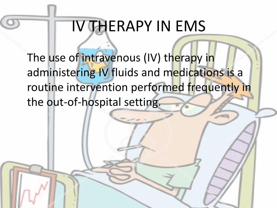
IV THERAPY IN EMS
The use of intravenous (IV) therapy in administering IV fluids and medications is a routine intervention performed frequently in the out-of-hospital setting.
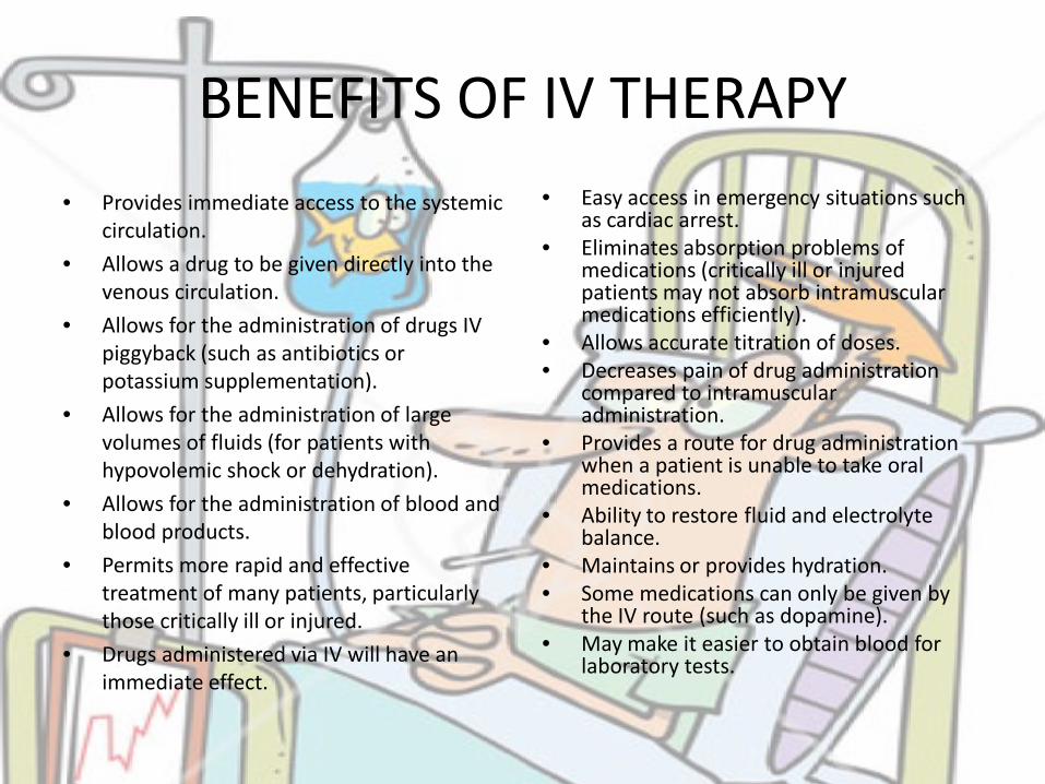
BENEFITS OF IV THERAPY
• Provides immediate access to the systemic circulation.
• Allows a drug to be given directly into the venous circulation.
• Allows for the administration of drugs IV piggyback (such as antibiotics or potassium supplementation).
• Allows for the administration of large volumes of fluids (for patients with hypovolemic shock or dehydration).
• Allows for the administration of blood and blood products.
• Permits more rapid and effective treatment of many patients, particularly those critically ill or injured.
• Drugs administered via IV will have an immediate effect.
• Easy access in emergency situations such as cardiac arrest.
• Eliminates absorption problems of medications (critically ill or injured patients may not absorb intramuscular medications efficiently).
• Allows accurate titration of doses. • Decreases pain of drug administration
compared to intramuscular administration.
• Provides a route for drug administration when a patient is unable to take oral medications.
• Ability to restore fluid and electrolyte balance.
• Maintains or provides hydration. • Some medications can only be given by
the IV route (such as dopamine). • May make it easier to obtain blood for
laboratory tests.

COMPLICATIONS OF IV THERAPY
Most complications of IV therapy can be prevented and/or rapidly treated. Complications to be aware of include both local and systemic.
LOCAL COMPLICATIONS
• Incompatibility between drugs– Although most emergency drugs are compatible with the
more commonly used IV fluids, many drugs are incompatible when administered at the same time (or very closely together) in the same IV line. This does not necessarily mean that more than one IV line is required; however, if you are unsure of drug compatibility when administering several medications, particularly in emergency situations, the line must be flushed well between drugs. Otherwise, the medications may precipitate when they mix. This can cause the drugs to crystallize and obstruct the IV line, necessitating changing the tubing or possibly the IV site itself.
LOCAL COMPLICATIONS
• Difficulty with access– Veins can collapse from hypovolemia or vasoconstriction.
Some patients have very small or scarred veins and access may be difficult.
• Iatrogenic infection– Improper technique when establishing an IV can lead to
infection. If aseptic technique was not used when starting the IV due to conditions at the scene, inform the health care provider at the facility to which you transported the patient. They may want to restart the IV to minimize the risk of infection. In addition, make sure you clean IV ports with alcohol swabs prior to drug administration. Dress IV sites according to protocol.

LOCAL COMPLICATIONS
• Venous irritation– Some solutions and drugs cause irritation to the vein.
Some examples are promethazine (Phenergan) and potassium. The patient may complain of pain at the site and up the arm. If venous irritation occurs, slow the infusion or dilute the drug.
LOCAL COMPLICATIONS
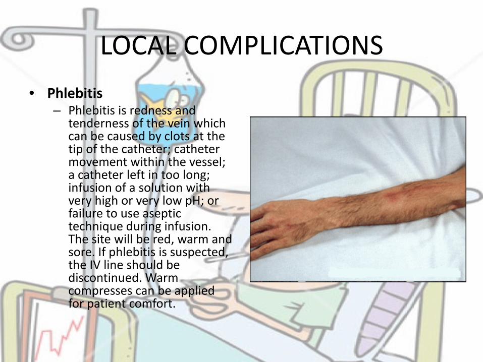
• Phlebitis– Phlebitis is redness and
tenderness of the vein which can be caused by clots at the tip of the catheter; catheter movement within the vessel; a catheter left in too long; infusion of a solution with very high or very low pH; or failure to use aseptic technique during infusion. The site will be red, warm and sore. If phlebitis is suspected, the IV line should be discontinued. Warm compresses can be applied for patient comfort.
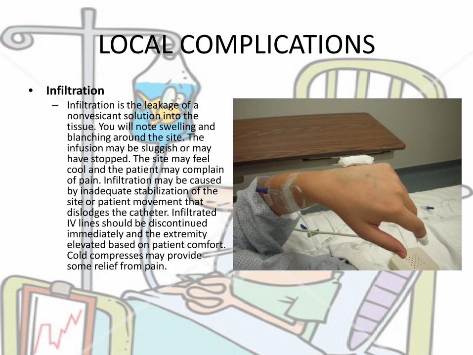
LOCAL COMPLICATIONS• Infiltration
– Infiltration is the leakage of a nonvesicant solution into the tissue. You will note swelling and blanching around the site. The infusion may be sluggish or may have stopped. The site may feel cool and the patient may complain of pain. Infiltration may be caused by inadequate stabilization of the site or patient movement that dislodges the catheter. Infiltrated IV lines should be discontinued immediately and the extremity elevated based on patient comfort. Cold compresses may provide some relief from pain.
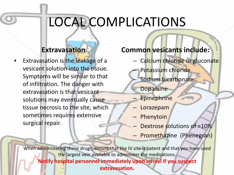
LOCAL COMPLICATIONS
Extravasation• Extravasation is the leakage of a
vesicant solution into the tissue. Symptoms will be similar to that of infiltration. The danger with extravasation is that vesicant solutions may eventually cause tissue necrosis to the site, which sometimes requires extensive surgical repair.
Common vesicants include: – Calcium chloride or gluconate
– Potassium chloride
– Sodium bicarbonate
– Dopamine
– Epinephrine
– Lorazepam
– Phenytoin
– Dextrose solutions of >10%
– Promethazine (Phenegran)
When administering these drugs, ensure that the IV site is patent and that you have used the largest vein available to administer the medications.
Notify hospital personnel immediately upon arrival if you suspect extravasation.

LOCAL COMPLICATIONS
• Severed catheter/Catheter Shear– A severed catheter piece can flow through the systemic
circulation and cause problems. To minimize the possibility of a severed catheter, do not reinsert the needle into the catheter. If you pull the catheter out and a portion is missing, retrieve it if it is visible. Otherwise, put a venous constricting band above the site and transport as quickly as possible.
LOCAL COMPLICATIONS
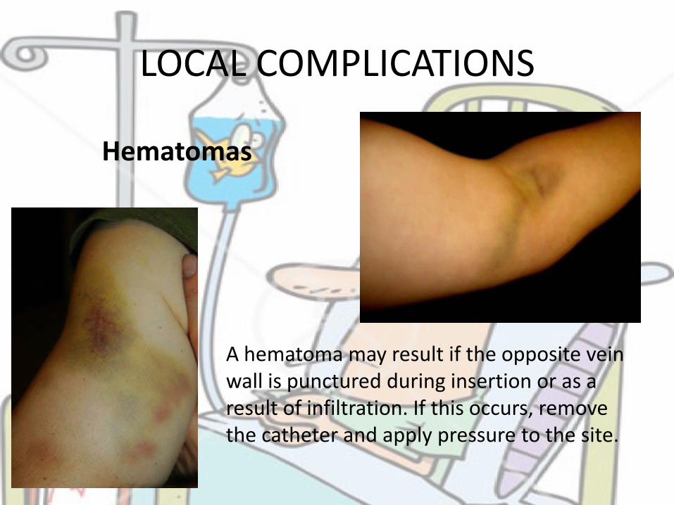
Hematomas
A hematoma may result if the opposite vein wall is punctured during insertion or as a result of infiltration. If this occurs, remove the catheter and apply pressure to the site.

LOCAL COMPLICATIONS
• Thrombosis– Any injury that roughens the venous wall allows platelets
to adhere and a thrombus to form. Thrombosis occurs when a local thrombus obstructs the flow of blood.
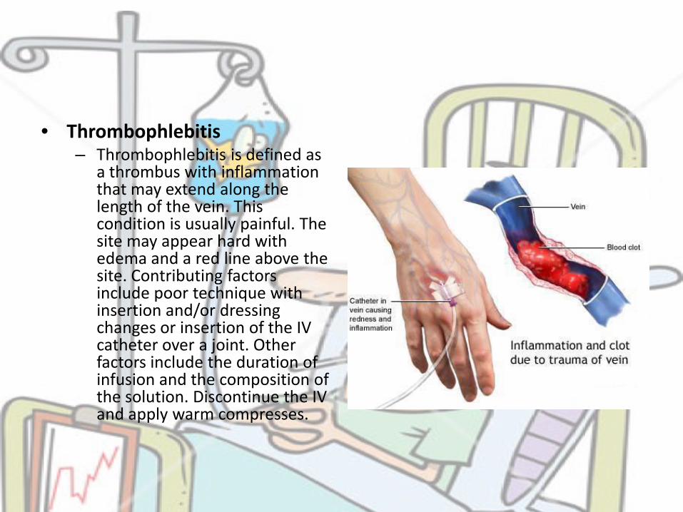
• Thrombophlebitis– Thrombophlebitis is defined as
a thrombus with inflammation that may extend along the length of the vein. This condition is usually painful. The site may appear hard with edema and a red line above the site. Contributing factors include poor technique with insertion and/or dressing changes or insertion of the IV catheter over a joint. Other factors include the duration of infusion and the composition of the solution. Discontinue the IV and apply warm compresses.
LOCAL COMPLICATIONS
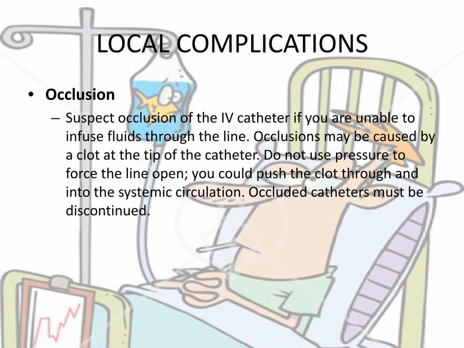
• Occlusion– Suspect occlusion of the IV catheter if you are unable to
infuse fluids through the line. Occlusions may be caused by a clot at the tip of the catheter. Do not use pressure to force the line open; you could push the clot through and into the systemic circulation. Occluded catheters must be discontinued.
SYSTEMIC COMPLICATIONS
• Circulatory overload– This results from administering too much fluid in too short
a period of time. This amount will vary according to the patient and their condition. For example, you would expect to be able to give a larger volume of fluid to a young trauma patient than you would to an elderly patient with a history of heart disease. Patients with circulatory overload may exhibit signs of heart failure (hypertension, crackles in the lung fields, shortness of breath, neck vein distention, etc.). If you suspect your patient is suffering from fluid overload, slow the flow rate down, administer oxygen and raise the head of the bed

SYSTEMIC COMPLICATIONS
• Allergic reaction to a drug– Allergic reactions to drugs
given via IV are evident immediately. The patient may complain of itching and difficulty breathing. You will note bronchospasm and wheezing. Stop the infusion immediately and treat per protocol (steroids, antihistamines, epinephrine).

PERSONAL SAFETY CONSIDERATIONS
• Everyone is familiar with the hazards of blood borne pathogens and the dangers associated with needle sticks. It is imperative that you protect yourself from exposure to blood and needle sticks.– Prevention methods
• BSI• Protective IV Catheters• Utilize a needleless system• Assure proper disposal of needles

SECURING IV ACCESS• Selecting a Site
– IV infusions are usually started in the hand or forearm when possible. However, during an emergency situation, such as a cardiac arrest or major trauma, IV access in the antecubitalspace may be preferable. Usually, larger bore IV catheters can be inserted in the antecubital space and access can be obtained more quickly and without interrupting resuscitation. For most patients, however, veins in the hand or forearm are preferable, as this will permit the patient more mobility. When initiating the IV, select a distal vein if possible; this will allow you to choose a higher vein in the event you miss the vein or the IV becomes dislodged.
The veins of the AC area may be the best site in critically ill or injured patients
SECURING IV ACCESS
• A constricting band placed about 6 inches above the desired site may make the vein easier to locate and enter. Leave the constricting band in place no longer than three minutes. If you cannot locate a vein and prepare the site in that time, release the constricting band and reapply after the site is prepared. The patient may pump his hand a few times if necessary. You may use a vein that is visible or palpable and of an adequate size to hold the IV catheter. Note that in patients with particularly fragile or rolling veins you may have a higher success rate if you do not use a constricting band. The constricting band, when released, can actually rupture the vein wall in these circumstances.
When starting an IV on an elderly patient, consider not using a constricting band
SECURING IV ACCESS
• Cleanse the area, hold the skin in the area for vein stabilization, and insert the catheter bevel up. Once a flash of blood is seen, advance the catheter slightly and while occluding the tip of the catheter, carefully withdraw the needle and attach the tubing device. If the catheter does not thread smoothly and/or blood flow stops, remove the catheter and apply pressure to the site. Attempt to insert an IV in another site using a new catheter.
• Never reinsert a needle into a catheter because the needle may sever the catheter.
• Dispose of the needle in a sharps container. Never stick the needle into a mattress or stretcher. Dress the IV site per protocol.
EXTERNAL JUGULAR IV ACCESS
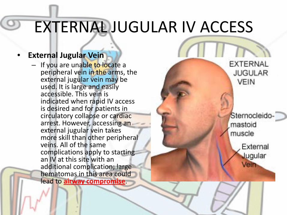
• External Jugular Vein– If you are unable to locate a
peripheral vein in the arms, the external jugular vein may be used. It is large and easily accessible. This vein is indicated when rapid IV access is desired and for patients in circulatory collapse or cardiac arrest. However, accessing an external jugular vein takes more skill than other peripheral veins. All of the same complications apply to starting an IV at this site with an additional complication; large hematomas in this area could lead to airway compromise.
EXTERNAL JUGULAR IV ACCESS
• If the patient’s clinical status will allow, the patient should be placed in the Trendelenberg position to enhance visualization of the vein.
• Turn the patient’s head to the opposite side (away from the jugular vein which will be used).
• With the bevel side up, aim the catheter toward the shoulder on the same side.
• Insert the catheter midway between the angle of the jaw and the midclavicular line.
• Hold the skin taught right above the clavicle to stabilize the vein. • Continue as with any other IV insertion. • In some patients, it is helpful to use a syringe to help identify
proper placement; attach the syringe to the end of the stylet and gently aspirate for blood return when you think you are in the lumen of the vein.
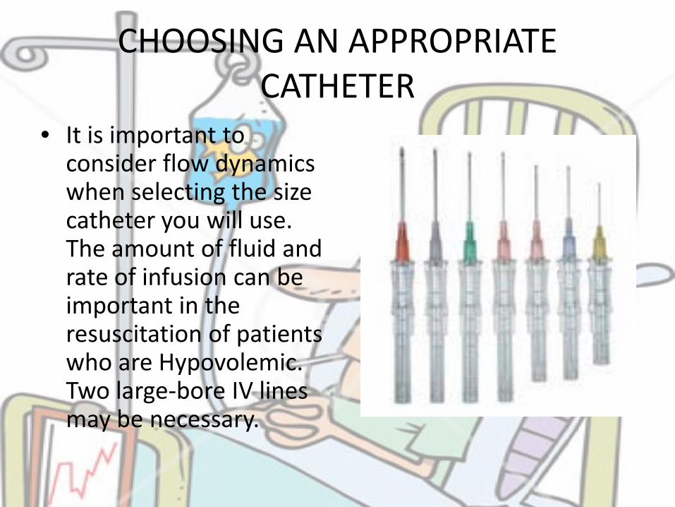
CHOOSING AN APPROPRIATE CATHETER
• It is important to consider flow dynamics when selecting the size catheter you will use. The amount of fluid and rate of infusion can be important in the resuscitation of patients who are Hypovolemic. Two large-bore IV lines may be necessary.
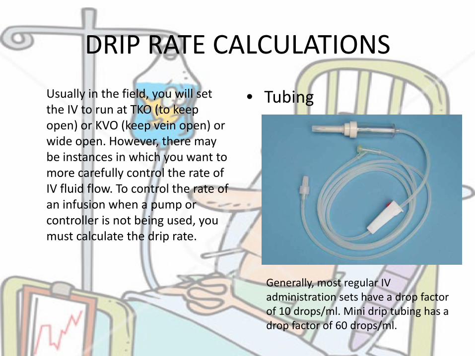
DRIP RATE CALCULATIONS
Usually in the field, you will set the IV to run at TKO (to keep open) or KVO (keep vein open) or wide open. However, there may be instances in which you want to more carefully control the rate of IV fluid flow. To control the rate of an infusion when a pump or controller is not being used, you must calculate the drip rate.
• Tubing
Generally, most regular IV administration sets have a drop factor of 10 drops/ml. Mini drip tubing has a drop factor of 60 drops/ml.

DRIP RATE CALCULATIONS
• Volume– the number mL to be infused
• Time– The time required or desired to
infuse the selected volume (Expressed in Minutes)
• Drip Factor– The tubing - #of drops=1mL
– found on the manufacturer's packaging. (Expressed as gtts/mL)
• Gtts/Min– The resulting drops (gtts) per minute
FORMULA
Example: You wish to deliver 1,000 mL of fluid over 12 hours and the drop factor is 10 drop/mL
(1000 × 10) ÷ (12 × 60) = 13.8 or 14 drops/min
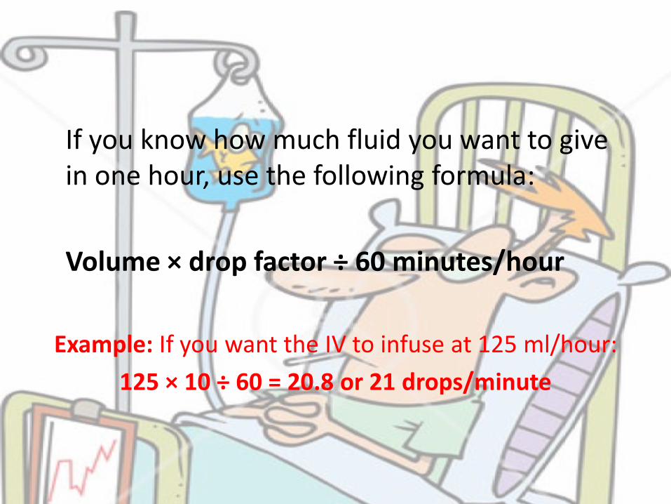
If you know how much fluid you want to give in one hour, use the following formula:
Volume × drop factor ÷ 60 minutes/hour
Example: If you want the IV to infuse at 125 ml/hour:
125 × 10 ÷ 60 = 20.8 or 21 drops/minute

DISCUSSION?