Embed Size (px)
Citation preview
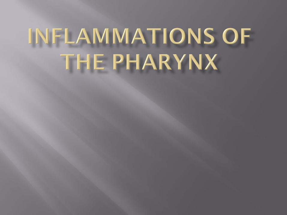
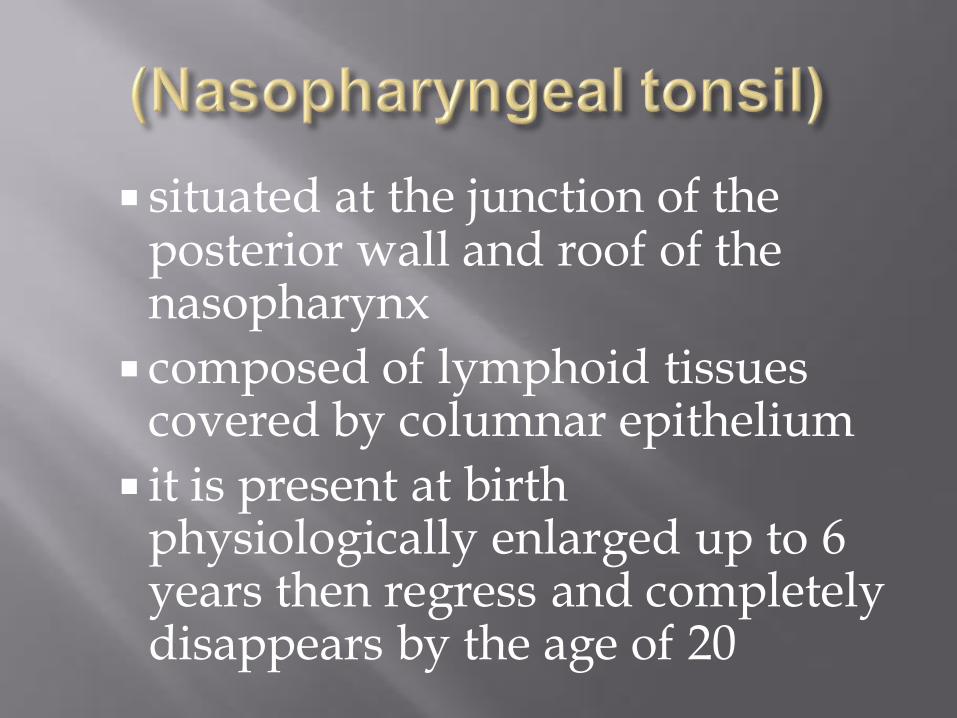
situated at the junction of the posterior wall and roof of the nasopharynx
composed of lymphoid tissues covered by columnar epithelium
it is present at birth physiologically enlarged up to 6 years then regress and completely disappears by the age of 20
1- Recurrent attacks of rhinitis and adenoid infection.
2- Allergy
3- Idiopathic
Nasal obstruction
Mouth breathing and snoring
Nasal discharge
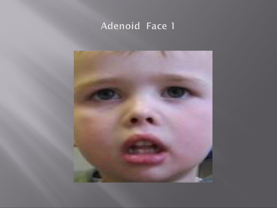
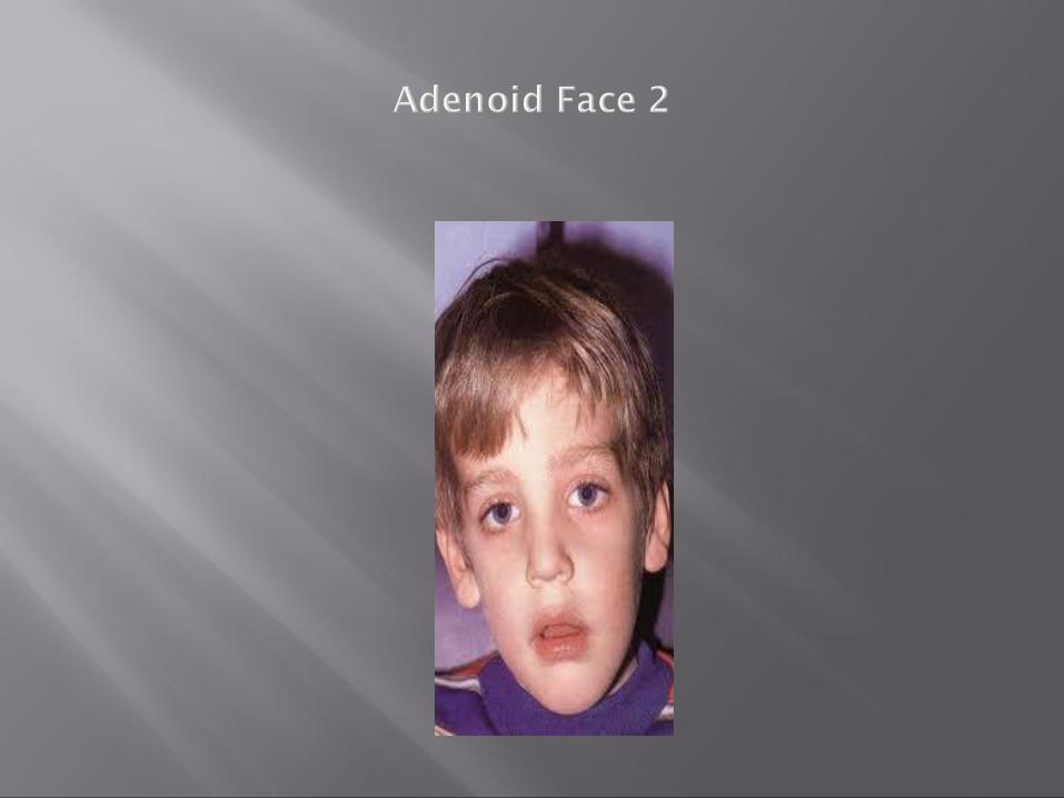
Adenoid face:
elongated face, dull expression, nasal discharge, open mouth, hitched-up upper lip, prominent and overcrowded upper teeth, high-arched palate
nasopharyngoscopy
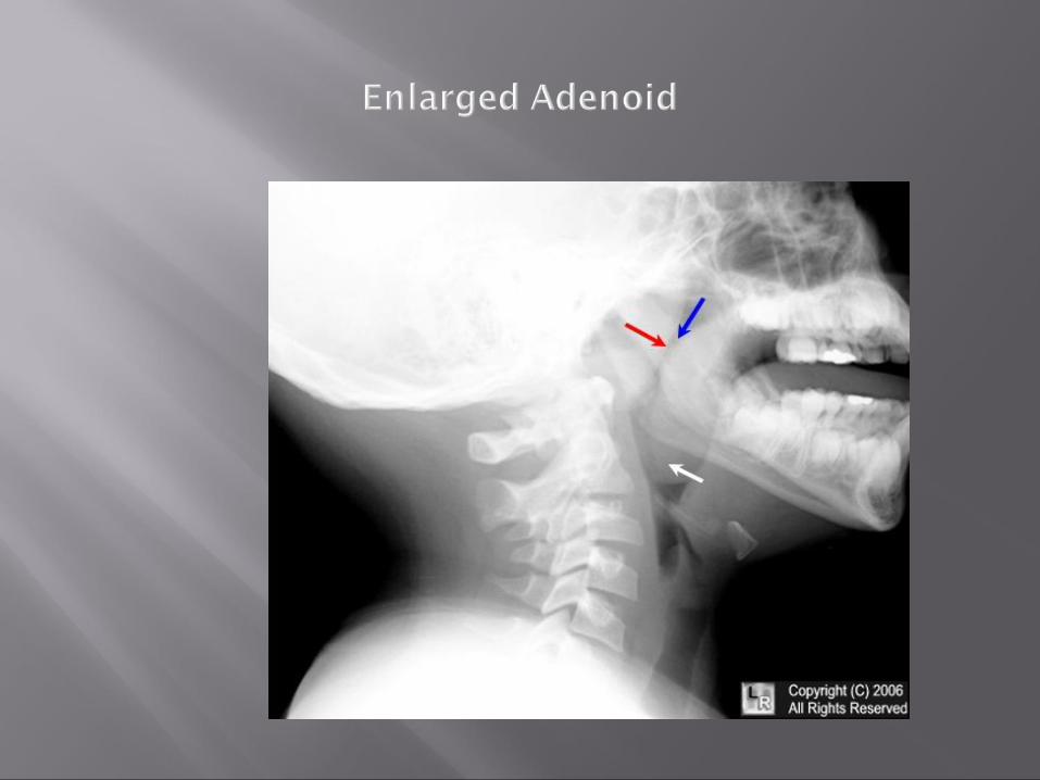
X-ray nasopharynx lateral view
When symptoms are not severe,
decongestant nasal drops + antihistamines is the treatment of choice. Antibiotics if there is bacterial infection.
Marked symptoms, treatment is adenoidectomy
Differential diagnosis:
1- other causes of nasal obstruction (septal deflection, nasal polyps, allergic rhinitis)
2- orthodontic anomaly
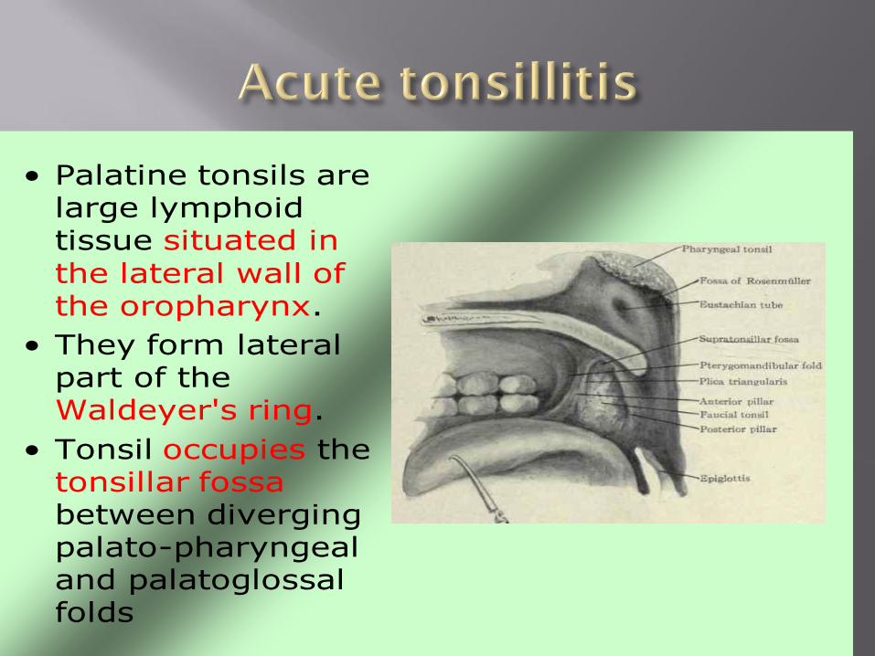
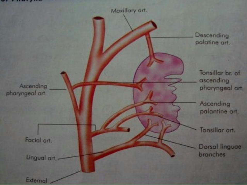
• Palatine tonsils are large lymphoid tissue situated in the lateral wall of the oropharynx.
• They form lateral part of the Waldeyer's ring.
• Tonsil occupies the tonsillar fossabetween diverging palato-pharyngeal and palatoglossalfolds
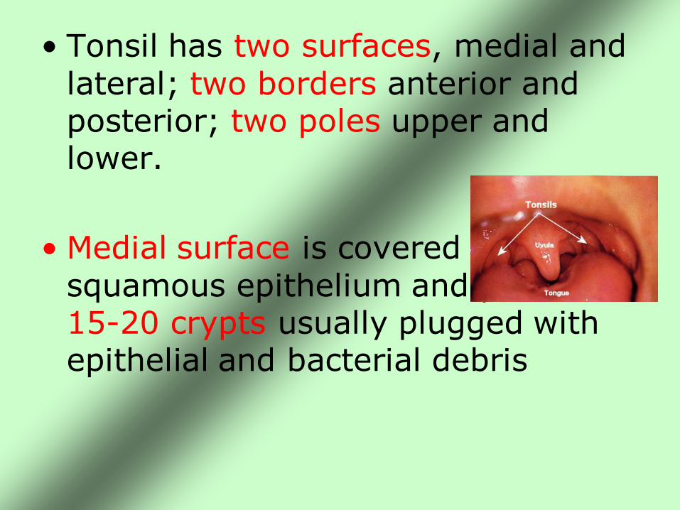
• Tonsil has two surfaces, medial and lateral; two borders anterior and posterior; two poles upper and lower.
• Medial surface is covered by squamous epithelium and presents 15-20 crypts usually plugged with epithelial and bacterial debris
• Lateral surface extends deep to surrounding boundaries. It is coated with a fibrous sheet, an extension of pharyngobasilar fascia called capsule of the tonsil.
• The capsule is loosely attached to the muscular wall but antero-inferiorly it is attached firmly to the side of the tongue just in front of insertion of palatoglossus and palatopharyngeus muscles
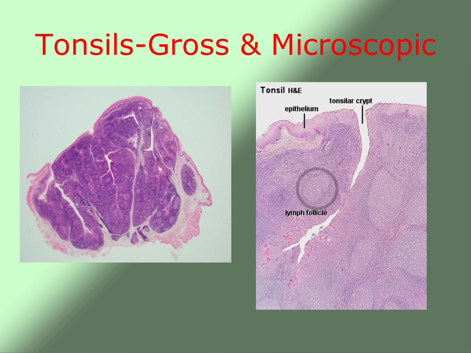
Tonsils-Gross & Microscopic
Acute tonsillitis Aetiology:
beta hemolytic strept.,viral inf.may be primary. Pathological types: 1- parynchymatous
2- follicular Clinical features:
1- sore throat 2- odynophagia
3- pyrexia 4- malaise
5- exam.: enlarged tender cervical LN.
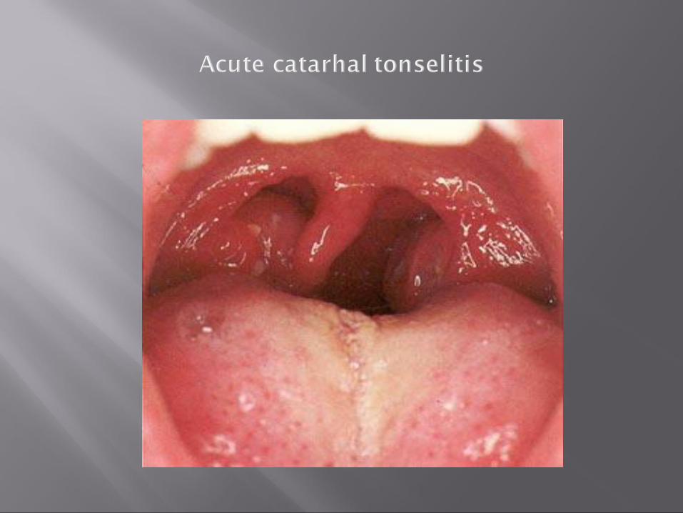
Classification (pathological types)1- Acute catarrhal or superficial tonsillitis: Here tonsillitis is a part of generalized pharyngitis and seen in viral infections
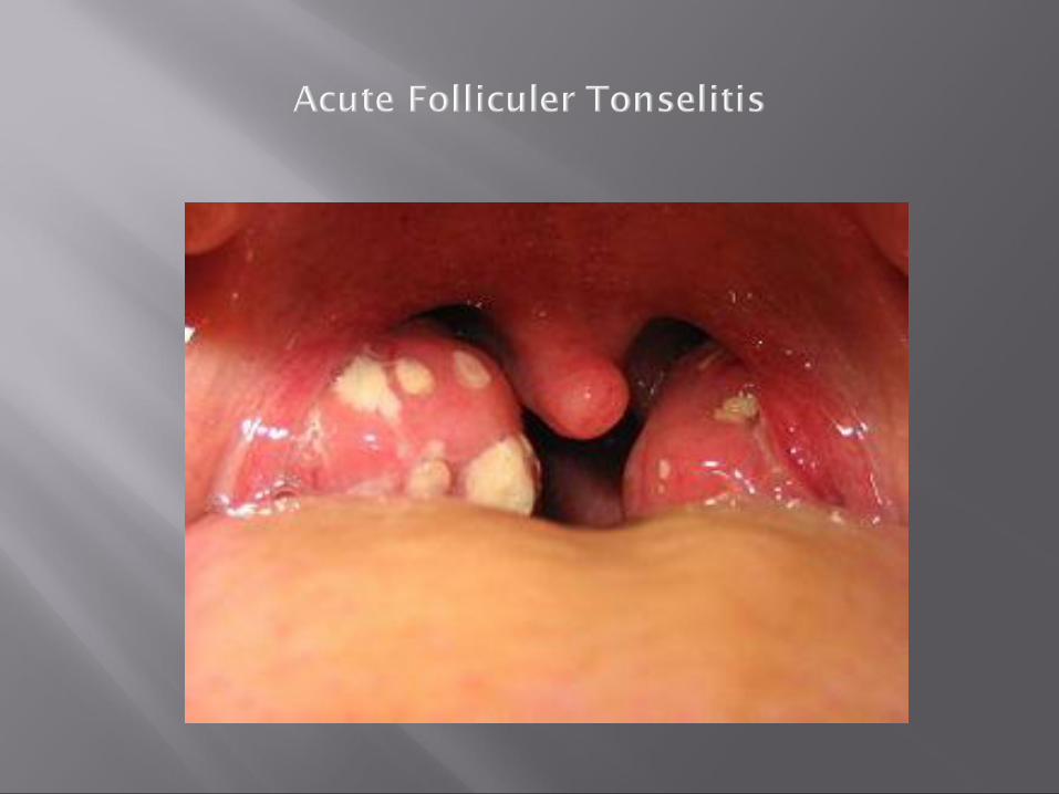
2-Acute follicular tonsillitis: In which tonsillar crypts become filled with purulent materials
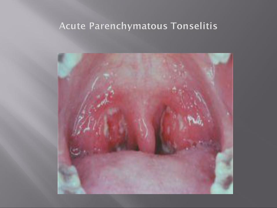
3-Acute parenchymatoustonsillitis: Here tonsils are uniformly enlarged and red
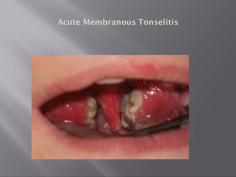
4- Acute membranous tonsillitis: The exudates in the crypts coalesces to form membrane on the surface
Affects school-age children but adults can also be affected. It is rare in infants (< 1 year age) and persons above 50 years.
More common in winter months.
Group A beta hemolytic streptococci
Haemophilus influenzae
Streptococcus pneumoniae
Staphylococci
Tuberculosis (in immunocompromised)
Viruses: adenovirus, Epstein-Bar virus and herpes simplex virus
sore throat
difficulty in swallowing + pain
fever (can be accompanied by rigors and chills)
ear ache
headache
generalized body fatigue
breath is foetid and tongue is coated hyperaemia of the pillars, soft palate
and uvula red and swollen tonsils with yellowish
spots in the crypts (follicular tonsillitis) , whitish membrane on the medial surface of the tonsils (membranous tonsillitis) or enlarged and congestive tonsils with swollen uvula (acute parenchymatous tonsillitis)
enlarged and tender jugulodigastric lymph nodes
bed rest + plenty of fluids
analgesia (Aspirin or Paracetamol)
antimicrobial (Penicillin is the drug of choice) should be continued for 7 -10 days
Differential diagnosis
1- Scarlet fever
2- Diphtheria
3- Infectious mononeucleosis
4- Blood dyscrasia:leukemia, agranulocytosis
5- Vincent’s angina
Complications: General 1-Rheumatic fever 2- Glomerulonephritis 3- Septicemia Local 1- Peritonsillar abscess(quinsy) 2-Paratonsillar abscess 3- Retropharyngeal abscess 4- Otitis media 5- Lower resp.tract infection
Acute rheumatic fever and glomerulonephritis:
• These diseases are of unknown aetiology and follow infection with Beta-haemolyticstreptococcus. The current belief is that antibodies produced against the streptococcus may in some instances cross react with patient’s own tissue.
• Thus the effect on tissue may be an arthritis, endocarditis or myocarditis or a dermatitis or rheumatic chorea (inflammation of cerebral cortex and basal ganglia).
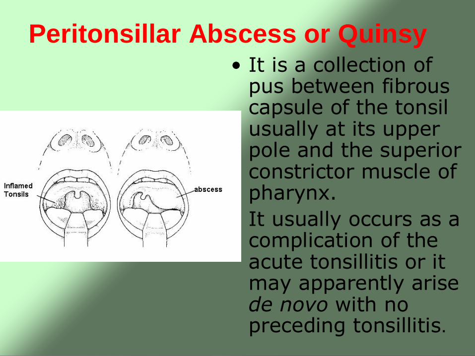
Peritonsillar Abscess or Quinsy• It is a collection of
pus between fibrous capsule of the tonsil usually at its upper pole and the superior constrictor muscle of pharynx.
• It usually occurs as a complication of the acute tonsillitis or it may apparently arise de novo with no preceding tonsillitis.
Bacteriology• The bacteriology of acute
tonsillitis and peritonsillar abscess is different although one is a complication of the other.
• The bacteriology of the quinsy is characterized by mixed flora with multiple organisms both aerobic and anaerobic.
Clinical Features• Fit and young adult with a prior history of
repeated attacks of acute tonsillitis.
• Preceded by a sore throat for 2-3 days which gradually becomes severe and unilateral.
• At this stage patient is ill with fever, often a headache and severe throat pain made worse by swallowing.
• There might be referred otalgia, pain and swelling in the neck due to infective lymphadenopathy. The patient’s voicedevelops a characteristic ‘plummy’ quality.
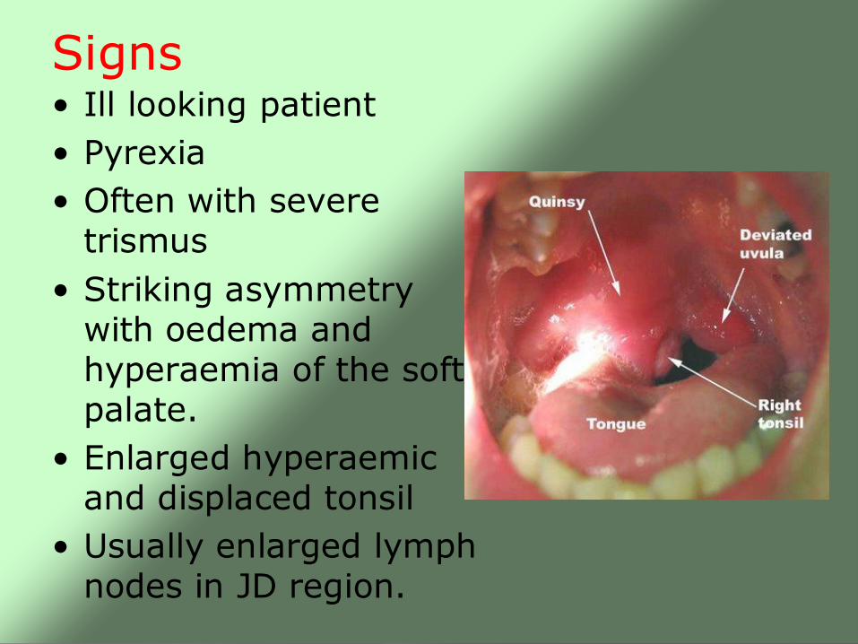
Signs• Ill looking patient
• Pyrexia
• Often with severe trismus
• Striking asymmetry with oedema and hyperaemia of the soft palate.
• Enlarged hyperaemic and displaced tonsil
• Usually enlarged lymph nodes in JD region.
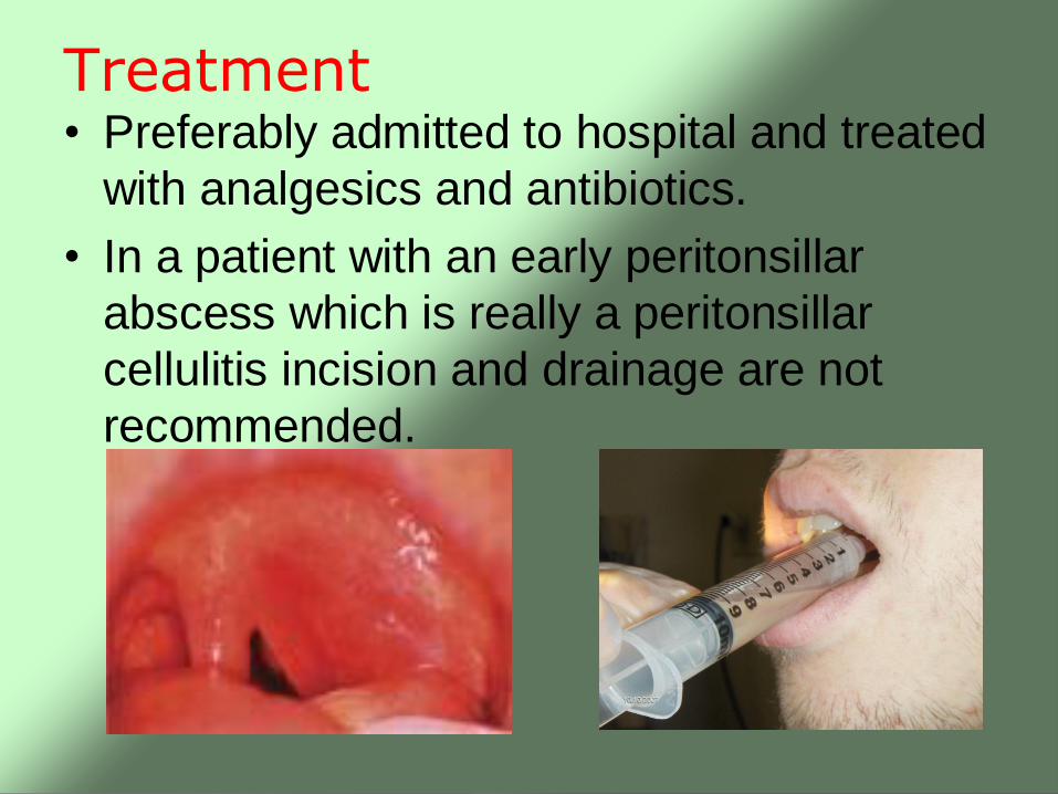
Treatment• Preferably admitted to hospital and treated
with analgesics and antibiotics.
• In a patient with an early peritonsillar
abscess which is really a peritonsillar
cellulitis incision and drainage are not
recommended.
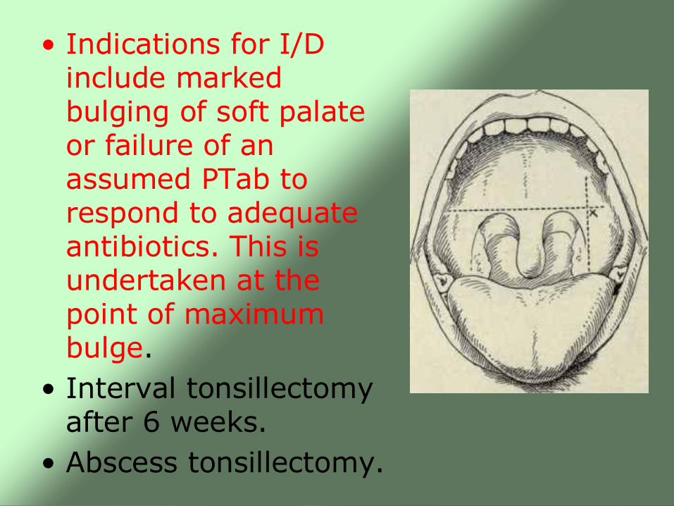
• Indications for I/D include marked bulging of soft palate or failure of an assumed PTab to respond to adequate antibiotics. This is undertaken at the point of maximum bulge.
• Interval tonsillectomy after 6 weeks.
• Abscess tonsillectomy.
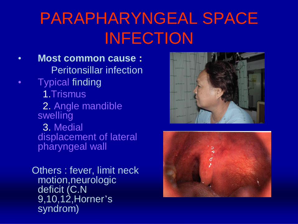
PARAPHARYNGEAL SPACE
INFECTION• Most common cause :
Peritonsillar infection
• Typical finding
1.Trismus
2. Angle mandible swelling
3. Medial displacement of lateral pharyngeal wall
Others : fever, limit neck motion,neurologicdeficit (C.N 9,10,12,Horner’s syndrom)
PARAPHARYNGEAL SPACE INFECTION
Treatment
1. Evaluate and maintain airway & fluid hydration
2. Parenteral antibiotic high dose 24-48 hrs.
3. If not improve, consider surgical drainage
RETROPHARYNGEAL SPACE INFECTION
Types: Acute (children)
Chronic (adults)
• In children: follows
retropharyngeal lymphadenitis from
upper respiratory tract infections
• In adult: follows
regional trauma and TB of the cervical
spines
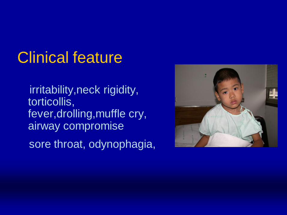
Clinical feature
irritability,neck rigidity, torticollis, fever,drolling,muffle cry, airway compromise
sore throat, odynophagia,
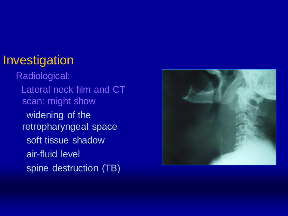
InvestigationRadiological:
Lateral neck film and CT
scan: might show
widening of the
retropharyngeal space
soft tissue shadow
air-fluid level
spine destruction (TB)
Treatment:
Hospital admission
Maintain the airway (intubation or tracheostomy)
Iv fluids and antibiotics
If abscess develop: drainage
1- Airway obstruction.
2- Septicemia.
3- neurological: cranial nerves palsy (9th and 10th) and Horner’s syndrome.
4- Carotid artery rupture.
5- Internal jugular vein thrombosis.