Embed Size (px)
Citation preview

Ultrasound Obstet Gynecol 2018; 52: 128–139Published online in Wiley Online Library (wileyonlinelibrary.com). DOI: 10.1002/uog.19072
isuog.org GUIDELINES
ISUOG Practice Guidelines: intrapartum ultrasound
Clinical Standards Committee
The International Society of Ultrasound in Obstetricsand Gynecology (ISUOG) is a scientific organization thatencourages sound clinical practice and high-quality teach-ing and research related to diagnostic imaging in women’shealthcare. The ISUOG Clinical Standards Committee(CSC) has a remit to develop Practice Guidelines and Con-sensus Statements as educational recommendations thatprovide healthcare practitioners with a consensus-basedapproach, from experts, for diagnostic imaging. They areintended to reflect what is considered by ISUOG to bethe best practice at the time at which they were issued.Although ISUOG has made every effort to ensure thatGuidelines are accurate when issued, neither the Societynor any of its employees or members accepts any liabilityfor the consequences of any inaccurate or misleading data,opinions or statements issued by the CSC. The ISUOGCSC documents are not intended to establish a legal stan-dard of care, because interpretation of the evidence thatunderpins the Guidelines may be influenced by individ-ual circumstances, local protocol and available resources.Approved Guidelines can be distributed freely with thepermission of ISUOG ([email protected]).
PURPOSE AND SCOPE
The purpose of these Guidelines is to review the publishedtechniques of ultrasound in labor and their practical appli-cations, to summarize the level of evidence regardingthe use of ultrasound in labor and to provide guid-ance to practitioners on when ultrasound in labor isclinically indicated and how the sonographic findingsmay affect labor management. We do not imply or sug-gest that ultrasound in labor is a necessary standardof care.
BACKGROUND AND INTRODUCTION
Traditionally, the assessment and management of awoman in labor is based upon clinical findings1–7. Thediagnosis of arrest of labor and decisions regarding thetiming or type of intervention rely mostly on digitalevaluation of cervical dilatation and fetal head stationand position8–17. However, clinical examination of head
station and position is inaccurate and subjective18–25,especially when caput succedaneum impairs palpation ofthe sutures and fontanels.
The use of ultrasound has been proposed to aid inthe management of labor. Several studies have demon-strated that ultrasound examination is more accurate andreproducible than clinical examination in the diagnosis offetal head position and station19–33 and in the predictionof arrest of labor34–42. Ultrasound examination can, tosome extent, distinguish those women destined for spon-taneous vaginal delivery and those destined for operativedelivery43–47. Furthermore, there is growing evidencethat ultrasound in labor may predict the outcome ofinstrumental vaginal delivery44–48.
Ultrasound in labor can be performed using a transab-dominal approach, mainly to determine head and spineposition49, or a transperineal approach, for assessment ofhead station and position at low stations. Several quantita-tive sonographic parameters have been proposed to assesshead station30–32,34,35,40,42,43,50,51. Currently, there is noconsensus regarding when in labor ultrasound should beperformed, which parameter(s) should be obtained andhow the sonographic findings should be integrated intoclinical practice in order to improve management of thepatient.
IDENTIFICATION AND ASSESSMENTOF EVIDENCE
The Cochrane Library and Cochrane Register of Con-trolled Trials were searched for relevant randomizedcontrolled trials, systematic reviews and meta-analyses.A search of Medline from 1966 to 2017 was alsocarried out. The date of the last search was 30 Septem-ber 2017. In addition, relevant conference proceedingsand abstracts were searched. Searches used the rele-vant MeSH terms, including all subheadings. This wascombined with a keyword search, including: ‘labor ultra-sound’, ‘transperineal ultrasound’, ‘fetal head station’,‘fetal occiput position’ and ‘instrumental vaginal deliv-ery’. When possible, recommendations in these Guidelinesare based on, and explicitly linked to, supporting evi-dence. Details of the grades of recommendation andlevels of evidence used in these Guidelines are given inAppendix 1.
Copyright © 2018 ISUOG. Published by John Wiley & Sons Ltd. ISUOG GUIDELINES

ISUOG Guidelines 129
GUIDELINES
Aims of ultrasound in the labor ward
These Guidelines address exclusively the use of ultrasoundin labor to determine fetal head station, position and atti-tude. All other applications of ultrasound in the laborward, such as assessment of cervical length or dilatationand fetal Doppler studies, are not covered. For the timebeing, ultrasound should be used as an adjunctive methodand not as a substitute for clinically indicated digitalvaginal examination.
Assessment of fetal head position
Precise knowledge of fetal occiput position in labor is ofparamount importance.
• Persistent occiput-posterior position is associated withhigher risk of operative delivery52 and maternal andperinatal morbidity53,54.
• Correct determination of head position is crucial beforeinstrumental delivery. An error in evaluation of headposition may result in inappropriate vacuum or forcepsplacement, increasing the potential for fetal injury andthe failure rate of the procedure55–58. Failed instru-mental delivery followed by Cesarean section is asso-ciated with an increased decision-to-delivery interval59
and an increased risk of maternal60,61 and fetal62–65
trauma.
Traditionally, clinicians determine fetal head positionby palpating the sagittal suture and the anterior andposterior fontanels. Several studies have evaluated theaccuracy of clinical diagnosis of fetal head position,using ultrasound19–28 or position-tracking technologysystems66 as reference; digital palpation was found to besubjective. Studies show consistently that digital exam-ination to determine head position is inaccurate, witha rate of error ranging from 20% to 70%, whenconsidering ultrasound as the standard19 (LEVEL OFEVIDENCE: 1–).
Clinical evaluation by palpation tends to be even lessaccurate in cases of abnormal head position, such asocciput posterior or transverse, when medical interven-tion is more likely to be needed19,20,22,23 (LEVEL OFEVIDENCE: 2++).
This inaccuracy may be exaggerated by the pres-ence of caput succedaneum and asynclitism, both ofwhich are frequently associated with obstructed labor.Several studies have failed to demonstrate a signifi-cant difference in accuracy between experienced andinexperienced obstetricians19,21,22, although this find-ing has been questioned by others20 (LEVEL OFEVIDENCE: 2+).
Various studies have demonstrated the superiority ofultrasound alone or in combination with digital exam-ination in the precise determination of fetal head rota-tion as compared with traditional digital examinationalone19–28,66 (LEVEL OF EVIDENCE: 1–).
Assessment of fetal head station
The fetal head station is the level of the fetal head in thebirth canal relative to the plane of the maternal ischialspines (non-cephalic presentation is not considered inthese Guidelines). The term ‘head engagement’ is usedwhen the widest part of the head passes into the pelvicinlet or two-fifths or less of the fetal head is palpableabdominally, corresponding to descent of the biparietalplane of the fetal head to a level below that of the pelvicinlet67. On digital vaginal examination, the fetal head isconsidered engaged when the leading part of the skull hasreached the imaginary line or plane between the maternalischial spines. This head station is referred to as station 0.Higher or lower head stations are expressed in centimetersabove (negative) or below (positive) this reference plane,respectively.
The subjectivity of transvaginal digital assessment offetal head station was demonstrated by Dupuis et al.18
(LEVEL OF EVIDENCE: 2+). Using a birth simula-tor equipped with a sensor, they placed a fetal headmannequin at defined stations according to the Amer-ican College of Obstetricians and Gynecologists, anda group of examiners of various levels of experienceused palpation to classify the fetal head station as high,mid-pelvis, low or outlet. The mean ‘category’ errorwas 30% for residents and 34% for obstetricians. Moreimportantly, the incorrect diagnosis of a mid-pelvic sta-tion rather than a true high-pelvic station accounted forthe majority of errors (88% and 67% by residents andobstetricians, respectively). In clinical practice, such mis-classification may impact adversely on the managementof labor.
Ultrasound examination documents objectively andprecisely the fetal head station in the birthcanal29–33,35,47,68 (LEVEL OF EVIDENCE: 2+).
A series of sonographic parameters have been sug-gested to describe the fetal head station; these havebeen demonstrated to have high intra- and interobserveragreement69–71 (LEVEL OF EVIDENCE: 2+).
Assessment of fetal head descent (progression)
Some observational studies36,37,39,72,73 have suggestedthat repeat ultrasound examinations to assess the changeof head station over time (‘progression’) performs bet-ter than does digital examination in documenting fetalhead descent and in demonstrating slow labor or lack ofprogress in both the first and second stages (LEVEL OFEVIDENCE: 2+).
Assessment of fetal head attitude
The fetal head attitude is the relationship of thefetal head to spine. Ultrasound has proved helpful invisual assessment of fetal head attitude74,75 (LEVELOF EVIDENCE: 2–) and in the objective diagnosis offetal head malpresentation in labor76–80 (LEVEL OFEVIDENCE: 3).
Copyright © 2018 ISUOG. Published by John Wiley & Sons Ltd. Ultrasound Obstet Gynecol 2018; 52: 128–139.

130 ISUOG Guidelines
Technique
Ultrasound assessment in labor may be performed using atransabdominal or transperineal approach, depending onthe parameter that is the aim of the examination (mainlyposition and station) and on the clinical indication. Atwo-dimensional ultrasound machine equipped with aconvex probe, such as that used for transabdominal fetalultrasound for biometry and assessment of anatomy, isused. Suggested requirements of equipment for use in thelabor ward are that it is quick to start up, and has bat-teries with a long life and that are quick to recharge. Awide-sector, low-frequency (< 4 MHz) insonation is bestsuited to ultrasound in labor.
Assessment of fetal head position
Sonographic assessment of fetal head position is best per-formed by transabdominal imaging in axial and sagittalplanes81. Placing the ultrasound probe transversely onthe maternal abdomen, an axial view of the fetal trunk isobtained at the level of the fetal upper abdomen or chest.The position of the fetal spine may then be determined.The ultrasound transducer is then moved downwards untilit reaches the maternal suprapubic region, visualizing the
fetal head. The landmarks depicting fetal occiput positionare the two fetal orbits for occiput posterior, the midlinecerebral echo for occiput transverse, and the occiput itselfand the cervical spine for occiput-anterior position81
(Figures 1 and 2). The choroid plexus, which divergestowards the occiput, can be helpful in determining fetalhead position47.
The midline structures in the fetal head may be difficultto visualize on transabdominal imaging at low fetal headstations. Combining a transabdominal with a transper-ineal ultrasound approach may be recommended in thesecases for precise determination of position.
Position can be described by depicting a circle, likea clock (Figure 3): positions ≥ 02.30 h and ≤ 03.30 hshould be recorded as left occiput transverse (LOT);positions ≥ 08.30 h and ≤ 09.30 h as right occiput trans-verse (ROT); positions > 03.30 h and < 08.30 h shouldbe recorded as occiput posterior; and positions > 09.30 hand < 02.30 h as occiput anterior25.
Assessment of fetal head station
Sonographic assessment of fetal head station is best per-formed by transperineal ultrasound in the midsagittal oraxial plane. The probe is placed between the two labia
Figure 1 Transabdominal ultrasound imaging (sagittal plane) in fetus with occiput-anterior position. (Reproduced from Youssef et al.81.)
Figure 2 Transabdominal ultrasound imaging (transverse plane) in fetus with occiput-posterior position. (Reproduced from Youssef et al.81.)
Copyright © 2018 ISUOG. Published by John Wiley & Sons Ltd. Ultrasound Obstet Gynecol 2018; 52: 128–139.

ISUOG Guidelines 131
majora or more caudally, at the level of the fourchette,with the woman in a semirecumbent position, with legsflexed at the hips and knees at 45◦ and 90◦ degrees,respectively. It is essential that her bladder is empty. Inthe midsagittal plane, the following anatomical landmarksare clearly depicted:
• pubic symphysis joint, as an oblong, irregular,echogenic structure; ideally displayed in a horizontalposition;
• fetal skull, with anterior and posterior tabula.
The traditional reference plane of vaginal palpation,the level of the ischial spines, cannot be seen in this view.However, there is a fixed anatomical relationship betweenthe lower end of the pubic symphysis and the interischialplane: the ‘infrapubic line’ is an imaginary line originatingfrom the caudal end of the symphysis pubis, perpendicularto its long axis, extending to the dorsal part of the birthcanal. In three-dimensional reconstructions of computedtomographic data from a normal female bony pelvis, the
OA
12.00 h
OP
06.00 h
LOT 03.00 hROT09.00 h
Figure 3 Classification of fetal occiput position based on positionsof hour hand on a clock face: positions ≥ 02.30 h and ≤ 03.30 hshould be recorded as left occiput transverse (LOT) and positions≥ 08.30 h and ≤ 09.30 h as right occiput transverse (ROT).Positions > 03.30 h and < 08.30 h are occiput posterior (OP) andpositions > 09.30 h and < 02.30 h are occiput anterior (OA)92,93.
Figure 4 Measurement of angle of progression, showing placement of transducer and how angle is measured (images courtesy of A. Youssef,E. A. Torkildsen and T.M. Eggebø).
infrapubic line has been shown to be 3 cm above the planeof the ischial spines42,82–84.
On transperineal imaging in the midsagittal plane, sev-eral parameters have been proposed that use the pubicsymphysis as landmark and reference point for quantita-tive measurements. Three indicate head station directly:the angle of progression (AoP), also called the ‘angle ofdescent’40,43; the progression distance (PD)30; and thetransperineal ultrasound head station41. Others indicateit indirectly: the head–symphysis distance (HSD) is anindirect parameter that changes with descent51; and thehead direction indicates the direction of the longest rec-ognizable axis of the fetal head with respect to the longaxis of the pubic symphysis42.
With simple clockwise rotation of the transducer by90◦, an axial plane is obtained, in which two addi-tional parameters can be evaluated and measured: thehead–perineum distance (HPD)34, as a marker of headstation; and the midline angle (MLA)31, which assessesrotation of the head.
Angle of progression (AoP)/angle of descent. The AoP isthe angle between the long axis of the pubic bone and aline from the lowest edge of the pubis drawn tangentialto the deepest bony part of the fetal skull (Figure 4). Itwas first described in 200940,43 and has been found to bean accurate and reproducible parameter for assessment offetal head descent40,41,69,70 (LEVEL OF EVIDENCE: 2+).Duckelmann et al.72 demonstrated that measurement ofAoP can be learned easily, regardless of the clinician’slevel of ultrasound experience (LEVEL OF EVIDENCE:2+). In their investigation of several different parame-ters, Tutschek et al.41 compared AoP and transperinealultrasound head station, finding that fetal [head station 0corresponds to an AoP of 116◦ (Table 1).
Fetal head direction. Head direction, an indirect markerof head station, was first described by Henrich et al.42, asthe angle between the longest recognizable axis of the fetalhead and the long axis of the pubic symphysis, measuredin a midsagittal transperineal view (Figure 5). It was
Copyright © 2018 ISUOG. Published by John Wiley & Sons Ltd. Ultrasound Obstet Gynecol 2018; 52: 128–139.

132 ISUOG Guidelines
Table 1 Conversion between angle of progression (AoP) andtransperineal ultrasound (TPU) head station
AoP ( ◦)Head
station (cm) AoP ( ◦)Head
station (cm)
84 –3.0 132 1.590 –2.5 138 2.095 –2.0 143 2.5100 –1.5 148 3.0106 –1.0 154 3.5111 –0.5 159 4.0116 0.0 164 4.5122 0.5 170 5.0127 1.0
Adapted from Tutschek et al.41. TPU head station calculated usingformula obtained by regression of head station over angle ofprogression (TPU head station (cm) = AoP (◦) × 0.0937 − 10.911).
classified categorically as ‘head down’ (angle < 0◦), ‘hor-izontal’ (angle 0◦ –30◦) and ‘head up’ (angle > 30◦). Theauthors noted an easily recognizable change in headdirection as it descends towards the pelvic floor, fromdownward to horizontal to upward. Head up immedi-ately before operative vaginal delivery (OVD) correlatedwith a successful and relatively easy (few tractions)procedure.
Sonographic head station. The transperineal ultrasoundhead station expresses head station on the scale con-ventionally used for palpatory assessment of progress oflabor (cm above or below the ischial spine plane) andincorporates the curvature of the birth canal. It requiresassessment of: (i) the head direction (see above) and (ii)the distance between the infrapubic plane (which is 3 cmabove the ischial plane) and the deepest presenting bonypart along the line of head direction (Figure 6). Transper-ineal ultrasound head station has been compared withother parameters of fetal head station. While it is morecomplex to measure (requiring both angle and distancemeasurements), it was found to correlate linearly withthe easily measurable AoP: the relationship between thesetwo parameters thus allows direct conversion of AoP mea-surements into centimeters on the conventional palpationscale (Table 1).
Symphysis
AoP
HSD
Head station
Headdirection
3 cm
Infrapubicplane
Level ofischial spines
Caput succedaneum
Figure 6 Transperineal ultrasound head station should bemeasured along line of head direction. Angle of progression (AoP),head–symphysis distance (HSD), and, as reference planes,measurable infrapubic plane and inferred ischial plane, are alsoshown (modified from Tutschek et al.32).
Head–perineum distance (HPD). HPD was firstdescribed by Eggebø et al.34 (Figure 7). The transducershould be placed between the labia majora (in the poste-rior fourchette), and the soft tissue compressed completelyagainst the pubic bone. The transducer should be angleduntil the skull contour is as clear as possible, indicatingthat the ultrasound beam is perpendicular to the fetalskull. HPD is measured in a frontal transperineal scanas the shortest distance from the outer bony limit of thefetal skull to the perineum. This distance represents thepart of the birth canal yet to be passed by the fetus.Women do not find this compression of the soft tissue tobe painful36.
HPD cannot be compared directly with the clinicalassessment of fetal head station (from –5 to +5) becauseHPD does not follow the curve of the birth canal36.Tutschek et al.32 found head station 0 to correspond to aHPD of 36 mm, Kahrs et al.47 found head station 0 to cor-respond to a HPD of 35 mm and Maticot-Baptista et al.85
found a HPD of 38 mm to correspond to midcavity.
Figure 5 Fetal head direction: horizontal (left) and head up (right).
Copyright © 2018 ISUOG. Published by John Wiley & Sons Ltd. Ultrasound Obstet Gynecol 2018; 52: 128–139.

ISUOG Guidelines 133
Perineum (transducerin perineum)
HPD (32 mm)
Skin
Molding
Midline
Skull contour
Figure 7 Measurement of head–perineum distance (HPD), showing placement of transducer and how distance is measured (images courtesyof S. Benediktsdottir, I. Frøysa and J. K. Iversen).
Midline angle
Midline angle
Figure 8 Measurement of midline angle, showing placement of transducer and how angle is measured.
Limits of agreement for interobserver measurement vari-ation were reported as –8.5 to +12.3 mm34.
Midline angle (MLA). MLA differs from the other param-eters as it utilizes the angle of head rotation as an indicatorof birth progress. First described by Ghi et al.31, it is mea-sured in the axial plane using a transperineal approach:the echogenic line interposed between the two cerebralhemispheres (midline) is identified, and MLA is the angle
between this line and the anteroposterior axis of the mater-nal pelvis (Figure 8). They found a significant correlationbetween head station assessed clinically and rotation asrepresented by MLA. After excluding occiput posteriorcases, they found a rotation ≥ 45◦ to correspond to ahead station of ≤ +2 cm in 70/71 (98.6%) cases and arotation < 45◦ to correspond to a head station of ≥+3 cmin 41/49 (83.7%) cases (P < 0.001) (LEVEL OF EVI-DENCE: 2+). Although MLA was originally described asan angle in relation to the maternal pelvis, head position
Copyright © 2018 ISUOG. Published by John Wiley & Sons Ltd. Ultrasound Obstet Gynecol 2018; 52: 128–139.
134 ISUOG Guidelines
Progression distance
Progression distance
Figure 9 Measurement of progression distance.
Fetal HSD
Figure 10 Measurement of head–symphysis distance (HSD), showing placement of transducer and how distance is measured. (Reproducedfrom Youssef et al.51.)
can be represented using positions on a clock face in thesame way as described for transabdominal imaging.
Additional parameters to assess fetal head station. Twofurther parameters have been proposed to measure thefetal head station in labor: progression distance (PD) andhead–symphysis distance (HSD). However, they have notbeen applied widely in research studies and their clinicalusefulness is less well established than that of the otherparameters.
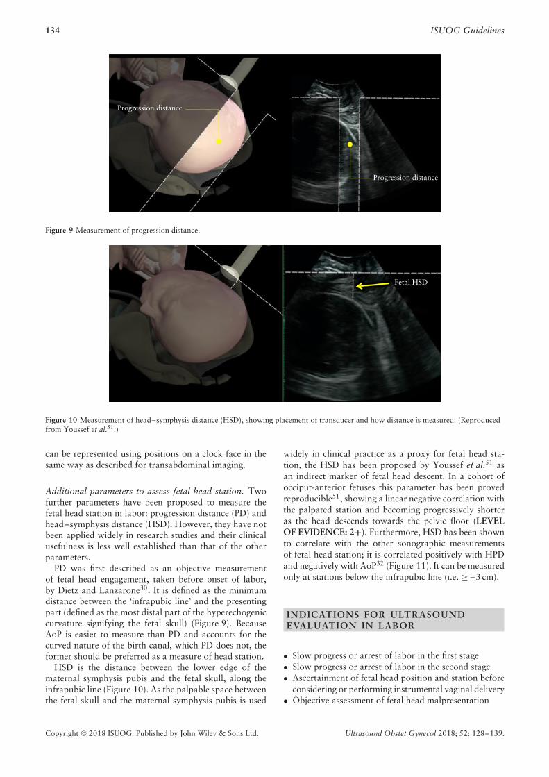
PD was first described as an objective measurementof fetal head engagement, taken before onset of labor,by Dietz and Lanzarone30. It is defined as the minimumdistance between the ‘infrapubic line’ and the presentingpart (defined as the most distal part of the hyperechogeniccurvature signifying the fetal skull) (Figure 9). BecauseAoP is easier to measure than PD and accounts for thecurved nature of the birth canal, which PD does not, theformer should be preferred as a measure of head station.
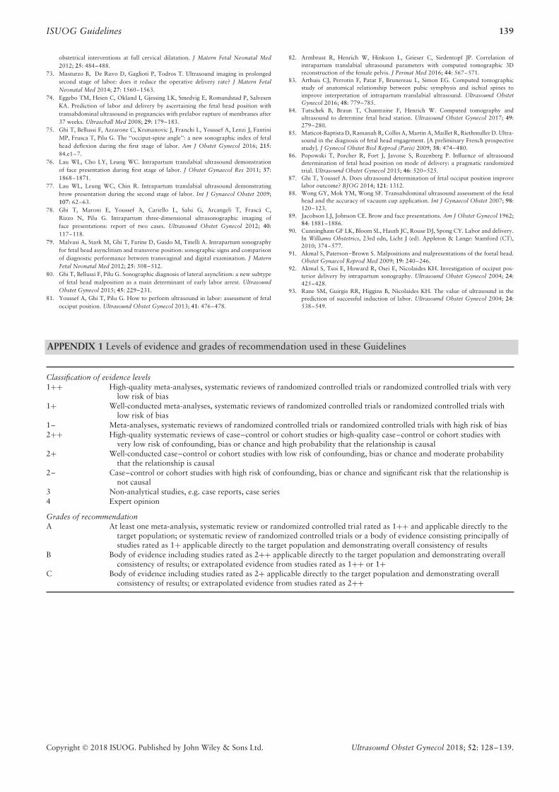
HSD is the distance between the lower edge of thematernal symphysis pubis and the fetal skull, along theinfrapubic line (Figure 10). As the palpable space betweenthe fetal skull and the maternal symphysis pubis is used
widely in clinical practice as a proxy for fetal head sta-tion, the HSD has been proposed by Youssef et al.51 asan indirect marker of fetal head descent. In a cohort ofocciput-anterior fetuses this parameter has been provedreproducible51, showing a linear negative correlation withthe palpated station and becoming progressively shorteras the head descends towards the pelvic floor (LEVELOF EVIDENCE: 2+). Furthermore, HSD has been shownto correlate with the other sonographic measurementsof fetal head station; it is correlated positively with HPDand negatively with AoP32 (Figure 11). It can be measuredonly at stations below the infrapubic line (i.e. ≥ –3 cm).
INDICATIONS FOR ULTRASOUNDEVALUATION IN LABOR
• Slow progress or arrest of labor in the first stage• Slow progress or arrest of labor in the second stage• Ascertainment of fetal head position and station before
considering or performing instrumental vaginal delivery• Objective assessment of fetal head malpresentation
Copyright © 2018 ISUOG. Published by John Wiley & Sons Ltd. Ultrasound Obstet Gynecol 2018; 52: 128–139.

ISUOG Guidelines 135
Fetal head station
0–2
AoP
(°)
HPD
and HSD
(mm
)1 2 3 4
0
10
20
30
40
50
60180
170
160
150
140
130
120
110
100
90
80
–1–3
Figure 11 Correlation of transperineal ultrasound (TPU) para-meters representative of fetal head station: angle of progression(AoP; ); head–perineum distance (HPD; ); and head–symphysisdistance (HSD; ). TPU head station is in cm above or below levelof ischial spines. Data are from Tutschek et al.32.
One study failed to demonstrate a benefit of routineuse of ultrasound in labor for determination of headposition (head station was not measured by ultrasoundin this study) among low-risk patients, in whom its usewas associated with a higher risk of Cesarean delivery86
(LEVEL OF EVIDENCE: 1–, GRADE OF RECOMMEN-DATION: A).
Although ultrasound has been demonstrated to be moreaccurate and reproducible than digital examination inthe determination of fetal head position and station inlabor, knowledge of these findings has not been shown toimprove the management of labor and delivery. Becauseof the rarity of adverse perinatal and maternal outcomesduring labor, very large randomized studies would benecessary to prove a clinical benefit of intrapartum sonog-raphy for the fetus or the mother with respect to severeperinatal or maternal morbidity. However, intrapartumultrasound allows more precise determination of positionand station and is more acceptable to women than digitalexamination72. Its use may be endorsed under the follow-ing circumstances as an adjunct to clinical examination.
Slow progress or arrest of labor in the first stage
Some consecutive studies have shown that HPD and AoPare more accurate than digital examination in predictingvaginal delivery in nulliparous women with prolonged firststage of labor36,39 (LEVEL OF EVIDENCE: 2+, GRADEOF RECOMMENDATION: B). In the largest multicentertrial, conducted on 150 women39, if HPD was < 40 mm,the likelihood of Cesarean delivery was 7%, whereas itwent up to 82% if HPD was > 50 mm. In the same study,if AoP was > 110◦, the likelihood of Cesarean deliverywas 12%, whereas this rose to 62% if AoP was < 100◦.
In a study of the same population of 150 womenwith prolonged first stage of labor37, the authorsshowed that occiput-posterior position, compared withnon-occiput-posterior position, was significantly associ-ated with the risk of Cesarean section (38% vs 17%,
P = 0.01) (LEVEL OF EVIDENCE: 2+, GRADE OFRECOMMENDATION: B).
Several case reports or small series76–80 have shownthat, in patients with prolonged first stage of labor, trans-abdominal or transperineal ultrasound may identify asa cause of labor arrest different types of head malpre-sentation, including deflexed presentation (brow or face)or asynclitism (LEVEL OF EVIDENCE: 3, GRADE OFRECOMMENDATION: C).
Slow progress or arrest of labor in the second stage
There is a paucity of studies addressing specifically theusefulness of ultrasound in predicting the chance of spon-taneous vaginal delivery compared with that of abdominaldelivery or OVD in patients with prolonged second stage.In 62 women with prolonged second stage examined bytransperineal ultrasound, Masturzo et al.73 found that afavorable head direction (head up) was associated withspontaneous vaginal delivery in the majority (16/20; 80%)of cases, in contrast to downward (4/20; 20%) or horizon-tal (9/22; 41%) head direction (LEVEL OF EVIDENCE:2+, GRADE OF RECOMMENDATION: B).
Ascertainment of fetal head position and station beforeinstrumental vaginal delivery
In a recent randomized controlled trial28, it was demon-strated that ultrasound assessment in addition to digitalexamination prior to instrumental vaginal delivery issignificantly more accurate compared with digital exam-ination alone in the diagnosis of fetal head position(ultrasound diagnosis incorrect in 1.6% of cases, com-pared with 20.2% in digital examination group) (LEVELOF EVIDENCE: 1–, GRADE OF RECOMMENDA-TION: A). While the study did not show significantdifferences in maternal or fetal morbidity, the main out-come was the accuracy of determining fetal position, andthe study was not powered to detect differences in theoccurrence of adverse events87.
In their randomized controlled trial, Wong et al.88
demonstrated that when fetal head position is determinedby ultrasound compared with by palpation, placementof the suction cup was significantly closer to the flexionpoint (LEVEL OF EVIDENCE: 1–, GRADE OF REC-OMMENDATION: A).
Head direction predicts the outcome of instrumentalvaginal delivery42. When evaluated before vacuum extrac-tion in protracted labor, the head-up sign is a positivepredictor of success. Among 11 women with fetal head upand an occiput-anterior position, all had successful simple(5/11) or moderately difficult (6/11) vacuum extraction. Incontrast, among the six cases with occiput-anterior fetuswith head horizontal or down, only one vacuum extrac-tion was simple, and the only case of failed extraction wasobserved in this group. The value of the head-up sign forprediction of vaginal delivery as well as its good intra- andinterobserver agreement were subsequently confirmed byothers41 (LEVEL OF EVIDENCE: 3, GRADE OF REC-OMMENDATION: C).
Copyright © 2018 ISUOG. Published by John Wiley & Sons Ltd. Ultrasound Obstet Gynecol 2018; 52: 128–139.

136 ISUOG Guidelines
AoP was investigated as a predictor of successful vac-uum delivery in 41 fetuses in occiput-anterior position. Acut-off value of 120◦ was found to predict an easy and suc-cessful vacuum extraction in 90% of cases43 (LEVEL OFEVIDENCE: 2+, GRADE OF RECOMMENDATION:B).
In 52 women with occiput-anterior fetus undergo-ing vacuum delivery, the combination of head-up sign,MLA < 45◦ and AoP > 120◦ were found to be significantsonographic predictors of a successful procedure45.
Cuerva et al.46 assessed the role of ultrasound inpredicting the outcome of forceps delivery in 30non-occiput-posterior fetuses. They found that the smallerthe AoP and the shorter the PD, the higher the risk offailure. AoP < 138◦ and PD < 4.8 cm were the strongestpredictors of the nine complicated procedures (defined asrequiring more than three tractions, failed procedure, ormaternal or neonatal trauma) (LEVEL OF EVIDENCE:2+, GRADE OF RECOMMENDATION: B).
A recent large study44 investigated the relationshipbetween vacuum extraction failure rate and AoP (imme-diately prior to application of the instrument) in 235women. In 30 (12%), the vacuum extraction failed,while in the remaining 205 it was successful. Failed vac-uum delivery was associated with a significantly smallermedian AoP (136.6◦ vs 145.9◦); interestingly, the pal-pated head station did not differ between the two groups(2 vs 2 cm) (LEVEL OF EVIDENCE: 2+, GRADE OFRECOMMENDATION: B).
In a European prospective study47, transperineal ultra-sound and the duration of vacuum extraction in a cohortof women with slow progress in the second stage oflabor were assessed. Among the 222 women included,the duration of the extraction procedure was signifi-cantly shorter in women with HPD ≤ 25 mm. The rateof Cesarean delivery was significantly lower amongcases with HPD ≤ 35 mm compared with those withHPD > 35 mm (3.9% vs 22.0%, P < 0.01) and, ifHPD > 35 mm was combined with occiput-posterior posi-tion, the rate of Cesarean delivery was 35%. Furthermore,the incidence of umbilical artery pH < 7.1 was signif-icantly higher in the infants which underwent vacuumdelivery with HPD > 35 mm.
In a prospective cohort study including 659 women,the HPD (in this study referred to as the perineum–skulldistance) was measured prior to OVD48. After adjust-ment for parity, presentation type and fetal macrosomia,HPD ≥ 40 mm was significantly associated with the occur-rence of a difficult extraction (odds ratio, 2.38; 95%CI, 1.51–3.74; P = 0.0002). Based on receiver–operatingcharacteristics curve analysis, perineum–skull distance onultrasound was a more accurate predictor of difficult OVDthan was digital vaginal examination (P = 0.036).
Visual confirmation of fetal head malpresentation
Deflexed cephalic presentation or asynclitism is a majorcause of obstructed labor13,14, estimated to account
for one-third of Cesarean deliveries for arrest oflabor4–6,8–10,15–17. In these cases the diagnosis is basedtraditionally upon digital examination in labor89–91,although the use of ultrasound to support the clini-cal diagnosis has been reported recently76–80 (LEVELOF EVIDENCE: 3, GRADE OF RECOMMENDATION:C).
SUMMARY
Ultrasound in active labor is not yet used widely, eventhough studies have shown that it is more precise andreproducible than clinical examination. Ultrasound allowsobjective measurement and precise documentation offindings obtained during the examination. Several sono-graphic parameters can be used during labor to assessmainly head station and position.
1. Head station can be measured objectively, for exampleby AoP or HPD, to assess current status and as a base-line for longitudinal measurements. It can also helpto predict whether OVD is likely to be successful.Head station should be assessed transperineally, nottransabdominally. HPD is straightforward to measureand is reproducible. AoP (in degrees) is equivalent tohead station expressed in centimeters, from –3 cmto +5 cm (direct conversion is possible), and hasthe potential to link ultrasound data to traditionalassessment by palpation. HPD and AoP/head sta-tion correlate linearly (for high station, i.e. higherthan 0 to +1).
2. Head (and spine) position is assessed more accuratelyby transabdominal ultrasound than by digital palpa-tion. Knowledge of head position in suspected delay orarrest of labor is important. Before OVD, knowledgeof head position is essential.
3. MLA is assessed by transverse transperineal ultra-sound and may help to decide whether OVD can beattempted safely.
4. Head direction is assessed by transperineal ultra-sound and may help to decide whether OVD canbe attempted safely.
There are two main situations in which ultrasoundassessment is likely to be of particular use in labor.
1. Suspected delay or arrest of first or second stage.We recommend measurement of either AoP or HPDtransperineally and assessment of head position trans-abdominally.
2. Potential need for performance of OVD. We recom-mend assessment of head position by transabdominalultrasound and suggest measurement of fetal headstation by transperineal ultrasound. The most reliablesonographic parameters to predict outcome of the pro-cedure are HPD and AoP. MLA and/or head directionmay also be useful to predict further the likelihood ofsuccess of the extraction.
Copyright © 2018 ISUOG. Published by John Wiley & Sons Ltd. Ultrasound Obstet Gynecol 2018; 52: 128–139.

ISUOG Guidelines 137
What we know and what we don’t
• We know that ultrasound allows more preciseexamination of fetal position and station thanclinical examination.
• We know that women prefer ultrasound to digitalexamination in labor.
• We know that transabdominal ultrasound is usedmost commonly for fetal lie and position, andtransperineal ultrasound can be used for head sta-tion.
• We don’t know how this knowledge impacts onmanagement of labor and maternal and neonataloutcomes.
REPORTING
If an ultrasound examination is performed in labor,its results should be added to the clinical notes of thepatient. For each sonographic evaluation, the followingdata should be noted:
• Fetal viability and heart rate• Presentation of the fetus (cephalic, transverse, breech,
oblique)• Whether any part of the placenta is seen between pre-
senting part and cervix• Occiput and spine position
Based upon the judgement of the clinician, the follow-ing transperineal ultrasound parameters can be added inthe second stage, especially before OVD (at rest or duringcontraction with maternal pushing; this should be noted):
• Angle of progression (AoP)• Head–perineum distance (HPD)• Head direction with respect to pubic symphysis• Midline angle (MLA)
GUIDELINE AUTHORS
These Guidelines were produced on behalf of the Interna-tional Society of Ultrasound in Obstetrics and Gynecology(ISUOG) by the following authors, and peer reviewed bythe Clinical Standards Committee.
T. Ghi, Obstetrics and Gynecology, University of Parma,Parma, ItalyT. Eggebø, National Center for Fetal Medicine, Trond-heim University Hospital (St Olavs Hospital), Trondheim,Norway; Department of Obstetrics and Gynecology,Stavanger University Hospital, Stavanger, NorwayC. Lees, Centre for Fetal Care, Queen Charlottes andChelsea Hospital, London, UKK. Kalache, Sidra Medical and Research Center, Doha,QatarP. Rozenberg, Centre Hospitalier Poissy Saint Germain,Obstetrics & Gynaecology, Paris, France
A. Youssef, Obstetrics and Gynecology, S. OrsolaMalpighi Hospital, Bologna, ItalyL. J. Salomon, Hopital Universitaire Necker-EnfantsMalades, AP-HP, Universite Paris Descartes, Maternite,Paris, France; Societe Francaise pour l’Amelioration desPratiques Echographiques, SFAPEB. Tutschek, Prenatal Zurich, Heinrich-Heine-Uni-versity, Medical Faculty, Zurich, Switzerland
Guideline external reviewers were V. Berghella,O. Dupuis and W. Lau. The final version is the responsi-bility of the Clinical Standards Committee of ISUOG. Theguideline review process will commence in 2023 unlessevidence requires earlier review.
CITATION
These Guidelines should be cited as: ‘Ghi T, Eggebø T,Lees C, Kalache K, Rozenberg P, Youssef A, SalomonLJ, Tutschek B. ISUOG Practice Guidelines: intra-partum ultrasound. Ultrasound Obstet Gynecol 2018;52: 128–139.’
REFERENCES
1. Friedman E. The graphic analysis of labor. Am J Obstet Gynecol 1954; 68:1568–1575.
2. Friedman EA. Primigravid labor; a graphicostatistical analysis. Obstet Gynecol 1955;6: 567–589.
3. Friedman EA. Labor in multiparas; a graphicostatistical analysis. Obstet Gynecol1956; 8: 691–703.
4. Zhang J, Troendle JF, Yancey MK. Reassessing the labor curve in nulliparous women.Am J Obstet Gynecol 2002; 187: 824–828.
5. Zhang J, Landy HJ, Branch DW, Burkman R, Haberman S, Gregory KD, HatjisCG, Ramirez MM, Bailit JL, Gonzalez-Quintero VH, Hibbard JU, Hoffman MK,Kominiarek M, Learman LA, Van Veldhuisen P, Troendle J, Reddy UM; Consortiumon Safe Labor. Contemporary patterns of spontaneous labor with normal neonataloutcomes: Consortium on safe labor. Obstet Gynecol 2010; 116: 1281–1287.
6. Segel SY, Carreno CA, Weiner SJ, Bloom SL, Spong CY, Varner MW, Rouse DJ,Caritis SN, Grobman WA, Sorokin Y, Sciscione A, Mercer BM, Thorp JM, MaloneFD, Harper M, Iams JD; Eunice Kennedy Shriver National Institute of Child Healthand Human Development Maternal-Fetal Medicine Units Network. Relationshipbetween fetal station and successful vaginal delivery in nulliparous women. Am JPerinatol 2012; 29: 723–730.
7. Hamilton EF, Simoneau G, Ciampi A, Warrick P, Collins K, Smith S, Garite TJ.Descent of the fetal head (station) during the first stage of labor. Am J Obstet Gynecol2016; 214: 360.e1–6.
8. American College of Obstetricians and Gynecologists, Society for Maternal-FetalMedicine, Caughey AB, Cahill AG, Guise JM, Rouse DJ. Safe prevention of theprimary cesarean delivery. Am J Obstet Gynecol 2014; 210: 179–193.
9. Barber EL, Lundsberg LS, Belanger K, Pettker CM, Funai EF, Illuzzi JL. Indicationscontributing to the increasing cesarean delivery rate. Obstet Gynecol 2011; 118:29–38.
10. Spong CY, Berghella V, Wenstrom KD, Mercer BM, Saade GR. Preventing the firstcesarean delivery: summary of a joint Eunice Kennedy Shriver National Institute ofChild Health and Human Development, Society for Maternal-Fetal Medicine, andAmerican College of Obstetricians and Gynecologists Workshop. Obstet Gynecol2012; 120: 1181–1193.
11. Cohen WR. Influence of the duration of second stage labor on perinatal outcomeand puerperal morbidity. Obstet Gynecol 1977; 49: 266–269.
12. Leveno KJ, Nelson DB, McIntire DD. Second-stage labor: how long is too long? AmJ Obstet Gynecol 2016; 214: 484–489.
13. Stitely ML, Gherman RB. Labor with abnormal presentation and position. ObstetGynecol Clin North Am 2005; 32: 165–179.
14. Boyle A, Reddy UM, Landy HJ, Huang CC, Driggers RW, Laughon SK. Primarycesarean delivery in the United States. Obstet Gynecol 2013; 122: 33–40.
15. Shin KS, Brubaker KL, Ackerson LM. Risk of cesarean delivery in nulliparous womenat greater than 41 weeks’ gestational age with an unengaged vertex. Am J ObstetGynecol 2004; 190: 129–134.
16. Oboro VO, Tabowei TO, Bosah JO. Fetal station at the time of labor arrest and riskof caesarean delivery. J Obstet Gynaecol 2005; 25: 20–22.
17. ACOG Practice Bulletin. Number 49, December 2003. Dystocia and augmentationof labor.
18. Dupuis O, Silveira R, Zentner A, Dittmar A, Gaucherand P, Cucherat M, RedarceT, Rudigoz RC. Birth simulator: reliability of transvaginal assessment of fetal headstation as defined by the American College of Obstetricians and Gynecologistsclassification. Am J Obstet Gynecol 2005; 192: 868–874.
Copyright © 2018 ISUOG. Published by John Wiley & Sons Ltd. Ultrasound Obstet Gynecol 2018; 52: 128–139.

138 ISUOG Guidelines
19. Dupuis O, Ruimark S, Corrine D, Simone T, Andre D, Rene- Charles R. Fetal headposition during the second stage of labor: comparison of digital and vaginal exam-ination and transabdominal ultrasonographic examination. Eur J Obstet GynecolReprod Biol 2005; 123: 193–197.
20. Akmal S, Kametas N, Tsoi E, Hargreaves C, Nicolaides KH. Comparison of transvagi-nal digital examination with intrapartum sonography to determine fetal head positionbefore instrumental delivery. Ultrasound Obstet Gynecol 2003; 21: 437–440.
21. Sherer DM, Miodovnik M, Bradley S, Langer O. Intrapartum fetal head positionI: comparison between transvaginal digital examination and transabdominal ultra-sound assessment during the active stage of labor. Ultrasound Obstet Gynecol 2002;19: 258–263.
22. Sherer DM, Miodovnik M, Bradley KS, Langer O. Intrapartum fetal head posi-tion II: comparison between transvaginal digital examination and transabdominalultrasound assessment during the second stage of labor. Ultrasound Obstet Gynecol2002; 19: 264–268.
23. Souka AP, Haritos T, Basayiannis K, Noikokyri N, Antsaklis A. Intrapartum ultra-sound for the examination of the fetal head position in normal and obstructed labor.J Matern Fetal Neonatal Med 2003; 13: 59–63.
24. Kreiser D, Schiff E, Lipitz S, Kayam Z, Avraham A, Achiron R. Determination offetal occiput position by ultrasound during the second stage of labor. J Matern FetalMed 2001; 10: 283–286.
25. Akmal S, Tsoi E, Nicolaides KH. Intrapartum sonography to determine fetal occipitalposition: interobserver agreement. Ultrasound Obstet Gynecol 2004; 24: 421–424.
26. Chou MR, Kreiser D, Taslimi MM, Druzin ML, El-Sayed YY. Vaginal versus ultra-sound examination of fetal occiput position during the second stage of labor. Am JObstet Gynecol 2004; 191: 521–524.
27. Ramphul M, Kennelly M, Murphy DJ. Establishing the accuracy and acceptabilityof abdominal ultrasound to define the foetal head position in the second stage oflabour: a validation study. Eur J Obstet Gynecol Reprod Biol 2012; 164: 35–39.
28. Ramphul M, Ooi PV, Burke G, Kennelly MM, Said SA, Montgomery AA, MurphyDJ. Instrumental delivery and ultrasound: a multicentre randomised controlled trial ofultrasound assessment of the fetal head position versus standard care as an approachto prevent morbidity at instrumental delivery. BJOG 2014; 121: 1029–1038.
29. Sherer DM, Abulafia O. Intrapartum assessment of fetal head engagement: com-parison between transvaginal digital and transabdominal ultrasound determinations.Ultrasound Obstet Gynecol 2003; 21: 430–436.
30. Dietz HP, Lanzarone V. Measuring engagement of the fetal head: validity and repro-ducibility of a new ultrasound technique. Ultrasound Obstet Gynecol 2005; 25:165–168.
31. Ghi T, Farina A, Pedrazzi A, Rizzo N, Pelusi G, Pilu G. Diagnosis of station androtation of the fetal head in the second stage of labor with intrapartum translabialultrasound. Ultrasound Obstet Gynecol 2009; 33: 331–336.
32. Tutschek B, Torkildsen EA, Eggebo TM. Comparison between ultrasound parame-ters and clinical examination to assess fetal head station in labor. Ultrasound ObstetGynecol 2013; 41: 425–429.
33. Duckelmann AM, Bamberg C, Michaelis SA, Lange J, Nonnenmacher A, Duden-hausen JW, Kalache KD. Measurement of fetal head descent using the ‘angle ofprogression’ on transperineal ultrasound imaging is reliable regardless of fetal headstation or ultrasound expertise. Ultrasound Obstet Gynecol 2010; 35: 216–222.
34. Eggebø TM, Gjessing LK, Heien C, Smedvig E, Økland I, Romundstad P, SalvesenKA. Prediction of labor and delivery by transperineal ultrasound in pregnancieswith prelabor rupture of membranes at term. Ultrasound Obstet Gynecol 2006; 27:387–391.
35. Eggebø TM, Heien C, Økland I, Gjessing LK, Romundstad P, Salvesen KA.Ultrasound assessment of fetal head-perineum distance before induction of labor.Ultrasound Obstet Gynecol 2008; 32: 199–204.
36. Torkildsen EA, Salvesen KA, Eggebø TM. Prediction of delivery mode with transper-ineal ultrasound in women with prolonged first stage of labor. Ultrasound ObstetGynecol 2011; 37: 702–708.
37. Eggebø TM, Hassan WA, Salvesen KA, Torkildsen EA, Østborg TB, Lees CC. Pre-diction of delivery mode by ultrasound-assessed fetal position in nulliparous womenwith prolonged first stage of labor. Ultrasound Obstet Gynecol 2015; 46: 606–610.
38. Eggebø TM, Wilhelm-Benartzi C, Hassan WA, Usman S, Salvesen KA, Lees CC. Amodel to predict vaginal delivery in nulliparous women based on maternal charac-teristics and intrapartum ultrasound. Am J Obstet Gynecol 2015; 213: 362.e1–6.
39. Eggebø TM, Hassan WA, Salvesen KA, Lindtjørn E, Lees CC. Sonographic predic-tion of vaginal delivery in prolonged labor: a two-center study. Ultrasound ObstetGynecol 2014; 43: 195–201.
40. Barbera AF, Pombar X, Perugino G, Lezotte DC, Hobbins JC. A new method toassess fetal head descent in labor with transperineal ultrasound. Ultrasound ObstetGynecol 2009; 33: 313–319.
41. Tutschek B, Braun T, Chantraine F, Henrich W. A study of progress of labor usingintrapartum translabial ultrasound, assessing head station, direction, and angle ofdescent. BJOG 2011; 118: 62–69.
42. Henrich W, Dudenhausen J, Fuchs I, Kamena A, Tutschek B. Intrapartum translabialultrasound (ITU): sonographic landmarks and correlation with successful vacuumextraction. Ultrasound Obstet Gynecol 2006; 28: 753–760.
43. Kalache KD, Duckelmann AM, Michaelis SA, Lange J, Cichon G, Dudenhausen JW.Transperineal ultrasound imaging in prolonged second stage of labor with occipi-toanterior presenting fetuses: how well does the ‘angle of progression’ predict themode of delivery? Ultrasound Obstet Gynecol 2009; 33: 326–330.
44. Bultez T, Quibel T, Bouhanna P, Popowski T, Resche-Rigon M, Rozenberg P. Angleof fetal head progression measured using transperineal ultrasound as a predictivefactor of vacuum extraction failure. Ultrasound Obstet Gynecol 2016; 48: 86–91.
45. Sainz JA, Borrero C, Aquise A, Serrano R, Gutierrez L, Fernandez-Palacın A. Util-ity of intrapartum transperineal ultrasound to predict cases of failure in vacuumextraction attempt and need of cesarean section to complete delivery. J Matern FetalNeonatal Med 2016; 29: 1348–1352.
46. Cuerva MJ, Bamberg C, Tobias P, Gil MM, De La Calle M, Bartha JL. Use ofintrapartum ultrasound in the prediction of complicated operative forceps deliveryof fetuses in non-occiput posterior position. Ultrasound Obstet Gynecol 2014; 43:687–692.
47. Kahrs BH, Usman S, Ghi T, Youssef A, Torkildsen EA, Lindtjørn E, Østborg TB,Benediktsdottir S, Brooks L, Harmsen L, Romundstad PR, Salvesen KA, Lees CC,Eggebø TM. Sonographic prediction of outcome of vacuum deliveries: a multicenter,prospective cohort study. Am J Obstet Gynecol 2017; 217: 69.e1–10.
48. Kasbaoui S, Severac F, Aıssi G, Gaudineau A, Lecointre L, Akladios C, FavreR, Langer B, Sananes N. Predicting the difficulty of operative vaginal delivery byultrasound measurement of fetal head station. Am J Obstet Gynecol 2017; 216:507.e1–9.
49. Blasi I, D’Amico R, Fenu V, Volpe A, Fuchs I, Henrich W, Mazza V. Sonographicassessment of fetal spine and head position during the first and second stages of laborfor the diagnosis of persistent occiput posterior position: a pilot study. UltrasoundObstet Gynecol 2010; 35: 210–215.
50. Barbera AF, Imani F, Becker T, Lezotte DC, Hobbins JC. Anatomic relationshipbetween the pubic symphysis and ischial spines and its clinical significance in theassessment of fetal head engagement and station during labor. Ultrasound ObstetGynecol 2009; 33: 320–325.
51. Youssef A, Maroni E, Ragusa A, De Musso F, Salsi G, Iammarino MT, PaccapeloA, Rizzo N, Pilu G, Ghi T. Fetal head-symphysis distance: a simple and reliableultrasound index of fetal head station in labor. Ultrasound Obstet Gynecol 2013;41: 419–424.
52. Carseldine WJ, Phipps H, Zawada SF, Campbell NT, Ludlow JP, Krishnan SY, DeVries BS. Does occiput posterior position in the second stage of labor increase theoperative delivery rate? Aust N Z J Obstet Gynaecol 2013; 53: 265–270.
53. Wu JM, Williams KS, Hundley AF, Connolly A, Visco AG. Occiput posterior fetalhead position increases the risk of anal sphincter injury in vacuum-assisted deliveries.Am J Obstet Gynecol 2005; 193: 525–528.
54. Pearl ML, Roberts JM, Laros RK, Hurd WW. Vaginal delivery from the persistentocciput posterior position. Influence on maternal and neonatal morbidity. J ReprodMed 1993; 38: 955–961.
55. Gei AF, Smith RA, Hankins GD. Brachial plexus paresis associated with fetal neckcompression from forceps. Am J Perinatol 2003; 20: 289–291.
56. Mola GD, Amoa AB, Edilyong J. Factors associated with success or failure in trialsof vacuum extraction. Aust N Z J Obstet Gynaecol 2002; 42: 35–39.
57. Vacca A, Keirse MJNC. Instrumental vaginal delivery. In Effective care in pregnancyand childbirth, Chalmers I, Enkin M, Keirse MJN (eds). Oxford University Press:Oxford, 1989; 1216–1233.
58. Dupuis O, Silveira R, Dupont C, Mottolese C, Kahn P, Dittmar A, Rudigoz RC.Comparison of ‘‘instrument-associated’’ and ‘‘spontaneous’’ obstetric depressed skullfractures in a cohort of 68 neonates. Am J Obstet Gynecol 2005; 192: 165–170.
59. Ramphul M, Kennelly MM, Burke G, Murphy DJ. Risk factors and morbidity asso-ciated with suboptimal instrument placement at instrumental delivery: observationalstudy nested within the Instrumental Delivery & Ultrasound randomised controlledtrial ISRCTN 72230496. BJOG 2015; 122: 558–563.
60. Donnelly V, Fynes M, Campbell D, Johnson H, O’Connell PR, O’Herlihy C. Obstetricevents leading to anal sphincter damage. Obstet Gynecol 1998; 92: 955–961.
61. MacLennan AH, Taylor AW, Wilson DH, Wilson D. The prevalence of pelvic floordisorders and their relationship to gender, age, parity and mode of delivery. BJOG2000; 107: 1460–1470.
62. Olagundoye V, MacKenzie IZ. The impact of a trial of instrumental delivery intheatre on neonatal outcome. BJOG 2007; 114: 603–608.
63. Towner D, Castro MA, Eby-Wilkens E, Gilbert WM. Effect of mode of deliveryin nulliparous women on neonatal intracranial injury. N Engl J Med 1999; 341:1709–1714.
64. Alexander JM, Leveno KJ, Hauth J, Landon MB, Thom E, Spong CY, VarnerMW, Moawad AH, Caritis SN, Harper M, Wapner RJ, Sorokin Y, Miodovnik M,O’Sullivan MJ, Sibai BM, Langer O, Gabbe SG; National Institute of Child Healthand Human Development Maternal–Fetal Medicine Units Network. Fetal injuryassociated with cesarean delivery. Obstet Gynecol 2006; 108: 885–890.
65. Murphy DJ, Liebling RE, Patel R, Verity L, Swingler R. Cohort study of operativedelivery in the second stage of labor and standard of obstetric care. BJOG 2003;110: 610–615.
66. Nizard J, Haberman S, Paltieli Y, Gonen R, Ohel G, Le Bourthe Y, Ville Y.Determination of fetal head station and position during labor: a new technique thatcombines ultrasound and a position-tracking system. Am J Obstet Gynecol 2009;200: 404.e1–5.
67. Cunningham F, MacDonald PC, Gant NF, Leveno KJ, Gilstrap LC 3rd, HankinsGDV, et al. Anatomy of the reproductive tract. In Williams Obstetrics, Licht J (ed).Appleton & Lange: Stamford (CT), 1997.
68. Bamberg C, Scheuermann S, Slowinski T, Duckelmann AM, Vogt M,Nguyen-Dobinsky TN, Streitparth F, Teichgraber U, Henrich W, DudenhausenJW, Kalache KD. Relationship between fetal head station established using an openmagnetic resonance imaging scanner and the angle of progression determined bytransperineal ultrasound. Ultrasound Obstet Gynecol 2011; 37: 712–716.
69. Ghi T, Contro E, Farina A, Nobile M, Pilu G. Three-dimensional ultrasound in mon-itoring progression of labor: a reproducibility study. Ultrasound Obstet Gynecol2010; 36: 500–506.
70. Molina FS, Terra R, Carrillo MP, Puertas A, Nicolaides KH. What is the most reli-able ultrasound parameter for assessment of fetal head descent? Ultrasound ObstetGynecol 2010; 36: 493–499.
71. Youssef A, Bellussi F, Montaguti E, Maroni E, Salsi G, Morselli-Labate AM, Pac-capelo A, Rizzo N, Pilu G, Ghi T. Agreement between two- and three-dimensionalmethods for the assessment of the fetal head-symphysis distance in active labor.Ultrasound Obstet Gynecol 2014; 43: 183–188.
72. Duckelmann AM, Michaelis SA, Bamberg C, Dudenhausen JW, Kalache KD. Impactof intrapartal ultrasound to assess fetal head position and station on the type of
Copyright © 2018 ISUOG. Published by John Wiley & Sons Ltd. Ultrasound Obstet Gynecol 2018; 52: 128–139.
ISUOG Guidelines 139
obstetrical interventions at full cervical dilatation. J Matern Fetal Neonatal Med2012; 25: 484–488.
73. Masturzo B, De Ruvo D, Gaglioti P, Todros T. Ultrasound imaging in prolongedsecond stage of labor: does it reduce the operative delivery rate? J Matern FetalNeonatal Med 2014; 27: 1560–1563.
74. Eggebo TM, Heien C, Okland I, Gjessing LK, Smedvig E, Romundstad P, SalvesenKA. Prediction of labor and delivery by ascertaining the fetal head position withtransabdominal ultrasound in pregnancies with prelabor rupture of membranes after37 weeks. Ultraschall Med 2008; 29: 179–183.
75. Ghi T, Bellussi F, Azzarone C, Krsmanovic J, Franchi L, Youssef A, Lenzi J, FantiniMP, Frusca T, Pilu G. The ‘‘occiput-spine angle’’: a new sonographic index of fetalhead deflexion during the first stage of labor. Am J Obstet Gynecol 2016; 215:84.e1–7.
76. Lau WL, Cho LY, Leung WC. Intrapartum translabial ultrasound demonstrationof face presentation during first stage of labor. J Obstet Gynaecol Res 2011; 37:1868–1871.
77. Lau WL, Leung WC, Chin R. Intrapartum translabial ultrasound demonstratingbrow presentation during the second stage of labor. Int J Gynaecol Obstet 2009;107: 62–63.
78. Ghi T, Maroni E, Youssef A, Cariello L, Salsi G, Arcangeli T, Frasca C,Rizzo N, Pilu G. Intrapartum three-dimensional ultrasonographic imaging offace presentations: report of two cases. Ultrasound Obstet Gynecol 2012; 40:117–118.
79. Malvasi A, Stark M, Ghi T, Farine D, Guido M, Tinelli A. Intrapartum sonographyfor fetal head asynclitism and transverse position: sonographic signs and comparisonof diagnostic performance between transvaginal and digital examination. J MaternFetal Neonatal Med 2012; 25: 508–512.
80. Ghi T, Bellussi F, Pilu G. Sonographic diagnosis of lateral asynclitism: a new subtypeof fetal head malposition as a main determinant of early labor arrest. UltrasoundObstet Gynecol 2015; 45: 229–231.
81. Youssef A, Ghi T, Pilu G. How to perform ultrasound in labor: assessment of fetalocciput position. Ultrasound Obstet Gynecol 2013; 41: 476–478.
82. Armbrust R, Henrich W, Hinkson L, Grieser C, Siedentopf JP. Correlation ofintrapartum translabial ultrasound parameters with computed tomographic 3Dreconstruction of the female pelvis. J Perinat Med 2016; 44: 567–571.
83. Arthuis CJ, Perrotin F, Patat F, Brunereau L, Simon EG. Computed tomographicstudy of anatomical relationship between pubic symphysis and ischial spines toimprove interpretation of intrapartum translabial ultrasound. Ultrasound ObstetGynecol 2016; 48: 779–785.
84. Tutschek B, Braun T, Chantraine F, Henrich W. Computed tomography andultrasound to determine fetal head station. Ultrasound Obstet Gynecol 2017; 49:279–280.
85. Maticot-Baptista D, Ramanah R, Collin A, Martin A, Maillet R, Riethmuller D. Ultra-sound in the diagnosis of fetal head engagement. [A preliminary French prospectivestudy]. J Gynecol Obstet Biol Reprod (Paris) 2009; 38: 474–480.
86. Popowski T, Porcher R, Fort J, Javoise S, Rozenberg P. Influence of ultrasounddetermination of fetal head position on mode of delivery: a pragmatic randomizedtrial. Ultrasound Obstet Gynecol 2015; 46: 520–525.
87. Ghi T, Youssef A. Does ultrasound determination of fetal occiput position improvelabor outcome? BJOG 2014; 121: 1312.
88. Wong GY, Mok YM, Wong SF. Transabdominal ultrasound assessment of the fetalhead and the accuracy of vacuum cup application. Int J Gynaecol Obstet 2007; 98:120–123.
89. Jacobson LJ, Johnson CE. Brow and face presentations. Am J Obstet Gynecol 1962;84: 1881–1886.
90. Cunningham GF LK, Bloom SL, Hauth JC, Rouse DJ, Spong CY. Labor and delivery.In Williams Obstetrics, 23rd edn, Licht J (ed). Appleton & Lange: Stamford (CT),2010; 374–577.
91. Akmal S, Paterson–Brown S. Malpositions and malpresentations of the foetal head.Obstet Gynaecol Reprod Med 2009; 19: 240–246.
92. Akmal S, Tsoi E, Howard R, Osei E, Nicolaides KH. Investigation of occiput pos-terior delivery by intrapartum sonography. Ultrasound Obstet Gynecol 2004; 24:425–428.
93. Rane SM, Guirgis RR, Higgins B, Nicolaides KH. The value of ultrasound in theprediction of successful induction of labor. Ultrasound Obstet Gynecol 2004; 24:538–549.
APPENDIX 1 Levels of evidence and grades of recommendation used in these Guidelines
Classification of evidence levels1++ High-quality meta-analyses, systematic reviews of randomized controlled trials or randomized controlled trials with very
low risk of bias1+ Well-conducted meta-analyses, systematic reviews of randomized controlled trials or randomized controlled trials with
low risk of bias1– Meta-analyses, systematic reviews of randomized controlled trials or randomized controlled trials with high risk of bias2++ High-quality systematic reviews of case–control or cohort studies or high-quality case–control or cohort studies with
very low risk of confounding, bias or chance and high probability that the relationship is causal2+ Well-conducted case–control or cohort studies with low risk of confounding, bias or chance and moderate probability
that the relationship is causal2– Case–control or cohort studies with high risk of confounding, bias or chance and significant risk that the relationship is
not causal3 Non-analytical studies, e.g. case reports, case series4 Expert opinion
Grades of recommendationA At least one meta-analysis, systematic review or randomized controlled trial rated as 1++ and applicable directly to the
target population; or systematic review of randomized controlled trials or a body of evidence consisting principally ofstudies rated as 1+ applicable directly to the target population and demonstrating overall consistency of results
B Body of evidence including studies rated as 2++ applicable directly to the target population and demonstrating overallconsistency of results; or extrapolated evidence from studies rated as 1++ or 1+
C Body of evidence including studies rated as 2+ applicable directly to the target population and demonstrating overallconsistency of results; or extrapolated evidence from studies rated as 2++
Copyright © 2018 ISUOG. Published by John Wiley & Sons Ltd. Ultrasound Obstet Gynecol 2018; 52: 128–139.

Ultrasound Obstet Gynecol 2018; 52: 128–139Published online in Wiley Online Library (wileyonlinelibrary.com). DOI: 10.1002/uog.19072
Directrices sobre pract icas de ISUOG: la ecograf ıa durante el parto
PROPOSITO Y ALCANCEEl proposito de estas Directrices es revisar las tecnicas publicadas sobre ecografıa durante el parto y sus aplicacionespracticas, resumir la calidad de la evidencia disponible con respecto al uso de la ecografıa durante el parto y proporcionarpautas a los profesionales sobre cuando se recomienda el uso de la ecografıa durante el parto por motivos clınicos ycomo los resultados ecograficos pueden afectar al cuidado durante el parto. No se infiere ni se sugiere que la ecografıadurante el parto sea un estandar necesario de asistencia medica.
Copyright © 2018 ISUOG. Published by John Wiley & Sons Ltd. ISUOG GUIDELINES